


Abstract
Termite-associated Ophiocordyceps species remain understudied despite the high diversity of the genus. Here we describe Ophiocordyceps minuta (holotype CXAC 0026) from termites (Termitidae, Macrotermitinae) collected in Yunnan, China. Phylogenetic analyses based on nrLSU (nuclear large subunit ribosomal RNA), tef1-α (translation elongation factor 1-alpha), and rpb2 (RNA polymerase II second largest subunit) sequences resolve this fungus as a distinct lineage. Notably, deep genetic divergence (65 bp in tef1-α) between the two ex-type strains of the allied O. fusiformis (BCC 93025 and BCC 93026) reveals cryptic diversity within that nominal species. Beyond the new species, a morphological assessment of termite-associated Ophiocordyceps indicates that perithecial immersion status, ascospore morphology, and conidial features are the most taxonomically valuable characters for this ecological group. These findings expand the known diversity in southwest China and underscore the importance of integrating phylogenetic data with key morphological traits for species delimitation in under-explored habitats.
1. Introduction
The genus Ophiocordyceps Petch (Ophiocordycipitaceae, Hypocreales) ranks among the most species-rich lineages of entomopathogenic fungi, with over 370 accepted names distributed globally (www.indexfungorum.org, accessed on 15 March 2026). Unlike their close relatives in Cordyceps sensu stricto, which typically produce brightly coloured, fleshy stromata, Ophiocordyceps species are characterized by dark-pigmented, wiry to pliant stromata and are associated with anamorphic genera including Hirsutella, Hymenostilbe, Paraisaria, and Syngliocladium [1,2,3]. Members of this genus exploit an exceptionally broad range of insect hosts, attacking various life stages across Blattodea, Coleoptera, Diptera, Hemiptera, Hymenoptera, Lepidoptera, Megaloptera, Neuroptera and Odonata [3,4,5,6,7]. However, the association with Blattodea is not restricted to termites; several Ophiocordyceps species, including the type species O. blattae, as well as O. asiatica, O. cf. blattae, and O. salganeicola, have been confirmed from non-termite Blattodea [8,9]. This ecological diversity is matched by morphological plasticity: perithecia may be superficial, immersed, or partially embedded, while ascospores either remain intact as multiseptate units or dissociate into individual part-spores [2,10].
Despite this overall diversity, the distribution of Ophiocordyceps species across host groups is markedly uneven [11,12,13]. Certain host associations, particularly with ants and beetle larvae, have yielded numerous described species, while others remain poorly documented [14,15]. Termites (Blattodea: Termitidae) represent one such understudied host group [16]. Termites were historically treated as a separate order, Isoptera, but molecular phylogenetic studies have firmly placed them within Blattodea [17], a placement further corroborated by recent genomic-scale analyses [18]. Termitidae is the most speciose termite family, comprising approximately 2000 described species [19]. As eusocial insects inhabiting subterranean nests in tropical and subtropical ecosystems worldwide, termites play critical roles in decomposition and nutrient cycling [20]. Their constant exposure to soil-borne microorganisms creates opportunities for fungal pathogens, yet the diversity of termite-associated Ophiocordyceps has only recently begun to receive sustained attention [8,21,22,23].
Historical records of termite-pathogenic Ophiocordyceps were limited to three species described over a span of six decades: O. bispora from East Africa, O. koningsbergeri from Java, and O. octospora from Mexico [24,25,26]. Following these early reports, progress remained slow until intensive surveys in Thailand during the past decade uncovered 11 additional species from forests [8,27]. The remaining species have been documented from other East and Southeast Asian localities: O. globiperitheciata, O. longistipes, O. ovatospora and O. taiwanensis from China, and O. puluongensis from Vietnam [21,22,23,28]. Thus, a total of 20 Ophiocordyceps species have been reported from termite hosts worldwide to date, not including the new species described in the present study [2,8,21,23,24,25,26,29]. These discoveries demonstrated that termite-pathogenic lineages harbor substantially greater diversity than previously recognized and suggested that similar surveys in other under-explored regions would likely yield additional taxa.
Southwest China, particularly Yunnan Province, has emerged as a promising area for discovering novel Cordyceps sensu lato species [30,31]. The region’s complex topography, elevational gradients, and high forest cover support diverse insect communities and their associated fungal pathogens [32,33]. Recent investigations have documented multiple new Ophiocordyceps taxa from Yunnan across various insect hosts, including two termite-associated species described in the past two years [21,22]. These findings indicate that the termite-pathogenic Ophiocordyceps diversity in this region remains incompletely sampled, with additional species awaiting discovery through continued field efforts. In the present study, we describe the new termite-associated species O. minuta using morphological and multigene phylogenetic evidence, reassess the key morphological characters for delimiting termite-associated Ophiocordyceps, and discuss the group’s diversity in southwest China.
2. Materials and Methods
2.1. Specimen Collection
Fungal specimens were collected from ground-dwelling termites (Termitidae, Macrotermitinae) in Taiyanghe National Nature Reserve, Yunnan Province, China. Specimens were carefully removed to preserve host integrity, placed in sterile plastic containers, and transported to the laboratory. Upon arrival, they were examined under a stereomicroscope (OLYMPUS SZ61, Olympus Corporation, Tokyo, Japan), cleaned of surface debris, assigned accession numbers CXAC 0026–0027, and air-dried. Hosts were identified using morphological characters and with guidance from termite taxonomists; finer identification was precluded by the condition of the fungus-infected specimens. Voucher specimens are deposited at the College of Agronomy Herbarium (CXAC), Chuxiong Normal University, China, for taxonomic identification and future studies.
2.2. Morphological Characterization
Specimen data, including host association and geographic origin, were documented. Sexual structures (perithecia, asci, ascospores, part-spores) were mounted in lactophenol cotton blue and measured under an Olympus BX53 compound microscope (Olympus Corporation, Tokyo, Japan). For each structure, 20–50 measurements were recorded, and the range was calculated.
2.3. Extraction of DNA, Polymerase Chain Reaction (PCR), and Molecular Sequencing
Genomic DNA was extracted from fungal mycelia using a commercial plant DNA isolation kit (FOREGENE, Chengdu, China) and amplified via polymerase chain reaction (PCR) for three genetic regions: nrLSU, tef1-α, and rpb2, with primers as following: the LSU region was amplified with the primer pair LR0R/LR5 [34,35], and the tef1-α and rpb2 genes using the primer sets EF1α-EF/EF1α-ER [2,36] and RPB2-5′F/RPB2-5′R [2,36] respectively. Each 25 μL PCR contained 2.5 μL of 10× buffer (2 mM Mg2+; Transgen Biotech, Beijing, China), 0.25 μL of Taq polymerase, 2 μL of dNTPs (2.5 mM each), 1 μL of DNA template (~500 ng/μL), 1 μL of each primer (10 μM), and 17.25 μL of deionized water. Amplification was conducted in a BIO-RAD T100™ Thermal Cycler following the gene-specific thermal cycling protocols detailed in Table 1. Successful amplification was verified by gel electrophoresis; the resulting amplicons were then purified prior to bidirectional Sanger sequencing at the Beijing Genomics Institute (Shenzhen, China) with the same PCR primers.
Table 1.
PCR cycling conditions for each gene.
2.4. Phylogenetic Analyses
To establish the phylogenetic placement of our taxa, we analyzed a three-gene matrix (nrLSU, tef1-α, and rpb2). Newly obtained sequences were confirmed via BLAST searches and aligned with reference sequences from GenBank (Table 2) using MAFFT v7.526 (L-INS-i strategy) (http://mafft.cbrc.jp/alignment/server/, accessed 15 March 2026) and refined manually in BioEdit v7.7.1. These alignments were concatenated into a supermatrix with FASconCAT-G v1.06 [37]. The partition homogeneity test in PAUP v5.0 (1000 reps, p > 0.01) revealed no significant conflict between gene partitions. Optimal partitioning schemes and models of evolution were selected under the BIC criterion using PartitionFinder2 v2.0.0 (greedy algorithm) [38]. Phylogenetic relationships were then inferred using two complementary methods. First, a Maximum Likelihood (ML) analysis was performed in IQ-TREE v3.0.1 under the optimal partition scheme (GTR + G + I model), with nodal support assessed via 1000 ultrafast bootstrap replicates [39]. Second, Bayesian Inference (BI) was conducted in MrBayes v3.2.7 using the GTR + G + I model (selected by MrModeltest v2.2) [40]. MCMC chains were run for 5 million generations, sampling every 1000 generations [41]. Convergence (ESS > 200) was verified in Tracer v1.7.2, after which the first 25% of samples were discarded as burn-in [42]. The resulting trees were visualized and annotated in FigTree v1.4.4 (https://tree.bio.ed.ac.uk/software/figtree/ (accessed on 18 may 2026), with the final artwork prepared in Adobe Illustrator CS6 according to the standards of Xie et al. [43].
Table 2.
Specimen information and GenBank accession numbers for sequences used in this study.
3. Results
3.1. Phylogenetic Analyses
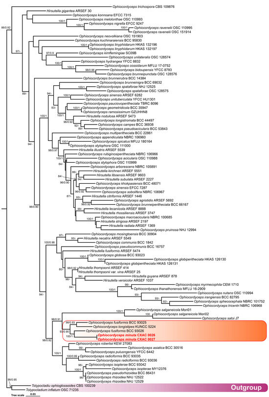
Phylogenetic analyses of Ophiocordyceps were based on a dataset of 92 sequences (Table 2). Tolypocladium inflatum OSC 71235 and T. ophioglossoides CBS 100239 were used as the outgroups. The final concatenated matrix comprised 2927 characters (including gaps), with three partitions: nrLSU 908 bp, tef1-α 957 bp, and rpb2 1062 bp. Both Bayesian inference (BI) and maximum likelihood (ML) analyses produced congruent trees, recovering Ophiocordyceps as distinct, well-supported clades (Figure 1). The new species Ophiocordyceps minuta formed a distinct lineage within a well-supported clade, clearly separated from its close relatives (Figure 1).
Figure 1.
Molecular phylogenetic analyses using the ML and BI based on combined nrLSU, tef1-α, and rpb2 sequence data. Statistical support values (BS ≥ 80% and PP ≥ 0.80) are shown at the nodes for ML bootstrap support (BS) and BI posterior probabilities (PP). Tolypocladium inflatum OSC 71235 and T. ophioglossoides CBS 100239 were used as outgroup taxa. Species in red type are those analyzed in this study. The scale bar represents the expected number of changes per site.
3.2. Taxonomy
Ophiocordyceps minuta Q.Y. Dong & C. D. Xu, sp. nov.
Figure 2.
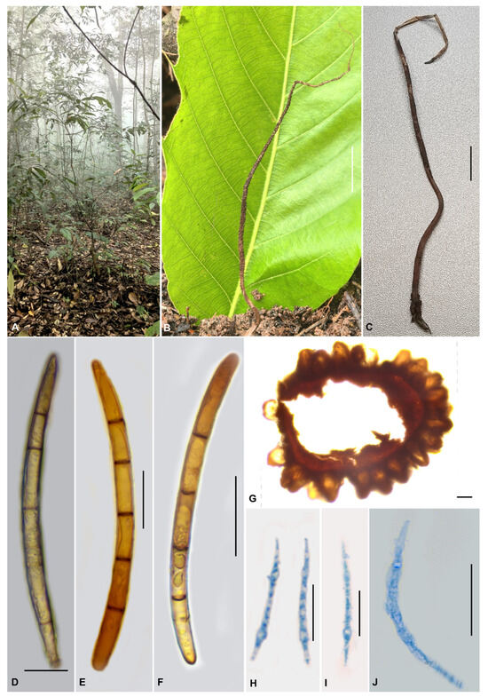
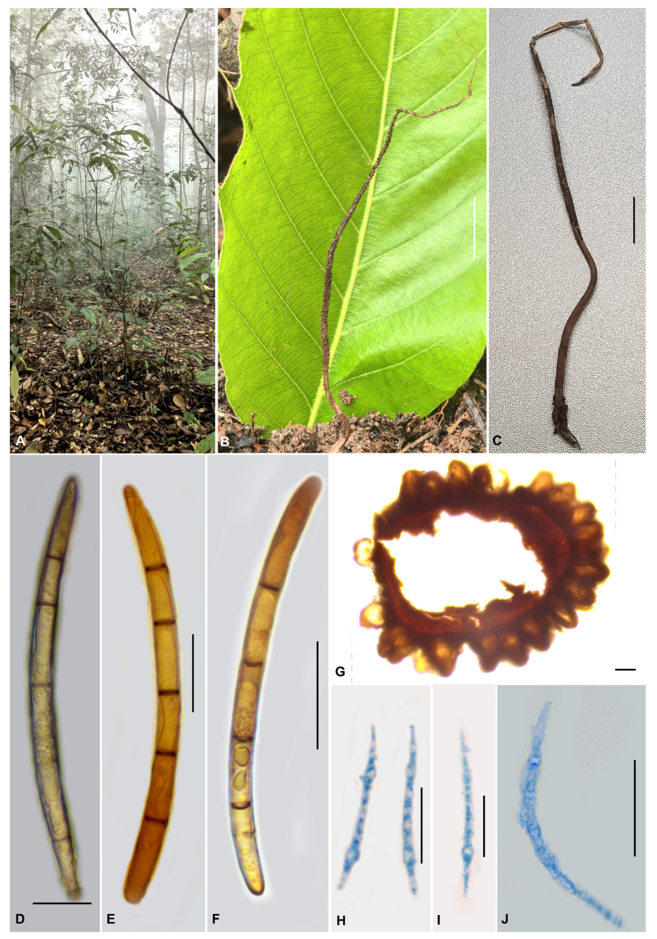
Morphological features of Ophiocordyceps minuta (holotype CXAC 0026). (A). Habitat; (B,C). Fungus on termite; (D–F). Ascospores; (G). Perithecia; (H–J). Ascospores. Scale bars: (B,C) = 10 mm; (D–F,H–J) = 20 µm; (G) = 200 µm.
MycoBank no: 862337
Etymology: minuta = minute, referring to the distinctly small size of the perithecia, a key diagnostic feature that distinguishes this species from its closely related congeners.
Holotype: China, Yunnan Province, Pu’er City, Simao District, Taiyanghe National Nature Reserve, on termite (Termitidae, Macrotermitinae), 15 August 2025, Quanying Dong (holotype CXAC 0026).
Stromata morph: Stromata arising from the segment between the prothorax and mesothorax of termite hosts buried in soil, solitary, simple, cylindrical, flexible, leathery, 6–14 cm long, 0.5–1.0 mm wide, brown to yellowish brown. Fertile parts cylindrical, yellowish brown, 3.5–5.5 cm long, 4–5 mm wide, located on the upper part of the stromata, with a spinous surface and a sterile apical tip 13–28 mm long, 0.5–1.0 mm wide. Perithecia superficial, pale yellow when young, turning brown at maturity, ovoid to flask-shaped, 151.5–221 × 115–188 µm, densely and irregularly scattered over the mid portion of the stromata. Ascospores dimorphic: type I fusiform, 80–92.5 × 5.0–6.5 µm, whole, hyaline, multiseptate, septa 8–14.5 µm long; type II vermiform or lanceolate, 32.5–52.5 × 2.0–4.5 µm.
Asexual morph: Undetermined.
Host: Parasitic on termites buried in soil.
Known distribution: Yunnan Province, China.
Additional specimens examined: China, Yunnan Province, Pu’er City, Simao District, Taiyanghe National Nature Reserve, on termite (Termitidae, Macrotermitinae), 15 August 2025, Quanying Dong (paratype CXAC 0027).
Commentary: The diagnostic morphology of Ophiocordyceps minuta conforms to the generic concept of Ophiocordyceps. The species produces dimorphic ascospores comprising two types: cylindrical, with pointed ends, and vermiform. The species can be distinguished by the following combination of morphological characteristics: perithecia distinctly small (151.5–221 × 115–188 µm), which is the smallest among its closely related congeners (Table 3); host specificity to termites (Termitidae, Macrotermitinae); producing dimorphic ascospores, type I fusiform and multiseptate (80–92.5 × 5.0–6.5 µm), type II vermiform or lanceolate (32.5–52.5 × 2.0–4.5 µm); and asci with conspicuous apical caps (3.5–5.5 μm high, 6.5–8.0 μm wide).
Table 3.
Morphological comparison between Ophiocordyceps species associated with termites.
Our multi-locus phylogenetic data (Figure 1) indicate that Ophiocordyceps minuta is related to O. fusiformis Tasan. et al. and O. longistipes Y.B. Wang et al. However, it forms a distinct clade separate from these taxa (Figure 1). Morphologically, O. minuta differs from related species in the following ways. Ophiocordyceps fusiformis, a species described from Thailand, also similar to O. minuta in appearance, has relatively larger perithecia 300–360 × 180–270 µm, longer asci 141–227 × 7–15 µm and shorter ascospores 6–78 × 5–6.5 µm, shorter septa 5–12 (19) µm long [27]. Moreover, in our phylogenetic tree, the two strains of O. fusiformis (BCC 93025 and BCC 93026) did not form a single clade but instead separated into two distinct branches. Comparison of the tef1-α gene sequences between the two strains revealed a 65 bp difference (out of a total length of 903 bp). Ophiocordyceps longistipes can be distinguished from O. minuta by its grayish white to yellowish brown with relatively longer stromata, larger perithecia measuring 390–420 × 295–350 µm; filiform asci, 160–195 × 4.5–6.5 µm; and filiform shorter ascospores 70–85 × 3.5–4.5 µm [21].
3.3. Morphological Characters and Their Taxonomic Significance in Termite-Associated Ophiocordyceps Species
Analysis of termite-associated Ophiocordyceps (Table 3) shows that morphological characters differ in taxonomic usefulness, a pattern seen in other Ophiocordycipitaceae. Host association is uniform (all on termites, Isoptera), so it does not help distinguish species but defines the group as a specialized ecological guild. Stromatal characters provide moderate to high resolution. Stromatal color (orange-brown, brown, yellowish-brown, grayish-white to yellowish-brown, or pale to red-brown) aids species recognition. Stromatal size shows clear extremes: diminutive in O. octospora and O. taiwanensis, elongate in O. asiatica, O. longistipes, O. pseudocommunis, and O. pseudorhizoidea. Multiple stromata (O. bispora: 20–30; O. globiperitheciata: 2–5) are also diagnostic. Perithecial immersion status (superficial, immersed, or pseudo-immersed) reliably separates species, as does perithecial size (large in O. communis and O. pseudocommunis; small in O. ovatospora and O. khokpasiensis). Ascus characters are less informative. Ascospores vary strikingly: most species have filiform, septate, whole ascospores; exceptions include elliptical to ellipsoidal–fusiform, non-disarticulating ascospores (O. bispora, O. octospora) and dimorphic ascospores (O. minuta), the latter being a unique autapomorphy. Asexual morphs (Hirsutella- or Hymenostilbe-like), conidiogenous cells (monophialidic or polyphialidic), and conidia (fusiform, globose, oval, or citriform) offer additional characters. In summary, perithecial immersion status, ascospore architecture, and conidial features are the most taxonomically valuable traits.
4. Discussion
The description of Ophiocordyceps minuta from termite hosts in Yunnan Province adds a new species to the entomopathogenic fungal diversity of Southeast Asia. The morphological and phylogenetic data presented here contribute to ongoing discussions on species delimitation, ascospore variation, and biogeography within the genus Ophiocordyceps.
Taxonomic implications of O. fusiformis strain divergence. In our phylogenetic analysis, the two ex-type strains of O. fusiformis (BCC 93025 and BCC 93026) occupied distinct positions (Figure 1), with a 65 bp divergence in the tef1-α region. Although these strains were originally described as conspecific from Thailand [27], this level of sequence divergence suggests that the current concept of O. fusiformis may encompass multiple species. Confirming species boundaries would require re-examination of type material and additional collections from the type locality. This finding reinforces the importance of including multiple representatives per putative species in phylogenetic studies, as single-strain typification may not fully capture intraspecific variation or detect cryptic diversity [48,60].
Ophiocordyceps minuta produces two morphologically distinct ascospore types. The vermiform Type II spores (32.5–52.5 × 2.0–4.5 µm) are similar in size and shape to microconidia produced by some Hirsutella species associated with termite-pathogenic Ophiocordyceps [8,21,22]. Whether the two ascospore types differ in biological function remains unknown; infection assays comparing their relative infectivity would help clarify their respective roles in the life cycle of this species.
Biogeographic patterns. The discovery of O. minuta in Yunnan adds to the growing number of termite-associated Ophiocordyceps species recorded from the Indochinese Peninsula, including recent reports from Thailand and Vietnam [21,22,27]. Multiple species from this ecological group have now been documented in the Mekong River basin and adjacent areas [6]. The 65 bp divergence observed between the two O. fusiformis strains, both collected in Thailand, further indicates that sequence variation exists within what is currently recognized as a single species. This observation highlights the need for more intensive sampling across the region, particularly collecting multiple specimens per morphospecies and from different host colonies [61].
Ophiocordyceps minuta is a morphologically and phylogenetically distinct species. The genetic divergence detected among O. fusiformis strains and the presence of dimorphic ascospores in O. minuta point to several directions for future research, including taxonomic revision of the O. fusiformis complex and functional studies of ascospore variation in O. minuta. These findings underscore the value of continued exploration of termite-associated Ophiocordyceps in Southeast Asia.
Author Contributions
Conceptualization, Q.-Y.D.; methodology, J.-L.L. and C.-K.L. validation, C.H. and W.-L.X.; formal analysis C.-D.X. and J.Y.; investigation, Q.-Y.D.; writing—review and editing, Q.-Y.D. All authors have read and agreed to the published version of the manuscript.
Funding
This research was funded by the Joint Program of Basic Research for Provincial Undergraduate Schools, Yunnan Provincial Department of Science and Technology (202401BA070001-139), the Ph.D. Research Start-Up Project of Chuxiong Normal University (BSQD2307), and the 2025 Self-funded Science and Technology Project of Chuxiong Prefecture (cxzc2025019).
Institutional Review Board Statement
Not applicable for studies not involving humans or animals.
Data Availability Statement
Data are contained within the article.
Conflicts of Interest
The authors declare no conflicts of interest.
References
- Dong, Q.Y.; Wang, Y.; Wang, Z.Q.; Tang, D.X.; Zhao, Z.Y.; Wu, H.J.; Yu, H. Morphology and phylogeny reveal five novel species in the genus Cordyceps (Cordycipitaceae, Hypocreales) from Yunnan, China. Front. Microbiol. 2022, 13, 846909. [Google Scholar] [CrossRef] [Scilit] [PubMed]
- Sung, G.H.; Hywel-Jones, N.L.; Sung, J.M.; Luangsa-ard, J.J.; Shrestha, B.; Spatafora, J.W. Phylogenetic classification of Cordyceps and the clavicipitaceous fungi. Stud. Mycol. 2007, 57, 5–59. [Google Scholar] [CrossRef] [Scilit]
- Araújo, J.P.M.; Evans, H.C.; Kepler, R.M.; Hughes, D.P. Zombie-ant fungi across continents: 15 new species and new combinations within Ophiocordyceps. I. Myrmecophilous hirsutelloid species. Stud. Mycol. 2018, 90, 119–160. [Google Scholar] [CrossRef] [Scilit]
- Thanakitpipattana, D.; Tasanathai, K.; Lamlertthon, S.; Luangsa-ard, J.J.; Mongkolsamrit, S.; Khonsanit, A. Fungal pathogens occurring on Orthopterida in Thailand. Persoonia 2020, 44, 140–160. [Google Scholar] [CrossRef] [Scilit]
- Zhang, X.; Peng, X.C.; Wei, D.P.; Wang, Y.; Norphanphoun, C.; Hongsanan, S.; Xie, N.; Liu, Z.L.; Mapook, A.; Kang, J.C.; et al. Reappraisal of the phylogenetic relationships of hirsutella-like fungi across Ophiocordycipitaceae and Polycephalomycetaceae (Hypocreales, Sordariomycetes), and the description of seven new species. IMA Fungus 2026, 17, 171084. [Google Scholar] [CrossRef] [Scilit]
- Dai, Y.D.; Guan, Y.H.; Wu, S.M.; Bibi, S.; Chen, H.; Loinheuang, C.; Liang, J.D.; Wang, Y. Taxonomic and phylogenetic insights into dipteran-parasitizing Ophiocordyceps: Descriptions of two new species and a new record from China and Laos. MycoKeys 2026, 127, 217–238. [Google Scholar] [CrossRef] [Scilit]
- Wang, Y.B.; Nguyen, T.T.; Dai, Y.D.; Yu, H.; Zeng, W.B.; Wu, C.K. Molecular phylogeny and morphology of Ophiocordyceps unituberculata sp. nov. (Ophiocordycipitaceae), a pathogen of caterpillars (Noctuidae, Lepidoptera) from Yunnan, China. Mycol. Prog. 2018, 17, 745–753. [Google Scholar] [CrossRef] [Scilit]
- Tasanathai, K.; Noisripoom, W.; Chaitika, T.; Khonsanit, A.; Hasin, S.; Luangsa-ard, J.J. Phylogenetic and morphological classification of Ophiocordyceps species on termites from Thailand. MycoKeys 2019, 56, 101–129. [Google Scholar] [CrossRef] [Scilit]
- Araujo, J.P.M.; Moriguchi, M.G.; Uchiyama, S.; Kinjo, N.; Matsuura, Y. Ophiocordyceps salganeicola, a parasite of social cockroaches in Japan and insights into the evolution of other closely-related Blattodea-associated lineages. IMA Fungus 2021, 12, 3. [Google Scholar] [CrossRef] [Scilit] [PubMed]
- Petch, T. Notes on entomogenous fungi. Trans. Br. Mycol. Soc. 1931, 16, 55–75. [Google Scholar] [CrossRef] [Scilit]
- Yang, Y.; Xiao, Y.P.; Jayawardena, R.S.; Hyde, K.D.; Nilthong, S.; Mapook, A.; Lu, Y.Z.; Xie, S.Q.; Al-Otibi, F.; Wang, X.; et al. Three new species of Ophiocordyceps (Hypocreales, Ophiocordycipitaceae) and a new host record for Hirsutella vermicola from China. MycoKeys 2025, 117, 289–313. [Google Scholar] [CrossRef] [Scilit]
- Xie, S.W.; Wei, D.P.; Qiu, J.Z.; Peng, X.C.; Kang, J.C.; He, Z.J.; Li, Z.Z.; Li, C.R.; Huang, S.K.; Zhang, X.; et al. Overview of hirsutella-like anamorphs in Ophiocordyceps (Sordariomycetes, Ophiocordycipitaceae): Introducing two new species and one new record from China. MycoKeys 2025, 119, 95–121. [Google Scholar] [CrossRef] [Scilit] [PubMed]
- Zou, W.Q.; Tang, D.X.; Xu, Z.H.; Huang, O.; Wang, Y.B.; Tran, N.-L.; Yu, H. Multigene phylogeny and morphology reveal Ophiocordyceps hydrangea sp. nov. and Ophiocordyceps bidoupensis sp. nov. (Ophiocordycipitaceae). MycoKeys 2022, 92, 109–130. [Google Scholar] [CrossRef] [Scilit]
- Sun, T.; Zou, W.Q.; Dong, Q.Y.; Huang, O.; Tang, D.X.; Yu, H. Morphology, phylogeny, mitogenomics and metagenomics reveal a new entomopathogenic fungus Ophiocordyceps nujiangensis (Hypocreales, Ophiocordycipitaceae) from Southwestern China. MycoKeys 2022, 94, 91–108. [Google Scholar] [CrossRef] [Scilit]
- Tang, D.X.; Huang, O.; Zou, W.Q.; Wang, Y.; Dong, Q.Y.; Sun, T.; Yang, G.; Yu, H. Six new species of zombie-ant fungi from Yunnan in China. IMA Fungus 2023, 14, 9. [Google Scholar] [CrossRef] [Scilit]
- Araujo, J.P.M.; Evans, H.C.; Fernandes, I.O.; Ishler, M.J.; Hughes, D.P. Zombie-ant fungi cross continents: II. Myrmecophilous hymenostilboid species and a novel zombie lineage. Mycologia 2020, 112, 1138–1170. [Google Scholar] [CrossRef] [Scilit]
- Inward, D.; Beccaloni, G.; Eggleton, P. Death of an order: A comprehensive molecular phylogenetic study confirms that termites are eusocial cockroaches. Biol. Lett. 2007, 3, 331–335. [Google Scholar] [CrossRef] [Scilit]
- Hellemans, S.; Rocha, M.M.; Wang, M.; Romero Arias, J.; Aanen, D.K.; Bagnères, A.-G.; Buček, A.; Carrijo, T.F.; Chouvenc, T.; Cuezzo, C.; et al. Genomic data provide insights into the classification of extant termites. Nat. Commun. 2024, 15, 6724. [Google Scholar] [CrossRef] [Scilit] [PubMed]
- Krishna, K.; Grimaldi, D.A.; Krishna, V.; Engel, M.S. Treatise on the Isoptera of the World. Bull. Am. Mus. Nat. Hist. 2013, 377, 1–200. [Google Scholar] [CrossRef] [Scilit]
- Wu, D.H.; Du, E.; Eisenhauer, N.; Mathieu, J.; Chu, C.J. Global engineering effects of soil invertebrates on ecosystem functions. Nature 2025, 640, 120–129. [Google Scholar] [CrossRef] [Scilit] [PubMed]
- Fan, Q.; Yang, T.; Li, H.; Wang, X.M.; Liao, H.F.; Shen, P.H.; Yang, Z.L.; Zeng, W.B.; Wang, Y.B. Molecular phylogeny and morphology reveal two new entomopathogenic species of Ophiocordyceps (Ophiocordycipitaceae, Hypocreales) parasitic on termites from China. MycoKeys 2024, 103, 1–24. [Google Scholar] [CrossRef] [Scilit]
- Xu, Z.H.; Tran, N.L.; Wang, Y.; Zhang, G.D.; Dao, V.M.; Nguyen, T.T.; Wang, Y.B.; Yu, H. Phylogeny and morphology of Ophiocordyceps puluongensis sp. nov. (Ophiocordycipitaceae, Hypocreales), a new fungal pathogen on termites from Vietnam. J. Invertebr. Pathol. 2022, 192, 107771. [Google Scholar] [CrossRef] [Scilit] [PubMed]
- Samarakoon, M.C.; Hsu, K.C.; Chiu, C.I.; Chen, C.Y.; Li, H.F.; Haituk, S. Ophiocordyceps taiwanensis sp. nov. (Ophiocordycipitaceae, Hypocreales) on Odontotermes formosanus (Termitidae, Blattodea). Phytotaxa 2024, 660, 97–111. [Google Scholar] [CrossRef] [Scilit]
- Stifler, C.B. A new genus of Hypocreales. Mycologia 1941, 33, 82–86. [Google Scholar] [CrossRef] [Scilit]
- Penzig, O.; Saccardo, P.A. Icones Fungorum Javanicorum; Buchhandlung and Druckerei EJ Brill: Leide, The Netherlands, 1904; pp. 55–57. [Google Scholar] [CrossRef] [Scilit]
- Blackwell, M.; Gilbertson, R.L. New information on Cordycepioideus bisporus and Cordycepioideus octosporus. Mycologia 1984, 76, 763–765. [Google Scholar] [CrossRef] [Scilit]
- Tasanathai, K.; Khonsanit, A.; Noisripoom, W.; Kobmoo, N.; Luangsa-ard, J.J. Hidden species behind Ophiocordyceps (Ophiocordycipitaceae, Hypocreales) on termites: Four new species from Thailand. Mycol. Prog. 2022, 21, 86–102. [Google Scholar] [CrossRef] [Scilit]
- Tang, D.X.; Zhu, J.Y.; Luo, L.J.; Hou, D.H.; Wang, Z.Q.; Yang, S.D.; Yu, H. Ophiocordyceps ovatospora sp. nov. (Ophiocordycipitaceae, Hypocreales), pathogenic on termites from China. Phytotaxa 2022, 574, 105–117. [Google Scholar] [CrossRef] [Scilit]
- Ochiel, G.S.; Evans, H.C.; Eilenberg, J. Cordycepioideus, a pathogen of termites in Kenya. Mycologist 1997, 11, 7–9. [Google Scholar] [CrossRef] [Scilit]
- Dai, Y.D.; Wu, C.K.; Yuan, F.; Wang, Y.B.; Huang, L.D.; Chen, Z.H.; Zeng, W.B.; Wang, Y.; Yang, Z.L.; Zeng, P.S.; et al. Evolutionary biogeography on Ophiocordyceps sinensis: An indicator of molecular phylogeny to geochronological and ecological exchanges. Geosci. Front. 2020, 11, 807–820. [Google Scholar] [CrossRef] [Scilit]
- Dong, Q.Y.; Gao, S.Y.; Zhou, J.N.; Xu, C.D.; Wang, Z.J.; Zeng, N.K. Verticillium-like anamorphic fungi in Sordariomycetes from Southwestern China: Two new taxa and a new record. J. Fungi 2025, 11, 598. [Google Scholar] [CrossRef] [Scilit] [PubMed]
- Liao, Z.Y.; Yang, D.R.; Kong, D.B.; Guan, Y.F.; Zhang, Y.; Zeng, J.; Wang, X.B.; Yuan, Y.L.; Deng, Z.J.; Zhang, Y. Distribution patterns of insect diversity along elevation gradients in Southeastern margin of Qinghai-Tibetan plateau. Res. J. Environ. Sci. 2024, 37, 2138–2147. [Google Scholar] [CrossRef]
- Ashton, L.A.; Nakamura, A.; Burwell, C.J.; Tang, Y.; Cao, M.; Whitaker, T.; Sun, Z.; Huang, H.; Kitching, R.L. Elevational sensitivity in an Asian ‘hotspot’: Moth diversity across elevational gradients in tropical, sub-tropical and sub-alpine China. Sci. Rep. 2016, 6, 26513. [Google Scholar] [CrossRef] [Scilit] [PubMed]
- Rehner, S.A.; Samuels, G.J. Taxonomy and phylogeny of Gliocladium analysed from nuclear large subunit ribosomal DNA sequences. Mycol. Res. 1994, 98, 625–634. [Google Scholar] [CrossRef] [Scilit]
- Vilgalys, R.; Hester, M. Rapid genetic identification and mapping of enzymatically amplified ribosomal DNA from several Cryptococcus species. J. Bacteriol. 1990, 172, 4238–4246. [Google Scholar] [CrossRef] [Scilit]
- Bischoff, J.F.; Rehner, S.A.; Humber, R.A. Metarhizium frigidum sp. nov.: A cryptic species of M. anisopliae and a member of the M. flavoviride complex. Mycologia 2006, 98, 737–745. [Google Scholar] [CrossRef]
- Kück, P.; Longo, G.C. FASconCAT-G: Extensive functions for multiple sequence alignment preparations concerning phylogenetic studies. Front. Zool. 2014, 11, 81. [Google Scholar] [CrossRef] [Scilit] [PubMed]
- Lanfear, R.; Frandsen, P.B.; Wright, A.M.; Senfeld, T.; Calcott, B. PartitionFinder 2: New methods for selecting partitioned models of evolution for molecular and morphological phylogenetic analyses. Mol. Biol. Evol. 2016, 34, 772–773. [Google Scholar] [CrossRef] [Scilit]
- Nguyen, L.T.; Schmidt, H.A.; von Haeseler, A.; Minh, B.Q. IQ-TREE: A fast and effective stochastic algorithm for estimating maximum-likelihood phylogenies. Mol. Biol. Evol. 2015, 32, 268–274. [Google Scholar] [CrossRef] [Scilit]
- Nylander, J.A.A. MrModeltest Version 2. Program Distributed by the Author; Uppsala University: Uppsala, Sweden, 2004. [Google Scholar]
- Ronquist, F.; Huelsenbeck, J.P. MrBayes 3: Bayesian phylogenetic inference under mixed models. Bioinformatics 2003, 19, 1572–1574. [Google Scholar] [CrossRef] [Scilit]
- Rambaut, A.; Drummond, A.J.; Xie, D.; Baele, G.; Suchard, M.A. Posterior Summarization in Bayesian Phylogenetics Using Tracer 1.7. Syst. Biol. 2018, 67, 901–904. [Google Scholar] [CrossRef] [Scilit]
- Xie, J.M.; Chen, Y.R.; Cai, G.J.; Cai, R.L.; Hu, Z.; Wang, H. Tree visualization by one table (tvBOT): A web application for visualizing, modifying and annotating phylogenetic trees. Nucleic Acids Res. 2023, 51, 587–592. [Google Scholar] [CrossRef] [Scilit]
- Simmons, D.R.; Kepler, R.M.; Rehner, S.A.; Groden, E. Phylogeny of Hirsutella species (Ophiocordycipitaceae) from the USA: Remedying the paucity of Hirsutella sequence data. IMA Fungus 2015, 6, 345–356. [Google Scholar] [CrossRef] [Scilit]
- Spatafora, J.W.; Sung, G.H.; Sung, J.M.; Hywel-Jones, N.L.; White, J.F. Phylogenetic evidence for an animal pathogen origin of ergot and the grass endophytes. Mol. Ecol. 2007, 16, 1701–1711. [Google Scholar] [CrossRef] [Scilit]
- Schoch, C.L.; Seifert, K.A.; Huhndorf, S.; Robert, V.; Spouge, J.L.; Levesque, C.A.; Chen, W.; Fungal Barcoding, C.; Fungal Barcoding Consortium Author, L. Nuclear ribosomal internal transcribed spacer (ITS) region as a universal DNA barcode marker for Fungi. Proc. Natl. Acad. Sci. USA 2012, 109, 6241–6246. [Google Scholar] [CrossRef] [Scilit]
- Ban, S.; Sakane, T.; Nakagiri, A. Three new species of Ophiocordyceps and overview of anamorph types in the genus and the family Ophiocordyceptaceae. Mycol. Prog. 2015, 14, 1017. [Google Scholar] [CrossRef] [Scilit]
- Luangsa-Ard, J.; Tasanathai, K.; Thanakitpipattana, D.; Khonsanit, A.; Stadler, M. Novel and interesting Ophiocordyceps spp. (Ophiocordycipitaceae, Hypocreales) with superficial perithecia from Thailand. Stud. Mycol. 2018, 89, 125–142. [Google Scholar] [CrossRef] [Scilit]
- Tasanathai, K.; Thanakitpipattana, D.; Himaman, W.; Phommavong, K.; Dengkhhamounh, N.; Luangsa-ard, J. Three new Ophiocordyceps species in the Ophiocordyceps pseudoacicularis species complex on Lepidoptera larvae in Southeast Asia. Mycol. Prog. 2020, 19, 1043–1056. [Google Scholar] [CrossRef] [Scilit]
- Xiao, Y.P.; Hongsanan, S.; Hyde, K.D.; Brooks, S.; Xie, N.; Long, F.Y.; Wen, T.C. Two new entomopathogenic species of Ophiocordyceps in Thailand. MycoKeys 2019, 47, 53–74. [Google Scholar] [CrossRef] [Scilit]
- Yang, Y.U.; Jayawardena, R.S.; Hyde, K.D.; Lu, Y.Z.; Mapook, A.; Nilthong, S.; Xie, S.Q.; Xiao, Y.P. Ophiocordyceps linyphiidarum sp. nov., a newly discovered spider-pathogenic fungus from China. Phytotaxa 2025, 715, 32–46. [Google Scholar] [CrossRef] [Scilit]
- Mongkolsamrit, S.; Noisripoom, W.; Arnamnart, N.; Lamlertthon, S.; Himaman, W.; Jangsantear, P.; Samson, R.A.; Luangsa-ard, J.J. Resurrection of Paraisaria in the Ophiocordycipitaceae with three new species from Thailand. Mycol. Prog. 2019, 18, 1213–1230. [Google Scholar] [CrossRef] [Scilit]
- Quandt, C.A.; Kepler, R.M.; Gams, W.; Araujo, J.P.; Ban, S.; Evans, H.C.; Hughes, D.; Humber, R.; Hywel-Jones, N.; Li, Z.; et al. Phylogenetic-based nomenclatural proposals for Ophiocordycipitaceae (Hypocreales) with new combinations in Tolypocladium. IMA Fungus 2014, 5, 121–134. [Google Scholar] [CrossRef] [Scilit]
- Johnson, D.; Sung, G.H.; Hywel-Jones, N.L.; Luangsa-ard, J.J.; Bischoff, J.F.; Kepler, R.M.; Spatafora, J.W. Systematics and evolution of the genus Torrubiella (Hypocreales, Ascomycota). Mycol. Res. 2009, 113, 279–289. [Google Scholar] [CrossRef] [Scilit]
- Wen, T.C.; Xiao, Y.P.; Li, W.J.; Kang, J.C.; Hyde, K.D. Systematic analyses of Ophiocordyceps ramosissimum sp. nov., a new species from a larvae of Hepialidae in China. Phytotaxa 2014, 161, 227–234. [Google Scholar] [CrossRef] [Scilit]
- Kepler, R.M.; Sung, G.H.; Ban, S.; Nakagiri, A.; Chen, M.J.; Huang, B.; Li, Z.; Spatafora, J.W. New teleomorph combinations in the entomopathogenic genus Metacordyceps. Mycologia 2012, 104, 182–197. [Google Scholar] [CrossRef] [Scilit]
- Zha, L.S.; Kryukov, V.Y.; Ding, J.H.; Jeewon, R.; Chomnunti, P. Novel taxa and species diversity of Cordyceps sensu lato (Hypocreales, Ascomycota) developing on wireworms (Elateroidea and Tenebrionoidea, Coleoptera). MycoKeys 2021, 78, 79–117. [Google Scholar] [CrossRef] [Scilit]
- Xiao, Y.P.; Wen, T.C.; Hongsanan, S.; Sun, J.Z.; Hyde, K.D. Introducing Ophiocordyceps thanathonensis, a new species of entomogenous fungi on ants, and a reference specimen for O. pseudolloydii. Phytotaxa 2017, 328, 115–126. [Google Scholar] [CrossRef] [Scilit]
- Sung, G.H.; Sung, J.M.; Hywel-Jones, N.L.; Spatafora, J.W. A multi-gene phylogeny of Clavicipitaceae (Ascomycota, Fungi); Identification of localized incongruence using a combinational bootstrap approach. Mol. Phylogen. Evol. 2007, 44, 1204–1223. [Google Scholar] [CrossRef] [Scilit]
- Dong, Q.Y.; Wang, Y.; Wang, Z.Q.; Liu, Y.F.; Yu, H. Phylogeny and systematics of the genus Tolypocladium (Ophiocordycipitaceae, Hypocreales). J. Fungi 2022, 8, 1158. [Google Scholar] [CrossRef] [Scilit]
- Cui, Y.Y.; Cai, Q.; Tang, L.P.; Liu, J.W.; Yang, Z.L. The family Amanitaceae: Molecular phylogeny, higher-rank taxonomy and the species in China. Fungal Divers. 2018, 91, 5–230. [Google Scholar] [CrossRef] [Scilit]

Disclaimer/Publisher’s Note: The statements, opinions and data contained in all publications are solely those of the individual author(s) and contributor(s) and not of MDPI and/or the editor(s). MDPI and/or the editor(s) disclaim responsibility for any injury to people or property resulting from any ideas, methods, instructions or products referred to in the content. |
© 2026 by the authors. Licensee MDPI, Basel, Switzerland. This article is an open access article distributed under the terms and conditions of the Creative Commons Attribution (CC BY) license.