


Abstract
Fluorescence is known to occur on varying surfaces across crustacea but is only cited in sporadic reports, despite being a potentially important visual signal and changing with development and within moult cycles. Here, we present the occurrence of fluorescence in some common Northeast Atlantic crustaceans and highlight differences by age and sex, to bring attention to this potentially important facet of their biology. Fluorescence was present on dactyli tips across most species, and setae on the ventral side of edible crabs and lobsters (with greater occurrence in females) and over the whole body in spider and hermit crabs.
Fluorescence is present in various forms across Arthropoda [1,2], with occurrence in some groups (particularly scorpions) widely known. Occurrence of fluorescence in crustacea is less well studied than other groups (however, see [3,4,5,6]), potentially due to the more easily detected bioluminescence across many terrestrial taxa in more accessible habitats [7]. Inducement of fluorescence depends on ambient light levels (particularly blue and UV, tied to depth in the sea [5]), while detection depends on the visual capacity of both the fluorescing species (for conspecific interaction) and potential predators or prey [8].
Knowledge of fluorescence and its drivers in crustaceans is limited, but studies on stomatopods (which are known to be sensitive to UV light) have indicated fluorescence as a potential enhancer for visual signalling in the more light-limited underwater environment [5], with the potential for patterns to aid in identification of conspecifics, though it is noted visual systems in this group are particularly sensitive. Fluorescence in Portunid crabs has been found to typically increase with growth stage (juveniles typically displaying lower levels or incomplete patterns compared to those found in adults) [6]. Simulated stress was shown to decrease fluorescence in some areas of Lithoid king crabs over short time scales [4]. The use of fluorescence to aid in crustacean identification has been noted [6], and increasing knowledge of decapod fluorescence has been garnered by underwater photography awards, such as the Underwater Photographer of the Year award for a photo of a fluorescing squat lobster Galathea strigosa (Linnaeus, 1761) [9], as well as other examples for other species and groups exhibiting fluorescence not covered here [10].
Here, we document the occurrence of this property among common species found in the Northeast Atlantic. Observations were made sporadically during rockpooling trips from 2025 to 2026, but all photos (excluding those for edible crabs and European lobsters ) were taken on 3/3/26 with individuals found in the intertidal of East Sands, St Andrews, Scotland. Photos for edible crabs and European lobsters were taken when the species were present in the Scottish Oceans Institute for study on 11/9/25. A commercially available UV torch (Lepro, Las Vegas, NV, USA) was used to induce fluorescence (at 395 nm), and photographs were taken using an OM TG-7 camera (OM Digital Solutions, Tokyo, Japan). Photos were taken in standard or macro mode, with automatic focus used at varying distances. A Zeikos 40.5 mm UV filter (Zeikos, NJ, USA) was used to block reflected UV light.
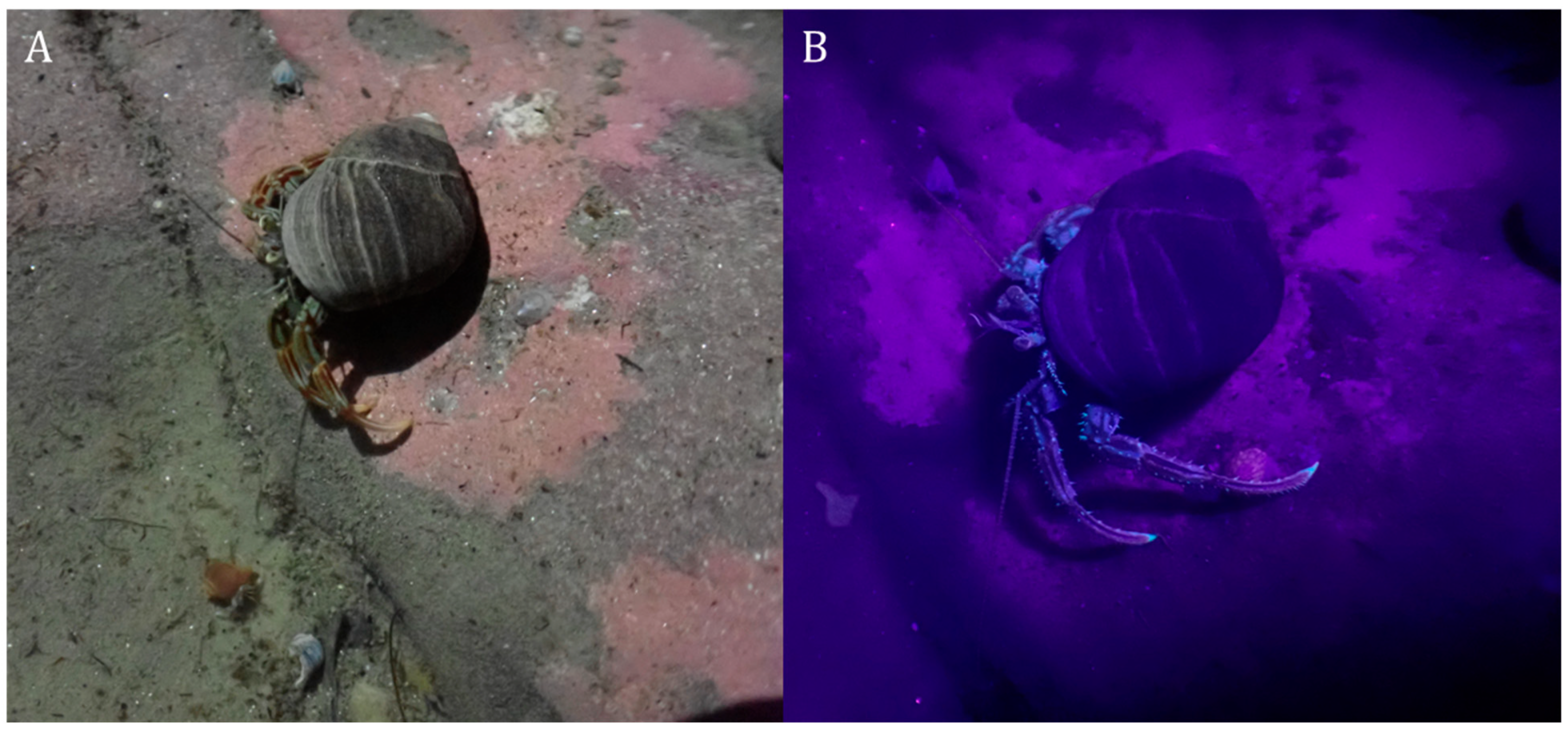
Fluorescence was generally localised in the setae and dactyli tips of pereiopods, the most visible found in the great spider crab Hyas araneus (Linnaeus, 1758; Brachyura: Oregoniidae; Figure 1) and the common hermit crab Pagurus bernhardus (Linnaeus, 1758; Anomura: Paguridae; Figure 2). All encountered H. araneus and P. bernhardus displayed fluorescence in the same areas and to the same degree.
Figure 1.
Dorsal (A,B) and ventral (C,D) views under normal (A,C) and UV (B,D) light of the great spider crab Hyas araneus. Setae fluorescence occurs across the body and limbs, with dactyli tips also fluorescing. Red fluorescing tufts are attached algae (largely Plocamium spp). The carapace width of the individual photographed was ~5 cm.
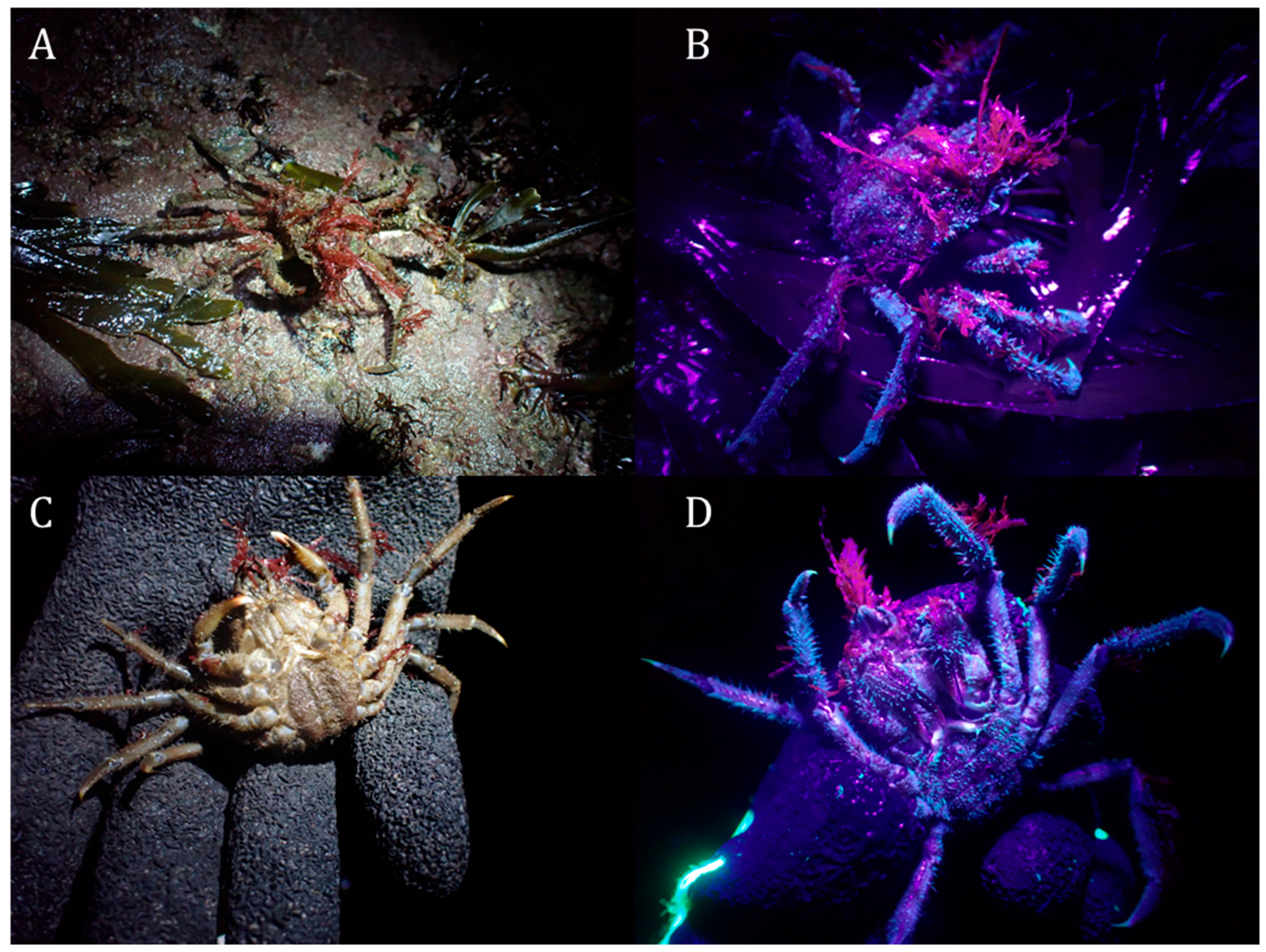
Figure 2.
Common hermit crab Pagurus bernhardus under normal (A) and UV light (B) with fluorescence on setae across the body and dactyli tips. The Littorina littorea shell occupied was ~3 cm at the coiling axis.
The brown crab Cancer pagurus (Linnaeus, 1758; Brachyura: Cancridae) exhibited no visible fluorescence as a juvenile but did so as an adult, with this generally limited to setae on the mouthparts, the underside of the body, and pereiopods except the chelae, in addition to the dactyli tips (Figure 3). Mature females had more and longer setae, resulting in greater levels of fluorescence than males of similar sizes. Juveniles only exhibited fluorescence at dactyli tips. Adult European lobsters Homarus gammarus (Linnaeus, 1758; Astacidea: Nephropidae) also fluoresced, largely from setae on the mouthparts, swimmerets, distal pereiopod segments (except for the chelae), pleon, and telson fringes (Figure 4). Higher setae density was also present on females of this species, but this divergence was to a lesser degree than C. pagurus and unclear in photos (TIB pers. obvs.). Preserved early benthic-phase lobsters (with a carapace length of < 2 cm) were also investigated for fluorescence and did not display it, though it has been noted that preservation can lessen levels [6].
The green crab Carcinus maenas (Linnaeus, 1758; Brachyura: Carcinidae) generally only fluoresced at the dactyli tips (Figure 5). Large individuals sometimes fluoresced on the carapace, possibly due to longer time in moult, driving degradation of the cuticle and exposure of fluorescent layers, especially at the edges and white points on the carapace (Figure 5A,B) or facilitating greater growth of encrusting species which may themselves fluoresce (Figure 5C,D).
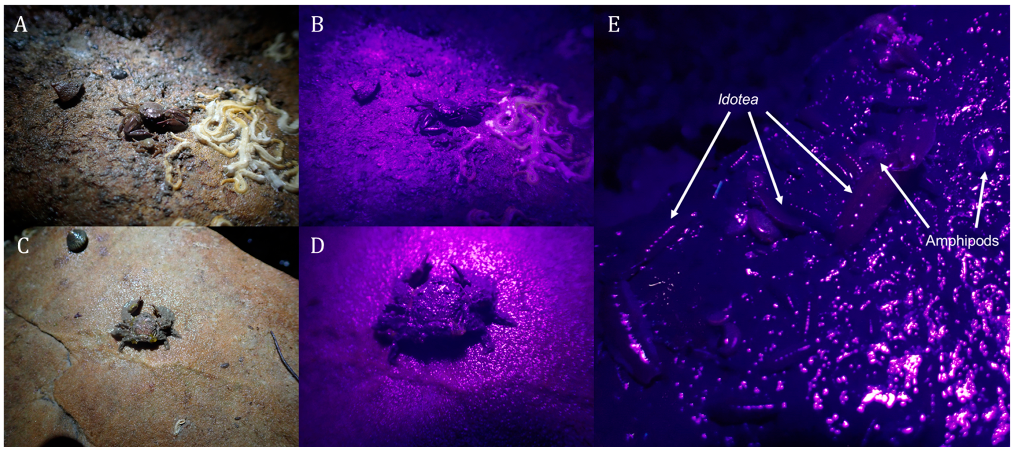
No fluorescence was noted in the long claw porcelain crab Pisidia longicornis (Linnaeus, 1767; Anomura: Porcellanidae), broad claw porcelain crab Porcellana platycheles (Pennant, 1777; Anomura: Porcellanidae), isopods Idotea spp. (Isopoda: Idoteidae), and some amphipods (likely Gammarus spp., Figure 6).
Figure 3.
Views of edible crab Cancer pagurus under normal (A,I) and UV light (B–H,I,K). Panels (A–D) display a female crab (carapace width, CW, ~12 cm) with focus on overall ventral surface (B), mouthparts (C), and pereiopod tips (D). (E–H) display a male (~13 cm CW) with reduced setal cover and fluorescence compared to the female in the ventral surface (F), mouthparts (G), and pereiopods (H). (I–K) displays juveniles (I,J ~6 cm CW, K ~2 cm CW), the colour in ambient light (I) and lack of fluorescence under UV on the dorsal (J,K) and ventral side, excluding dactyli tips (K).
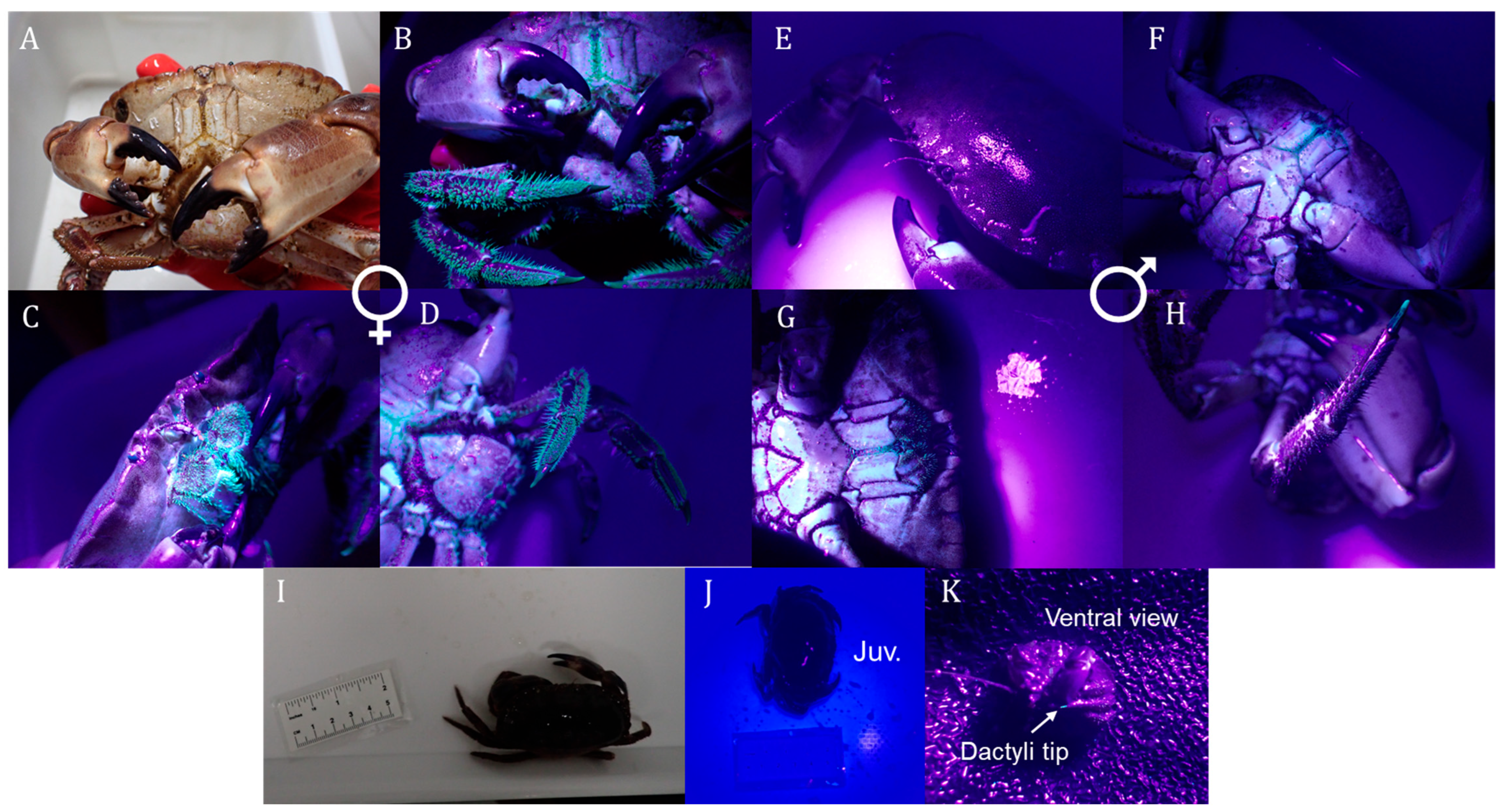
Figure 4.
European lobster Homarus gammarus viewed in normal (A) and UV (B–G) light. Fluorescence was typically limited to setae at the rostrum (B) on the dorsal side but not the rest of the carapace (C). Ventral setal fluorescence was present across the mouthparts (D), pereiopods (E; excluding chelae), telson (F), and pleopods (G). The lobster pictures here had a carapace length of ~9 cm.
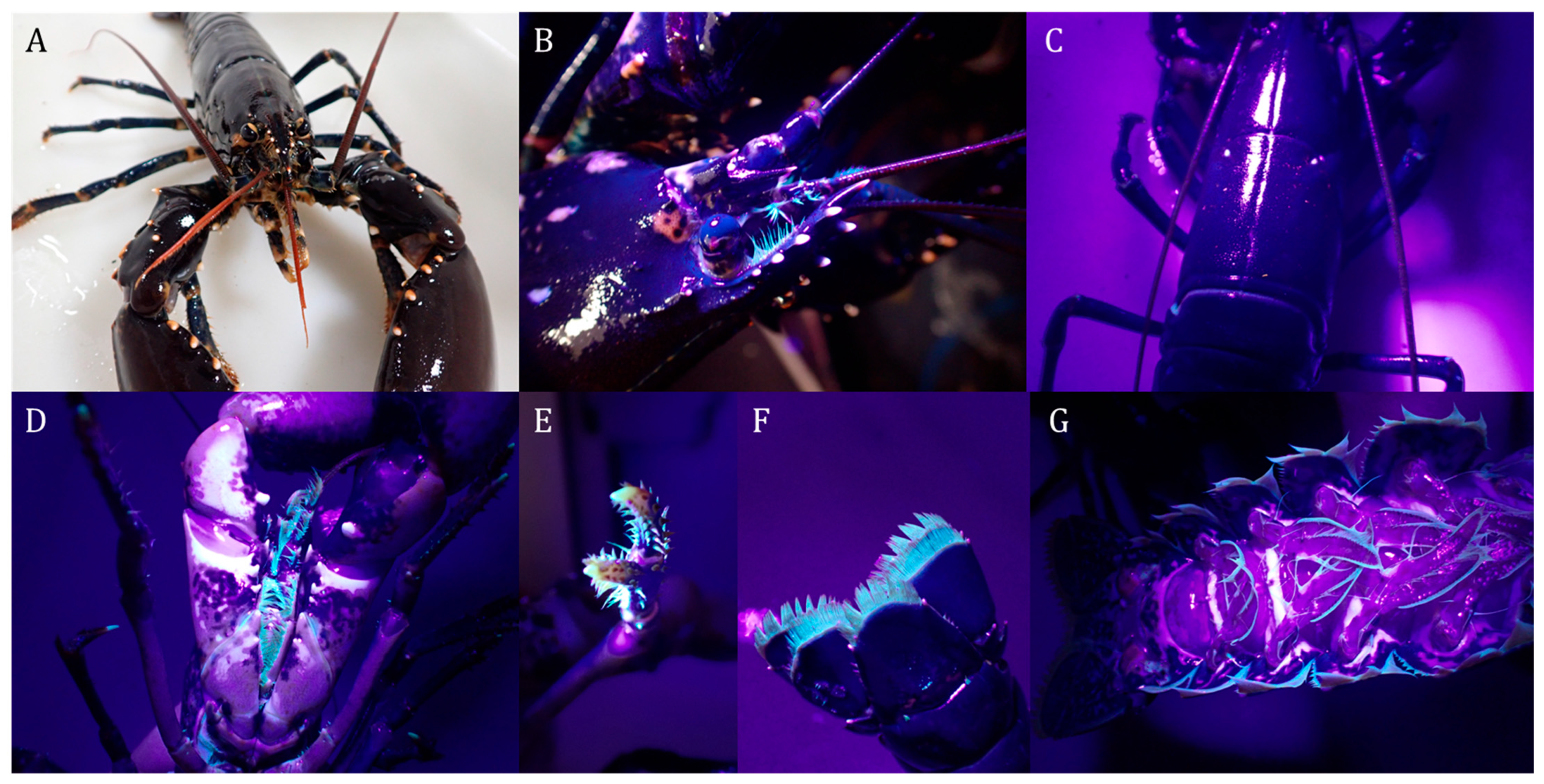
Figure 5.
Views of three green crabs Carcinus maenas (A,B ~9 cm CW, C,D ~6 cm CW, E–G ~5 cm CW) under ambient (A,C,E) and UV light (B,D,F,G). Fluorescence was seen most clearly on the largest (and oldest) crab, matching carapace patterning (B), though it was also seen on dactyli tips (G). White growth on the carapace also fluoresced (C,D).
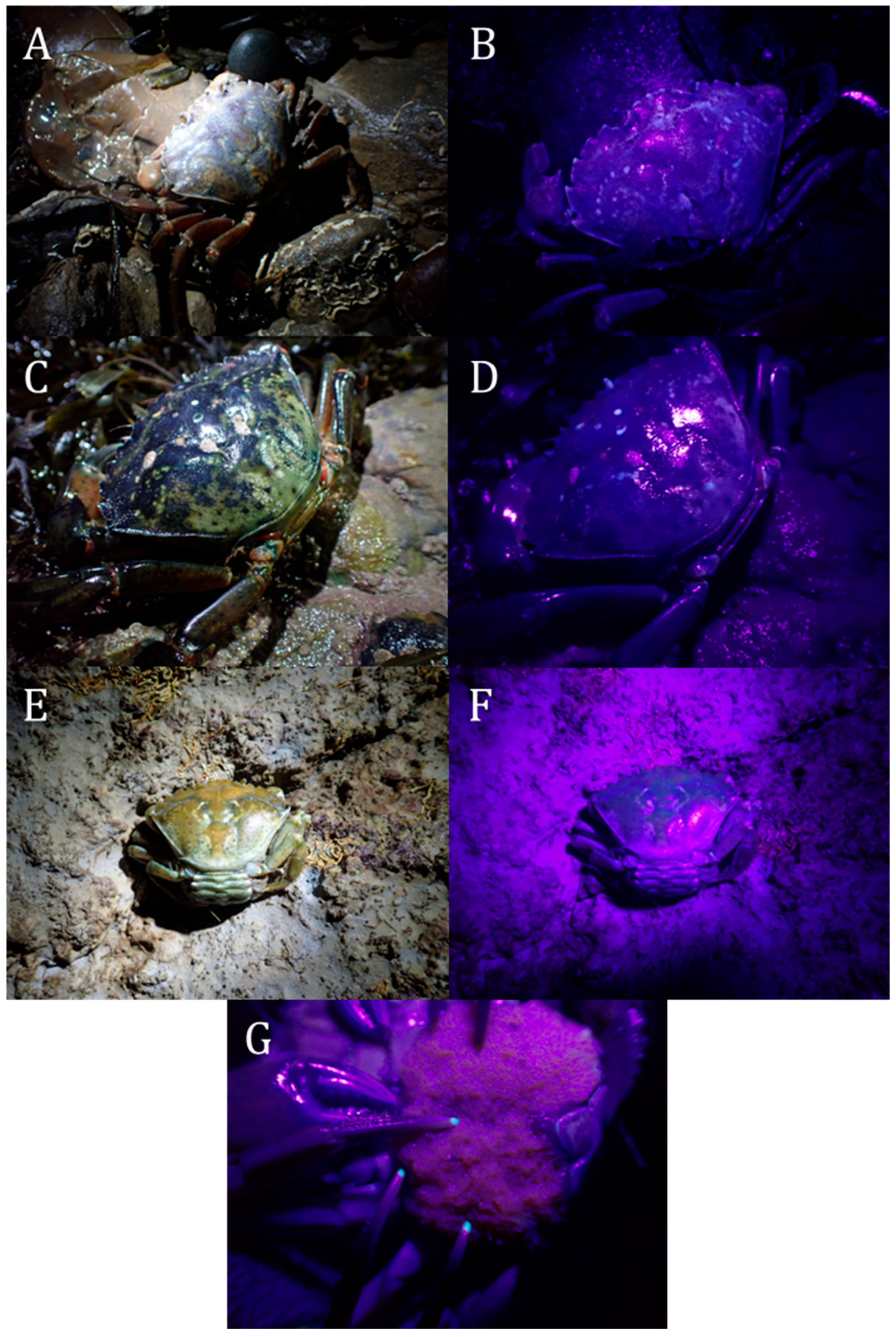
Figure 6.
Views of the long claw porcelain crab Pisidia longicornis (A,B, <1 cm CW) and broad claw porcelain crab Porcellana platycheles (C,D, ~1 cm CW) under normal (A,C) and UV (B,D) light, displaying no fluorescence. Idotea isopods and amphipods under UV (E) also displaying no fluorescence (individual sizes ~1 cm).
Some crustaceans can detect UV [11], and fluorescence in such species can be used for visual signalling [5]. Development of fluorescent structures at maturity and differentially between sexes could function to aid in mate detection, or may simply be a product of differential setae investment between sexes. Consistent localization at the dactyli tips may also be a consequence of cuticle structure at this point, but may aid in increasing the visibility of threat displays, as in stomatopods [5]. Potential predators, such as fish [12], birds [13], and other crustaceans, can also detect UV, and this may constrain fluorescence in small species or juvenile stages which do not have the size-based escape (large decapods) or defences (hermit crab shell, spider crab camouflage) of the other fluorescing species; fluorescence is often lower in juveniles than adults of crustaceans [6] and other arthropods [2]. Species investigated here were predominantly intertidal, where light is present across the spectrum; colour and fluorescence may be more important in these species than crustaceans that inhabit deeper water or pelagic environments, which have more blue-biassed vision due to the reduced spectrum experienced [14].
The lack of observation of fluorescence in some species at any particular stage does not rule it out: the relevant moult stage, age, or sex may not have been observed, or the signal strength of fluorescence in some areas may not have been detected by the observers. If fluorescence development is seasonal, it also may have been missed. UV examination in other wavelengths may also yield results not found here. This is also the reverse in the case of fluorescing species: stages or sexes not examined may not fluoresce in the same manner as adults, and no pelagic (larval) stages were assessed of any species.
Overall, we highlight the variability of fluorescence within and between species to bring attention to its potential impact in ecology. Fluorescence is helpful in locating camouflaged species such as H. araneus (TIB pers. obvs.) and has the capacity to aid identification of similar species [6]. More widespread knowledge of this phenomenon could aid in guiding future experimentation and surveys of species with fluorescent properties.
Author Contributions
Concept, investigation, writing—original draft: T.I.B. Supervision, writing—review and editing: D.M.P. and A.J.B. All authors have read and agreed to the published version of the manuscript.
Funding
This research received no external funding.
Institutional Review Board Statement
Crustaceans, while recognised as sentient in the UK, are afforded no protection under the law, and ethical assessments do not need to be undertaken for them. All animals photographed for this paper were handled with care and returned to the wild once photographed (except in the cases of adult Cancer pagurus and Homarus gammarus, which were in use for a different experiment).
Data Availability Statement
The original contributions presented in this study are included in the article. Further inquiries can be directed to the corresponding author.
Acknowledgments
TIB would like to thank Ian Baxter, who gifted the UV torch, Boyd Dunster and William Lewis, for joining the rockpooling jaunt which resulted in the project, and Peter Crighton and Matthew Nicholson for aiding in the subsequent photography trip. Thanks to Vicki Balfour and Mark James for access to their crabs and lobsters.
Conflicts of Interest
The authors declare no conflicts of interest.
References
- Berthier, S.; Schöllhorn, B. Fluorescence. In Light-Arthropod Interactions; Berthier, S., Schöllhorn, B., Eds.; Springer Nature: Cham, Switzerland, 2026; pp. 295–341. ISBN 978-3-031-76224-6. [Google Scholar]
- Lawrence, R.F. Fluorescence in Arthropoda. J. Entomol. Soc. South. Afr. 1954, 17, 167–170. [Google Scholar]
- Glenn, D.; Pakes, M.J.; Caldwell, R.L. Fluorescence in Arthropoda Informs Ecological Studies in Anchialine Crustaceans, Remipedia, and Atyidae. J. Crustac. Biol. 2013, 33, 620–626. [Google Scholar] [CrossRef] [Scilit]
- Juhasz-Dora, T.; Thesslund, T.; Maguire, J.; Doyle, T.K.; Lindberg, S.-K. Investigation of Biofluorescence Produced by the Red King Crab Paralithodes Camtschaticus. Aquac. Fish Fish. 2024, 4, e159. [Google Scholar] [CrossRef] [Scilit]
- Mazel, C.H.; Cronin, T.W.; Caldwell, R.L.; Marshall, N.J. Fluorescent Enhancement of Signaling in a Mantis Shrimp. Science 2004, 303, 51. [Google Scholar] [CrossRef] [Scilit] [PubMed]
- Ze-Lin, W.; Ngan-Kee, N.; Teo, S.L.-M.; Parra-Velandia, F.J. Fluorescent Patterns in Some Portunus Species (Custacea: Brachyura: Portunidae). Contrib. Mar. Sci. 2012, 35, 135–143. [Google Scholar]
- Herring, P.J. Bioluminescence in Decapod Crustacea. J. Mar. Biol. Assoc. UK 1976, 56, 1029–1047. [Google Scholar] [CrossRef] [Scilit]
- Cronin, T.W. The Visual Ecology of Predator-Prey Interactions. In Ecology of Predator-Prey Interactions; Oxford University Press: New York, NY, USA, 2005; pp. 105–138. [Google Scholar]
- Lynott, J. Behind the Shot: Fluoro Spiny Squat Lobster. Available online: https://www.divephotoguide.com/underwater-photography-special-features/article/behind-shot-fluoro-spiny-squat-lobster-scotland/ (accessed on 8 April 2026).
- Lynott, J. FluoDiving Scotland—James Lynott Photography. Available online: https://jameslynott.smugmug.com/Underwater-Fluorescence/FluoDiving-Scotland (accessed on 13 March 2026).
- Cronin, T.W.; Hariyama, T. Spectral Sensitivity in Crustacean Eyes. In Proceedings of the The Crustacean Nervous System; Wiese, K., Ed.; Springer: Berlin/Heidelberg, Germany, 2002; pp. 499–511. [Google Scholar]
- Losey, G.S.; Cronin, T.W.; Goldsmith, T.H.; Hyde, D.; Marshall, N.J.; McFarland, W.N. The UV Visual World of Fishes: A Review. J. Fish Biol. 1999, 54, 921–943. [Google Scholar] [CrossRef]
- Rajchard, J. Ultraviolet (UV) Light Perception by Birds: A Review. Vet. Med. 2009, 54, 351–359. [Google Scholar] [CrossRef] [Scilit]
- Johnson, M.L.; Gaten, E.; Shelton, P.M.J. Spectral Sensitivities of Five Marine Decapod Crustaceans and a Review of Spectral Sensitivity Variation in Relation to Habitat. J. Mar. Biol. Assoc. UK 2002, 82, 835–842. [Google Scholar] [CrossRef] [Scilit]
Disclaimer/Publisher’s Note: The statements, opinions and data contained in all publications are solely those of the individual author(s) and contributor(s) and not of MDPI and/or the editor(s). MDPI and/or the editor(s) disclaim responsibility for any injury to people or property resulting from any ideas, methods, instructions or products referred to in the content. |
© 2026 by the authors. Licensee MDPI, Basel, Switzerland. This article is an open access article distributed under the terms and conditions of the Creative Commons Attribution (CC BY) license.