


Abstract
The obligate symbiosis between aphids and their primary bacterial symbionts (Buchnera) is ecologically and evolutionarily significant, yet the genetic patterns underlying these associations require further clarification. This study investigated the coevolutionary relationships of two aphid species, Cinara formosana and Tuberolachnus salignus, with their Buchnera symbionts using COI (mitochondrial cytochrome c oxidase subunit I) and bacterial 16S rRNA (16S ribosomal RNA) markers revealed substantial genetic divergence between the two aphid species, with interspecific genetic distances ranging from 0.131 to 0.138. In contrast, populations of T. salignus from different regions showed minimal intraspecific variation (genetic distance 0.006). Phylogenetic analyses confirmed that populations of each aphid species formed distinct, monophyletic clusters. Crucially, the phylogenetic relationships inferred from the aphid COI gene were fully congruent with those derived from the Buchnera 16S rRNA gene sequences. This concordance further supports the application of the COI gene as a reliable marker for species identification within the studied Lachninae aphids. Our findings provide novel insights into the coupled genetic divergence and coevolution of aphids and their obligate symbionts, offering a molecular framework for the precise identification and population monitoring of these aphids, which can inform sustainable management strategies.
1. Introduction
Aphids (Hemiptera: Aphididae) are globally distributed pests that cause significant economic losses in agriculture by transmitting plant viruses and reducing crop yields [1]. They are a diverse group, with approximately 4400 known species [2]. Research suggests that aphids have existed since the Middle Triassic period and are predominantly found in temperate and subtropical regions of the Northern Hemisphere [3]. Aphids typically congregate on seedlings, tender leaves, young stems, and leaves near the ground, using their piercing–sucking mouthparts to feed on the sap of host plants [4]. They also secrete honeydew, which contaminates leaves and fruits, impairs photosynthesis and promotes the growth of sooty molds [5]. In addition to these damaging effects, aphids serve as key vectors for plant viral diseases, often causing reductions in crop yields or even complete crop failure, resulting in significant economic impacts [6]. Due to their high reproductive capacity and remarkable adaptability to adverse environmental conditions, aphids are particularly challenging to control [4].
Almost all aphids harbor endosymbionts bacteria within their bodies [7], which play crucial roles in various biological processes of their hosts [8]. Among these bacteria, Buchnera sp. is found in nearly all aphid species [9]. The genus Buchnera belongs to the phylum Proteobacteria, class Gammaproteobacteria, and family Enterobacteriaceae. Through long-term coevolution, Buchnera sp. has established a stable symbiotic relationship with aphids unable to survive independently outside its host [10]. Studies have shown that Buchnera sp. plays a vital role in synthesizing essential amino acids for aphids, such as histidine, lysine, tryptophan, and methionine—compounds that aphids cannot produce themselves or obtain from their diet [11]. Moreover, Buchnera sp. is believed to influence interactions between aphids and plants [10]. In recent years, the endosymbiotic relationship between aphids and Buchnera sp. has become a focal point of research [12,13], providing valuable insights into the origin of cellular organelles and the evolution of inter-organism relationships. Understanding the coevolution of aphids and Buchnera sp. is not only fundamental to insect biology but also has significant implications for pest management strategies. The coevolution between aphids and their obligate symbiont Buchnera sp. is a cornerstone of insect adaptation, yet the genetic mechanisms underlying host–symbiont specificity in ecologically divergent aphid species remain poorly understood.
Cinara formosana Takahashi, 1924 (Hemiptera: Aphididae) primarily infests Pinus tabuliformis Carr. (Pinales: Pinaceae), with both adults and nymphs feeding on the sap of the branches and trunks of the host tree [4]. In severe infestations, the tips of the affected pine needles turn red and dry out, displaying reddish-yellow spots, leading to significant defoliation [14]. It is multivoltine, with population peaks typically occurring in spring and summer. Cinara formosana secretes large amounts of honeydew, leading to the development of sooty mold. This impairs photosynthesis, weakens the tree.
Tuberolachnus salignus Gmelin, 1790 (Hemiptera: Aphididae) is widely distributed across China and primarily infests various willow species of the order Salicales, family Salicaceae, including Salix alba L., 1753, S. babylonica L., 1753, S. fragilis L., and S. koreensis Miq. Like C. formosana, it is also multivoltine, exhibiting complex population dynamics across the growing season [15]. When present in large numbers, the honeydew secreted by T. salignus promotes the growth of sooty mold, severely affecting the host plant growth and development.
Both P. tabuliformis and Salix spp. species are ecologically and economically valuable [16,17]. Their extensive root systems make them important pioneer species for soil and water conservation and landscaping. However, the damage caused by C. formosana and T. salignus to these trees presents significant threats to ecological stability. Currently, chemical control is the primary method used to manage aphid infestations. However, the long-term reliance on chemical treatments may lead to pesticide resistance in aphid populations [18], resulting in more severe problems, such as environmental contamination. To address these issues, it is essential to develop integrated, environmentally friendly control strategies that combine multiple approaches. Managing C. formosana and T. salignus is particularly challenging due to their short life cycles, high reproductive rates, rapid dispersal, and small size—all of which complicate species identification. These factors hinder effective control efforts. Therefore, accurate species identification is a prerequisite for developing scientifically sound management strategies. However, limited research has been conducted on these two aphid species. Consequently, the patterns of genetic divergence and coevolutionary history between these ecologically distinct aphids and their obligate Buchnera symbionts remain unclear. In this study, we employed molecular approaches to identify C. formosana and T. salignus specimens collected from Lanzhou City. The 16S rRNA (16S ribosomal RNA) gene of the primary symbiotic bacterium Buchnera was amplified, and phylogenetic trees were constructed based on the mitochondrial COI (cytochrome c oxidase subunit I) gene sequences of the aphids and the 16S rRNA gene sequences of Buchnera. By examining the genetic relationships within the COI gene of C. formosana and T. salignus, as well as the phylogenetic patterns of Buchnera across these two aphid species, this study aims to elucidate the evolutionary dynamics between aphids and their symbiotic bacterium Buchnera. Furthermore, the findings provide a solid foundation for the molecular identification of aphids using the COI gene, offering valuable technical support for the identification and management of C. formosana and T. salignus.
To elucidate how host plant association may be linked to genomic divergence in aphids and their symbionts, this study integrates to test three interconnected hypotheses: (i) that aphid genetic divergence between the conifer-feeding C. formosana and willow-feeding T. salignus exceeds intraspecific variation by an order of magnitude, corresponding to their distinct host plant affiliations; (ii) that Buchnera strain variation correlates with aphid lineage differentiation despite nutritional redundancy; and (iii) that congruent phylogenetic signals from these dual markers can operationalize molecular identification of host-adapted pest populations. By establishing these genotype-ecology linkages, we aim to bridge fundamental coevolutionary theory with actionable tools for sustainable pest management.
2. Materials and Methods
2.1. Study Materials
Field collections of aphid specimens were conducted during spring and summer 2023 in Anning District, Gansu Agricultural University (36.0985° N, 103.7345° E) and Yuzhong County Xinglong Mountain (35.3856° N, 103.5023° E), Gansu Province, China. Specimens were sampled from P. tabuliformis and S. alba. These locations were selected as representative habitats of their respective host plant species within the region. Following DNA extraction and PCR amplification of the mitochondrial COI gene (Section 2.2.2), species identification was performed by comparing obtained sequences with reference data in the NCBI database using BLASTn (Basic Local Alignment Search Tool; https://blast.ncbi.nlm.nih.gov/Blast.cgi, accessed on 15 October 2024 via the NCBI website, Bethesda, MD, USA). Molecular identification first confirmed species assignment of 60 specimens in total (comprising both C. formosana and T. salignus), from which qualified samples meeting quality thresholds (sequence length > 650 bp, identity > 98%) were subsequently selected for Buchnera 16S rRNA gene analysis.
2.2. Laboratory Methods
2.2.1. DNA Extraction
DNA extraction was performed using the STE (Sodium Tris EDTA) method [19]. The aphid samples, preserved in 99.8% ethanol and stored at −20 °C, were first rinsed six times with absolute ethanol. After rinsing, the samples were placed in a clean petri dish lined with absorbent paper and allowed to air-dry for 45 min. Once dried, individual aphids were carefully placed into labeled 1.5 mL centrifuge tubes (Eppendorf, Hamburg, Germany). A suitable amount of STE extraction buffer was then added, and the aphids were ground thoroughly. Following this, 2 μL of proteinase K (TaKaRa, Dalian, China) was added to each tube and mixed well. The tubes were incubated in a metal bath (TC-100, BIOER, Hangzhou, China) at 45 °C for 10 min per cycle, with three cycles followed by one additional cycle at 95 °C for 5 min. After incubation, the tubes were centrifuged at 12,000 rpm for 1 min using a centrifuge (5424 R, Eppendorf). The supernatant was collected and used as the DNA template for subsequent analyses.
2.2.2. PCR Amplification
To amplify the mitochondrial COI gene, 1 μL of DNA template was added to a new centrifuge tube, along with the following reagents: 2.5 μL of 10× PCR Buffer, 2.5 μL of MgCl2, 2.5 μL of dNTPs mixture, 0.2 μL of 5 U/μL rTaq polymerase (all from TaKaRa, Dalian, China), 1 μL each of 10 μmol/L primers HCO2198 and LCO1490 (synthesized by Sangon Biotech, Shanghai, China), and 14.3 μL of ddH2O. The reaction mixture was mixed thoroughly and then placed in a PCR machine (T100, Bio-Rad, Hercules, CA, USA) programmed with the following conditions: initial denaturation at 94 °C for 4 min; 35 cycles of denaturation at 94 °C for 1 min, annealing at 51 °C for 1 min, and extension at 72 °C for 2 min; final extension at 72 °C for 10 min; and hold at 16 °C. After amplification, 5 μL of the PCR product was analyzed by 1% agarose gel electrophoresis. Samples displaying clear target bands were selected and sent for bidirectional sequencing.
To amplify the Buchnera 16S rRNA gene, the following reagents were added to 1 μL of DNA template: 2.5 μL of 10× PCR Buffer, 2.5 μL of ddH2O, 2.5 μL of dNTPs mixture, 0.2 μL of 5 U/μL rTaq polymerase (all from TaKaRa, Dalian, China), 1 μL each of 10 μmol/L primers BuF700n and BuR1363n (synthesized by Sangon Biotech, Shanghai, China), and 14.3 μL of ddH2O. The reaction mixture was thoroughly mixed and then placed in a PCR machine(T100, Bio-Rad, Hercules, CA, USA) programmed with the following conditions: initial denaturation at 95 °C for 2 min; 35 cycles of denaturation at 95 °C for 30 s, annealing at 56 °C for 30 s, and extension at 72 °C for 2 min; final extension at 72 °C for 10 min; and hold at 16 °C. Following amplification, 5 μL of the PCR product was analyzed by 1% agarose gel electrophoresis. Samples displaying clear target bands were selected and sent for bidirectional sequencing.
2.2.3. Data Processing
The sequences obtained from sequencing were compared with reference sequences from the NCBI database using the Nucleotide BLAST (Basic Local Alignment Search Tool; https://blast.ncbi.nlm.nih.gov/Blast.cgi, accessed on 15 October 2024 via the NCBI website, Bethesda, MD, USA) to identify the aphid species. Once aligned, the sequences were downloaded for further phylogenetic analysis. The COI sequences from the experimental aphids were compared with those of Cinara formosana (Hebei Weixian population, NCBI accession number: JX034952.1), Tuberolachnus salignus (Tibet Linzhi population, NCBI accession number: JX034916.2), and 23 other populations of the same two species. Additionally, the 16S rRNA sequences of Buchnera from C. formosana (Beijing population, NCBI accession number: KP866426.1) and T. salignus (Yunnan Chuxiong population, NCBI accession number: KP866506.1), were compared with those from 25 other populations. Phylogenetic trees were constructed using both the Neighbor-joining (NJ) and Maximum Likelihood (ML) methods in MEGA software, version 6.22 (Molecular Evolutionary Genetics Analysis, Pennsylvania State University, State College, PA, USA) The NJ tree was constructed using the p-distance model and bootstrap analysis with 1000 replicates. Intraspecific genetic distances were calculated based on the results of these analyses with the same MEGA software, version 6.22, using MEGA 6 software to maintain consistency with the analytical pipeline of comparable earlier studies.
3. Results
3.1. Sequence Conservation and Variation in COI and Buchnera 16S rRNA Genes
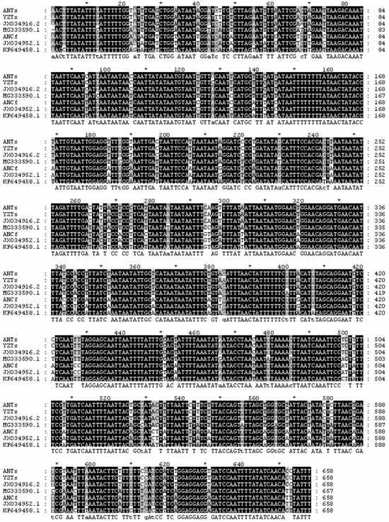
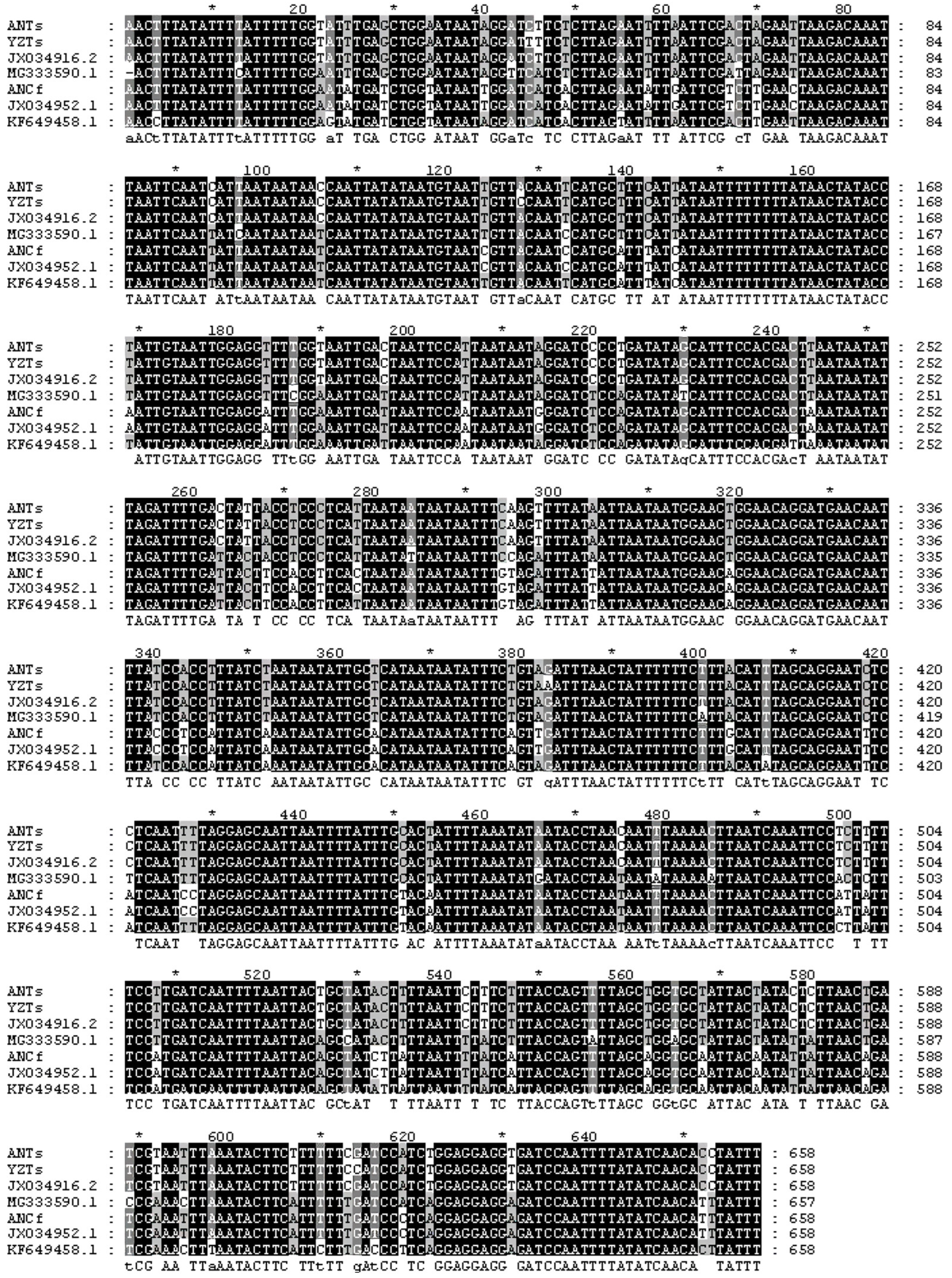
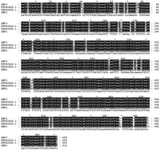
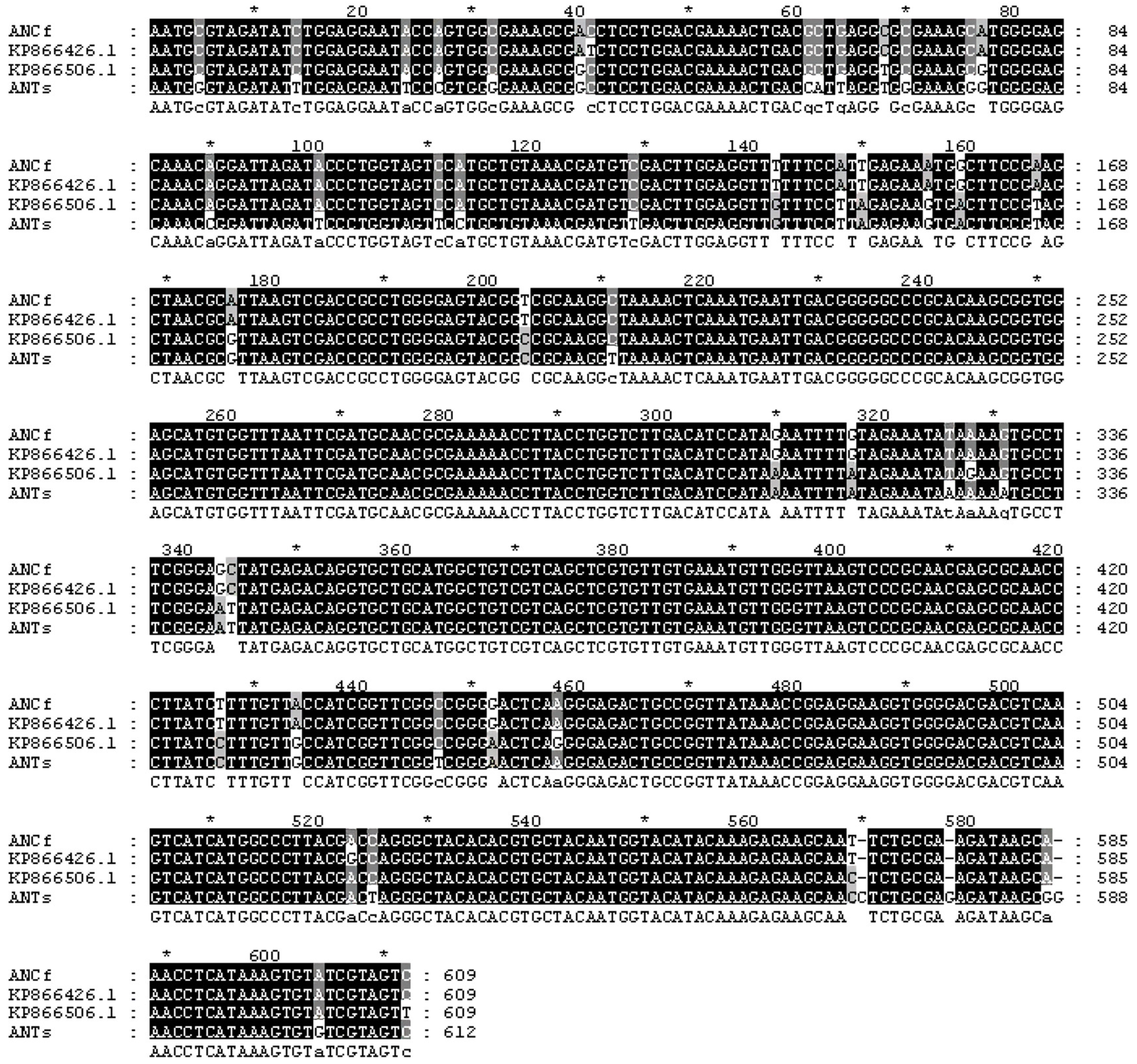
The COI and Buchnera 16S rRNA gene sequences of the experimental aphids were aligned with those of multiple aphid populations. A 658 bp fragment of the COI gene and a 612 bp fragment of the Buchnera 16S rRNA gene were obtained, as shown in Figure 1 and Figure 2, respectively. Alignment of the COI and Buchnera 16S rRNA gene sequences revealed clear genetic patterns (Figure 1 and Figure 2). Intraspecific variation was minimal; for example, COI sequences were identical among T. salignus populations from Anning, Yuzhong, and Tibet. In contrast, pronounced interspecific divergence was observed between C. formosana and T. salignus for both markers. The Buchnera 16S rRNA gene also showed greater sequence divergence between the two aphid species than within them.
Figure 1.
Multiple Sequence Alignment of COI (cytochrome c oxidase subunit I) Gene in Aphididae (* in the figure denote fully conserved positions; Colors: Black denotes fully conserved nucleotide sites, gray denotes partially conserved nucleotide sites, and white denotes variable nucleotide sites in the alignment).
Figure 2.
Multiple Sequence Alignment of Buchnera 16S rRNA (16S ribosomal RNA) Gene (* in the figure denote fully conserved positions; Colors: Black denotes fully conserved nucleotide sites, gray denotes partially conserved nucleotide sites, and white denotes variable nucleotide sites in the alignment).
Analysis of the nucleotide composition of the aphid COI gene fragment revealed the following average percentages: G (9.9%), A (33.84%), C (14.49%), and T (42.7%). The average G + C content was 24.42%, while the average A + T content was 75.58%, indicating a significant A + T bias (Table 1).
Table 1.
Characteristics of COI (cytochrome c oxidase subunit I) Gene Sequences in the three Experimental Aphid Species.
3.2. Nucleotide Substitution Values of COI in Experimental Aphids
An analysis of nucleotide substitutions, including transition and transversion rates, in the COI gene sequences of the experimental aphids revealed that the total number of transition sites was 23, while the number of transversion sites was 26, resulting in a transition/transversion ratio (R value) of 0.88. Transitions predominantly occurred between adenine (A) and thymine (T), while transversions primarily occurred between cytosine (C) and guanine (G). Substitution rates for the 1st, 2nd, and 3rd codon positions were 40 and 0.67, 9 and 3.14, and 1 and 0.48, respectively. These findings suggest that nucleotide substitutions primarily occurred at the 1st codon position, followed by the 2nd position, with the 3rd codon position remaining relatively conserved (Table 2).
Table 2.
Nucleotide Substitution Values of COI Gene in three Aphid Populations.
3.3. Genetic Distances and Divergence Between Aphid Species
Based on the data in Table 3, the genetic distances between the different aphid populations were as follows: The genetic distance between the C. formosana Anning population and the T. salignus Anning population was 0.131, while the genetic distance between the C. formosana Anning population and the T. salignus Yuzhong population was 0.138. The genetic distance between the T. salignus Anning and Yuzhong populations was 0.006. Furthermore, the genetic distance between the C. formosana Anning population and the C. formosana Hebei Weixian population was 0, and the genetic distance between the T. salignus Anning population and the T. salignus Tibet Linzhi population was also 0. The minimal intraspecific variation observed, particularly in T. salignus, may reflect its predominantly asexual reproduction mode and/or recent evolutionary history, as suggested for some Lachninae aphids [20] between C. formosana and T. salignus, potentially driven by their adaptation to distinct host plants.
Table 3.
Genetic Distance Analysis of 13 Aphid Populations Based on COI Sequences.
3.4. Phylogenetic Analysis
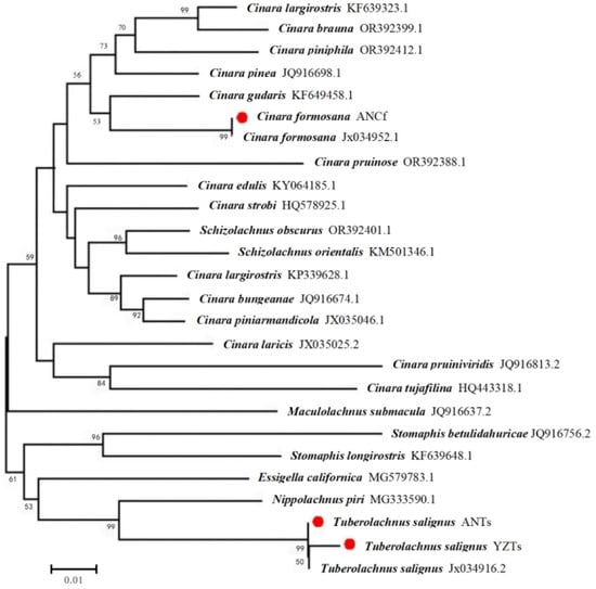
In this study, phylogenetic analyses were conducted using the COI gene sequences from two aphid species and the 16S rRNA gene sequences of Buchnera. As shown in Figure 3, the COI gene sequences of the C. formosana Anning population and the C. formosana Hebei Weixian population clustered together on the same branch. In contrast, the COI sequences of the T. salignus populations from Anning, Yuzhong, and Tibet (Linzhi) formed a separate cluster. These results indicate that the C. formosana Anning and Hebei Weixian populations are more closely related to each other, while the T. salignus Yuzhong population is more closely related to the Anning and Tibet (Linzhi) populations.
Figure 3.
Neighbor-joining phylogenetic tree based on COI Gene sequences (0.005 substitutions per site; Red dots indicate the Tuberolachnus salignus and Cinara formosana samples analyzed in this study).
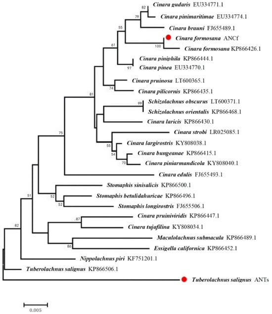
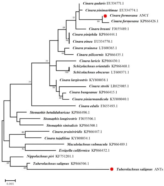
The neighbor-joining phylogenetic tree based on the 16S rRNA gene sequences of the aphid symbiotic bacterium Buchnera sp. (Figure 4) showed that the C. formosana Anning and C. formosana Beijing populations clustered together on the same branch. In contrast, T. salignus Anning population formed a distinct branch, while the T. salignus Yunnan Chuxiong population grouped into a separate branch. The maximum likelihood phylogenetic tree (Figure 5) produced similar results, with the C. formosana Anning and Beijing populations clustered together, and the T. salignus Anning and Yunnan Chuxiong populations forming separate branches. These findings suggest that the C. formosana Anning population is most closely related to the C. formosana Beijing population, while the T. salignus Anning population is most closely related to the T. salignus Yunnan Chuxiong population.
Figure 4.
Neighbor-joining phylogenetic tree based on Buchnera 16S rRNA Gene sequences (0.005 substitutions per site; Red dots indicate the Tuberolachnus salignus and Cinara formosana samples analyzed in this study).
Figure 5.
Maximum Likelihood Phylogenetic Tree Based on Buchnera 16S rRNA Sequences. (0.005 substitutions per site; Red dots indicate the Tuberolachnus salignus and Cinara formosana samples analyzed in this study).
4. Discussion
The results revealed an imbalance in the nucleotide composition of the COI genes in C. formosana from the Anning District and Yuzhong County of Lanzhou City, and T. salignus from Anning. Specifically, the average A + T content was significantly higher than the G + C content, highlighting a pronounced A + T bias The observed pattern could be attributed to selective pressures operating on these genomic regions. This finding is consistent with nucleotide composition patterns observed in the COI genes of other insect species, further confirming that it aligns with the characteristic nucleotide composition features of insect mitochondrial genes [21]. These happened because the mitochondrial DNA of C. formosana and T. salignus from Anning and Yuzhong follows an evolutionary pattern similar to other insect taxa.
Analysis of codon positions in the COI gene revealed significant variation in substitution rates. The transition/transversion ratio (R value) at the third codon position was notably lower than at the first and second positions, indicating a faster rate of nucleotide substitution. This result is consistent with findings in other insect groups, such as Chrysopidae [22], where the third codon position evolves more rapidly due to reduced functional constraints. The increased evolutionary rate at the third codon position suggests that this region is under less selective pressure, contributing to its higher variability. This supports the utility of the COI gene as a molecular marker for aphid species identification, as both conserved and variable regions enable effective differentiation among species and populations. The accelerated evolution at the third codon position of COI parallels the high variability in Buchnera 16S rRNA, suggesting synchronized host–symbiont genetic drift. This may reflect relaxed selection on symbiont genes due to nutritional redundancy in aphids feeding on divergent hosts.
The suitability of the COI gene for species identification depends on the degree of separation between interspecific and intraspecific genetic distances. An ideal molecular marker displays significantly greater interspecific divergence than intraspecific variation [23]. In this study, the genetic distance between the C. formosana Anning population and the T. salignus populations from Anning and Yuzhong was over ten times greater than the distance between the T. salignus populations themselves. This supports the principle that interspecific genetic distances should exceed intraspecific ones by at least an order of magnitude [24]. Furthermore, there was no overlap between the interspecific and intraspecific genetic distances among the C. formosana Anning, T. salignus Anning, and T. salignus Yuzhong populations, confirming the reliability of COI sequences as molecular markers for distinguishing between these species.
The maximum likelihood phylogenetic tree further validated the genetic relationships among populations. Both the COI and Buchnera gene sequences from T. salignus populations in Anning and Yuzhong clustered closely, forming a single branch. Similarly, the C. formosana Anning population grouped with the C. formosana Beijing population. In contrast, the neighbor-joining phylogenetic tree based on Buchnera 16S rRNA sequences showed that the T. salignus Anning population was distantly related to the T. salignus Yunnan Chuxiong population, nearly forming an outgroup. The overall congruence between the phylogenetic topologies derived from the aphid COI and the Buchnera 16S rRNA genes (i.e., the monophyletic grouping of each aphid species with its associated Buchnera strains) supports a history of co-divergence, despite the noted topological discrepancies within the Buchnera trees regarding certain populations. Such patterns of host–symbiont phylogenetic congruence, alongside observed Buchnera sequence divergence between aphid species exploiting different host plants, resonate with findings in other aphid groups where Buchnera genetic variation is associated with host plant use and life cycle differences [25]. These discrepancies suggest that the maximum likelihood method may provide a more accurate representation of evolutionary relationships in Buchnera sequences, consistent with findings from previous studies, such as the work of Chak et al. [26] on cockroach–bacterial endosymbiont coevolution.
The strong topological congruence between the aphid and Buchnera phylogenies is not merely a methodological validation. It provides concrete molecular evidence for their long-term co-evolution and parallel diversification (co-cladogenesis). This pattern underscores the depth of their obligate symbiotic integration, where the evolutionary fates of the host and its primary symbiont are intrinsically linked over geological timescales.
These findings have important implications for the development of molecular markers for rapid aphid identification—a critical component of effective pest management strategies. However, this study is limited by a relatively small sample size and the lack of comparative data from other geographic regions. Future research should incorporate larger datasets and include samples from a wider range of locations, as well as from closely related aphid species or host races specializing on different plants, to validate these results and better distinguish the effects of deep phylogenetic history from those of contemporary ecological adaptation on host–symbiont coevolution.
5. Conclusions
This study demonstrates clear genetic divergence between the conifer-feeding Cinara formosana and the willow-feeding Tuberolachnus salignus, and reveals congruent phylogenetic patterns between these aphids and their obligate Buchnera symbionts. Phylogenetic analyses of the COI gene in these aphids, alongside the 16S rRNA gene of their obligate symbiotic bacterium of the genus Buchnera sp., revealed clear and consistent evolutionary relationships. These findings provide a solid theoretical basis for the molecular identification of aphids using the COI gene, confirming its reliability as a species-specific marker. Our dual-marker system (COI + 16S rRNA) not only confirms the utility of COI for aphid identification but, from the perspective of symbiont divergence, reveals that the evolutionary history of Buchnera sp. genotypes is congruent with that of their aphid hosts, despite the deep phylogenetic split between C. formosana and T. salignus. This congruence suggests coupled evolution and offers a molecular toolkit for tracking pest populations and exploring symbiont-targeted control strategies.
Furthermore, the potential to use both COI and 16S rRNA genes as dual markers presents a promising approach to improve the accuracy of aphid identification and enhancing pest management strategies. The study also suggests that Buchnera sp. genotypes may influence aphid adaptation to host plants and contribute to pest virulence—an intriguing area for future research. Further studies should focus on the functional characterization of Buchnera sp. genes and their roles in aphid–plant interactions. This knowledge could facilitate the development of targeted, sustainable pest control strategies, ultimately benefiting both agricultural productivity and ecological health. While our study provides initial insights, the limited sequencing depth of Buchnera genomes (single-gene analysis) may overlook functional genomic adaptations. Future studies should employ metagenomic sequencing to resolve strain-level variation and metabolic complementarity.
Author Contributions
Writing—original draft, C.A.; Investigation, C.A. and L.L.; Data curation, C.A. and L.L.; Visualization, C.A. and L.W.; Methodology, C.A., L.L., Y.D. and S.S.; Writing—review and editing, H.X., Y.D. and S.S.; Supervision, H.X.; Conceptualization, L.L. and H.X.; Funding acquisition, H.X. All authors have read and agreed to the published version of the manuscript.
Funding
This work was funded by the National Key Research and Development Program: InterGovernmental Science and Technology Innovation Program, No. 2022YFE0115200; Insect Resources Investigation of Liancheng National Nature Reserve of Gansu Province, GSAU-JSFW-2022-082.
Data Availability Statement
The data that support the findings of this study are openly available.
Conflicts of Interest
All authors declare that they have no conflicts of interest.
References
- Ng, J.C.K.; Falk, B.W. Virus-vector interactions mediating nonpersistent and semipersistent transmission of plant viruses. Annu. Rev. Phytopathol. 2006, 44, 183–212. [Google Scholar] [CrossRef] [Scilit]
- Liang, Y.; Yonezawa, T.; von Dohlen, C.D. Phylogeny of Rhus gall aphids (Hemiptera: Aphididae) reveals an earlier origin than their primary host plants. Syst. Entomol. 2025, 50, 736–749. [Google Scholar] [CrossRef] [Scilit]
- Węgierek, P.; Malik, K.; Hutyra, P.; Depa, Ł. What does the morphological diversity of siphunculi tell us about the evolution of aphids (Insecta, Hemiptera, Aphidoidea). Eur. Zool. J. 2025, 92, 85–96. [Google Scholar] [CrossRef] [Scilit]
- Guo, H.J.; Sun, Y.C. Interplay between aphid salivary proteins, plant immunity and virus spread. Entomol. Gen. 2025, 45, 1271–1283. [Google Scholar] [CrossRef] [Scilit]
- Wu, S.; Huang, Z.; Rebeca, C.L.; Zhu, X.; Guo, Y.; Lin, Q.; Hu, X.; Wang, R.; Liang, G.; Guan, X.; et al. De novo characterization of the pine aphid Cinara pinitabulaeformis Zhang et Zhang transcriptome and analysis of genes relevant to pesticides. PLoS ONE 2017, 12, e0178496. [Google Scholar] [CrossRef] [Scilit] [PubMed]
- Dedryver, C.A.; Le Ralec, A.; Fabre, F. The conflicting relationships between aphids and men: A review of aphid damage and control strategies. Comptes Rendus Biol. 2010, 333, 539–553. [Google Scholar] [CrossRef] [Scilit]
- Zug, R.; Hammerstein, P. Still a host of hosts for Wolbachia: Analysis of recent data suggests that 40% of terrestrial arthropod species are infected. PLoS ONE 2012, 7, e38544. [Google Scholar] [CrossRef] [Scilit]
- Wu, D.; Daugherty, S.C.; E Van Aken, S.; Pai, G.H.; Watkins, K.L.; Khouri, H.; Tallon, L.J.; Zaborsky, J.M.; E Dunbar, H.; Tran, P.L.; et al. Metabolic complementarity and genomics of the dual bacterial symbiosis of sharpshooters. PLoS Biol. 2006, 4, e188. [Google Scholar] [CrossRef] [Scilit]
- Buchner, P. Endosymbiosis of Animals with Plant Microorganisms; Interscience Publishers: New York, NY, USA, 1965. [Google Scholar]
- Hansen, A.K.; Moran, N.A. Aphid genome expression reveals host symbiont cooperation in the production of amino acids. Proc. Natl. Acad. Sci. USA 2011, 108, 2849–2854. [Google Scholar] [CrossRef] [Scilit]
- Zhang, Y.; Su, X.; Harris, A.J.; Caraballo-Ortiz, M.A.; Ren, Z.; Zhong, Y. Genetic Structure of the Bacterial Endosymbiont Buchnera aphidicola from Its Host Aphid Schlechtendalia chinensis and Evolutionary Implications. Curr. Microbiol. 2018, 75, 309–315. [Google Scholar] [CrossRef] [Scilit]
- Wilson, A.C.; Ashton, P.D.; Calevro, F.; Charles, H.; Colella, S.; Febvay, G.; Jander, G.; Kushlan, P.F.; Macdonald, S.J.; Schwartz, J.F.; et al. Genam ic insight into the am ino acid relations ofpisum, with its symbiotic bacteriumthe pea aphid Acyrthosiphon Buchnera aphidicola. Insect Mol. Biol. 2010, 19, 249–258. [Google Scholar] [CrossRef] [Scilit]
- Łukasik, P.; Dawid, M.A.; Ferrari, J.; Godfray, H.C.J. The diversity and fitness effects of infection with facultative endosymbionts in the grain aphid, Sitobion avenae. Oecologia 2013, 173, 985–996. [Google Scholar] [CrossRef] [Scilit]
- Cao, P.; Gao, J. Analysis of the control effect of different pesticides on Cinara formosana. Inn. Mong. For. 2017, 14–15. [Google Scholar]
- Sopow, S.; Wardhaugh, C.; Turner, R.; Gresham, B.; Sutherland, R.; Woodall, G.; Withers, T. Host specificity testing of Pauesia nigrovaria (Hymenoptera: Braconidae: Aphidiinae) for classical biological control of Tuberolachnus salignus (Hemiptera: Aphididae: Lachninae) in New Zealand. BioControl 2021, 66, 739–751. [Google Scholar] [CrossRef] [Scilit]
- Janicka, M.; Kutkowska, A.; Paderewski, J. Diversity of segetal flora in Salix viminalis L. crops established on former arable and fallow lands in central Poland. Agriculture 2021, 11, 25. [Google Scholar] [CrossRef] [Scilit]
- Zhang, M. In depth analysis of the growth habits, application value, and cultivation techniques of Pinus tabuliformis. Seed World 2024. (In Chinese) [Google Scholar]
- Kirkland, L.S.; Chirgwin, E.; Ward, S.E.; Congdon, B.S.; van Rooyen, A.; Umina, P.A. P450-mediated resistance in Myzus persicae (Sulzer) (Hemiptera: Aphididae) reduces the efficacy of neonicotinoid seed treatments in Brassica napus. Pest Manag. Sci. 2023, 79, 1851–1859. [Google Scholar] [CrossRef] [Scilit]
- Murthy, M.K.; Khandayataray, P.; Tara, M.; Buragohain, P.; Giri, A.; Gurusubramanian, G. Optimisation of DNA isolation and PCR techniques for beetle (Order: Coleoptera) specimens. Int. J. Trop. Insect Sci. 2022, 42, 2761–2771. [Google Scholar] [CrossRef] [Scilit]
- Kanturski, M.; Lee, Y. Hyojoong KimPhylogenetic reconstruction of Tuberolachnini and Lachninae (Insecta, Hemiptera): Morphological and molecular analyses revealed a new tribe. Front. Zool. 2024, 21, 9. [Google Scholar] [CrossRef] [Scilit] [PubMed]
- Schorderet-Weber, S.; Noack, S.; Selzer, P.M.; Kaminsky, R. Blocking transmission of vector-borne diseases. Int. J. Parasitol. Drugs Drug Resist. 2017, 7, 90–109. [Google Scholar] [CrossRef] [Scilit] [PubMed]
- Hu, H.; Zhang, Z.; Lu, Z.; Ren, X.; Ma, X.; Ma, Y.; Song, X.; Ma, Y. Phylogenetic Analysis of Three Green Lacewing Species (Neuropetra, Chrysopidae) Based on the Mitochondrial COI Gene. J. Yunnan Agric. Univ. 2021, 36, 937–943. (In Chinese) [Google Scholar]
- Aliabadian, M.; Kaboli, M.; Nijman, V.; Vences, M. Molecular identification of birds: Performance of distance-based DNA barcoding in three genes to delimit parapatric species. PLoS ONE 2009, 4, e4119. [Google Scholar] [CrossRef] [Scilit]
- Hebert, P.D.N.; Penton, E.H.; Burns, J.M.; Janzen, D.H.; Hallwachs, W. Ten species in one: DNA barcoding reveals cryptic species in the neotropical skipper butterfly Astraptes fulgerator. Proc. Natl. Acad. Sci. USA 2004, 101, 14812–14817. [Google Scholar] [CrossRef] [Scilit] [PubMed]
- Kaszyca-Taszakowska, N.; Depa, Ł. Microbiome of the Aphid Genus Dysaphis Börner (Hemiptera: Aphidinae) and Its Relation to Ant Attendance. Insects 2022, 13, 1089. [Google Scholar] [CrossRef] [Scilit] [PubMed]
- Clark, J.W.; Hossain, S.; Burnside, C.A.; Kambhampati, S. Coevoli tion between a cockroach and its bacterial endosymbiont a biogeognphical penpective. Proc. R. Soe. Lond. Ser. B 2000, 268, 393–398. [Google Scholar] [CrossRef] [Scilit] [PubMed]

Disclaimer/Publisher’s Note: The statements, opinions and data contained in all publications are solely those of the individual author(s) and contributor(s) and not of MDPI and/or the editor(s). MDPI and/or the editor(s) disclaim responsibility for any injury to people or property resulting from any ideas, methods, instructions or products referred to in the content. |
© 2026 by the authors. Licensee MDPI, Basel, Switzerland. This article is an open access article distributed under the terms and conditions of the Creative Commons Attribution (CC BY) license.