


Abstract
The genus Metarhizium is one of the most significant entomopathogenic fungi with diverse morphological characteristics and host species. Species of Metarhizium have been widely used for pest control as an environmentally safe alternative to chemical pesticides. This study reports a new species of Metarhizium and a new record of M. bibionidarum from China. The taxonomic positions of the two species within Metarhizium were assessed by morphological and multi-gene phylogenetic data. This assessment confirmed that the new species M. dianzhongense on white grubs (Coleoptera) is a sister to M. ellipsoideum on adult leafhoppers (Hemiptera) and represents a distinctive fungus according to the morphological and phylogenetic evidence. The two species, M. dianzhongense and M. bibionidarum, were described and illustrated. Pathogenicity tests by M. bibionidarum and M. dianzhongense were performed on early instar larvae of the significant agricultural pest Spodoptera frugipera (Lepidoptera). The results demonstrated that both M. bibionidarum and M. dianzhongense exhibit significant insecticidal activity against larvae of S. frugipera, providing new fungal resources for the development of an eco-friendly biocontrol agent against this pest.
1. Introduction
Metarhizium is one of the most interesting genera of entomopathogenic fungi (EPF); it is known as “green muscardine fungus” because it mostly produces green conidia on its arthropod hosts. The genus was established by Sorokīn (1879) based on the type species M. anisopliae (Metschn.) Sorokīn attacking the wheat cockchafer Anisoplia austriaca (Coleoptera) from Russia. Tulloch [1] revised the genus and recognized only two species, M. anisopliae and M. flavoviride W. Gams & Rozsypal, along with two varieties, M. anisopliae var. anisopliae and M. anisopliae var. majus (J.R. Johnst.) M.C. Tulloch. Species of Metarhizium were historically described by morphological characteristics such as colony color, conidial structure, and sporulation patterns [2], which led to considerable debate regarding their taxonomic statuses.
The advancement of molecular systematics, especially multi-gene phylogenetic analyses that include rDNA gene fragments and functional protein genes, has revolutionized the taxonomy of Metarhizium. The genus Metacordyceps G.H. Sung et al. was erected to include Cordyceps spp. linked to anamorphic Metarhizium and Pochonia Bat. & O.M. Fonsa within the family Clavicipitaceae [3]. The recombination to Metacordyceps was made by adding several teleomorphic species, which accommodated 15 species and 1 variety [4]. Following the adoption of the “One Fungus One Name” standard, Kepler et al. [5] delimited the boundary of Metarhizium by combining the majority of species in Metacordyceps, Nomuraea Maubl., and Chamaeleomyces Sigler, recognizing 34 species. In recent years, numerous new taxa have been added to the genus Metarhizium and enriched its species diversity [6,7,8,9,10,11,12,13,14]. In the most complete taxonomic treatment of Metarhizium and its related genera, Mongkolsamrit et al. [11] reconstructed the phylogenetic framework of Clavicipitaceae focusing on the core Metarhizium clade and established six genera (Keithomyces Samson et al., Marquandomyces Samson et al., Papiliomyces Luangsa-ard et al., Purpureomyces Luangsa-ard et al., Sungia Luangsa-ard et al., and Yosiokobayasia Samson et al.) belonging to the core Metarhizium clade. To date, the genus Metarhizium accommodates 74 species, including 23 species in the M. anisopliae species complex and 13 species in the M. flavoviride species complex [11,14,15,16,17,18,19,20,21,22,23].
As an important and widespread group of EPF, Metarhizium has been used for pest control worldwide for approximately 140 years [24,25]. Various Metarhizium-based biocontrol products are available on the market and have been effectively implemented in managing a broad spectrum of pests worldwide [11,26]. Consequently, precise identifications for Metarhizium species and their host ranges are critical to the high-efficiency biocontrol strategy.
During surveys of EPF in southwestern China, we found a new species and a new record of Metarhizium attacking white grubs and March fly larvae, respectively. Collected specimens of the two species were identified by combining morphological and seven-gene (ITS, nrSSU, nrLSU, tef-1α, rpb1, rpb2, and 5′tef) phylogenetic analyses. The two species were described and illustrated. In addition, pathogenicity tests of the new species M. bibionidarum Nishi & Hiroki Sato and new record M. dianzhongense were performed on larvae of Spodoptera frugipera J. E. Smith.
2. Materials and Methods
2.1. Sample Collection and Isolation
Inst pathogenic specimens were meticulously collected in Yunnan, China, in 2022, with a keen examination of the forest understory, leaf litter, and the undersides of leaves for instances of EPF infection. Specimens were noted and photographed in the field, carefully placed in plastic boxes, and then transported to field stations or laboratories for detailed examination. The material was examined under an Olympus SZ60 microscope. Isolation from the material was conducted as described by Wang et al. [27] with modifications: the conidia from sporulating structures were touched by using a flame-sterilized inoculation needle and streaked on potato–dextrose agar (PDA: potato 200 g/L, dextrose 20 g/L, agar 20 g/L, in 1 L distilled water) plates. Purified strains were maintained in a culture room at 25 °C. Subculturing was performed on new PDA plates to ensure the development of pure colonies. Pure cultures were transplanted onto PDA slants and stored at 4 °C. Dried specimens were deposited in the Cryptogamic Herbarium of the Kunming Institute of Botany, Chinese Academy of Sciences (KUN-HKAS). Pure cultures were deposited in the Culture Collection of Kunming Institute of Botany, Chinese Academy of Sciences (KUNCC).
2.2. Test Material
The initial bioassay experiments were conducted using the selected strains of M. bibionidarum (KUNCC 10806) and M. dianzhongense (KUNCC 10811), which were collected, isolated, and purified in this study. Concurrently, field surveys were conducted to collect insect samples of S. frugiperda in 2022. The 4th-instar larvae of S. frugiperda were bred using lab colonies of the pest noctuids. The test strains were cultured on PDA plates for 14 d at 25 °C under a light/dark condition (L/D = 14:10) and the humidity of 70%. To harvest fungal spores, a solution containing 0.1% Tween 80 was poured onto a culture plate, and the surface of the fungal colony was gently scraped to release the spores. The spore concentration was estimated using a hemocytometer and light microscope. For the bioassay, the spore suspensions were diluted to achieve a concentration of 1 × 10⁸ spores/mL.
2.3. Morphological Observation
Specimens were photographed and examined in the laboratory using a Canon 750D camera and an Olympus SZ60 microscope. For descriptions of colony appearance, the isolated strains were cultured on PDA plates for 14 d at 25 °C under a light/dark condition (L/D = 14:10). The characteristics of colonies (size, texture, and color) were photographed with a Canon 750D camera. For morphological evaluation, microscope slide cultures were prepared by placing small amounts of mycelia on 5 mm diameter PDA medium blocks overlaid by a cover slip. The cultures were then incubated in a Petri dish at 25 °C with a small volume of sterile water. The asexual micro-morphological characteristics (hypha, conidiogenous structure, and conidia) were observed and examined under a light Olympus BX53 microscope.
2.4. DNA Extraction, PCR Amplification, and Sequencing
The genomic DNA of the two species was extracted using the modified CTAB method [27]. The extracted DNA used for PCR amplification was stored at −20 °C. Primer details used for amplifying seven genes (ITS, nrSSU, nrLSU, tef-1α, rpb1, rpb2, and 5′tef) in this study are provided in Table 1. Each 25 µL PCR reaction contained 12.5 µL of 2 × Taq PCR Master Mix (Tiangen Biotech Co., Ltd., Beijing, China), 9.5 µL of RNase-free water (Sangon Bio Co., Ltd., Shanghai, China), 1 µL of each forward and reverse primer (10 µmol/L), 1 µL of DNA template (500 ng/µL). PCR reactions were placed in a LongGene T20 multi-block thermal cycler (Hangzhou LongGene Scientific Instruments Co., Ltd., Hangzhou, China) under the following conditions: for ITS, (1) 3 min at 95 °C, (2) 35 cycles of denaturation at 95 °C for 30 s, annealing at 55 °C for 30 s, and extension at 72 °C for 1 min, (3) extension at 72 °C for 10 min and 14 °C soak; for tef-1α, (1) 3 min at 95 °C, (2) 35 cycles of denaturation at 95 °C for 30 s, annealing at 55 °C for 30 s, and extension at 72 °C for 1 min, (3) extension at 72 °C for 10 min and 14 °C soak; for rpb1, (1) 4 min at 95 °C, (2) 30 cycles of denaturation at 94 °C for 50 s, annealing at 52 °C for 50 s, and extension at 72 °C for 1 min, followed by (3) 8 cycles of denaturation at 94 °C for 50 s, annealing at 51 °C for 50 s, and extension at 72 °C for 1 min, (4) extension at 72 °C for 10 min and 14 °C soak; for rpb2, (1) 4 min at 95 °C, (2) 37 cycles of denaturation at 94 °C for 30 s, annealing at 60 °C for 30 s, and extension at 72 °C for 1 min, (3) extension at 72 °C for 10 min and 14 °C soak; for nrLSU, (1) 4 min at 95 °C, (2) 30 cycles of denaturation at 95 °C for 1 min, annealing at 50 °C for 1 min, and extension at 72 °C for 2 min, (3) extension at 72 °C for 8 min and 14 °C soak; for nrSSU, (1) 2 min at 95 °C, (2) 35 cycles of denaturation at 94 °C for 1 min, annealing at 51 °C for 30 s, and extension at 72 °C for 1 min, (3) extension at 72 °C for 10 min and 14 °C soak; for 5′tef, (1) 3 min at 95 °C, (2) 35 cycles of denaturation at 95 °C for 30 s, annealing at 55 °C for 30 s, and extension at 72 °C for 1 min, (3) extension at 72 °C for 10 min and 14 °C soak. Standard DNA markers (Sangon Bio Co., Ltd., Shanghai, China) of known size and weight were used to quantify the PCR products. The PCR products were separated by electrophoresis in 1% agarose gels, purified using the Gel Band Purification Kit (Bio Teke Co., Ltd., Beijing, China), and then sent to Tsing ke Biotechnology Co., Ltd. for sequencing after the selection of impurity-free and distinct bands. The seven genes of two Metarhizium species generated in this study were submitted to GenBank (https://www.ncbi.nlm.nih.gov/genbank, accessed on 26 February 2024).
Table 1.
Sequences of primers used in this study.
2.5. Phylogenetic Analyses
Based on molecular data from recent research in the genus Metarhizium, seven gene sequences (ITS, nrSSU, nrLSU, tef-1α, rpb1, rpb2, and 5′tef) were retrieved and downloaded from the GenBank database. Detailed information is provided in Table 2. Along with the sequencing results from this study, gene sequences for 76 taxonomic units were ultimately obtained. Sequences of seven genes were aligned using MEGA v. 7.0 [32,33]. After sequence alignments, the aligned sequences of seven genes were concatenated. Ambiguously aligned sites were manually eliminated, and gaps were treated as missing data. The sequences of Pochonia Bat. & O.M. Fonsa (P. boninensis Nonaka et al. JCM 18597 and P. chlamydosporia (Goddard) Zare & W. Gams CBS 101244) were selected as the outgroup. Under the Akaike Information Criterion (AIC), ModelFinder is used to select the most appropriate nucleotide substitution model for Bayesian inference (BI) and maximum-likelihood (ML) analyses. The BI analysis was conducted using MrBayes v. 3.2 [34], employing the GTR+G+I model. Four simultaneous Markov chains were run for 2,000,000 generations with a sub-sampling frequency every 100 generations. A burn-in of the first 25% of the total run was discarded. The ML analysis was conducted using IQ-TREE v. 2.1.3 [35] under the TNe+I+R3 model with 1000 ultrafast bootstrap [36]. Trees were visualized with Bayesian posterior probability (BI-PP) and maximum-likelihood bootstrap proportions (ML-BS) in FigTree v. 1.4.4 and edited with Adobe Illustrator CS6.0.
Table 2.
Voucher information and GenBank accession numbers of sequences used in this study.
2.6. Pathogenicity Test
The test strain’s spore suspension with a concentration of 1 × 10⁸ spores/mL was used to immerse the selected and consistent-sized healthy 4th-instar larvae of S. frugiperda for approximately 10 s. Subsequently, the larvae were removed and allowed to air dry. They were then placed in a controlled artificial climate chamber at 25 °C and a humidity level of 65% and were individually reared in plastic rearing containers measuring 3 cm in diameter and height, with corn seeds and leaves provided as their food source. The experiment was conducted with three repetitions, each consisting of 12 S. frugiperda larvae.
A 0.1% Tween-80 sterile water treatment was used as the blank control group. Infected and control samples were then reared under standard insect-rearing conditions (as mentioned above) for a duration of nine days. Deceased insects were promptly removed and placed in the moist culture room to confirm their infection. Mortality rates and adjusted mortality rates were calculated as follows. The mortality rate (%) was determined by the following formula: (number of deceased insects/total number of test insects) × 100%. The adjusted mortality rate (%) was calculated using the following formula: (mortality rate in treatment group—mortality rate in control group)/(1—mortality rate in control group) × 100%.
3. Results
3.1. Phylogenetic Analyses
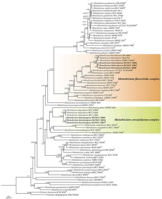
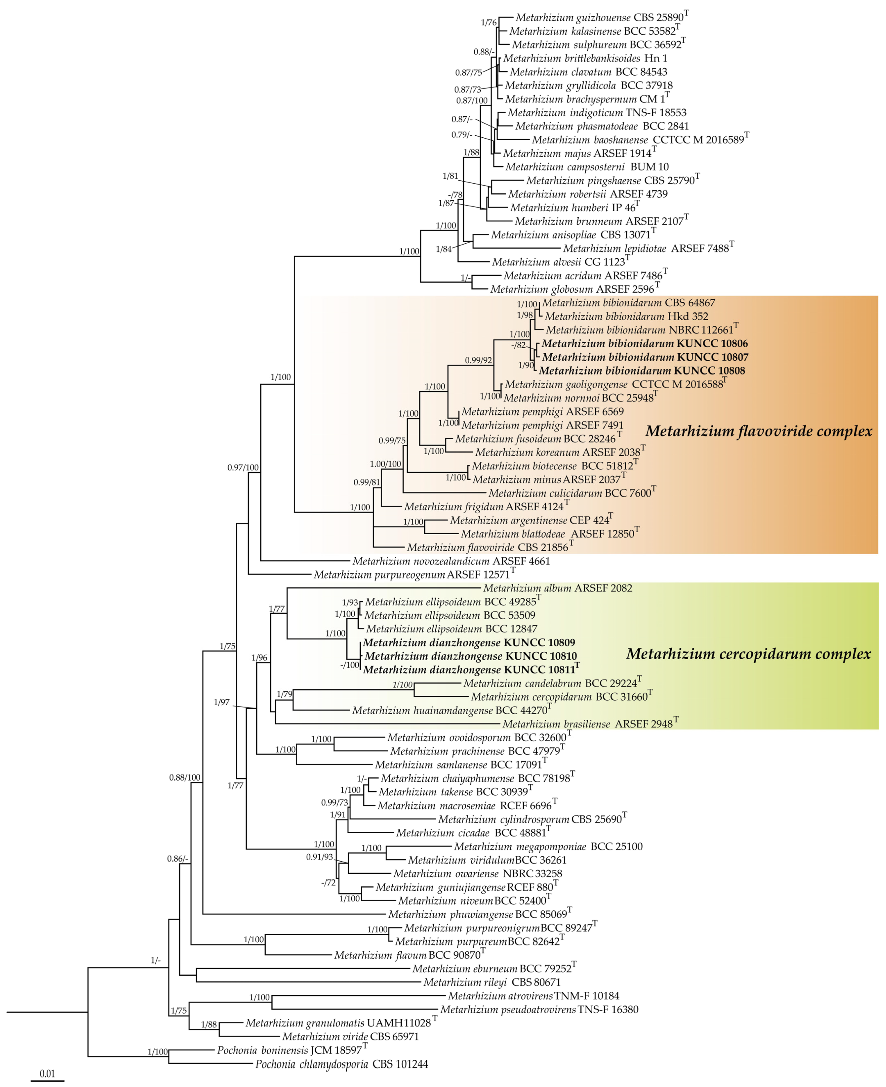
The combined seven-gene dataset comprised 5581 base pairs (bp) after alignments (ITS 581 bp, nrSSU 968 bp, nrLSU 764 bp, tef-1α 871 bp, rpb1 738 bp, rpb2 919 bp, and 5′tef 740 bp). Phylogenetic trees from ML and BI analyses based on the combined seven-gene dataset of 78 taxa showed nearly congruent topologies (Figure 1). It was revealed that all Metarhizium species formed a monophyletic group with high support. The samples of M. bibionidarum collected in this study and described by Nishi et al. [20] both have the same hosts, the March fly larvae (Diptera: Bibionidae). Based on the seven-gene phylogeny, they clustered into a clade with credible bootstrap support (BI-PP = 1 and ML-BP = 100%). The new species M. dianzhongense was sister to M. ellipsoideum Luangsa-ard et al. in the M. cercopidarum complex. However, the three strains of M. dianzhongense (KUNCC 10809, KUNCC 10810, and KUNCC 10811) formed a single derived clade (BI-PP = 1 and ML-BP = 100%).
Figure 1.
Phylogenetic placements of Metarhizium dianzhongense and Metarhizium bibionidarum in the genus Metarhizium inferred from BI and ML analyses based on multi-gene dataset (ITS, nrSSU, nrLSU, tef-1α, rpb1, rpb2, and 5′tef). Values at the nodes before and after the backslash are BI posterior probabilities (BI-PP > 0.70) and ML bootstrap proportions (ML-BP > 70%), respectively.
3.2. Taxonomy
Metarhizium bibionidarum Nishi & Hiroki Sato, Mycological Progress 16(10): 993 (2017) [20], Index Fungorum number: IF819797, Figure 2.
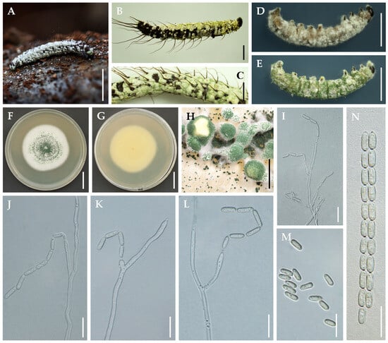
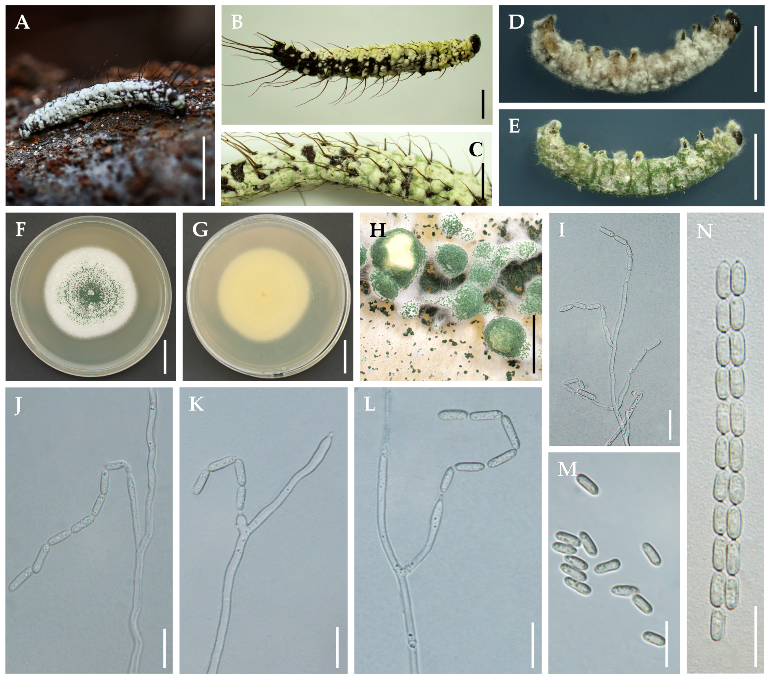
Figure 2.
Metarhizium bibionidarum. (A–C) Larvae of March fly infected by M. bibionidarum; (D,E) 4th-instar larvae of S. frugiperda killed by M. bibionidarum; (F–H) colonies on PDA; (I–L) conidiophores and phialides on PDA; (M) conidia on PDA; (N) conidia on insect host. Scale bars: (A) = 5 mm, (B,C) = 2 mm, (D,E) =5 mm, (F,G) = 2 cm, (H) = 5 mm, (I) = 20 µm, (J–N) = 10 µm.
Description: Specimens were found on March fly larvae (Diptera: Bibionidae). The head and thorax of March fly larvae were covered with white mycelia and powdery conidia. The conidia were smooth-walled, yellowish-green to green, cylindrical to ellipsoid with semi-papillate apices, 2.5–3.5 × 5.5–7 μm. Colonies on PDA grew well, attaining a diameter of 27–29 mm in 7 d, and were white, fluffy, and cottony in appearance. Sporulation with peacock-green conidia produced on mycelia started at 7 d after inoculation, reverse white. Hyphae were smooth-walled, hyaline, branched, septate. Conidiophores arising from aerial mycelia were erect and smooth-walled. Phialides were smooth-walled, cylindrical or ellipsoidal, with papillate apices, 7.6–27.3 × 2–3.5 μm. Conidia were smooth-walled, cylindrical to ellipsoidal with semi-papillate apices, arranged in chains, 7.8–10.4 × 2–3 μm.
Host: March fly larvae (Bibionidae, Diptera).
Habitat: On the March fly larvae buried in soil or attached to fallen leaves.
Distribution: Known from France, Japan, and China.
Material examined: China. Yunnan Province: Kunming City, Heilongtan Park, alt. 2150 m, on a March fly larva, 5 September 2022, C.Y. Wei (HKAS 126221, KUNCC 10806); Ibid. 5 September 2022, C.Y. Wei (HKAS 126222, KUNCC 10807); Ibid. 5 September 2022, C.Y. Wei (HKAS 126223, KUNCC 10808).
Notes: Our newly collected three samples are phylogenetically clustered with Metarhizium bibionidarum including strain NBRC 112661 (ex-type, Japan) and CBS 648.67 (France) within the M. flavoviride complex with strong statistical support. M. bibionidarum is closely related to M. gaoligongense Z.H. Chen & L. Xu, M. nornnoi Luangsa-ard et al., and M. pemphigi (Driver & Milner) Kepler et al. The three samples parasitize March fly larvae and produce similar-sized cylindrical to ellipsoidal conidia on PDA. The morphological characteristics on other media lack a clear distinction. M. bibionidarum is known to parasitize Diptera and Coleoptera, in addition to being commonly found in soil [20].
Metarhizium dianzhongense C.Y. Wei, Zhu L. Yang & Y.B. Wang, sp. nov. Fungal Names number: FN571888; Figure 3.
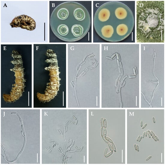
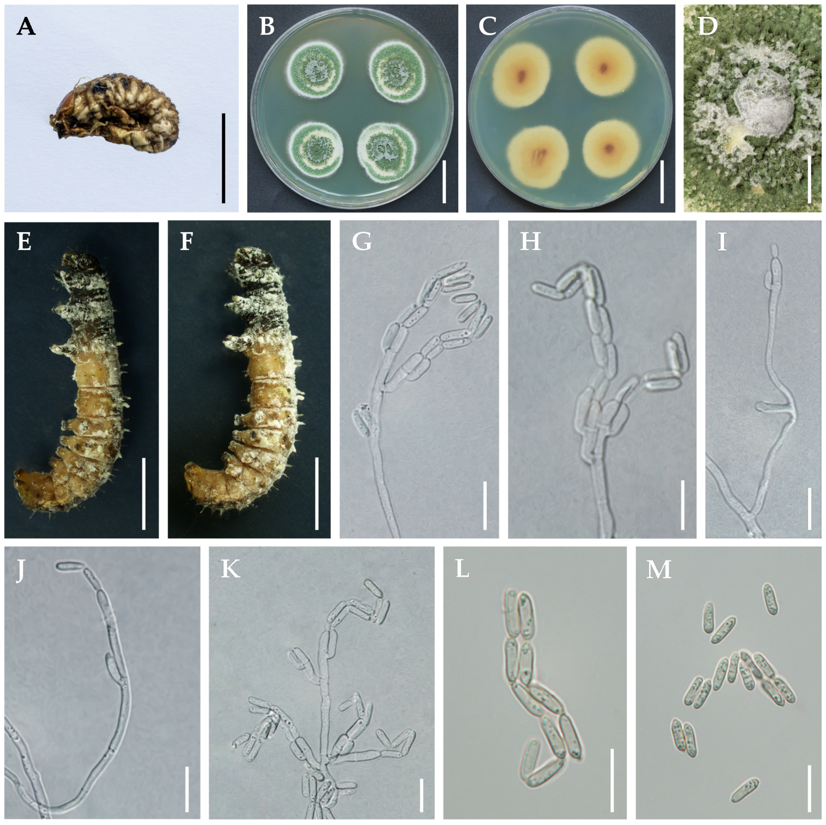
Figure 3.
Metarhizium dianzhongense. (A) White grub infected by M. dianzhongense; (B–D) colonies on PDA; (E,F) 4th-instar larvae of S. frugiperda killed by M. dianzhongense; (G–K) conidiophores and phialides on PDA; (L,M) conidia on PDA. Scale bars: (A) = 1 cm, (B,C) = 2 cm, (D–F) = 5 mm, (G–M) = 10 µm.
Etymology: The epithet ‘dianzhong’ refers to central Yunnan Province in Chinese, where the holotype was collected.
Holotype: China. Yunnan Province, Chuxiong City, Sanjie Town, 100°69′233 E, 24°52′322 N, alt. 1880 m, on a white grub buried in soil, 5 September 2022, C.Y. Wei (HKAS 126224, holotype; KUNCC 10811, ex-holotype culture). GenBank accession Nos.: ITS = PP256142, nrLSU = PP256150, tef-1α = PP328483, rpb1 = PP294692, rpb2 = PP314010.
Description: Specimens were found on larvae of Scarabaeidae (Coleoptera). The insect body curled up, stiffened, and formed sclerotia. Colonies on PDA attained a diameter of 23–25 mm in 14 d, with mycelia closely appressed to the agar surface, dense, flat. Margins were initially pale yellow to white, white turning to parrot-green with powdery conidia, becoming raised at the center of colonies. Sporulation started 3 d after inoculation, reverse pale yellow. Hyphae were smooth-walled. Conidiophores arising from aerial mycelia were erect, smooth-walled, cylindrical, 7.8–10.4 × 2–3 μm. Phialides were smooth-walled, cylindrical or ellipsoidal with semi-papillate apices, 4–7.5 × 1–3 μm. Conidia were smooth-walled, parrot-green, ellipsoidal or cylindrical with rounded apices, arranged in chains, 7–9 × 2–3 μm.
Host: White grubs, larvae of Scarabaeidae (Coleoptera).
Habitat: On the larvae of Scarabaeidae buried in soil from farmland.
Distribution: Known from Chuxiong City, Yunnan Province, China.
Other material examined: China. Yunnan Province, Chuxiong City, Sanjie Town, 100°53′346 E, 23°58′465 N, alt. 1873 m, on a white grub buried in soil, 13 November 2021, C.Y. Wei (HKAS 126224; KUNCC 10809); Ibid. 13 November 2021, C.Y. Wei (HKAS 126225, KUNCC 10810).
Notes: Phylogenetically, M. dianzhongense is sister to M. ellipsoideum with strong statistical support, forming a separate clade from other Metarhizium species in the M. cercopidarum complex. It is similar to M. ellipsoideum, M. candelabrum, M. cercopidarum, and M. huainamdangense due to the cylindrical and parrot-green conidia with rounded apices [11]. However, M. dianzhongense (14 d, 24 mm) grows faster than M. ellipsoideum (14 d, 14–15 mm), M. candelabrum (14 d, 15 mm), M. cercopidarum (14 d, 15 mm), and M. huainamdangense (14 d, 15 mm) on PDA. The distinctiveness of M. dianzhongense is indicated by its larger conidial size (7–9 × 2–3 μm) compared with M. ellipsoideum (5–7 × 1.5–2 μm) and M. cercopidarum (6–8 × 2 μm). In their natural habitats, M. ellipsoideum, M. candelabrum, M. cercopidarum, and M. huainamdangense parasitize insects of leafhoppers (Hemiptera). However, M. dianzhongense specifically targets larvae of Scarabaeidae (Coleoptera) in farmland and has been observed to infect larvae of S. frugiperda (Lepidoptera) during controlled experiments.
Additionally, there exist significant distinctions between M. dianzhongense and other unmentioned Metarhizium species in this study, namely M. cicadinum (Höhn.) Petch, M. dendrolimatilis Z.Q. Liang et al., M. lepidopterorum W.H. Chen et al., M. lymantriidae Z.H. Chen & L. Xu, M. putuoense Yi Li et al., and M. rongjiangense W.H. Chen et al. [11,15,42,43,44,45]. The six species are mainly parasitic on Hemiptera (M. cicadinum) and Lepidoptera (M. dendrolimatilis, M. lepidopterorum, M. lymantriidae, M. putuoense, and M. rongjiangense) hosts, which distinguishes them from M. dianzhongense (Coleoptera). The morphological characteristics of M. dianzhongense significantly differ from those of the six species in terms of colonies, conidiogenous structures, and conidia. Phylogenetically, M. dianzhongense, belonging to the M. cercopidarum complex clade, is distinct from M. cicadinum (the M. anisopliae complex), M. putuoense (the M. purpureum complex), and M. dendrolimatilis and M. lymantriidae (the M. rileyi complex) [43]. Particularly, M. lepidopterorum and M. rongjiangense now belong to the genus Marquandomyces, which was previously described as Metarhizium [44].
3.3. Pathogenicity to Spodoptera frugipera
After nine days of infection, the M. bibionidarum strain KUNCC 10806 exhibited the highest overall mortality in instar larvae of S. frugiperda, followed by the M. dianzhongense strain KUNCC 108011. The findings showed that mortality in S. frugiperda larvae caterpillars treated with KUNCC 10806 and KUNCC 10811 predominantly occurred within the initial three days after infection. The mortality rates of 94.44% and 86.11% were observed in S. frugiperda larvae caterpillars nine days after infection with KUNCC 10806 and KUNCC 10811, respectively, resulting in induced corrected mortality rates of 93.97% and 84.85%.
4. Discussion
The current species delimitation of fungal complex groups emphasizes the necessity of employing multi-gene phylogeny, as relying solely on morphological characteristics is insufficient for accurate species identification [3,5,11,46]. In this study, two Metarhizium species were identified by combining morphological and seven-gene phylogenetic evidence. The new record of M. bibionidarum infecting March fly larvae and belonging to the M. flavoviride complex is closely related to M. gaoligongense, M. nornnoi, and M. pemphigi. The taxonomic position of the new species M. dianzhongense infecting larvae of Scarabaeidae was determined based on phylogenetic analyses. Metarhizium dianzhongense is clustered in the M. cercopidarum complex and is clearly distinguished from its related species M. ellipsoideum.
In the M. flavoviride complex, thirteen species have been recognized, namely M. argentinense A.C. Gutierrez et al., M. bibionidarum, M. biotecense Luangsa-ard et al., M. blattodeae C. Montalva et al., M. culicidarum Luangsa-ard et al., M. flavoviride, M. frigidum J.F. Bisch. & S.A. Rehner, M. fusoideum Luangsa-ard et al., M. gaoligongense, M. koreanum Kepler et al., M. minus (Rombach, Humber & D.W. Roberts) Kepler et al., M. nornnoi Luangsa-ard et al., and M. pemphigi. The asexual states of all species are naturally observed, while no instances of sexual states have been documented. Some species (M. bibionidarum, M. flavoviride, and M. pemphigi) appear to exhibit a worldwide distribution, while others (M. minus, M. biotecense, M. culicidarum, M. fusoideum, and M. nornnoi) have only been reported in tropical regions [2,3,5,8,11,17,20,22,37]. The M. cercopidarum complex comprises six recognized species, including M. album Petch, M. brasiliense Kepler et al., M. candelabrum Luangsa-ard et al., M. cercopidarum, M. dianzhongense, M. ellipsoideum, and M. huainamdangense Luangsa-ard et al. All species belonging to this group have been reported in tropical and subtropical regions, especially Thailand and China [5,11]. Some species (M. album, M. brasiliense, M. candelabrum, M. cercopidarum, M. ellipsoideum, and M. huainamdangense) parasitize insects of Hemiptera, while the new species M. dianzhongense parasitizes the larvae of Coleoptera [3,5,11,16]. These species, particularly, exhibit a candelabrum-like arrangement of cylindrical phialides, forming a densely packed hymenium [11].
Previous studies have demonstrated that M. anisopliae, M. favoviride, M. majus (J.R. Johnst.) J.F. Bisch. et al., M. rileyi (Farl.) Kepler et al., and Beauveria bassiana (Bals. -Criv.) Vuill. exhibit significant insecticidal activity against various insect pests [23,47,48,49]. M. favoviride is an effective microbial insecticidal against S. litura, with the fungus significantly impairing the immune response of the insect. The larval mortality rate reached 80% when exposed to treatment concentrations of 1 × 10⁹ conidia/mL [50]. Reports have revealed the detection of a significant number of S. frugiperda larvae infected by M. rileyi in various maize-growing regions, particularly in countries such as Mexico and Brazi [49,51]. In laboratory conditions, Perumal et al. [23] observed that M. majus exhibited significant insecticidal activity against S. frugiperda larvae within nine days. The mortality rate of Colorado potato beetle adults caused by discovered native strains reached up to 100% when beetles were treated with conidia containing a concentration of 1 × 10⁷ spores/mL [52]. Our findings demonstrate that the strains of M. bibionidarum and M. dianzhongense, which were isolated from naturally infested larvae of Bibionidae and Scarabaeidae, exhibited significant virulence against S. frugiperda larvae. These results suggest that these two species could serve as viable alternatives to broad-spectrum chemical insecticidal treatment.
Author Contributions
Conceptualization, S.-K.S. and Z.-L.Y.; supervision and validation, Q.F.; software and investigation, L.-Y.X.; methodology, formal analysis, and resources, C.-Y.W.; data management, M.T.; writing—original draft preparation, C.-Y.W. and M.T.; writing—review and editing, Y.-B.W. and C.-Y.W.; project administration and funding acquisition, Y.-B.W. and G.D. All authors have read and agreed to the published version of the manuscript.
Funding
This work was financially supported by the Science and Technology Planning Project of Yunnan Province (202207AB110016, 202401AS070030); the High Level Talent Introduction Plan, Kunming Institute of Botany, CAS (E16N61); and the Innovation Project of Guangxi Graduate Education (YCBZ2022028).
Institutional Review Board Statement
This article does not contain any studies with human participants or animals performed by any of the authors.
Data Availability Statement
The datasets used and/or analyzed during the current study are available from the corresponding authors on request. Moreover, sequences were deposited in GenBank http://www.ncbi.nlm.nih.gov/ (accessed on 20 August 2023) under the accession numbers mentioned in the text.
Acknowledgments
The authors gratefully acknowledge Baozheng Chen and Xinling Lai. of the Yunnan Agricultural University for their invaluable assistance and support during the sample collection process; and thanks Hui Xu. of the Yunnan University for technical support.
Conflicts of Interest
The authors declare no conflicts of interest.
References
- Tulloch, M. The genus metarhizium. Trans. Br. Mycol. Soc. 1976, 66, 407–411. [Google Scholar] [CrossRef] [Scilit]
- Chen, Z.; Yang, X.; Sun, N.; Xu, L.; Zheng, Y.; Yang, Y. Species diversity and vertical distribution characteristics of Metarhizium in Gaoligong Mountains, southwestern China. Biodivers. Sci. 2018, 26, 1308. [Google Scholar] [CrossRef] [Scilit]
- Sung, G.H.; Hywel-Jones, N.L.; Sung, J.M.; Luangsa-Ard, J.J.; Shrestha, B.; Spatafora, J.W. Phylogenetic classification of Cordyceps and the clavicipitaceous fungi. Stud. Mycol. 2007, 57, 5–59. [Google Scholar] [CrossRef] [Scilit] [PubMed]
- Kepler, R.; Sung, G.-H.; Ban, S.; Nakagiri, A.; Chen, M.-J.; Huang, B.; Li, Z.Z.; Spatafora, J. New teleomorph combinations in the entomopathogenic genus Metacordyceps. Mycologia 2012, 104, 182–197. [Google Scholar] [CrossRef] [Scilit] [PubMed]
- Kepler, R.M.; Humber, R.A.; Bischoff, J.F.; Rehner, S.A. Clarification of generic and species boundaries for Metarhizium and related fungi through multigene phylogenetics. Mycologia 2014, 106, 811–829. [Google Scholar] [CrossRef] [Scilit] [PubMed]
- Sharma, S.K.; Gautam, N. Metacordyceps dhauladharensis sp. nov., a new entomopathogenic fungus from India. Turk. J. Bot. 2015, 39, 520–526. [Google Scholar] [CrossRef] [Scilit]
- Chu, H.L.; Chen, W.H.; Wen, T.C.; Liang, Z.Q.; Zheng, F.C.; Liang, J.D.; Han, Y.F. Delimitation of a novel member of genus Metarhizium (Clavicipitaceae) by phylogenetic and network analysis. Phytotaxa 2016, 288, 51–60. [Google Scholar] [CrossRef] [Scilit]
- Luangsa-Ard, J.J.; Mongkolsamrit, S.; Thanakitpipattana, D.; Khonsanit, A.; Tasanathai, K.; Noisripoom, W.; Humber, R.A. Clavicipitaceous entomopathogens: New species in Metarhizium and a new genus Nigelia. Mycol. Prog. 2017, 16, 369–391. [Google Scholar] [CrossRef] [Scilit]
- Lopes, R.B.; Souza, D.A.; Rocha, L.F.; Montalva, C.; Luz, C.; Humber, R.A.; Faria, M. Metarhizium alvesii sp. nov.: A new member of the Metarhizium anisopliae species complex. J. Invertebr. Pathol. 2018, 151, 165–168. [Google Scholar] [CrossRef] [Scilit]
- Luz, C.; Rocha, L.F.; Montalva, C.; Souza, D.A.; Botelho, A.B.R.; Lopes, R.B.; Faria, M.; Júnior, I.D. Metarhizium humberi sp. nov. (Hypocreales: Clavicipitaceae), a new member of the PARB clade in the Metarhizium anisopliae complex from Latin America. J. Invertebr. Pathol. 2019, 166, 107216. [Google Scholar] [CrossRef] [Scilit] [PubMed]
- Mongkolsamrit, S.; Khonsanit, A.; Thanakitpipattana, D.; Tasanathai, K.; Noisripoom, W.; Lamlertthon, S.; Himaman, W.; Houbraken, J.; Samson, R.; Luangsa-Ard, J. Revisiting Metarhizium and the description of new species from Thailand. Stud. Mycol. 2020, 95, 171–251. [Google Scholar] [CrossRef] [Scilit]
- Thanakitpipattana, D.; Tasanathai, K.; Mongkolsamrit, S.; Khonsanit, A.; Lamlertthon, S.; Luangsa-Ard, J. Fungal pathogens occurring on Orthopterida in Thailand. Persoonia-Mol. Phylogeny Evol. Fungi 2020, 44, 140–160. [Google Scholar] [CrossRef] [Scilit]
- Yamamoto, K.; Ohmae, M.; Orihara, T. Metarhizium brachyspermum sp. nov. (Clavicipitaceae), a new species parasitic on Elateridae from Japan. Mycoscience 2020, 61, 37–42. [Google Scholar] [CrossRef] [Scilit]
- Chen, M.-J.; Lin, Y.; Wang, T.; Zhang, S.-L.; Huang, B. Metarhizium macrosemiae sp. nov. and the anamorph of M. guniujiangense on adult cicada from Guniujiang Nature Preserve, southeastern China. Phytotaxa 2022, 575, 68–78. [Google Scholar] [CrossRef] [Scilit]
- Chen, Z.H.; Dai, Y.D.; Chen, K.; Zhang, Y.F.; Xu, L.; Wang, Y.B. Papiliomyces puniceum and Metarhizium lymantriidae: Two new species from the Gaoligong Mountains in southwestern China. Phytotaxa 2023, 594, 53–63. [Google Scholar] [CrossRef] [Scilit]
- Driver, F.; Milner, R.J.; Trueman, J.W. A taxonomic revision of Metarhizium based on a phylogenetic analysis of rDNA sequence data. Mycol. Res. 2000, 104, 134–150. [Google Scholar] [CrossRef] [Scilit]
- Bischoff, J.F.; Rehner, S.A.; Humber, R.A. Metarhizium frigidum sp. nov.: A cryptic species of M. anisopliae and a member of the M. flavoviride complex. Mycologia 2006, 98, 737–745. [Google Scholar] [CrossRef]
- Bischoff, J.F.; Rehner, S.A.; Humber, R.A. A multilocus phylogeny of the Metarhizium anisopliae lineage. Mycologia 2009, 101, 512–530. [Google Scholar] [CrossRef] [Scilit]
- Nishi, O.; Iiyama, K.; Yasunaga-Aoki, C.; Shimizu, S. Phylogenetic status and pathogenicity of Metarhizium majus isolated from a fruit beetle larva in Japan. Mycol. Prog. 2015, 14, 58. [Google Scholar] [CrossRef] [Scilit]
- Nishi, O.; Shimizu, S.; Sato, H. Metarhizium bibionidarum and M. purpureogenum: New species from Japan. Mycol. Prog. 2017, 16, 987–998. [Google Scholar] [CrossRef] [Scilit]
- Chen, Z.; Xu, L.; Yang, X.; Zhang, Y.; Yang, Y. Metarhizium baoshanense sp. nov., a new entomopathogen fungus from southwestern China. Pak. J. Zool. 2018, 50, 1739–1746. [Google Scholar] [CrossRef] [Scilit]
- Chen, Z.H.; Zhang, Y.G.; Yang, X.N.; Chen, K.; Liu, Q.; Xu, L. A new fungus Metarhizium gaoligongense from China. Int. J. Agric. Biol. 2018, 20, 2271–2276. [Google Scholar] [CrossRef]
- Perumal, V.; Kannan, S.; Alford, L.; Pittarate, S.; Geedi, R.; Elangovan, D.; Marimuthu, R.; Krutmuang, P. First report on the enzymatic and immune response of Metarhizium majus bag formulated conidia against Spodoptera frugiperda: An ecofriendly microbial insecticide. Front. Microbiol. 2023, 14, 1104079. [Google Scholar] [CrossRef] [Scilit] [PubMed]
- Nishi, O.; Iiyama, K.; Yasunaga-Aoki, C.; Shimizu, S. Comparison of the germination rates of Metarhizium spp. conidia from Japan at high and low temperatures. Lett. Appl. Microbiol. 2013, 57, 554–560. [Google Scholar] [CrossRef] [Scilit] [PubMed]
- González-Hernández, G.A.; Padilla-Guerrero, I.E.; Martínez-Vázquez, A.; Torres-Guzmán, J.C. Virulence Factors of the Entomopathogenic Genus Metarhizium. Curr. Protein Pept. Sci. 2020, 21, 324–330. [Google Scholar] [CrossRef] [Scilit] [PubMed]
- Kumar, K.K.; Sridhar, J.; Murali-Baskaran, R.K.; Senthil-Nathan, S.; Kaushal, P.; Dara, S.K.; Arthurs, S. Microbial biopesticides for insect pest management in India: Current status and future prospects. J. Invertebr. Pathol. 2019, 165, 74–81. [Google Scholar] [CrossRef] [Scilit] [PubMed]
- Wang, Y.B.; Wang, Y.; Fan, Q.; Duan, D.E.; Zhang, G.D.; Dai, R.Q.; Dai, Y.D.; Zeng, W.B.; Chen, Z.H.; Li, D.D. Multigene phylogeny of the family Cordycipitaceae (Hypocreales): New taxa and the new systematic position of the Chinese cordycipitoid fungus Paecilomyces hepiali. Fungal Divers. 2020, 103, 1–46. [Google Scholar] [CrossRef] [Scilit]
- White, T.J.; Bruns, T.; Lee, S.; Taylor, J. Amplification and Direct Sequencing of Fungal Ribosomal RNA Genes for Phylogenetics; PCR Protoc: A Guide Methods; Academic Press: San Diego, CA, USA, 1990; pp. 315–322. [Google Scholar] [CrossRef] [Scilit]
- Vilgalys, R.; Hester, M. Rapid genetic identification and mapping of enzymatically amplified ribosomal DNA from several Cryptococcus species. J. Bacteriol. 1990, 172, 4238–4246. [Google Scholar] [CrossRef] [Scilit] [PubMed]
- Rehner, S.A.; Samuels, G.J. Taxonomy and phylogeny of Gliocladium analysed from nuclear large subunit ribosomal DNA sequences. Mycol. Res. 1994, 98, 625–634. [Google Scholar] [CrossRef] [Scilit]
- Wang, Y.B.; Yu, H.; Dai, Y.D.; Wu, C.K.; Zeng, W.B.; Yuan, F.; Liang, Z.Q. Polycephalomyces agaricus, a new hyperparasite of Ophiocordyceps sp. infecting melolonthid larvae in southwestern China. Mycol. Prog. 2015, 14, 70. [Google Scholar] [CrossRef] [Scilit]
- Katoh, K.; Standley, D.M. MAFFT multiple sequence alignment software version 7: Improvements in performance and usability. Mol. Biol. Evol. 2013, 30, 772–780. [Google Scholar] [CrossRef] [Scilit] [PubMed]
- Kumar, S.; Stecher, G.; Tamura, K. MEGA7: Molecular evolutionary genetics analysis version 7.0 for bigger datasets. Mol. Biol. Evol. 2016, 33, 1870–1874. [Google Scholar] [CrossRef] [Scilit]
- Ronquist, F.; Teslenko, M.; Van Der Mark, P.; Ayres, D.L.; Darling, A.; Höhna, S.; Larget, B.; Liu, L.; Suchard, M.A.; Huelsenbeck, J.P. MrBayes 3.2: Efficient Bayesian phylogenetic inference and model choice across a large model space. Syst. Biol. 2012, 61, 539–542. [Google Scholar] [CrossRef] [Scilit] [PubMed]
- Nguyen, L.T.; Schmidt, H.A.; Von Haeseler, A.; Minh, B.Q. IQ-TREE: A fast and effective stochastic algorithm for estimating maximum-likelihood phylogenies. Mol. Biol. Evol. 2015, 32, 268–274. [Google Scholar] [CrossRef] [Scilit] [PubMed]
- Hoang, D.T.; Chernomor, O.; Von Haeseler, A.; Minh, B.Q.; Vinh, L.S. UFBoot2: Improving the ultrafast bootstrap approximation. Mol. Biol. Evol. 2018, 35, 518–522. [Google Scholar] [CrossRef] [Scilit] [PubMed]
- Gutierrez, A.C.; Leclerque, A.; Manfrino, R.G.; Luz, C.; Ferrari, W.A.; Barneche, J.; García, J.J.; Lastra, C.C.L. Natural occurrence in Argentina of a new fungal pathogen of cockroaches, Metarhizium argentinense sp. nov. Fungal Biol. 2019, 123, 364–372. [Google Scholar] [CrossRef] [Scilit] [PubMed]
- Montalva, C.; Collier, K.; Rocha, L.F.N.; Inglis, P.W.; Lopes, R.B.; Luz, C.; Humber, R.A. A natural fungal infection of a sylvatic cockroach with Metarhizium blattodeae sp. nov., a member of the M. flavoviride species complex. Fungal Biol. 2016, 120, 655–665. [Google Scholar] [CrossRef] [Scilit] [PubMed]
- Liu, Z.Y.; Liang, Z.; Whalley, A.; Yao, Y.; Liu, A. Cordyceps brittlebankisoides, a new pathogen of grubs and its anamorph, Metarhizium anisopliae var. majus. J. Invertebr. Pathol. 2001, 78, 178–182. [Google Scholar] [CrossRef] [Scilit]
- Sigler, L.; Gibas, C.F.C.; Kokotovic, B.; Bertelsen, M.F. Disseminated mycosis in veiled chameleons (Chamaeleo calyptratus) caused by Chamaeleomyces granulomatis, a new fungus related to Paecilomyces viridis. J. Clin. Microbiol. 2010, 48, 3182–3192. [Google Scholar] [CrossRef] [Scilit] [PubMed]
- Nonaka, K.; Ōmura, S.; Masuma, R.; Kaifuchi, S.; Masuma, R. Three new Pochonia taxa (Clavicipitaceae) from soils in Japan. Mycologia 2013, 105, 1202–1218. [Google Scholar] [CrossRef] [Scilit] [PubMed]
- Petch, T. Notes on entomogenous fungi. Trans. Br. Mycol. Soc. 1931, 16, 55–75. [Google Scholar] [CrossRef] [Scilit]
- Chen, W.; Han, Y.; Liang, J.; Liang, Z.; Jin, D. Metarhizium dendrolimatilis, a novel Metarhizium species parasitic on Dendrolimus sp larvae. Mycosphere 2017, 8, 31–37. [Google Scholar] [CrossRef] [Scilit]
- Chen, W.-H.; Han, Y.-F.; Liang, J.-D.; Liang, Z.-Q. Morphological and phylogenetic characterization of novel Metarhizium species in Guizhou, China. Phytotaxa 2019, 419, 189–196. [Google Scholar] [CrossRef] [Scilit]
- Li, Y.; Zhao, X.-C.; Wu, L.-X.; Wang, Y.; Xu, A.; Lin, W.-F. Blackwellomyces kaihuaensis and Metarhizium putuoense (Hypocreales), Two New Entomogenous Fungi from Subtropical Forests in Zhejiang Province, Eastern China. Forests 2023, 14, 2333. [Google Scholar] [CrossRef] [Scilit]
- Kepler, R.M.; Sung, G.H.; Harada, Y.; Tanaka, K.; Tanaka, E.; Hosoya, T.; Bischoff, J.F.; Spatafora, J.W. Host jumping onto close relatives and across kingdoms by Tyrannicordyceps (Clavicipitaceae) gen. nov. and Ustilaginoidea (Clavicipitaceae). Am. J. Bot. 2012, 99, 552–561. [Google Scholar] [CrossRef] [Scilit] [PubMed]
- Borisade, O.; Magan, N. Growth and sporulation of entomopathogenic Beauveria bassiana, Metarhizium anisopliae, Isaria farinosa and Isaria fumosorosea strains in relation to water activity and temperature interactions. Biocontrol Sci. Technol. 2014, 24, 999–1011. [Google Scholar] [CrossRef] [Scilit]
- Li, M.; Li, S.; Xu, A.; Lin, H.; Chen, D.; Wang, H. Selection of Beauveria isolates pathogenic to adults of Nilaparvata lugens. J. Insect Sci. 2014, 14, 32. [Google Scholar] [CrossRef] [Scilit] [PubMed]
- Ruiz-Nájera, R.E.; Ruiz-Estudillo, R.A.; Sánchez-Yáñez, J.M.; Molina-Ochoa, J.; Skoda, S.R.; Coutiño-Ruiz, R.; Pinto-Ruiz, R.; Guevara-Hernández, F.; Foster, J.E. Occurrence of entomopathogenic fungi and parasitic nematodes on Spodoptera frugiperda (Lepidoptera: Noctuidae) larvae collected in central Chiapas, México. Fla. Entomol. 2013, 96, 498–503. [Google Scholar] [CrossRef] [Scilit]
- Vivekanandhan, P.; Swathy, K.; Alford, L.; Pittarate, S.; Subala, S.P.R.R.; Mekchay, S.; Elangovan, D.; Krutmuang, P. Toxicity of Metarhizium flavoviride conidia virulence against Spodoptera litura (Lepidoptera: Noctuidae) and its impact on physiological and biochemical activities. Sci. Rep. 2022, 12, 16775. [Google Scholar] [CrossRef] [Scilit] [PubMed]
- Ordóñez-García, M.; Bustillos-Rodríguez, J.C.; Loya-Márquez, J.; Ríos-Velasco, C.; Jacobo-Cuellar, J.L. Parasitoides de Spodoptera frugiperda (JE Smith)(Lepidoptera: Noctuidae) en Chihuahua, México. Métodos En Ecol. Y Sist. 2015, 10, 67–72. [Google Scholar]
- Zemek, R.; Konopická, J.; Jozová, E.; Skoková Habuštová, O. Virulence of Beauveria bassiana strains isolated from cadavers of Colorado potato beetle, Leptinotarsa decemlineata. Insects 2021, 12, 1077. [Google Scholar] [CrossRef] [Scilit]

Disclaimer/Publisher’s Note: The statements, opinions and data contained in all publications are solely those of the individual author(s) and contributor(s) and not of MDPI and/or the editor(s). MDPI and/or the editor(s) disclaim responsibility for any injury to people or property resulting from any ideas, methods, instructions or products referred to in the content. |
© 2024 by the authors. Licensee MDPI, Basel, Switzerland. This article is an open access article distributed under the terms and conditions of the Creative Commons Attribution (CC BY) license (https://creativecommons.org/licenses/by/4.0/).