


Abstract
During an ongoing research survey of saprobic fungi in Thailand, four coelomycetous strains were isolated from decaying leaves in Chiang Mai and Phitsanulok Provinces. Morphological characteristics demonstrated that these taxa are typical of Cytospora in forming multi-loculate, entostromatic conidiomata, branched or unbranched conidiophores, with enteroblastic, phialidic conidiogenous cells and hyaline, allantoid, aseptate conidia. Multiloci phylogeny of ITS, LSU, ACT, RPB2, TEF1-α and TUB2 confirmed these taxa are distinct new species in Cytospora in Cytosporaceae (Diaporthales, Sordariomycetes), viz., Cytospora chiangmaiensis, C. phitsanulokensis and C. shoreae. Cytospora chiangmaiensis has a close phylogenetic relationship with C. shoreae, while C. phitsanulokensis is sister to C. acaciae. These three novel species were also preliminary screened for their antagonistic activity against five plant pathogenic fungi: Colletotrichum fructicola, Co. siamense, Co. artocarpicola, Co. viniferum and Fusarium sambucinum. Cytospora shoreae and C. phitsanulokensis showed >60% inhibition against Co. viniferum and F. sambucinum, while C. chiangmaiensis had moderate inhibition activity against all pathogens.
1. Introduction
Cytospora Ehrenb. is mainly characterized by the coelomycetous asexual morph producing single or labyrinthine, loculate stromata, filamentous conidiophores, with enteroblastic, phialidic conidiogenous cells and hyaline, allantoid, aseptate conidia [1,2,3]. The sexual morph of Cytospora is characterized as solitary, immersed to erumpent ascostromata, composed of prosenchymatous or pseudoparenchymatous cells of valsoid or diatrypelloid configurations, embedded in ectostromatic disc, present or absent paraphyses, J-, ellipsoid to clavate asci with refractive apical ring, and hyaline, ellipsoid to allantoid, aseptate ascospores [1,4,5]. Ehrenberg [6] initially introduced the genus Cytospora to accommodate four species, C. betulina, C. epimyces, C. resinae and C. ribis. The type species, C. chrysosperma, was introduced by Donk [7] as the asexual morph of Valsa sordida [8]. Cytospora was previously treated as the asexual morph of Valsa and other related genera, Leucocytospora, Leucostoma, Valsella and Valseutypella [4,8,9]. Based on the International Code of Nomenclature for Algae, Fungi, and Plants (ICN) [10], Rossman et al. [8] recommended to use Cytospora instead of Valsa as Cytospora was prior introduced and thus Valsa was treated as a synonym of Cytospora, together with the genera Leucocytospora, Leucostoma, Valsella and Valseutypella [4,8].
Species identification in Cytospora was problematic in the past due to overlapping morphological characteristics and wide host ranges [11,12,13,14,15]. A polyphasic approach including host association, geographical distribution, morphological characteristics and multiloci phylogeny has been widely accepted for identifying Cytospora species [1,2,3,5,9,16,17,18]. To date, 671 species epithets are listed in Index Fungorum [19], but less than a quarter of taxa in this genus are clarified with phylogenetic placement. In recent years, about 117 species have been accepted based on morpho-molecular approaches [20], and these species have primarily been described from China, Italy, Russia and Thailand [1,2,3,5,9,16,17,18,21].
Cytospora is a cosmopolitan genus with diverse ecological niches, including plant pathogens, endophytes and saprobes, colonizing decaying wood substrates [1,2,17,18]. Cytospora species are important causal agents of diebacks and stem cankers in numerous woody plants such as Betulaceae, Pinophyta, Fabaceae, Juglandaceae, Rosaceae, Salicaceae, Tiliaceae and Ulmaceae [1,3,16,17,18,22,23]. Furthermore, Cytospora has also been reported to produce potential secondary metabolites with various biological activities, such as antibacterial, antifungal and antiviral activities [24,25,26]. Investigation of the endophytic fungus Cytospora sp. from Costa Rica yielded novel antibiotic compounds: cytoskyrins and cytosporones [24,25]. Upon screening novel secondary metabolites produced by the endophytic fungus Cytospora sp. from Chinese mangrove, new bicyclic sesquiterpene and seiricardine D were isolated and they showed effective inhibition of human and plant pathogens [27].
During a survey of saprobic fungi in Thailand, three Cytospora species were collected and isolated. Based on morphological characteristics and phylogenetic analyses, our strains were identified as new species of Cytospora. In addition, these fungi were tested in vitro as the first step for screening new biocontrol agents against pathogenic fungi.
2. Materials and Methods
2.1. Collection and Isolation of Fungi
Decaying leaves were collected from disturbed forests in Chiang Mai and Phitsanulok Provinces in Thailand. The forests were disturbed due to the utilization of bioresources by the local communities. Samples were kept in plastic bags with labels of location, date, host and collector details before being taken to laboratory for morphological observation. We followed Senanayake et al. [28] for single spore isolation by using potato dextrose agar (PDA) and incubating at room temperature (28 °C). Pure cultures were deposited in Mae Fah Luang University Culture Collection (MFLUCC) and type specimens were deposited in the Herbarium of Mae Fah Luang University (MFLU). New taxa were registered in Faces of Fungi [29] and Index Fungorum databases [19].
2.2. Morphological Observation
Conidiomata on host surface were examined using a Motic SMZ 168 Series stereo microscope (Motic Incorporation Ltd., Hong Kong). Hand-sectioning of conidiomata was carried out and the sections were mounted on a slide with a drop of distilled water. Morphological characteristics including structure and size of stromata, ectostromatic disc, ostioles as well as shape and size of conidiogenous cells, conidiophores and conidia were observed and photographed using a Nikon ECLIPSE 80i compound microscope equipped with a Canon EOS 600D digital camera. Microscopic elements were measured using the Tarosoft (R) Image Frame Work program. The measurements of each structure were represented as minimum value-maximum value ( = sum of all measurements/n, n = number of measurements). The figures were processed using Adobe Photoshop CS6 Extended version 10.0 software (Adobe Systems, San Jose, CA, USA).
2.3. DNA Extraction, PCR Amplification and Sequencing
Genomic DNA was extracted from fresh mycelium which was grown on PDA for 1–2 weeks using the Biospin Fungus Genomic DNA Extraction Kit (BioFlux®, Hangzhou, China) following the manufacturer’s protocol. The amplification of specific ribosomal DNA regions was carried out using two gene regions, including the internal transcribed spacers region of ribosomal DNA (ITS) [30] and the partial 28S large subunit nuclear ribosomal DNA (LSU) [31], and four protein coding gene regions: the RNA polymerase II second largest subunit (RPB2) [32], α-actin (ACT) [33], the translation elongation factor 1-α (TEF1-α) [33] and beta-tubulin (TUB2) [34]. The final volume of PCR mixtures was 25 μL, including 8.5 μL ddH2O, 12.5 μL 2× PCR MasterMix (TIANGEN Co., Beijing, China), 2 μL DNA template and 1 μL of each forward and reverse primer. The PCR primers and conditions for each gene regions are described in Table 1. The purification and sequencing of PCR products were conducted by TsingKe Company (Kunming, China).
Table 1.
Partial marker regions, primers and PCR conditions used in this study.
2.4. Phylogenetic Analyses
Sequences generated in this study were analyzed with other sequences retrieved from the GenBank (www.ncbi.nlm.nih.gov/blast/, accessed on 1 August 2021) and recent publications [1,2,17] (Table 2). Alignments of the individual locus were aligned with MAFFT v.7 (http://mafft.cbrc.jp/alignment/server/index.html, accessed on 3 August 2021; [35]) and automatically trimmed using TrimAl (http://trimal.cgenomics.org accessed on 1 August 2021; [36]). The single-locus datasets of ITS, LSU, ACT, RPB2, TEF1-α and TUB2 regions were initially analyzed by maximum likelihood (ML) criterion with 120, 72, 69, 50, 55 and 41 sequences, respectively. The tree topologies obtained from single-locus analyses were checked for incongruence and the results did not show any topological conflicts. Phylogenetic analyses of the final concatenated ITS, LSU, ACT, RPB2, TEF1-α and TUB2 sequence datasets were performed via maximum likelihood (ML), maximum parsimony (MP) and Bayesian inference (BI).
Table 2.
Taxa used in the phylogenetic analyses and their corresponding GenBank accession numbers. The newly generated sequences are indicated in red bold. The ex-type strains are bold. N/A = unavailable sequence data.
Maximum likelihood (ML) analysis was performed by RAxML-HPC2 (v.8.2.12) on XSEDE implemented in the CIPRES Science Gateway web server (http://www.phylo.org accessed on 1 August 2021; [37] using 1000 rapid bootstrap replicates and the GTR + GAMMA + I substitution model. Maximum parsimony (MP) analysis was generated by PAUP (Phylogenetic Analysis Using Parsimony) v.4.0b10 [38] using the heuristic search option and 1000 random sequence additions. The branch-swapping was analysed using tree-bisection reconnection (TBR) algorithm. Maxtrees was set up at 1000 and all characters were unordered and of equal weight. The branches of zero length were collapsed and gaps were treated as missing data. All multiple and equally parsimonious trees were saved. The stability of the most parsimonious tree was evaluated by a bootstrap analysis with 1000 replicates, each with 100 replicates of random stepwise addition of taxa. The descriptive parsimonious tree, Tree Length [TL], Consistency Index [CI], Retention Index [RI], Relative Consistency Index [RC] and Homoplasy Index [HI] were calculated.
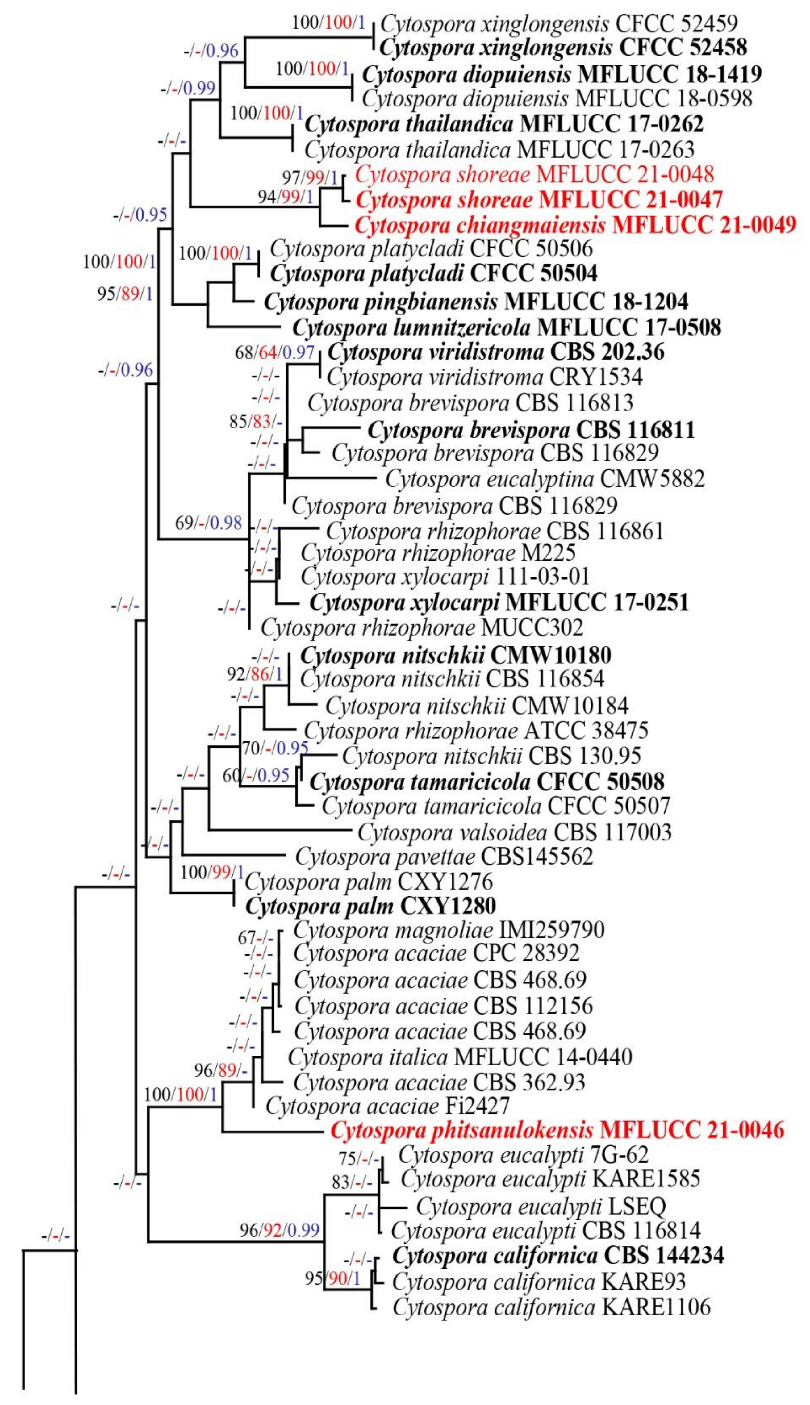
The evolutionary models for Bayesian inference (BI) analysis were determined for each locus using MrModeltest v.2.3 [39]. GTR + I + G was selected as the best-fitting model for LSU, RPB2, TEF-α and TUB2 datasets, SYM + I + G for the ITS dataset and HKY + I + G for the ACT dataset. BI analysis was conducted by Markov chain Monte Carlo sampling (BMCMC) to assess posterior probabilities (PP) [40,41] using MrBayes v3.1.2 [42]. Six simultaneous Markov chains were run for random trees for 10,000,000 generations, and trees were sampled every 1000th generation. The effective sampling sites (ESS) of initial trees were checked using the Tracer v. 1.6 [43]. The first 10% of generated trees were discarded, and the remaining trees were used to calculate posterior probabilities (PP) in the majority rule consensus tree (the standard deviation of split frequency lower than 0.01). Bootstrap support values for ML and MP equal to or greater than 60% and Bayesian posterior probabilities (PP) equal to or greater than 0.95 were given above the nodes in the phylogenetic tree (Figure 1).
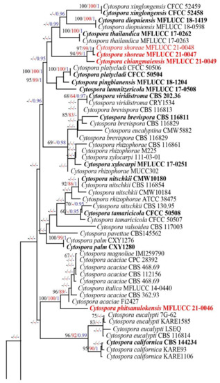
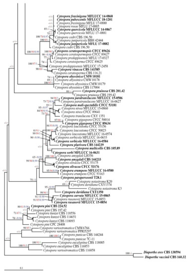
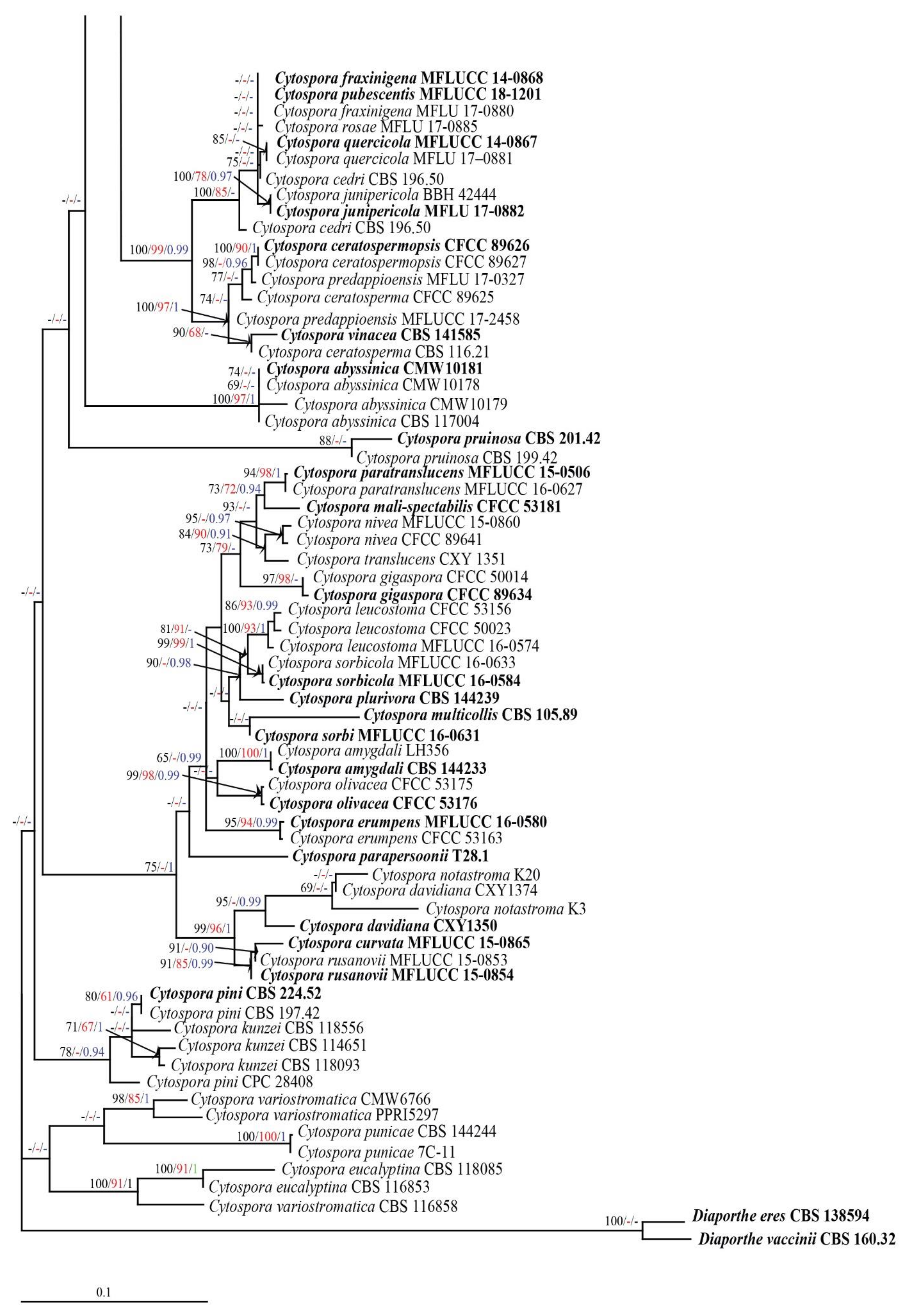
Figure 1.
Phylogenetic tree obtained from RAxML analyses of a combined ITS, LSU, ACT, RPB2, TEF1-α and TUB2 sequence dataset. Bootstrap support values for ML, MP equal to or greater than 60% and PP equal to greater than 0.95 are indicated at the nodes as ML/MP/PP. The ex-type strains are in bold and the new isolates of this study are in red. The tree is rooted to Diaporthe eres (CBS138594) and D. vaccinii (CBS16032).
Phylograms were visualized by FigTree v1.4.0 program [44] and reorganized by Microsoft PowerPoint 2013 (Microsoft Inc., Redmond, WA, USA) and Adobe Photoshop CS6 software (Adobe Systems, San Jose, CA, USA). The generated sequences of the new taxa were registered for the GenBank accession numbers. The final alignment and phylogram were submitted in TreeBASE (http://purl.org/phylo/treebase/phylows/study/TB2:S28804 accessed on 1 August 2021).
2.5. Genealogical Concordance Phylogenetic Species Recognition Analysis
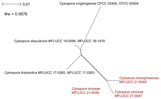
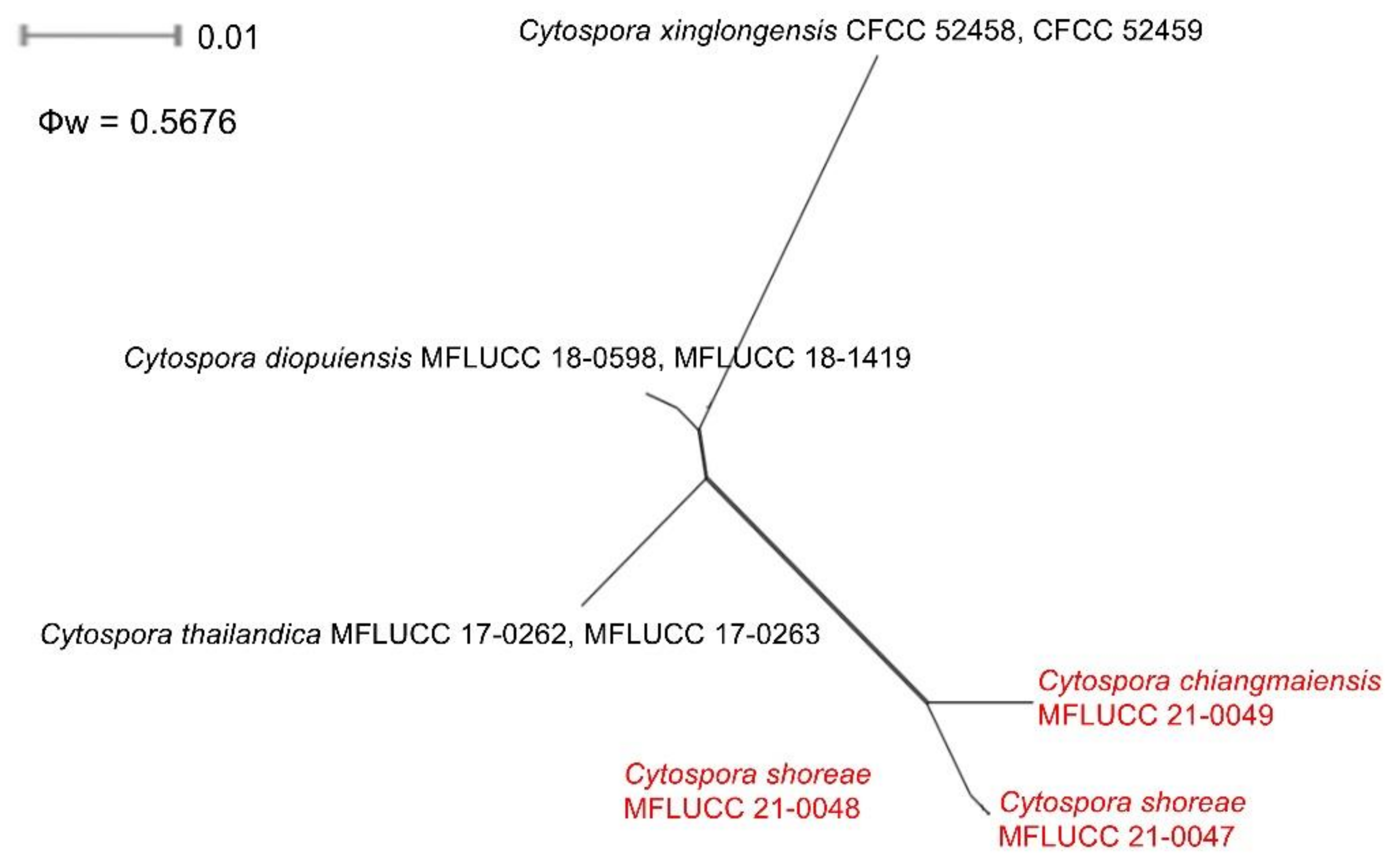
New species and phylogenetically related species were analyzed using the Genealogical Concordance Phylogenetic Species Recognition (GCPSR) model by conducting a pairwise homoplasy index (PHI) test as described by Bruen et al. [45] and Quaedvlieg et al. [46]. The PHI test was conducted in SplitsTree4 [47,48] to examine the recombination level within phylogenetically closely related species using a six-locus concatenated dataset (ITS, LSU, ACT, RPB2, TEF1-α and TUB2). The significant recombination in the dataset was indicated by PHI value below 0.05 (Φw < 0.05). The results were visualized by generating a split graph, using both the LogDet transformation and split decomposition options.
2.6. Preliminary Screening of Antagonistic Activity against Fungal Pathogens
The fungal pathogens, Colletotrichum artocarpicola (MFLUCC 18-1167), Co. fructicola (MFLUCC 18-1160), Co. siamense (MFLUCC 18-1162), Co. viniferum (MFLUCC 18-1179) and Fusarium sambucinum (MFLUCC 17-1056) were obtained from MFLUCC and used for the antagonistic activity test. Fungal isolates were screened using in vitro dual culture assays for their ability to suppress the mycelial growth of fungal pathogens. An antagonism test was performed with 10-day-old cultures of pathogens and new strains. Fresh cultures (pathogens and our strains) 10 days after incubation were used for the antagonism test. A fungal pathogen disc (5 mm) was placed 3 cm from the margin of the PDA plate (9 cm in diam.). An antagonist fungus disc (5 mm) was also placed in a similar manner but on the direct opposite of the pathogen disc. The plate was incubated at room temperature (28 °C) for 10 days. Plates inoculated with a fungal pathogen in the absence of an antagonistic fungus were used as negative controls. The assay was replicated three times. Observations were carried out for the 3rd, 5th, 7th and 10th days. Clear inhibition zone was recorded and the percentage inhibition in mycelial growth was calculated using the following formula [49]: I% = [(R1–R2)/R1] × 100, where I% = the percentage inhibition, R1 = the radial growth of test pathogen in a control plate and R2 = the radial growth of test pathogen in the direction of antagonistic fungus. Data were statistically analyzed with ANOVA using SPSS version 22 (SPSS, Inc., Chicago, IL, USA). Tukey’s HSD test was used to determine the significant differences between treatments at p ≤ 0.05.
3. Results
3.1. Phylogenetic Analyses
The concatenated sequence dataset of ITS, LSU, ACT, RPB2, TEF1-α and TUB2 gene regions comprised 120 strains of Cytospora and two outgroup taxa, Diaporthe eres (CBS 138594) and D. vaccinia (CBS 160.32). The dataset contained 3339 characters according to the order of ITS: 1–416, LSU: 417–1199, ACT: 1200–1429, RPB2: 1430–2027, TEF-α: 2028–2624, β-TUB: 2625–3339. The tree topologies of combined sequence data obtained from ML, MP and BI analyses were not significantly different.
The RAxML analysis of the combined dataset yielded a best scoring tree with a final ML optimization likelihood value of −24983.102448 (Figure 1). The aligned sequence matrix comprises 1399 distinct alignment patterns, with 55.00% of undetermined characters or gaps. Estimated base frequencies were as follows: A = 0.239305, C = 0.269679, G = 0.260287, T = 0.230728; substitution rates AC = 1.822160, AG = 4.370331, AT = 1.771054, CG = 1.304544, CT = 8.574513, GT = 1.000000; gamma distribution shape parameter α = 0.219288. The maximum parsimonious dataset consisted of 3354 characters, of which 2127 were constant, 1052 were parsimony-informative and 175 were parsimony-uninformative. The descriptive statistics of the most parsimony tree was TL = 4294, CI = 0.468, RI = 0.780, RC = 0.365, HI = 0.532.
In the phylogenetic analyses (Figure 1), two new species, Cytospora chiangmaiensis (MFLUCC 21-0049) and Cytospora shoreae (MFLUCC 21-0047, MFLUCC 21-0048), clustered in a monophyletic lineage but well separated branch with strong bootstrap support (94% ML/99% MP/1.00 PP). Another new species, Cytospora phitsanulokensis (MFLUCC 21-0046), formed an independent branch adjacent to C. acaciae, C. magnoliae and C. italica with high bootstrap support (100% ML, 100% MP, 1.00 PP; Figure 1).
A pairwise homoplasy index (PHI) test revealed no significant recombination event between Cytospora phitsanulokensis and the closely related taxa, C. acaciae, C. italica and C. magnoliae (Figure 2). There was also no significant recombination among Cytospora chiangmaiensis, C. shoreae, C. thailandica, C. diopuiensis and C. xinglongensis (Figure 2). This evidence supports that they are different species. The significant recombination between two strains of Cytospora shoreae (MFLUCC 21-0047 and MFLUCC 21-0048) indicate that they are conspecific (Figure 3).
Figure 2.
Results of the pairwise homoplasy index (PHI) test of Cytospora phitsanulokensis and closely related species using both LogDet transformation and splits decomposition. PHI test results (Φw) < 0.05 indicate significant recombination within the dataset. New species described in this study are indicated in red.
Figure 3.
Results of the pairwise homoplasy index (PHI) test of Cytospora chiangmaiensis and C. shoreae and closely related species using both LogDet transformation and splits decomposition. PHI test results (Φw) < 0.05 indicate significant recombination within the dataset. New species described in this study are indicated in red.
3.2. Taxonomy
3.2.1. Cytospora chiangmaiensis Monkai and K.D. Hyde, sp. nov.
Index Fungorum number: IF558524; Facesoffungi number: FoF 09935; Figure 4.
Figure 4.
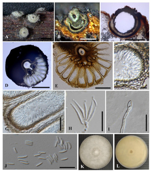
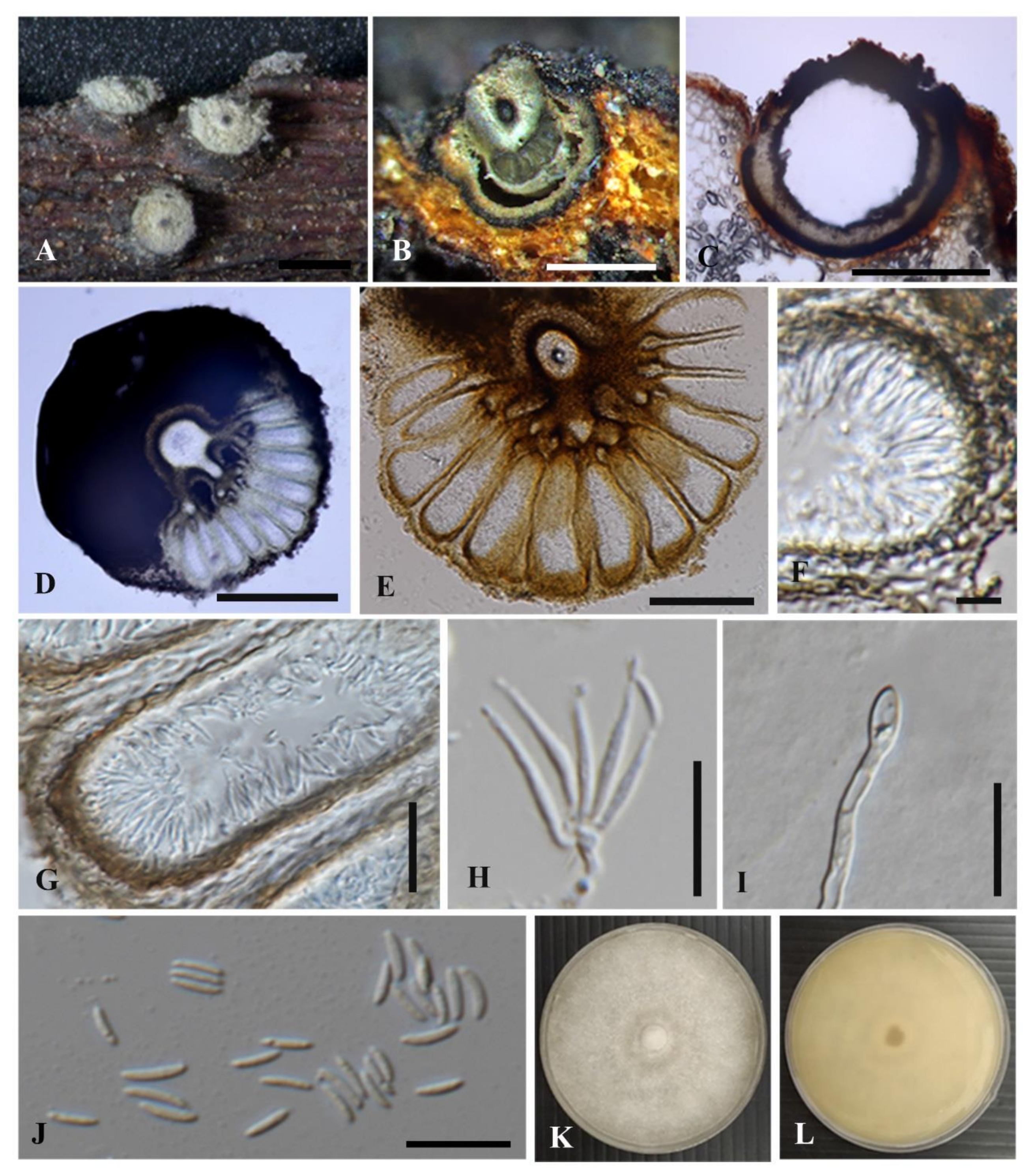
Cytospora chiangmaiensis (MFLU 21-0048, holotype). (A) Conidiomata on host substrate. (B–D) Longitudinal sections through conidioma. (E) Conidiomata with ostiole and the arrangement of locules. (F,G) Peridium. (H) Conidiophore and conidiogenous cells with attached conidia. (I) Germinating conidia. (J) Conidia. (K,L) Colony on PDA plate (9 cm diam.) ((K) from above, (L) from reverse). Scale bars: (A–C) = 500 μm, (D,E) = 100 μm, (G) = 50 μm, (F,H–J) = 10 μm.
Etymology: Name reflects the locality, Chiang Mai Province, Thailand, where the holotype was collected.
Holotype: MFLU 21-0048
Saprobic on decaying leaves (vein and petioles) of Shorea sp. Sexual morph: Undetermined. Asexual morph: Conidiomata 650–800 µm diam., pycnidial, solitary, semi-immersed to erumpent, circular to ovoid, multi-loculate. Conceptacle black. Ectostromatic disc 170–230 µm diam., brown to dark brown, circular to ovoid, one ostiole per disc. Ostioles 55–65 µm diam., conspicuous, circular, dark brown, at the same level as the disc surface. Peridium comprising few layers of cells, textura globosa, dark brown to black. Locules numerous, regularly arranged with a column, independent wall. Conidiophores hyaline, unbranched or occasionally branched at the bases, formed from the innermost layer of pycnidial wall, embedded in a gelatinous layer. Conidiogenous cells 8.5–12.2 × 1–1.7 μm ( = 10.4 × 1.3 μm, n = 20), enteroblastic, phialidic, sub-cylindrical to cylindrical, tapering towards apices. Conidia 5–7 × 0.9–1.7 µm ( = 6 × 1.2 µm, n = 30), hyaline, unicellular, elongate-allantoid, guttulate, smooth, thin-walled.
Culture characteristics: Conidia germinating on PDA within 24 h germ tubes produced from one pole. Colonies on PDA reached at 5 cm diam. after 7 days at 28 °C, irregular in shape, surface slightly rough, effuse, slightly raised, with undulate margin, medium dense, white to cream, in reverse pale yellowish to white.
Material examined: THAILAND, Chiang Mai, Doi Lor district, Yang Kram, on decaying leaves of Shorea sp. (Dipterocarpaceae), 15 October 2019, J. Monkai, CH1-2 (MFLU 21-0048, holotype), ex-type living culture, MFLUCC 21-0049.
Notes: Cytospora chiangmaiensis formed an independent lineage sister to Cytospora shoreae with 94% ML/99% MP/1.00 PP bootstrap support (Figure 1). Based on phylogenetic analyses, Cytospora chiangmaiensis grouped in the same clade with C. diopuiensis, C. lumnitzericola, C. pingbianensis, C. platycladi C. shoreae, C. thailandica and C. xinglongensis (Figure 1). Cytospora diopuiensis and C. pingbianensis are only known from their sexual morph [2], and our strain was found as an asexual morph. Thus, we are unable to compare the morphological characters of those species. However, Cytospora lumnitzericola can be distinguished from C. chiangmaiensis by its shorter conidia [5] (Table 3). Cytospora platycladi is similar to C. chiangmaiensis in having multi-loculate conidiomata with central ostiole but it has smaller conidiomata and shorter conidia [1] (Table 3). Cytospora thailandica differs from C. chiangmaiensis in having larger conidiomata with an ostiolar neck, shorter conidiogenous cell and shorter conidia [5] (Table 3). Cytospora xinglongensis differs from C. chiangmaiensis in having undivided locules, inconspicuous ostioles and shorter conidiogenous cells and longer conidia [16] (Table 3).
Table 3.
Synopsis of asexual morph of Cytospora species discussed in this study.
3.2.2. Cytospora shoreae Monkai and K.D. Hyde, sp. nov.
Index Fungorum number: IF558525; Facesoffungi number: FoF 09936; Figure 5.
Figure 5.
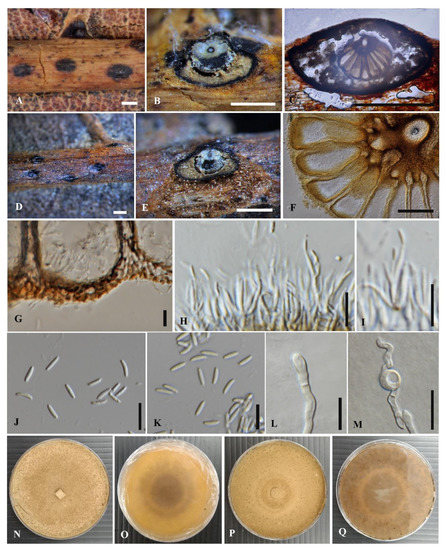
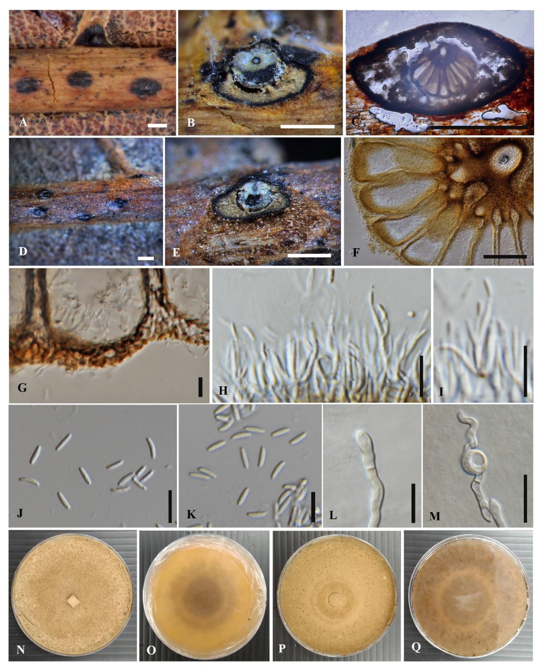
Cytospora shoreae (MFLU 21-0049, holotype (C–E,G,H,J,L,N,O) and MFLU 21-0050, paratype (A,B,F,I,K,M,P,G)). (A,D) Conidiomata on host substrate. (B,C,E,F) Longitudinal sections through conidioma. (G) Peridium. (H,I) Conidiophores and conidiogenous cells with attached conidia. (J,K) Conidia. (L,M) Germinating conidia. (N–Q) Colonies on PDA plate (9 cm diam.) ((N,P) from above, (O,Q) from reverse). Scale bars: (A–E) = 500 μm, (F) = 50 μm, (G–M) = 10 μm.
Etymology: The specific epithet shoreae refers to host plant genus Shorea, on which the fungus was first collected.
Holotype: MFLU 21-0049
Saprobic on decaying leaves (vein and petioles) of Shorea sp. Sexual morph: Undetermined. Asexual morph: Conidiomata 400–1000 µm diam., pycnidial, semi-immersed in host tissue, solitary, erumpent, flask shaped to ovoid, multi-loculate. Conceptacle black. Ectostromatic disc 120–580 µm diam., brown to dark brown, circular to ovoid, one ostiole per disc. Ostioles 19–52 µm diam., conspicuous, circular, dark brown, at the same level as the disc surface. Peridium comprising few layers of cells, textura globosa, dark brown to black. Locules numerous, regularly arranged with a column, independent wall. Conidiophores hyaline, unbranched or occasionally branched at the bases, formed from the innermost layer of pycnidial wall, embedded in a gelatinous layer. Conidiogenous cells 6.7–11.8 × 1–1.9 μm ( = 10 × 1.4 μm, n = 10), enteroblastic, phialidic, subcylindrical to cylindrical, tapering towards apices. Conidia 5–7 × 1–1.6 µm ( = 6 × 1.3 µm, n = 30), hyaline, unicellular, elongate-allantoid, guttulate, smooth, thin-walled.
Culture characteristics: Conidia germinating on PDA within 24 h germ tubes produced from both poles. Colonies on PDA reached at 6 cm diam. after 7 days at 28 °C, irregular in shape, surface slightly rough, effuse, slightly raised, with undulate margin, medium dense, pale brown to white, in reverse pale yellowish to white.
Material examined: THAILAND, Chiang Mai, Omkoi district, Yang Piang, on decaying leaves of Shorea sp. (Dipterocarpaceae), 16 October 2019, J. Monkai, CH2-2 (MFLU 21-0049, holotype), ex-type living culture, MFLUCC 21-0047; THAILAND, Chiang Mai, Omkoi district, Yang Piang, on unidentified decaying leaves, 16 October 2019, J. Monkai, CH2-3 (MFLU 21-0050, paratype), ex-paratype living culture MFLUCC 21-0048.
Notes: Cytospora shoreae formed a sister clade to C. chiangmaiensis with 94% ML, 99% MP and 1.00 PP statistical support (Figure 1). Cytospora chiangmaiensis and C. shoreae share similar morphology in the size and characteristics of conidiogenous cells and conidia. However, C. chiangmaiensis has circular shaped conidiomata. Cytospora shoreae has larger, flask-shaped conidiomata. The single gene comparison of ACT, RPB2, TEF1-α and TUB2 showed that there are significant nucleotide differences (more than 1.5%) between Cytospora chiangmaiensis and two strains of C. shoreae (MFLUCC 21-0047 and MFLUCC 21-0048; Table 4) and this provides evidence that they are different species [50]. Cytospora shoreae differs from C. lumnitzericola and C. platycladi by its longer conidia [1,5] (Table 3). Cytospora shoreae is distinguished from C. thailandica by having longer conidiogenous cell and conidia [5] (Table 3). Cytospora shoreae is distinct from C. xinglongensis in having longer conidiogenous cells and shorter conidia [16] (Table 3). Cytospora shoreae, strains MFLUCC 21-0047 and MFLUCC 21-0048 showed similar characteristics of conidiomata, conidiogenous cells, conidia and culture characteristics, with no significant differences. They were also collected from the same site with the notes of different hosts, where the holotype was isolated from Shorea sp. and the paratype was isolated from unidentified host. The single gene comparison of ITS, LSU, ACT, RPB2, TEF1-α and TUB2 showed that there is no significant difference between two strains of Cytospora shoreae (MFLUCC 21-0047 and MFLUCC 21-0048; Table 4), and this confirms that these two strains are the same species [50].
Table 4.
Nucleotide differences of Cytospora species discussed in this study.
3.2.3. Cytospora phitsanulokensis Monkai and K.D. Hyde, sp. nov.
Index Fungorum number: IF558526; Facesoffungi number: FoF 09937; Figure 6.
Figure 6.
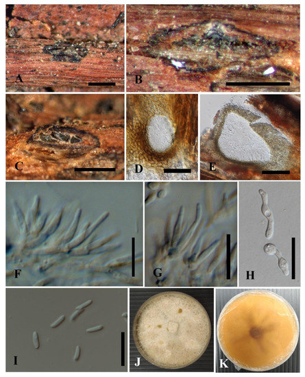
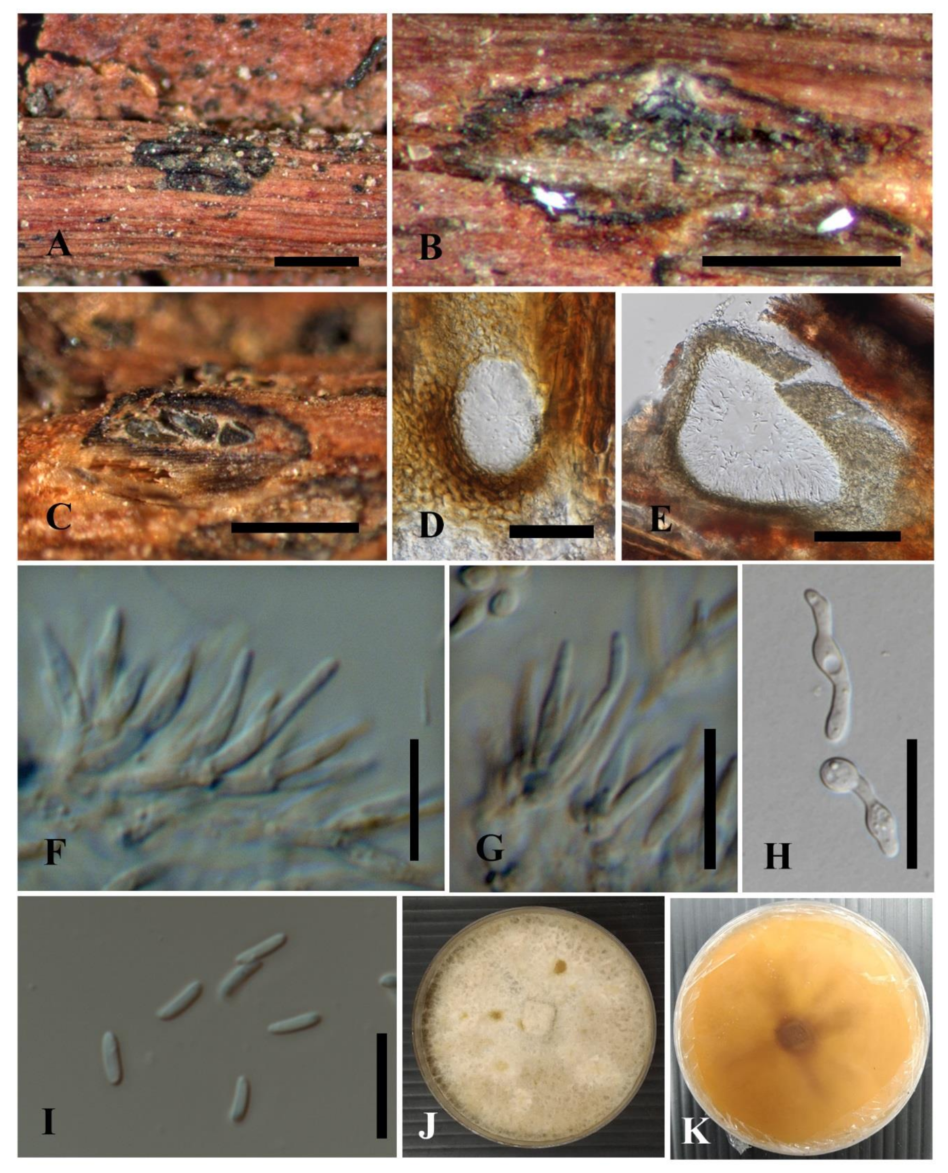
Cytospora phitsanulokensis (MFLU 21-0051, holotype). (A) Conidiomata on host substrate. (B,C) Longitudinal sections through conidioma. (D,E) Longitudinal sections of locules. (F,G) Conidiophores and conidiogenous cells with attached conidia. (H) Germinating conidia. (I) Conidia. (J,K) Colonies on PDA plate (9 cm diam.) ((J) from above, (K) from reverse). Scale bars: (A–C) = 500 μm, (D) = 30 μm, (E) = 50 μm, (F–I) = 10 μm.
Etymology: Name reflects the locality, Phitsanulok Province, Thailand, where the holotype was collected.
Holotype: MFLU 21-0051
Saprobic on unidentified decaying leaves (vein and petioles). Sexual morph: Undetermined. Asexual morph: Conidiomata 900–1100 µm diam., pycnidial, semi-immersed in host tissue, solitary, erumpent, nearly flat, multi-loculate. Conceptacle black. Ectostromatic disc 500–640 µm diam., brown to dark brown, circular to ovoid, one ostiole per disc. Ostioles inconspicuous, black. Peridium comprising few layers of cells of textura angularis, brown to dark brown. Locules numerous, irregular, subdivided frequently by invaginations with independent walls. Conidiophores hyaline, unbranched or occasionally branched at the bases, formed from the innermost layer of pycnidial wall, embedded in a gelatinous layer. Conidiogenous cells 6.7–14.1 × 1.1–2.5 μm ( = 9.8 × 1.9 μm, n = 30), enteroblastic, phialidic, sub-cylindrical to cylindrical. Conidia 5.2–6.9 × 1.2–1.7 µm ( = 6 × 1.5 µm, n = 30), hyaline, unicellular, allantoid, guttulate, smooth, thin-walled.
Culture characteristics: Conidia germinating on PDA within 24 h germ tubes produced from both poles. Colonies on PDA reached at 8 cm diam. after 7 days at 28 °C, circular in shape, effuse, slightly raised, with entire margin, medium dense, floccose, white, in reverse pale yellowish to white.
Material examined: THAILAND, Phitsanulok, Wang Thong, on unidentified decaying leaves, 25 July 2019, E. Yasanthika, E5-2 (MFLU 21-0051, holotype), ex-type living culture, MFLUCC 21-0046.
Notes: Phylogenetically, Cytospora phitsanulokensis forms a distinct lineage and is closely related to C. acaciae, C. magnoliae and C. italica with 100% ML, 100% MP and 1.00 PP statistical support (Figure 1). Cytospora phitsanulokensis differs from C. italica by its larger conidiomata, shorter conidiogenous cell and longer conidia [51] (Table 3). Complete descriptions of C. acaciae and C. magnoliae were not available for morphological comparison [11,52]. Adam et al. [11] reported that Cytospora acaciae produces phialides with a long narrow channel of apical pores with lipid globules at one end of conidium; however, these characteristics were not observed in C. phitsanulokensis.
3.3. Preliminary Screening of Antagonistic Activity against Fungal Pathogens
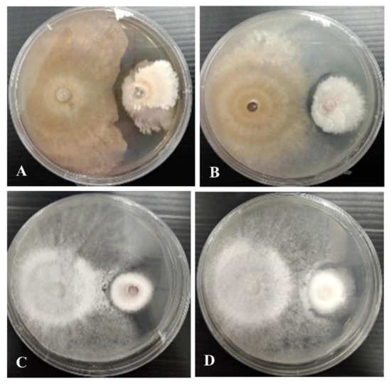
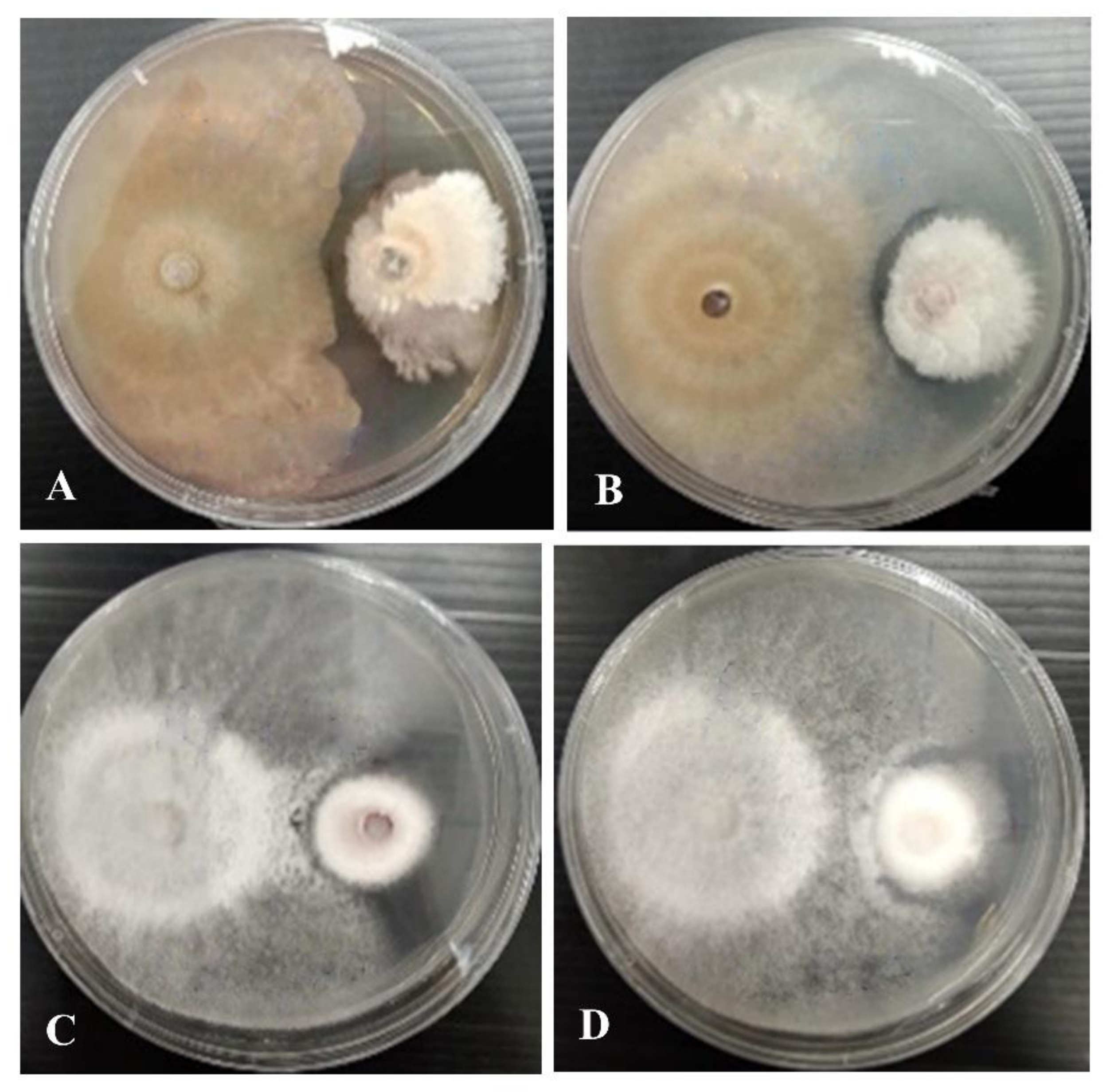
Cytospora phitsanulokensis (MFLUCC 21-0046) exhibited the highest inhibition against Co. viniferum and F. sambucinum at 75.1 ± 4.0% and 67.5 ± 1.3%, respectively (Table 5, Figure 7). Cytospora shoreae (MFLUCC 21-0047 and MFLUCC 21-0048) showed a high inhibition effect against Co. viniferum at 64.0 ± 1.5% and 61.8 ± 1.4%, respectively (Table 3, Figure 7). However, Cytospora chiangmaiensis (MFLUCC 21-0049) had moderate inhibition activity against five tested plant pathogenic fungi (Table 5).
Table 5.
In vitro antagonistic activity of Cytospora spp. against five plant pathogenic fungi after 10 days incubation at 28 °C.
Figure 7.
The antagonistic activity of Cytospora species (left colonies) against fungal pathogens (right colonies). (A) Cytospora shoreae (MFLUCC 21-0047) vs. Colletotrichum viniferum (MFLUCC 18-1179). (B) C. shoreae (MFLUCC 21-0048) vs. Co. viniferum (MFLUCC 18-1179). (C) C. phitsanulokensis (MFLUCC 21-0046) vs. Co. viniferum (MFLUCC 18-1179). (D) C. phitsanulokensis (MFLUCC 21-0046) vs. Fusarium sambucinum (MFLUCC 17-1056).
4. Discussion
This study provides taxonomic novelties of Cytospora species discovered from Thailand and their antagonistic activities against fungal pathogens. We show that six-locus phylogeny (ITS, LSU, ACT, RPB2, TEF1-α and TUB2) facilitates species delineation in Cytospora which is consistent with previous studies [1,3,17,18]. In addition, Cytospora chiangmaiensis and C. shoreae are phylogenetically closely related to C. diopuiensis, which was also collected from Chiang Mai, Thailand [2], indicating the close geographical relationship of these taxa. Based on the Fungus-Host USDA database [53], two new species, Cytospora chiangmaiensis and C. shoreae, are recorded for the first time on Shorea sp. (Dipterocarpaceae). Cytospora phitsanulokensis collected from Phitsanulok, Thailand, is phylogenetically closely related to C. acaciae, C. italica and C. magnoliae. The GCPSR analyses provided evidence for Cytospora phitsanulokensis as a separate species. However, the presence of recombination among strains of C. acaciae, C. italica and C. magnoliae shown in the spilt graph (Figure 2) may be caused by the lack of some gene regions in the dataset. Thus, more strains and new sequence data of C. italica and C. magnoliae should be obtained to better confirm their taxonomic placements in the C. acaciae clade.
Cytospora species are discovered in woody substrates such as bark, branches and twigs [1]. It should be noted that the stromata of Cytospora were only detected on vein and petioles, but not on leaf lamina for all our samples. Adams et al. [11] mentioned that the formation of stroma of Cytospora could possibly decrease in leaves compared to bark. Cytospora species are known as saprobes on dead plants and are one of the important plant pathogens causing dieback and canker diseases on a wide range of hosts [1,2,3,5,9,16,17,18,23]. Considering that the species number of Cytospora is ever-increasing and related taxonomic knowledge continues to expand [1], more extensive sampling of both fresh and dead plant samples in selected hosts and locations should be implemented to improve and stabilize the identification and classification of Cytospora species. In addition, Cytospora are interesting fungal taxa for screening potential biological activities [24,25,27]. Previous studies reported novel bioactive compounds produced by Cytospora spp. [24,25,27,54]; however, species identification of these potential strains was not established. The taxonomic classification and phylogenetic relationships can be used as important tools for the screening of biologically active strains and their biological activities for further applications [55,56].
In this study, we identified new Cytospora species and demonstrated that different strains and/or species can inhibit fungal pathogens. However, our study only conducted a preliminary screening of Cytospora that can inhibit fungal pathogens, using the dual culture method. Although our new species were identified as saprobes based on their occurrence on decaying leaf substrates, their pathogenicity should be clarified, as this genus is well known for pathogens. Therefore, to confirm whether our Cytospora strains are true saprobes, further research is needed. Further studies will be carried out in the near future to check the pathogenicity of the new Cytospora species, elucidate the biology of these fungi and screen secondary metabolites.
Author Contributions
Conceptualization, J.M., A.M. (Ausana Mapook) and C.N.; Methodology, J.M., S.T., A.M. (Areerat Manowong), A.M. (Ausana Mapook) and C.N.; Formal analysis, J.M. and C.N.; Data curation, J.M.; Resources, J.M., S.T., K.D.H. and I.P.; Supervision, K.D.H. and I.P.; Writing—original draft, J.M., S.T., A.M. (Areerat Manowong), A.M. (Ausana Mapook), C.N., K.D.H. and I.P.; Writing—review and editing, J.M., S.T., A.M. (Ausana Mapook), C.N., K.D.H. and I.P. All authors have read and agreed to the published version of the manuscript.
Funding
Kevin D. Hyde thanks the Thailand Research Fund “Impact of climate change on fungal diversity and biogeography in the Greater Mekong Subregion” (project no. RDG6130001) for supporting this research. Saowaluck Tibpromma would like to thank the Inter-national Postdoctoral Exchange Fellowship Program (number Y9180822S1), CAS President’s International Fellowship Initiative (PIFI) (number 2020PC0009), China Postdoctoral Science Foundation and Yunnan Human Resources, and the Social Security Department Foundation for fund-ing her postdoctoral research.
Informed Consent Statement
Not applicable.
Data Availability Statement
Data can be found within the manuscript.
Acknowledgments
Jutamart Monkai would like to thank the Postdoctoral Fellowship from Mae Fah Luang University. Jutamart Monkai is grateful to Rungtiwa Phookamsak, Ruvishika Jayawardena, Naruemon Huanraluek, Erandi Yasanthika, Guang-Cong Ren, Gao Ying and Mark S. Calabon for their assistance during this research. Shaun Pennycook from Landcare Research, Auckland, New Zealand, is thanked for advising on the taxon name. Austin Smith at World Agroforestry (ICRAF), Kunming Institute of Botany, China, is thanked for English editing. We also acknowledge the Biology Experimental Center, Germplasm Bank of Wild Species, Kunming Institute of Botany, Chinese Academy of Sciences for providing the molecular laboratory facilities. Itthayakorn Promputtha is grateful to Chiang Mai University for partial support this research.
Conflicts of Interest
The authors declare no conflict of interest.
References
- Fan, X.; Bezerra, J.D.P.; Tian, C.; Crous, P. Cytospora (Diaporthales) in China. Persoonia-Mol. Phylogeny Evol. Fungi 2020, 45, 1–45. [Google Scholar] [CrossRef] [Scilit] [PubMed]
- Shang, Q.J.; Hyde, K.D.; Camporesi, E.; Maharachchikumbura, S.S.N.; Norphanphoun, C.; Brooks, S.; Liu, J.K. Additions to the genus Cytospora with sexual morph in Cytosporaceae. Mycosphere 2020, 11, 189–224. [Google Scholar] [CrossRef] [Scilit]
- Zhu, H.; Pan, M.; Bezerra, J.D.P.; Tian, C.; Fan, X. Discovery of Cytospora species associated with canker disease of tree hosts from Mount Dongling of China. MycoKeys 2020, 62, 97–121. [Google Scholar] [CrossRef] [Scilit]
- Maharachchikumbura, S.S.N.; Hyde, K.D.; Jones, E.B.G.; McKenzie, E.H.C.; Bhat, J.D.; Dayarathne, M.C.; Huang, S.-K.; Norphanphoun, C.; Senanayake, I.C.; Perera, R.H.; et al. Families of Sordariomycetes. Fungal Divers. 2016, 79, 1–317. [Google Scholar] [CrossRef] [Scilit]
- Norphanphoun, C.; Raspé, O.; Jeewon, R.; Wen, T.-C.; Hyde, K.D. Morphological and phylogenetic characterisation of novel Cytospora species associated with mangroves. MycoKeys 2018, 38, 93–120. [Google Scholar] [CrossRef] [Scilit]
- Ehrenberg, C.G. Sylvae Mycologicae Berolinenses; Formis Teophili Bruschcke: Berlin, Germany, 1818; pp. 1–32. [Google Scholar]
- Donk, M.A. Nomina conservanda proposita I. Propos. Fungi. Deuteromycetes Regnum Veg. 1964, 34, 7–15. [Google Scholar]
- Rossman, A.Y.; Crous, P.W.; Hyde, K.D.; Hawksworth, D.L.; Aptroot, A.; Bezerra, J.L.; Bhat, J.D.; Boehm, E.; Braun, U.; Boonmee, S.; et al. Recommended names for pleomorphic genera in Dothideomycetes. IMA Fungus 2015, 6, 507–523. [Google Scholar] [CrossRef] [Scilit]
- Norphanphoun, C.; Doilom, M.; Daranagama, D.A.; Phookamsak, R.; Wen, T.C.; Bulgakov, T.S.; Hyde, K.D. Revisiting the genus Cytospora and allied species. Mycosphere 2017, 8, 51–97. [Google Scholar] [CrossRef] [Scilit]
- McNeill, J.; Barrie, F.R.; Buck, W.R.; Demoulin, V.; Greuter, W.; Hawksworths, D.L.; Herendeen, P.S.; Knapp, S.; Marhold, K.; Prado, J.; et al. International code of nomenclature for algae, fungi and plants (Melbourne Code) adopted by the Eighteenth International Botanical Congress Melbourne, Australia, July 2011. Regnum Veg. 2012, 154, 1–140. [Google Scholar]
- Adams, G.C.; Wingfield, M.J.; Common, R.; Roux, J. Phylogenetic relationships and morphology of Cytospora species and related teleomorphs (Ascomycota, Diaporthales, Valsaceae) from Eucalyptus. Stud. Mycol. 2005, 52, 1–144. [Google Scholar]
- Wang, X.; Wei, J.; Huang, L.; Kang, Z. Re-evaluation of pathogens causing Valsa canker on apple in China. Mycologia 2011, 103, 317–324. [Google Scholar] [CrossRef] [Scilit]
- Fan, X.; Hyde, K.D.; Liu, M.; Liang, Y.; Tian, C. Cytospora species associated with walnut canker disease in China, with description of a new species C. gigalocus. Fungal Biol. 2015, 119, 310–319. [Google Scholar] [CrossRef] [Scilit] [PubMed]
- Hyde, K.D.; Hongsanan, S.; Jeewon, R.; Bhat, D.J.; McKenzie, E.H.C.; Jones, E.B.G.; Phookamsak, R.; Ariyawansa, H.; Boonmee, S.; Zhao, Q.; et al. Fungal diversity notes 367–490: Taxonomic and phylogenetic contributions to fungal taxa. Fungal Divers. 2016, 80, 1–270. [Google Scholar] [CrossRef] [Scilit]
- Lawrence, D.P.; Holland, L.A.; Nouri, M.T.; Travadon, R.; Abramians, A.; Michailides, T.J.; Trouillas, F.P. Molecular phylogeny of Cytospora species associated with canker diseases of fruit and nut crops in California, with the descriptions of ten new species and one new combination. IMA Fungus 2018, 9, 333–369. [Google Scholar] [CrossRef] [Scilit] [PubMed]
- Jiang, N.; Yang, Q.; Fan, X.-L.; Tian, C.-M. Identification of six Cytospora species on Chinese chestnut in China. MycoKeys 2020, 62, 1–25. [Google Scholar] [CrossRef] [Scilit]
- Pan, M.; Zhu, H.; Bonthond, G.; Tian, C.; Fan, X. High diversity of Cytospora associated with canker and dieback of Rosaceae in China, with 10 new species described. Front. Plant. Sci. 2020, 11, 690. [Google Scholar] [CrossRef] [Scilit]
- Pan, M.; Zhu, H.; Tian, C.; Huang, M.; Fan, X. Assessment of Cytospora isolates from conifer cankers in china, with the descriptions of four new Cytospora species. Front. Plant. Sci. 2021, 12. [Google Scholar] [CrossRef] [Scilit]
- Index Fungorum. Available online: http://www.indexfungorum.org (accessed on 1 August 2021).
- Hyde, K.D.; Norphanphoun, C.; Maharachchikumbura, S.S.N.; Bhat, D.J.; Jones, E.B.G.; Bundhun, D.; Chen, Y.J.; Bao, D.F.; Boonmee, S.; Calabon, M.S.; et al. Refined families of Sordariomycetes. Mycosphere 2020, 11, 305–1059. [Google Scholar] [CrossRef] [Scilit]
- Senanayake, I.; Crous, P.; Groenewald, J.; Maharachchikumbura, S.; Jeewon, R.; Phillips, A.; Bhat, J.; Perera, R.; Li, Q.; Li, W.; et al. Families of Diaporthales based on morphological and phylogenetic evidence. Stud. Mycol. 2017, 86, 217–296. [Google Scholar] [CrossRef] [Scilit]
- Adams, G.C.; Roux, J.; Wingfield, M.J. Cytospora species (Ascomycota, Diaporthales, Valsaceae): Introduced and native pathogens of trees in South Africa. Australas. Plant. Pathol. 2006, 35, 521–548. [Google Scholar] [CrossRef] [Scilit]
- Wang, Y.-L.; Lu, Q.; Decock, C.; Li, Y.-X.; Zhang, X.-Y. Cytospora species from Populus and Salix in China with C. davidiana sp. nov. Fungal Biol. 2015, 119, 420–432. [Google Scholar] [CrossRef] [Scilit] [PubMed]
- Brady, S.F.; Wagenaar, M.M.; Singh, M.P.; Janso, J.E.; Clardy, J. The Cytosporones, new octaketide antibiotics isolated from an endophytic fungus. Org. Lett. 2000, 2, 4043–4046. [Google Scholar] [CrossRef] [Scilit] [PubMed]
- Singh, M.P.; Janso, J.E.; Brady, S.F. Cytoskyrins and cytosporones produced by Cytospora sp. CR200: Taxonomy, fermentation and biological activities. Mar. Drugs 2007, 5, 71–84. [Google Scholar] [CrossRef] [Scilit] [PubMed]
- Sadorn, K.; Saepua, S.; Boonyuen, N.; Boonruangprapa, T.; Rachtawee, P.; Pittayakhajonwut, P. Antimicrobial activity and cytotoxicity of xanthoquinodin analogs from the fungus Cytospora eugeniae BCC42696. Phytochemistry 2018, 151, 99–109. [Google Scholar] [CrossRef] [Scilit]
- Deng, Q.; Li, G.; Sun, M.; Yang, X.; Xu, J. A new antimicrobial sesquiterpene isolated from endophytic fungus Cytospora sp. from the Chinese mangrove plant Ceriops tagal. Nat. Prod. Res. 2018, 34, 1404–1408. [Google Scholar] [CrossRef] [Scilit]
- Senanayake, I.C.; Rathnayaka, A.R.; Marasinghe, D.S.; Calabon, M.S.; Gentekaki, E.; Lee, H.B.; Hurdeal, V.G.; Pem, D.; Dissanayake, L.S.; Wijesinghe, S.N.; et al. Morphological approaches in studying fungi: Collection, examination, isolation, sporulation and preservation. Mycosphere 2020, 11, 2678–2754. [Google Scholar] [CrossRef] [Scilit]
- Jayasiri, S.C.; Hyde, K.D.; Ariyawansa, H.; Bhat, J.; Buyck, B.; Cai, L.; Dai, Y.-C.; Abd-Elsalam, K.A.; Ertz, D.; Hidayat, I.; et al. The faces of fungi database: Fungal names linked with morphology, phylogeny and human impacts. Fungal Divers. 2015, 74, 3–18. [Google Scholar] [CrossRef] [Scilit]
- White, T.J.; Bruns, S.; Lee, S.; Taylor, J. Amplification and direct sequencing of fungal ribosomal RNA genes for phylogenetics. PCR Protoc. A Guid. to Methods Appl. 1990, 18, 315–322. [Google Scholar]
- Vilgalys, R.; Hester, M. Rapid genetic identification and mapping of enzymatically amplified ribosomal DNA from several Cryptococcus species. J. Bacteriol. 1990, 172, 4238–4246. [Google Scholar] [CrossRef] [Scilit]
- Liu, Y.J.; Whelen, S.; Hall, B.D. Phylogenetic relationships among ascomycetes: Evidence from an RNA polymerse II subunit. Mol. Biol. Evol. 1999, 16, 1799–1808. [Google Scholar] [CrossRef] [Scilit]
- Carbone, I.; Kohn, L.M. A method for designing primer sets for speciation studies in filamentous ascomycetes. Mycologia 1999, 91, 553–556. [Google Scholar] [CrossRef] [Scilit]
- Glass, N.L.; Donaldson, G.C. Development of primer sets designed for use with the PCR to amplify conserved genes from filamentous ascomycetes. Appl. Environ. Microbiol. 1995, 61, 1323–1330. [Google Scholar] [CrossRef] [Scilit] [PubMed]
- Katoh, K.; Rozewicki, J.; Yamada, K.D. MAFFT online service: Multiple sequence alignment, interactive sequence choice and visualization. Briefings Bioinform. 2017, 20, 1160–1166. [Google Scholar] [CrossRef] [Scilit]
- Capella-Gutierrez, S.; Silla-Martinez, J.M.; Gabaldon, T. TrimAl: A tool for automated alignment trimming in large-scale phylogenetic analyses. Bioinformatics 2009, 25, 1972–1973. [Google Scholar] [CrossRef] [Scilit] [PubMed]
- Miller, M.A.; Pfeiffer, W.; Schwartz, T. Creating the CIPRES science gateway for inference of large phylogenetic trees. In Proceedings of the 2010 Gateway Computing Environments Workshop (GCE), New Orleans, LA, USA, 14 November 2010; IEEE: New Orleans, LA, USA, 2010; pp. 1–8. [Google Scholar]
- Swofford, D.L. PAUP* Phylogenetic Analysis Using Parsimony * (and Other methods); Version 4.0; Sinauer Associates: Sunderland, UK, 2002. [Google Scholar]
- Nylander, J.A. MrModeltest 2. Program Distributed by the Author. Department of Systematic Zoology; Evolutionary Biology Centre, Uppsala University: Uppsala, Sweden, 2004. [Google Scholar]
- Huelsenbeck, J.P.; Ronquist, F. MRBAYES: Bayesian inference of phylogenetic trees. Bioinformatics 2001, 17, 754–755. [Google Scholar] [CrossRef] [Scilit]
- Zhaxybayeva, O.; Gogarten, J.P. Bootstrap, Bayesian probability and maximum likelihood mapping: Exploring new tools for comparative genome analyses. BMC Genomic. 2002, 3, 1–15. [Google Scholar] [CrossRef] [Scilit]
- Ronquist, F.; Teslenko, M.; Van Der Mark, P.; Ayres, D.L.; Darling, A.; Hoehna, S.; Larget, B.; Liu, L.; Suchard, M.A.; Huelsenbeck, J.P. MrBayes 3.2: Efficient bayesian phylogenetic inference and model choice across a large model space. Syst. Biol. 2012, 61, 539–542. [Google Scholar] [CrossRef] [Scilit] [PubMed]
- Rambaut, A.; Drummond, A. Tracer v1.4. Available online: http://beast.bio.ed.ac.uk/Tracer (accessed on 3 August 2021).
- Rambaut, A.; Drummond, A.J. FigTree: Tree Figure Drawing Tool; Institute of Evolutionary Biology, University of Edinburgh: Edinburgh, Scotland, 2012. [Google Scholar]
- Bruen, T.C.; Philippe, H.; Bryant, D. A simple and robust statistical test for detecting the presence of recombination. Genetics 2006, 172, 2665–2681. [Google Scholar] [CrossRef] [Scilit]
- Quaedvlieg, W.; Binder, M.; Groenewald, J.; Summerell, B.; Carnegie, A.; Burgess, T.; Crous, P. Introducing the consolidated species concept to resolve species in the Teratosphaeriaceae. Persoonia-Mol. Phylogeny Evol. Fungi 2014, 33, 1–40. [Google Scholar] [CrossRef] [Scilit]
- Huson, D.H. SplitsTree: Analyzing and visualizing evolutionary data. Bioinformatics 1998, 14, 68–73. [Google Scholar] [CrossRef] [Scilit]
- Huson, D.H.; Bryant, D. Application of phylogenetic networks in evolutionary studies. Mol. Biol. Evol. 2005, 23, 254–267. [Google Scholar] [CrossRef] [Scilit]
- Hamzah, T.N.T.; Lee, S.Y.; Hidayat, A.; Terhem, R.; Faridah-Hanum, I.; Mohamed, R. Diversity and characterization of endophytic fungi isolated from the tropical mangrove species, Rhizophora mucronata, and identification of potential antagonists against the soil-borne fungus, Fusarium solani. Front. Microbiol. 2018, 9, 1707. [Google Scholar] [CrossRef] [Scilit] [PubMed]
- Jeewon, R.; Hyde, K.D. Establishing species boundaries and new taxa among fungi: Recommendations to resolve taxonomic ambiguities. Mycosphere 2016, 7, 1669–1677. [Google Scholar] [CrossRef] [Scilit]
- Thambugala, K.M.; Daranagama, D.A.; Phillips, A.; Bulgakov, T.; Bhat, D.J.; Camporesi, E.; Bahkali, A.H.; Eungwanichayapant, P.D.; Liu, Z.-Y.; Hyde, K.D. Microfungi on Tamarix. Fungal Divers 2016, 82, 239–306. [Google Scholar] [CrossRef] [Scilit]
- Kepley, J.B.; Reeves, F.B.; Jacobi, W.R.; Adams, G.C. Species associated with Cytospora canker on Populus tremuloides. Mycotaxon 2015, 130, 783–805. [Google Scholar] [CrossRef] [Scilit]
- Farr, D.F.; Rossman, A.Y. Fungal Databases, U.S. National Fungus Collections, ARS, USDA. Available online: https://nt.ars-grin.gov/fungaldatabases/ (accessed on 10 August 2021).
- Wei, C.; Deng, Q.; Sun, M.; Xu, J. Cytospyrone and cytospomarin: Two new polyketides isolated from mangrove endophytic fungus, Cytospora sp. Molecules 2020, 25, 4224. [Google Scholar] [CrossRef] [Scilit] [PubMed]
- Raja, H.A.; Miller, A.N.; Pearce, C.J.; Oberlies, N.H. Fungal identification using molecular tools: A primer for the natural products research community. J. Nat. Prod. 2017, 80, 756–770. [Google Scholar] [CrossRef] [Scilit]
- Doilom, M.; Guo, J.-W.; Phookamsak, R.; Mortimer, P.E.; Karunarathna, S.C.; Dong, W.; Liao, C.-F.; Yan, K.; Pem, D.; Suwannarach, N.; et al. Screening of phosphate-solubilizing fungi from air and soil in Yunnan, China: Four novel species in Aspergillus, Gongronella, Penicillium, and Talaromyces. Front. Microbiol. 2020, 11, 585215. [Google Scholar] [CrossRef] [Scilit]



Publisher’s Note: MDPI stays neutral with regard to jurisdictional claims in published maps and institutional affiliations. |
© 2021 by the authors. Licensee MDPI, Basel, Switzerland. This article is an open access article distributed under the terms and conditions of the Creative Commons Attribution (CC BY) license (https://creativecommons.org/licenses/by/4.0/).