


Abstract
Astaxanthin can be derived from various sources, including petrochemical synthesis, natural sourcing from green algae, or microbial fermentation. As one of the strongest antioxidants known by nature, astaxanthin is attracting attention as an active ingredient in cosmetic products designed to protect the skin against oxidative stress. In contrast to widely performed chemical antioxidant activity assays, this study compares synthetic, algal, and corynebacterial astaxanthin in a physiologically relevant test setting: the intracellular antioxidant activity in cultured human skin cells (keratinocytes). The astaxanthin-rich corynebacterial oleoresin demonstrated superior antioxidant properties in the assay with an EC50 of 2.7 µM, whereas the synthetic and algal-based variants showed no significant effect. Given the potential application of such raw materials, it is therefore tempting to speculate that astaxanthin-containing corynebacterial oleoresins could serve as a natural, superior active ingredient for skin health applications in the future.
1. Introduction
Oxidative stress is closely linked to inflammatory processes and is therefore associated with a range of health issues ranging from cardiovascular and neurodegenerative diseases, diabetes, and cancer to skin diseases and aging [1,2,3,4]. Environmental triggers such as pollution and UV light create ubiquitous oxidative stress in biological systems [5,6,7,8]. Oxidative stress can harm cells in multiple ways, impacting different key processes associated with various locations within the cell. At the plasma membrane/cell envelope, lipid peroxidation causes membrane alterations and interference with receptor signaling [9]. Furthermore, intracellular oxidative stress can interfere with various cellular processes, including DNA repair [10] and protein folding [11,12], as well as affecting subcellular structures [13]. Antioxidants are therefore a dominant group of active ingredients [14] for various applications, ranging from nutrition to cosmetics and pharmaceuticals [15,16,17].
Astaxanthin, known as “the queen of carotenoids”, is one of the most powerful antioxidants found in nature [18]. Its excellent activity can be explained by the conjugated double bond system and the further oxyfunctionalized groups at the β-ionone rings of its structure [18,19]. The astaxanthin market share is dominated by natural astaxanthin, with a share of >600 Mio. USD in 2024 and a CAGR of 8.8% in the forecasted period till 2034 [20]. Natural variants on the markets are, for example, sources from the red yeast Phaffia rhodozyma [21,22,23], the bacterium Paracoccus carotinifaciens [24], and the green microalgae Haematococcus pluvialis [25]. In addition to these well-established production hosts, strain and bioprocess engineering allow us to tackle the astaxanthin market with other microbial cell factories such as Escherichia coli [26,27] and Yarrowia lipolytica [28,29]. In recent years, Corynebacterium glutamicum has been presented as an alternative microbial cell factory for astaxanthin production, with fermentative protocols [30,31,32] and downstream processes [33,34] having been established and optimized.
Besides their different sources, the synthesized astaxanthins differ in their stereochemistry and esterification. Synthetic astaxanthin is present as a racemate (a 1:2:1 mixture of (3S,3′S), (3S,3′R), and (3R,3′R)) in its free form [35]. Natural biosynthesis typically results in either the 3S,3′S enantiomer (dominantly algal, bacterial, and plant sources) [36,37] or the 3R,3′R enantiomer (dominantly yeast sources) [38]. Depending on the specific production host, astaxanthin may be present in its free form or esterified with fatty acids [39,40]. This means that a series of structurally different astaxanthin variants exists, which may have different functional properties. In vitro assays, such as the 2,2-diphenyl-1-picrylhydrazyl (DPPH) assay, allow the antioxidant activity to be easily determined based on the color change during quenching of the free radical DPPH in the presence of a ROS scavenger [41]. This assay is widely applied due to its easy, fast, and cheap handling. However, the interpretation of such chemical assays for real-world application is limited. Active (pharmaceutical) ingredients have to be bioavailable [42,43,44] in order to fulfill their function at the desired cellular location.
As a promising active ingredient in skincare products, topical application of astaxanthin has been shown to reduce wrinkles, improve elasticity and pigmentation [45], support wound healing [46], and mitigate UV-induced skin damage [47]. Therefore, the scope of this work is on the investigation of the antioxidant activity of three different astaxanthin sources, namely, synthetic, algal-based, and corynebacterial astaxanthin (as present in a corynebacterial natural extract), using a more physiologically relevant test setting involving human keratinocytes.
2. Results
2.1. Intracellular Antioxidant Activity Testing of Different Astaxanthins
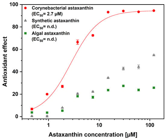
The human keratinocyte cell line HaCaT is a widely used model of the epidermis and is therefore employed to investigate skin physiology and evaluate novel cosmetic ingredients [48,49]. As astaxanthin is a promising ingredient for skin applications [50], the assay was performed on human keratinocytes (HaCaT). The assay uses the fluorescent dye thiazole orange, which is taken up by the cells. When illuminated, it triggers the generation of ROS, such as singlet oxygen and hydroxyl radicals. These ROS can be neutralized by the addition of an antioxidant [51]. The results of the AOP1 assay are shown in Figure 1. The half-maximal effective concentration (EC50) of corynebacterial astaxanthin (CA) was determined to be 2.7 µM. By contrast, no calculable EC50 was obtained for algal-based astaxanthin (AA) or synthetic astaxanthin (SA), as only partial activity was observed even at the highest concentrations examined (120 µM).
Figure 1.
Intracellular antioxidant activity of astaxanthin from different sources. Corynebacterial astaxanthin-containing oleoresin (CA), synthetic and algal (SA) astaxanthin-containing oleoresin (AA) are shown in red, gray, and green, respectively. Values are given as mean ± standard deviation (n = 3).
2.2. De-Esterification of Algal-Based Astaxanthin and Its Intracellular Activity
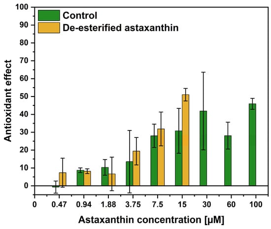
We aimed to de-esterify the astaxanthin-(di)esters in the algal-based oleoresin in order to test the activity of an algal-derived free-form astaxanthin. Therefore, astaxanthin derived from H. pluvialis was de-esterified using cholesterol esterase, as confirmed by HPLC analysis (Figure S1). The AOP1 assay, which compared the de-esterified and the esterified astaxanthin, was performed as previously described, and the results are depicted in Figure 2. No statistical differences were observed between the control and de-esterified samples. Notably, the samples containing the de-esterified astaxanthin exhibited cytotoxic effects at concentrations > 15 µM, leading to incomplete results.
Figure 2.
Intracellular antioxidant activity of de-esterified astaxanthin. Extract from H. pluvialis was treated with BSA (control; shown in green) or cholesterol esterase (de-esterified; shown in yellow). Values are given as mean ± standard deviation (n = 3).
3. Discussion
This work demonstrates that the source and molecular form of astaxanthin significantly impact its antioxidant activities within cultured human keratinocytes. Astaxanthin-containing corynebacterial oleoresin exhibited a pronounced intracellular antioxidant effect, with an EC50 of 2.7 µM.
In a previous study, we have shown that corynebacterial and algal astaxanthin exhibit comparable antioxidant activities using the widely accepted DPPH assay [33]. While natural corynebacterial and algal astaxanthins showed a comparable EC50 of 3.2–3.7 µg mL−1, the racemic mixture of the synthetic astaxanthin had approximately 10-fold lower antioxidant activity (correspondingly a higher EC50 of 42 µg mL−1) [33]. These findings are consistent with previous reports indicating that the enantiomer pure (3S,3′S) astaxanthin exhibits a higher antioxidant activity than the (3R,3′R) enantiomer and the synthetic racemic mixture in different chemical assays such as ABTS and ORAC, as well as in cell- and organism-based in vivo studies [52].
The discrepancy between DPPH-based antioxidant activity and the intracellular AOP1 assay highlights the importance of complementing chemical assays for specific application evaluations [15]. While chemical antioxidant assays excel at quantifying radical-scavenging capacity in cell-free solutions, they lack information on bioavailability and cell uptake, which are practical hurdles that influence the antioxidant activity in cellular systems [14,53].
Astaxanthin itself is a lipophilic compound [44], but the solubility and bioavailability of the esterified (algal) and free form (corynebacterial and synthetic) astaxanthins [54], as well as the enantiomer composition, are considered to affect the stability and bioavailability [55,56]. In vitro and in vivo studies have shown that free astaxanthin is absorbed faster, since astaxanthin esters must be hydrolyzed by digestive enzymes and fluids prior to uptake [56,57,58].
As the conditions of the AOP1 assay do not favor ester hydrolysis, it is tempting to speculate that the algal-derived astaxanthin was not properly taken up by the HaCaT cells. A similar situation may apply to the cosmetic use of algal-derived astaxanthin: unlike free astaxanthin, the esterified form of algal astaxanthin is less bioavailable to the skin due to the absence of hydrolyzing enzymes, which makes it less suitable for topical applications [39,56].
Therefore, it was questioned whether the intracellular antioxidant activity of the algal-based astaxanthin could be increased after de-esterification. Although the de-esterification was successfully validated by HPLC analysis (Figure S1), the intracellular antioxidant activity in cultured keratinocytes did not differ from that of the control algal-based oleoresin. Cytotoxic effects of the de-esterified astaxanthin were observed at concentrations > 15 µM, leading to incomplete results. This cytotoxicity may be due to the release of free fatty acids during the de-esterification, which are known to exhibit in vitro cytotoxicity for cell cultures [59,60].
Astaxanthin exhibits skin-protective effects in a concentration-dependent manner. Cellular in vitro studies typically report efficacy in the low micromolar range (1–10 µM), while topical formulations commonly contain around 0.1%, and oral supplementation is usually applied at doses of 4–12 mg/day [61,62]. Overall, astaxanthin is considered a safe ingredient for both topical and oral use. At the concentrations typically used for topical application, adverse effects such as skin discoloration are unlikely to occur. Safety assessments by the European Food Safety Authority, as well as human intervention studies involving daily doses of 8–12 mg, have not identified any toxicological concerns [62,63,64]. Nevertheless, high concentrations of carotenoids may influence gastrointestinal function and lipid metabolism and can facilitate pro-oxidative effects under certain conditions [65]. Currently, there is no evidence of isomer-specific adverse dermatological or systemic effects of astaxanthin in humans. This indicates that differences between astaxanthin enantiomers primarily affect bioavailability and biological activity rather than safety. While synthetic astaxanthin is registered and widely used as a feed colorant in aquaculture, only natural astaxanthin derived from algae has been registered as a novel food for human consumption.
Despite the clear activity differences observed, some limitations should be considered. As the corynebacterial astaxanthin was tested as part of the oleoresin rather than as a purified compound, the synergistic effects of co-extracted compounds cannot be excluded. The oleoresin matrix itself may account for the superior activity, as byproducts in the extract may promote the solubility and/or delivery of the lipophilic astaxanthin [66]. It is known that lipid-rich matrices and formulation strategies can improve the bioavailability of carotenoids as compared to purified compounds [50,57,67]. Additionally, microneedle-based delivery systems can bypass the outermost layer of the skin, thereby potentially facilitating transdermal absorption [68].
In this study, the corynebacterial oleoresin was demonstrated to be the most promising source of astaxanthin for the intracellular ROS-scavenging in keratinocytes. The cell-based assay was developed by Gironde et al. (2020), who obtained the similar partial activity of synthetic astaxanthin [51] as observed in this study. Although the synthetic astaxanthin was most likely taken up by the cells due to its free form, the low activity was likely due to its less active enantiomers. When compared to the results of Gironde et al. 2020 [51], it should be noted that the astaxanthin-rich corynebacterial oleoresin is one of the most effective antioxidants identified in the AOP1 assay, with an EC50 of 2.7 µM in HaCaT cells (Figure 1).
As these results highlight the significance of alternative antioxidant resources from microbial fermentation [26], the real-world applicability should be further evaluated in other cell lines and skin penetration assays to strengthen the conclusion [50]. From an application perspective, C. glutamicum is a platform system offering access to other astaxanthin derivatives, such as astaxanthin-diglucoside [32], which may exhibit distinct bioactivities compared to the here tested astaxanthin variants.
4. Materials and Methods
4.1. Production and Extraction of Corynebacterial Astaxanthin
Astaxanthin-producing C. glutamicum strain ASTA** was cultivated for 48 h in shake flasks as described by [32] using the optimized trace element solution [31]. After cultivation, the biomass was harvested by centrifugation at 10,000× g for 20 min. Subsequently, the cell pellet was extracted using 90% (v v−1) ethanol at a solvent-to-biomass ratio of 15 mgCDW mL−1 at 60 °C for 30 min in a 1 L stirred bottle reactor equipped with an anchor stirrer (DWK Life Sciences, Mainz, Germany) at 500 rpm. The astaxanthin oleoresin was prepared as described in [33] and was stored at −20 °C until further usage. The astaxanthin content in the oleoresin was 11.6 mg g−1 as determined by HPLC [32].
4.2. Intracellular In Vivo Antioxidant Assay
For the intracellular in vivo antioxidant assay (AOP1), the corynebacterial astaxanthin (as oleoresin) (CA) was compared to two other astaxanthin sources: synthetic astaxanthin (SA) (Sigma-Aldrich, St. Louis, MA, USA; catalog number 1044200) and esterified astaxanthin (as oleoresin; containing 100 mg g−1 astaxanthin (calculated as free astaxanthin); astaxanthin is present as 75% monoester, 20% diester, and 5% free) from Haematococcus pluvialis (AA) (Sigma-Aldrich, St. Louis, MA, USA; catalog number 1044210). All samples were dissolved in DMSO to a concentration of 3 mM (calculated as free astaxanthin) at 40 °C and 1000 rpm (Thermomixer comfort, Eppendorf, Wesseling, Germany). Stock solutions were shipped frozen to Antioxidant Power (Toulouse, France). Nine different astaxanthin concentrations were obtained by serial factor 2 dilutions, with the highest concentration of astaxanthin in the cell culture medium being 120 µM, corresponding to 4% DMSO. The assay was performed as described by [51] using human keratinocytes (HaCaT).
4.3. AOP1 Assay with De-Esterified Algal Astaxanthin
In order to verify that the low activity of the algal astaxanthin is based on the esters, a second AOP1 assay was performed, comparing both the free and the esterified astaxanthin from algae. To obtain the free version of the esterified astaxanthin from H. pluvialis, an enzymatic cleavage was performed, which was adapted from [69]. Per mL of acetone, 1.5 mg of H. pluvialis oleoresin was dissolved. The enzyme solution consisted of 2 U mL−1 cholesterol esterase from Pseudomonas fluorescencs (Sigma-Aldrich, St. Louis, MA, USA; catalog number: C9281) in 0.05 M Tris HCl, pH 7. The astaxanthin-containing acetone and the enzyme solution were mixed at a ratio of 3:2, and the reaction mixture was shaken at 37 °C in the dark for 4 h. The cholesterol esterase was replaced by bovine serum albumin (BSA) as a negative control. After incubation, 5 mL of hexane was added to the reaction mixture and mixed vigorously. The hexane layer was transferred into a new tube and subsequently evaporated (Concentrator Plus, Eppendorf, Germany). Successful de-esterification (>99%) was verified by HPLC (Figure S1). BSA-treated samples (control) and the cholesterol esterase-treated samples (de-esterified astaxanthin) were dissolved in DMSO at concentrations of 2.5 mM and 2.9 mM (calculated as free astaxanthin), respectively.
4.4. Quantification of Astaxanthin by HPLC
Astaxanthin was quantified by using the Agilent 1200 series (Agilent Technologies, Santa Clara, CA, USA) equipped with a reversed-phase precolumn (LiChrospher 100 RP18 EC-5, 40 × 4 mm) (CS-Chromatographie, Langerwehe, Germany), a reversed-phase main column (LiChrospher 100 RP18 EC-5, 125 × 4 mm) (CS-Chromatographie, Langerwehe, Germany), and a diode array detector (DAD) recording the absorption at λ = 470 nm. A defined amount of sample was dissolved in a 7:3 methanol:acetone mixture, of which 50 µL was injected. The mobile phases consisted of methanol:water (9:1) (A) and methanol (B). A gradient at a flow rate of 1.5 mL min−1 was used as follows: 0 min B: 0%, 10 min B: 100%, and 32.5 min B: 100%. Synthetic astaxanthin was used as a reference standard.
5. Conclusions
In conclusion, the results demonstrate that the astaxanthin-rich corynebacterial extract exhibits superior intracellular antioxidant activity in keratinocytes compared to algal and synthetic forms, highlighting the importance of molecular form and stereoisomer composition for cellular efficacy. Effective doses observed here for antioxidation in keratinocytes (EC50 of 2.7 µM) are consistent with concentrations reported in the literature. It can be hypothesized that the differences between tested astaxanthin variants primarily affect bioavailability and efficacy rather than safety.
Supplementary Materials
The following supporting information can be downloaded at: https://www.mdpi.com/article/10.3390/ijms27083638/s1.
Author Contributions
Conceptualization, N.A.H. and J.S.; methodology, N.A.H. and J.S.; formal analysis, J.S.; investigation, J.S.; data curation, J.S.; writing—original draft preparation, N.A.H. and J.S.; writing—review and editing, N.A.H. and J.S.; visualization, J.S.; supervision, N.A.H.; project administration, N.A.H.; funding acquisition, N.A.H. All authors have read and agreed to the published version of the manuscript.
Funding
This research was funded by the German Federal Ministry of Education and Research (BMBF) project KaroTec (grant number: 03VP09460). N.A.H. acknowledges support by the KIT-Publication Fund of Karlsruhe Institute of Technology.
Institutional Review Board Statement
Not applicable.
Informed Consent Statement
Not applicable.
Data Availability Statement
The original contributions presented in this study are included in the article/Supplementary Materials. Further inquiries can be directed to the corresponding author.
Acknowledgments
We acknowledge scientific discussion with Rainer Figge and Volker F. Wendisch.
Conflicts of Interest
The authors declare no conflicts of interest.
Abbreviations
The following abbreviations are used in this manuscript:
| AOP | Antioxidant power test |
| AA | Algal astaxanthin |
| CA | Corynebacterial astaxanthin |
| CDW | Cell dry weight |
| SA | Synthetic astaxanthin |
References
- Zuo, L.; Prather, E.R.; Stetskiv, M.; Garrison, D.E.; Meade, J.R.; Peace, T.I.; Zhou, T. Inflammaging and Oxidative Stress in Human Diseases: From Molecular Mechanisms to Novel Treatments. Int. J. Mol. Sci. 2019, 20, 4472. [Google Scholar] [CrossRef] [Scilit] [PubMed]
- Altanam, S.Y.; Darwish, N.; Bakillah, A. Exploring the Interplay of Antioxidants, Inflammation, and Oxidative Stress: Mechanisms, Therapeutic Potential, and Clinical Implications. Diseases 2025, 13, 309. [Google Scholar] [CrossRef] [Scilit]
- Valaitienė, J.; Laučytė-Cibulskienė, A. Oxidative Stress and Its Biomarkers in Cardiovascular Diseases. Artery Res. 2024, 30, 18. [Google Scholar] [CrossRef] [Scilit]
- Reddy, V.P. Oxidative Stress in Health and Disease. Biomedicines 2023, 11, 2925. [Google Scholar] [CrossRef] [Scilit]
- Petruk, G.; Del Giudice, R.; Rigano, M.M.; Monti, D.M. Antioxidants from Plants Protect against Skin Photoaging. Oxid. Med. Cell Longev. 2018, 2018, 1454936. [Google Scholar] [CrossRef] [Scilit]
- Hooda, R.; Madke, B.; Choudhary, A. Photoaging: Reversal of the Oxidative Stress Through Dietary Changes and Plant-Based Products. Cureus 2023, 15, e37321. [Google Scholar] [CrossRef] [Scilit] [PubMed]
- Yang, W.; Omaye, S.T. Air pollutants, oxidative stress and human health. Mutat. Res. 2009, 674, 45–54. [Google Scholar] [CrossRef] [Scilit]
- Kammeyer, A.; Luiten, R.M. Oxidation events and skin aging. Ageing Res. Rev. 2015, 21, 16–29. [Google Scholar] [CrossRef] [Scilit] [PubMed]
- Yin, H.; Xu, L.; Porter, N.A. Free Radical Lipid Peroxidation: Mechanisms and Analysis. Chem. Rev. 2011, 111, 5944–5972. [Google Scholar] [CrossRef] [Scilit]
- Cadet, J.; Davies, K.J.A. Oxidative DNA damage & repair: An introduction. Free Radic. Biol. Med. 2017, 107, 2–12. [Google Scholar] [CrossRef] [Scilit]
- Sies, H.; Berndt, C.; Jones, D.P. Oxidative Stress. Annu. Rev. Biochem. 2017, 86, 715–748. [Google Scholar] [CrossRef] [Scilit]
- Sies, H.; Jones, D.P. Reactive oxygen species (ROS) as pleiotropic physiological signalling agents. Nat. Rev. Mol. Cell Biol. 2020, 21, 363–383. [Google Scholar] [CrossRef] [Scilit]
- Forrester, S.J.; Kikuchi, D.S.; Hernandes, M.S.; Xu, Q.; Griendling, K.K. Reactive Oxygen Species in Metabolic and Inflammatory Signaling. Circ. Res. 2018, 122, 877–902. [Google Scholar] [CrossRef] [Scilit]
- Gulcin, İ. Antioxidants: A comprehensive review. Arch. Toxicol. 2025, 99, 1893–1997. [Google Scholar] [CrossRef] [Scilit]
- Forman, H.J.; Zhang, H. Targeting oxidative stress in disease: Promise and limitations of antioxidant therapy. Nat. Rev. Drug Discov. 2021, 20, 689–709, Correction in Nat. Rev. Drug Discov. 2021, 20, 652. https://doi.org/10.1038/s41573-021-00267-5. [Google Scholar] [CrossRef] [Scilit]
- Dini, I.; Laneri, S. Nutricosmetics: A brief overview. Phytother. Res. 2019, 33, 3054–3063. [Google Scholar] [CrossRef] [Scilit] [PubMed]
- Budzianowska, A.; Banaś, K.; Budzianowski, J.; Kikowska, M. Antioxidants to Defend Healthy and Youthful Skin—Current Trends and Future Directions in Cosmetology. Appl. Sci. 2025, 15, 2571. [Google Scholar] [CrossRef] [Scilit]
- Dutta, S.; Kumar, S.P.J.; Banerjee, R. A comprehensive review on astaxanthin sources, structure, biochemistry and applications in the cosmetic industry. Algal Res. 2023, 74, 103168. [Google Scholar] [CrossRef] [Scilit]
- Brotosudarmo, T.H.P.; Limantara, L.; Setiyono, E.; Heriyanto. Structures of Astaxanthin and Their Consequences for Therapeutic Application. Int. J. Food Sci. 2020, 2020, 2156582. [Google Scholar] [CrossRef] [Scilit]
- Global Market Insights. Astaxanthin Market Size & Share 2025–2034: Market Size by Source, by Application, Industry Analysis, Growth Forecast; Global Market Insights: Selbyville, DE, USA, 2025. [Google Scholar]
- Sun, J.; Zhang, Z.; Gao, L.; Yang, F. Advances and trends for astaxanthin synthesis in Phaffia rhodozyma. Microb. Cell Factories 2025, 24, 100. [Google Scholar] [CrossRef] [Scilit]
- Ji, W.; Wang, W.; Chen, X.; Sun, M.; Zhang, J.; Nan, B.; Li, X.; Wang, Y.; Wang, Y.; Piao, C. Stress-induced astaxanthin biosynthesis in Phaffia rhodozyma: Bridging mechanistic understanding to industrial Feasibility. Bioresour. Technol. 2025, 435, 132957. [Google Scholar] [CrossRef] [Scilit]
- Luna-Flores, C.H.; Wang, A.; von Hellens, J.; Speight, R.E. Towards commercial levels of astaxanthin production in Phaffia rhodozyma. J. Biotechnol. 2022, 350, 42–54. [Google Scholar] [CrossRef] [Scilit]
- Mussagy, C.U.; Pereira, J.F.B.; Dufossé, L. Astaxanthin production using Paracoccus carotinifaciens: A way forward? Trends Biotechnol. 2023, 41, 996–999. [Google Scholar] [CrossRef] [Scilit]
- Ariyadasa, T.U.; Thevarajah, B.; Anthonio, R.A.D.P.; Nimarshana, P.H.V.; Wasath, W.A.J. From present to prosperity: Assessing the current status and envisioning opportunities in the industrial-scale cultivation of Haematococcus pluvialis for astaxanthin production. Phytochem. Rev. 2024, 23, 749–779. [Google Scholar] [CrossRef] [Scilit]
- Wang, C.; Hong, Z.; Song, M.; Zheng, H.; Zhou, Q.; Yang, H.; Li, H.; Huang, D. Production of astaxanthin with high purity and activity based on engineering improvement strategies. J. Biotechnol. 2025, 405, 139–149. [Google Scholar] [CrossRef] [Scilit]
- Lu, Q.; Bu, Y.-F.; Liu, J.-Z. Metabolic Engineering of Escherichia coli for Producing Astaxanthin as the Predominant Carotenoid. Mar. Drugs 2017, 15, 296. [Google Scholar] [CrossRef] [Scilit]
- Li, N.; Han, Z.; O’Donnell, T.J.; Kurasaki, R.; Kajihara, L.; Williams, P.G.; Tang, Y.; Su, W.W. Production and excretion of astaxanthin by engineered Yarrowia lipolytica using plant oil as both the carbon source and the biocompatible extractant. Appl. Microbiol. Biotechnol. 2020, 104, 6977–6989. [Google Scholar] [CrossRef] [Scilit]
- Tramontin, L.R.R.; Kildegaard, K.R.; Sudarsan, S.; Borodina, I. Enhancement of Astaxanthin Biosynthesis in Oleaginous Yeast Yarrowia lipolytica via Microalgal Pathway. Microorganisms 2019, 7, 472. [Google Scholar] [CrossRef] [Scilit]
- Meyer, F.; Schmitt, I.; Schäffer, T.; Wendisch, V.F.; Henke, N.A. Design-of-Experiment-Guided Establishment of a Fermentative Bioprocess for Biomass-Bound Astaxanthin with Corynebacterium glutamicum. Fermentation 2023, 9, 969. [Google Scholar] [CrossRef] [Scilit]
- Meyer, F.; Schmitt, I.; Wendisch, V.F.; Henke, N.A. Response surface-based media optimization for astaxanthin production in Corynebacterium glutamicum. Front. Bioeng. Biotechnol. 2025, 13, 1516522. [Google Scholar] [CrossRef] [Scilit] [PubMed]
- Göttl, V.L.; Meyer, F.; Schmitt, I.; Persicke, M.; Peters-Wendisch, P.; Wendisch, V.F.; Henke, N.A. Enhancing astaxanthin biosynthesis and pathway expansion towards glycosylated C40 carotenoids by Corynebacterium glutamicum. Sci. Rep. 2024, 14, 8081. [Google Scholar] [CrossRef] [Scilit] [PubMed]
- Seeger, J.; Wendisch, V.F.; Henke, N.A. Extraction and Purification of Highly Active Astaxanthin from Corynebacterium glutamicum Fermentation Broth. Mar. Drugs 2023, 21, 530. [Google Scholar] [CrossRef] [Scilit]
- Seeger, J.; Zäh, M.; Wendisch, V.F.; Brandenbusch, C.; Henke, N.A. Supercritical carbon dioxide extraction of astaxanthin from Corynebacterium glutamicum. Bioresour. Bioprocess. 2025, 12, 46. [Google Scholar] [CrossRef] [Scilit]
- Higuera-Ciapara, I.; Félix-Valenzuela, L.; Goycoolea, F.M. Astaxanthin: A review of its chemistry and applications. Crit. Rev. Food Sci. Nutr. 2006, 46, 185–196. [Google Scholar] [CrossRef] [Scilit]
- Zhang, C.; Yao, W.; Wen, D.; Li, X.; Wu, S.; Leng, X. Dietary Adonis. aestivalis extract improved the flesh pigmentation, antioxidative status and shelf-life of rainbow trout (Oncorhynchus mykiss). Aquac. Nutr. 2020, 26, 2032–2042. [Google Scholar] [CrossRef] [Scilit]
- Renstrøm, B.; Borch, G.; Skulberg, O.M.; Liaaen-Jensen, S. Optical purity of (3S,3′S)-astaxanthin from Haematococcus pluvialis. Phytochemistry 1981, 20, 2561–2564. [Google Scholar] [CrossRef] [Scilit]
- Andrewes, A.G.; Starr, M.P. (3R,3′R)-astaxanthin from the yeast Phaffia rhodozyma. Phytochemistry 1976, 15, 1009–1011. [Google Scholar] [CrossRef] [Scilit]
- Holtin, K.; Kuehnle, M.; Rehbein, J.; Schuler, P.; Nicholson, G.; Albert, K. Determination of astaxanthin and astaxanthin esters in the microalgae Haematococcus pluvialis by LC-(APCI)MS and characterization of predominant carotenoid isomers by NMR spectroscopy. Anal. Bioanal. Chem. 2009, 395, 1613–1622. [Google Scholar] [CrossRef] [Scilit]
- Miao, F.; Lu, D.; Li, Y.; Zeng, M. Characterization of astaxanthin esters in Haematococcus pluvialis by liquid chromatography–atmospheric pressure chemical ionization mass spectrometry Anal. Anal. Biochem. 2006, 352, 176–181. [Google Scholar] [CrossRef] [Scilit]
- Blois, M.S. Antioxidant Determinations by the Use of a Stable Free Radical. Nature 1958, 181, 1199–1200. [Google Scholar] [CrossRef] [Scilit]
- Stielow, M.; Witczyńska, A.; Kubryń, N.; Fijałkowski, Ł.; Nowaczyk, J.; Nowaczyk, A. The Bioavailability of Drugs-The Current State of Knowledge. Molecules 2023, 28, 8038. [Google Scholar] [CrossRef] [Scilit]
- Chen, M.L.; Shah, V.; Patnaik, R.; Adams, W.; Hussain, A.; Conner, D.; Mehta, M.; Malinowski, H.; Lazor, J.; Huang, S.M.; et al. Bioavailability and bioequivalence: An FDA regulatory overview. Pharm. Res. 2001, 18, 1645–1650. [Google Scholar] [CrossRef] [Scilit]
- Wang, W.; Cui, Y.; Liu, H.; Wang, Y.; Nan, B.; Li, X.; Wang, Y. Progress in the bioavailability of natural astaxanthin: Influencing factors, enhancement strategies, evaluation methods, and limitations of current research. Trends Food Sci. Technol. 2025, 160, 104998. [Google Scholar] [CrossRef] [Scilit]
- Tominaga, K.; Hongo, N.; Karato, M.; Yamashita, E. Cosmetic benefits of astaxanthin on humans subjects. Acta Biochim. Pol. 2012, 59, 43–47. [Google Scholar] [CrossRef] [Scilit] [PubMed]
- Meephansan, J.; Rungjang, A.; Yingmema, W.; Deenonpoe, R.; Ponnikorn, S. Effect of astaxanthin on cutaneous wound healing. Clin. Cosmet. Investig. Dermatol. 2017, 10, 259–265. [Google Scholar] [CrossRef] [Scilit]
- Hama, S.; Takahashi, K.; Inai, Y.; Shiota, K.; Sakamoto, R.; Yamada, A.; Tsuchiya, H.; Kanamura, K.; Yamashita, E.; Kogure, K. Protective effects of topical application of a poorly soluble antioxidant astaxanthin liposomal formulation on ultraviolet-induced skin damage. J. Pharm. Sci. 2012, 101, 2909–2916. [Google Scholar] [CrossRef] [Scilit]
- Blanchard, G.; Pich, C.; Hohl, D. HaCaT cells as a model system to study primary cilia in keratinocytes. Exp. Dermatol. 2022, 31, 1276–1280. [Google Scholar] [CrossRef] [Scilit]
- McLean, P.; Marshall, J.; García-Bilbao, A.; Beal, D.; Katsumiti, A.; Carrière, M.; Boyles, M.S.P. A comparison of dermal toxicity models; assessing suitability for safe(r)-by-design decision-making and for screening nanomaterial hazards. Toxicol. Vitr. 2024, 97, 105792. [Google Scholar] [CrossRef] [Scilit]
- Lima, S.G.M.; Freire, M.; Oliveira, V.D.S.; Solisio, C.; Converti, A.; de Lima, Á.A.N. Astaxanthin Delivery Systems for Skin Application: A Review. Mar. Drugs 2021, 19, 511. [Google Scholar] [CrossRef] [Scilit] [PubMed]
- Gironde, C.; Rigal, M.; Dufour, C.; Furger, C. AOP1, a New Live Cell Assay for the Direct and Quantitative Measure of Intracellular Antioxidant Effects. Antioxidants 2020, 9, 471. [Google Scholar] [CrossRef] [Scilit]
- Liu, X.; Luo, Q.; Rakariyatham, K.; Cao, Y.; Goulette, T.; Liu, X.; Xiao, H. Antioxidation and anti-ageing activities of different stereoisomeric astaxanthin in vitro and in vivo. J. Funct. Foods 2016, 25, 50–61. [Google Scholar] [CrossRef] [Scilit]
- Apak, R. Current Issues in Antioxidant Measurement. J. Agric. Food Chem. 2019, 67, 9187–9202. [Google Scholar] [CrossRef] [Scilit] [PubMed]
- Oninku, B.; Lomas, M.W.; Burr, G.; Aryee, A.N.A. Astaxanthin: An overview of its sources, extraction methods, encapsulation techniques, characterization, and bioavailability. J. Agric. Food Res. 2025, 21, 101869. [Google Scholar] [CrossRef] [Scilit]
- Snell, T.W.; Carberry, J. Astaxanthin Bioactivity Is Determined by Stereoisomer Composition and Extraction Method. Nutrients 2022, 14, 1522. [Google Scholar] [CrossRef] [Scilit] [PubMed]
- Yang, L.; Qiao, X.; Gu, J.; Li, X.; Cao, Y.; Xu, J.; Xue, C. Influence of molecular structure of astaxanthin esters on their stability and bioavailability. Food Chem. 2021, 343, 128497. [Google Scholar] [CrossRef] [Scilit]
- Li, Z.; Zhong, S.; Kopec, R.E. Carotenoid Bioaccessibility and Caco-2 Cell Uptake Following Novel Encapsulation Using Medium Chain Triglycerides. J. Diet. Suppl. 2024, 21, 756–770. [Google Scholar] [CrossRef] [Scilit]
- Zhou, Q.; Xu, J.; Yang, L.; Gu, C.; Xue, C. Thermal stability and oral absorbability of astaxanthin esters from Haematococcus pluvialis in Balb/c mice. J. Sci. Food Agric. 2019, 99, 3662–3671. [Google Scholar] [CrossRef] [Scilit]
- Ulloth, J.E.; Casiano, C.A.; De Leon, M. Palmitic and stearic fatty acids induce caspase-dependent and -independent cell death in nerve growth factor differentiated PC12 cells. J. Neurochem. 2003, 84, 655–668. [Google Scholar] [CrossRef] [Scilit] [PubMed]
- Alsabeeh, N.; Chausse, B.; Kakimoto, P.A.; Kowaltowski, A.J.; Shirihai, O. Cell culture models of fatty acid overload: Problems and solutions. Biochim. Biophys. Acta Mol. Cell Biol. Lipids 2018, 1863, 143–151. [Google Scholar] [CrossRef] [Scilit]
- Ito, N.; Seki, S.; Ueda, F. The Protective Role of Astaxanthin for UV-Induced Skin Deterioration in Healthy People-A Randomized, Double-Blind, Placebo-Controlled Trial. Nutrients 2018, 10, 817. [Google Scholar] [CrossRef] [Scilit]
- Zhang, X.-C.; Shu, M.-Y.; Li, K.-M.; Wang, N.; Wang, X.-Y.; Shao, L.; Yang, Y.; Liu, W.; Zhu, S.; Zuo, L.; et al. Effects of 4-week astaxanthin supplementation on athletic performance and body composition in young male taekwondo athletes: A randomized, double-blind, placebo-controlled trial. Front. Nutr. 2025, 12, 173899. [Google Scholar] [CrossRef] [Scilit]
- Efsa Panel on Nutrition, N.F.; Food, A.; Turck, D.; Castenmiller, J.; de Henauw, S.; Hirsch-Ernst, K.I.; Kearney, J.; Maciuk, A.; Mangelsdorf, I.; McArdle, H.J.; et al. Safety of astaxanthin for its use as a novel food in food supplements. EFSA J. 2020, 18, e05993. [Google Scholar] [CrossRef] [Scilit]
- EFSA Panel on Dietetic Products, Nutrition and Allergies (NDA). Scientific Opinion on the safety of astaxanthin-rich ingredients (AstaREAL A1010 and AstaREAL L10) as novel food ingredients. EFSA J. 2014, 12, 3757. [Google Scholar] [CrossRef] [Scilit]
- Farhana, A.; Khan, Y.S.; Alsrhani, A.; Manni, E.; Alameen, A.A.M.; Derafa, W.; Alhathloul, N.; Atif, M.; Eltayeb, L.B. Antioxidant and Prooxidant Functions of Carotenoids in Human Health: Trigger Factors, Mechanism and Application. In Recent Advances in Phytochemical Research; Khan, M.K., Ed.; IntechOpen: London, UK, 2025. [Google Scholar]
- Yang, L.; Gu, J.; Luan, T.; Qiao, X.; Cao, Y.; Xue, C.; Xu, J. Influence of oil matrixes on stability, antioxidant activity, bioaccessibility and bioavailability of astaxanthin ester. J. Sci. Food Agric. 2020, 101, 1609–1617. [Google Scholar] [CrossRef] [Scilit]
- Yan, X.; Huang, J.; Huang, L.; Luo, C.; Li, Z.; Xu, P.; Tan, K.; Cheong, K.-L.; Tan, K. Effects of dietary lipids on bioaccessibility and bioavailability of natural carotenoids. LWT 2024, 200, 116171. [Google Scholar] [CrossRef] [Scilit]
- Kaur, R.; Arora, S.; Goswami, M. Evaluation of fabricated solid microneedles as smart approach for transdermal drug delivery system of astaxanthin. Int. J. Appl. Pharm. 2023, 15, 255–262. [Google Scholar] [CrossRef] [Scilit]
- Gómez, P.I.; Inostroza, I.; Pizarro, M.; Pérez, J. From genetic improvement to commercial-scale mass culture of a Chilean strain of the green microalga Haematococcus pluvialis with enhanced productivity of the red ketocarotenoid astaxanthin. AoB Plants 2013, 5, plt026. [Google Scholar] [CrossRef] [Scilit] [PubMed]
Disclaimer/Publisher’s Note: The statements, opinions and data contained in all publications are solely those of the individual author(s) and contributor(s) and not of MDPI and/or the editor(s). MDPI and/or the editor(s) disclaim responsibility for any injury to people or property resulting from any ideas, methods, instructions or products referred to in the content. |
© 2026 by the authors. Licensee MDPI, Basel, Switzerland. This article is an open access article distributed under the terms and conditions of the Creative Commons Attribution (CC BY) license.