


Abstract
Serum amyloid A (SAA) proteins increase significantly in the blood following inflammation. Recently, SAAs were increased in humans following stroke and in ischemic animal models. However, the impact of SAAs on whether this signal is critical in the ischemic brain remains unknown. Therefore, we investigated the role of SAA and SAA signaling in the ischemic brain. Wild-type and SAA-deficient mice were exposed to middle cerebral artery occlusion and reperfusion and examined to determine the impact of infarct volumes, behavioral changes, inflammatory markers, TUNEL staining, and BBB changes. The underlying mechanisms were investigated using SAA-deficient mice, transgenic mice, and viral vectors. SAA levels were significantly increased following MCAo, and mice deficient in SAA showed reduced infarct volumes and improved behavioral outcomes. SAA-deficient mice showed a reduction in TUNEL staining, inflammation, and decreased glial activation. Mice lacking acute phase SAAs demonstrated a reduction in the expression of the NLRP3 inflammasome, and SAA/NLRP3 KO mice showed improvement. The restoration of SAA expression via SAA tg mice or adenoviral expression re-established the detrimental effects of SAA. A reduction in BBB permeability was seen in SAA KO mice, and anti-SAA antibody treatment reduced the effects on ischemic injury. SAA signaling plays a critical role in regulating NLRP3-induced inflammation and glial activation in the ischemic brain. Blocking this signal will be a promising approach for treating ischemic stroke.
1. Introduction
Stroke is the second leading cause of death worldwide and is the primary cause of disability in the USA [1,2]. The pathophysiology of cerebral ischemic injury is complex, and numerous studies have demonstrated that oxidative stress and inflammation are key in the underlying mechanisms [3,4]. Over the years, studies have implicated various signaling pathways that are involved in the production of reactive oxygen species (ROS), mitochondrial dysfunction, and activation of apoptotic pathways [5,6]. These include the role of NADPH oxidase and the activation of the mitogen-activated protein kinase (MAPK) pathways in cell death mechanisms and glial activation [7,8]. In addition, the generation of inflammatory pathways and mediators, as well as the role of the inflammasome in ischemic injury, has been elucidated [9,10].
Acute-phase serum amyloid A proteins (A-SAAs) are secreted during the acute phase of inflammation [11]. SAA proteins, the most significant acute-phase reactants, are associated with high-density lipoproteins [12]. SAA proteins are normally maintained at 1–5 μg/mL in plasma, but during an acute-phase response, levels can increase to 500–1000 μg/mL [13,14]. SAA biosynthesis takes place primarily in the liver. SAA expression has been detected in the normal brain, but to a limited extent [15,16]. These proteins have several roles, including the transport of cholesterol to the liver for secretion into bile, the recruitment of immune cells to inflammatory sites, and the induction of enzymes that degrade the extracellular matrix [15,17,18,19,20]. A-SAAs are implicated in several chronic inflammatory diseases, such as amyloidosis, atherosclerosis, and rheumatoid arthritis [21,22,23,24]. Three acute-phase SAA isoforms have been reported in mice, called SAA1, SAA2, and SAA3 [25]. During inflammation, Saa1 and Saa2 are expressed and induced principally in the liver, whereas SAA3 is induced in many distinct tissues [26]. The mouse Saa1 and Saa2 genes are regulated in liver cells by the proinflammatory cytokines IL-1β, IL-6, and TNF-α [27,28].
Inflammation is a response by the body to infection and injury. In the nervous system, microglial cells react to these signals, propagate, and switch to different cellular states depending on the types of signals [29,30,31]. NOD-like receptor proteins (NLRPs) are one of the forms of sensors in the cell membrane that are activated upon ligand binding [32,33,34]. The NLRP3 inflammasome complex has been associated with inflammatory responses in the brain, and inhibition or genetic deletion attenuates neuroinflammation and limits the extent of injury [35,36,37]. Triggers that elicit NLRP3 activation result in the stimulation and release of IL-1β from microglia cells in a pro-inflammatory response to the injury [38,39]. IL-1β, in return, can further activate inflammation in the brain, as well as contribute to the degeneration and death of neuronal cells [9,40]. Further validating that the inflammasome plays a critical role in the pathogenesis of many neurological disorders.
Previous studies have shown that SAA levels increase in the plasma following cerebral ischemia and other brain injuries, suggesting they may play a role in the outcomes [41,42]. Inflammasome activation may also contribute to the response to injury and may be a target for intervention. To understand the mechanisms associated with SAA expression in the context of cerebral ischemia, mice deficient in various SAAs were examined. We showed that mice deficient in acute-phase SAA proteins had reduced inflammation and significantly less infarct volumes compared to wild-type mice. In addition, when SAAs were reintroduced by transgenics or viruses, we were able to restore the deleterious effects of SAA proteins. These data suggest that SAAs participate in the exacerbation of brain injury via enhanced inflammation mediated, in part, by the inflammasome complex.
2. Results
2.1. Cerebral Ischemia/Reperfusion Injury Increases SAA Plasma Levels
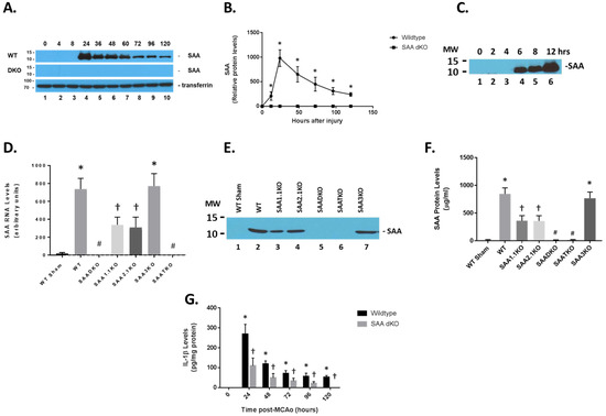
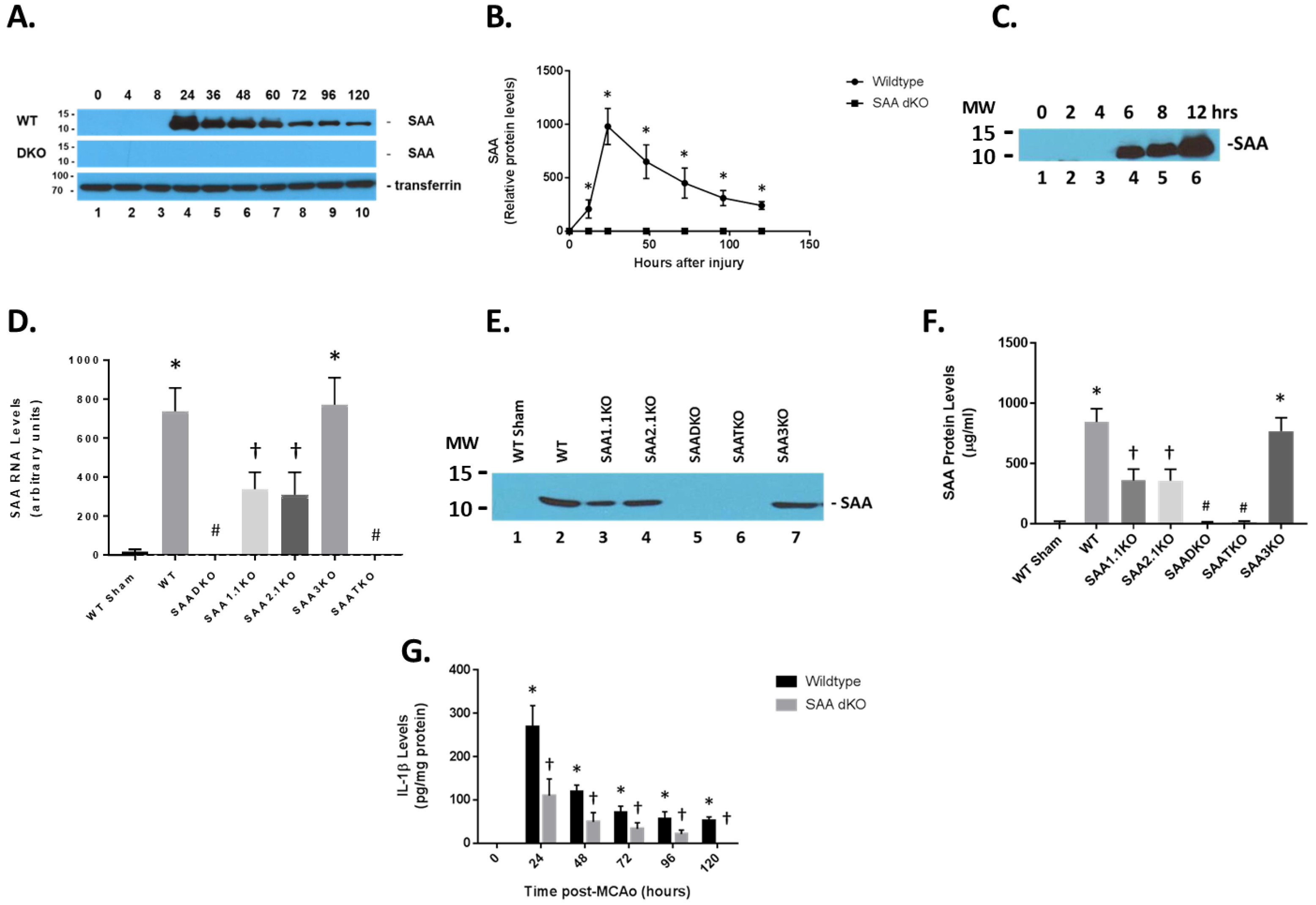
We and others have previously shown that plasma SAA levels are increased following cerebral ischemia and reperfusion injury in mice [41,42]. We assessed the expression of SAA in mice at different times of reperfusion following 1 h of ischemia (Figure 1). Remarkably, the SAA protein was expressed at high levels in the blood in ischemic/reperfused animals. As seen in the figure, SAA levels were low in control (0 h) mice but increased significantly at 24 h of reperfusion and were maintained for at least 120 h (Figure 1A). Transferrin was used as a control protein for the blood of the mice. Furthermore, mice deficient in both Saa1 and Saa2 genes showed no SAA protein at all when compared to circulating transferrin levels. The quantitation of SAA in the blood showed a 1000-fold increase in the protein at the 24 h time point (Figure 1B). Examination of the early time points indicated that SAA is present as early as 4–6 h following ischemia and reperfusion injury (Figure 1C, longer exposure to earlier time points). Additionally, we determined that the majority of SAA in the blood was generated by expression in the liver (Figure 1D). Different SAA isoforms are all expressed in the plasma after 1 h of ischemia and 24 h of reperfusion (Figure 1E,F). The marked difference in SAA expression in the liver suggests that the overall release of cytokines from the brain can trigger a systemic response in plasma inflammatory proteins that could have an impact on stroke outcomes (1G, IL-1β is shown here).
Figure 1.
Expression of SAA proteins following ischemia and reperfusion injury. (A) Mice were subjected to 1 h of ischemia and different times of reperfusion, and blood was collected and subjected to Western blot analysis (1/100 dilution of plasma in loading buffer). Wild-type (WT) and SAA double KO (DKO) mice were used and analyzed for SAA levels. Transferrin was used as a loading control for plasma proteins. (B) Graphical representation of data in (A). (C) Early time course of SAA protein expression in the blood of WT mice subjected to ischemia and reperfusion injury. (D) Liver SAA RNA levels (24 h) in the different SAA-deficient mice (as determined by RT-PCR for specific mRNAs). (E) Western blot for different SAA levels in ischemic mice (time in h). (F) Quantitation of (E). (G) Brain IL-1β levels in the different KOs were determined by ELISA. MW—molecular weight: upper band is 15 kDa; lower band is 10 kDa. Data are presented as mean ± S.E. The measurements are from 8–10 mice per group (time point). *, p < 0.001 compared to control (0 time point). #, p < 0.001 compared to the WT ischemic group. †, p < 0.001 compared to WT sham and ischemic groups. Data are considered significantly different at p < 0.05.
2.2. SAADKO Mice Have Reduced IL-1β Levels
Compared to wild-type (WT) mice, SAADKO showed complete obliteration of SAA expression in plasma (Figure 1A,E,F). However, individual KO mice (SAA1.1 and SAA2.1) had about 50% of the total SAA expression observed in WT mice. SAA3KO mice showed similar SAA plasma levels compared to WT animals. Finally, SAA triple KO (SAATKO) had no detectable plasma SAA quantities.
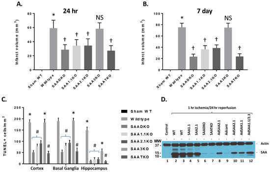
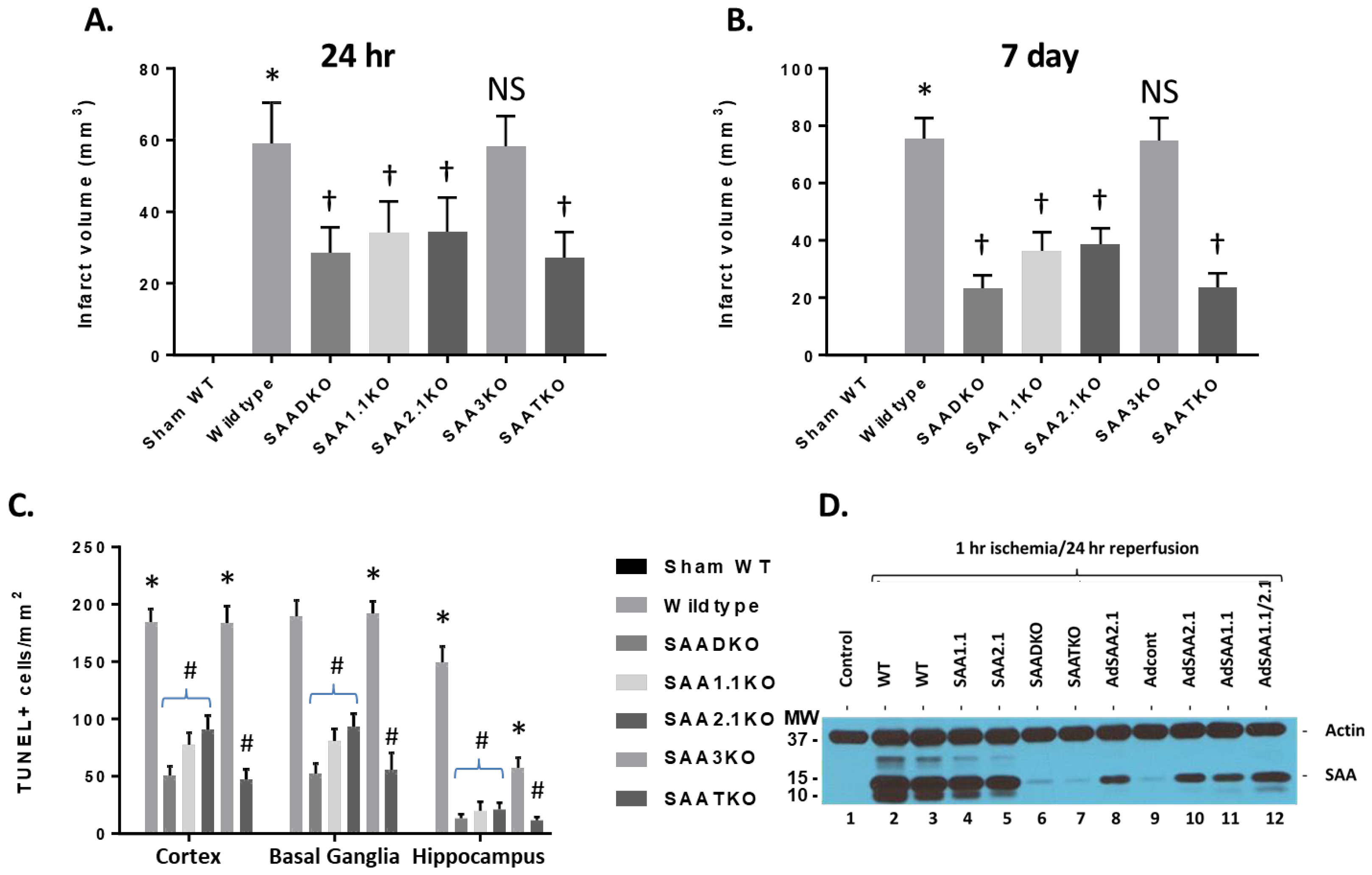
To determine the impact of SAA expression following ischemia and reperfusion injury, mice were subjected to 1 h of ischemia and various times of reperfusion and the brains were isolated and examined for IL-1β levels by ELISA (Figure 1G). IL-1β concentrations increased significantly in the brain at the 24-h time point and remained elevated up to 120 h after the start of reperfusion. IL-1β quantities in SAA DKO mice were significantly reduced and decreased to undetectable levels by 120 h. The increase in IL-1β coincides with the expression of SAA in the mouse model, and the data suggests that the augmentation of IL-1β is linked to SAA expression. Additionally, TUNEL analysis of the number of dead cells following ischemia and reperfusion injury in the different KO mice (and different brain regions) demonstrates the importance of SAA proteins in neuronal cell death (Figure 2C). Finally, to validate the presence of SAAs in the brain following cerebral ischemia and reperfusion injury, following 1 h of ischemia and 24 h of reperfusion, the mice were perfused to clear blood from the cerebrovasculature and the brain was isolated for Western blot analysis. Figure 2D shows that both wild-type and individual KO mice had SAA protein present in the brain. SAADKO and SAATKO mice had little if any SAA in the brain. When SAA was expressed in the liver using adenoviral vectors, SAA was again seen in the brain following ischemic injury. Thus, SAA expression following MCAo in wild-type mice allows for continued deterioration.
Figure 2.
Infarct volumes and TUNEL staining in wild-type and SAA KO mice. (A) Mice were subjected to 1 h of ischemia and 24 h of reperfusion, and brains were analyzed for infarct volume using TTC staining. DKO–double KO; TKO–triple KO. (B) Mice were subjected to 1 h of ischemia and 7 days of reperfusion, and brains were analyzed for infarct volume using TTC staining. (C) Cell death in different brain regions as analyzed by TUNEL immunostaining at 7 days after MCAo (TUNEL-positive cells per square millimeter). (D) Western blot analysis of SAA accumulation in the brain following IRI. AdSAAs are adenoviral vectors. Data are presented as mean ± S.E. The measurements are 10 mice per group. *, p < 0.001 compared to control. † or #, p < 0.01, compared to wild-type mice. NS = not significant.
2.3. Infarct Volume Is Reduced in SAA KO Mice
To further understand the role of SAA in cerebral ischemia/reperfusion injury or stroke, different mouse strains were subjected to 1 h of ischemia and 24 h of reperfusion and examined for infarct volumes (Figure 2A). Wild-type C57BL6 mice showed a typical infarct volume profile with 59.08 ± 3.27 mm3. While SAADKO mice showed a significant decrease in infarct size (28.67 ± 2.03 mm3), the individual KO mice (SAA1.1 and SAA2.1) had a more modest reduction (34.17 ± 2.50 mm3 vs. 34.5 ± 2.77 mm3, respectively). SAA3KO mice did not show a change in damage compared to wild-type mice (58.42 ± 2.40 mm3). Finally, SAATKO or triple knockout mice, which lack SAA1.1, SAA2.1, and SAA3, demonstrated an equivalent level of injury compared to SAADKO mice (27.25 ± 2.05 mm3). At 7 days after the start of reperfusion, wild-type and SAA3KO mice showed a continued increase in infarct volume (75.6 ± 2.22 mm3 and 74.9 ± 2.48 mm3, respectively), while SAAD-, SAA1.1-, and SAA2.1-deficient mice showed a further reduction in or equivalent infarct size (23.4 ± 1.39 mm3, 36.5 ± 2.02 mm3, and 38.9 ± 1.74 mm3, respectively) (Figure 2B).
The absence of SAAs had no significant effects on blood pH or heart rate (Table 1). Compared with pre-MCAo mice, no significant changes were noted in brain temperatures or MABP for any of the treatment groups (Table 1). Comparing the pre- and post-MCAo values of blood gases revealed that MCAo showed no reduction in PCO2 or increase in PO2 in KO mice.
Table 1.
Physiological measurements in the SAA mice. Physiological measurements were taken before, during and post ischemia and reperfusion injury in the mice. Mean arterial blood pressure (MABP), heart rate (HR), body temperature (Temp) and pH were determined in the study for the different mouse strains. Data are presented as mean ± SEM.
We also determined cell death in the ipsilateral cortex, basal ganglia, and hippocampus at 7 days following MCAo by TUNEL staining. SAADKO, SAATKO, and individual SAA-deficient mice (1.1 and 2.1) showed a significant reduction in cell death in the basal ganglia, cortex, and hippocampus compared to WT and SAA3KO mice. Further, compared to individual SAA-deficient mice, SAADKO and SAATKO mice had significantly less cell death in the cortex, basal ganglia, and hippocampus. There were no significant differences between SAADKO and SAATKO mice across the three brain regions (Figure 2C).
2.4. SAA Expression on Permanent Ischemia and MCAo in Rats
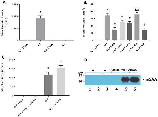
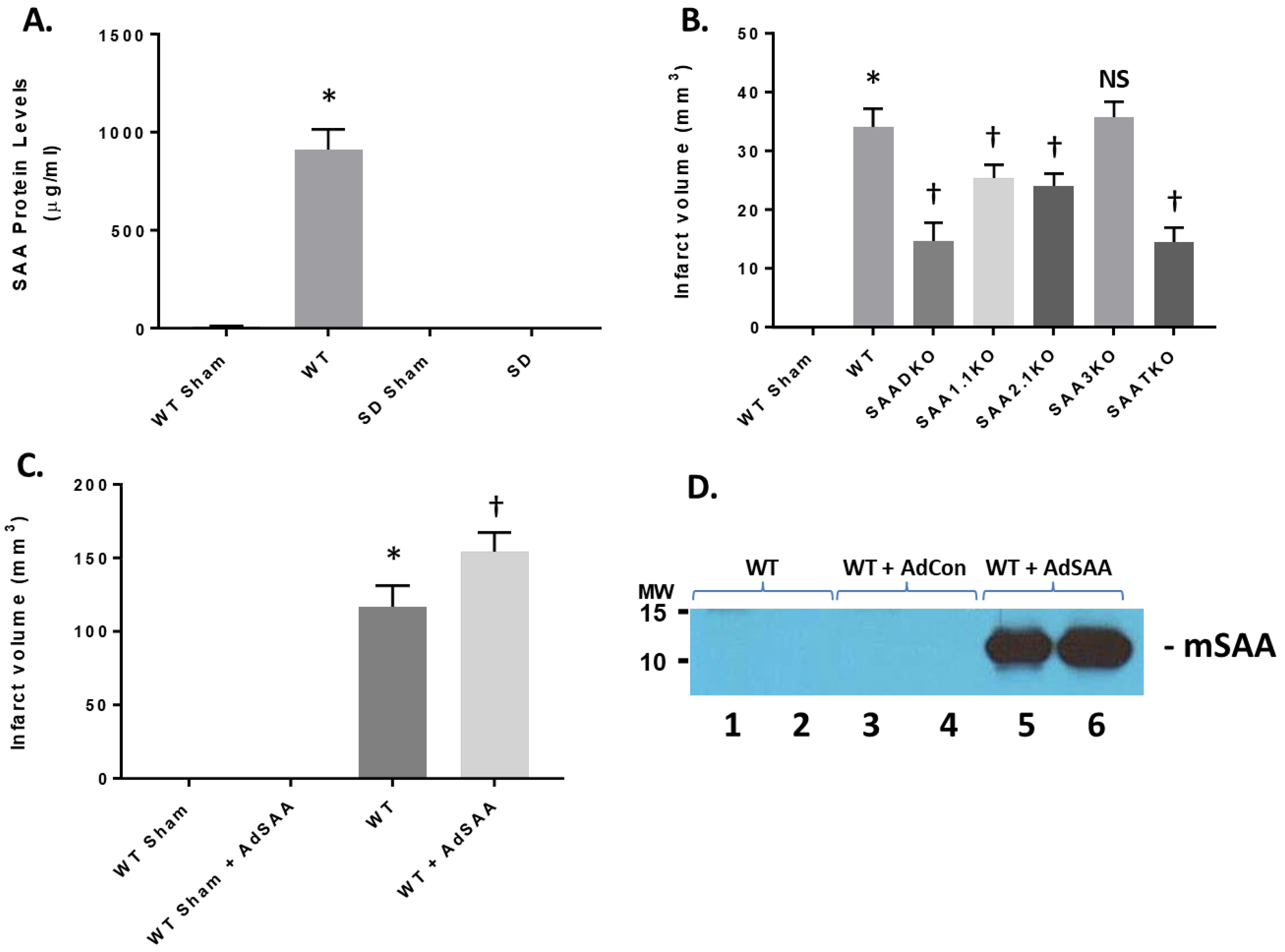
To determine the impact on SAA expression in permanent ischemia, mice were subjected to 24 h of ischemia with no reperfusion (Figure 3). When mice were treated with permanent ischemia, the plasma SAA levels increased in a similar fashion to ischemia and reperfusion injury (Figure 3A). WT animals presented with typical infarct volumes (33.4 ± 1.68 mm3) compared to sham mice (Figure 3B). However, SAA KO mice showed a reduction in infarct volume, commensurate with the level of SAA expression (SAADKO—15.3 ± 1.45 mm3; SAA1.1KO—24.5 ± 1.59 mm3; SAA2.1KO—22.8 ± 1.73 mm3; SAA3KO—34.3 ± 1.99 mm3; SAATKO—14.6 ± 1.19 mm3). As seen in Figure 3A,D, the rat does not express a viable SAA isoform; this is due to mutations or evolutionary changes. Therefore, when rats were subjected to 2 h of ischemia and 24 h of reperfusion, the infarct volume in the rat was in the absence of SAA proteins (Figure 3C, 111.4 ± 2.73 mm3). However, when murine SAA is delivered back using adenoviral vectors (Figure 3D), the infarct volume is significantly increased (154.8 ± 2.94 mm3), indicating that SAA contributes to the exacerbation of ischemic injury.
Figure 3.
Impact of SAA in permanent ischemia in mice and I/R in in rats. (A) Mice and rats were subjected to permanent ischemia or ischemia/reperfusion injury and examined for SAA expression. (B) Mice (WT and SAA-deficient mice) were subjected to 24 h of permanent ischemia and examined for infarct volumes at 24 h. (C) Rats (Sprague-Dawley) were either Sham or I/R (2 h ischemia and 24 h reperfusion) +/− adenoviral SAA and the infarct volume was determined at 24 h. (D) Representative plasma mSAA levels from rats injected with vehicle, AdCon, or AdSAA vectors, and Western blotted for mSAA expression. Data are presented as mean ± S.E. The measurements are 10 mice per group. *, p < 0.001 compared to WT Sham. †, p < 0.01, compared to wildtype I/R mice. NS = not significant.
2.5. Decreased Microglial and Astrocytic Markers in SAA KO Mice
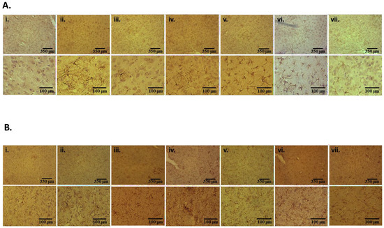
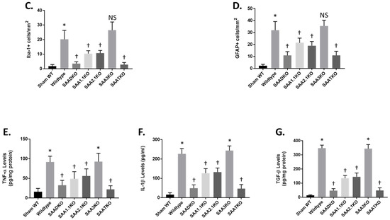
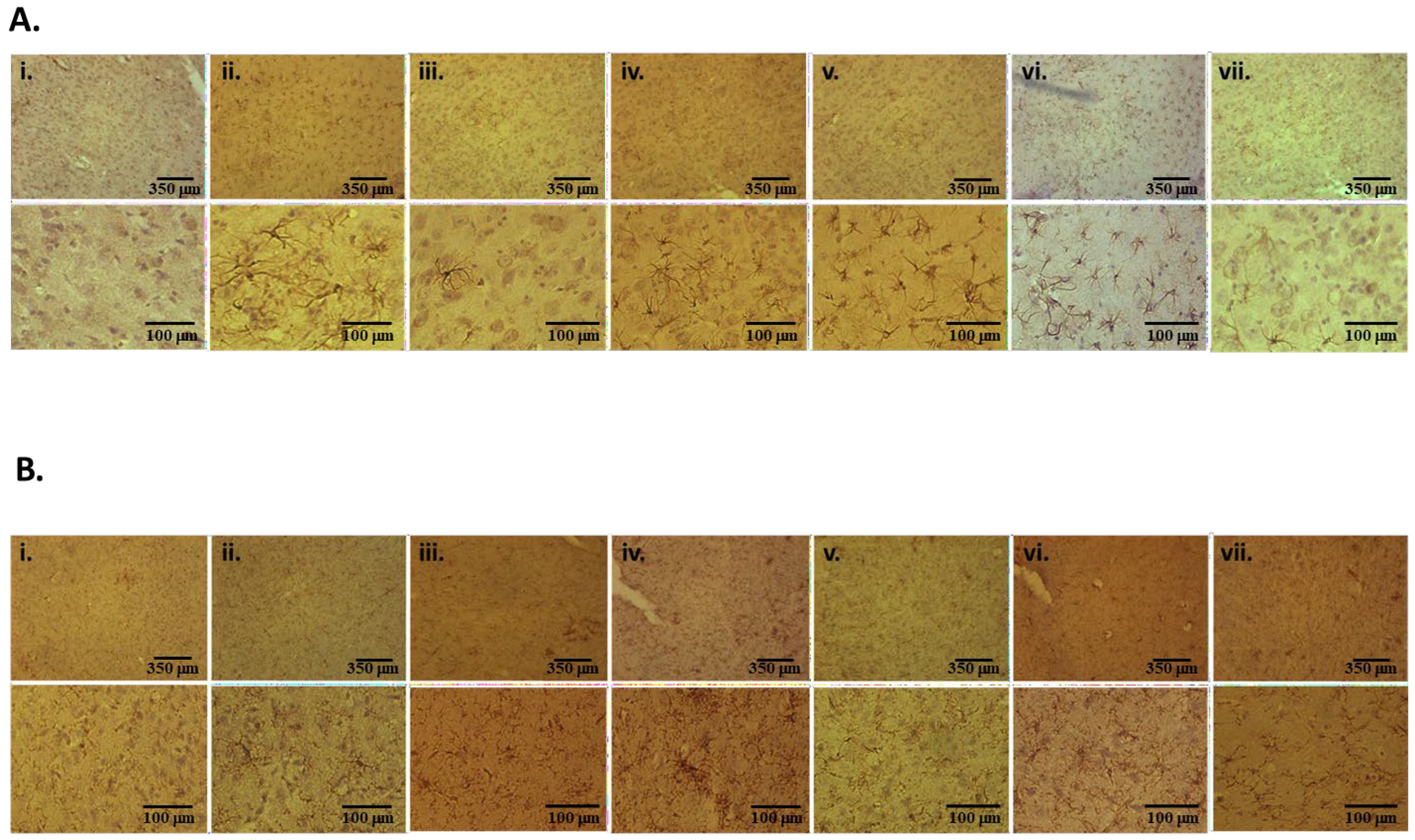
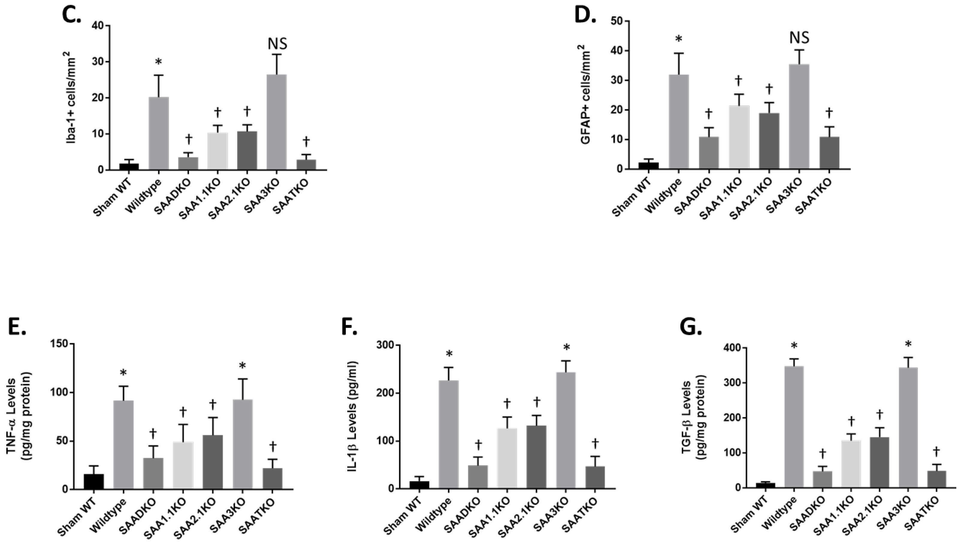
In response to MCAo, microglia and astrocytes are activated, and microglial stimulation and astrogliosis are general hallmarks of inflammation within the brain. This process can persist for many days following a stroke and may contribute to the disease process. Microglial activation and astrogliosis were assessed in the brain at 7 days post-MCAo by immunohistochemical detection of Iba-1 and GFAP markers in the cortex, respectively (Figure 4). Compared to WT sham animals, there was a significant reduction in both activated microglia (Figure 4A–C) and reactive astrocytes (Figure 4B–D) in the SAADKO, SAATKO, and, to a lesser extent, in the individual SAA-deficient mice. In contrast, there was no significant difference in the detection of either marker in SAA3KO mice compared with WT mice. No differences were observed in the contralateral hemispheres between the different groups. These data indicate that SAA deficiency reduced glial cell activation after MCAO.
Figure 4.
Microglial, astrocytic, and inflammatory activation following IRI in SAA-deficient mice. (A) Brains of mice from 1 h ischemia and 24 h reperfusion were sectioned and stained with iba-1 antibody. (i) Sham WT; (ii) WT; (iii) SAADKO; (iv) SAA1.1KO; (v) SAA2.1KO; (vi) SAA3KO; (vii) SAATKO (top panel 10×, bottom panel 40×). (B) Brains of mice from 1 h ischemia and 24 h reperfusion stained with gfap antibody. Order is the same as (A) (top panel 10×, bottom panel 40×). (C) Quantification of data in (A). (D) Quantification of data in (B). Mice were subjected to 1 h ischemia and 24 h of reperfusion, and the brain was isolated for cytokine analysis. (E) Brains were analyzed for TNF-α levels. (F) Brains were analyzed for IL-1β levels. (G) Brains were analyzed for TGF-β levels. Data are presented as mean ± S.E. The measurements are 10 mice per group. *, p < 0.001 compared to control. †, p < 0.001, compared to wild-type mice or other groups. NS = not significant.
To determine the impact of SAA deficiency on neuroinflammation in the MCAo mouse brain, we determined the expression of inflammatory markers. We measured the levels of specific cytokines, namely tumor necrosis factor-α (TNF-α), interleukin-1β (IL-1β), and transforming growth factor-β (TGF-β) 24 h after reperfusion (Figure 4E–G). The data showed that when SAAs were removed from the equation, there was a significant reduction in TNF-α (E), IL-1β (F), and TGF-β (G) levels.
2.6. Attenuated Behavioral Changes Are Associated with SAA Deficiency
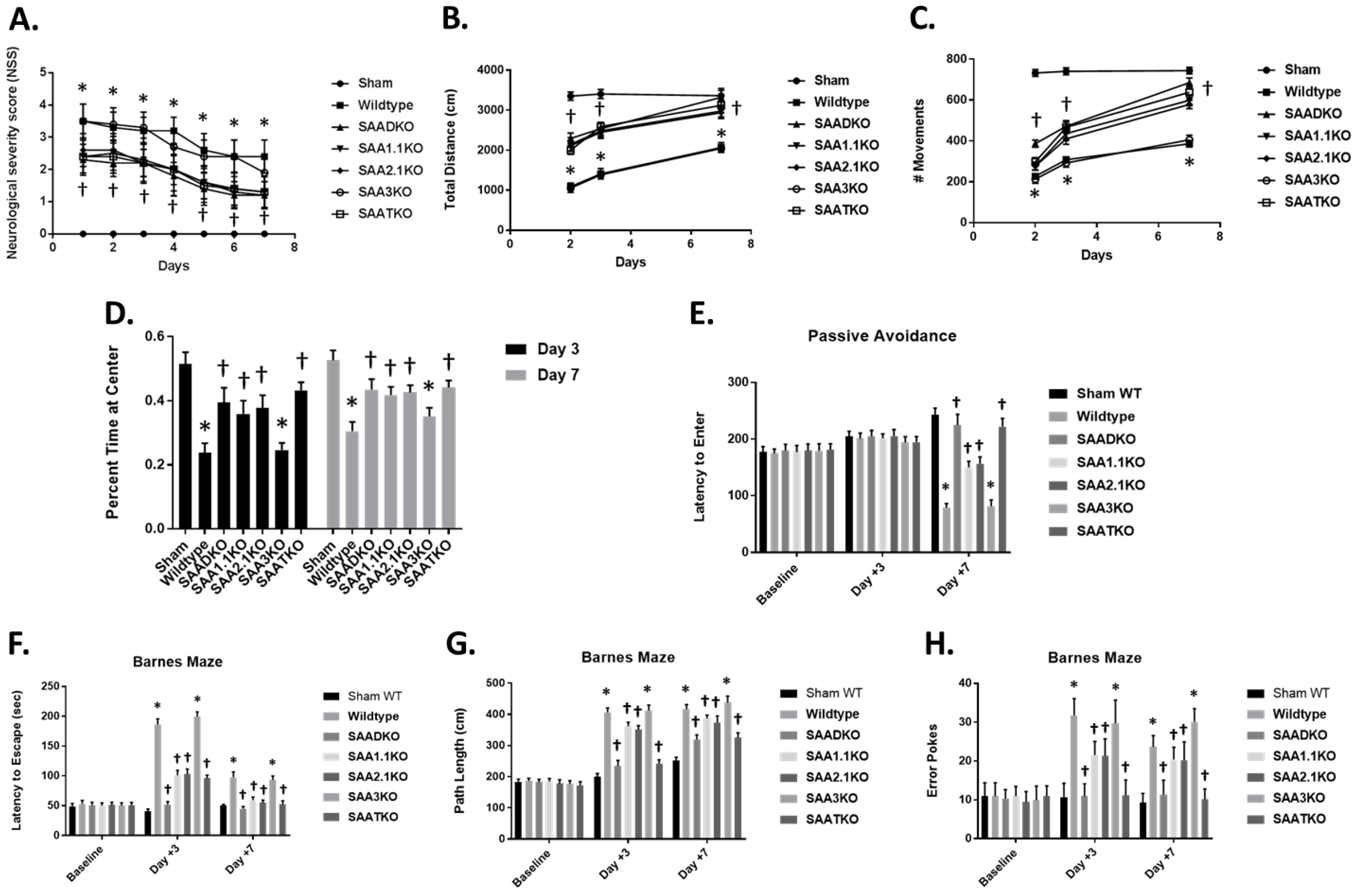
To better understand the impact of SAA deficiency on stroke outcomes, we measured neurological severity score via locomotor activity in an open-field activity monitor (Figure 5). There was a significant difference in neurological severity score (NSS) between the groups: WT and SAA3KO mice started out with higher scores that did improve slightly over time (Figure 5A). SAADKO, SAA1.1KO, SAA2KO, and SAATKO started off with a better (lower) score and improved to a greater degree than WT mice. In the locomotor activity chambers, SAA-deficient mice showed significantly increased locomotor activity (total distance and # of movements) compared to WT and SAA3KO mice (Figure 5B,C). Studies have suggested that anxiety may impact the outcomes of behavioral testing in mice; therefore, we evaluated the anxiety levels across the groups by measuring the percentage of time spent at the center of the open field. We found that MCAo did increase the anxiety in WT and SAA3KO mice significantly compared to other SAA-deficient mice, which reduced anxiety levels in different groups on days 3 and 7 post-MCAO (Figure 5D). The data implicates SAA as a factor that is involved in the evolution of injury to the brain following MCAo and suggests it may be a potential target for therapeutic intervention.
Figure 5.
Effect of SAA deficiency on behavioral deficit and locomotor function after MCAo. (A) Neurological deficit scores (0–4) within the first week after MCAo. (B,C) Locomotor activity measured as total distance moved (B) or number of movements (C), using an open field activity monitor. Determinations were made 2, 3, and 7 days after MCAO. (D) Anxiety assessment across the different groups measured as the percent time spent at the center of an open field at 3 and 7 days. (E) SAA deficiency improves performance on passive avoidance tasks 7 days after MCAo. Shown is the latency to enter a dark box associated with a shock. Mice were given a trial to associate a shock with the dark side of an apparatus, and latency to enter the dark side was evaluated on days 3 and 7 post-MCAo. (F–H) SAA deficiency reduces spatial memory deficits in the Barnes maze task in the subacute phase after MCAo. Mice were trained on the maze for 5 days before surgery and then tested on days 3 and 7 after MCAo for latency to escape (F), path length (G), and number of error pokes (H). Data are presented as mean ± S.E. The measurements are 10 mice per group. *, p < 0.001 compared to control. †, p < 0.01, compared to wild-type mice. At baseline, there were no differences in animals.
2.7. Lack of SAA Improves Behavior in the Subacute Phase After Stroke
Based on the significant sparing of tissue resulting from SAA deficiency, we performed a more comprehensive investigation of cognitive outcome by investigating spatial reference memory and avoidance learning using the Barnes maze and passive avoidance tasks. In the passive avoidance task, there were no significant differences between WT and SAA KO mice at either baseline or 3 days post-MCAo, with mice from all groups recording latency to enter times similar to the times that were recorded at baseline (Figure 5E). However, on day 7 post-MCAo, there was a significant reduction in performance of the WT group, whereas the performance of SAA-deficient mice was significantly different to the baseline and day 3.
In the Barnes maze task 3 days post-MCAo, the performance of WT mice was significantly worse, as demonstrated by an increase in latency to escape, path length, and number of error pokes compared to baseline. On day 7 post-MCAo, the performance in the WT group was improved but remained significantly poorer compared to the baseline (p < 0.001). In contrast, SAA-deficient mice displayed no or moderate impairment on the Barnes maze performance at either 3 or 7 days post-MCAo compared to baseline (Figure 5F–H). Consequently, compared to WT, SAA-deficient mice had significantly better spatial reference memory through the subacute phase after ischemic stroke.
2.8. SAA Inflammatory Processes Are Mediated by NLRP3
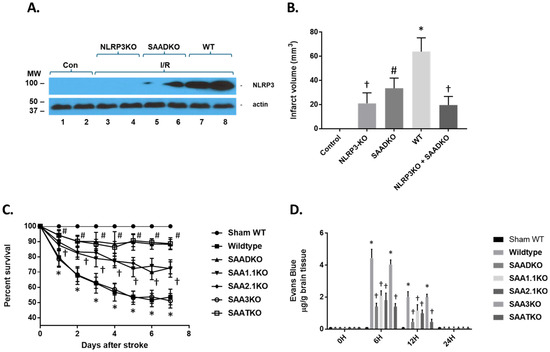
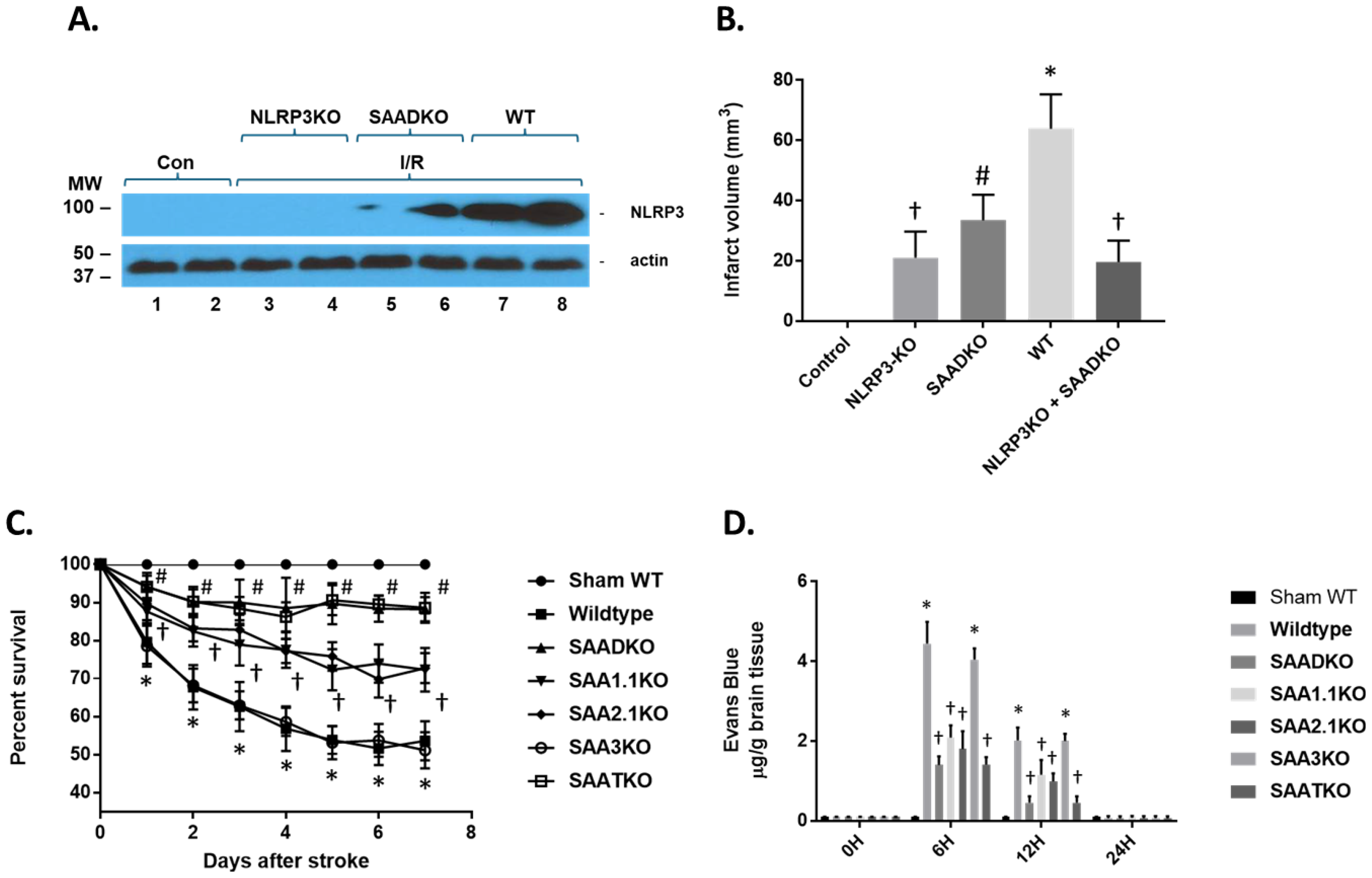
Previous studies have implicated SAA action via the node-like receptor protein 3 (NLRP3) and activation of the inflammasome [41]. To determine the impact of SAA in the MCAo model, WT, SAADKO, NLRP3KO, and NLRP3/SAADKO mice were examined for the expression of NLRP3 following 1 h of ischemia and 24 h of reperfusion (Figure 6A). As seen in the figure, in WT mice, NLRP3 significantly increased compared to the non-ischemic mice. However, in NLRP3-deficient mice, obviously, NLRP3 is non-existent, while in SAADKO, NLRP3 is significantly reduced. When assessing the infarct volume in the mice, NLRP3-deficient mice have reduced infarct volumes (as previously shown) and are significantly lower than SAADKO mice (Figure 6B). In addition, when NLRP3 and SAAD-deficient mice were crossed together, the infarct volume was comparable to NLRP3-deficient mice alone, suggesting that the SAA effect is partially mediated through the inflammasome.
Figure 6.
NLRP3 levels following cerebral ischemia and reperfusion injury. (A) Wild-type (WT), SAA double knockout (SAADKO), and NLRP3 KO mice were subjected to 1 h ischemia and 24 h of reperfusion and the brain was isolated for protein analysis. Protein was subjected to Western blot analysis for NLRP3 and actin. Con (control); I/R (ischemia and reperfusion). (B) Graphical representation of data in A and additional strains. NLRP3 + SAADKO are KO for NLRP3, SAA1.1, and SAA2.1. (C) Kaplan–Meier survival analysis of wild-type and different SAA-deficient mice over a 7-day period post-MCAO. (D) Mice were subjected to 1 h ischemia and different times of reperfusion, from 24 h and the brain was isolated for Evans blue measurement. Mice were injected via the tail 30 min prior to euthanasia. Data are presented as mean ± S.E. The measurements are 10 mice per group. †, p < 0.001 compared to control (non-ischemic). #, p < 0.001 compared to control and NLRP3KO. *, p < 0.001, compared to control and all other groups.
Complications from a stroke are a major issue in patients following strokes and can increase the risk for death and disability. Since SAA levels are increased not only in response to injury but also because of infection and suppression of the immune system, we examined the impact of SAA deficiencies on the survival rate in our model system (Figure 6C). MCAo in WT and SAA3KO mice showed a significant effect on survival in that by 7 days, almost 50% of the animals did not survive. However, mice deficient in SAA1.1, SAA2.1, or both showed a significant improvement in survival out to 7 days, with the SAADKO and SAATKO demonstrating a greater survival rate than the individual SAAs. These data indicate that treatment to reduce or impede the role of SAA in MCAo might reduce not only the infarct volume but also decrease susceptibility to secondary complications seen in experimental ischemic stroke.
To determine the impact of SAAs on BBB dysfunction post-stroke, C57/BL6 male mice underwent MCAo and were injected with 2% Evans blue as a marker for BBB penetration and permeability 30 min prior to euthanization (Figure 6D). Evans blue penetrated the BBB at 6 h and 12 h, then returned to normal levels. Quantification of Evans blue extravasation into the hemispheres revealed a four-fold increase at 6 h post-stroke in the right hemisphere of the brain where the occlusion occurred. There was no significant Evans blue extravasation observed at 24 h post-stroke. The SAA-deficient mice have a reduced BBB penetration of Evans blue at both the 6- and 12-h time points. SAA3KO mice, on the other hand, showed no difference in Evans blue extravasation compared with WT animals. These data suggest that BBB opening is partially dependent on the presence of SAA.
2.9. Restoration of SAA in SAA KO Mice Re-Establishes Activity
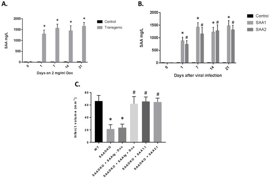
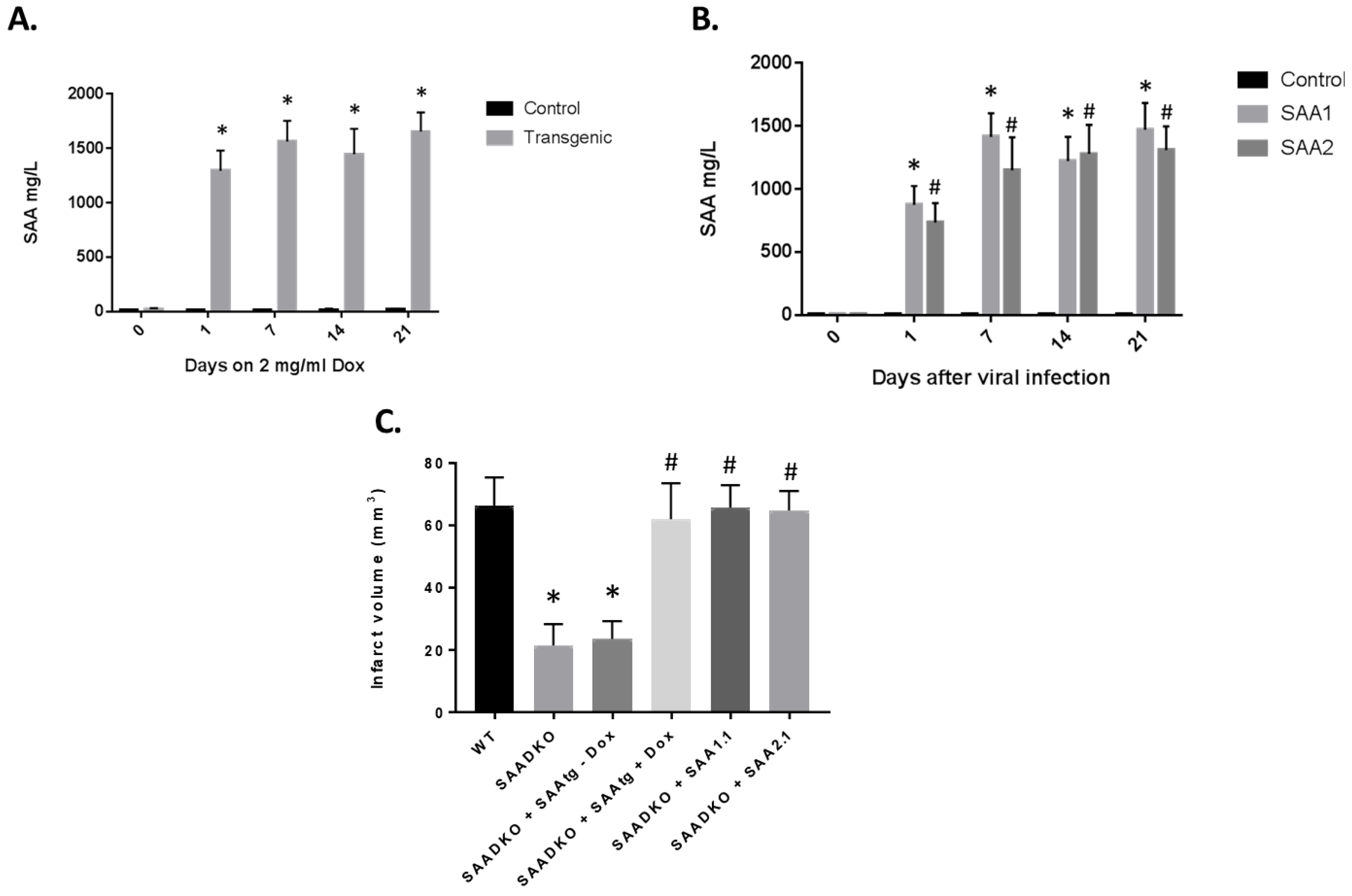
In order to determine the direct role of SAA in the pathogenesis of stroke, we used two different approaches. We obtained SAA transgenic (mSAAtg) mice with the transgene under an inducible promoter with doxycycline (dox) (Figure 7A). When the mice were given dox (2 mg/mL) in their drinking water, the amount of SAA in the blood increased significantly to well over 1000 mg/L. As long as the animals are on the dox, the SAA levels remain elevated. Previously, we developed adenoviral vectors that express the different mouse SAA (1.1 and 2.1) proteins. When the adenoviruses were injected into mice, the expression levels were similar to those induced by LPS and the transgenic mouse (Figure 7B). To validate the role of SAA in stroke, a mSAAtg mouse was crossed with a SAADKO mouse, and the SAADKO mice were injected with SAA adenoviruses. As seen previously, the SAADKO mice had reduced infarct volumes compared to WT animals (Figure 7C). However, when SAA was expressed via transgene or viral vector, the infarct volumes returned to WT levels. These studies demonstrate that SAA plays a critical role in the outcomes from stroke.
Figure 7.
Expression of SAA via transgene or adenoviral vectors restores the detrimental effects of the proteins. (A) SAA transgenic mice were crossed with the SAADKO background and subjected to dox (2 mg/mL) in drinking water, and plasma was examined for SAA protein levels by ELISA. (B) SAADKO mice were injected with adenoviruses containing SAA1.2 or SAA2.1 cDNAs, and plasma was examined for SAA protein levels by ELISA. (C) Mice were examined for infarct volumes in the presence of SAA mediated through the activated transgene or viral vectors. Data are presented as mean ± S.E. The measurements are 10 mice per group. *, p < 0.001 compared to the control group. #, p < 0.001 compared to the control group or to treated groups.
2.10. Protection from IRI with SAA Neutralizing Antibodies
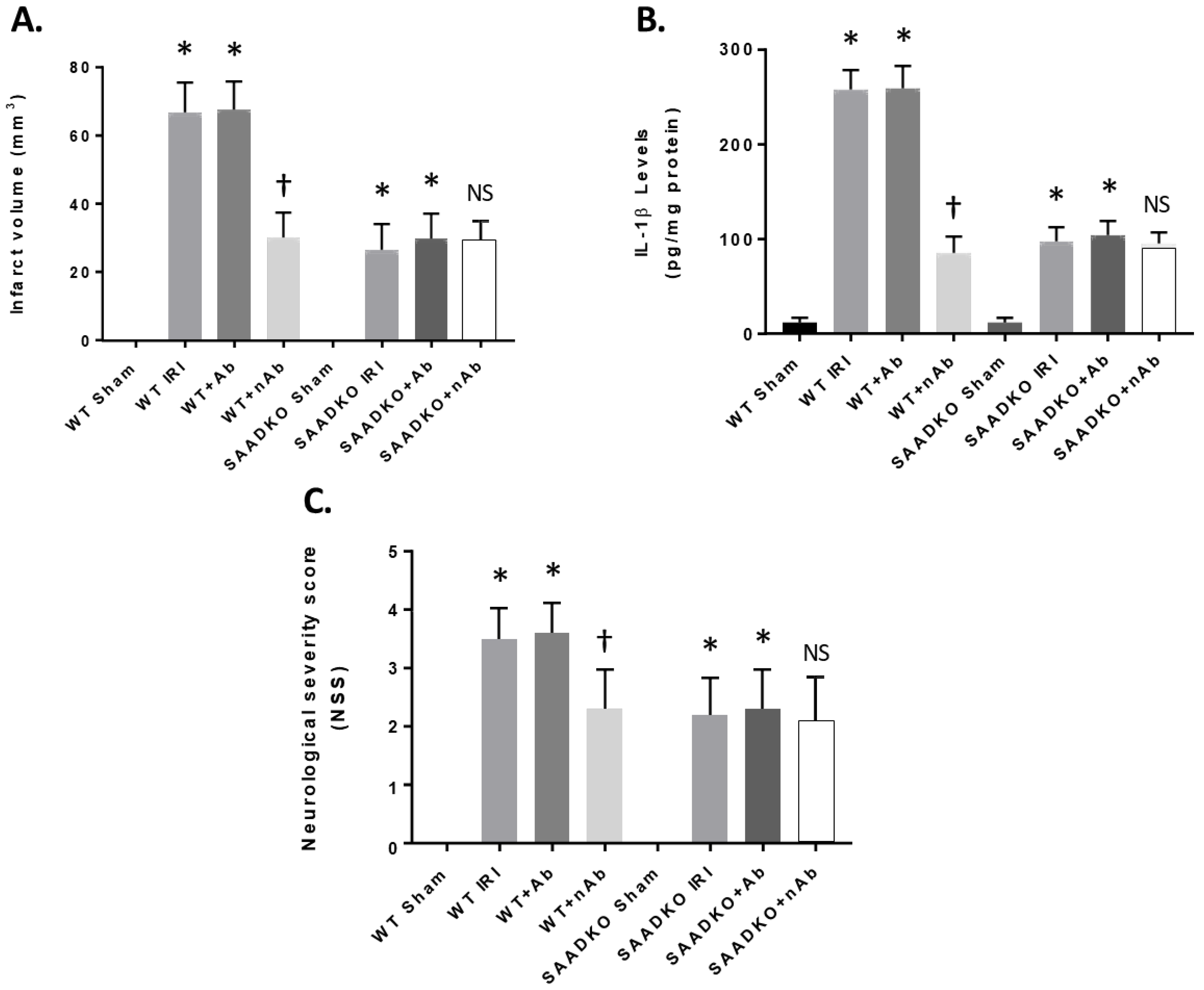
We tested the therapeutic potential of interfering with liver SAA expression in the MCAo mouse model, which develops increased SAA levels, infarct volume, and inflammation following cerebral ischemia and reperfusion injury. We administered the specific mouse anti-SAA blocking antibody (25 mg/kg body weight via intraperitoneal injection) beginning at the time of reperfusion. After 24 h of reperfusion, NSS and IL-1β and infarct volume were determined (Figure 8). As seen in the figure, the blocking or neutralizing antibodies (nAb) showed a reduction in infarct volume in the WT mice after MCAo, while the control antibody (Ab) had no effect (Figure 8A). In addition, in SAADKO mice, the nAb showed no improvement over the lack of SAA in the MCAo model.
Figure 8.
Reduced effects of SAA by neutralizing antibodies. Mice were subjected to 1 h ischemia and were injected with anti-mouse SAA antibodies (neutralizing) at 50 mg/kg (i.p.) and reperfusion for 24 h. Mice were examined for infarct volume (A), IL-1β expression (B), and neurological severity score (C). Data are presented as mean ± S.E. The measurements are 10 mice per group. *, p < 0.001 compared to the sham group. †, p < 0.001 compared to other groups. NS, not significant compared to other groups.
Figure 8B shows that nAb reduced IL-1β levels in the brain following MCAo when compared to the control and Ab treatment, having little effect in SAADKO mice. Finally, we showed that SAA blocking Abs reduced the NSS and behavioral detriment seen in the MCAo model (Figure 8C). These data indicate that blocking SAA inhibits inflammation in the brain following MCAo and attenuates the development of the infarct volume and neurological deficits seen in a well-established animal model of MCAo.
3. Discussion
Our data show that SAAs are a critical constituent of the inflammatory mechanism that contributes to the pathogenesis of stroke [41,42,43]. These data confirm the functional significance of SAA expression in the brain and identify SAAs as a decisive component that regulate the inflammasome pathway [44]. Thus, the SAA–inflammasome axis appears to be a major route mediating inflammation in the brain via the systemic circulation, which is in contrast to microglia-to-neuron interactions that dominate most neurological disorders [45,46]. It was of major interest that SAA proteins were significantly elevated in ischemia and reperfusion injury in the mouse model (as well as seen in stroke patients). It is likely that SAA proteins, which are for the most part elaborated from the liver, are increased in response to the release of cytokines (IL-1β, IL-6, TNF-α, etc.) from the brain following ischemic injury [47]. Release into the bloodstream and transport to the liver will trigger the increase in SAA expression, which in turn would be transported to the brain via the blood either on high-density lipoprotein (HDL) particles or other particles or even as non-lipidated entities [48,49]. Entrance into brain circulation, either by the breakdown of the BBB or transport across the BBB, would allow for access to the neurons and glial cells [41,45,49]. This, in turn, would elicit the responses seen in the present study. Therefore, inhibition of SAA expression might provide a mechanism to downregulate the SAA–inflammasome axis and afford protection following ischemia and reperfusion injury.
Compared to WT mice, SAA-deficient mice showed a significant decrease in neuronal cell death following acute ischemic injury, as well as in ischemia and reperfusion injury [41]. The studies presented here were carried out using constitutive knockout mice, and SAA expressions were deleted from all organs, so we cannot rule out the potential impact of SAA expression in glial cells or neurons on the outcomes [50,51]. However, several pieces of evidence strongly point to the role of liver-expressed SAAs in the pathogenesis of stroke. First, ischemia and reperfusion injury result in a significant increase in SAA expression from the liver, as determined by both mRNA and protein levels. Second, SAA KO mice show a significant reduction in neuronal injury in ischemia and reperfusion animals, and even though SAAs have been shown to be expressed in the brain, its expression levels are quite low. Third, the restoration of liver-specific expression of SAAs via transgenic mice and adenoviral vectors implicates the majority of neuronal injuries at this specific level of articulation. Fourth, inhibition of SAAs via specific immunotherapy can provide protection to the brain following ischemia and reperfusion injury, although it could be argued that this could interfere with brain expression as well. Finally, in the rat model of ischemia and reperfusion injury, longer ischemia times are required to generate similar levels of injury as in the mouse, and this may be attributed to the lack of SAA expression in the rat. We validated these results by expressing mSAAs in the rat and were able to restore the efficacy of SAA proteins in exacerbated neuronal injury, suggesting that SAA plays a critical role in brain injury. These studies indicate that the inhibition of SAA expression following a stroke, via immunotherapy, antisense oligonucleotides, or specific inhibitors, might afford protection to the brain [52,53].
Our data suggest that targeting SAAs might be a potential therapeutic approach to reduce the impact of stroke and to extend the window of opportunity for stroke recovery. Currently, there are no therapeutic treatments for ischemic stroke, although numerous neuroprotectants, anti-inflammatory agents, hypothermia, etc., have shown to be effective in various animal models and even in phase I and II studies [54,55]. Patients with occlusion of the major arteries of the brain are often treated with intravenous tissue-type plasminogen activator (IV tPA), thrombolysis, and/or mechanical thrombectomy (MTE) [56]. However, the time period for the delivery of these agents or mechanical disruption is limited, and time is of the essence [57]. The damage to the brain continues from the core into the penumbra region and beyond that results in poor clinical outcomes. By inhibiting or reducing SAA expression immediately following this stroke may attenuate the damage and allow for time for recanalization and recovery from the progression of neurological deficits.
Moreover, our data show that neuroprotection will be efficacious in the stage of secondary brain injury within the context of ischemia and reperfusion injury, and that blocking SAA entry into the brain might provide a powerful approach as an adjuvant therapy in acute stroke to reduce the damage and improve outcomes [41]. Complications that occur following ischemia and reperfusion injury are hemorrhagic transformation, bleeding, and infections that worsen outcomes and lead to disability and death [58]. A number of therapeutic approaches have met their demise due to increased complications associated with bleeding. This includes tissue plasminogen activator (tPA) even when delivered under the prescribed conditions.
In contrast, SAA-deficient mice experienced reduced bleeding in the brain following ischemia and reperfusion injury, and bleeding times were slightly reduced in the mice. Finally, SAA-deficient mice showed improved long-term survival compared to WT mice, suggesting a reduction in infection-related outcomes. Therefore, the inhibition of SAA might provide an opportunistic therapeutic approach for acute stroke patients by reducing inflammation, lesion volumes, bleeding, and response to infection.
In addition to stroke, the inhibition of SAAs might be a novel treatment for other neurological and neurodegenerative disorders in which disturbances in SAA homeostasis comprise a major pathogenic feature [59,60]. The recognition of SAAs as a significant constituent of stroke and their response to cytokine triggers released by the brain provides a more viable target for pharmacological intervention. Given that the SAA-deficient mice are healthy in their appearance and are unaffected in their general cognitive behavior or other physiological aspects, the inhibition of SAAs might be a rather safe approach to treat these disorders [61]. In addition, the lack of side effects might prove beneficial in the treatment paradigm.
4. Materials and Methods
4.1. Study Design
All experiments were approved by the Institutional Animal Care and Use Committee of the University of South Florida (4613 and 8883) and conducted in accordance with the University of South Florida Guidelines, which are based on the National Institutes of Health’s Guide for the Care and Use of Laboratory Animals and Animal Research: Reporting of In Vivo Experiments guidelines. All procedures and histological and gene expression analyses were performed by examiners who were blinded to the experimental conditions. The number of animals was 80% powered to detect 25% changes with a 2-sided α value of 0.05. Mice that showed the following were excluded: spasm after craniotomy (n = 3), spontaneous complete recovery of the middle cerebral artery during surgery (n = 2), failure of the training test in the hanging wire test (n = 3), and no abnormality in the hanging wire test on day 1 (n = 5). None of the mice died after surgery. For all of the experiments (western blots and immunohistochemical analyses), all samples were subject to the analysis described.
4.2. Animals
Adult C57BL/6 male and female (50:50) mice (30–35 gms, 5–6 months of age), SAA1.1KO, SAA2.1KO, SAA3KO, SAA-double KO (SAADKO), and SAA-triple KO (SAATKO) (all on a C57BL/6 background) were bred as needed at the University of South Florida [62,63,64]. Five to six-month-old animals were used to better represent individuals who have strokes. Normally, 10–12-week-old animals were used, which is too young. Animals were randomly assigned to different groups, and the individuals who performed the animal studies were blinded to different strains. The behavioral testing experimenters were also blinded to different groups. All the studies followed the STAIR and ARRIVE guidelines for preclinical studies [65,66]. The experimental procedures were approved by the Institutional Animal Care and Use Committee of the University of South Florida.
4.3. Ischemic Injury and Infarct Volumes
Transient (60 min) focal ischemia was induced by suture occlusion of the middle cerebral artery (MCAo), as described previously [67,68]. Mice were anesthetized using a mixture of 1.5% isoflurane, 70% N2O, and 28.5% O2. After ischemia (1 h), reperfusion started and continued until the end of the study. Body temperatures were maintained at 37 °C by a water-jacketed heating pad and an anal thermometer. Transcranial laser Doppler was used to monitor any changes in cerebral blood flow. Mice were included in the study when blood flow dropped below 20% during ischemia and returned to 90% during reperfusion, relative to the normal value for data analysis. After the indicated times of ischemia and reperfusion injury (IRI), mice were euthanized, and the brains were removed for analysis. Coronal sections at 1–2 mm intervals were prepared and stained with 2% 2,3,5-triphenyltetrazolium chloride (vital dye). Infarct volumes were calculated by summing the infarcted areas (pale) of all sections and multiplying by the thickness of the sections.
Male and female Sprague-Dawley rats (250 to 275 g, Harlan, Indianapolis, IN, USA) were given free access to food and water before the experiment [68]. All procedures were within institutional guidelines for animal use. Rats were anesthetized with halothane (1% in 70%/30% NO2/O2), and their bilateral femoral arteries were cannulated with PE-50 tubing for the monitoring of mean arterial blood pressure (MABP) and for blood collection. The right femoral veins were cannulated for infusion of test articles. Body and brain temperatures were monitored with a rectal thermometer and a thermistor probe inserted into the temporalis muscle, respectively, and maintained at 37 °C using a water-jacketed heating pad. Temperatures and MABP were monitored continuously from 1 h before to 6 h after ischemia. The left common carotid artery of each rat was exposed through a midline incision in the neck. The superior thyroid and occipital arteries were electrocoagulated and divided. A microsurgical clip was placed around the origin of the external carotid artery (ECA). The distal end of the ECA was ligated with 6-0 silk and transected, and 6-0 silk was tied loosely around the ECA stump. The clip was removed, and the blunted tip of a 4-0 nylon suture was inserted into the ECA stump. The loop of the 6-0 silk was tightened around the stump, and the nylon suture was advanced into and through the internal carotid artery until it rested in the anterior cerebral artery. After the nylon suture had been in place for 2 h, it was pulled back into the ECA, and the incision was closed.
4.4. Measurement of Cerebral Blood Flow
Regional cerebral blood flow (rCBF) was analyzed by laser Doppler flowmetry every 30 min over a period of 1 h before to 6 h after MCAo [69]. Mice were anesthetized with isoflurane (1.5% in 70%/28.5% NO2/O2), and a 2-mm hole was drilled into the skull, with the probe positioned at 0.1 mm above the dura over the cortical surface. In the hemisphere ipsilateral to the occlusion, coordinates were as follows: point A, 1 mm posterior to the bregma and 5.4 mm lateral to the midline; point B, 1 mm posterior to the bregma and 2.1 mm lateral to the midline; point C, 1 mm anterior to the bregma and 3.4 mm lateral to the midline. The mean values of rCBF were measured before MCAO as the baseline, and the data thereafter were expressed as percentages of the baseline value. The rCBF data in the present report was taken from reference point A.
4.5. TUNEL Staining
Following ischemia and reperfusion injury (7 days), the terminal deoxynucleotidyl transferase (TdT)-mediated dUTP-digoxygenin nick-end labeling (TUNEL) technique was used to determine the number of apoptotic cells (ApopTag® Peroxidase In Situ Apoptosis Detection Kit, Chemicon International, Inc., Temecula, CA, USA) [70]. The preparation of the cryosectioned tissue was the same as described above. TUNEL assays were executed according to the manufacturer’s instructions. In brief, the sections were fixed in 1% paraformaldehyde for 10 min at room temperature, then post-fixed in pre-cooled ethanol: acetic acid (2:1) for 5 min at −20 °C. The endogenous peroxidase enzymes were quenched in 3.0% H2O2 for 5 min at room temperature (RT). Subsequently, the sections were incubated with buffer for at least 30 s at RT, which was followed by incubation with TdT in a humidified chamber at 37 °C for at least 1 h. The reaction was terminated by adding the stop/wash buffer for 10 min at RT. Sections were then incubated with anti-digoxigenin peroxidase conjugate in a humidified chamber for 30 min at RT and developed with 3,3′-diaminobenzidine (DAB) for 5 min at RT. Sections were then counterstained with 0.5% methyl green and cover-slipped. Sections from 8 to 10 separate animals were used to assess the number of TUNEL-positive cells.
4.6. Neurological Outcomes
Neurological deficits were determined following MCAo by blinded observers using a five-tiered scoring system, as previously described [71,72]. Locomotor activity was automatically quantified using the open field activity monitor (Any Maze, San Diego Instruments, San Diego, CA, USA). Mice were placed in a random corner and allowed to acclimate for 10 min prior to a 60-min testing period. External noise, lights, and other stimuli were minimized to reduce bias. A number of measures were automatically obtained during the task, including total distance, number of movements, time spent at the periphery, and time spent in the center of the enclosure. Activity readings acquired prior to the sham procedure were used to establish baseline activities. The duration that the animal spent at the periphery vs. the center was used to assess anxiety level during the task.
4.7. Behavioral Testing
Animals were tested for their performance on both the Barnes maze and passive avoidance tasks [72]. To evaluate spatial reference memory, mice were educated on the Barnes maze for 5 days before surgery, as previously described, then tested again on days 3 and 7 after the start of reperfusion for the time needed to escape into the hole, the number of error pokes, and the length of the animal’s path prior to escape. An automated passive avoidance apparatus (San Diego Instruments) was used to assess avoidance learning with automated sensing and shock systems (Gemini, San Diego Instruments). The instrument included a double-compartment chamber with one light and one dark compartment. Mice were allowed to explore the chamber for 5 min for orientation. Following habituation, the mice were given one trial where a shock was associated with the dark side, allowed 48 h of rest, and then tested for retention, measured as latency to enter the dark side. Testing was repeated on days 3 and 7 post-reperfusion with no shock delivered during the test phase.
4.8. Immunohistochemical Analysis
Immunohistochemical staining was conducted on 8-μm paraffin sections and assessed by a blinded observer by light microscopy (Olympus BX61, Center Valley, PA, USA) [72]. Following antigen retrieval (IHC World, Ellicott City, MD, USA), the following primary antibodies were used: anti-ionized calcium-binding adaptor molecule 1 (Iba-1, 1:250; Abcam, Cambridge, UK) and anti-mouse glial fibrillary acidic protein (GFAP, 1:1000; Dako, Agilent, Santa Clara, CA, USA). Primary antibodies were detected with ImmPress-HRP kit and NovaRed peroxidase chromagen (Vector Laboratories, Newark, CA, USA), and primary antibodies were omitted for negative controls. Terminal deoxynucleotidyl transferase dUTP nick end labeling (TUNEL) staining was performed using ApopTag Peroxidase Staining Kit (Millipore, Lakeland, FL, USA) per manufacturer’s instructions. The investigators stained multiple sections in and around the infarct region and used an unbiased and randomized imaging approach for the quantification of the data.
4.9. RNA Isolation and Analysis
Total RNA was isolated from mouse livers using TRIzol Reagent (Invitrogen/ThermoFisher, Waltham, MA, USA) according to the manufacturer’s instructions. RNA samples (10 μg) were treated with DNase 1 (TURBO DNA-free TM kit (Invitrogen/ThermoFisher AM 1907)) for 30 min at 37 °C. RNA from liver (0.5 μg) was reverse transcribed into cDNA using the Reverse Transcription System (Promega 3500, Madison, WI, USA). After a 5-fold dilution, 5 μL were used as a template for real-time RT-PCR. Amplification was carried out for 40 cycles using the SYBRTM Green PCR Master Mix (ThermoFisher, 4309155). Quantification of mRNA was performed using the ΔΔCT method and normalized to GAPDH for the liver. Primer sequences are as follows: GAPDH (Accession number NM_008084) 5′-CTC ATG ACC ACA GTC CAT GCC A-3′; 5′-GGA TGA CCT TGC CCA CAG CCT T-3′; SAA1.1/2/1 (Accession number NM_009117.3) 5′-CTC CTA TTA GCT CAG TAG GTT GTG-3′; 5′-CAC TTC CAA GTT TCT GTT TAT TAC CC-3; SAA3 (Accession number NM_011315.3) 5′-TTT CTC TTC CTG TTG TTC CCA GTC-3′; 5′-TCA CAA GTA TTT ATT CAG CAC ATT GGG A-3′. For specific mRNAs, the primer sequences used in this study were as follows: SAA1.1 (accession no., NM009117), sense 5′-ATG AAG GAA GCT AAC TGG AAA AAC TC-3′, antisense 5′-TCC TCC TCA AGC AGT TAC TAC TGC AA-3′; SAA2.1 (accession no., NM011314), sense 5′-ATG AAG GAA GCT GGC TGG AAA GAT GG-3′, antisense 5′-TCC TCC TCA AGC AGT TAC TAC TGC TC-3′; SAA3 (accession no., NM 011315), sense 5′-GCC ACC ATG AAG CCT TCC ATT GCC ATC ATT-3′.
4.10. Cytokine Analysis
For quantitative analysis of cytokines, ELISA was used to measure the levels of tumor necrosis factor-α (TNF-α), interleukin-1β (IL-1β), or transforming growth factor-β (TGF-β) in brain tissue [67]. Cytokines were extracted from mouse brains as follows: frozen hemibrains were placed in tissue homogenization buffer containing protease inhibitor cocktail (Sigma, St. Louis, MO, USA) at a 1:1000 dilution immediately before use and homogenized using polytron. Tissue sample suspensions were distributed in aliquots and snap-frozen in liquid nitrogen for later measurements. Invitrogen/ThermoFisher ELISA kits were then used according to the manufacturer’s directions (Carlsbad, CA, USA).
4.11. Western Blot Assay
Coronal slices were dissected after the indicated reperfusion time, and the right hemisphere (the infarct/ischemic side) was selected. For western blotting, proteins were extracted from mouse brains using lysis buffer (50 mM Tris–HCl, pH 7.4, 0.5% Triton X-100, 4 mM EGTA, 10 mM EDTA, 1 mM Na3VO4, 40 mM Na2P2O7·10H2O, 50 mM NaF, 100 nM calyculin A, 50 μg/mL leupeptin, 25 μg/mL pepstatin A, 50 μg/mL trypsin inhibitor, and 1 mM dithiothreitol), as previously described [72]. For the plasma samples, diluted plasma was added to 5× samples buffer and run on gels. The protein concentrations were quantified using Bradford’s assay and normalized. Equal quantities of protein were loaded and separated by 4–12% SDS–polyacrylamide gels (SDS/PAGE) and transferred to polyvinylidene difluoride (PVDF) or nitrocellulose membranes. Membranes were probed with primary antibodies overnight at 4 °C and then with appropriate secondary antibodies. Thereafter, membranes were detected with an enhanced chemiluminescence (ECL) immunoblotting detection system (Amersham Biosciences, Piscataway, NJ, USA) using a Luminescent Image Analyzer (LAS-4000 mini, Fuji Film, Tokyo, Japan). The densities of the bands were analyzed with ImageJ software (version 1.54, NIH, Bethesda, MA, USA). Primary antibodies were SAA (R&D systems, AF2948), transferrin (Abcam, ab84036), NLRP3 (Abcam, ab214185), and actin (Abcam, ab179467). Secondary antibodies were Goat anti-rabbit (Sigma, A6154).
4.12. BBB Leakage Analysis
Evans blue (Sigma, CA, 2% in saline; 4 mL/kg) was used to determine the BBB permeability [36,73]. For Evans blue detection, the dye was intravenously administered through the tail vein 30 min prior to euthanization. Animals were transcardially perfused with saline, brains were sectioned with a 2 mm brain matrix, and images were taken by a photo scanner. Hemisphere samples were weighed, homogenized with 400 uL PBS, and precipitated with 50% trichloroacetic acid (Sigma, CA) overnight. All samples were centrifuged at 1000× g rpm for 30 min to separate out the brain tissue in the pellet prior to measuring. Absorption was quantified at 610 nm with a plate reader (BMG Labtech, Cary, NC, USA). To quantify Evans Blue penetration through the brain barrier, an Evans blue standardized curve was used. Results were quantified as microgram/gram brain tissue.
4.13. Adenoviral Preparation
The production of replication-defective adenoviral vectors expressing mouse SAA proteins has been described using second-generation adenoviral vectors [74,75]. Briefly, recombinant adenoviruses were expanded in 293 human embryonic kidney (HEK) cells. The virus was used to infect 293 cells, and viral particles were isolated from the cells after 72 h of infection and purified by density gradient cesium chloride centrifugation. Virus particles were centrifuged through a two-step gradient of cesium chloride (1.45 g/mL and 1.20 g/mL) for 2 h and 14 h, respectively. The virus particles were collected and chromatographed on a 10-mL Econo-Pac DG column, as described by the manufacturer (Bio-Rad, Hercules, CA, USA). The number of particles was determined by optical density.
4.14. Statistics and Reproducibility
Statistical differences between parametric data (infarct volumes, activity values, ELISA values, cell counts, densitometry, and RNA data) were assessed using a one-way analysis of variance (ANOVA) test with Bonferroni’s multi-group comparison, and non-parametric data (neurological deficits) were compared with the Kruskal–Wallis test with Dunn’s comparison (Prism 7.0, GraphPad). Sample sizes and number of replicates are included in the figure legends or text. Survival was compared in all mice subjected to MCAo using the Kaplan–Meier test. Differences between data were considered statistically significant when p < 0.05.
5. Conclusions
Our findings establish SAAs as a major mediator of inflammation in the brain that plays an important role in ischemia/reperfusion injury. Increased SAA following a stroke can exacerbate the damage via further activation of the inflammasome pathway. These findings may provide evidence for the development of therapeutic approaches using anti-SAA treatments in stroke [76,77].
6. Limitations
There are some limitations to the experimental protocol. First, for the antibody treatment experiments, the antibody was administered only at the start of reperfusion, and the effect of delayed administration was not explored. In the current study, even though we used both male and female mice, we did not dissect out the differences in gender. These can have an impact on clinical trials and potential outcomes. Those experiments will be followed up on in subsequent studies.
Author Contributions
Designed the experiments: J.Y. and M.S.K. Conducted the experiments: J.Y., H.Z., S.T. and M.S.K. Analyzed and interpreted the data: J.Y., S.T., D.M.D., C.K., J.-Y.L. and M.S.K. Wrote the manuscript: M.S.K. All authors have read and agreed to the published version of the manuscript.
Funding
This work was partially supported by grants from the National Institutes of Health (R01 ES016774-01, R21AG043718), VA Merit Award (RR&D, I01RX001450 & BLR&D, 1I01BX006259-01A1), an AHA SFRN grant (15SFDRN25710468), and AHA Transformation Award (19TPA34910015) to MSK. Dr. Kindy is a Senior Research Career Scientist (BLR&D, IK6 BX005239-01) in the VA. The views expressed in this article are those of the authors and do not necessarily reflect the position or policy of the Department of Veterans Affairs or the United States government.
Institutional Review Board Statement
All experiments were approved by the Institutional Animal Care and Use Committee of the University of South Florida (protocol code 12241, approval date 12 May 2023 and protocol code 8883, approval date 4 May 2021) and conducted in accordance with the University of South Florida Guidelines, which are based on the National Institutes of Health’s Guide for the Care and Use of Laboratory Animals and Animal Research: Reporting of In Vivo Experiments guidelines.
Informed Consent Statement
Not applicable.
Data Availability Statement
Data, datasets used, and/or analyzed during the current study will be provided by the corresponding author on reasonable request.
Acknowledgments
We thank Frederick DeBeer and Nancy Webb for the SAA1.1, 2.1 and DKO mice, Alan Chait for the SAA3 KO mice, Dan Littman for the SAATKO mice, Doug Golenbock for the NLRP3 KO mice, and Paul Simons for the SAA tg mice. This work is dedicated to the memory of C. Kirstein, who passed away during the completion of this study.
Conflicts of Interest
Dr. Kindy receives funding from the American Heart Association (AHA); he also serves on several review committees for the AHA, NIH, and the VA, and is a member of the Professional Education Committee for the AHA. The other authors declare no competing interests.
Correction Statement
This article has been republished with a minor correction to the Informed Consent Statement. This change does not affect the scientific content of the article.
Abbreviations
The following abbreviations are used in this manuscript:
| IL-1β | Interleukin-1β |
| MAPK | Mitogen-Activated Protein Kinase |
| MCAo | Middle Cerebral Artery Occlusion |
| NLRP3 | Nod-Like Receptor Protein 3 |
| rCBF | Regional Cerebral Blood Flow |
| SAA | Serum Amyloid A |
| TNF-α | Tumor Necrosis Factor-α |
References
- Lim, H.; Choue, R. Impact of nutritional status and dietary quality on stroke: Do we need specific recommendations? Eur. J. Clin. Nutr. 2013, 67, 548–554. [Google Scholar] [CrossRef] [Scilit]
- Mukherjee, D.; Patil, C.G. Epidemiology and the global burden of stroke. World Neurosurg. 2011, 76, S85–S90. [Google Scholar] [CrossRef] [Scilit]
- Rivers-Auty, J.; Ashton, J.C. Neuroinflammation in ischemic brain injury as an adaptive process. Med. Hypotheses 2014, 82, 151–158. [Google Scholar] [CrossRef] [Scilit]
- Radak, D.; Resanovic, I.; Isenovic, E.R. Link between oxidative stress and acute brain ischemia. Angiology 2014, 65, 667–676. [Google Scholar] [CrossRef] [Scilit] [PubMed]
- Chan, P.H. Reactive oxygen radicals in signaling and damage in the ischemic brain. J. Cereb. Blood Flow Metab. 2001, 21, 2–14. [Google Scholar] [CrossRef] [Scilit] [PubMed]
- Jung, J.E.; Kim, G.S.; Chen, H.; Maier, C.M.; Narasimhan, P.; Song, Y.S.; Niizuma, K.; Katsu, M.; Okami, N.; Yoshioka, H.; et al. Reperfusion and neurovascular dysfunction in stroke: From basic mechanisms to potential strategies for neuroprotection. Mol. Neurobiol. 2010, 41, 172–179. [Google Scholar] [CrossRef] [Scilit]
- Yasuda, S.; Sugiura, H.; Tanaka, H.; Takigami, S.; Yamagata, K. p38 MAP kinase inhibitors as potential therapeutic drugs for neural diseases. Cent. Nerv. Syst. Agents Med. Chem. 2011, 11, 45–59. [Google Scholar] [CrossRef] [Scilit]
- Kahles, T.; Brandes, R.P. NADPH oxidases as therapeutic targets in ischemic stroke. Cell. Mol. Life Sci. 2012, 69, 2345–2363. [Google Scholar] [CrossRef] [Scilit]
- Gao, L.; Dong, Q.; Song, Z.; Shen, F.; Shi, J.; Li, Y. NLRP3 inflammasome: A promising target in ischemic stroke. Inflamm. Res. 2017, 66, 17–24. [Google Scholar] [CrossRef] [Scilit]
- Lu, Y.; Xiao, G.; Luo, W. Minocycline Suppresses NLRP3 Inflammasome Activation in Experimental Ischemic Stroke. Neuroimmunomodulation 2016, 23, 230–238. [Google Scholar] [CrossRef] [Scilit]
- Schrödl, W.; Büchler, R.; Wendler, S.; Reinhold, P.; Muckova, P.; Reindl, J.; Rhode, H. Acute phase proteins as promising biomarkers: Perspectives and limitations for human and veterinary medicine. Proteom. Clin. Appl. 2016, 10, 1077–1092. [Google Scholar]
- Kim, M.H.; de Beer, M.C.; Wroblewski, J.M.; Charnigo, R.J.; Ji, A.; Webb, N.R.; de Beer, F.C.; van der Westhuyzen, D.R. Impact of individual acute phase serum amyloid A isoforms on HDL metabolism in mice. J. Lipid Res. 2016, 57, 969–979. [Google Scholar] [PubMed]
- Han, C.Y.; Tang, C.; Guevara, M.E.; Wei, H.; Wietecha, T.; Shao, B.; Subramanian, S.; Omer, M.; Wang, S.; O’Brien, K.D.; et al. Serum amyloid A impairs the antiinflammatory properties of HDL. J. Clin. Investig. 2016, 126, 796. [Google Scholar] [CrossRef] [Scilit]
- Feingold, K.R.; Grunfeld, C. Effect of inflammation on HDL structure and function. Curr. Opin. Lipidol. 2016, 27, 521–530. [Google Scholar]
- Soriano, S.; Moffet, B.; Wicker, E.; Villapol, S. Serum Amyloid A is Expressed in the Brain After Traumatic Brain Injury in a Sex-Dependent Manner. Cell. Mol. Neurobiol. 2020, 40, 1199–1211. [Google Scholar] [CrossRef] [Scilit] [PubMed]
- Ji, A.; Trumbauer, A.C.; Noffsinger, V.P.; de Beer, F.C.; Webb, N.R.; Tannock, L.R.; Shridas, P. Serum amyloid A augments the atherogenic effects of cholesteryl ester transfer protein. J. Lipid Res. 2023, 64, 100365. [Google Scholar] [CrossRef] [Scilit] [PubMed]
- de Beer, M.C.; Wroblewski, J.M.; Noffsinger, V.P.; Ji, A.; Meyer, J.M.; van der Westhuyzen, D.R.; de Beer, F.C.; Webb, N.R. The Impairment of Macrophage-to-Feces Reverse Cholesterol Transport during Inflammation Does Not Depend on Serum Amyloid A. J. Lipids 2013, 2013, 283486. [Google Scholar] [CrossRef] [Scilit]
- Eklund, K.K.; Niemi, K.; Kovanen, P.T. Immune functions of serum amyloid A. Crit. Rev. Immunol. 2012, 32, 335–348. [Google Scholar] [CrossRef] [Scilit]
- Faty, A.; Ferré, P.; Commans, S. The acute phase protein Serum Amyloid A induces lipolysis and inflammation in human adipocytes through distinct pathways. PLoS ONE 2012, 7, e34031. [Google Scholar] [CrossRef] [Scilit]
- Seidl, S.E.; Pessolano, L.G., Jr.; Bishop, C.A.; Best, M.; Rich, C.B.; Stone, P.J.; Schreiber, B.M. Toll-like receptor 2 activation and serum amyloid A regulate smooth muscle cell extracellular matrix. PLoS ONE 2017, 12, e0171711. [Google Scholar] [CrossRef] [Scilit] [PubMed]
- de Beer, M.C.; de Beer, F.C.; McCubbin, W.D.; Kay, C.M.; Kindy, M.S. Structural prerequisites for serum amyloid A fibril formation. J. Biol. Chem. 1993, 268, 20606–20612. [Google Scholar] [CrossRef] [Scilit]
- Yan, S.D.; Zhu, H.; Zhu, A.; Golabek, A.; Du, H.; Roher, A.; Yu, J.; Soto, C.; Schmidt, A.M.; Stern, D.; et al. Receptor-dependent cell stress and amyloid accumulation in systemic amyloidosis. Nat. Med. 2000, 6, 643–645. [Google Scholar] [CrossRef] [Scilit]
- Banka, C.L.; Yuan, T.; de Beer, M.C.; Kindy, M.; Curtiss, L.K.; de Beer, F.C. Serum amyloid A (SAA): Influence on HDL-mediated cellular cholesterol efflux. J. Lipid Res. 1995, 36, 1058–1065. [Google Scholar] [CrossRef] [Scilit]
- Hwang, Y.G.; Balasubramani, G.K.; Metes, I.D.; Levesque, M.C.; Bridges, S.L., Jr.; Moreland, L.W. Differential response of serum amyloid A to different therapies in early rheumatoid arthritis and its potential value as a disease activity biomarker. Arthritis Res. Ther. 2016, 18, 108. [Google Scholar] [CrossRef] [Scilit]
- Liao, F.; Lusis, A.J.; Berliner, J.A.; Fogelman, A.M.; Kindy, M.; de Beer, M.C.; de Beer, F.C. Serum amyloid A protein family. Differential induction by oxidized lipids in mouse strains. Arterioscler. Thromb. 1994, 14, 1475–1479. [Google Scholar] [CrossRef] [Scilit] [PubMed]
- Rienhoff, H.Y., Jr.; Huang, J.H.; Li, X.X.; Liao, W.S. Molecular and cellular biology of serum amyloid A. Mol. Biol. Med. 1990, 7, 287–298. [Google Scholar]
- De Buck, M.; Gouwy, M.; Wang, J.M.; Van Snick, J.; Opdenakker, G.; Struyf, S.; Van Damme, J. Structure and Expression of Different Serum Amyloid A (SAA) Variants and their Concentration-Dependent Functions During Host Insults. Curr. Med. Chem. 2016, 23, 172555. [Google Scholar] [CrossRef] [Scilit] [PubMed]
- Sellar, G.C.; Oghene, K.; Boyle, S.; Bickmore, W.A.; Whitehead, A.S. Organization of the region encompassing the human serum amyloid A (SAA) gene family on chromosome 11p15.1. Genomics 1994, 23, 492–495. [Google Scholar] [CrossRef] [Scilit]
- Herman, F.J.; Pasinetti, G.M. Principles of inflammasome priming and inhibition: Implications for psychiatric disorders. Brain Behav. Immun. 2018, 73, 66–84. [Google Scholar] [CrossRef] [Scilit] [PubMed]
- Herman, F.J.; Simkovic, S.; Pasinetti, G.M. Neuroimmune nexus of depression and dementia: Shared mechanisms and therapeutic targets. Br. J. Pharmacol. 2019, 176, 3558–3584. [Google Scholar] [CrossRef] [Scilit]
- Hong, P.; Gu, R.N.; Li, F.X.; Xiong, X.X.; Liang, W.B.; You, Z.J.; Zhang, H.F. NLRP3 inflammasome as a potential treatment in ischemic stroke concomitant with diabetes. J. Neuroinflamm. 2019, 16, 121. [Google Scholar] [CrossRef] [Scilit]
- Ye, Z.; Ting, J.P. NLR, the nucleotide-binding domain leucine-rich repeat containing gene family. Curr. Opin. Immunol. 2008, 20, 3–9. [Google Scholar] [CrossRef] [Scilit]
- Pedra, J.H.; Cassel, S.L.; Sutterwala, F.S. Sensing pathogens and danger signals by the inflammasome. Curr. Opin. Immunol. 2009, 21, 10–16. [Google Scholar] [CrossRef] [Scilit]
- Martinon, F.; Tschopp, J. Inflammatory caspases and inflammasomes: Master switches of inflammation. Cell Death Differ. 2007, 14, 10–22. [Google Scholar] [CrossRef] [Scilit]
- Zhou, K.Q.; Green, C.R.; Bennet, L.; Gunn, A.J.; Davidson, J.O. The Role of Connexin and Pannexin Channels in Perinatal Brain Injury and Inflammation. Front. Physiol. 2019, 10, 141. [Google Scholar] [CrossRef] [Scilit]
- Sun, J.; Chi, L.; He, Z.; Gao, Y.; Gao, Y.; Huang, Y.; Nan, G. NLRP3 inflammasome contributes to neurovascular unit damage in stroke. J. Drug Target. 2019, 27, 866–875. [Google Scholar] [CrossRef] [Scilit]
- Luo, Y.; Reis, C.; Chen, S. NLRP3 Inflammasome in the Pathophysiology of Hemorrhagic Stroke: A Review. Curr. Neuropharmacol. 2019, 17, 582–589. [Google Scholar] [CrossRef] [Scilit] [PubMed]
- Heneka, M.T.; McManus, R.M.; Latz, E. Inflammasome signalling in brain function and neurodegenerative disease. Nat. Rev. Neurosci. 2018, 19, 610–621, Erratum in Nat. Rev. Neurosci. 2019, 20, 187. [Google Scholar] [CrossRef] [Scilit] [PubMed]
- Yang, S.J.; Shao, G.F.; Chen, J.L.; Gong, J. The NLRP3 Inflammasome: An Important Driver of Neuroinflammation in Hemorrhagic Stroke. Cell. Mol. Neurobiol. 2018, 38, 595–603. [Google Scholar] [CrossRef] [Scilit] [PubMed]
- Barrington, J.; Lemarchand, E.; Allan, S.M. A brain in flame; do inflammasomes and pyroptosis influence stroke pathology? Brain Pathol. 2017, 27, 205–212. [Google Scholar] [CrossRef] [Scilit] [PubMed]
- Yu, J.; Zhu, H.; Taheri, S.; Mondy, W.; Bonilha, L.; Magwood, G.S.; Lackland, D.; Adams, R.J.; Kindy, M.S. Serum amyloid A-mediated inflammasome activation of microglial cells in cerebral ischemia. J. Neurosci. 2019, 39, 9465–9476. [Google Scholar] [CrossRef] [Scilit]
- Zhao, J.; Piao, X.; Wu, Y.; Xu, P.; He, Z. Association of SAA gene polymorphism with ischemic stroke in northern Chinese Han population. J. Neurol. Sci. 2017, 380, 101–105. [Google Scholar] [CrossRef] [Scilit] [PubMed]
- Boehme, A.K.; McClure, L.A.; Zhang, Y.; Luna, J.M.; Del Brutto, O.H.; Benavente, O.R.; Elkind, M.S. Inflammatory Markers and Outcomes After Lacunar Stroke: Levels of Inflammatory Markers in Treatment of Stroke Study. Stroke 2016, 47, 659–667. [Google Scholar] [CrossRef] [Scilit] [PubMed]
- Shridas, P.; De Beer, M.C.; Webb, N.R. High-density lipoprotein inhibits serum amyloid A-mediated reactive oxygen species generation and NLRP3 inflammasome activation. J. Biol. Chem. 2018, 293, 13257–13269. [Google Scholar] [CrossRef] [Scilit] [PubMed]
- Facci, L.; Barbierato, M.; Zusso, M.; Skaper, S.D.; Giusti, P. Serum amyloid A primes microglia for ATP-dependent interleukin-1β release. J. Neuroinflamm. 2018, 15, 164. [Google Scholar] [CrossRef] [Scilit]
- Migita, K.; Izumi, Y.; Jiuchi, Y.; Kozuru, H.; Kawahara, C.; Nakamura, M.; Nakamura, T.; Agematsu, K.; Masumoto, J.; Yasunami, M.; et al. Serum amyloid A induces NLRP-3-mediated IL-1β secretion in neutrophils. PLoS ONE 2014, 9, e96703. [Google Scholar] [CrossRef] [Scilit]
- Fullerton, H.J.; deVeber, G.A.; Hills, N.K.; Dowling, M.M.; Fox, C.K.; Mackay, M.T.; Kirton, A.; Yager, J.Y.; Bernard, T.J.; Hod, E.A.; et al. Inflammatory Biomarkers in Childhood Arterial Ischemic Stroke: Correlates of Stroke Cause and Recurrence. Stroke 2016, 47, 2221–2228. [Google Scholar] [CrossRef] [Scilit]
- Wilson, P.G.; Thompson, J.C.; Shridas, P.; McNamara, P.J.; de Beer, M.C.; de Beer, F.C.; Webb, N.R.; Tannock, L.R. Serum Amyloid A Is an Exchangeable Apolipoprotein. Arter. Thromb. Vasc. Biol. 2018, 38, 1890–1900. [Google Scholar] [CrossRef] [Scilit]
- Wicker, E.; Benton, L.; George, K.; Furlow, W.; Villapol, S. Serum Amyloid A Protein as a Potential Biomarker for Severity and Acute Outcome in Traumatic Brain Injury. BioMed Res. Int. 2019, 2019, 5967816. [Google Scholar] [CrossRef] [Scilit]
- Guo, J.T.; Yu, J.; Grass, D.; de Beer, F.C.; Kindy, M.S. Inflammation-dependent cerebral deposition of serum amyloid a protein in a mouse model of amyloidosis. J. Neurosci. 2002, 22, 5900–5909. [Google Scholar] [CrossRef] [Scilit]
- Tucker, P.C.; Sack, G.H., Jr. Expression of serum amyloid A genes in mouse brain: Unprecedented response to inflammatory mediators. FASEB J. 2001, 15, 2241–2246. [Google Scholar] [CrossRef] [Scilit]
- Sayed, R.H.; Hawkins, P.N.; Lachmann, H.J. Emerging treatments for amyloidosis. Kidney Int. 2015, 87, 516–526. [Google Scholar] [CrossRef] [Scilit]
- Pepys-Vered, M.E.; Pepys, M.B. Targeted treatment for amyloidosis. Isr. Med. Assoc. J. 2014, 16, 277–280. [Google Scholar]
- Kaur, H.; Prakash, A.; Medhi, B. Drug therapy in stroke: From preclinical to clinical studies. Pharmacology 2013, 92, 324–334. [Google Scholar] [CrossRef] [Scilit]
- Xu, S.Y.; Pan, S.Y. The failure of animal models of neuroprotection in acute ischemic stroke to translate to clinical efficacy. Med. Sci. Monit. Basic Res. 2013, 19, 37–45. [Google Scholar] [CrossRef] [Scilit] [PubMed]
- Xiong, Y.; Manwani, B.; Fisher, M. Management of Acute Ischemic Stroke. Am. J. Med. 2019, 132, 286–291. [Google Scholar] [CrossRef] [Scilit]
- Gonzalez-Aquines, A.; Cordero-Pérez, A.C.; Cristobal-Niño, M.; Pérez-Vázquez, G.; Góngora-Rivera, F. Contribution of Onset-to-Alarm Time to Prehospital Delay in Patients with Ischemic Stroke. J. Stroke Cerebrovasc. Dis. 2019, 28, 104331. [Google Scholar] [CrossRef] [Scilit] [PubMed]
- Guenego, A.; Lecler, A.; Raymond, J.; Sabben, C.; Khoury, N.; Premat, K.; Botta, D.; Boisseau, W.; Maïer, B.; Ciccio, G.; et al. Hemorrhagic transformation after stroke: Inter-, intrarater agreement. Eur. J. Neurol. 2019, 26, 476–482. [Google Scholar] [CrossRef] [Scilit]
- Richards, D.B.; Cookson, L.M.; Berges, A.C.; Barton, S.V.; Lane, T.; Ritter, J.M.; Fontana, M.; Moon, J.C.; Pinzani, M.; Gillmore, J.D.; et al. Therapeutic Clearance of Amyloid by Antibodies to Serum Amyloid P Component. N. Engl. J. Med. 2015, 373, 1106–1114. [Google Scholar] [CrossRef] [Scilit] [PubMed]
- Richards, D.B.; Cookson, L.M.; Barton, S.V.; Liefaard, L.; Lane, T.; Hutt, D.F.; Ritter, J.M.; Fontana, M.; Moon, J.C.; Gillmore, J.D.; et al. Repeat doses of antibody to serum amyloid P component clear amyloid deposits in patients with systemic amyloidosis. Sci. Transl. Med. 2018, 10, eaan3128. [Google Scholar] [CrossRef] [Scilit]
- Shridas, P.; Tannock, L.R. Role of serum amyloid A in atherosclerosis. Curr. Opin. Lipidol. 2019, 30, 320–325. [Google Scholar] [CrossRef] [Scilit]
- de Beer, M.C.; Webb, N.R.; Wroblewski, J.M.; Noffsinger, V.P.; Rateri, D.L.; Ji, A.; van der Westhuyzen, D.R.; de Beer, F.C. Impact of serum amyloid A on high density lipoprotein composition and levels. J. Lipid Res. 2010, 51, 3117–3125. [Google Scholar] [CrossRef] [Scilit]
- Sano, T.; Huang, W.; Hall, J.A.; Yang, Y.; Chen, A.; Gavzy, S.J.; Lee, J.Y.; Ziel, J.W.; Miraldi, E.R.; Domingos, A.I.; et al. An IL-23R/IL-22 Circuit Regulates Epithelial Serum Amyloid A to Promote Local Effector Th17 Responses. Cell 2015, 163, 381–393. [Google Scholar] [CrossRef] [Scilit]
- den Hartigh, L.J.; Wang, S.; Goodspeed, L.; Ding, Y.; Averill, M.; Subramanian, S.; Wietecha, T.; O’Brien, K.D.; Chait, A. Deletion of serum amyloid A3 improves high fat high sucrose diet-induced adipose tissue inflammation and hyperlipidemia in female mice. PLoS ONE 2014, 9, e108564. [Google Scholar]
- Lapchak, P.A.; Zhang, J.H.; Noble-Haeusslein, L.J. RIGOR guidelines: Escalating STAIR and STEPS for effective translational research. Transl. Stroke Res. 2013, 4, 279–285. [Google Scholar] [CrossRef] [Scilit]
- Kilkenny, C.; Browne, W.J.; Cuthill, I.C.; Emerson, M.; Altman, D.G. Improving bioscience research reporting: The ARRIVE guidelines for reporting animal research. PLoS Biol. 2010, 8, e1000412. [Google Scholar]
- Elvington, A.; Atkinson, C.; Zhu, H.; Yu, J.; Takahashi, K.; Stahl, G.L.; Kindy, M.S.; Tomlinson, S. The alternative complement pathway propagates inflammation and injury in murine ischemic stroke. J. Immunol. 2012, 189, 4640–4647. [Google Scholar] [CrossRef] [Scilit] [PubMed]
- Ellsworth, J.L.; Garcia, R.; Yu, J.; Kindy, M.S. Fibroblast growth factor-18 reduced infarct volumes and behavioral deficits after transient occlusion of the middle cerebral artery in rats. Stroke 2003, 34, 1507–1512. [Google Scholar] [CrossRef] [Scilit]
- Yun, X.; Maximov, V.D.; Yu, J.; Zhu, H.; Vertegel, A.A.; Kindy, M.S. Nanoparticles for targeted delivery of antioxidant enzymes to the brain after cerebral ischemia and reperfusion injury. J. Cereb. Blood Flow Metab. 2013, 33, 583–592. [Google Scholar] [CrossRef] [Scilit] [PubMed]
- Yu, J.; Zhu, H.; Ko, D.; Kindy, M.S. Motoneuronotrophic factor analog GM6 reduces infarct volume and behavioral deficits following transient ischemia in the mouse. Brain Res. 2008, 1238, 143–153. [Google Scholar] [CrossRef] [Scilit]
- Hata, R.; Mies, G.; Wiessner, C.; Fritze, K.; Hesselbarth, D.; Brinker, G.; Hossmann, K.A. A reproducible model of middle cerebral artery occlusion in mice. Hemodynamic, biochemical, and magnetic resonance imaging. J. Cereb. Blood Flow Metab. 1998, 18, 367–375. [Google Scholar] [CrossRef] [Scilit] [PubMed]
- Alawieh, A.; Elvington, A.; Zhu, H.; Yu, J.; Kindy, M.S.; Atkinson, C.; Tomlinson, S. Modulation of post-stroke degenerative and regenerative processes and subacute protection by site-targeted inhibition of the alternative pathway of complement. J. Neuroinflamm. 2015, 12, 247. [Google Scholar] [CrossRef] [Scilit] [PubMed]
- Hu, H.; Hone, E.A.; Provencher, E.A.P.; Sprowls, S.A.; Farooqi, I.; Corbin, D.R.; Sarkar, S.N.; Hollander, J.M.; Lockman, P.R.; Simpkins, J.W.; et al. MiR-34a Interacts with Cytochrome c and Shapes Stroke Outcomes. Sci. Rep. 2020, 10, 3233. [Google Scholar] [CrossRef] [Scilit] [PubMed]
- Yu, J.; Guo, J.T.; Zhu, H.; Kindy, M.S. Amyloid formation in the rat: Adenoviral expression of mouse serum amyloid A proteins. Amyloid 2000, 7, 32–40. [Google Scholar] [CrossRef] [Scilit]
- Kindy, M.S.; King, A.R.; Yu, J.; Gerardot, C.; Whitley, J.; de Beer, F.C. Adenoviral expression of murine serum amyloid A proteins to study amyloid fibrillogenesis. Biochem. J. 1998, 332, 721–728. [Google Scholar] [CrossRef] [Scilit]
- Sun, P.; Bu, F.; Min, J.W.; Munshi, Y.; Howe, M.D.; Liu, L.; Koellhoffer, E.C.; Qi, L.; McCullough, L.D.; Li, J. Inhibition of calcium/calmodulin-dependent protein kinase kinase (CaMKK) exacerbates impairment of endothelial cell and blood-brain barrier after stroke. Eur. J. Neurosci. 2019, 49, 27–39. [Google Scholar] [CrossRef] [Scilit]
- Yabuki, Y.; Fukunaga, K. Oral administration of glutathione improves memory deficits following transient brain ischemia by reducing brain oxidative stress. Neuroscience 2013, 250, 394–407. [Google Scholar] [CrossRef] [Scilit]

Disclaimer/Publisher’s Note: The statements, opinions and data contained in all publications are solely those of the individual author(s) and contributor(s) and not of MDPI and/or the editor(s). MDPI and/or the editor(s) disclaim responsibility for any injury to people or property resulting from any ideas, methods, instructions or products referred to in the content. |
© 2026 by the authors. Licensee MDPI, Basel, Switzerland. This article is an open access article distributed under the terms and conditions of the Creative Commons Attribution (CC BY) license.