


Abstract
High Mobility Group AT-hook 2 (HMGA2), a recognized oncogene in myeloproliferative neoplasm (MPN), is regulated by dozens of microRNAs (miRNAs), notably, let-7a. We hypothesize that other miRNAs might influence HMGA2-associated phenotypic characteristics in MPN, as well. To explore further, we applied a miRNA library screening system to identify potential HMGA2 mRNA 3′-untranslated region (3′UTR)-targeting candidates and evaluated their functional effects in MPN cell models. MicroRNA-93 (miR-93) was among the few HMGA2 3′UTR-targeting miRNAs differentially expressed in MPN cells, being downregulated in CALR-mutated cells but not in JAK2V617F-mutated cells. Correspondingly, HMGA2 overexpression was observed in CALR-mutated cells. Luciferase reporter assays confirmed that miR-93 could target a highly conserved sequence within the HMGA2 3′UTR region. Upregulation of miR-93 in CALR-mutated cells significantly impeded cellular growth and increased apoptosis in vitro. Rapid development of myelofibrosis phenotypes was observed in immune-deficient mice transplanted with CALR-mutated cells. Ectopic expression of miR-93 in these cells attenuated myelofibrosis-like phenotypes in vivo and prolonged the survival of these mice. Clinically, higher miR-93 expression was associated with features suggesting a more indolent disease course in a small cohort of CALR-mutated MPN patients, although these observations were exploratory. Collectively, our experimental findings support a potential tumor-suppressive role for miR-93 in CALR-mutated MPN, while the clinical associations require further validation in larger patient cohorts.
Keywords:
myeloproliferative neoplasms; CALR mutations; HMGA2; microRNAs; miR-93; ASID mice; phenotype 1. Introduction
Philadelphia chromosome-negative myeloproliferative neoplasms (MPNs), which encompass three major subtypes in polycythemia vera (PV), essential thrombocythemia (ET), and primary myelofibrosis (PMF), are distinguished by specific driver mutations, including those that affect JAK2, CALR, and MPL genes [1]. Pathogenic CALR mutations cluster in exon 9 and consist of numerous variants, with type 1 (52 bp deletion; DEL) and type 2 (“TTGTC” insertion; INS) mutations being most prevalent [2]. These MPN-associated driver mutations instigate aberrant activation of the JAK-STAT (signal transducer and activator of transcription) pathways that constitute the pathogenic hallmark of the disease [3]. While it is bewildering that both ET and MF populations comprise diseases driven by three distinct driver mutations, it is equally perplexing why the same acquired mutation in one of these genes results in different MPN subtypes or similar clinical entities with distinct phenotypes. An emerging hypothesis posits that the development of the disease and its progression may necessitate the confluence of multiple collaborative genetic alterations, given that mutations in signaling molecules alone are insufficient for disease development in humans [4].
The High Mobility Group AT-hook 2 (HMGA2) gene, which encodes a non-histone chromatin protein known for its ability to modulate chromatin structure and facilitate transcriptional regulation in various cellular processes including differentiation and oncogenesis, is highly and preferentially expressed in hematopoietic stem cells (HSCs) [5,6]. The 3′-untranslated region (3′UTR) of HMGA2 messenger RNA (mRNA) harbors specific binding sites for the let-7 family of microRNAs (miRNAs), which exert a posttranscriptional inhibitory effect on gene expression [7]. Recent investigations have documented HMGA2 as an aberrantly expressed proto-oncogene in numerous human malignancies [8,9]. Transgenic mice expressing Hmga2 with truncation of its 3′UTR were shown to exhibit a myeloproliferative phenotype [10]. In our previous study, we found that HMGA2 overexpression could be observed in both JAK2- and CALR-mutated MPN cells, with the escalation more robust in JAK2 mutant cells [11]. Ectopic expression of JAK2V617F also led to concurrently suppressed let-7a, which provided JAK2 mutant cells with a survival advantage through inhibiting apoptosis. In our MPN patient cohort, those with HMGA2 overexpression did exhibit unique clinical characteristics [11]. These data suggest that, within a specific subset of MPN patients, the let-7–HMGA2 axis plays a substantial role in the disease’s pathogenesis, resulting in distinct clinical phenotypes. However, there was only a modest inverse correlation between the expression levels of let-7a and HMGA2 in our patients, and our in vitro experiment demonstrated that HMGA2 expression could not be completely abrogated by let-7a. Consequently, we speculate that other miRNAs might play roles in HMGA2-associated phenotypic specification in MPN, as well. Although Guglielmelli et al. were the pioneers that identified aberrant HMGA2 activity in MPN patients, they did not specifically address the issues on miRNA expression [12]. Indeed, clinical data on this aspect was scarce. Therefore, the exploration of HMGA2-targeting miRNAs could be intriguing and may lead to better delineation of the pathogenesis in MPNs.
Beyond the let-7 family, a number of other miRNAs, such as miR-33b, miR-145, and miR-93, have been identified as regulators of HMGA2 [13,14,15]. To appraise potential miRNAs that could mitigate the expression of HMGA2, we employed a Tet-on miRNA library screening system and identified several HMGA2 3′UTR-targeting miRNA candidates. Intriguingly, we found that miR-93 was the major candidate exhibiting differential expression in CALR-mutated cells, whereas this phenomenon was not observed in JAK2-mutated cells. Through in vitro functional assays and an in vivo transplant model, we evaluated the phenotypic effects of enforced miR-93 expression in CALR-mutated MPN models. Because these experiments predominantly use a gain-of-function approach, the data support a functional association but do not establish that endogenous miR-93 loss or HMGA2 mediation is necessary for the observed phenotypes.
2. Results
2.1. Exploring HMGA2 3′UTR-Targeting miRNA Candidates: miR-93 Emerges as a Potential Player in the Pathophysiology of CALR-Mutated MPN
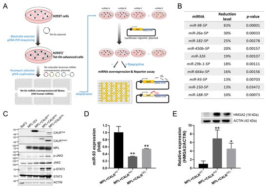
To unearth yet to be identified miRNAs that could target 3′UTR of HMGA2, we employed a Tet-on miRNA library screening system (containing 594 miRNAs) in conjunction with a luciferase reporter assay to search for potential miRNA candidates (Figure 1A). Excluding the well-known let-7a, the top 10 HMGA2 3′UTR-targeting miRNAs that exhibited the most prominent reduction of luciferase activity are listed in Figure 1B. We next examined the significance of these miRNAs in JAK2-mutated MPN cells; yet, none were differentially expressed as compared to the wild-type (WT) cells. In our earlier work, we showed that HMGA2 overexpression could also be seen in CALR-mutated MPN cells [11]. Therefore, we tried to scrutinize the expression levels of the identified miRNAs in these cells. We previously engineered CALR-mutated MPN cell models through co-transduction of an RFP-containing, MPLWT-expressing lentiviral vector and a GFP-harboring MSCV vector, cloned with either an empty vector (EV), CALRWT, CALRINS, or CALRDEL into BaF3 cells, to obtain four stable clones, including two mutant clones and two control clones [16]. Figure 1C demonstrates the adequacy of these cells, including either WT or mutant protein expression in respective cells, and the presence of JAK-STAT signaling activation in the mutant cells. Among the top 10 HMGA2 3′UTR-targeting miRNAs identified, only miR-93 showed differential expression between WT and CALR mutant cells, as it was downregulated in both CALRINS and CALRDEL cells (Figure 1D). Intriguingly, there was an inverse trend of HMGA2 expression in both mutant cells (Figure 1E). These findings suggest a potential correlation between HMGA2 and miR-93 in CALR-mutated MPN that could be pathobiologically relevant.
Figure 1.
Exploring HMGA2 3′UTR-targeting miRNA candidates. (A) A schema depicting the process of the Tet-On miRNA screening library and the luciferase reporter assay. (B) Identification of miRNA candidates that target 3′UTR of HMGA2. Candidate miRNAs were selected based on reduced luciferase activity, which was indicative of their effectiveness in targeting the HMGA2 3′UTR regions. Excluding the well-known let-7a, the table here lists the top ten HMGA2 3′UTR-targeting candidate miRNAs. (C) Western blots examining the expression levels of various proteins, including MPL, wild-type and mutant CALR proteins, and total as well as phosphorylated JAK2 and STAT3 proteins, in our previously established CALRWT and CALRMUT cell lines. Images shown are cropped regions of specific membranes. Full uncropped blots are provided in the Supplementary Materials dataset. (D) Expression levels of miR-93 assessed with quantitative RT-PCR (qRT-PCR) in CALRWT and CALRMUT cells. U6 small nuclear RNA was used for normalization. (E) Results for Western blot analysis of HMGA2. Representative data are shown in the upper panel. Expression levels of HMGA2 are quantified and illustrated in the lower panel. Data on mean ± standard deviation (SD) values from three independent experiments are depicted in (D) and (E). * p < 0.05; ** p < 0.01 by Student’s t-test.
2.2. Effects of miR-93 Overexpression on Cellular Phenotypes in CALR-Mutated MPN Cells In Vitro
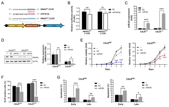
To assess if miR-93 does target HMGA2 3′UTR and regulate its expression, we employed a luciferase reporter assay for this purpose. Specifically, we cloned two reporter gene plasmids containing either the WT HMGA2 3′UTR sequence (HMGA2WT 3′UTR) or a mutated sequence within the 3′UTR region that became non-complementary to miR-93 (HMGA2MUT 3′UTR) into a luciferase reporter vector (Figure 2A). As demonstrated in Figure 2B, miR-93 significantly reduced luciferase activity in the HMGA2WT 3′UTR vector compared to the scramble control sequence. Conversely, mutations in the “GCACUUU” sequence within 3′UTR of HMGA2 mRNA led to indiscernible luciferase activity between miR-93 and the control sequence, indicating selective inhibition of HMGA2 mRNA expression by miR-93 through its binding to the conserved 3′UTR region.
Figure 2.
MiR-93 induces translational repression of HMGA2 while leading to suppressed proliferation in CALR-mutated MPN cells. (A) Illustration of viral constructs for luciferase reporter assay. A wild-type HMGA2 3′UTR region (HMGA2WT 3′UTR), which contained a sequence complementary to miR-93 or a mutated non-complementary 3′UTR (HMGA2MUT 3′UTR) sequence, was cloned into the pmirGLO plasmid. (B) Luciferase reporter assay. The 293T cells transfected with either HMGA2WT 3′UTR-containing or HMGA2MUT 3′UTR-containing plasmids (left and right panels, respectively) were treated with miR-93 or a scramble control sequence. Relative luminescence was measured at 48 h post transfection. (C) Ectopic expression of miR-93 in CALR-mutated MPN cells. Cells were transduced with a lentiviral vector containing either miR-93 (CALRMUT miR-93) or a scramble control sequence (CALRMUT SC). Successful overexpression was confirmed with qRT-PCR analysis. U6 was used for normalization. (D) Western blots on the expression levels of HMGA2 in CALRMUT miR-93 and CALRMUT SC cells. β-Actin was used as the loading control. (E) Time-dependent cell viability of miR-93-transducing CALRMUT cells, as measured with the XTT assay. Statistical significance was determined by two-way ANOVA, followed by Tukey’s multiple comparisons test. Asterisks indicate statistically significant differences between the miR-93 and SC groups. (F) Flow cytometric analyses on cellular proliferation indices of miR-93-transducing CALRMUT cells, assessed with Ki67 staining. (G) Rates of apoptosis in miR-93-transducing CALRMUT cells. Cells were treated with 7-AAD and APC-Annexin V and subsequently subjected to flow cytometric analyses. Cells positively stained for Annexin V only were defined as being in early apoptosis, whereas those with positive stains for both Annexin V and 7-AAD were considered as being late apoptotic cells. All experiments were done in triplicates, and results are shown as mean ± SD. * p < 0.05; ** p < 0.01; *** p < 0.001; ns: no significance. Statistical significance was determined by Student’s t-test, unless otherwise indicated.
To further assess the effect of miR-93 on HMGA2 expression in vitro, we transduced miR-93 into CALRMUT cells to obtain three respective cell lines: untreated controls, scramble control (SC)-treated cells, and miR-93-overexpressing CALRMUT cells. Overexpression of miR-93 was validated with quantitative RT-PCR (qRT-PCR) (Figure 2C). Western blot analysis showed lower HMGA2 expression in miR-93-overexpressing CALRMUT cells (Figure 2D). As mentioned earlier, HMGA2 is widely considered a proto-oncogene in various human malignancies, including MPN [8,9,10,11,17]. Therefore, we were intrigued to see what impacts miR-93 overexpression might have on these MPN cells. Cell viability assays revealed that both CALRINS and CALRDEL cells transduced with miR-93 exhibited significantly lower cell viability as compared with either non-transduced cells or SC-treated cells (Figure 2E). The finding could be further supplemented in the flow cytometric analysis showing suppressed activity in the proliferation marker Ki-67 in both miR-93-overexpressing CALRMUT cells (Figure 2F). Additionally, Annexin-V/7-AAD dual staining demonstrated increased apoptotic rates in these cells, implicating miR-93 could render CALR-mutated MPN cells more prone to apoptotic death (Figure 2G). Together, these results indicate that enforced miR-93 expression could potentially attenuate the exaggerated proliferation induced by CALR mutants in MPN cells through impeding cellular growth and inducing more apoptosis.
2.3. Ectopic miR-93 Expression Attenuates CALR Mutant-Associated Phenotypes in an In Vivo Transplantation Model
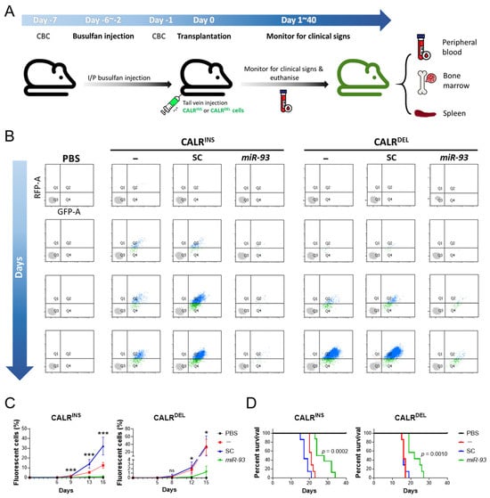
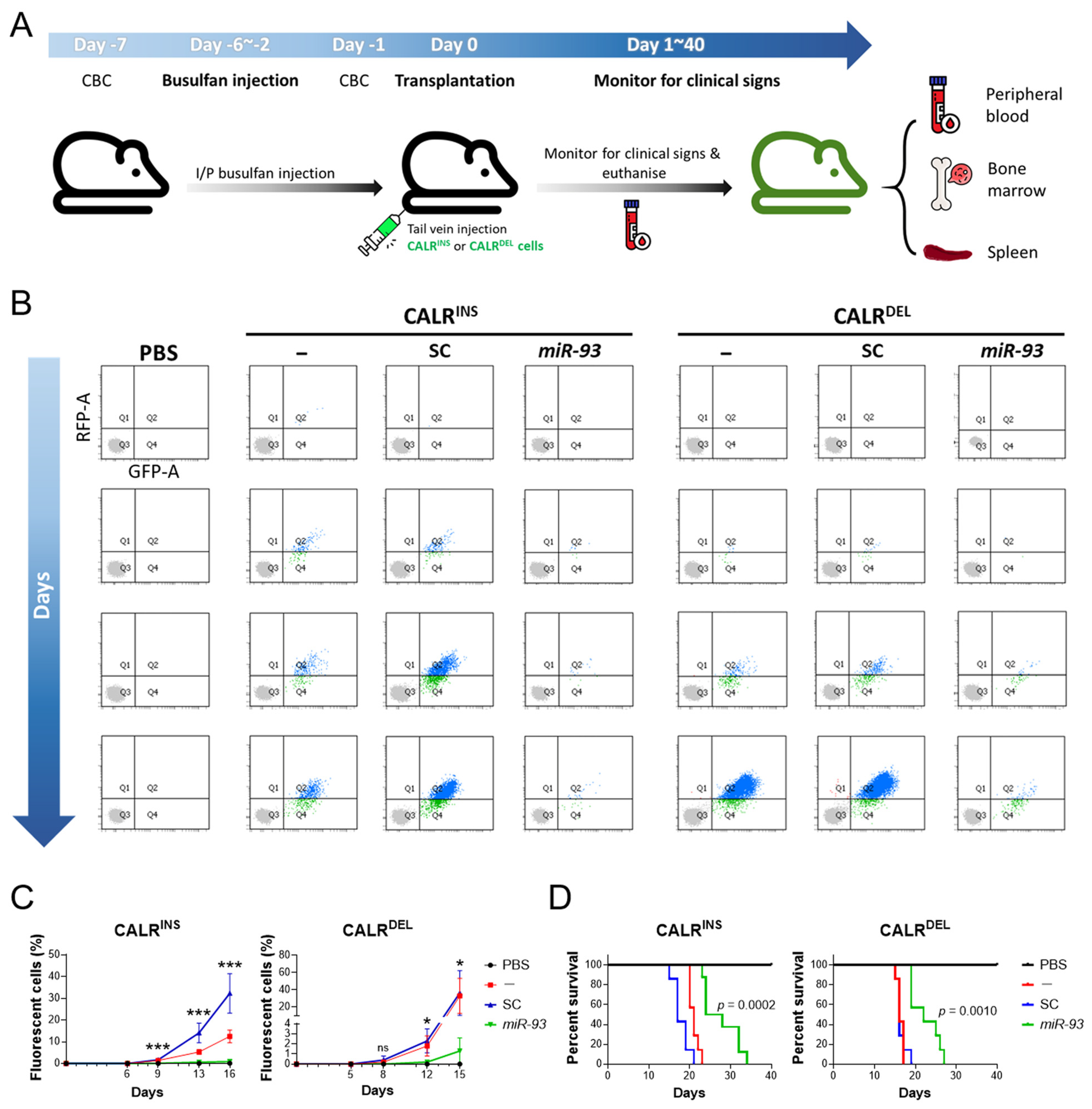
To gain further insights into the impact of miR-93 on the development of MPN and to validate our in vitro findings, we next employed in vivo mice transplantation models to explore further. Following intraperitoneal busulfan injection, we infused both CALR mutant cells as well as their control comparators into respective ASID mice groups and monitored their clinical condition closely (Figure 3A). In ASID mice transplanted with either CALRMUT or CALRMUT SC cells (Figure 3B, second, third, fifth, and sixth columns), dual-fluorescent MPN cells began to appear in their peripheral blood (PB) after approximately one week, and these cells rapidly expanded by week 2 (Figure 3B,C), suggesting successful engraftment. In contrast, miR-93 overexpression in CALRMUT MPN cells was associated with a delayed appearance and lower abundance of dual-fluorescent cells (Figure 3B, fourth and seventh columns, and Figure 3C). These results suggested that miR-93 could suppress the expansion of CALR-mutated MPN cells in vivo. In addition, mice transplanted with either CALRMUT or CALRMUT SC cells exhibited gradually emerging debilitating symptoms, including rapid weight loss, hunching, reduced activity, huddling for warmth, and labored breathing, whereas a subset of mice also demonstrated symptoms of hind-limb paralysis. All these manifestations could be translated into the survival outcome, as mice receiving miR-93-overexpressing MPN cells had significantly longer survival, as highlighted in Figure 3D.
Figure 3.
In vivo modeling of CALR-mutated MPN and impacts of miR-93 overexpression on engraftment and survival of transplanted mice. (A) Schematic diagram of in vivo cell transplantation strategy in advanced severe immunodeficiency (ASID) mice. Both CALRDEL and CALRINS arms contained three groups of mice: mice infused with either CALRMUT, CALRMUT SC, and CALRMUT miR-93 cells. An independent group of mice infused with PBS only was used as control. Sample sizes are n = 4 (PBS), n = 7 (untreated), n = 7 (SC), and n = 7 (miR-93). Some icons were adapted from Flaticon.com graphics (https://www.flaticon.com/, accessed on 21 May 2026). (B) Twice-weekly monitoring for MPN cell engraftment in transplanted mice employing peripheral blood (PB) samples. Our CALR mutant cells harbor both green and red fluorescence, which can be used for clonal monitoring with flow cytometry. Gradual appearance of dual-fluorescent cells was noted in PB of some transplanted mice, indicating successful engraftment. (C) Percentages of dual-fluorescent cells in respective mice, depicted in linear graph. Statistical significance was determined using a mixed-effects model, followed by Dunnett’s multiple comparisons test. Asterisks indicate statistically significant differences of the miR-93 group compared to the SC control group. * p < 0.05; *** p < 0.001; ns: no significance. (D) Survival curves for transplanted mice in respective groups.
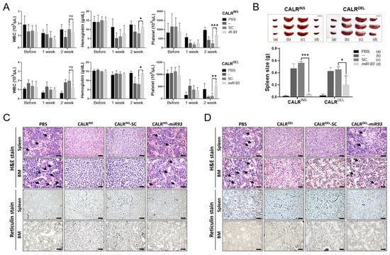
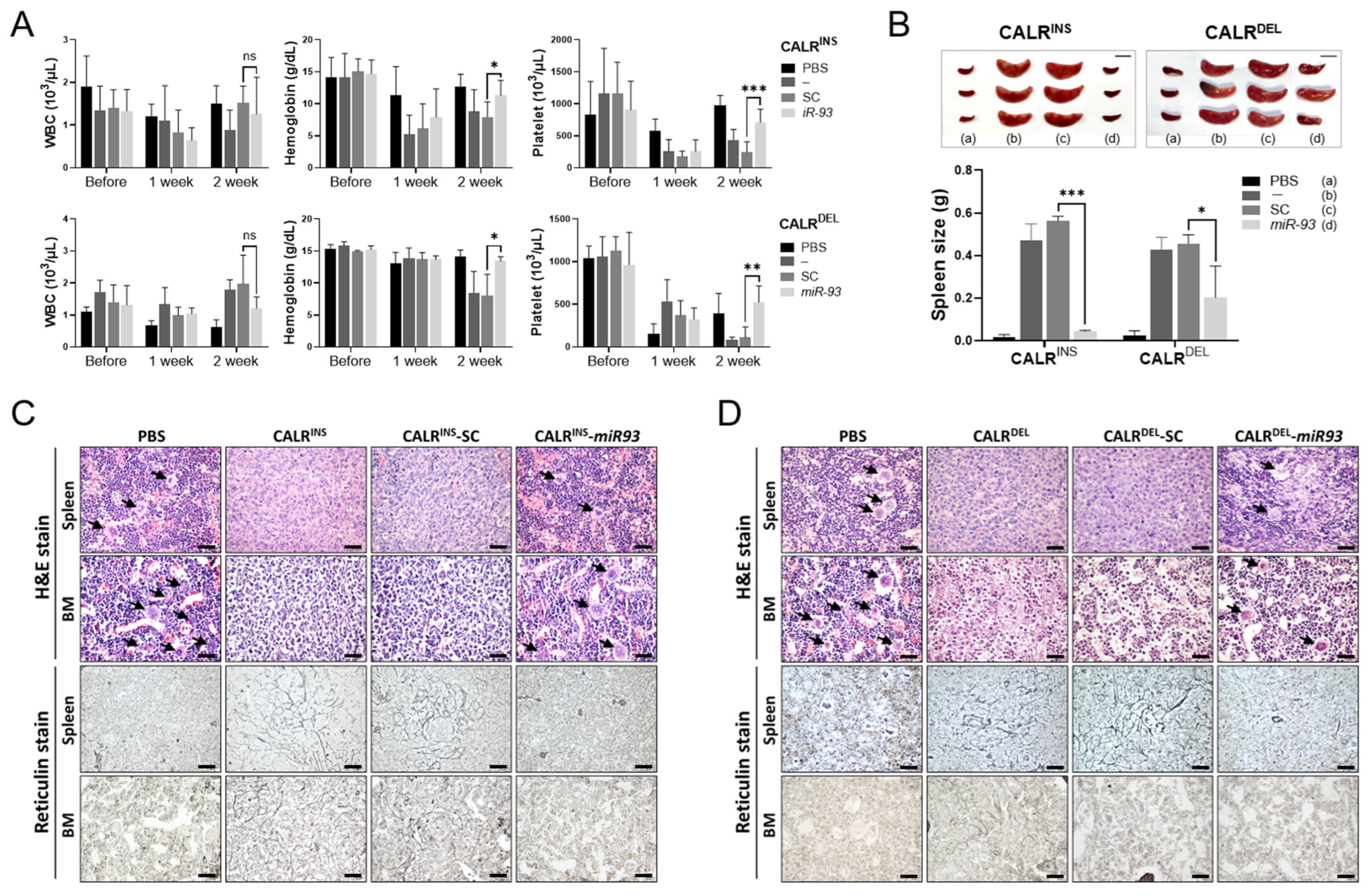
We next investigated disease-specific phenotypes in these transplanted mice. We first looked at three key hematological parameters and found some differences, as well. Although the white blood cell counts were not disparate among various transplanted groups (Figure 4A, left panel), mice transplanted with either CALRMUT or CALRMUT SC cells manifested gradual anemia and thrombocytopenia by week 2 (Figure 4A, middle and right panels, respectively). However, the cytopenia could be ameliorated by the presence of miR-93 (Figure 4A, rightmost columns in the middle and right panels, respectively).
Figure 4.
In vivo phenotypes of mice transplanted with CALR-mutated MPN, in the presence or absence of miR-93 overexpression. (A) Hematological profiles of transplanted mice. Complete blood counts were monitored at baseline and 1-week interval after transplantation. Results for white cell count (WBC), hemoglobin, and platelet counts are shown. Sample sizes are n = 3 (PBS), n = 7 (untreated), n = 7 (SC), and n = 7 (miR-93). Statistical significance was determined using a mixed-effects model, followed by Dunnett’s multiple comparisons test. Asterisks indicate statistically significant differences of the miR-93 group compared to the SC control group. * p < 0.05; ** p < 0.01; *** p < 0.001; ns: no significance. (B) The size of spleen in transplanted mice, demonstrated with photo images and weight in the bar graph. Scale bars, 1 cm. Sample sizes are n = 3 per group. * p < 0.05; *** p < 0.001; ns: no significance by Student’s t-test. (C,D) Histological sections and staining with H&E (upper panel) and reticulin (lower panel) of spleen and bone marrow (BM) in the transplanted mice. Arrows indicate megakaryocytes. Scale bars, 40 μm. In (B,C), transplanted mice categorized within the same arm (either CALRINS or CALRDEL mutation) were humanely euthanized simultaneously when the first mice within that arm succumbed to MPN disease. Following extraction of their spleens and both femurs, comparison was made across different groups with regards to spleen weight and results of H&E, as well as reticulin stains on the BM and spleen.
In a separate experiment to assess the spleen size in transplanted mice, we sacrificed all the mice once the first mice died of apparent disease (confirmed through the presence of dual-fluorescent cells in PB). As demonstrated in Figure 4B, mice infused with either CALRMUT or CALRMUT SC cells (shown in the middle two columns of both panels) all harbored significant splenomegaly, whereas miR-93 overexpression was associated with lower spleen weight. On histological examination of the bone marrow (BM) and spleen of sacrificed mice, we found that those transplanted with either CALRINS or CALRINS SC cells manifested significantly less megakaryopoiesis and increased reticulin fibrosis in their spleen and BM, respectively (Figure 4C, middle two columns; as compared to mice infused with PBS only in the left column). On the other hand, miR-93 attenuated these aberrant histological phenotypes in the BM and spleen (Figure 4C, right column). Comparable findings could be observed in mice transplanted with CALRDEL cells (Figure 4D). These results suggested that in our immunodeficient mice model transplanted with MPN cells, both CALR mutations would lead to a myelofibrosis-like phenotype, whereas miR-93 could attenuate the development of these phenotypes and support hematopoietic recovery.
2.4. Exploratory Associations Between miR-93 Expression and Clinical Characteristics in CALR-Mutated MPN
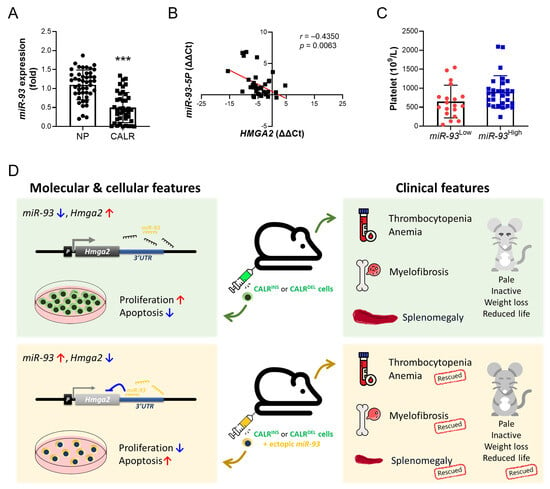
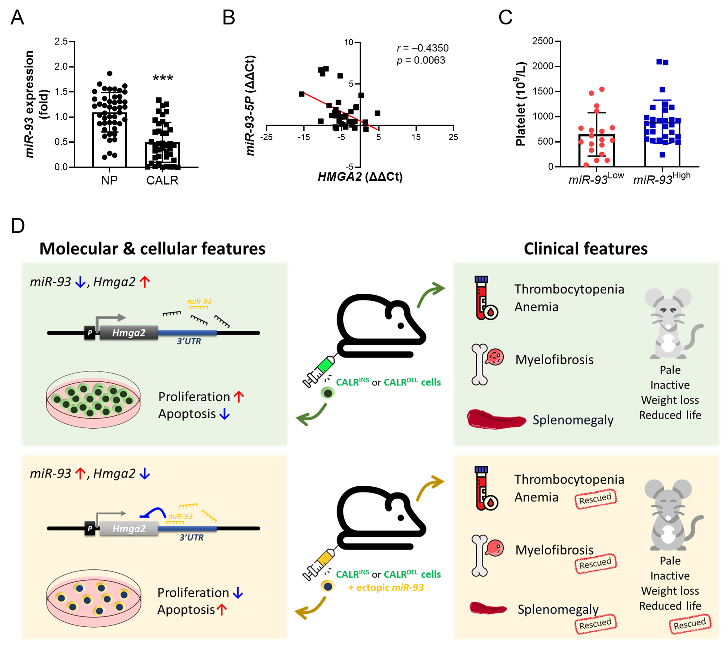
To explore the potential clinical relevance of our experimental findings, we examined miR-93 expression and its associations with clinical characteristics in patients with CALR-mutated MPN. Compared with normal healthy populations (NP), CALR-mutated MPN patients expressed lower levels of miR-93 (Figure 5A). In addition, there was an inverse correlation between the expression levels of miR-93 and HMGA2 in those CALR-mutated patients (Figure 5B). We further stratified these patients into miR-93High and miR-93Low groups based on whether their miR-93 expression levels exceeded twice the mean value observed in healthy controls, using this reference-based threshold for exploratory comparisons rather than as a validated prognostic cutoff. Table 1 lists the comparison between the baseline characteristics of these two groups of patients. There was no significant difference between the two groups of patients with regards to age, gender, type of CALR mutations, spleen size, white cell count, hemoglobin, marrow cellularity, and either MF or acute myeloid leukemia (AML) transformation. On the other hand, it seemed that ET and pre-PMF patients were more enriched in the miR-93High group, whereas most MF patients expressed lower miR-93 levels. However, because of the limited case number, the comparison was not statistically significant. Similar findings could be observed in the comparison of platelet counts; there was a trend for miR-93High patients to harbor higher platelet counts (p = 0.054) than their counterparts (Table 1 and Figure 5C). This trend could echo results from our mice transplantation model, in which miR-93 attenuated myelofibrosis-like and increased megakaryopoiesis. Lastly, our miR-93High patients had apparently fewer thrombotic events and enjoyed a prolonged thrombosis-free survival, although this finding should be interpreted as exploratory given the inclusion of thrombotic events present at diagnosis. These data suggest that CALR-mutated MPN patients with relatively higher miR-93 expression might run a more indolent clinical course and could exhibit unique clinical phenotypes that closely resemble what we observed in the immunodeficient mice transplanted with CALR-mutated MPN cells.
Figure 5.
Validation of significance of miR-93 expression in clinical CALR-mutated MPN patients and schematic illustration of pathognomonic roles of miR-93 in CALR-mutated MPN. (A) Quantification of miR-93 in clinical samples. MiR-93 expression in the PB granulocytes of CALR-mutated MPN patients and normal population (NP) was assessed with qRT-PCR. *** p < 0.001 by Student’s t-test. (B) Scatter plot illustrating the correlation between the expression levels of miR-93 and HMGA2 transcripts in the granulocytes of CALR-mutated MPN patients (correlation coefficient r = −0.4350; Pearson’s correlation, p = 0.0063). Data are shown in ΔΔCt. (C) Platelet counts in CALR-mutated MPN patients, stratified by the expression level of miR-93. We categorized these patients into miR-93High and miR-93Low groups based on whether their miR-93 expression levels were two times higher than the average value of the NP population. (D) Schematic summary of the proposed association between miR-93/HMGA2 regulation and phenotypic features in CALR-mutated MPN. Some icons were adapted from Flaticon.com graphics (https://www.flaticon.com/, accessed on 21 May 2026).
Table 1.
Clinical and laboratory features of CALR-mutated MPN patients, stratified by the expressional level of miR-93.
Overall, the findings of our current study are summarized in Figure 5D. MiR-93 targets a highly conserved sequence within HMGA2 mRNA 3′UTR, leading to its translational repression and reduced protein expression. Overexpression of miR-93 is accompanied by lower HMGA2 expression, reduced proliferation, and augmented apoptosis in CALR-mutated MPN cells. Transplantation of CALR-mutated MPN cells into severely immunodeficient mice induces rapidly progressive myelofibrosis phenotypes mainly manifested as anemia, thrombocytopenia, splenomegaly, and increased reticulin fibrosis within the BM and spleen. Explicitly, ectopic expression of miR-93 in CALR-mutated MPN cells retrogresses these manifestations and prolongs the survival of transplanted mice.
3. Discussion
Aiming to explore previously uncharacterized HMGA2-targeting miRNAs and delineate their roles in the pathogenesis of MPN, we inadvertently identified miR-93 as a candidate regulator in CALR-mutated MPN in this study. We demonstrate that miR-93 targets a highly conserved region within the HMGA2 3′UTR sequence, leading to its translational repression. Overexpression of miR-93 mitigates the aggressiveness and expansion of CALR-mutated MPN clones both in vitro and in vivo. These findings support a potential tumor-suppressive effect of miR-93 in CALR-mutated MPN. However, because the present study predominantly uses gain-of-function experiments, the data do not establish that endogenous miR-93 loss is required for disease progression or that HMGA2 is the necessary downstream mediator of all observed effects.
Co-transcribed in the context of the MCM7 primary transcript, miR-93 is encoded by a sequence within intron 13 of the host gene MCM7 on chromosome 7q22.1 [18]. It belongs to the miRNA-106b-25 cluster, which consists of three highly conserved miRNAs (miR-25, miR-93, and miR-106b) [19] and regulates genes non-redundantly involved in mediating cellular processes such as the cell cycle, proliferation, and apoptosis [18,20]. Reports have indicated that miR-93 may serve as a proto-oncogene by promoting tumor growth, invasion, and metastasis in various human cancers [20,21]. However, there is ample evidence demonstrating that miR-93 also functions as a tumor suppressor by inhibiting migration as well as invasion in pancreatic cancer [22] and colon cancer [23,24], while also leading to sensitization of ovarian cancer cells to chemotherapy [25], among other outcomes. The dual functionality highlights the complex and context-dependent nature of miR-93′s role in cancer biology.
In contrast to what has been identified in solid tumors, there is a scarcity of data on potential pathogenic or tumor suppressive roles of miR-93 in hematological malignancies, especially myeloid neoplasms. It has been found that miR-93 is upregulated in AML [26,27], and overexpression of miR-93 leads to accelerated growth in AML cell lines [27]. Although our findings on the protective role of miR-93 in CALR-mutated MPN pathogenesis are innovative, there is growing evidence that deregulated miRNAs contribute to MPN biology [28]. While most research on this aspect mainly focused on the correlation between miRNA expression and its putative targets without functional validation [28], some in vivo models indicated that enforced expression of miR-125a [29], miR-125b [30], or miR-155 [31] or downregulation of miR-146a [32] in the hematopoietic cells would result in myeloproliferation in mice. Given the crucial roles of miRNAs in HSC function and the specific involvement of distinct miRNAs in various lineage differentiations [28], it is likely that deregulated miRNAs may lead to lineage bias and the development of MPN phenotypes. Interestingly, a recent study demonstrated that miR-106 could mediate the differentiation of megakaryocytes [33]. Considering that miR-93 is categorized within the same miRNA-106b-25 cluster and shares structural homology with miR-106 [19], it is plausible that miR-93 might participate in the megakaryocytic commitment within the HSC compartment, as well, and its deregulation could contribute to MPN pathogenesis.
In our present study, we unearthed the downregulation of miR-93 in CALR-mutated MPN cells by exploring potential HMGA2 3′UTR-targeting candidate miRNAs, a finding further supplemented with clinical samples validation. However, it is not immediately clear what the molecular mechanism driving miR-93 repression in CALR-mutated MPN is. While both JAK2 and CALR mutations activate the STAT5 pathway, the differential expression of miR-93 in CALR-mutated cells versus JAK2-mutated cells suggests that the upstream regulatory mechanisms may be distinct. This divergence could be linked to the unique chaperone-like function of mutant CALR or its specific impact on the MCM7 host gene, which may not be triggered by JAK2V617F kinase activity. Most of the time, the expression of intronic miRNAs is expected to be in agreement with the expression of their respective host genes [34]. Therefore, miR-93 is commonly regulated with MCM7 in a concordant manner. Together, their abnormal expression can go through either altered promoter methylation [35], aberrant transcription factor activities [19], or deregulated functioning of transcriptional activators [19]. On the other hand, recent studies have identified discordant expression between MCM7 and its hosted miRNAs in both normal and malignant cells [19]. In hematopoietic stem and progenitor cells, the differential expression of MCM7 and its intronic miRNA cluster is especially important in megakaryopoiesis, as Haldar S. et al. elegantly demonstrated, showing that megakaryocytic endomitosis (repetitive DNA synthesis without cell division) proceeded with the uncoupling of expression between MCM7 and miR-106b-25, which was attributed to the presence of a MCM7 transcript variant that underwent nonsense-mediated decay [36]. Essentially, upregulation of the miR-106b-25 cluster was found to promote megakaryopoiesis in their study, a finding affirmatively echoed by our miR-93 data in the CALR-mutated MPN model.
With regards to how miR-93 mediates its oncogenic or tumor suppressive activity, there have been many downstream pathways identified in various cancers [20,21]. Among the plausible targets, HMGA2 could be a biologically relevant candidate downstream mediator in the present context. Truncation of Hmga2 3′UTR was shown to lead to an MPN phenotype in transgenic mice [10] and promoted better repopulating ability in serial bone marrow transplants [37], whereas knockdown of Hmga2 in MPN cells impeded clonal growth and increased cellular apoptosis [11]. Essentially, deletion of Hmga2 ameliorated anemia and splenomegaly in a JAK2V617F mice model [37]. These observations are directionally consistent with our finding that miR-93 overexpression, accompanied by HMGA2 downregulation, attenuated MF-like phenotypes in CALR-mutated cell-transplanted mice. Nevertheless, miR-93 has multiple potential targets, and we did not perform Hmga2 rescue experiments or direct loss-of-function studies of miR-93. Therefore, the present data support HMGA2 as a candidate downstream mediator but do not establish that the miR-93/HMGA2 axis is necessary or sufficient for the observed phenotypes. Integrating these findings, we propose a hypothesis of mutation-associated differences in miRNA–HMGA2 regulation. Our previous work demonstrated that JAK2V617F mutation induces a robust 8-fold upregulation of Hmga2, primarily driven by the profound suppression of let-7a [11]. In contrast, CALR mutations elicit a more modest 2-fold increase in Hmga2 transcripts [11]. This disparity raises the possibility that miR-93-mediated regulation may have greater relative relevance in CALR-mutated cells, and divergence in miRNA-mediated control may partially explain the distinct clinical manifestations observed between JAK2- and CALR-mutated MPN patients. However, direct comparative mechanistic studies will be required to test this hypothesis.
While our findings on miR-93 on retarding MF progression are unprecedented, there have been several reports illustrating the protective roles of miR-93 against organ fibrosis. The level of circulating miR-93 was shown to be negatively correlated with liver fibrosis in patients with chronic hepatitis C [38]. Overexpression of miR-93 prevented renal fibrosis through inhibition of either Ccnd1 [39] or Cdkn1a [40] in mice models of nephropathy. Similarly, miR-93 upregulation also ameliorated TGF-β1-mediated fibrosis in a human proximal tubule cell line [41] and mitigated HMGA2-induced fibrosis and inflammation in human glomerular mesangial cells [15]. Intriguingly, miR-93 can be upregulated in response to inflammatory stimuli and is capable of targeting immune pathways in macrophages to revascularize ischemic organs [42], whereas multiple studies have implied that miR-93-endued suppression of inflammation provides clinical benefits [43,44,45]. Considering that inflammation and TGF-β signaling are pivotal in the pathogenesis of myelofibrosis [46,47], these observations raise the possibility that miR-93 may influence both cell-intrinsic and microenvironment-related pathways. Further exploratory work is imperative to delineate how miR-93 regulates the states and fates of CALR-mutated MPN cells.
From a clinical perspective, reduced miR-93 expression seemed to be more enriched in the MF subtype in our patient cohort, and those miR-93 low expressors had comparably lower platelets counts. These data might support the potential role of miR-93 in mitigating the phenotypic characteristics of CALR-mutated MPN, although we have to interpret these exploratory observations with caution, as neither comparison reached statistical significance. The associations with thrombosis and thrombosis-free survival also require confirmation in larger, independent cohorts, with adjustment for established clinical risk factors. Importantly, the present study relies predominantly on gain-of-function models. Future rescue experiments restoring HMGA2 despite miR-93 overexpression, as well as miR-93 loss-of-function approaches using inhibitors or antagomirs in CALR-mutated cell models or primary MPN cells, will be required to determine whether the proposed miR-93/HMGA2 relationship is necessary for the observed phenotypes. Several limitations should be considered when interpreting the clinical findings. First, the relatively small cohort (n = 48) and limited number of major thrombotic events (n = 9) restrict the statistical power and precision of the analysis and preclude reliable multivariable adjustment for established thrombotic risk factors, including age, disease subtype, previous thrombosis, and treatment exposure. Second, the miR-93 expression cutoff was selected post hoc, and its robustness across alternative thresholds has not been evaluated. Third, the thrombosis-related time-to-event analysis included both thrombotic events present at diagnosis and incident events during follow-up, limiting its interpretation as a conventional prospective thrombosis-free survival outcome. Furthermore, multiple clinical variables were examined without formal adjustment for multiple comparisons, increasing the possibility of chance associations. Consequently, the observed clinical associations should be regarded as exploratory and hypothesis generating, requiring confirmation in larger independent cohorts with prespecified thresholds, clearly defined incident thrombotic outcomes, and appropriately adjusted survival analyses.
The ASID mice employed in our in vivo experiment are considered equivalent to NSG mice and bear the same genetic background of the NOD strain [48,49]. This inherent immunodeficient nature of these mice, including a complete lack of B cells, T cells, and NK cells and high defectiveness in their dendritic cells and macrophages, makes them ideal candidates for in vivo modeling of tumorigenesis. Considering the lack of adequate commercially available transgenic mice models, our novel approach represents a reasonable alternative for in vivo studies on CALR-mutated MPN. Nevertheless, this system has several important limitations. First, it is a transplantation model based on engineered murine BaF3 cells rather than primary human MPN hematopoietic stem/progenitor cells and therefore does not reproduce the clonal architecture, lineage hierarchy, or chronic natural history of human CALR-mutated MPN. Second, the profoundly immunodeficient host lacks key immune and inflammatory components of the human marrow microenvironment that contribute to MPN-associated fibrosis. Third, the unusually rapid disease course likely reflects the proliferative behavior of the transplanted cells and permissive host environment; preliminary observations in small pilot cohorts suggest that reducing the number of infused cells by 50–90% may not substantially alter disease onset or survival (Supplementary Materials Figure S1). Accordingly, the anemia, thrombocytopenia, splenomegaly, and reticulin fibrosis observed here are best regarded as MF-like surrogate phenotypes rather than a complete model of CALR-mutated myelofibrosis. The model nevertheless permits controlled comparison of otherwise matched CALR-mutated cells with or without enforced miR-93 expression. The attenuation of these phenotypes by miR-93 overexpression therefore supports a cell-intrinsic phenotypic effect in this experimental setting, but it does not by itself establish the mechanism, the requirement for endogenous miR-93 loss, or the clinical efficacy of targeting this pathway.
Collectively, we have demonstrated that miR-93, capable of targeting the HMGA2 mRNA 3′UTR sequence, is deregulated in CALR-mutated MPN. Reduced miR-93 expression is associated with CALR mutant cells, whereas enforced miR-93 expression reduces proliferation and increases apoptosis in vitro. Through impeding the growth and expansion of CALR-mutated MPN clones, ectopic expression of miR-93 effectively attenuates MF-associated phenotypes in an in vivo mice model transplanted with CALR-mutated MPN. Together with the clinical associations observed in our cohort, these findings provide new insights and lay the groundwork for further mechanistic studies of the miR-93/HMGA2 relationship in CALR-mutated MPN.
4. Materials and Methods
4.1. Advanced Tet-On Inducible Expression System and Luciferase Reporter Assays
The study utilized an advanced Tet-on vector system and a Tet-on miRNA overexpression cell library (Academia Sinica, Taipei, Taiwan) to identify candidate miRNAs that may target 3′UTR of HMGA2. After transfection and selection processes, H293T cells were transduced with lentiviral vectors encoding various miRNAs. HMGA2 3′UTR was amplified and cloned into a pmirGLO luciferase reporter vector (pmirGLO-HMGA2WT 3′UTR) for analysis. The luminescence results were measured to evaluate the interactions and effects of miRNAs on HMGA2 3′UTR. The detailed procedures (Figure 1A) are also listed in the Supplementary Materials.
4.2. Animals and Transplantation of MPN
Advanced severe immunodeficiency (ASID, NOD.Cg-PrkdcscidIl2rgtm1Wjl/YckNarl) mice were purchased from the National Laboratory Animal Center in Taiwan. They were housed in a sterile environment constructed within a maximum-barrier facility and individually ventilated cages at our Laboratory Animal Center, where the environmental temperature was maintained at 22–24 °C. All experiments were conducted using 5–8-week-old female mice weighing approximately 20 g. All processes were conducted in accordance with the institutional animal welfare guidelines of Chang-Gung Memorial Hospital, Chiayi (IACUC No.2021122406). Mouse experiments are described in the Supplementary Materials.
4.3. Study Population
Patients with CALR-mutated MPN followed at our institute were enrolled in this study. Adult healthy individuals were also included as controls. The detection of CALR exon 9 mutations in clinical samples was performed as previously described [11]. For exploratory clinical comparisons, patients were categorized into miR-93High and miR-93Low groups using a threshold of twice the mean miR-93 expression level in healthy controls. This reference-based threshold was chosen to identify patients with relatively elevated miR-93 expression, consistent with the commonly applied twofold-change convention in gene expression studies. The cutoff was selected during exploratory data analysis rather than prespecified and was not intended as a clinically validated prognostic threshold. For the thrombosis-related time-to-event analysis, the date of MPN diagnosis was used as time zero, and thrombotic events present at diagnosis were counted as time-zero events. Subsequent thrombotic events were also included during follow-up. Accordingly, this analysis incorporated both thrombosis present at diagnosis and incident thrombotic events, rather than exclusively evaluating prospective thrombosis-free survival.
4.4. Statistical Analysis
Statistical tests were two-sided. For comparisons displayed as pairwise contrasts, Student’s t-test was used as indicated in the corresponding figure legends. Longitudinal measurements were analyzed using two-way repeated-measures ANOVA or a mixed-effects model (to accommodate missing values), followed by appropriate multiple comparisons tests, at the prespecified time points shown in the figures, with comparisons made within the same CALR mutant background. Survival distributions were estimated by the Kaplan–Meier method and compared using the log-rank test. Correlations were assessed using Pearson’s correlation coefficient. Given the limited number of major thrombotic events (n = 9), multivariable survival analysis was not performed because of concerns regarding overfitting and unstable parameter estimates. Clinical comparisons were considered exploratory, and the reported p-values were interpreted as nominal, without adjustment for multiple comparisons. Statistical calculations were performed using the Statistical Package of Social Sciences 18.0 (SPSS, Inc., Chicago, IL, USA) and GraphPad Prism 9.0 (GraphPad Software, Inc., San Diego, CA, USA). All in vitro experiments were independently repeated at least three times. For blot analysis, data from three independent experiments were quantified and analyzed collectively. For qPCR analysis, technical triplicates were performed within each independent experiment, and the results were reproduced across at least three independent experiments. Results are presented as mean ± S.D., with horizontal lines in dot plots representing the mean value of each group. Asterisks in graphs denote the significance of p-values compared with controls (* p < 0.05; ** p < 0.01; *** p < 0.001; ns: no significance), unless otherwise specified.
4.5. Others
Information on the Advanced Tet-on inducible expression system and luciferase reporter assays, MPN cell lines and cell culture, virus transduction, quantitative RT-PCR, Western blot, cell viability, proliferation, apoptosis assays, animal transplantation, and tissue staining are listed in the Supplementary Materials.
Supplementary Materials
The following supporting information can be downloaded at: https://www.mdpi.com/article/10.3390/ijms272210390/s1.
Author Contributions
Y.-H.W.: Validation, Formal Analysis, Data Curation, Writing—Original Draft, Writing Review and Editing, Visualization, Project Administration. W.-C.T.: Investigation, Validation, Writing—Original Draft, Writing Review and Editing. Y.-J.C.: Conceptualization, Methodology, Investigation, Data Curation. C.-E.H.: Investigation, Resources, Supervision. Y.-Y.C.: Investigation, Resources, Funding Acquisition. Y.-Y.W.: Investigation, Resources. Y.-R.Y.: Investigation, Resources. C.-H.W.: Investigation, Resources. W.-Y.T.: Investigation, Resources. H.-Y.T.: Investigation, Data Curation. C.-C.C.: Formal Analysis, Conceptualization, Investigation, Resources, Writing—Original Draft, Writing Review and Editing, Supervision, Project Administration, Funding Acquisition. All authors have read and agreed to the published version of the manuscript.
Funding
The study was supported by grants from the National Science and Technology Council (Taiwan) to Chih-Cheng Chen (NSTC 112-2314-B-A49-082-MY3 and NSTC 115-2314-B-A49-046) and Yi-Yang Chen (NSTC 114-2314-B-182A-092) and from Chang-Gung Memorial Hospital to Chih-Cheng Chen (CMRPG6J0213) and Yi-Yang Chen (CORPG6N0191).
Institutional Review Board Statement
The study was conducted in accordance with the Declaration of Helsinki and approved by the Institutional Review Board of Chang-Gung Memorial Hospital (Approval No.: 201900953B0, approved on 1 October 2019). All animal experiments were approved by the Institutional Animal Care and Use Committee (IACUC) of Chang-Gung Memorial Hospital (Approval No.: 2021122406, approved on 1 August 2022) and conducted in accordance with institutional and national guidelines for the care and use of laboratory animals.
Informed Consent Statement
All participants provided informed written consent in accordance with the Declaration of Helsinki.
Data Availability Statement
All relevant data are available from the corresponding author upon reasonable request.
Acknowledgments
The authors thank Pei-Wen Tsai for assistance in data collection, Chian-Pei Li, Yi-Hsuan Lin and Xian-Zhan Cai for assistance in experiments. We also acknowledge the Precious Instrumentation Core Laboratory at Chang Gung Memorial Hospital (Chiayi, Taiwan) for the BD FACSCanto II flow cytometer service and the BD FACSAria Fusion cell sorter service, Laboratory Animal Center at Chang Gung Memorial Hospital (Chiayi, Taiwan) for animal care service, and Laboratory Animal Center at Chang Gung Memorial Hospital (Linkou, Taiwan) for mouse CBC analyses. This study was supported in part by a research grant from the Taiwan Clinical Oncology Research Foundation.
Conflicts of Interest
The authors declare no conflicts of interest.
References
- Barbui, T.; Tefferi, A.; Vannucchi, A.M.; Passamonti, F.; Silver, R.T.; Hoffman, R.; Verstovsek, S.; Mesa, R.; Kiladjian, J.J.; Hehlmann, R.; et al. Philadelphia chromosome-negative classical myeloproliferative neoplasms: Revised management recommendations from European LeukemiaNet. Leukemia 2018, 32, 1057–1069. [Google Scholar] [CrossRef] [Scilit]
- Merlinsky, T.R.; Levine, R.L.; Pronier, E. Unfolding the Role of Calreticulin in Myeloproliferative Neoplasm Pathogenesis. Clin. Cancer Res. 2019, 25, 2956–2962. [Google Scholar] [CrossRef] [Scilit]
- Grabek, J.; Straube, J.; Bywater, M.; Lane, S.W. MPN: The Molecular Drivers of Disease Initiation, Progression and Transformation and their Effect on Treatment. Cells 2020, 9, 1901. [Google Scholar] [CrossRef] [Scilit]
- Vainchenker, W.; Delhommeau, F.; Constantinescu, S.N.; Bernard, O.A. New mutations and pathogenesis of myeloproliferative neoplasms. Blood 2011, 118, 1723–1735. [Google Scholar] [CrossRef] [Scilit]
- Ozturk, N.; Singh, I.; Mehta, A.; Braun, T.; Barreto, G. HMGA proteins as modulators of chromatin structure during transcriptional activation. Front. Cell Dev. Biol. 2014, 2, 5. [Google Scholar] [CrossRef] [Scilit]
- Kumar, P.; Beck, D.; Galeev, R.; Thoms, J.A.I.; Talkhoncheh, M.S.; de Jong, I.; Unnikrishnan, A.; Baudet, A.; Subramaniam, A.; Pimanda, J.E.; et al. HMGA2 promotes long-term engraftment and myeloerythroid differentiation of human hematopoietic stem and progenitor cells. Blood Adv. 2019, 3, 681–691. [Google Scholar] [CrossRef] [Scilit]
- Lee, Y.S.; Dutta, A. The tumor suppressor microRNA let-7 represses the HMGA2 oncogene. Genes. Dev. 2007, 21, 1025–1030. [Google Scholar] [CrossRef] [Scilit]
- Di Cello, F.; Hillion, J.; Hristov, A.; Wood, L.J.; Mukherjee, M.; Schuldenfrei, A.; Kowalski, J.; Bhattacharya, R.; Ashfaq, R.; Resar, L.M. HMGA2 participates in transformation in human lung cancer. Mol. Cancer Res. 2008, 6, 743–750. [Google Scholar] [CrossRef] [Scilit]
- Fang, C.Y.; Liew, P.L.; Chen, C.L.; Lin, Y.H.; Fang, C.L.; Chen, W.Y. High HMGA2 Expression Correlates with Reduced Recurrence-free Survival and Poor Overall Survival in Oral Squamous Cell Carcinoma. Anticancer Res. 2017, 37, 1891–1899. [Google Scholar] [CrossRef] [Scilit]
- Ikeda, K.; Mason, P.J.; Bessler, M. 3’UTR-truncated Hmga2 cDNA causes MPN-like hematopoiesis by conferring a clonal growth advantage at the level of HSC in mice. Blood 2011, 117, 5860–5869. [Google Scholar] [CrossRef] [Scilit]
- Chen, C.C.; You, J.Y.; Lung, J.; Huang, C.E.; Chen, Y.Y.; Leu, Y.W.; Ho, H.Y.; Li, C.P.; Lu, C.H.; Lee, K.D.; et al. Aberrant let7a/HMGA2 signaling activity with unique clinical phenotype in JAK2-mutated myeloproliferative neoplasms. Haematologica 2017, 102, 509–518. [Google Scholar] [CrossRef] [Scilit]
- Guglielmelli, P.; Zini, R.; Bogani, C.; Salati, S.; Pancrazzi, A.; Bianchi, E.; Mannelli, F.; Ferrari, S.; Le Bousse-Kerdiles, M.C.; Bosi, A.; et al. Molecular profiling of CD34+ cells in idiopathic myelofibrosis identifies a set of disease-associated genes and reveals the clinical significance of Wilms’ tumor gene 1 (WT1). Stem Cells 2007, 25, 165–173. [Google Scholar] [CrossRef] [Scilit]
- Szymczyk, A.; Macheta, A.; Podhorecka, M. Abnormal microRNA expression in the course of hematological malignancies. Cancer Manag. Res. 2018, 10, 4267–4277. [Google Scholar] [CrossRef] [Scilit]
- Pattanayak, B.; Garrido-Cano, I.; Adam-Artigues, A.; Tormo, E.; Pineda, B.; Cabello, P.; Alonso, E.; Bermejo, B.; Hernando, C.; Martinez, M.T.; et al. MicroRNA-33b Suppresses Epithelial-Mesenchymal Transition Repressing the MYC-EZH2 Pathway in HER2+ Breast Carcinoma. Front. Oncol. 2020, 10, 1661. [Google Scholar] [CrossRef] [Scilit]
- Wang, X.; Liu, Y.; Rong, J.; Wang, K. LncRNA HCP5 knockdown inhibits high glucose-induced excessive proliferation, fibrosis and inflammation of human glomerular mesangial cells by regulating the miR-93-5p/HMGA2 axis. BMC Endocr. Disord. 2021, 21, 134. [Google Scholar] [CrossRef] [Scilit]
- Wang, Y.H.; Chen, Y.J.; Lai, Y.H.; Wang, M.C.; Chen, Y.Y.; Wu, Y.Y.; Yang, Y.R.; Tsou, H.Y.; Li, C.P.; Hsu, C.C.; et al. Mutation-Driven S100A8 Overexpression Confers Aberrant Phenotypes in Type 1 CALR-Mutated MPN. Int. J. Mol. Sci. 2023, 24, 8747. [Google Scholar] [CrossRef] [Scilit]
- Dreux, N.; Marty, M.; Chibon, F.; Velasco, V.; Hostein, I.; Ranchere-Vince, D.; Terrier, P.; Coindre, J.M. Value and limitation of immunohistochemical expression of HMGA2 in mesenchymal tumors: About a series of 1052 cases. Mod. Pathol. 2010, 23, 1657–1666. [Google Scholar] [CrossRef] [Scilit]
- Petrocca, F.; Vecchione, A.; Croce, C.M. Emerging role of miR-106b-25/miR-17-92 clusters in the control of transforming growth factor beta signaling. Cancer Res. 2008, 68, 8191–8194. [Google Scholar] [CrossRef] [Scilit]
- Mehlich, D.; Garbicz, F.; Wlodarski, P.K. The emerging roles of the polycistronic miR-106b approximately 25 cluster in cancer—A comprehensive review. Biomed. Pharmacother. 2018, 107, 1183–1195. [Google Scholar] [CrossRef] [Scilit]
- Hussen, B.M.; Abdullah, S.R.; Rasul, M.F.; Jawhar, Z.H.; Faraj, G.S.H.; Kiani, A.; Taheri, M. MiRNA-93: A novel signature in human disorders and drug resistance. Cell Commun. Signal 2023, 21, 79. [Google Scholar] [CrossRef] [Scilit]
- Ashrafizadeh, M.; Najafi, M.; Mohammadinejad, R.; Farkhondeh, T.; Samarghandian, S. Flaming the fight against cancer cells: The role of microRNA-93. Cancer Cell Int. 2020, 20, 277. [Google Scholar] [CrossRef] [Scilit]
- Cheng, Y.; Yang, H.; Sun, Y.; Zhang, H.; Yu, S.; Lu, Z.; Chen, J. RUNX1 promote invasiveness in pancreatic ductal adenocarcinoma through regulating miR-93. Oncotarget 2017, 8, 99567–99579. [Google Scholar] [CrossRef] [Scilit]
- Wei, H.; Yang, Z.; Lin, B. Overexpression of long non coding RNA CA3-AS1 suppresses proliferation, invasion and promotes apoptosis via miRNA-93/PTEN axis in colorectal cancer. Gene 2019, 687, 9–15. [Google Scholar] [CrossRef] [Scilit]
- Tang, Q.; Zou, Z.; Zou, C.; Zhang, Q.; Huang, R.; Guan, X.; Li, Q.; Han, Z.; Wang, D.; Wei, H.; et al. MicroRNA-93 suppress colorectal cancer development via Wnt/beta-catenin pathway downregulating. Tumour Biol. 2015, 36, 1701–1710. [Google Scholar] [CrossRef] [Scilit]
- Fu, X.; Tian, J.; Zhang, L.; Chen, Y.; Hao, Q. Involvement of microRNA-93, a new regulator of PTEN/Akt signaling pathway, in regulation of chemotherapeutic drug cisplatin chemosensitivity in ovarian cancer cells. FEBS Lett. 2012, 586, 1279–1286. [Google Scholar] [CrossRef] [Scilit]
- Wang, J.; Wu, Y.; Uddin, M.N.; Hao, J.P.; Chen, R.; Xiong, D.Q.; Ding, N.; Yang, J.H.; Wang, J.H.; Ding, X.S. Identification of MiR-93-5p Targeted Pathogenic Markers in Acute Myeloid Leukemia through Integrative Bioinformatics Analysis and Clinical Validation. J. Oncol. 2021, 2021, 5531736. [Google Scholar] [CrossRef] [Scilit]
- Huang, J.; Xiao, R.; Wang, X.; Khadka, B.; Fang, Z.; Yu, M.; Zhang, L.; Wu, J.; Liu, J. MicroRNA-93 knockdown inhibits acute myeloid leukemia cell growth via inactivating the PI3K/AKT pathway by upregulating DAB2. Int. J. Oncol. 2021, 59, 81. [Google Scholar] [CrossRef] [Scilit]
- Zhan, H.; Cardozo, C.; Raza, A. MicroRNAs in myeloproliferative neoplasms. Br. J. Haematol. 2013, 161, 471–483. [Google Scholar] [CrossRef] [Scilit]
- Guo, S.; Bai, H.; Megyola, C.M.; Halene, S.; Krause, D.S.; Scadden, D.T.; Lu, J. Complex oncogene dependence in microRNA-125a-induced myeloproliferative neoplasms. Proc. Natl. Acad. Sci. USA 2012, 109, 16636–16641. [Google Scholar] [CrossRef] [Scilit]
- O’Connell, R.M.; Chaudhuri, A.A.; Rao, D.S.; Gibson, W.S.; Balazs, A.B.; Baltimore, D. MicroRNAs enriched in hematopoietic stem cells differentially regulate long-term hematopoietic output. Proc. Natl. Acad. Sci. USA 2010, 107, 14235–14240. [Google Scholar] [CrossRef] [Scilit]
- O’Connell, R.M.; Rao, D.S.; Chaudhuri, A.A.; Boldin, M.P.; Taganov, K.D.; Nicoll, J.; Paquette, R.L.; Baltimore, D. Sustained expression of microRNA-155 in hematopoietic stem cells causes a myeloproliferative disorder. J. Exp. Med. 2008, 205, 585–594. [Google Scholar] [CrossRef] [Scilit]
- Zhao, J.L.; Rao, D.S.; Boldin, M.P.; Taganov, K.D.; O’Connell, R.M.; Baltimore, D. NF-kappaB dysregulation in microRNA-146a-deficient mice drives the development of myeloid malignancies. Proc. Natl. Acad. Sci. USA 2011, 108, 9184–9189. [Google Scholar] [CrossRef] [Scilit]
- Dahariya, S.; Raghuwanshi, S.; Sangeeth, A.; Malleswarapu, M.; Kandi, R.; Gutti, R.K. Megakaryoblastic leukemia: A study on novel role of clinically significant long non-coding RNA signatures in megakaryocyte development during treatment with phorbol ester. Cancer Immunol. Immunother. 2021, 70, 3477–3488. [Google Scholar] [CrossRef] [Scilit]
- Baskerville, S.; Bartel, D.P. Microarray profiling of microRNAs reveals frequent coexpression with neighboring miRNAs and host genes. RNA 2005, 11, 241–247. [Google Scholar] [CrossRef] [Scilit]
- Bao, C.; Chen, J.; Chen, D.; Lu, Y.; Lou, W.; Ding, B.; Xu, L.; Fan, W. MiR-93 suppresses tumorigenesis and enhances chemosensitivity of breast cancer via dual targeting E2F1 and CCND1. Cell Death Dis. 2020, 11, 618. [Google Scholar] [CrossRef] [Scilit]
- Haldar, S.; Roy, A.; Banerjee, S. Differential regulation of MCM7 and its intronic miRNA cluster miR-106b-25 during megakaryopoiesis induced polyploidy. RNA Biol. 2014, 11, 1137–1147. [Google Scholar] [CrossRef] [Scilit]
- Ueda, K.; Ikeda, K.; Ikezoe, T.; Harada-Shirado, K.; Ogawa, K.; Hashimoto, Y.; Sano, T.; Ohkawara, H.; Kimura, S.; Shichishima-Nakamura, A.; et al. Hmga2 collaborates with JAK2V617F in the development of myeloproliferative neoplasms. Blood Adv. 2017, 1, 1001–1015. [Google Scholar] [CrossRef] [Scilit]
- El-Hefny, M.; Fouad, S.; Hussein, T.; Abdel-Hameed, R.; Effat, H.; Mohamed, H.; Abdel Wahab, A.H. Circulating microRNAs as predictive biomarkers for liver disease progression of chronic hepatitis C (genotype-4) Egyptian patients. J. Med. Virol. 2019, 91, 93–101. [Google Scholar] [CrossRef] [Scilit]
- Ma, H.; Li, X.; Yu, S.; Hu, Y.; Yin, M.; Zhu, F.; Xu, L.; Wang, T.; Wang, H.; Li, H.; et al. Deletion of the miR-25/93/106b cluster induces glomerular deposition of immune complexes and renal fibrosis in mice. J. Cell Mol. Med. 2021, 25, 7922–7934. [Google Scholar] [CrossRef] [Scilit]
- Yang, J.; Shen, Y.; Yang, X.; Long, Y.; Chen, S.; Lin, X.; Dong, R.; Yuan, J. Silencing of long noncoding RNA XIST protects against renal interstitial fibrosis in diabetic nephropathy via microRNA-93-5p-mediated inhibition of CDKN1A. Am. J. Physiol. Ren. Physiol. 2019, 317, F1350–F1358. [Google Scholar] [CrossRef] [Scilit]
- Ma, J.; Zhang, L.; Hao, J.; Li, N.; Tang, J.; Hao, L. Up-regulation of microRNA-93 inhibits TGF-beta1-induced EMT and renal fibrogenesis by down-regulation of Orai1. J. Pharmacol. Sci. 2018, 136, 218–227. [Google Scholar] [CrossRef] [Scilit]
- Ganta, V.C.; Choi, M.H.; Kutateladze, A.; Fox, T.E.; Farber, C.R.; Annex, B.H. A MicroRNA93-Interferon Regulatory Factor-9-Immunoresponsive Gene-1-Itaconic Acid Pathway Modulates M2-Like Macrophage Polarization to Revascularize Ischemic Muscle. Circulation 2017, 135, 2403–2425. [Google Scholar] [CrossRef] [Scilit]
- Yan, X.T.; Ji, L.J.; Wang, Z.; Wu, X.; Wang, Q.; Sun, S.; Lu, J.M.; Zhang, Y. MicroRNA-93 alleviates neuropathic pain through targeting signal transducer and activator of transcription 3. Int. Immunopharmacol. 2017, 46, 156–162. [Google Scholar] [CrossRef] [Scilit]
- Liu, J.; Jiang, M.; Deng, S.; Lu, J.; Huang, H.; Zhang, Y.; Gong, P.; Shen, X.; Ruan, H.; Jin, M.; et al. miR-93-5p-Containing Exosomes Treatment Attenuates Acute Myocardial Infarction-Induced Myocardial Damage. Mol. Ther. Nucleic Acids 2018, 11, 103–115. [Google Scholar] [CrossRef] [Scilit]
- Xiong, L.; Yu, K.H.; Zhen, S.Q. MiR-93 blocks STAT3 to alleviate hepatic injury after ischemia-reperfusion. Eur. Rev. Med. Pharmacol. Sci. 2018, 22, 5295–5304. [Google Scholar] [CrossRef] [Scilit]
- Luque Paz, D.; Kralovics, R.; Skoda, R.C. Genetic basis and molecular profiling in myeloproliferative neoplasms. Blood 2023, 141, 1909–1921. [Google Scholar] [CrossRef] [Scilit]
- Melo-Cardenas, J.; Bezavada, L.; Crawford, J.C.; Gurbuxani, S.; Cotton, A.; Kang, G.; Gossett, J.; Marinaccio, C.; Weinberg, R.; Hoffman, R.; et al. IL-13/IL-4 signaling contributes to fibrotic progression of the myeloproliferative neoplasms. Blood 2022, 140, 2805–2817. [Google Scholar] [CrossRef] [Scilit]
- Shultz, L.D.; Lyons, B.L.; Burzenski, L.M.; Gott, B.; Chen, X.; Chaleff, S.; Kotb, M.; Gillies, S.D.; King, M.; Mangada, J.; et al. Human lymphoid and myeloid cell development in NOD/LtSz-scid IL2R gamma null mice engrafted with mobilized human hemopoietic stem cells. J. Immunol. 2005, 174, 6477–6489. [Google Scholar] [CrossRef] [Scilit]
- Ito, M.; Hiramatsu, H.; Kobayashi, K.; Suzue, K.; Kawahata, M.; Hioki, K.; Ueyama, Y.; Koyanagi, Y.; Sugamura, K.; Tsuji, K.; et al. NOD/SCID/gamma(c)(null) mouse: An excellent recipient mouse model for engraftment of human cells. Blood 2002, 100, 3175–3182. [Google Scholar] [CrossRef] [Scilit]

Disclaimer/Publisher’s Note: The statements, opinions and data contained in all publications are solely those of the individual author(s) and contributor(s) and not of MDPI and/or the editor(s). MDPI and/or the editor(s) disclaim responsibility for any injury to people or property resulting from any ideas, methods, instructions or products referred to in the content. |
© 2026 by the authors. Licensee MDPI, Basel, Switzerland. This article is an open access article distributed under the terms and conditions of the Creative Commons Attribution (CC BY) license.