


Abstract
Gene therapy has emerged as a promising approach for the treatment and prevention of genetic disorders through targeted modification of cellular DNA. The clustered regularly interspaced short palindromic repeats-CRISPR-associated protein 9 (CRISPR-Cas9) system, in particular, has enabled precise correction of disease-causing mutations. Recently, increasing interest has been directed toward in utero gene therapy, which offers a unique therapeutic window to prevent irreversible pathological changes before birth. This narrative review explores the current preclinical landscape of in utero CRISPR-Cas9-mediated genome editing. A literature search was conducted using PubMed/MEDLINE and Google Scholar databases to identify relevant studies, focusing primarily on delivery strategies, temporal considerations, therapeutic efficacy, and safety outcomes. Preclinical evidence demonstrates that prenatal delivery of CRISPR-Cas9 components in animal models can achieve durable genome editing in multiple fetal tissues, leading to phenotypic improvements in metabolic and early-onset lethal disorders. However, significant challenges remain, including off-target effects, mosaicism, maternal–fetal immune interactions, and the risk of unintended germline transmission. Ultimately, while in utero CRISPR-Cas9 gene therapy holds substantial therapeutic potential for severe congenital pathologies, rigorous safety evaluation, delivery optimization, and resolution of ethical and regulatory dilemmas are strictly required before clinical translation can be considered.
1. Introduction
Gene therapy aims to treat or prevent disease by modifying the genetic material of a patient’s cells. Early approaches delivered functional gene copies, most often with adeno-associated viral (AAV) or other viral vectors, and have produced the first clinical successes in monogenic disease: subretinal AAV2 gene replacement restores vision in RPE65-associated inherited retinal dystrophy [1], and a single intravenous AAV9 infusion improves survival and motor function in infants with spinal muscular atrophy type 1 [2]. Gene-modified cells are also used clinically, for example, CD19-directed chimeric antigen receptor (CAR)-T cells in B-cell lymphoblastic leukaemia [3]. Programmable nucleases, above all clustered regularly interspaced short palindromic repeats-CRISPR-associated protein 9 (CRISPR-Cas9), have since shifted the field from adding gene copies to editing endogenous sequences, and this is now reaching the clinic: editing of the erythroid BCL11A enhancer reactivates fetal haemoglobin in sickle cell disease and β-thalassaemia [4], CRISPR-Cas9 knockout of hepatic transthyretin is being tested in hereditary transthyretin (ATTR) amyloidosis [5], and a patient-specific base-editing therapy has been used in an infant with carbamoyl-phosphate synthetase 1 deficiency (CPS1) [6]. All of these interventions, however, take place after birth.
For context, postnatal strategies have been explored in many tissues. Ex vivo editing of hematopoietic stem cells is the most advanced route, as illustrated by BCL11A enhancer editing [4]. Preclinical studies have also used CRISPR-Cas9 to correct CYBA and CYBB for chronic granulomatous disease [7] and patient-derived induced pluripotent stem cells to model and correct severe combined immunodeficiency (SCID) [8]. In vivo, AAV9-delivered CRISPR-Cas9 partially restored dystrophin in a mouse model of Duchenne muscular dystrophy after birth [9], and homology-directed repair of the tyrosinase T373K variant in rabbit zygotes restored pigmentation [10]. Most of this work is preclinical or ex vivo and, apart from the zygote-based rabbit model, it occurs after birth, when damage in early-onset disease may already be established. It therefore provides the technical background, but not the answer, for diseases that begin in development.
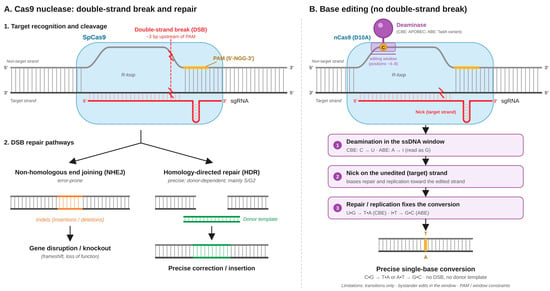
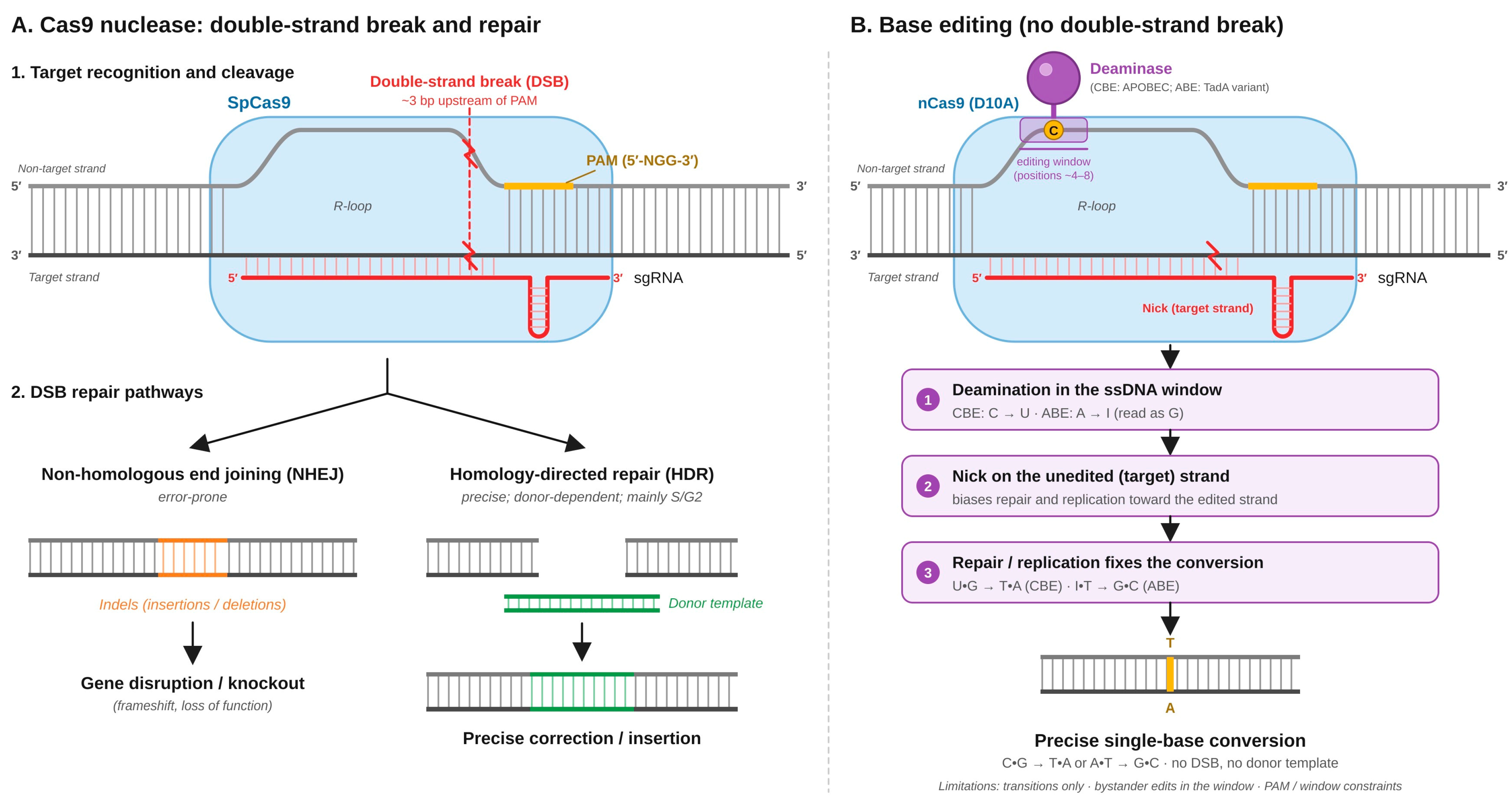
The CRISPR-Cas9 system utilizes a single guide RNA (sgRNA) to direct the Cas9 endonuclease toward a specific complementary DNA target sequence located next to a protospacer adjacent motif (PAM). Once bound, Cas9 introduces a targeted double-strand break (DSB), which in turn activates the cell’s endogenous DNA repair machinery. This repair proceeds primarily through one of two pathways: non-homologous end joining (NHEJ) or homology-directed repair (HDR). NHEJ is an error-prone process that typically generates small insertions or deletions (indels) at the cleavage site, resulting in gene disruption or knockout. HDR, on the other hand, is a highly precise mechanism that requires an exogenously supplied donor DNA template to seamlessly introduce specific sequence corrections. Furthermore, unlike traditional Cas9, base editors do not rely on generating DSBs. These tools consist of a catalytically impaired Cas9 (such as a Cas9 nickase) fused to a nucleotide deaminase. This configuration enables the direct and irreversible conversion of one target DNA base into another, for example, from C-G to T-A, without the need for double-strand breaks or donor templates. Consequently, this approach significantly reduces the risk of large unintended deletions or complex insertions, making base editors exceptionally valuable for the correction of point mutations [11,12] (Figure 1).
Figure 1.
Mechanistic pathways of CRISPR-Cas9 nuclease and base editing. Abbreviations: ABE, adenine base editor; CBE, cytosine base editor; DBS, double-strand break; HDR, homology-directed repair; NHEJ, non-homologus end joining; PAM, protospacer adjacent motif; sgRNA, single guide RNA; SpCas9, Streptococcus pyogenes Cas9.
Postnatal therapy has an inherent limitation for diseases that begin in development: many severe congenital disorders cause irreversible cellular and organ damage before or soon after birth, often before a diagnosis is made, and such damage may not be reversible by later treatment [13,14]. This has drawn attention to in utero gene therapy, which may offer four theoretical advantages. The small size of the fetus allows a high effective dose per kilogram of body weight. The fetal immune system is immature and may be more tolerant to vectors and edited cells. Proliferating stem and progenitor cells are abundant in the liver, haematopoietic system and brain, so edited cells may expand and contribute to tissue maintenance after birth. Finally, treatment could prevent disease rather than mitigate it [14,15].
Preclinical work supports the feasibility of this approach. In fetal mice, delivery of Cas9 or a base editor lowered plasma PCSK9 and cholesterol and rescued a lethal model of hereditary tyrosinaemia type 1 [16]. Intra-amniotic delivery improved survival in a model of monogenic lung disease [17], transplacental delivery of editing components produced mutations in embryonic cardiomyocytes [18], lipid nanoparticles delivered mRNA to the fetal brain [19], and nanoparticles delivered editing reagents in utero [20].
Prenatal editing also raises specific problems. Maternal antibodies against AAV cross the placenta and reduce fetal editing in mice, whereas immunity against Cas9 appears less important [21]. Off-target edits, mosaicism and unintended modification of primordial germ cells are particular concerns given the rapid cell division of the fetus [13]. Informed consent, maternal and fetal risk, and the societal implications of altering the human genome complicate clinical translation [22,23].
This narrative review summarises current preclinical research on in utero CRISPR-Cas9 gene therapy, with emphasis on delivery strategies, reported efficacy and safety outcomes, and discusses the translational, ethical and regulatory challenges that must be resolved before clinical use.
2. Preclinical Evidence and Current Challenges
2.1. Preclinical In Utero Genome Editing
Preclinical studies, mostly in rodents, show that in utero delivery of CRISPR-Cas9 components can produce targeted, stable edits in fetal tissues with measurable therapeutic benefit.
In fetal mice, viral-vector delivery of Cas9 or a base editor targeting Pcsk9 lowered plasma PCSK9 and cholesterol, and editing of Hpd rescued a lethal model of hereditary tyrosinaemia type 1. Edited cells persisted after birth [16]. Intra-amniotic delivery of CRISPR-Cas9 targeting mutant Sftpc^I73T in fetal mice edited about 20% of the lung epithelium, improved lung morphology and increased postnatal survival [17]. Transplacental delivery of genome-editing components induced mutations in embryonic cardiomyocytes of mid-gestational mouse fetuses, supporting feasibility for cardiac disorders [18]. Reviews conclude that proof-of-concept studies have shown the feasibility and safety of in utero editing across several organ systems and disease entities [15].
As a comparator to Cas9-based methods, non-nuclease platforms induce targeted gene correction without introducing targeted double-strand breaks. Instead, these systems rely on molecules such as peptide nucleic acids (PNAs), which bind strongly to the target genomic sequence to form a localized PNA-DNA triplex. The cell recognizes this unusual structural distortion as a DNA lesion, which recruits endogenous DNA repair machinery to initiate homologous recombination using a co-delivered single-stranded donor DNA template. Utilizing this mechanism, biodegradable poly(lactic-co-glycolic acid) (PLGA) nanoparticles carrying PNAs and donor DNA successfully corrected a β-globin mutation in a mouse model of β-thalassaemia, where a single prenatal dose resulted in a lasting improvement in anaemia [20]. AAV-mediated gene addition (human PQBP1 delivered to pregnant mice at embrionic day 10 (E10)) rescued microcephaly in a conditional Pqbp1 knockout, although behavioural recovery was incomplete. The authors named transduction efficiency, virulence, tumorigenesis and inflammation as future translational problems [24]. A review of this field describes adenoviral CRISPR/base-editing approaches, polymeric PNA nanoparticles and ionizable lipid nanoparticle (LNP)-mRNA, and reports preclinical efficacy alongside barriers to clinical translation [25].
Lipid nanoparticles have extended in utero delivery to the nervous system and muscle. Acid-degradable lipid nanoparticles (ADP-LNPs) carrying Cas9 mRNA and guide RNA were injected intracerebroventricularly into fetal mouse brains to target the Ube3a-ats region in a model of Angelman syndrome. This edited more than 40% of cortical neurons and about 60% of hippocampal neurons in selected experiments [19]. In a separate study, LNPs delivered mRNA in utero to the heart, diaphragm and skeletal muscle with low toxicity. That study demonstrated efficient mRNA transport and expression, not correction of a mutation or treatment of Duchenne muscular dystrophy [26]. Together, these data show that fetal haematopoietic and hepatic [16,20], neural [19] and, for mRNA, muscle tissues [26] are accessible, although therapeutic editing in muscle has not yet been shown.
Reviews of genome editing in post-implantation mouse fetuses describe mid-gestation delivery with viral vectors and intravenous administration [27]. They also describe microinjection, electroporation, Genome-editing via Oviductal Nucleic Acids Delivery (GONAD) and improved GONAD (i-GONAD), transplacental delivery and AAV vectors [28]. Broader reviews of in utero gene therapy cover small and large animal models, distinguish NHEJ from HDR in the prenatal setting, and call for further studies before clinical translation [29]. Overviews of fetal therapy [30] and of the opportunities and challenges of fetal gene therapy [31] describe the translational potential of treating severe congenital disease before birth. A summary of representative preclinical studies demonstrating these interventions is provided in Table 1.
Table 1.
Representative Preclinical Studies of In Utero Genome Editing.
2.2. Delivery Platforms
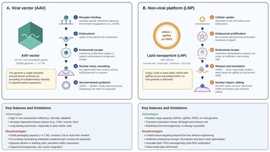
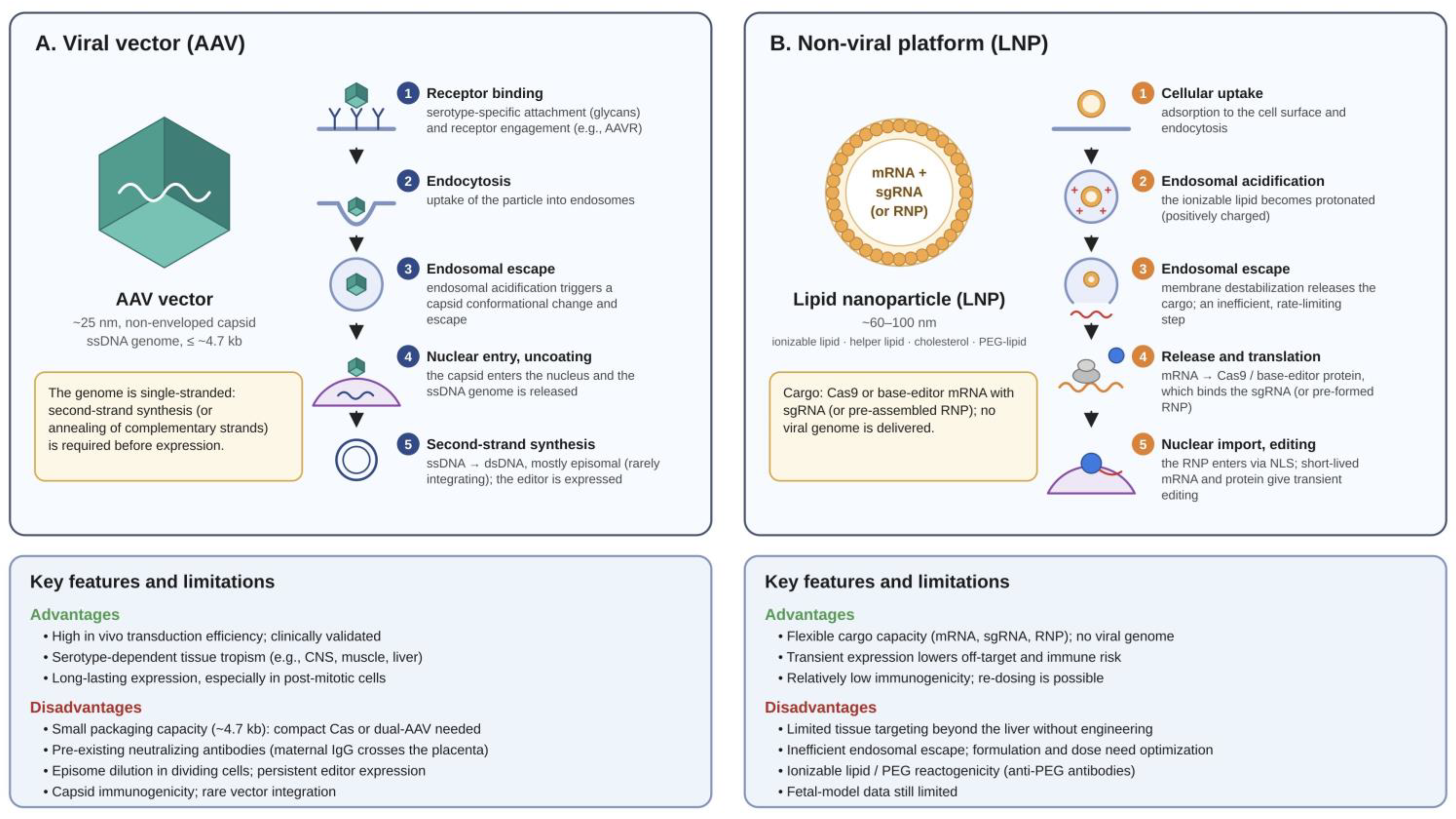
Targeted delivery of CRISPR-Cas9 components to specific fetal tissues remains the main technical problem. Delivery systems generally fall into two categories: viral and non-viral. Each operates through a fundamentally different mechanism. Viral vectors, including AAVs and adenoviruses, use natural infection pathways. They enter host cells and deliver their genetic material directly into the nucleus. Their main advantages are high transduction efficiency and well-defined tissue tropism. On the downside, viral vectors have a limited packaging capacity (~4.7 kb for AAV). This makes it difficult to deliver large editing systems like Streptococcus pyogenes Cas9 (SpCas9). They can also trigger unwanted maternal and fetal immune responses. Non-viral platforms, such as LNPs, take a different approach. They package mRNA or ribonucleoprotein (RNP) complexes inside a lipid shell. LNPs enter target cells through endocytosis and release their cargo into the cytoplasm. This type of delivery is transient. It limits the duration of nuclease activity, which helps lower the risk of off-target edits. LNPs also bypass the strict size limits of viral vectors. However, standard LNPs typically show lower tissue specificity and require careful formulation to prevent cellular toxicity (Figure 2).
Figure 2.
Delivery platforms for in utero gene therapy: viral vs. non-viral. Abbreviations: AAV, adeno-associated virus; AAVR, adeno-associated virus receptor; CNS, central nervous system; dsDNA, double-stranded DNA; IgG, immunoglobulin G; kb, kilobases; LNP, lipid nanoparticle; NLS, nuclear localization signal; PEG, polyethylene glycol; sgRNA, single guide RNA; ssDNA, single-stranded DNA.
Reviews of delivery technologies for in utero gene therapy discuss viral vectors and lipid nanoparticles. They highlight tissue access, vehicle toxicity and maternal–fetal immune responses as the main constraints, and treat choice of carrier as central to development [32]. CRISPR-Cas9 can be delivered as plasmid DNA, as sgRNA plus Cas9 mRNA, or as sgRNA plus Cas9 protein, through physical, viral or non-viral systems [33]. AAV and lentiviral vectors are the most widely used viral carriers [33,34].
AAV vectors have low immunogenicity, serotype specificity and a good safety profile, although mild toxicity has been reported at high doses in animal models. Lentiviral vectors offer high transduction efficiency and can be pseudotyped [33]. Adenoviral vectors give high editing efficiency but provoke stronger innate immune responses. AAV vectors have limited cargo capacity and may be blocked by pre-existing immunity [34,35]. Non-viral platforms such as LNPs offer flexible biodistribution and lower immune activation, but dosing, organ specificity and long-term safety need further optimisation [19,26,32] (Table 2).
Table 2.
Comparison of Delivery Platforms for Fetal Gene Therapy.
Standard LNPs have shown toxicity, which motivated the acid-degradable formulations [19].
2.3. Maternal–Fetal Immunity and Timing of Intervention
The fetal immune system is considered immature and potentially tolerogenic, which may favour in utero editing [13,14,15]. Maternal immunity can nevertheless influence the outcome. In mice, pre-existing maternal anti-AAV IgG crosses the placenta and reduces fetal editing in a titre-dependent manner, whereas maternal immunity against Cas9 appears less consequential. Antibody transfer is less efficient in mid-gestation than near term, which suggests a preferable window for prenatal intervention [21] (Table 3).
Table 3.
Immunological and Temporal Considerations in Prenatal Intervention.
These findings support maternal screening for vector-specific antibodies and careful choice of the timing of intervention.
Therapeutic benefit in preclinical studies has been judged by several kinds of outcome:
- −
- Biochemical markers and survival [16,17].
- −
- Organ morphology [17].
- −
- The proportion of edited neurons [19].
- −
- Anaemia [20].
- −
- Behaviour [24].
Long-term follow-up in some rodent studies shows persistence of edited cells and durable rescue [16], whereas comparable data from large animals remain limited [29]. The Introduction also cites the need for careful monitoring of off-target effects after prenatal editing [13].
2.4. Safety: Mosaicism, Off-Target and Large Genomic Alterations
Viral vectors carry risks of insertional mutagenesis, immunogenicity and unintended integration. Non-viral methods reduce immune activation but may be less efficient [34]. In utero studies with viral vectors show durable persistence of edited cells, but they also point to dose-dependent toxicity and the need to balance transduction efficiency against maternal–fetal safety [13,16,34]. Invasive delivery itself may cause embryotoxicity or developmental abnormalities, which favours precise, minimally invasive approaches [13,34].
Mosaicism arises when editing occurs after the first embryonic divisions and creates heterogeneous cell populations that may limit phenotypic correction. In bovine embryos, mosaicism and off-target mutations were evaluated after injecting Cas9 mRNA or protein, and high mosaicism was observed with both formats [36]. Reviews of editing in primate embryos likewise identify mosaicism and off-target mutations as limits on precision [37]. In human tripronuclear (3PN) zygotes targeting HBB, Cas9 cut the gene but HDR efficiency was low. The authors also found mosaicism, off-target editing and unwanted mutations caused by use of the homologous HBD gene as a repair template [38]. Introducing the CCR5-Δ32 variant into human 3PN embryos succeeded only partially, with indels and incomplete modification of both gene copies [39]. In early human embryos, frequent loss of heterozygosity near Cas9 cut sites was reported, which can mean loss of chromosome segments or structural changes that standard short-fragment analyses do not always detect [40]. A review of CRISPR-Cas9 in oocytes and early embryos summarises the same problems: off-target effects, mosaicism, chromosomal alterations and low efficiency of precise repair [41]. Stem-cell-based trophoblast models underline that aneuploidy and mosaicism can also affect placental development and pregnancy maintenance [42].
Off-target editing, even when minimal, remains a concern, especially for germline cells [43,44]. Proposed mitigation includes improved gRNA design, high-fidelity or modified nucleases, and better delivery methods [44,45]. Together, the embryo data argue for sequencing methods that detect large structural changes, and for comprehensive genomic assessment before any clinical use of fetal editing.
2.5. Postnatal and Clinical CRISPR-Based Benchmarks
Postnatal and ex vivo studies provide translational and safety benchmarks (Table 4).
Table 4.
Postnatal and Clinical CRISPR-Cas9 Benchmarks.
In the exagamglogene autotemcel update, ex vivo editing of haematopoietic stem cells for sickle cell disease and β-thalassaemia showed negligible off-target editing [4]. In vivo CRISPR-Cas9 knockout of hepatic transthyretin lowered serum hepatic transthyretin (TTR) in hereditary ATTR with polyneuropathy [5]. A patient-specific base-editing therapy was used in an infant with CPS1 deficiency, and after 7 weeks the patient tolerated an increased protein intake without major adverse effects [6]. Preclinical work shows that choice of nuclease strategy can limit off-target effects when correcting CYBA and CYBB in chronic granulomatous disease [7]. Correction of JAK3 in patient-derived stem cells restored lymphocyte development in a model of SCID [8]. After birth, AAV9-delivered CRISPR-Cas9 partially restored dystrophin in cardiac and skeletal muscle in a Duchenne mouse model [9]. In rabbit zygotes, homology-directed repair of the tyrosinase T373K variant restored melanin production [10]. These studies show the methodological and safety standards that prenatal editing will have to meet, but they act after birth, apart from the zygote-based rabbit model.
2.6. Ethical, Germline and Regulatory Context
Interventions on fetuses involve proxy consent by parents or the pregnant person, and they expose both mother and fetus to risk [22,23]. Fetal editing may unintentionally affect primordial germ cells, which distinguishes it from postnatal somatic therapy [13,22]. Animal studies so far have not shown a significant effect of prenatal gene therapy on fetal germ cells or maternal organs [12]. AAV, though the preferred carrier for neurodevelopmental targets, carries a risk of transducing germ cells [22].
The Society for Maternal-Fetal Medicine statement and other expert discussions propose that clinical application, if pursued, should be limited to severe, early-onset monogenic diseases that cause death or irreversible morbidity before or shortly after birth [13,46]. Such trials would need regulatory oversight, robust preclinical efficacy and safety data, and long-term follow-up [13,46]. Systematic analysis of the ethics of human embryo editing identifies risk, oversight, consent, justice and eugenics as the main concerns [23].
2.7. Biological Rationale for Fetal Editing
The Introduction lists four theoretical advantages: a high effective dose per kilogram, an immature and possibly tolerogenic immune system, abundant proliferating progenitor cells, and the chance to prevent rather than mitigate disease [13,14,15]. Preclinical work supports the last two points in particular. In the lung model, correction before birth improved lung architecture and survival [17]. Edited progenitors can expand with growing tissues [16]. The same features also increase safety, ethical and germline concerns.
2.8. Summary of Evidence
Preclinical studies provide proof of principle that in utero CRISPR-Cas9 and base editing can rescue lethal metabolic and lung disease in mice and produce durable, organ-specific editing [16,17], and that LNPs can reach the fetal brain and muscle [19,26]. Delivery, maternal–fetal immunity, timing, mosaicism and off-target effects remain the key challenges [13,21,36,37,38,39,40,41]. Large-animal data are limited [29], and the embryo studies show risks that conventional genotyping may miss [40]. Substantial work on precision, delivery and ethical frameworks is needed before any clinical use [46].
3. Discussion
The findings of this narrative review indicate that in utero genome editing using CRISPR-Cas9 and related technologies is a promising experimental approach for the treatment of severe congenital genetic disorders. The strongest evidence currently comes from preclinical studies demonstrating that prenatal delivery of genome-editing components can modify target sequences in fetal tissues and, in selected disease models, produce measurable therapeutic benefits. However, the available evidence does not yet establish the safety or clinical efficacy of prenatal genome editing in humans. The principal challenge is therefore no longer limited to demonstrating whether fetal genome editing is technically feasible but rather determining whether it can be performed with sufficient specificity, durability and safety to justify clinical application [13,14,15,29,31].
An important distinction emerging from the reviewed literature is that delivery of a therapeutic payload, successful genome editing and correction of a disease phenotype are separate outcomes. For example, the study by Nakamura et al. demonstrated that transplacental delivery could induce mutations in embryonic cardiomyocytes, supporting the feasibility of reaching fetal cardiac tissue [18]. However, evidence of target-site modification alone does not establish that a disease-causing variant has been corrected or that cardiac function has improved. Similarly, the delivery of mRNA to fetal heart, diaphragm and skeletal muscle using lipid nanoparticles demonstrates the accessibility of these tissues to a therapeutic payload but does not by itself demonstrate successful genome editing or treatment of muscular dystrophy [26]. These distinctions are essential when evaluating the maturity of different applications and identifying the next steps required for translation.
3.1. Delivery Platforms and Tissue-Specific Targeting
Selecting the optimal in utero gene therapy approach requires balancing editing efficiency, genomic safety, and delivery feasibility. Traditional CRISPR-Cas9 nucleases provide high efficiency for gene disruption (NHEJ) but pose significant risks of structural variants and off-target mutations [12]. HDR offers precise correction but suffers from low efficiency in post-mitotic tissues and requires co-delivery of a donor template. In contrast, base editors enable highly efficient and precise point mutation correction without double-strand breaks, significantly improving the safety profile [11,12]. However, their large size complicates viral delivery [34,35].
Regarding delivery systems, viral vectors (such as AAVs) achieve high transduction efficiency and sustained expression, which is crucial for systemic or neurodevelopmental targets [32]. Yet, their limited cargo capacity restricts the use of large editors, and they carry risks of insertional mutagenesis or neutralization by maternal antibodies [21,34]. Non-viral platforms, such as LNPs, bypass size constraints and offer transient expression. This transient nature enhances safety by minimizing off-target nuclease activity. Nevertheless, LNPs currently demonstrate lower targeting specificity and require further optimization to match the in vivo efficiency of AAVs [32,34].
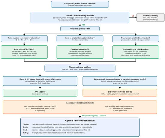
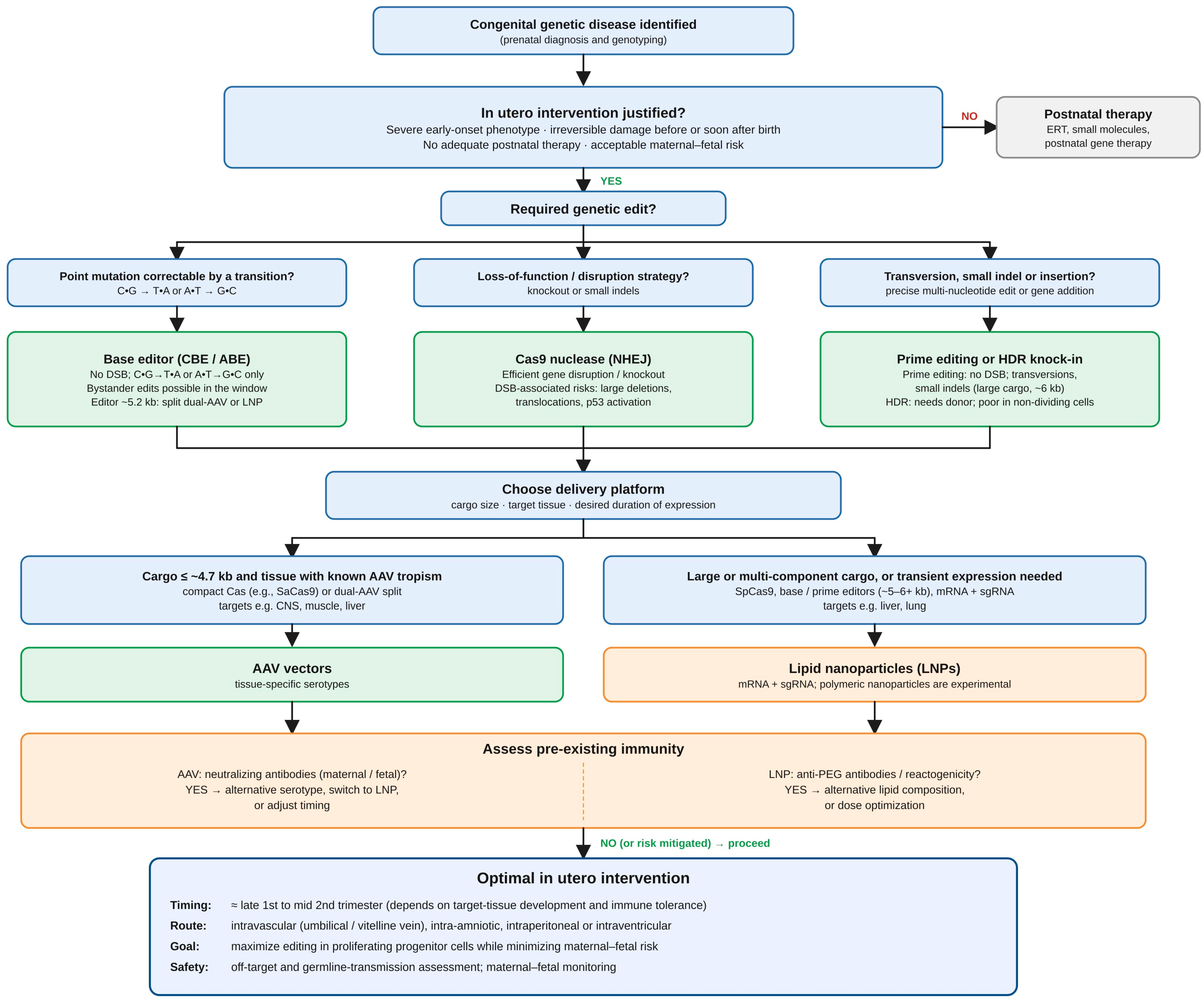
To navigate these trade-offs, we propose a decision-making scheme (Figure 3) for selecting the most appropriate strategy. The algorithm begins with the genetic disease mechanism (e.g., point mutation versus large deletion), determining the choice of genome-editing technology. Next, the target tissue and the physical size of the editing machinery dictate the optimal delivery system. Finally, maternal immune status and the fetal developmental stage guide the timing of intervention to maximize both efficacy and maternal–fetal safety [13,21].
Figure 3.
Decision-making scheme for in utero gene therapy strategy. Abbreviations: AAV, adeno-associated virus; ABE, adenine base editor; CBE, cytosine base editor; CNS, central nervous system; DBS, double-strand break; ERT, enzyme replacement therapy; HDR, homology-directed repair; kb, kilobases; LNP, lipid nanoparticle; NHEJ, non-homologous end joining; PEG, polyethylene glycol; SaCas9, Staphylococcus aureus Cas9; sgRNA, single guided RNA; SpCas9, Streptococcus pyogenes Cas9.
The choice of delivery platform remains one of the principal determinants of the feasibility and safety of in utero genome editing. Viral vectors, particularly AAVs, offer efficient transduction and tissue tropism, while adenoviral vectors provide greater cargo capacity. However, their use is constrained by immunogenicity, dose-dependent toxicity, pre-existing maternal antibodies and, in some circumstances, the possibility of unintended vector integration. AAVs also have limited packaging capacity, which can restrict the delivery of larger editing systems. These limitations are particularly relevant in prenatal applications, where exposure may affect both the fetus and the pregnant individual [32,33,34,35].
Non-viral delivery systems, including LNPs and biodegradable polymeric nanoparticles, offer an alternative approach. Their composition can be adjusted to influence biodistribution, cellular uptake and the release of therapeutic cargo. Acid-degradable LNPs carrying Cas9 mRNA and guide RNA have enabled substantial editing in selected regions of the fetal mouse brain, including cortical and hippocampal neurons [19]. In addition, nanoparticle-mediated delivery of peptide nucleic acids and donor DNA has demonstrated prenatal correction of a β-globin mutation in a mouse model of β-thalassaemia, accompanied by sustained improvement in anaemia [20]. Although these approaches differ mechanistically from conventional CRISPR-Cas9 nuclease editing, they illustrate the broader potential of non-viral prenatal genome-modification strategies.
Despite these advances, the advantages of non-viral delivery should not be interpreted as evidence that these platforms are universally safer or more effective than viral vectors. Delivery efficiency and tissue specificity depend on the formulation, route of administration, developmental stage and target organ. Furthermore, evidence that a carrier reaches a particular tissue does not necessarily demonstrate that sufficient editing occurs in the relevant cell population to produce a therapeutic effect. Future studies should therefore evaluate delivery systems using comparable endpoints, including the proportion of relevant cells edited, the distribution of editing across organs, the duration of editor activity, functional outcomes and maternal and fetal toxicity [19,26,32].
The development of transient delivery systems is particularly important because permanent genomic modifications may result from even a brief exposure to an editing reagent. Delivering Cas9 as mRNA or ribonucleoprotein complexes may limit the duration of editor activity compared with sustained expression from some viral vectors. Nevertheless, transient exposure does not eliminate the risk of off-target editing or unintended structural changes. The optimal platform should consequently be selected according to the target disease, the required degree of correction, the accessibility of the affected tissue and the potential consequences of unintended editing [34,44,45].
3.2. Developmental Timing and Maternal–Fetal Immunity
The prenatal period offers a potentially valuable therapeutic window because intervention may precede the development of irreversible organ damage. In addition, the relatively small fetal size may facilitate systemic exposure at a lower absolute dose, while proliferating stem and progenitor cells may provide opportunities for the expansion of edited cell populations during subsequent development. These considerations form the biological rationale for in utero treatment of severe congenital disorders, particularly those in which pathology begins before birth or progresses rapidly during early infancy [13,14,15].
However, the advantages of early intervention must be balanced against developmental and safety considerations. Editing during early embryonic development may affect a larger proportion of descendant cells, but it may also increase the consequences of unintended modifications and create a risk of mosaicism if editing is incomplete or occurs after cell divisions have begun. Later fetal intervention may permit more targeted delivery to differentiated tissues, but it may occur after some pathological changes have already developed. Therefore, an optimal treatment window cannot be assumed to be the same for every disease or delivery platform. It must be established experimentally by considering disease progression, tissue development, editing kinetics and potential maternal and fetal complications [13,27,28,29,36].
Maternal immunity introduces an additional constraint, particularly for viral delivery systems. Riley et al. demonstrated in mice that pre-existing maternal antibodies against AAV could cross the placenta and reduce the efficiency of fetal genome editing, whereas maternal immunity against Cas9 had a less pronounced effect in the investigated setting [21]. This finding challenges the assumption that the fetal immune environment necessarily protects prenatal interventions from immune-related barriers. Although the fetal immune system may be relatively immature and can exhibit tolerogenic responses under certain conditions, maternal antibodies and other immune mechanisms remain relevant to treatment outcomes [13,14,15,21].
These observations suggest that maternal immune status, vector selection and gestational timing should be incorporated into preclinical study design. Nevertheless, the results obtained in mice cannot directly establish the optimal timing of treatment in humans because placental antibody transfer and fetal immune development differ between species. Before clinical translation, further research will be needed to determine how maternal immunity influences biodistribution, editing efficiency, inflammatory responses and the safety of both the pregnant individual and the fetus.
3.3. Genomic Safety: Off-Target Effects, Mosaicism and Structural Alterations
Genomic safety remains a central obstacle to prenatal genome editing. CRISPR-Cas9 can produce unintended modifications at sequences other than the intended target, while the repair of DNA double-strand breaks may generate insertions, deletions or larger genomic alterations. Such events are of particular concern in fetal development because an unintended modification may be propagated to multiple descendant cells and potentially affect tissue development or function [13,40,43,44].
Studies involving animal and human embryos provide important safety information, although their findings should not be treated as direct estimates of the risk associated with therapeutic editing of an established fetus. Research in bovine embryos has identified mosaicism and evaluated off-target mutations following the delivery of Cas9 mRNA or protein [36]. Studies of human tripronuclear zygotes have also reported incomplete editing, mosaicism and unintended sequence changes associated with the use of homologous genomic sequences as repair templates [38,39]. These findings illustrate the biological complexity of genome editing during early development and the limitations of assuming that cleavage at the intended target will necessarily produce a precise and uniform correction.
Of particular concern, Alanis-Lobato et al. reported frequent loss of heterozygosity near Cas9 cleavage sites in edited early human embryos [40]. This observation highlights the possibility that short-range genotyping may fail to detect all relevant genomic consequences of editing. Consequently, an apparently successful edit based on routine sequencing of a small target region may not exclude larger deletions, chromosomal rearrangements or loss of genetic information. Comprehensive safety assessment should therefore combine targeted sequencing with methods capable of detecting larger structural variants and chromosomal abnormalities, as appropriate to the editing strategy and biological model [40,41,44].
Mosaicism also complicates the interpretation of therapeutic efficacy. A measurable average editing rate does not necessarily indicate that the required proportion of disease-relevant cells has been corrected. Different tissues may contain different proportions of edited and unedited cells, and the relationship between editing frequency and clinical benefit may vary by disease. For this reason, future studies should report not only overall editing efficiency but also its distribution across cell populations and organs, the frequency and nature of unintended changes, and the relationship between these findings and functional outcomes [13,36,41].
Several strategies have been proposed to improve genomic safety, including optimized guide RNA design, high-fidelity Cas9 variants, alternative editing systems and improved delivery methods that limit unnecessary exposure to editing components [43,44,45]. Base editing may avoid the double-strand breaks associated with conventional Cas9 editing in suitable applications, but it should not be considered free of unintended genomic or bystander changes. No single strategy eliminates all potential risks. The appropriate approach will depend on the target sequence, the type of genetic defect and the balance between therapeutic benefit and the consequences of unintended modification.
3.4. Germline Exposure and Ethical Considerations
The possibility of unintended germline modification distinguishes prenatal genome editing from many established forms of postnatal somatic gene therapy. If primordial germ cells are exposed to editing components, genomic changes could potentially be transmitted to future generations. Although the extent of this risk will depend on the delivery platform, target tissue and developmental stage, it cannot be dismissed solely because the intended therapeutic target is a somatic organ [13,22,23].
This concern must be considered alongside the potential benefits of treating severe congenital disease before irreversible damage occurs. Prenatal intervention involves decisions made on behalf of a fetus that cannot provide consent while the pregnant individual may also be exposed to procedural, immunological or other treatment-related risks. Ethical assessment must therefore consider the severity and natural history of the disease, the availability of alternative treatments, the likelihood of meaningful benefit, the uncertainty of long-term outcomes and the proportionality of risks to both the fetus and the pregnant individual [22,23].
The current evidence supports a cautious distinction between therapeutic fetal intervention and heritable genome editing undertaken for reproductive or enhancement purposes. Discussions of clinical translation have proposed restricting initial consideration to severe, early-onset monogenic disorders associated with death or substantial irreversible morbidity before or shortly after birth, particularly where effective alternatives are unavailable [13,46]. Any such approach would require rigorous preclinical evidence, independent ethical and regulatory review, appropriate consent procedures and long-term follow-up.
Regulatory oversight should also address the possibility of delayed adverse effects, including developmental abnormalities, organ dysfunction and consequences that become apparent only after birth. Long-term monitoring would be necessary to evaluate durability of therapeutic benefit and identify adverse outcomes that may not be evident during pregnancy or the neonatal period. Because potential germline exposure raises questions extending beyond the treated individual, governance frameworks should also define how such risks are assessed and communicated. Ethical and regulatory safeguards must develop alongside the technology rather than being considered only after technical feasibility has been established [22,23,46].
3.5. Translational Relevance and Limitations of the Evidence
The progress of postnatal and ex vivo genome-editing therapies provides useful benchmarks for prenatal research. Clinical studies of BCL11A enhancer editing have demonstrated the therapeutic potential of CRISPR-modified haematopoietic stem cells in sickle cell disease and β-thalassaemia [4]. In vivo editing of hepatic transthyretin has also shown that targeted genomic intervention can reduce circulating disease-associated protein in patients with hereditary transthyretin amyloidosis [5]. Furthermore, the use of individualized in vivo base editing in an infant with carbamoyl-phosphate synthetase 1 deficiency illustrates the growing capacity to develop highly tailored interventions for rare genetic disorders [6]. These developments establish that genome editing can achieve clinically relevant outcomes in selected postnatal settings, but they do not demonstrate that fetal genome editing is ready for clinical use.
Prenatal interventions introduce additional variables, including placental transport, maternal immune responses, developmental changes in tissue accessibility and the possibility of affecting germline cells. Moreover, the consequences of an unintended genomic modification may differ when it occurs in a developing organism rather than in a more restricted population of postnatal somatic cells. Clinical success in postnatal settings should therefore inform the design of prenatal studies without being interpreted as direct evidence of prenatal safety [13,21,22,46].
Several limitations of the evidence reviewed here must also be acknowledged. First, the literature is heterogeneous in terms of animal models, developmental stages, delivery routes, editing systems and outcome measures. This prevents direct quantitative comparison of editing efficiency or therapeutic benefit across studies and limits the conclusions that can be drawn about the relative superiority of different platforms. Second, most evidence for therapeutic in utero genome editing comes from animal studies, particularly rodent models, while data from large-animal models remain limited [29,31]. Third, some studies demonstrate delivery or target-site editing without establishing correction of a disease phenotype, and these outcomes should not be regarded as equivalent to therapeutic efficacy.
The present review also has methodological limitations. It was conducted as a narrative review rather than a systematic review, and no formal risk-of-bias assessment was performed. Although the literature search included multiple databases and supplementary sources, the selection process and the heterogeneity of the available evidence may have influenced the studies included and the emphasis placed on particular findings. The conclusions should therefore be understood as a qualitative synthesis of current research rather than a quantitative estimate of treatment effectiveness or safety.
Future research should prioritize reproducible comparisons of delivery platforms, comprehensive genomic safety testing, evaluation of maternal and fetal outcomes, and long-term follow-up extending beyond birth. Studies in larger and more clinically relevant animal models will be important for assessing tissue distribution, dose requirements, immune interactions and procedural risks. Standardized reporting of editing efficiency, functional benefit, off-target effects, structural genomic changes and potential germline involvement would also improve the comparability of results between research groups [29,32,40,44].
Overall, the evidence supports the biological feasibility of prenatal genome editing and demonstrates therapeutic benefit in selected preclinical models. However, the transition from proof of concept to clinical application will require a substantially stronger evidence base, particularly regarding long-term safety, tissue specificity and the prevention of unintended genomic alterations. In utero CRISPR-Cas9 therapy should therefore be regarded as a promising but experimental strategy whose future clinical role will depend on whether its potential benefits can be demonstrated to outweigh its distinctive developmental, immunological and ethical risks.
4. Conclusions
In utero genome editing using CRISPR-Cas9 and related technologies offers a potential strategy for treating severe congenital genetic disorders before irreversible pathological changes develop. Preclinical studies have demonstrated targeted genome modification and therapeutic benefits in selected animal models, including improvements in metabolic parameters, lung morphology and survival. Additional studies have shown that viral and non-viral delivery systems can reach a range of fetal tissues, including the liver, lung, heart and brain, although successful delivery does not always translate into therapeutic genome correction.
Despite these advances, prenatal genome editing remains experimental. The principal barriers to clinical translation include tissue-specific delivery, maternal–fetal immune interactions, mosaicism, off-target editing, large genomic alterations and the possibility of unintended germline modification. The limited availability of long-term data and studies in large-animal models further restricts conclusions about safety and clinical applicability.
Future research should focus on improving delivery specificity, minimizing unintended genomic changes, establishing standardized safety assessments and demonstrating durable functional benefit in clinically relevant models. Ethical and regulatory frameworks must also ensure that maternal and fetal risks are proportionate to the expected benefit, that parental consent is appropriately informed, and that long-term outcomes are monitored.
In conclusion, in utero CRISPR-Cas9 genome editing has the potential to expand the treatment options for selected severe congenital disorders by enabling intervention before or during the earliest stages of disease development. However, its promise must be balanced against the particular risks of modifying the genome of a developing organism. Clinical application should be considered only when robust evidence demonstrates sufficient therapeutic benefit, reliable genomic precision and acceptable maternal and fetal safety.
Author Contributions
Conceptualization, W.O., A.K.P., K.G. and Ż.K.-T.; methodology, W.O.; validation, W.O., A.K.P. and K.G.; data curation, W.O., A.K.P. and K.G.; writing—original draft preparation, W.O., A.K.P. and K.G.; writing—review and editing, W.O. and Ż.K.-T.; supervision, Ż.K.-T. All authors have read and agreed to the published version of the manuscript.
Funding
This research received no external funding.
Institutional Review Board Statement
Not applicable.
Informed Consent Statement
Not applicable.
Data Availability Statement
No new data were created or analyzed in this study. Data sharing is not applicable to this article.
Conflicts of Interest
The authors declare no conflicts of interest.
Abbreviations
The following abbreviations are used in this manuscript:
| AAV | Adeno-associated virus |
| AAV2 | Adeno-associated virus serotype 2 |
| AAV9 | Adeno-associated virus serotype 9 |
| ADP-LNPs | Acid-degradable lipid nanoparticles |
| ATTR | Transthyretin amyloidosis |
| CAR-T | Chimeric antigen receptor T cells |
| Cas9 | CRISPR-associated protein 9 |
| CRISPR | Clustered regularly interspaced short palindromic repeats |
| HDR | Homology-directed repair |
| HT1 | Hereditary tyrosinemia type 1 |
| SCID | Severe combined immunodeficiency |
| TTR | Transthyretin |
References
- Russell, S.; Bennett, J.; Wellman, J.A.; Chung, D.C.; Yu, Z.F.; Tillman, A.; Wittes, J.; Pappas, J.; Elci, O.; McCague, S.; et al. Efficacy and safety of voretigene neparvovec (AAV2-hRPE65v2) in patients with RPE65-mediated inherited retinal dystrophy: A randomised, controlled, open-label, phase 3 trial. Lancet 2017, 390, 849–860, Correction in Lancet 2017, 390, 848. [Google Scholar] [CrossRef] [Scilit] [PubMed]
- Mendell, J.R.; Al-Zaidy, S.; Shell, R.; Arnold, W.D.; Rodino-Klapac, L.R.; Prior, T.W.; Lowes, L.; Alfano, L.; Berry, K.; Church, K.; et al. Single-dose gene-replacement therapy for spinal muscular atrophy. N. Engl. J. Med. 2017, 377, 1713–1722. [Google Scholar] [CrossRef] [Scilit] [PubMed]
- Maude, S.L.; Laetsch, T.W.; Buechner, J.; Rives, S.; Boyer, M.; Bittencourt, H.; Bader, P.; Verneris, M.R.; Stefanski, H.E.; Myers, G.D.; et al. Tisagenlecleucel in children and young adults with B-cell lymphoblastic leukemia. N. Engl. J. Med. 2018, 378, 439–448. [Google Scholar] [CrossRef] [Scilit] [PubMed]
- Yen, A.; Boitano, T.; Zappala, Z.; Mahajan, S.; Willems, T.; Majarian, T.D.; Thunga, P.; Altshuler, D. Specificity of CRISPR-Cas9 Editing in Exagamglogene Autotemcel—Update. N. Engl. J. Med. 2025, 393, 1440–1442. [Google Scholar] [CrossRef] [Scilit] [PubMed]
- Gillmore, J.D.; Gane, E.; Täubel, J.; Pilebro, B.; Echaniz-Laguna, A.; Kao, J.; Litchy, W.; Shahda, S.; Haagensen, A.; Walsh, L.; et al. Nexiguran Ziclumeran Gene Editing in Hereditary ATTR with Polyneuropathy. N. Engl. J. Med. 2025, 393, 1375–1386. [Google Scholar] [CrossRef] [Scilit] [PubMed]
- Musunuru, K.; Grandinette, S.A.; Wang, X.; Hudson, T.R.; Briseno, K.; Berry, A.M.; Hacker, J.L.; Hsu, A.; Silverstein, R.A.; Hille, L.T.; et al. Patient-Specific In Vivo Gene Editing to Treat a Rare Genetic Disease. N. Engl. J. Med. 2025, 392, 2235–2243. [Google Scholar] [CrossRef] [Scilit] [PubMed]
- Wolff, J.H.; Skov, T.W.; Haslund, D.; Dorset, S.R.; Revenfeld, A.L.S.; Aussel, C.; Jørgensen, S.E.; Holm, M.; Thomsen, M.K.; Ammann, S.; et al. Targeted gene editing and near-universal cDNA insertion of CYBA and CYBB as a treatment for chronic granulomatous disease. Nat. Commun. 2025, 16, 7475. [Google Scholar] [CrossRef] [Scilit] [PubMed]
- Chang, C.W.; Lai, Y.S.; Westin, E.; Khodadadi-Jamayran, A.; Pawlik, K.M.; Lamb, L.S.; Goldman, F.D.; Townes, T.M. Modeling Human Severe Combined Immunodeficiency and Correction by CRISPR/Cas9-Enhanced Gene Targeting. Cell Rep. 2015, 12, 1668–1677. [Google Scholar] [CrossRef] [Scilit] [PubMed]
- Long, C.; Amoasii, L.; Mireault, A.A.; McAnally, J.R.; Li, H.; Sanchez-Ortiz, E.; Bhattacharyya, S.; Shelton, J.M.; Bassel-Duby, R.; Olson, E.N. Postnatal genome editing partially restores dystrophin expression in a mouse model of muscular dystrophy. Science 2016, 351, 400–403. [Google Scholar] [CrossRef] [Scilit] [PubMed]
- Song, Y.; Zhang, Y.; Chen, M.; Deng, J.; Sui, T.; Lai, L.; Li, Z. Functional validation of the albinism-associated tyrosinase T373K SNP by CRISPR/Cas9-mediated homology-directed repair (HDR) in rabbits. EBioMedicine 2018, 36, 517–525. [Google Scholar] [CrossRef] [Scilit] [PubMed]
- Jinek, M.; Chylinski, K.; Fonfara, I.; Hauer, M.; Doudna, J.A.; Charpentier, E. A programmable dual-RNA-guided DNA endonuclease in adaptive bacterial immunity. Science 2012, 337, 816–821. [Google Scholar] [CrossRef] [Scilit] [PubMed]
- Peranteau, W.H.; Flake, A.W. The Future of In Utero Gene Therapy. Mol. Diagn. Ther. 2020, 24, 135–142. [Google Scholar] [CrossRef] [Scilit] [PubMed]
- Yung, N.K.; Maassel, N.L.; Ullrich, S.J.; Ricciardi, A.S.; Stitelman, D.H. A narrative review of in utero gene therapy:Advances, challenges, and future considerations. Transl. Pediatr. 2021, 10, 1486–1496. [Google Scholar] [CrossRef] [Scilit] [PubMed]
- Schwab, M.E.; MacKenzie, T.C. Prenatal Gene Therapy. Clin. Obstet. Gynecol. 2021, 64, 876–885. [Google Scholar] [CrossRef] [Scilit] [PubMed]
- Bose, S.K.; Kennedy, K.; Peranteau, W.H. Foetal genome editing. Curr. Opin. Obstet. Gynecol. 2023, 35, 134–139. [Google Scholar] [CrossRef] [Scilit] [PubMed]
- Rossidis, A.C.; Stratigis, J.D.; Chadwick, A.C.; Hartman, H.A.; Ahn, N.J.; Li, H.; Singh, K.; Coons, B.E.; Li, L.; Lv, W.; et al. In utero CRISPR-mediated therapeutic editing of metabolic genes. Nat. Med. 2018, 24, 1513–1518. [Google Scholar] [CrossRef] [Scilit] [PubMed]
- Alapati, D.; Zacharias, W.J.; Hartman, H.A.; Rossidis, A.C.; Stratigis, J.D.; Ahn, N.J.; Coons, B.; Zhou, S.; Li, H.; Singh, K.; et al. In utero gene editing for monogenic lung disease. Sci. Transl. Med. 2019, 11, eaav8375. [Google Scholar] [CrossRef] [Scilit] [PubMed]
- Nakamura, S.; Ishihara, M.; Ando, N.; Watanabe, S.; Sakurai, T.; Sato, M. Transplacental delivery of genome editing components causes mutations in embryonic cardiomyocytes of mid-gestational murine fetuses. IUBMB Life 2019, 71, 835–844. [Google Scholar] [CrossRef] [Scilit] [PubMed]
- Gao, K.; Han, H.; Cranick, M.G.; Zhao, S.; Xu, S.; Yin, B.; Song, H.; Hu, Y.; Clarke, M.T.; Wang, D.; et al. Widespread Gene Editing in the Brain via In Utero Delivery of mRNA Using Acid-Degradable Lipid Nanoparticles. ACS Nano 2024, 18, 30293–30306. [Google Scholar] [CrossRef] [Scilit] [PubMed]
- Ricciardi, A.S.; Bahal, R.; Farrelly, J.S.; Quijano, E.; Bianchi, A.H.; Luks, V.L.; Putman, R.; López-Giráldez, F.; Coşkun, S.; Song, E.; et al. In utero nanoparticle delivery for site-specific genome editing. Nat. Commun. 2018, 9, 2481. [Google Scholar] [CrossRef] [Scilit] [PubMed]
- Riley, J.S.; Luks, V.L.; Berkowitz, C.L.; Dumitru, A.M.; Kus, N.J.; Dave, A.; Menon, P.; De Paepe, M.E.; Jain, R.; Li, L.; et al. Preexisting maternal immunity to AAV but not Cas9 impairs in utero gene editing in mice. J. Clin. Investig. 2024, 134, e179848. [Google Scholar] [CrossRef] [Scilit] [PubMed]
- Major, R.M.; Juengst, E.T. Prenatal gene editing for neurodevelopmental diseases: Ethical considerations. Am. J. Hum. Genet. 2025, 112, 201–214. [Google Scholar] [CrossRef] [Scilit] [PubMed]
- Wiley, L.; Cheek, M.; LaFar, E.; Ma, X.; Sekowski, J.; Tanguturi, N.; Iltis, A. The Ethics of Human Embryo Editing via CRISPR-Cas9 Technology: A Systematic Review of Ethical Arguments, Reasons, and Concerns. HEC Forum 2025, 37, 267–303. [Google Scholar] [CrossRef] [Scilit] [PubMed]
- Ito, H.; Shiwaku, H.; Yoshida, C.; Homma, H.; Luo, H.; Chen, X.; Fujita, K.; Musante, L.; Fischer, U.; Frints, S.G.; et al. In utero gene therapy rescues microcephaly caused by Pqbp1-hypofunction in neural stem progenitor cells. Mol. Psychiatry 2015, 20, 459–471. [Google Scholar] [CrossRef] [Scilit] [PubMed]
- Bose, S.K.; Menon, P.; Peranteau, W.H. In Utero Gene Therapy: Progress and Challenges. Trends Mol. Med. 2021, 27, 728–730. [Google Scholar] [CrossRef] [Scilit] [PubMed]
- Gao, K.; Li, J.; Song, H.; Han, H.; Wang, Y.; Yin, B.; Farmer, D.L.; Murthy, N.; Wang, A. In utero delivery of mRNA to the heart, diaphragm and muscle with lipid nanoparticles. Bioact. Mater. 2023, 25, 387–398. [Google Scholar] [CrossRef] [Scilit] [PubMed]
- Nakamura, S.; Inada, E.; Saitoh, I.; Sato, M. Recent Genome-Editing Approaches toward Post-Implanted Fetuses in Mice. BioTech 2023, 12, 37. [Google Scholar] [CrossRef] [Scilit] [PubMed]
- Sato, M.; Takabayashi, S.; Akasaka, E.; Nakamura, S. Recent Advances and Future Perspectives of In Vivo Targeted Delivery of Genome-Editing Reagents to Germ cells, Embryos, and Fetuses in Mice. Cells 2020, 9, 799. [Google Scholar] [CrossRef] [Scilit] [PubMed]
- Hartman, H.A.; Rossidis, A.C.; Peranteau, W.H. In Utero Gene Therapy and Genome Editing. Curr. Stem Cell Rep. 2018, 4, 52–60. [Google Scholar] [CrossRef] [Scilit]
- Shear, M.A.; Massa, A. In Utero Fetal Therapy: Stem Cells, Cell Transplantation, Gene Therapy, and CRISPR-Cas9. Clin. Obstet. Gynecol. 2021, 64, 861–875. [Google Scholar] [CrossRef] [Scilit] [PubMed]
- Jeanne, M.; Chung, W.K. Opportunities and Challenges of Fetal Gene Therapy. Prenat. Diagn. 2025, 45, 764–771. [Google Scholar] [CrossRef] [Scilit] [PubMed]
- Palanki, R.; Peranteau, W.H.; Mitchell, M.J. Delivery technologies for in utero gene therapy. Adv. Drug Deliv. Rev. 2021, 169, 51–62. [Google Scholar] [CrossRef] [Scilit] [PubMed]
- Sharma, G.; Sharma, A.R.; Bhattacharya, M.; Lee, S.S.; Chakraborty, C. CRISPR-Cas9: A Preclinical and Clinical Perspective for the Treatment of Human Diseases. Mol. Ther. 2021, 29, 571–586. [Google Scholar] [CrossRef] [Scilit] [PubMed]
- Liu, C.; Zhang, L.; Liu, H.; Cheng, K. Delivery strategies of the CRISPR-Cas9 gene-editing system for therapeutic applications. J. Control. Release 2017, 266, 17–26. [Google Scholar] [CrossRef] [Scilit] [PubMed]
- Uddin, F.; Rudin, C.M.; Sen, T. CRISPR Gene Therapy: Applications, Limitations, and Implications for the Future. Front. Oncol. 2020, 10, 1387. [Google Scholar] [CrossRef] [Scilit] [PubMed]
- Hennig, S.L.; Owen, J.R.; Lin, J.C.; Young, A.E.; Ross, P.J.; Van Eenennaam, A.L.; Murray, J.D. Evaluation of mutation rates, mosaicism and off target mutations when injecting Cas9 mRNA or protein for genome editing of bovine embryos. Sci. Rep. 2020, 10, 22309. [Google Scholar] [CrossRef] [Scilit] [PubMed]
- Guo, X.; Li, X.J. Targeted genome editing in primate embryos. Cell Res. 2015, 25, 767–768. [Google Scholar] [CrossRef] [Scilit] [PubMed]
- Liang, P.; Xu, Y.; Zhang, X.; Ding, C.; Huang, R.; Zhang, Z.; Lv, J.; Xie, X.; Chen, Y.; Li, Y.; et al. CRISPR/Cas9-mediated gene editing in human tripronuclear zygotes. Protein Cell 2015, 6, 363–372. [Google Scholar] [CrossRef] [Scilit] [PubMed]
- Kang, X.; He, W.; Huang, Y.; Yu, Q.; Chen, Y.; Gao, X.; Sun, X.; Fan, Y. Introducing precise genetic modifications into human 3PN embryos by CRISPR/Cas-mediated genome editing. J. Assist. Reprod. Genet. 2016, 33, 581–588, Correction in J. Assist. Reprod. Genet. 2017, 34, 963. [Google Scholar] [CrossRef] [Scilit] [PubMed]
- Alanis-Lobato, G.; Zohren, J.; McCarthy, A.; Fogarty, N.M.E.; Kubikova, N.; Hardman, E.; Greco, M.; Wells, D.; Turner, J.M.A.; Niakan, K.K. Frequent loss of heterozygosity in CRISPR-Cas9-edited early human embryos. Proc. Natl. Acad. Sci. USA 2021, 118, e2004832117. [Google Scholar] [CrossRef] [Scilit] [PubMed]
- Zhang, Y.R.; Yin, T.L.; Zhou, L.Q. CRISPR/Cas9 technology: Applications in oocytes and early embryos. J. Transl. Med. 2023, 21, 746. [Google Scholar] [CrossRef] [Scilit] [PubMed]
- Nikitina, T.V.; Lebedev, I.N. Stem Cell-Based Trophoblast Models to Unravel the Genetic Causes of Human Miscarriages. Cells 2022, 11, 1923. [Google Scholar] [CrossRef] [Scilit] [PubMed]
- Guo, C.; Ma, X.; Gao, F.; Guo, Y. Off-target effects in CRISPR/Cas9 gene editing. Front. Bioeng. Biotechnol. 2023, 11, 1143157. [Google Scholar] [CrossRef] [Scilit] [PubMed]
- Kalter, N.; Fuster-García, C.; Silva, A.; Ronco-Díaz, V.; Roncelli, S.; Turchiano, G.; Gorodkin, J.; Cathomen, T.; Benabdellah, K.; Lee, C.; et al. Off-target effects in CRISPR-Cas genome editing for human therapeutics:Progress and challenges. Mol. Ther. Nucleic Acids 2025, 36, 102636. [Google Scholar] [CrossRef] [Scilit] [PubMed]
- Han, H.A.; Pang, J.K.S.; Soh, B.S. Mitigating off-target effects in CRISPR/Cas9-mediated in vivo gene editing. J. Mol. Med. 2020, 98, 615–632. [Google Scholar] [CrossRef] [Scilit] [PubMed]
- Shanahan, M.A.; Aagaard, K.M.; McCullough, L.B.; Chervenak, F.A.; Shamshirsaz, A.A. Society for Maternal-Fetal Medicine Special Statement: Beyond the scalpel: In utero fetal gene therapy and curative medicine. Am. J. Obstet. Gynecol. 2021, 225, B9–B18. [Google Scholar] [CrossRef] [Scilit] [PubMed]

Disclaimer/Publisher’s Note: The statements, opinions and data contained in all publications are solely those of the individual author(s) and contributor(s) and not of MDPI and/or the editor(s). MDPI and/or the editor(s) disclaim responsibility for any injury to people or property resulting from any ideas, methods, instructions or products referred to in the content. |
© 2026 by the authors. Licensee MDPI, Basel, Switzerland. This article is an open access article distributed under the terms and conditions of the Creative Commons Attribution (CC BY) license.