


Abstract
Decompression sickness (DCS) is an occupational health problem fundamentally driven by the formation of gas bubbles in the human body, which could be a threat to the life of divers, astronauts and hyperbaric workers. The phenomenon of bubble formation in organisms after decompression provided the earliest experimental evidence for decompression theory. Since then, researchers across different eras have conducted extensive work on biological decompression bubbles, which brought about the establishment of gas nuclei theory. While Yount’s quantitatively calculable Varying Permeability Model (VPM) has been successfully applied to decompression algorithms, its core assumption—the “surface-active skin”—remains experimentally unverified. But recent developments in research on nanobubbles and the Active Hydrophobic Spots (AHS) hypothesis have provided new physicochemical perspectives. This review critically evaluates the theoretical evolution and experimental progress in this field, integrating recent advances in molecular dynamics and interfacial physics. To contribute to shedding light on the unresolved paradox of biological decompression bubbles in vivo, we highlight the significance of the paradigm shift from macroscopic gas dissolution to interfacial mechanics, which may help identify the missing links in current DCS research and outline a new multidisciplinary path forward.
1. Introduction
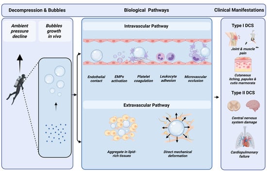
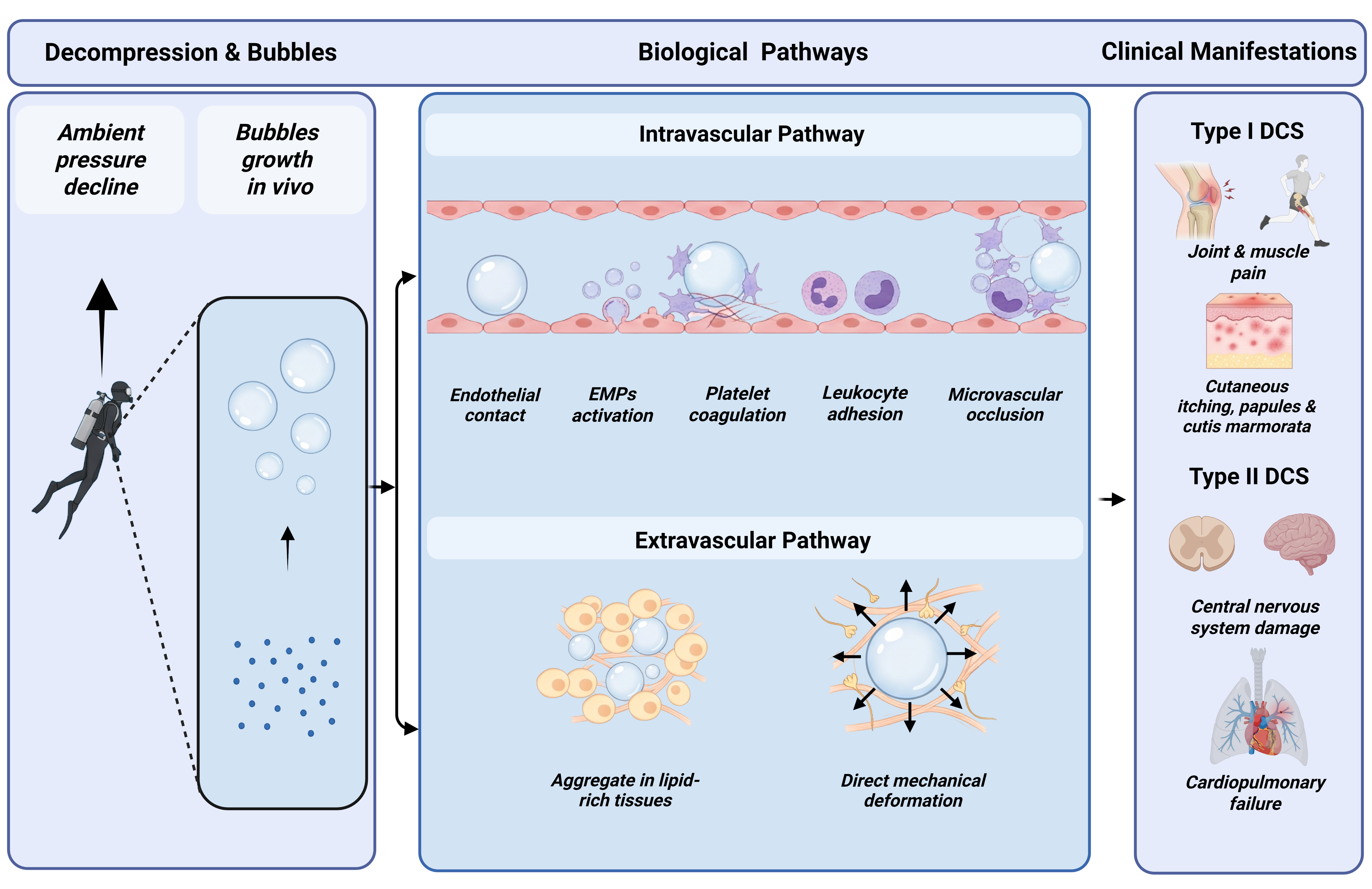
Decompression sickness (DCS) is a condition in which, after exposure to a high-pressure environment, ambient pressure drops too rapidly, causing dissolved gases in blood and tissues to precipitate as bubbles, leading to circulatory impairment and tissue damage. It affects special occupational groups such as underwater divers, astronauts during extravehicular activities, and workers in high-pressure shield tunneling, with diving-related DCS being the most common [1,2,3,4]. DCS presents with diverse and nonspecific symptoms and is classified by severity into mild (Type I) and severe (Type II) (Figure 1). Mild symptoms include skin itching, papules, joint and muscle pain, dizziness, and paresthesia, while severe cases can cause central nervous system damage and cardiopulmonary failure [5,6]. The pathological mechanisms are complex: beyond direct mechanical obstruction of blood vessels and tissue compression by bubbles, DCS involves biochemical cascades such as activation of the coagulation system, complement activation, and release of endothelial microparticles, which induce coagulation and inflammation [7,8]. In nature, gas bubble disease in fish, shrimp, and other aquatic organisms is essentially caused by environmental changes leading to tissue gas supersaturation, bubble formation, and subsequent organ damage [9,10,11]. Its biophysical mechanism resembles that of human diving DCS, both stemming from the generation and pathogenic action of decompression bubbles. Thus, from an etiological standpoint, the core physical factor in DCS is the appearance of decompression bubbles; therapeutically, the effective approach is to eliminate them; and preventively, the key issue is to control them. Therefore, research on decompression bubbles has always occupied a central position in DCS research.
Figure 1.
Physical and pathological mechanisms and typical symptoms of DCS in divers for instance.
In the early days, the understanding of biological decompression bubbles relied mostly on animal experiments and macroscopic observation. In recent years, nonlinear dynamics and mathematical modeling have been increasingly applied to the study of bubble evolution, providing novel mathematical tools for understanding complex phase transitions and interfacial dynamics [12,13]. These mathematical frameworks offer transferable methodologies for conceptualizing the interfacial and geometric constraints that may govern gas nuclei behavior in biological systems.
2. Theoretical Evolution
2.1. Early Exploration of Decompression Theory (1670–1908)
People’s understanding of decompression bubbles deepened alongside early research on DCS and decompression theory. In 1670, Boyle [14] published experimental results on the physiological changes of snakes under vacuum, describing how a viper’s body swelled and blisters appeared on its back after the container was evacuated—the earliest experimental record of organ damage caused by decompression bubbles. Subsequently, fundamental studies on gas laws and dissolution were carried out. In 1798, Dalton [15] proposed the law of partial pressures, describing how partial pressures of mixed gas components behave. In 1803, Henry [16] clarified the basic law of gas dissolution in liquids, establishing the relationship among solubility, gas partial pressure, and dissolved gas content. However, given the limited development of diving and high-pressure operations at the time, although diving-related discomfort had long been recorded among sponge divers, severe DCS cases were still rare. Research on decompression bubbles therefore remained largely within the physical and chemical sciences.
In the mid-19th century, high-pressure operation technology advanced significantly. In 1841, French mining engineer Triger [17] designed and built the world’s first caisson for underwater construction in a Loire Valley coal mine and recorded cases of paralysis and death related to pressure changes. Although the cause was unclear, these cases were observed mainly among divers and caisson workers, and the condition was termed “caisson disease” or “diver’s disease”. Triger was among the first to address the occupational health of workers in special environments, hiring physicians Pol and Watelle for physical examinations and on-site medical support [18]. In 1854, Pol and Watelle conducted a retrospective study of typical caisson cases and noted that DCS occurrence was related to decompression speed [19]. As operation depth and exposure time increased, incidence and severity of DCS rose, prompting researchers to shift focus back to the key cause—decompression bubbles. In 1857, Hoppe Seyler repeated Boyle’s viper experiment using guinea pigs and cats. Through dissection, he observed bubbles in the vena cava and heart and recorded pulmonary vessel obstruction causing heart failure. He speculated that sudden release of intravascular bubbles caused sudden death in caisson workers and proposed recompression therapy [4]. In 1869, Le Roy de Méricourt [20] noted that the occupational disease of sponge divers using diving gear and the “caisson disease” of caisson workers were both caused by breathing compressed air. By this period, accumulating clinical cases had established a causal link between rapid decompression after high-pressure exposure and DCS, though the fundamental cause remained unclear.
In 1870, Paul Bert [21] used rats, dogs, and birds as experiment materials, pressurizing them to 10 atmospheres and then rapidly decompressing, and he observed numerous bubbles in veins and the right heart. Based on extensive evidence, Bert clearly stated that the root cause of DCS was the release of inert gas dissolved in blood and tissues under high pressure to form bubbles during decompression. He also found that oxygen accelerated decompression and proposed oxygen recompression therapy. However, Boycott and Haldane et al. [22,23] argued that Bert did not explain why DCS symptoms appeared 10–30 min after decompression, and although Bert proposed that slower decompression could prevent bubbles and caisson disease, his experiments did not determine a safe rate, nor were his conclusions validated in practice. From the late 19th to early 20th century, Moir used more conservative decompression rates in the New York Hudson River tunnel caisson project and administered recompression to symptomatic patients, reducing DCS mortality from 25% to 2% [24]. In the early 20th century, Heller and Schrotter, based on Bert’s work, proposed a uniform decompression theory of 20 min/atm to prevent bubble formation [25]. Hill and Greenwood, to verify its safety, personally entered a hyperbaric chamber and, after brief exposure at about 5–6 atmospheres, decompressed uniformly without obvious DCS [26]. Since uniform decompression remained controversial, in 1908 Haldane et al. [22], based on extensive clinical and experimental studies, proposed staged decompression and founded the classical dissolved-tissue model, which effectively controlled DCS incidence in practice.
In summary, early diving and hyperbaric medicine primarily used decompression bubbles as observational indicators to describe DCS pathology and verify decompression safety. Research focused on macroscopic pathological manifestations after leaving the high-pressure environment. The classical dissolved-tissue model solved the engineering problem of safe decompression but did not answer the fundamental biophysical question of where bubbles originate. Only in the early 20th century did research on the microscopic mechanisms of bubble formation move from the periphery to the core of diving and hyperbaric medicine.
2.2. Establishment of Gas Nuclei Theory (1912–1986)
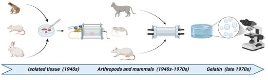
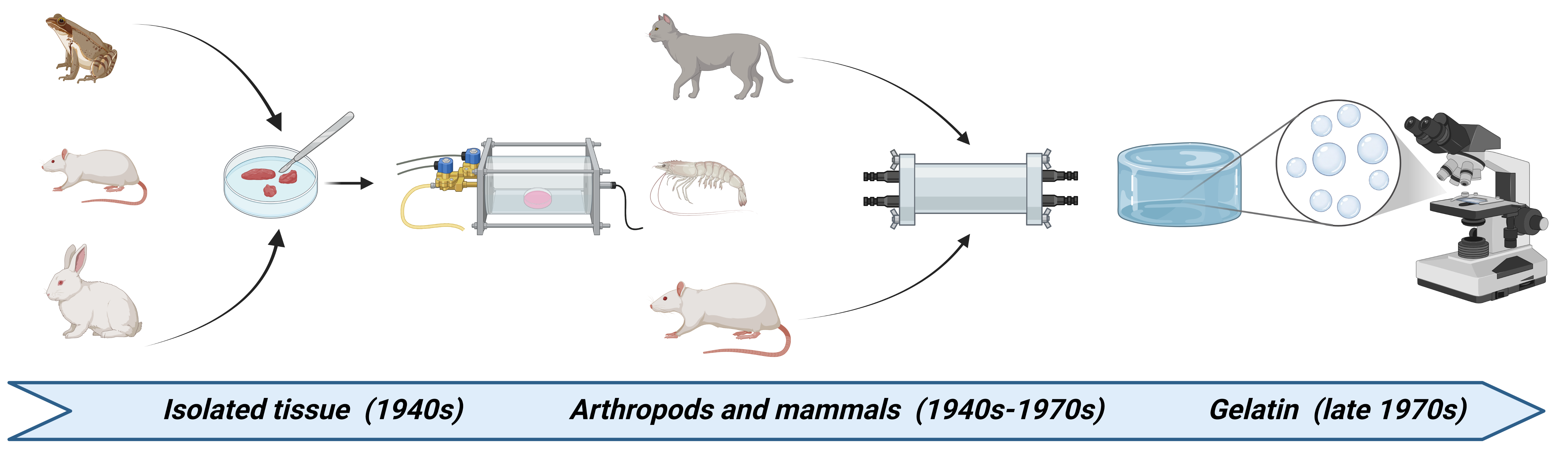
In the early 20th century, controversies over the origin of decompression bubbles abounded, but gas nuclei theory gradually gained dominance. In 1912, Hill [27] pointed out that supersaturation alone was insufficient to produce bubbles and speculated that certain “weak points” must exist in liquids for bubbles to form there. This “weak point” concept was later developed into gas nuclei theory, becoming the foundation of modern bubble decompression theory. After that, a series of experiments with materials such as isolated tissues, animals and gelatin has been carried out since the 1940s, laying the foundation for the establishment of the nuclei theory of biological decompression bubbles (Figure 2).
Figure 2.
Development of experiment materials and methods since 1940s.
In 1944, Harvey et al. [28], addressing Hill’s “pre-existing gas nuclei” hypothesis, conducted systematic research on bubble formation. Harvey [29] designed high-pressure precompression experiments, applying extremely high hydrostatic pressure to animals and isolated tissues before decompression to observe bubble formation. Results showed that high-pressure pretreatment significantly reduced or eliminated decompression bubbles, indirectly proving that bubbles originate from pre-existing gas nuclei. Harvey [30] further studied gas nuclei distribution in blood and tissues, proposing that bubbles originate at the vascular endothelial–tissue interface. Using cats, he analyzed the effects of gas tension and hydrostatic pressure difference on bubble formation [31].
In 1969, Evans and Walder [32] used brine shrimp to study decompression bubble nucleation. Evans directly observed bubble formation through the translucent shell. In the high-pressure pretreatment group, about 39.2 MPa hydrostatic pressure was applied for several minutes, followed by decompression to extremely low pressure; the control group was decompressed directly to the same low pressure. Results showed that 96% of control shrimp developed bubbles versus only 8% in the pretreated group. This directly demonstrated Harvey’s hypothesis that pre-existing gas nuclei can be eliminated by high pressure, but physiological differences between arthropods and mammals required validation in mammals.
In 1980, Vann et al. [33] used rats. The experimental group received 1000 fsw high-pressure pretreatment followed by 240 fsw exposure for 2 h. DCS incidence in the pretreated group was 64%, significantly lower than 83% in controls. This further supported Harvey’s in vivo pre-existing gas nuclei hypothesis. However, quantitative description of microscale gas nuclei encountered the stability paradox: according to the classical Laplace equation, extremely small nuclei with very high surface tension cannot theoretically exist stably, contradicting observations.
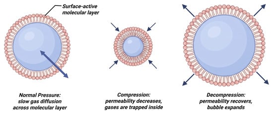
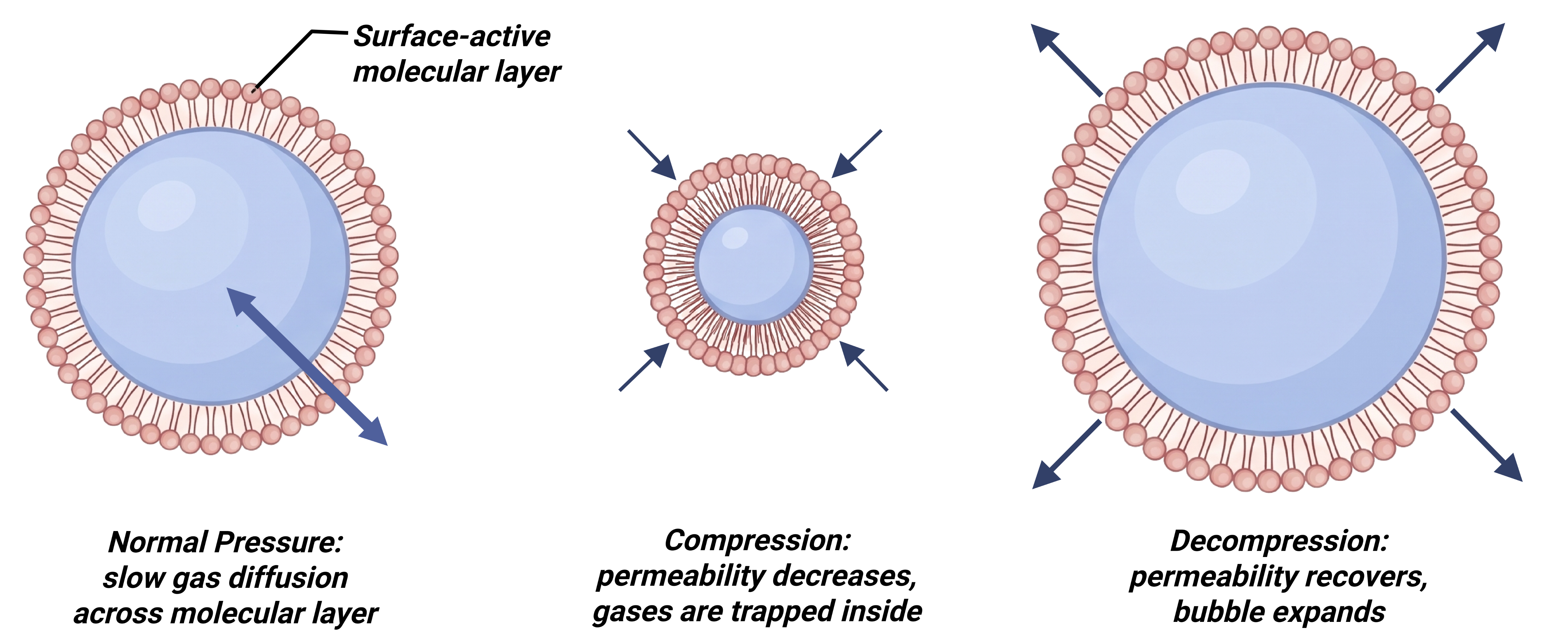
To address this problem, from the late 1970s to early 1980s, Yount et al. [34] at the University of Hawaii used gelatin to establish a controllable, observable platform for direct bubble nucleation observation. Yount chose transparent gelatin because its high viscosity immobilizes bubbles, preventing flotation, while the transparent medium allows optical observation and individual measurement, with precise control of supersaturation and pressure. Through theoretical analysis and gelatin experiments, Yount proposed that gas nuclei are stabilized by a “skin” of surface-active molecules [35], later developed into the varying permeability theory [36], forming the basis of the Varying Permeability Model (VPM). According to this theory, the elastic coating of surface-active molecules changes its gas permeability with pressure: at normal pressure, it is permeable and gas slowly passes; under rapid compression, it is compressed, permeability drops sharply, and internal gas is “locked in” and stabilized; during decompression, permeability returns and gas diffuses outward, expanding the nucleus (Figure 3). Mathematically, the theory introduces a skin compression term Γ into the Laplace equation (Equation (1)):
pgas + 2Γ/r = pamb + 2γ/r
Figure 3.
Mechanism of Varying Permeability Model. Under normal pressure (left), gas slowly diffuses through the surface-active layer. Upon rapid compression (middle), the layer is compressed, decreasing its permeability and trapping gas inside to stabilize the nucleus. During decompression (right), permeability recovers, allowing gas to diffuse outward and the bubble to expand.
In Equation (1), pgas is the gas pressure inside the nucleus, pamb is the ambient pressure, r is the nucleus radius, γ is the surface tension of the gas–liquid interface, and Γ is the reduction in surface tension contributed by the surface-active film. Physically, the term 2Γ/r represents the mechanical counteraction of the compressed surface-active molecular layer, which offsets a portion of the classical Laplace pressure to stabilize the nucleus. This equation was originally derived by Yount [36]. The term 2γ/r is the classical Laplace pressure, whereas 2Γ/r is the skin-compression term introduced by Yount. Equation 1 can be re-arranged as Equation (2):
pgas = pamb + 2(γ − Γ)/r
This shows that the surface-active film reduces the effective surface tension from γ to γ − Γ. When Γ = 0, the equation reduces to the classical Young–Laplace equation. When Γ increases, the effective Laplace pressure decreases, allowing a gas nucleus to remain mechanically stable at a radius smaller than that predicted for a bare bubble. In the VPM, Γ is not constant: rapid compression compresses the film, changes its permeability, and effectively locks gas inside the nucleus; decompression restores permeability and allows the nucleus to expand. This pressure-dependent behavior of Γ is the central mechanism of the Varying Permeability Model.
Yount applied the model to rat and human decompression data, achieving good fits [37] and demonstrating cross-species consistency of parameters. To determine the physical scale of gas nuclei, Yount filtered gelatin with Nuclepore membranes to remove nuclei larger than a given pore size. Results showed that the initial cutoff radius for bubble formation equaled the membrane pore size (0.4–3.0 μm), proving gas nuclei are physically real [38]. By tracking radius changes with pressure, Yount proposed mechanisms for gas nuclei generation and regeneration [39]. In 1984, Yount, with Gillary and Hoffman, used light and electron microscopy to directly observe gas nuclei of about 1 μm or less in distilled water and gelatin, estimating the surface-active film monolayer thickness at about (20 ± 7) Å [40]. These provided key microscopic evidence. Then, by replacing M-values with four global nucleation parameters, Yount and Hoffman applied the VPM to diving decompression table calculations for the first time, which was thought to lay the parametric foundation for subsequent bubble-physics-based algorithms [41].
2.3. Development of Gas Nuclei Theory
Yount’s varying permeability theory provided the first quantitatively predictive mechanism for the pre-existing gas nuclei hypothesis, but its core assumption—the chemical identity of the surface-active “skin”—has not been clearly identified in vivo. To address this problem, subsequent researchers attempted to verify the hypothesis or develop alternatives.
2.3.1. Nanobubbles and the AHS Hypothesis
According to Epstein–Plesset theory, a 100 nm surface bubble has a Laplace pressure of about 14 atmospheres, which should drive rapid gas diffusion and bubble disappearance within milliseconds. Yet experiments showed that nanobubbles on hydrophobic surfaces can remain stable for days or weeks. This contradiction initiated the beginning of nanobubble research [42].
Classical DLVO theory describes colloidal interactions as a balance between van der Waals attraction and electrostatic double-layer repulsion. In 1994, Parker et al. [43] used a surface force apparatus to measure the attraction between two hydrophobic spheres in water and discovered a stepped long-range attraction that could not be explained by this classical theory. To resolve this discrepancy, they proposed the existence of nanoscale gas domains at the interface—a concept that subsequently became foundational in nanobubble research. In 1999, Miller’s team [44] immersed hydrophobically modified silicon wafers in butane-saturated water and used FTIR to detect the first experimental evidence of interfacial nanobubbles, finding characteristic butane peaks that decayed slowly over tens of minutes—far exceeding Epstein–Plesset predictions. Currently, the mainstream explanation for surface nanobubble stability is contact line pinning [45]. According to this theory, the three-phase contact line is pinned by physical defects or chemical heterogeneities, keeping the contact radius constant while the curvature radius changes freely with gas exchange. Once pinned, curvature radius is determined by contact angle, not volume. Theoretical calculations indicate that stability requires local gas supersaturation and sufficiently strong pinning force. Pinning force depends on substrate roughness and wettability, while the required pinning force depends on wettability and pinning radius. Only when the required force falls within the substrate’s capability can nanobubbles exist stably. Unlike surface nanobubbles, the long-term stability of bulk nanobubbles is predominantly hypothesized to arise from a combination of electrostatic double-layer repulsion and local gas supersaturation. It is suggested that bulk nanobubbles adsorb negative ions (e.g., OH−) to form an electric double layer, where electrostatic repulsion may inhibit coalescence, and local gas supersaturation could counteract the internal Laplace pressure to maintain a dynamic equilibrium [46]. However, it should be acknowledged that the exact stabilization mechanism of bulk nanobubbles remains a subject of active debate in colloid science. The conditions under which gas supersaturation alone can sustain stability are highly dependent on specific ionic environments, and these proposed mechanisms require further rigorous validation.
The core contribution of nanobubble theory to decompression bubble research is providing a physically testable mechanism for Arieli’s AHS hypothesis. Arieli combined the “pre-existing gas nuclei” hypothesis with nanobubble theory and proposed the “Active Hydrophobic Spots” (AHS) hypothesis. Arieli [47] found that smooth hydrophobic surfaces immersed in gas-containing water spontaneously form 5–100 nm nanobubbles, suggesting that these might be the gas micronuclei predicted by Harvey and Yount but never directly identified. He further hypothesized that hydrophobic lipid spots (AHSs) on vascular endothelium might stabilize these nanobubbles, which could expand into macroscopic bubbles during decompression and cause DCS. Before Arieli, Hills [48] studied the origin of vascular endothelial hydrophilicity in the 1990s, finding that sheep pulmonary vein, left ventricle, and aorta, and human umbilical vein endothelium, exhibited obvious hydrophobicity, and cerebral vascular endothelium showed surface-active lamellar bodies under electron microscopy. He speculated that hydrophobic surface-active substances (phospholipids) might enter vascular walls of other tissues from lung tissue via pulmonary circulation, forming bubble generation sites. Hills’ findings and contemporaneous nanobubble discoveries might have inspired Arieli. Through characterization of sheep blood vessels, Arieli found that after decompression, bubbles appeared only at specific spots in aorta, pulmonary artery, pulmonary vein, and vena cava; these spots stained positive for lipids and were named AHSs [49]. According to his study, the average bubble detachment diameter was about 0.7–1.0 mm, with biphasic expansion: a slow initiation phase (peaking at about 45 min post-decompression), followed by rapid diffusion-controlled growth. In addition, Arieli [50] detected dipalmitoylphosphatidylcholine (DPPC), a major pulmonary surfactant component, at AHSs. DPPC content in AHSs producing ≥4 bubbles was significantly higher than in low-producing AHSs and controls. Arieli’s finding of DPPC corresponds exactly to the candidate “surface-active skin” molecule in Yount’s model; however, further attempts to predict individual bubbling tendency via plasma DPPC failed—plasma DPPC levels did not correlate with bubble production, suggesting AHS formation depends on local DPPC deposition in the vascular wall, not systemic concentration [51].
2.3.2. Tissue Elasticity Stabilization Theory
Yount’s varying permeability theory assumes a surface-active molecular layer, but Yount himself admitted its in vivo composition was unidentified. To address this problem, Goldman proposed the tissue elasticity stabilization theory, using tissue shear modulus (rigidity) instead of Yount’s “surface-active skin” to explain long-term gas nuclei stability. According to the Young–Laplace equation, internal bubble pressure must exceed external pressure by 2γ/R. But Goldman noted that in vivo bubbles mainly contain nitrogen while surrounding tissue contains nitrogen and oxygen, so internal pressure should be lower than atmospheric. If so, classical theory predictions would collapse. Goldman [52] proposed a generalized Young–Laplace equation treating tissue as a soft elastic solid with nonzero shear modulus. Elastic deformation resists bubble contraction, enabling mechanical stability even when internal pressure is lower than external. This theory applies mainly to sufficiently large bubbles (surface tension treated macroscopically, radius >> interface thickness). For submicron nuclei, interfacial microscopic behavior becomes significant and elastic approximation loses accuracy. Goldman extended the Epstein–Plesset dissolution/growth rate equation to soft elastic solids, finding that nonzero shear modulus accelerates growth and slows dissolution, predicting metastability or growth even in undersaturated elastic media [53]. Goldman also proposed tribonucleation: rapid separation of adhering surfaces generates instantaneous negative pressure, forming new bubbles. This provides a physical mechanism for exercise increasing DCS risk—muscle activity and joint movement continuously replenish gas nuclei via tribonucleation, offsetting dissolution loss [54].
2.3.3. RGBM Theory and Applications
In 1990, Bruce Wienke [55] engineered Yount’s VPM into the Reduced Gradient Bubble Model (RGBM), the first bubble-physics-based diving algorithm integrated into commercial dive computers. RGBM assumes: (1) exponentially distributed bubble seeds, with small seeds far more numerous; and (2) full-pressure permeability, allowing gas transfer across interfaces at all pressures, which is fundamentally different from Yount’s variable permeability. Wienke introduced a “phase volume” constraint across the dive profile, tracking accumulated “excited” bubble volume to ensure residual volume at surface does not exceed a safety limit, to directly determine stop depth and duration [56]. RGBM is generally conservative, it produces deeper stops to suppress bubble growth at depth rather than allocating long decompression time at shallow water. It has been adopted by Suunto, Mares, and others, representing one of the few commercially deployed bubble models.
3. Advances in Experimental Methodology
3.1. High-Pressure Precompression Experiments
High-pressure precompression is the classical method for verifying the pre-existing gas nuclei hypothesis. Harvey [29] first applied extremely high hydrostatic pressure to animals and isolated tissues before decompression, which showed significant reduction in bubbles after decompression and laid the methodological foundation in this field. Evans and Walder [32] used arthropods and direct observation through translucent shells, providing the first quantitative evidence of high-pressure elimination of gas nuclei in vivo. Vann et al. [33] confirmed this in mammals. The method is simple and produces clear effects, but it can only assess the presence/absence of bubbles after precompression and decompression, not the detailed nucleation or stability mechanisms.
3.2. Gelatin Model and Membrane Filtration
Unlike previous studies using animals or biological materials, Yount [34] used transparent gelatin to establish a controllable, observable nucleation platform. Gelatin has the following advantages: firstly, gelatin’s high viscosity immobilizes bubbles; secondly, the transparency of gelatin allows optical observation and individual measurement. In addition, supersaturation and pressure are precisely controlled in gelatin. To determine the physical scale of gas nuclei, Yount [38] filtered gelatin with Nuclepore membranes—track-etched, uniform cylindrical pores with small nominal error. Using 0.4, 0.6, 0.8, 1.0, and 3.0 μm membranes, he confirmed that the initial cutoff radius equaled pore size within measurement accuracy, which proved the physical existence of gas nuclei. This method provided the first controllable in vitro platform, though gelatin is a simplified model that cannot fully reflect biological interface complexity.
3.3. Surface Characterization and Spectroscopic Methods
On a micro-scale, atomic force microscopy (AFM) and Fourier Transform Infrared Spectroscopy (FTIR) moved nanobubbles from theoretical speculation to experimentally verifiable existence. For example, with the use of AFM, Lou [57] and Ishida [58] first directly imaged surface nanobubbles. Miller’s team [44] detected interfacial butane peaks on hydrophobic silicon with FTIR, supporting the existence of nanobubbles. These techniques provide direct evidence of nanobubbles but face practical difficulties when used in biological decompression research. For example, AFM probes cannot even approach rough vascular surfaces in practical use.
3.4. Molecular Dynamics Simulation
Molecular dynamics (MD) simulation provides microscopic insight into nanoscale nucleation. Liu et al. [59] revealed a two-step pathway for nanobubble nucleation on rough hydrophobic surfaces: gas cavity formation in pits, then coalescence into pinned nanobubbles. MD can analyze effects of roughness, pit width, and contact position. Yu [60] systematically studied surface hydrophobicity and gas supersaturation effects on nucleation kinetics. MD offers molecular-level analysis of complex biological interfaces but the use of it is limited by simulation time scales, and further experimental validation after is still required.
3.5. In Situ Characterization of Vascular AHS
Arieli [49] developed an ex vivo sheep blood vessel decompression system and provided chemical characterization via lipid staining and DPPC detection [50]. Arieli [61] further provided morphological evidence of endothelial cell loss near AHSs using electron and light microscopy, supporting mechanical damage from bubble detachment tearing endothelial membranes. This method first localized nucleation sites on biological tissues, but since it cannot directly observe nanobubbles or prove that they are precursors to the observed macro-bubbles, the effectiveness of it remains controversial.
4. Controversial Issues
4.1. Controversy over the AHS Hypothesis
In 2019, Doolette [62] published a letter in Diving and Hyperbaric Medicine arguing that the AHS hypothesis relied on the assumption that nanobubbles were the source of gas nuclei, yet no one had directly observed nanobubbles on biological tissue surfaces by AFM. Doolette contended that Arieli’s experiments only showed macroscopic bubble formation after decompression in sheep vessels, without evidence of nanobubbles on the vascular lumen or proof that they were precursors. Doolette further noted that surface nanobubbles typically form on atomically smooth hard surfaces, whereas biological surfaces were far from smooth. He pointed out that venous bubbles could be detected in humans after decompression from only 138 kPa air saturation, implying very low required supersaturation. However, according to the Young–Laplace equation, de novo formation of a 10 nm bubble under blood surface tension (0.056 N·m−1) requires over 11.2 MPa supersaturation—an order-of-magnitude gap. In response, Arieli [63] argued that nanobubble formation and stability on hydrophobic surfaces were well established, and the Young–Laplace equation did not apply at the nanoscale, and that the hydrophobic silicon wafer experiment could fully support the AHS hypothesis. Up to now, the core disagreement still exists: Doolette demands direct AFM evidence on biological tissues, while Arieli considers that hydrophobic surface bubbling, DPPC presence, and former nanobubble researches are sufficient enough as indirect evidence.
4.2. Controversy over the Surface-Active Skin
Yount believed gas nuclei were stabilized by a surface-active “skin” with variable permeability. Goldman objected that the surfactant had never been identified in vivo, and that tissue shear modulus might provide an alternative stabilization mechanism. In fact, the two theories are not necessarily mutually exclusive. Combined with Arieli’s work, one might speculate a more complete mechanism: hydrophobic surfaces provide nucleation sites, while tissue elasticity provides stabilization.
5. Discussion
Compared with dissolved-tissue theory, bubble theory approaches DCS pathogenesis from a physically intuitive perspective, offering a more scientific basis for decompression models and algorithms. From Hill’s “defect point” hypothesis, gas nuclei theory evolved into the VPM and found application in safe diving decompression, yet its core assumption—the “surface-active skin”—remains experimentally unidentified. After decades of research, the question of gas nuclei stability in vivo still remains unresolved. Nanobubble theory introduced concepts of nanoscale ultrastability from physical chemistry into the research of biological decompression bubbles. Although the experimental fact that nanobubbles form on hydrophobic surfaces readily links them to hydrophobic sites in the body, current experiments have not captured evidence of surface or bulk nanobubbles in biological tissues, so direct evidence of biological nanobubbles is still lacking. The AHS theory therefore requires substantial further evidence to clarify the origin of decompression bubbles.
Recent advances in interfacial rheology may offer a novel perspective for dealing with the above unresolved question about biological decompression bubbles. As implied by former research, AHS spots are supposed to be enriched with DPPC from circulation, so this mixed lipid layer is very likely to play a more complicated role in the stability of biological gas bubbles rather than acting as a simple static hydrophobic surface. In addition, molecular dynamics simulations and classical nucleation theory have shown that reentrant cavities drastically lower the energy barrier for nucleation. Specifically, MD simulations by Zhang et al. [64] revealed that reentrant cavities exhibit the shortest bubble initial nucleation time among various nanostructure morphologies. Furthermore, free energy MD combined with CNT by Giacomello et al. [65] demonstrated that reentrant geometries sustain a confined vapor phase and modulate the nucleation barrier. These findings suggest that reentrant surface irregularities may serve as promoted origins for biological decompression bubbles, lowering the energetic cost of gas nucleus formation at the vascular interface. Therefore, the physical geometry of the nucleation site is supposed to be an important factor affecting the generation and detachment of gas bubbles. Future research should focus on how the interplay between the geometry of nucleation site and the physical properties of interface govern the dynamics of biological decompression bubbles. Meanwhile, biological decompression bubble research has expanded from generation and stability to growth and movement patterns [66,67,68]: the transition from a stable micro-nucleus to a macroscopic pathogenic bubble involves complex nonlinear dynamics under variable ambient pressure. Future integration of molecular dynamics and hemodynamics into decompression bubble dynamics promises to bridge microscopic and macroscopic studies of biological decompression bubbles.
6. Conclusions
In conclusion, the study of biological decompression bubbles has evolved from early empirical observations to the current molecular and interfacial investigations in the course of hundreds of years. The VPM, RGBM and AHS theories have significantly advanced our understanding of biological decompression bubbles; however, the direct evidence for the “surface-active skin” and nanobubbles in vivo remains elusive. To resolve these long-standing paradoxes, future research should focus on the integration of interfacial rheology, biomimetics, microfluidics, and multiscale mathematical modeling. By embracing multidisciplinary approaches combining physical chemistry, molecular dynamics, and diving physiology, we may finally decode the physical identity of gas nuclei and develop safer and more personalized decompression algorithms.
Author Contributions
Conceptualization, L.W., J.Y., X.L., N.Z., H.Z., G.H. and Y.F.; methodology, L.W. and J.Y. formal analysis, L.W.; data curation, G.H.; writing—original draft preparation, L.W., J.Y. and X.L.; writing—review and editing, L.W., H.Z. and Y.F.; All authors have read and agreed to the published version of the manuscript.
Funding
This research was funded by the National Natural Science Foundation of China, grant number 82603647.
Institutional Review Board Statement
Not applicable.
Informed Consent Statement
Not applicable.
Data Availability Statement
No new data were created or analyzed in this study. Data sharing is not applicable to this article.
Conflicts of Interest
The authors declare no conflicts of interest.
Abbreviations
The following abbreviations are used in this manuscript:
| DCS | Decompression sickness |
| VPM | Varying Permeability Model |
| AHS | Active Hydrophobic Spots |
| DLVO | Derjaguin–Landau–Verwey–Overbeek |
| AFM | Atomic force microscopy |
| FTIR | Fourier Transform Infrared Spectroscopy |
| MD | Molecular dynamics |
| RGBM | Reduced Gradient Bubble Model |
| DPPC | dipalmitoylphosphatidylcholine |
References
- Vann, R.D.; Butler, F.K.; Mitchell, S.J.; Moon, R.E. Decompression illness. Lancet 2010, 377, 157–164. [Google Scholar] [CrossRef] [Scilit]
- Mitchell, S.J. Decompression illness: A comprehensive overview. Diving Hyperb. Med. 2024, 54, 1–53. [Google Scholar] [CrossRef] [Scilit]
- Mitchell, S.J.; Bennett, M.H.; Moon, R.E. Decompression Sickness and Arterial Gas Embolism. N. Engl. J. Med. 2022, 386, 1254–1264. [Google Scholar] [CrossRef] [Scilit]
- Edmonds, C.; Bennett, M.; Lippmann, J.; Mitchell, S.J. Decompression sickness: Manifestations, Diving and Subaquatic Medicine, 5th ed.; CRC Press: Boca Raton, FL, USA, 2015; pp. 125–126. [Google Scholar] [CrossRef] [Scilit]
- Klass, S.; Burton, A.; Price, C. SCUBA Diver’s Knee: A Case Report. Curr. Sports Med. Rep. 2022, 21, 109–111. [Google Scholar] [CrossRef] [Scilit]
- Sundal, E.; Lygre, S.H.L.; Irgens, Å.; Troland, K.; Grønning, M. Long-term neurological sequelae after decompression sickness in retired professional divers. J. Neurol. Sci. 2022, 434, 120181. [Google Scholar] [CrossRef] [Scilit]
- Pontier, J.M.; Vallée, N.; Ignatescu, M.; Bourdon, L. Pharmacological intervention against bubble-induced platelet aggregation in a rat model of decompression sickness. J. Appl. Physiol. 2011, 110, 724–729. [Google Scholar] [CrossRef] [Scilit]
- Thom, S.R.; Yang, M.; Bhopale, V.M.; Huang, S.; Milovanova, T.N. Microparticles initiate decompression-induced neutrophil activation and subsequent vascular injuries. J. Appl. Physiol. 2011, 110, 340–351. [Google Scholar] [CrossRef] [Scilit]
- Chong, S.M. Gas bubble disease. In Aquaculture Pathophysiology; Academic Press: Cambridge, MA, USA, 2022; pp. 741–743. [Google Scholar] [CrossRef] [Scilit]
- Machova, J.; Faina, R.; Randak, T.; Valentova, O.; Steinbach, C.; Kroupova, H.K.; Svobodova, Z. Fish death caused by gas bubble disease: A case report. Vet. Med. 2017, 62, 231–237. [Google Scholar] [CrossRef] [Scilit]
- Cao, C.; Deng, Y.; Yin, Q.; Li, N.; Liu, X.; Shi, H.; Yang, Y.; Xu, L. Effects of continuous acute and intermittent exposure on the tolerance of juvenile yellow catfish (Pelteobagrus fulvidraco) in total dissolved gas supersaturated water. Ecotoxicol. Environ. Saf. 2020, 201, 110855. [Google Scholar] [CrossRef] [Scilit]
- Mohammadein, S.A.; Mohamed, K.G. Growth of a Gas Bubble in a Steady Diffusion Field in a Tissue Undergoing Decompression. Math. Model. Anal. 2016, 21, 762–773. [Google Scholar] [CrossRef] [Scilit]
- Abu-Nab, A.K.; Abu-Bakr, A.F. Magnetically Induced Nonlinear Multicavitation Dynamics Near a Rigid Wall in (MWCNT-SWCNT-CuO-TiO2-MgO) Penta-Hybrid Nanofluids: Investigation of Thermophysical Properties. J. Nonlinear Math. Phys. 2026, 33, 19. [Google Scholar] [CrossRef] [Scilit]
- Boyle, R. New pneumatical experiments about respiration. Philos. Trans. 1670, 5, 2013–2015. [Google Scholar] [CrossRef] [Scilit]
- Dalton, J. Experimental essays on the constitution of mixed gases; on the force of steam or vapour from water and other liquids in different temperatures, both in a Torricellian vacuum and in air; on evaporation; and on the expansion of gases by heat. Mem. Lit. Philos. Soc. Manch. 1798, 5, 535–602. [Google Scholar]
- Henry, W. Experiments on the quantity of gases absorbed by water at different temperatures, and under different pressures. Philos. Trans. R. Soc. Lond. 1803, 93, 29–42. [Google Scholar] [CrossRef] [Scilit]
- Triger, C. Lettre à M. Arago. Comptes Rendus Hebd. Séances l’Académie Des Sci. 1845, 20, 445–449. [Google Scholar]
- Ninokawa, S.; Nordham, K. Discovery of caisson disease: A dive into the history of decompression sickness. Proc. Bayl. Univ. Med. Cent. 2022, 35, 129–132. [Google Scholar] [CrossRef] [Scilit]
- Pol, B.; Watelle, T.J. Mémoire sur les effets de la compression de l’air appliquée au creusement des puits à houille. Ann. Hyg. Public 1845, 2, 241–279. [Google Scholar]
- de Méricourt, L.R. Considérations sur l’hygiène des pêcheurs d’éponges. Ann. Hyg. Pub. Med. Leg. 1869, 31, 274–286. [Google Scholar]
- Bert, P. Barometric Pressure: Researches in Experimental Physiology; College Book Company: Columbus, OH, USA, 1943; pp. 150–188. [Google Scholar] [CrossRef] [Scilit]
- Boycott, A.E.; Damant, G.C.; Haldane, J.S. The prevention of compressed-air illness. J. Hyg. 1908, 8, 342–443. [Google Scholar] [CrossRef] [Scilit]
- Boycott, A.E.; Damant, G.C. Experiments on the influence of fatness on susceptibility to caisson disease. J. Hyg. 1908, 8, 445–456. [Google Scholar] [CrossRef] [Scilit]
- Butler, W.P. Caisson disease during the construction of the Eads and Brooklyn Bridges: A review. Undersea Hyperb. Med. 2004, 31, 445–459. [Google Scholar]
- Edmonds, C.; Bennett, M.; Lippmann, J.; Mitchell, S.J. Decompression sickness: Pathophysiology, Diving and Subaquatic Medicine, 5th ed.; CRC Press: Boca Raton, FL, USA, 2015; pp. 58–76. [Google Scholar] [CrossRef] [Scilit]
- Hill, L.; Greenwood, M. On the formation of bubbles in the tissues of animals exposed to high atmospheric pressure. Proc. R. Soc. Lond. B 1910, 82, 453–457. [Google Scholar]
- Hill, L. Caisson Sickness and the Physiology of Work in Compressed Air; E. Arnold: London, UK, 1912. [Google Scholar] [CrossRef] [Scilit]
- Harvey, E.N. Decompression sickness and bubble formation in blood and tissues. Bull. N. Y. Acad. Med. 1945, 21, 505–536. [Google Scholar]
- Harvey, E.N.; Barnes, D.K.; McElroy, W.D.; Whiteley, A.H.; Pease, D.C.; Cooper, K.W. Bubble formation in animals. I. Physical factors. J. Cell. Comp. Physiol. 1944, 24, 1–22. [Google Scholar] [CrossRef] [Scilit]
- Harvey, E.N.; Barnes, D.K.; McElroy, W.D.; Pease, D.C.; Barnes, D.K. Bubble formation in animals. II. Gas nuclei and their distribution in blood and tissues. J. Cell. Comp. Physiol. 1944, 24, 23–34. [Google Scholar] [CrossRef] [Scilit]
- Harvey, E.N.; McElroy, W.D.; Whiteley, A.H.; Warren, G.H.; Pease, D.C. Bubble formation in animals. III. An analysis of gas tension and hydrostatic pressure in cats. J. Cell. Comp. Physiol. 1944, 24, 117–132. [Google Scholar] [CrossRef] [Scilit]
- Evans, A.; Walder, D.N. Significance of gas micronuclei in the aetiology of decompression sickness. Nature 1969, 222, 251–252. [Google Scholar] [CrossRef] [Scilit]
- Vann, R.D.; Grimstad, J.; Nielsen, C.H. Evidence for gas nuclei in decompressed rats. Undersea Biomed. Res. 1980, 7, 107–112. [Google Scholar]
- Yount, D.E. Bubble formation in gelatin: A model for decompression sickness. J. Appl. Phys. 1976, 47, 5081–5089. [Google Scholar] [CrossRef] [Scilit]
- Yount, D.E.; Kunkle, T.D.; D’Arrigo, J.S.; Ingle, F.W.; Yeung, C.M.; Beckman, E.L. Stabilization of gas cavitation nuclei by surface-active compounds. Aviat. Space Environ. Med. 1977, 48, 185–189. [Google Scholar]
- Yount, D.E. Skins of varying permeability: A stabilization mechanism for gas cavitation nuclei. J. Acoust. Soc. Am. 1979, 65, 1429–1439. [Google Scholar] [CrossRef] [Scilit]
- Yount, D.E. Application of a bubble formation model to decompression sickness in rats and humans. Aviat. Space Environ. Med. 1979, 50, 44–50. [Google Scholar]
- Yount, D.E.; Yeung, C.M.; Ingle, F.W. Determination of the radii of gas cavitation nuclei by filtering gelatin. J. Acoust. Soc. Am. 1979, 65, 1440–1450. [Google Scholar] [CrossRef] [Scilit]
- Yount, D.E. On the evolution, generation, and regeneration of gas cavitation nuclei. J. Acoust. Soc. Am. 1982, 71, 1473–1481. [Google Scholar] [CrossRef] [Scilit]
- Yount, D.E.; Gillary, E.W.; Hoffman, D.C. A microscopic investigation of bubble formation nuclei. J. Acoust. Soc. Am. 1984, 76, 1511–1521. [Google Scholar] [CrossRef] [Scilit]
- Yount, D.E.; Hoffman, D.C. On the use of a bubble formation model to calculate diving tables. Aviat. Space Environ. Med. 1986, 57, 149–156. [Google Scholar]
- Hong, P.; Birkett, G.R.; Nguyen, A.V. Progress on the surface nanobubble story: What is in the bubble? Why does it exist? Adv. Colloid Interface Sci. 2015, 222, 573–580. [Google Scholar] [CrossRef] [Scilit]
- Parker, J.L.; Claesson, P.M.; Attard, P. Bubbles, cavities, and the long-ranged attraction between hydrophobic surfaces. J. Phys. Chem. B 1994, 98, 8468–8480. [Google Scholar] [CrossRef] [Scilit]
- Miller, J.D.; Hu, Y.; Veeramasuneni, S.; Lu, Y. In-situ detection of butane gas at a hydrophobic silicon surface. Colloids Surf. A 1999, 154, 137–147. [Google Scholar] [CrossRef] [Scilit]
- Liu, Y.; Wang, J.; Zhang, X.; Wang, W. Stabilization of nanobubbles on a substrate by contact line pinning. J. Chem. Phys. 2014, 140, 054705. [Google Scholar] [CrossRef] [Scilit]
- Chen, C.; Gao, Y.; Zhang, X. The existence and stability mechanism of bulk nanobubbles: A review. Nanomaterials 2025, 15, 314. [Google Scholar] [CrossRef] [Scilit]
- Arieli, R. Nanobubbles form at active hydrophobic spots on the luminal aspect of blood vessels: Consequences for decompression illness in diving and possible implications for autoimmune disease—An overview. Front. Physiol. 2017, 8, 591. [Google Scholar] [CrossRef] [Scilit]
- Hills, B.A. A hydrophobic oligolamellar lining to the vascular lumen in some organs. Undersea Biomed. Res. 1992, 19, 107–120. [Google Scholar]
- Arieli, R.; Marmur, A. Ex vivo bubble production from ovine large blood vessels: Size on detachment and evidence of “active spots”. Respir. Physiol. Neurobiol. 2014, 200, 110–117. [Google Scholar] [CrossRef] [Scilit]
- Arieli, R.; Khatib, S.; Vaya, J. Presence of dipalmitoylphosphatidylcholine from the lungs at the active hydrophobic spots in the vasculature where bubbles are formed on decompression. J. Appl. Physiol. 2016, 121, 811–815. [Google Scholar] [CrossRef] [Scilit]
- Arieli, R.; Khatib, S.; Vaya, J. Ovine plasma dipalmitoylphosphatidylcholine does not predict decompression bubbling. Respir. Physiol. Neurobiol. 2019, 259, 26–29. [Google Scholar] [CrossRef] [Scilit]
- Goldman, S. Generalizations of the Young-Laplace equation for the pressure of a mechanically stable gas bubble in a soft elastic material. J. Chem. Phys. 2009, 131, 184502. [Google Scholar] [CrossRef] [Scilit]
- Solano-Altamirano, J.M.; Malcolm, J.D.; Goldman, S. Gas bubble dynamics in soft materials. Soft Matter 2015, 11, 202–210. [Google Scholar] [CrossRef] [Scilit]
- Goldman, S. Free energy wells for small gas bubbles in soft deformable materials. J. Chem. Phys. 2010, 132, 164509. [Google Scholar] [CrossRef] [Scilit]
- Wienke, B.R. Reduced gradient bubble model. Int. J. Biomed. Comput. 1990, 26, 237–256. [Google Scholar] [CrossRef] [Scilit]
- Wienke, B.R. Computer validation and statistical correlations of a modern decompression diving algorithm. Comput. Biol. Med. 2010, 40, 252–260. [Google Scholar] [CrossRef] [Scilit]
- Lou, S.T.; Ouyang, Z.Q.; Zhang, Y.; Li, X.-J.; Hu, J.; Li, M.-Q.; Yang, F.-J. Nanobubbles on solid surface imaged by atomic force microscopy. J. Vac. Sci. Technol. B 2000, 18, 2573–2575. [Google Scholar] [CrossRef] [Scilit]
- Ishida, N.; Inoue, T.; Miyahara, M.; Higashitani, K. Nanobubbles on a hydrophobic surface in water observed by tapping-mode atomic force microscopy. Langmuir 2000, 16, 6377–6380. [Google Scholar] [CrossRef] [Scilit]
- Liu, Y.; Zhang, X. Molecular dynamics simulation of nanobubble nucleation on rough surfaces. J. Chem. Phys. 2017, 146, 164704. [Google Scholar] [CrossRef] [Scilit]
- Yang, H.; Jiang, H.; Cheng, Y.; Xing, Y.; Cao, Y.; Gui, X. Solid-liquid interfacial nanobubble nucleation dynamics influenced by surface hydrophobicity and gas oversaturation. J. Mol. Liq. 2024, 411, 125758. [Google Scholar] [CrossRef] [Scilit]
- Arieli, R. Endothelial injury in diving: Atomic force-, electronic-, and light-microscopy study of the ovine decompressed blood vessels. Front. Physiol. 2021, 12, 767435. [Google Scholar] [CrossRef] [Scilit]
- Doolette, D.J. Gas micronuclei that underlie decompression bubbles and decompression sickness have not been identified. Diving Hyperb. Med. 2019, 49, 64. [Google Scholar] [CrossRef] [Scilit]
- Arieli, R. Gas micronuclei that underlie decompression bubbles and decompression sickness have most probably been identified—In response to the Letter to the Editor from Dr David Doolette. Diving Hyperb. Med. 2019, 49, 311–312. [Google Scholar] [CrossRef] [Scilit]
- Zhang, L.; Yang, Y.; Han, J. Microscopic mechanism of effects of nanostructure morphology on bubble nucleation: A molecular dynamics simulation. Numer. Heat Transf. Part A Appl. 2024, 85, 187–202. [Google Scholar] [CrossRef] [Scilit]
- Giacomello, A.; Amabili, M.; Casciola, C.M. How to control bubble nucleation from superhydrophobic surfaces. J. Phys. Conf. Ser. 2015, 656, 012124. [Google Scholar] [CrossRef] [Scilit]
- Chappell, M.A.; Payne, S.J. A physiological model of gas pockets in crevices and their behavior under compression. Respir. Physiol. Neurobiol. 2006, 152, 100–114. [Google Scholar] [CrossRef] [Scilit]
- Chappell, M.A.; Payne, S.J. A crevice bubble growth model for the analysis of decompression sickness. Conf. Proc. IEEE Eng. Med. Biol. Soc. 2005, 3, 2240–2243. [Google Scholar] [CrossRef] [Scilit]
- Chappell, M.A.; Uzel, S.; Payne, S.J. Modeling the detachment and transport of bubbles from nucleation sites in small vessels. IEEE Trans. Biomed. Eng. 2007, 54, 2106–2108. [Google Scholar] [CrossRef] [Scilit]
Disclaimer/Publisher’s Note: The statements, opinions and data contained in all publications are solely those of the individual author(s) and contributor(s) and not of MDPI and/or the editor(s). MDPI and/or the editor(s) disclaim responsibility for any injury to people or property resulting from any ideas, methods, instructions or products referred to in the content. |
© 2026 by the authors. Licensee MDPI, Basel, Switzerland. This article is an open access article distributed under the terms and conditions of the Creative Commons Attribution (CC BY) license.