


Abstract
Nephrogenic syndrome of inappropriate antidiuresis (NSIAD) is a rare disorder of water homeostasis caused by constitutive activation of the renal vasopressin V2 receptor and may closely mimic syndrome of inappropriate antidiuresis (SIAD). Recognition in infancy is challenging because severe hyponatremia may remain clinically silent and conventional biochemical findings may overlap with other disorders of water balance. We report the clinical, biochemical, and molecular characterization of a 7-month-old male infant with severe hypotonic hyponatremia incidentally identified during evaluation for bronchiolitis, which persisted after resolution of the acute respiratory illness. Serum sodium was 112 mmol/L and serum osmolality was 231 mOsm/kg (calculated), with preserved renal and hepatic function. Adrenal insufficiency was excluded by ACTH stimulation testing, while a low copeptin concentration (2 pmol/L) supported appropriately suppressed vasopressin secretion. Whole-exome sequencing identified the pathogenic activating hemizygous AVPR2 c.409C>T, p.(Arg137Cys) variant, confirming NSIAD, together with a pathogenic heterozygous ACVRL1 c.270C>A, p.(Cys90*) variant associated with hereditary hemorrhagic telangiectasia. The co-occurrence of these two pathogenic variants is clinically relevant for surveillance and genetic counselling but is not proposed as a mechanistic explanation of the water-balance disorder. To the best of our knowledge, this represents the first reported case of genetically confirmed NSIAD in Romania and the first documented coexistence of these two pathogenic variants in a single patient. Although mechanistically unrelated, their coexistence highlights the clinical relevance of comprehensive genetic testing in identifying additional actionable diagnoses. Fluid restriction and oral urea were followed by an improvement in serum sodium to 130 mmol/L. This case illustrates the diagnostic value of integrating copeptin assessment with genomic testing in severe unexplained hypotonic hyponatremia in infancy, and highlights the need for independent surveillance of concomitant pathogenic findings identified by broad genomic testing.
1. Introduction
Maintenance of water balance depends on the coordinated regulation of fluid intake and renal free water excretion. Arginine vasopressin (AVP) plays a central role in this process by binding to V2 receptors on the basolateral membrane of principal cells in the renal collecting duct, thereby activating adenylate cyclase through Gs proteins, increasing intracellular cyclic AMP, and promoting insertion of aquaporin-2 water channels into the apical membrane. This cascade increases water permeability and allows for the generation of concentrated urine [1]. Hyponatremia, the most common electrolyte abnormality encountered in clinical practice, most often reflects an impairment in the kidney’s ability to excrete free water [2]. In euvolemic patients, the combination of hypotonic hyponatremia, inappropriately concentrated urine, and natriuresis is typically attributed to the syndrome of inappropriate antidiuresis [3], a disorder most commonly associated with nonosmotic AVP release in the setting of medications [4], pulmonary disease [5], central nervous system disorders [6], or malignancy [7].
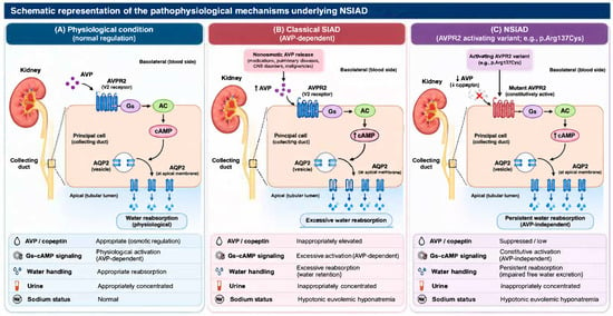
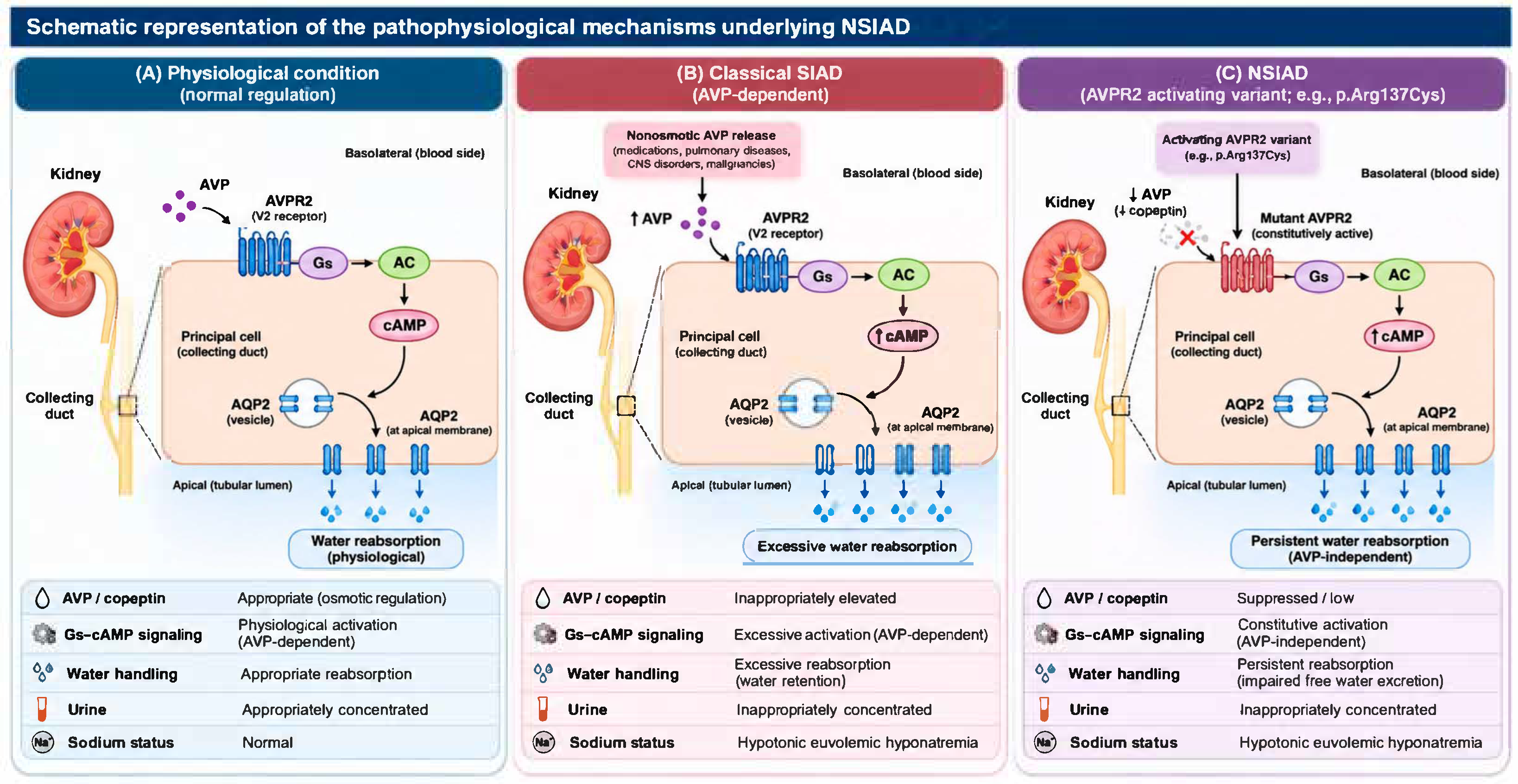
However, accumulating evidence indicates that, in rare instances, a similar biochemical phenotype may arise in the absence of detectable abnormalities in AVP secretion [1]. In such cases, dysregulation of the antidiuretic pathway occurs at the level of the kidney, resulting in persistent water retention despite physiologically appropriate or suppressed circulating AVP concentrations [8]. The distinction between physiological AVP-mediated antidiuresis, classical SIAD, and AVPR2-related NSIAD is illustrated in Figure 1, highlighting the constitutive activation of renal antidiuretic signaling despite suppressed AVP secretion in NSIAD.
Figure 1.
Schematic representation of the pathophysiological mechanisms underlying NSIAD. (A) Under physiological conditions, arginine vasopressin (AVP) binds to the V2 receptor (AVPR2) on the basolateral membrane of renal collecting duct principal cells, activating the Gs protein–adenylyl cyclase–cyclic adenosine monophosphate (cAMP) signaling pathway. This promotes aquaporin-2 (AQP2) insertion into the apical membrane, increasing water reabsorption and allowing appropriate regulation of urine concentration. (B) In classical syndrome of inappropriate antidiuresis (SIAD), nonosmotic AVP release, potentially triggered by medications, pulmonary diseases, central nervous system disorders, or malignancies, results in inappropriate activation of the same signaling pathway, leading to excessive water retention and hypotonic euvolemic hyponatremia. (C) In NSIAD, activating variants of AVPR2, such as p.Arg137Cys, cause constitutive receptor activation independently of AVP binding. Consequently, persistent Gs–cAMP signaling promotes AQP2-mediated water reabsorption despite physiologically suppressed AVP secretion and low circulating copeptin concentrations. This results in impaired free water excretion, inappropriately concentrated urine, and hypotonic euvolemic hyponatremia. Abbreviations: AVP, arginine vasopressin; AVPR2, arginine vasopressin receptor 2; AQP2, aquaporin-2; cAMP, cyclic adenosine monophosphate; NSIAD, nephrogenic syndrome of inappropriate antidiuresis; SIAD, syndrome of inappropriate antidiuresis.
We describe a patient with recurrent euvolemic hyponatremia in whom detailed evaluation revealed an uncommon mechanism of impaired water excretion, highlighting the importance of considering alternative pathophysiologic processes when standard diagnostic frameworks do not fully account for the clinical presentation.
2. Case Report
A 7-month-old male infant was admitted for clinical and biochemical evaluation after severe hyponatremia (114 mmol/L) was incidentally identified during laboratory testing performed for an episode of bronchiolitis. Two weeks after the complete resolution of the respiratory illness, the 7-month-old male infant returned for further clinical and biochemical evaluation, which confirmed persistent hyponatremia despite clinical recovery. His past medical history was notable for mid-penile hypospadias and right cryptorchidism. Renal ultrasonography revealed mild left-sided hydronephrosis (grade I–II according to Society for Fetal Urology [9]), characterized by dilatation of the renal pelvis with minimal involvement of the central calyces. No ureteral dilatation was observed, and renal parenchymal thickness and corticomedullary differentiation were preserved. There was no history of prior electrolyte disturbances.
The family history was notable for nonconsanguineous parents, both 37 years of age and reportedly healthy. His maternal grandmother died of cardiorespiratory arrest at the age of 62. Maternal history is significant for a prior pregnancy loss at 10 weeks’ gestation (spontaneous miscarriage). Perinatal history was notable for placental abruption. The patient was the second child, born at term with a birth weight of 3160 g, length of 51 cm, and an Apgar score of 9. He has a 7-year-old sister with myopic astigmatism. He was breastfed, with complementary feeding initiated at 6 months. Vaccinations and rickets prophylaxis were administered according to national guidelines. There was no known family history of endocrine or genetic disorders. On admission, the infant appeared in good general condition. Physical examination did not reveal major dysmorphic features suggestive of a specific syndromic diagnosis. Minor anomalies mentioned in the genetic report (sacral fossa, metatarsus adductus) were considered non-specific and not clinically defining. He was afebrile, with normal hydration status and stable hemodynamic and respiratory parameters. The child presented with anthropometric measurements of 9985 g in weight and 70 cm in length. According to WHO growth standards, the weight-for-length Z-score is estimated to be above +2 SD, indicating a weight-for-length above the normal range consistent with overweight status (high weight-for-length). Skin and mucous membranes were normally colored. The abdomen was soft, non-tender, and without organomegaly. Bowel movements were infrequent (once every 2–3 days). Genitourinary examination revealed: mid-penile hypospadias with curvature, right undescended testis, normal left testis in scrotum. Neurologic development was appropriate for age, with no signs of meningeal irritation.
Initial laboratory evaluation (3 June 2025) revealed severe hyponatremia (112 mmol/L), a serum potassium concentration of 5.0 mmol/L, which subsequently normalized spontaneously without specific treatment, hypochloremia, microcytic hypochromic anemia (Hb 8.8 g/dL, low MCV), low ferritin (<6 µg/L) and low serum iron. As direct measurement of serum and urine osmolality was not available in our laboratory, these parameters were estimated using established calculation formulas based on the available biochemical measurements. The estimated serum osmolality was markedly reduced (231 mOsm/kg; reference range: 285–295 mOsm/kg), as calculated using the standard formula: Osm = 2 × Na + Glucose/18 + BUN/2.8 [9].
Urinary osmolarity was estimated as twice the sum of urinary sodium and potassium concentrations plus the urinary urea concentration [10,11], with all values expressed in mmol/L. Based on urinary sodium (16.9 mmol/L), potassium (15.5 mmol/L), and urea (501 mg/dL, equivalent to 83.4 mmol/L), the estimated urinary osmolarity was 148.2 mOsm/L.
Although these calculated estimates cannot replace direct osmometric measurements, the estimated urinary osmolarity suggests incomplete urinary dilution in the setting of profound hypotonic hyponatremia.
Renal and hepatic function tests were within normal limits. Urinary electrolyte analysis showed low urinary sodium (<10 mmol/L, reference range 54–150 mmol/L) and potassium excretion (8.25 mmol/L, reference range 20–80 mmol/L). Stool examination and urinalysis were unremarkable. Scrotal ultrasound demonstrated a right testis located within the inguinal canal, with a normal-appearing left testis. Abdominal ultrasound findings were unremarkable, showing normal liver, spleen, and kidneys, with no evidence of urinary tract obstruction or intra-abdominal free fluid.
Given the severe hyponatremia, primary adrenal insufficiency was initially suspected. Baseline hormonal evaluation is detailed in Table 1. Reference ranges are those provided by the laboratory in which the samples were analyzed and are age- and sex-specific for the patient.
Table 1.
Endocrine evaluation at presentation.
An ACTH stimulation test (Synacthen test) performed on 6 June 2025 demonstrated an appropriate cortisol response:
- •
- Baseline: 10 µg/dL;
- •
- 30 min: 13.2 µg/dL;
- •
- 60 min: 20.4 µg/dL.
These findings excluded adrenal insufficiency. Due to persistent hyponatremia, treatment was initiated with oral sodium chloride supplementation (2 g/day), hydrocortisone (Alkindi), later discontinued after exclusion of adrenal insufficiency, and fludrocortisone (Astonin 0.1 mg daily). Following initiation of mineralocorticoid therapy and sodium supplementation, serum sodium levels gradually improved: 119 mmol/L (June 10), 120 mmol/L (June 12), 123 mmol/L (June 13).
A normal male karyotype (46,XY) was confirmed. Sequence analysis using the Blueprint Genetics (BpG) Whole Exome identified a hemizygous missense variant AVPR2 c.409C>T, p.(Arg137Cys), consistent with NSIAD, and a heterozygous nonsense variant ACVRL1 c.270C>A, p.(Cys90*), associated with hereditary hemorrhagic telangiectasia, which are both pathogenic. Also, the patient is heterozygous for PITPNB c.284_289+3del, which is a variant of uncertain significance (VUS).
Given the diagnosis of NSIAD, fluid intake was restricted to approximately 800 mL/day (100 mL/kg/day), calculated according to the Holliday–Segar maintenance formula for a body weight of 8 kg, with adjustment for insensible losses. Oral urea therapy was subsequently initiated according to the protocol described by Huang et al. [12], using a 30% oral urea solution at an initial dose of 0.1 g/kg/day, which was subsequently progressively increased to 1 g/kg/day. The solution was prepared using ureNa™ 15-g lemon-flavored sachets. Following initiation of oral urea therapy, serum urea levels increased to 130 mg/dL. No neurological abnormalities were observed, and the child continued to demonstrate normal neurodevelopment for age. At the initiation of treatment, the patient’s serum urea concentration was 36 mg/dL (6.0 mmol/L), within the age-specific reference limit for infants younger than one year (<41 mg/dL or <6.8 mmol/L), and remained within normal limits throughout the subsequent treatment period.
Genomic DNA was extracted from peripheral blood and whole-exome sequencing (WES) was performed using the Blueprint Genetics Whole-Exome Test, which includes sequencing of all protein-coding genes and deletion/duplication (CNV) analysis, aligned to the GRCh37/hg19 human reference genome. The whole-exome sequencing was performed using an Illumina sequencing-by-synthesis system with 2 × 150-bp paired-end reads and alignment to GRCh37/hg19. Illumina, Inc. is headquartered in San Diego, CA, USA, and Blueprint Genetics performed the test in Espoo, Finland. Variant nomenclature follows HGVS guidelines using the following reference transcripts: NM_000054.6 (AVPR2), NM_000020.3 (ACVRL1) and NM_012399.5 (PITPNB). Variants were classified according to the ACMG/AMP 2015 guidelines, with ClinGen HHT-specific criteria applied for ACVRL1. Sequence variants classified as pathogenic were confirmed by bidirectional Sanger sequencing when they did not meet stringent NGS quality metrics.
WES identified a hemizygous AVPR2 c.409C>T, p.(Arg137Cys) variant (ClinVar Variation ID 10854) and a heterozygous ACVRL1 c.270C>A, p.(Cys90*) variant (ClinVar Variation ID 654296), both classified as pathogenic [13]. A heterozygous variant of uncertain significance (VUS), PITPNB c.284_289+3del, was also identified.
Targeted testing for the AVPR2 c.409C>T variant was performed in the patient’s mother, who was found to be a heterozygous carrier, confirming the X-linked recessive inheritance of the variant in this family. Given her carrier status, the recurrence risk for future male offspring is 50%, and genetic counselling was provided. Genetic testing for the patient’s sister, who may be a heterozygous carrier, is planned as part of ongoing family follow-up. Parental testing for the ACVRL1 variant was recommended for family assessment.
3. Discussion
The present case illustrates the diagnostic complexity of severe hyponatremia in infancy and highlights nephrogenic syndrome of inappropriate antidiuresis (NSIAD) as an important, although rare, differential diagnosis of a syndrome of inappropriate antidiuresis (SIAD)-like biochemical phenotype. In this infant, severe hypotonic hyponatremia occurred in the absence of clinical evidence of volume depletion or fluid overload, while adrenal insufficiency was subsequently excluded by an appropriate cortisol response to ACTH stimulation. The combination of profound hypotonicity, markedly low copeptin concentrations, and the identification of a hemizygous activating AVPR2 variant, c.409C>T, p.(Arg137Cys), provides a coherent pathophysiological explanation for impaired renal free-water excretion despite suppressed vasopressin signaling.
3.1. From an SIAD-like Phenotype to AVP-Independent Antidiuresis
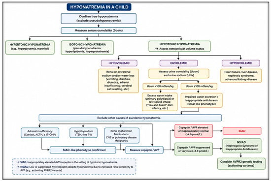
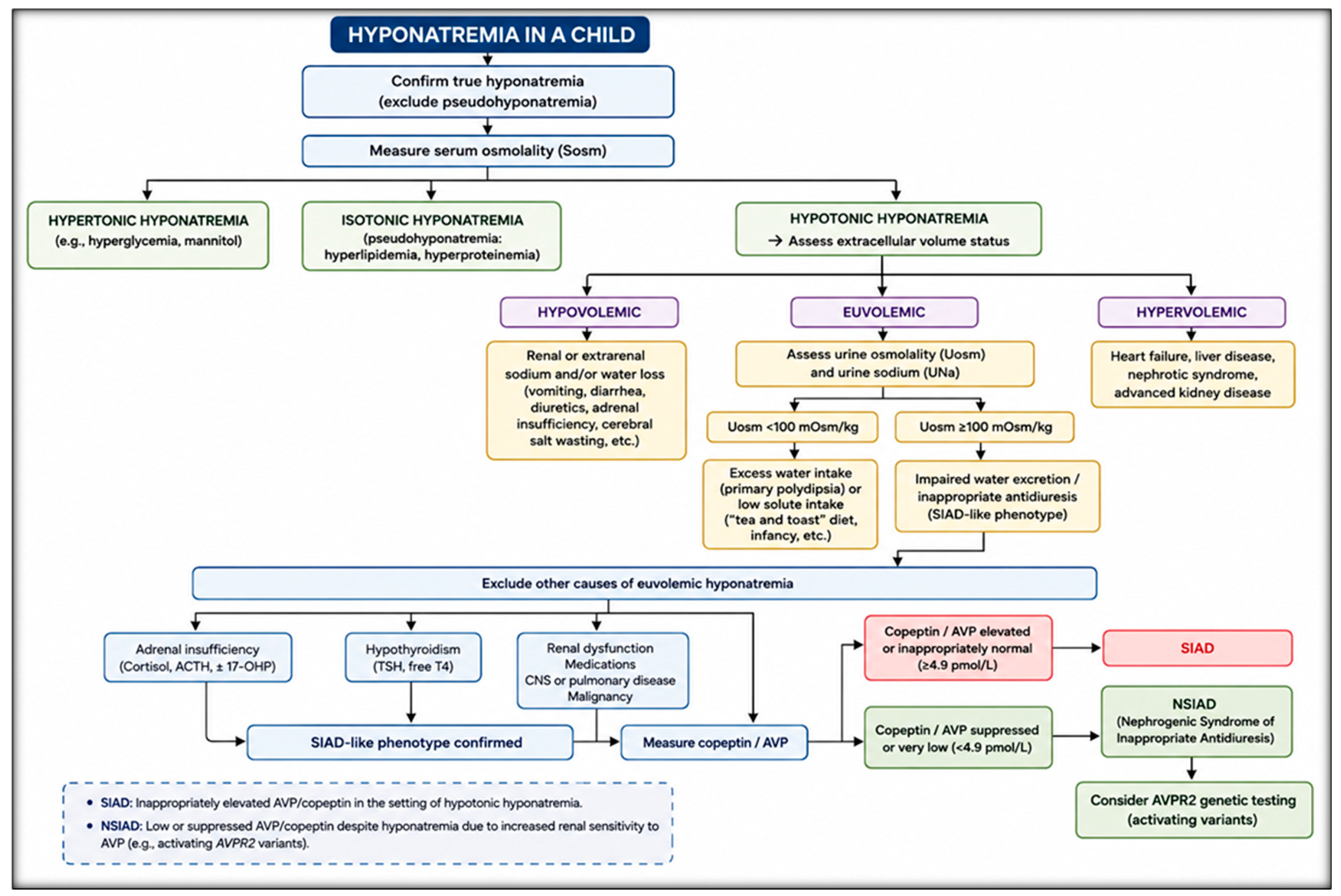
Hyponatremia is fundamentally a disorder of water balance, most commonly resulting from impaired renal free-water excretion [14]. In a clinically euvolemic patient, the coexistence of hypotonic hyponatremia with impaired urinary dilution generally raises the suspicion of SIAD [15]. However, NSIAD can produce an almost indistinguishable biochemical phenotype because both disorders ultimately converge on persistent activation of the renal vasopressin V2 receptor (V2R) pathway and increased aquaporin-2-mediated water reabsorption [8]. Consequently, the distinction between SIAD and NSIAD cannot be made reliably on the basis of serum sodium concentration or clinical volume status alone; rather, it requires assessment of the upstream regulation of the vasopressin pathway. A systematic approach to the differential diagnosis of hyponatremia in children, integrating serum osmolality, volume status, and urine osmolality, is presented in Figure 2.
Figure 2.
Proposed diagnostic approach to hyponatremia in children.
In classical SIAD, persistent or inappropriate secretion of arginine vasopressin (AVP) activates V2R in the renal collecting duct, stimulates intracellular cAMP production, and promotes aquaporin-2 insertion into the apical membrane [16]. In NSIAD, in contrast, activating variants of AVPR2 result in constitutive receptor activity, allowing antidiuretic signaling to persist despite appropriately suppressed circulating AVP [17]. NSIAD therefore represents a form of renal antidiuresis that is functionally independent of the circulating AVP concentration. This distinction is particularly relevant in the present patient.
At a serum sodium concentration of 112 mmol/L and a serum osmolality of 231 mOsm/kg, the copeptin concentration was only 2 pmol/L. Because copeptin is secreted in equimolar amounts with AVP and closely reflects circulating AVP concentrations, its marked suppression in the setting of profound hypotonicity provides strong evidence against persistent inappropriate AVP secretion as the primary mechanism of water retention. Although copeptin should not be considered an absolute surrogate for tissue-level AVP activity, its very low concentration in this biochemical context is highly informative [18]. The subsequent identification of the activating AVPR2 p.(Arg137Cys) variant provides molecular confirmation of an AVP-independent mechanism [19].
The low urinary sodium concentration (<10 mmol/L) initially appears less typical of the classical biochemical description of SIAD, in which urinary sodium excretion is frequently increased [20]. However, urinary sodium concentration is influenced by dietary sodium intake, renal sodium handling, age, and the preceding clinical and therapeutic context [21]. These factors are particularly relevant in infants, in whom adult-derived diagnostic thresholds may be less reliable. Therefore, low urinary sodium should not be regarded as an exclusion criterion for NSIAD when the overall clinical, biochemical, hormonal, and molecular findings support constitutive V2R activation. The present case consequently reinforces the need to interpret urinary sodium within the complete physiological context rather than as an isolated diagnostic discriminator. Transient secondary pseudohypoaldosteronism (PHA) represents another important differential diagnosis of hyponatremia in infancy, particularly in the presence of urinary tract abnormalities such as hydronephrosis [22]. This condition results from transient renal tubular resistance to aldosterone, frequently associated with urinary tract infections or obstructive uropathies, and typically manifests with renal sodium wasting, hyponatremia, hyperkalemia, metabolic acidosis, and elevated renin and aldosterone levels [23]. In our patient, the presence of mild left-sided hydronephrosis (grade I–II) warranted consideration of this diagnosis. However, the low urinary sodium concentration (<10 mmol/L) was less suggestive of active renal salt wasting, although this finding should be interpreted in its clinical and therapeutic context. Furthermore, the markedly suppressed copeptin concentration and identification of the activating hemizygous AVPR2 p.(Arg137Cys) variant supported NSIAD as the primary mechanism underlying the severe hyponatremia.
3.2. The Molecular Basis of NSIAD
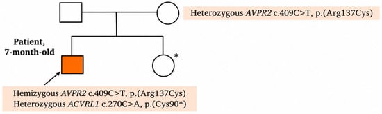
The molecular diagnosis represents the central finding of this case. The hemizygous AVPR2 c.409C>T, p.(Arg137Cys) variant affects a highly conserved residue within the DRY-type motif at the cytoplasmic end of transmembrane helix 3 (TM3), near the TM3/ICL2 boundary, and has been previously associated with NSIAD [24,25,26,27]. Only one heterozygous individual has been reported in gnomAD v2 [28]. The affected amino acid is well conserved in mammals, as well as in the evolutionarily more distant species, which suggests that this position may not tolerate variation. In silico analysis using the Rare Exome Variant Ensemble Learner (REVEL) provided additional computational evidence supporting the potential pathogenicity of the identified variant [29]. REVEL is a machine learning-based ensemble prediction tool that integrates information from 13 individual computational predictors, incorporating sequence conservation and functional annotations to estimate the likelihood that a missense variant is disease-associated [29]. REVEL scores range from 0 to 1, with higher values indicating a greater predicted likelihood of pathogenicity. However, REVEL predictions should be interpreted as supportive evidence rather than definitive proof of pathogenicity, as the tool does not distinguish between gain-of-function and loss-of-function mechanisms [29]. The p.(Arg137Cys) is a well-established gain-of-function variant that affects a conserved DRY/H motif at the cytoplasmic end of TM3, near the TM3/ICL2 boundary [24]. It has been proposed that the disruption of a hydrogen bond between the residues Thr269 and Arg137 may promote stabilization of the receptor in its active conformation [25]; however, this remains a hypothesis-generating structural model based on molecular modelling rather than a directly demonstrated mechanism. The functional importance of p.(Arg137Cys) to NSIAD has been reviewed in detail [27]. The AVPR2 variant is inherited in an X-linked recessive manner. Males hemizygous for the variant are affected, whereas heterozygous females are typically asymptomatic carriers [30]. However, female individuals heterozygous for the variant have been diagnosed with NSIAD and were confirmed to have an unskewed X-linked inactivation pattern in blood or in epithelial cells of the renal collecting duct, whereas an asymptomatic heterozygous woman was reported to present skewed X-linked inactivation of the mutated allele in the blood [26]. This phenotypic variability, attributable to X-chromosome inactivation, does not alter the X-linked recessive classification of the disease. In the present family, the mother was confirmed to be a heterozygous carrier, and a pedigree illustrating the family history and carrier status has been added (Figure 3).
Figure 3.
Family pedigree illustrating the segregation of the AVPR2 c.409C>T (p.Arg137Cys) variant. * Genetic testing for the AVPR2 c.409C>T variant is currently pending.
Previously, the AVPR2 c.409C>T, p.(Arg137Cys) variant has been reported in the literature in several males and females affected with NSIAD during childhood and adulthood, with the reported cases summarized in Table 2. For the summary table, PubMed, Embase, and Scopus were searched from database inception through 24 October 2026, using combinations of the terms ‘nephrogenic syndrome of inappropriate antidiuresis’, ‘NSIAD’, ‘AVPR2’, ‘Arg137Cys’, ‘Arg137Leu’, ‘activating variants’, ‘GNAS’, and ‘Gs alpha’. The reference lists of relevant publications were also screened to identify additional cases. Cases were included when a germline genetic cause of NSIAD was identified; cases of purely drug-associated antidiuresis were excluded from the genetic table. Clinical details were extracted from the original case reports. Importantly, the clinical severity of NSIAD is variable. Reported patients have ranged from asymptomatic hyponatremia detected incidentally to severe manifestations including seizures, irritability, lethargy, and recurrent symptomatic episodes. This phenotypic variability suggests that genotype alone does not completely determine the clinical presentation and that environmental and physiological factors, particularly fluid intake, intercurrent illness, and the ability to regulate water consumption, may influence the severity of hyponatremia.
The infantile presentation in our patient is therefore clinically important. Early life NSIAD may be particularly difficult to recognize because symptoms may be nonspecific or attributed to intercurrent illnesses. In the present case, severe hyponatremia was identified during evaluation for bronchiolitis rather than because of neurological manifestations attributable to hyponatremia. This reinforces previous observations that NSIAD may remain clinically silent until an intercurrent illness or increased free-water intake unmasks the underlying defect [31]. The potential consequences of profound hyponatremia in infancy, including seizures and neurological injury, make early recognition particularly important.
3.3. The Concomitant ACVRL1 Variant: Pathogenicity Versus Phenotypic Relevance
An additional finding in the present case was the heterozygous nonsense ACVRL1 c.270C>A, p.(Cys90*) variant. This variant is absent in The Genome Aggregation Database (gnomAD), a large reference population database (n > 120,000 exomes and >15,000 genomes) which aims to exclude individuals with severe pediatric disease [28] and is predicted to result in loss of normal protein function through premature termination and/or nonsense-mediated mRNA decay [32]. The variant generates a premature stop codon in exon 3 (of a total of 10 exons) and is predicted to lead to a loss of normal protein function, either through protein truncation or nonsense-mediated mRNA decay [32]. Loss-of-function is an established disease mechanism for ACVRL1, which is a well-established gene associated with hereditary hemorrhagic telangiectasia type 2 [33]. This variant has been previously reported in two unrelated patients with HHT [34] and is annotated in ClinVar (Variation ID 654296) [35].
ACVRL1 gene (MIM *601284) on chromosome 12q13.13 encodes a type I cell-surface receptor for the TGF-beta superfamily of ligands [36]. The encoded protein, sometimes termed ALK1, shares similar domain structures with other closely related ALK or activin receptor-like kinase proteins that form a subfamily of receptor serine/threonine kinases [37]. In addition to the canonical transcript, NM_000020.3, which encodes a 503-amino-acid protein, ACVRL1 has an alternative RefSeq transcript encoding a distinct protein isoform [38]. Autosomal dominant variants in ACVRL1 are associated with hereditary hemorrhagic telangiectasia (HHT) type 2 (MIM #600376, also known as Rendu-Osler-Weber syndrome 2 [39]. There are more than 475 variants in ACVRL1 annotated as disease-causing (DM) in the HGMD Professional variant database (version), including all kinds of variants [40]. The vast majority of the DM variants are associated with hereditary haemorrhagic telangiectasia. In a study of 23 disease-causing ACVLR1 variants, the missense variants were found to have a critical effect on the protein structure and abolish or partially reduce the protein function [41]. According to ClinGen, the association with HHT is definitive [42] and there is sufficient evidence for haploinsufficiency [43]. In addition, ClinGen has published a recommended HHT-specific classification for ACVRL1 variants [43].
Importantly, however, there is currently no established mechanistic basis linking heterozygous ACVRL1 loss-of-function variants to NSIAD or to the patient’s severe hyponatremia. In the context of the present case, the ACVRL1 finding should therefore be regarded as a clinically relevant concomitant genetic diagnosis rather than as the molecular explanation for the disorder of water homeostasis. This distinction is important to avoid attributing the complete phenotype to a second pathogenic variant simply because it was identified by broad genomic testing.
Nevertheless, the ACVRL1 finding has independent clinical significance. Hereditary hemorrhagic telangiectasia may have age-dependent manifestations, and clinically overt features may be absent during infancy or early childhood [44]. Therefore, the identification of a pathogenic ACVRL1 variant warrants appropriate genetic counselling and longitudinal clinical assessment for manifestations associated with ACVRL1-related hereditary hemorrhagic telangiectasia, according to established multidisciplinary recommendations [45].
The absence of current clinical manifestations of HHT in this infant does not exclude later phenotypic expression. Because HHT is an age-dependent disorder with full penetrance, the identification of this variant warrants a structured surveillance plan according to current HHT guidelines [39]. This plan should include annual clinical review, screening for pulmonary arteriovenous malformations by contrast echocardiography with agitated saline (followed by CT angiography if positive), screening for cerebral arteriovenous malformations by MRI/MRA, and gastrointestinal screening, initiated in childhood according to institutional protocols [39].
The severe microcytic hypochromic anaemia observed in this patient (Hb 8.8 g/dL, ferritin <6 µg/L) requires iron replacement and longitudinal follow-up. Although nutritional iron deficiency is the most likely explanation in this infant, chronic gastrointestinal blood loss from HHT-related telangiectasias may contribute to iron deficiency later in life and should be considered during follow-up.
Parental testing for the ACVRL1 variant was recommended for family assessment and counselling, as an affected parent would confirm familial HHT and allow appropriate screening of at-risk relatives. Finally, ACVRL1 loss-of-function variants are associated with a rare but severe form of pulmonary arterial hypertension (PAH), and pulmonary arteriovenous malformations are present in approximately 40% of patients with HHT [46]. Awareness of these associations is therefore important for long-term multidisciplinary follow-up.
3.4. Therapeutic Considerations and Long-Term Management
The therapeutic objective in NSIAD differs conceptually from that in SIAD. Because the primary abnormality is constitutive activation of the renal V2R, treatment is directed toward limiting the consequences of persistent renal water retention rather than suppressing AVP secretion [1]. Fluid restriction is therefore the principal therapeutic strategy and has been successfully used in both pediatric and adult patients with NSIAD [47]. In infants, however, fluid restriction requires particular caution because fluid intake is largely caregiver-controlled and excessive restriction may compromise adequate nutrition, growth, and hydration [48].
Oral urea has also been reported as an effective option, as its osmotic effect increases renal solute excretion and promotes free-water clearance [31]. In pediatric patients, doses of approximately 0.1 g/kg/day divided into four administrations have been reported, with doses up to 2 g/kg/day or a maximum total daily dose of 60 g described in the literature [49]. By increasing osmotic water excretion, urea may enhance free-water clearance and reduce the degree of hyponatremia associated with impaired renal water excretion [50]. An important practical consideration in infants is the palatability and acceptability of oral urea. Preparation in a formulation that can be administered reliably by caregivers may facilitate adherence, although the taste and gastrointestinal tolerability of urea can limit long-term use. In the present patient, a commercially available flavored oral urea preparation was used, allowing gradual dose escalation. Caregivers should receive clear instructions regarding preparation, dosing, and administration, as dosing errors may result in either inadequate correction of hyponatremia or excessive osmotic diuresis.
Other therapeutic approaches used in SIAD require careful consideration in NSIAD. Pharmacological V2-receptor antagonism (vaptans) was initially proposed on the mechanistic rationale of constitutive V2-receptor activation [51]. However, functional studies have demonstrated that the constitutive activity of the p.(Arg137Cys) and p.(Arg137Leu) receptors is not inhibited by vaptans, indicating that these variants confer resistance to V2-receptor antagonism [23]. Vaptans should therefore not be presented as an established mechanistic solution for NSIAD caused by these activating AVPR2 variants. Experience with vasopressin receptor antagonists in infants with genetically confirmed NSIAD remains limited [52], and their use in this age group may be constrained by safety, pharmacokinetic, and regulatory considerations [44]. Although vaptans have shown efficacy in adults with SIADH [45], their role in NSIAD caused by activating AVPR2 variants is fundamentally different, as these mutant receptors are resistant to antagonism [23]. Similarly, sodium supplementation alone does not correct the underlying defect because the primary disturbance is excess water retention rather than an isolated sodium deficit [53]. In the present case, the initial increase in serum sodium following sodium chloride and mineralocorticoid administration occurred before the molecular diagnosis was established, but these interventions were subsequently reconsidered after adrenal insufficiency and primary mineralocorticoid deficiency had been excluded.
Gain-of-function variants in GNAS, encoding the Gsα protein, represent an additional downstream genetic cause of NSIAD. Because these variants constitutively activate the Gsα pathway downstream of the V2 receptor, V2-receptor antagonists would not be expected to correct the defect. The diagnostic algorithm for NSIAD should therefore consider testing for both AVPR2 and GNAS, and future case series should include GNAS-related cases alongside AVPR2-related cases.
The clinical course also illustrates an important principle in the management of severe hyponatremia: correction should be controlled and individualized. In chronic or longstanding hyponatremia, an excessively rapid increase in serum sodium may carry a risk of neurological complications associated with overly rapid correction [54]. Conversely, persistent severe hyponatremia exposes the infant to the risk of cerebral edema and neurological dysfunction [55]. Serial measurements of serum sodium are therefore essential during initiation and adjustment of therapy, particularly when fluid restriction and osmotic therapy are combined [56]. The rate of sodium correction should be considered together with the estimated duration of hyponatremia and the presence of risk factors for neurological complications [57].
Long-term management should consequently include individualized fluid-intake guidance, regular monitoring of serum sodium, and education of caregivers regarding situations associated with an increased risk of water intoxication.
Table 2.
Summary of Reported Cases of Nephrogenic Syndrome of Inappropriate Antidiuresis.
4. Conclusions
Nephrogenic syndrome of inappropriate antidiuresis should be considered in infants presenting with severe hypotonic hyponatremia when conventional causes of impaired water excretion have been excluded, particularly when circulating vasopressin activity is appropriately suppressed. In this setting, a markedly low copeptin concentration may provide an important diagnostic clue to an AVP-independent mechanism, while molecular analysis of AVPR2 can establish the diagnosis by demonstrating constitutive V2 receptor activation.
The AVPR2 p.(Arg137Cys) variant identified in our patient is a well-established gain-of-function cause of NSIAD. Its presence in this infant illustrates that NSIAD may present with profound yet clinically silent hyponatraemia during infancy and may be uncovered incidentally in the context of an intercurrent illness, particularly when hyponatraemia persists after resolution of the acute illness and is accompanied by suppressed copeptin. The concomitant identification of a pathogenic ACVRL1 variant represents an independent clinical diagnosis requiring HHT-specific surveillance and family counselling. This finding emphasizes the value of broad genomic testing in selected patients without implying a shared mechanism with the electrolyte disorder, and underscores the importance of distinguishing the molecular basis of the presenting condition from additional actionable genetic findings.
Management of NSIAD requires an individualized balance between limiting free-water intake and maintaining adequate hydration and nutrition. Fluid restriction and oral urea represent practical therapeutic strategies, but their use in infancy requires close biochemical and clinical monitoring to avoid both persistent severe hyponatremia and excessively rapid correction. Importantly, V2-receptor antagonists should not be regarded as an established therapeutic option for NSIAD caused by the p.(Arg137Cys) or p.(Arg137Leu) variants, as functional studies have demonstrated that these mutant receptors are resistant to vaptan inhibition. Long-term management should include regular assessment of serum sodium, individualized fluid-intake recommendations, caregiver education, and surveillance during intercurrent illnesses. The identification of GNAS gain-of-function variants as an additional downstream cause of NSIAD further supports a diagnostic algorithm that considers both AVPR2 and GNAS, and informs therapeutic expectations, since receptor antagonists would not be expected to correct a constitutively activated downstream Gsα pathway.
Integration of clinical phenotype, serum osmolality, copeptin concentrations, and molecular analysis of the AVP–V2 receptor pathway may facilitate earlier recognition of NSIAD and prevent potentially serious neurological complications. Recognition of this rare disorder is particularly relevant in infancy, when severe hyponatremia may remain clinically unapparent despite substantial biochemical abnormalities. Finally, our case illustrates that comprehensive genomic testing in infants with severe unexplained hyponatraemia may uncover additional actionable diagnoses unrelated to the presenting phenotype, which require independent surveillance and family counselling.
Author Contributions
Conceptualization, I.V., E.-L.S., L.I.B. and L.M.T.; methodology, I.V., E.-L.S., L.I.B. and L.M.T.; software, I.V., E.-L.S., L.I.B. and L.M.T.; validation, I.V.; formal analysis, L.M.T.; investigation, I.V., E.-L.S., L.I.B. and L.M.T.; resources, I.V.; data curation, I.V.; writing—original draft preparation I.V., E.-L.S., L.I.B. and L.M.T.; writing—review and editing, I.V., E.-L.S., L.I.B. and L.M.T.; visualization, I.V., E.-L.S., L.I.B. and L.M.T.; supervision, L.M.T.; project administration, L.M.T.; funding acquisition, I.V. All authors have read and agreed to the published version of the manuscript.
Funding
This research received no external funding.
Institutional Review Board Statement
The study was conducted in accordance with the Declaration of Helsinki, and approved by the Ethics Committee of “Sfânta Maria” Emergency Children Hospital Iasi Romania (No. 22447/9.07.2026).
Informed Consent Statement
Written informed consent has been obtained from the legal guardian of the patient to publish this paper.
Data Availability Statement
The data presented in this study are available from the corresponding author upon reasonable request and with the permission of the patient’s legal guardian. The data are not publicly available due to privacy and ethical restrictions.
Acknowledgments
During the preparation of this manuscript, ChatGPT (GPT-5.6 Luna; OpenAI) was used solely to improve the English language, including grammar, clarity, and readability. It was not used for data generation, data analysis, interpretation of the findings, or scientific conclusions. The authors critically reviewed and revised all AI-assisted text and take full responsibility for the accuracy and integrity of the manuscript.
Conflicts of Interest
The authors declare no conflicts of interest.
Abbreviations
The following abbreviations are used in this manuscript:
| 17-OHP | 17-Hydroxyprogesterone |
| ACMG | American College of Medical Genetics and Genomics |
| ACTH | Adrenocorticotropic hormone |
| ACVRL1 | Activin A receptor-like type 1 |
| ALK1 | Activin receptor-like kinase 1 |
| AMH | Anti-Müllerian hormone |
| AMP | Association for Molecular Pathology |
| AVP | Arginine vasopressin |
| AVPR2 | Arginine vasopressin receptor 2 |
| BUN | Blood urea nitrogen |
| cAMP | Cyclic adenosine monophosphate |
| CNV | Copy number variation |
| CT | Computed tomography |
| CT-proAVP | C-terminal pro-arginine vasopressin |
| DHEAS | Dehydroepiandrosterone sulfate |
| fT4 | Free thyroxine |
| gnomAD | Genome Aggregation Database |
| Gsα | Stimulatory G-protein alpha subunit |
| HGMD | Human Gene Mutation Database |
| HGVS | Human Genome Variation Society |
| HHT | Hereditary hemorrhagic telangiectasia |
| ICL2 | Second intracellular loop |
| MCV | Mean corpuscular volume |
| MIM | Mendelian Inheritance in Man |
| MRA | Magnetic resonance angiography |
| NGS | Next-generation sequencing |
| NSIAD | Nephrogenic syndrome of inappropriate antidiuresis |
| PAH | Pulmonary arterial hypertension |
| PHA | Pseudohypoaldosteronism |
| PITPNB | Phosphatidylinositol transfer protein beta |
| REVEL | Rare Exome Variant Ensemble Learner |
| SIAD | Syndrome of inappropriate antidiuresis |
| SIADH | Syndrome of inappropriate antidiuretic hormone secretion |
| TGF-β | Transforming growth factor beta |
| TM3 | Third transmembrane helix |
| TRACE | Time-Resolved Amplified Cryptate Emission |
| TSH | Thyroid-stimulating hormone |
| V2R | Vasopressin V2 receptor |
| VUS | Variant of uncertain significance |
| WES | Whole-exome sequencing |
| WHO | World Health Organization |
References
- Feldman, B.J.; Rosenthal, S.M.; Vargas, G.A.; Fenwick, R.G.; Huang, E.A.; Matsuda-Abedini, M.; Lustig, R.H.; Mathias, R.S.; Portale, A.A.; Miller, W.L.; et al. Nephrogenic Syndrome of Inappropriate Antidiuresis. N. Engl. J. Med. 2005, 352, 1884–1890. [Google Scholar] [CrossRef] [Scilit] [PubMed]
- Verbalis, J.G. Hyponatremia: Pathophysiology of Different Etiologies. Best Pract. Res. Clin. Endocrinol. Metab. 2026, 40, 102084. [Google Scholar] [CrossRef] [Scilit] [PubMed]
- Martin-Grace, J.; Tomkins, M.; O’Reilly, M.W.; Thompson, C.J.; Sherlock, M. Approach to the Patient: Hyponatremia and the Syndrome of Inappropriate Antidiuresis (SIAD). J. Clin. Endocrinol. Metab. 2022, 107, 2362–2376. [Google Scholar] [CrossRef] [Scilit] [PubMed]
- Shepshelovich, D.; Schechter, A.; Calvarysky, B.; Diker-Cohen, T.; Rozen-Zvi, B.; Gafter-Gvili, A. Medication-induced SIADH: Distribution and Characterization According to Medication Class. Br. J. Clin. Pharmacol. 2017, 83, 1801–1807. [Google Scholar] [CrossRef] [Scilit] [PubMed]
- Al Zaman, K.; Alhebsi, A.A.; Almheiri, A.; Alhosani, H.; Alshehhi, N.T.; Alwheibi, E.S. Syndrome of Inappropriate Antidiuretic Hormone (SIADH) in Chronic Respiratory Diseases: A Comprehensive Review. Cureus 2025, 17, e77407. [Google Scholar] [CrossRef] [Scilit] [PubMed]
- Cui, H.; He, G.; Yang, S.; Lv, Y.; Jiang, Z.; Gang, X.; Wang, G. Inappropriate Antidiuretic Hormone Secretion and Cerebral Salt-Wasting Syndromes in Neurological Patients. Front. Neurosci. 2019, 13, 1170. [Google Scholar] [CrossRef] [Scilit] [PubMed]
- Goldvaser, H.; Rozen-Zvi, B.; Yerushalmi, R.; Gafter-Gvili, A.; Lahav, M.; Shepshelovich, D. Malignancy Associated SIADH: Characterization and Clinical Implications. Acta Oncol. 2016, 55, 1190–1195. [Google Scholar] [CrossRef] [Scilit] [PubMed]
- Decaux, G.; Vandergheynst, F.; Bouko, Y.; Parma, J.; Vassart, G.; Vilain, C. Nephrogenic Syndrome of Inappropriate Antidiuresis in Adults: High Phenotypic Variability in Men and Women from a Large Pedigree. J. Am. Soc. Nephrol. 2007, 18, 606–612. [Google Scholar] [CrossRef] [Scilit] [PubMed]
- Available online: https://radiopaedia.org/articles/sfu-grading-system-of-hydronephrosis (accessed on 24 October 2026).
- Rasouli, M. Basic Concepts and Practical Equations on Osmolality: Biochemical Approach. Clin. Biochem. 2016, 49, 936–941. [Google Scholar] [CrossRef] [Scilit] [PubMed]
- Pande, A.R.; Aggarwal, A.; Chandra, K.P. Inappropriate Use of Serum Osmolarity Formulas in the Measurement of Urine Osmolality. Indian J. Endocrinol. Metab. 2025, 29, 347–348. [Google Scholar] [CrossRef] [Scilit] [PubMed]
- Huang, E.A.; Feldman, B.J.; Schwartz, I.D.; Geller, D.H.; Rosenthal, S.M.; Gitelman, S.E. Oral Urea for the Treatment of Chronic Syndrome of Inappropriate Antidiuresis in Children. J. Pediatr. 2006, 148, 128–131. [Google Scholar] [CrossRef] [Scilit] [PubMed]
- ClinVar. Available online: https://www.ncbi.nlm.nih.gov/clinvar/ (accessed on 26 October 2026).
- Sumi, H.; Tominaga, N.; Fujita, Y.; Verbalis, J.G.; The Electrolyte Winter Seminar, Collaborative Group. Pathophysiology, Symptoms, Outcomes, and Evaluation of Hyponatremia: Comprehension and Best Clinical Practice. Clin. Exp. Nephrol. 2025, 29, 134–148. [Google Scholar] [CrossRef] [Scilit] [PubMed]
- Reddy, P. Clinical Approach to Euvolemic Hyponatremia. Cureus 2023, 15, e35574. [Google Scholar] [CrossRef] [Scilit] [PubMed]
- Robertson, G.L. Regulation of Arginine Vasopressin in the Syndrome of Inappropriate Antidiuresis. Am. J. Med. 2006, 119, S36–S42. [Google Scholar] [CrossRef] [Scilit] [PubMed]
- Erdélyi, L.S.; Alexander Mann, W.; Morris-Rosendahl, D.J.; Groß, U.; Nagel, M.; Várnai, P.; Balla, A.; Hunyady, L. Mutation in the V2 Vasopressin Receptor Gene, AVPR2, Causes Nephrogenic Syndrome of Inappropriate Diuresis. Kidney Int. 2015, 88, 1070–1078. [Google Scholar] [CrossRef] [Scilit] [PubMed]
- Refardt, J.; Winzeler, B.; Christ-Crain, M. Copeptin and Its Role in the Diagnosis of Diabetes Insipidus and the Syndrome of Inappropriate Antidiuresis. Clin. Endocrinol. 2019, 91, 22–32. [Google Scholar] [CrossRef] [Scilit] [PubMed]
- ClinVar ClinVar. Available online: https://www.ncbi.nlm.nih.gov/clinvar/RCV000011601/ (accessed on 8 September 2026).
- Pliquett, R.U.; Obermüller, N.; Feingold, K.R.; Adler, R.A.; Ahmed, S.F.; Anawalt, B.; Blackman, M.R.; Chrousos, G.; Corpas, E.; De Herder, W.W.; et al. Endocrine Testing for the Syndrome of Inappropriate Antidiuretic Hormone Secretion (SIADH); MDText.com, Inc.: Dartmouth, MA, USA, 2000. [Google Scholar]
- Xie, H.; Wen, X.; Fan, F.; Jia, J.; Huo, Y.; Gong, Y.; Zhang, Y. Association between Urinary Sodium Excretion and All-Cause Mortality: A Cohort Study in a Chinese Community-Based Population. BMC Cardiovasc. Disord. 2025, 25, 177. [Google Scholar] [CrossRef] [Scilit] [PubMed]
- Betti, C.; Lavagno, C.; Bianchetti, M.G.; Kottanattu, L.; Lava, S.A.G.; Schera, F.; Lacalamita, M.C.; Milani, G.P. Transient Secondary Pseudo-Hypoaldosteronism in Infants with Urinary Tract Infections: Systematic Literature Review. Eur. J. Pediatr. 2024, 183, 4205–4214. [Google Scholar] [CrossRef] [Scilit] [PubMed]
- Rodríguez-Soriano, J.; Vallo, A.; Oliveros, R.; Castillo, G. Transient Pseudohypoaldosteronism Secondary to Obstructive Uropathy in Infancy. J. Pediatr. 1983, 103, 375–380. [Google Scholar] [CrossRef] [Scilit] [PubMed]
- Tiulpakov, A.; White, C.W.; Abhayawardana, R.S.; See, H.B.; Chan, A.S.; Seeber, R.M.; Heng, J.I.; Dedov, I.; Pavlos, N.J.; Pfleger, K.D.G. Mutations of Vasopressin Receptor 2 Including Novel L312S Have Differential Effects on Trafficking. Mol. Endocrinol. 2016, 30, 889–904. [Google Scholar] [CrossRef] [Scilit] [PubMed]
- Powlson, A.S.; Challis, B.G.; Halsall, D.J.; Schoenmakers, E.; Gurnell, M. Nephrogenic Syndrome of Inappropriate Antidiuresis Secondary to an Activating Mutation in the Arginine Vasopressin Receptor AVPR2. Clin. Endocrinol. 2016, 85, 306–312. [Google Scholar] [CrossRef] [Scilit] [PubMed]
- Mammadova, J.; Kara, C.; Çelebi Bitkin, E.; İzci Güllü, E.; Aydın, M. Nephrogenic Syndrome of Inappropriate Antidiuresis Mimicking Hyporeninemic Hypoaldosteronism: Case Report of Two Infants. J. Clin. Res. Pediatr. Endocrinol. 2023, 15, 214–219. [Google Scholar] [CrossRef] [Scilit] [PubMed]
- Rochdi, M.D.; Vargas, G.A.; Carpentier, E.; Oligny-Longpré, G.; Chen, S.; Kovoor, A.; Gitelman, S.E.; Rosenthal, S.M.; von Zastrow, M.; Bouvier, M. Functional Characterization of Vasopressin Type 2 Receptor Substitutions (R137H/C/L) Leading to Nephrogenic Diabetes Insipidus and Nephrogenic Syndrome of Inappropriate Antidiuresis: Implications for Treatments. Mol. Pharmacol. 2010, 77, 836–845. [Google Scholar] [CrossRef] [Scilit] [PubMed]
- Chen, S.; Francioli, L.C.; Goodrich, J.K.; Collins, R.L.; Kanai, M.; Wang, Q.; Alföldi, J.; Watts, N.A.; Vittal, C.; Gauthier, L.D.; et al. A Genomic Mutational Constraint Map Using Variation in 76,156 Human Genomes. Nature 2024, 625, 92–100, Erratum in Nature 2024, 626, E1. [Google Scholar] [CrossRef] [Scilit] [PubMed]
- Ioannidis, N.M.; Rothstein, J.H.; Pejaver, V.; Middha, S.; McDonnell, S.K.; Baheti, S.; Musolf, A.; Li, Q.; Holzinger, E.; Karyadi, D.; et al. REVEL: An Ensemble Method for Predicting the Pathogenicity of Rare Missense Variants. Am. J. Hum. Genet. 2016, 99, 877–885. [Google Scholar] [CrossRef] [Scilit] [PubMed]
- Hague, J.; Casey, R.; Bruty, J.; Legerton, T.; Abbs, S.; Oddy, S.; Powlson, A.S.; Majeed, M.; Gurnell, M.; Park, S.-M.; et al. Adult Female with Symptomatic AVPR2-Related Nephrogenic Syndrome of Inappropriate Antidiuresis (NSIAD). Endocrinol. Diabetes Metab. Case Rep. 2018, 2018, 17–139. [Google Scholar] [CrossRef] [Scilit] [PubMed]
- Morin, D.; Tenenbaum, J.; Ranchin, B.; Durroux, T. Nephrogenic Syndrome of Inappropriate Antidiuresis. Int. J. Pediatr. 2012, 2012, 937175. [Google Scholar] [CrossRef] [Scilit] [PubMed]
- DeMille, D.; McDonald, J.; Bernabeu, C.; Racher, H.; Olivieri, C.; Cantarini, C.; Sbalchiero, A.; Thompson, B.A.; Jovine, L.; Shovlin, C.L.; et al. Specifications of the ACMG/AMP Variant Curation Guidelines for Hereditary Hemorrhagic Telangiectasia Genes—ENG and ACVRL1. Hum. Mutat. 2024, 2024, 3043736. [Google Scholar] [CrossRef] [Scilit] [PubMed]
- McDonald, J.; Gossage, J.R.; Stevenson, D.A. Hereditary Hemorrhagic Telangiectasia. In GeneReviews(®); University of Washington: Seattle, WA, USA, 1993. [Google Scholar]
- McDonald, J.; Damjanovich, K.; Millson, A.; Wooderchak, W.; Chibuk, J.M.; Stevenson, D.A.; Gedge, F.; Bayrak-Toydemir, P. Molecular Diagnosis in Hereditary Hemorrhagic Telangiectasia: Findings in a Series Tested Simultaneously by Sequencing and Deletion/Duplication Analysis. Clin. Genet. 2011, 79, 335–344. [Google Scholar] [CrossRef] [Scilit] [PubMed]
- VCV000065429.4—ClinVar—NCBI. Available online: https://www.ncbi.nlm.nih.gov/clinvar/variation/65429/ (accessed on 26 October 2026).
- Iwasa, T.; Urasaki, A.; Kakihana, Y.; Nagata-Akaho, N.; Harada, Y.; Takeda, S.; Kawamura, T.; Shiraishi, I.; Kurosaki, K.; Morisaki, H.; et al. Computational and Experimental Analyses for Pathogenicity Prediction of ACVRL1 Missense Variants in Hereditary Hemorrhagic Telangiectasia. J. Clin. Med. 2023, 12, 5002. [Google Scholar] [CrossRef] [Scilit] [PubMed]
- ACVRL1 Activin A Receptor like Type 1 [Homo Sapiens (Human)]—Gene—NCBI. Available online: https://www.ncbi.nlm.nih.gov/gene/94?utm_source=chatgpt.com (accessed on 17 September 2026).
- Homo Sapiens Activin A Receptor like Type 1 (ACVRL1), Transcript Varia—Nucleotide—NCBI. Available online: https://www.ncbi.nlm.nih.gov/nuccore/NM_000020 (accessed on 17 September 2026).
- Available online: https://omim.org/entry/600376 (accessed on 17 September 2026).
- HGMD® Gene Result. Available online: https://www.hgmd.cf.ac.uk/ac/gene.php?gene=ACVRL1 (accessed on 1 May 2026).
- Alaa el Din, F.; Patri, S.; Thoreau, V.; Rodriguez-Ballesteros, M.; Hamade, E.; Bailly, S.; Gilbert-Dussardier, B.; Abou Merhi, R.; Kitzis, A. Functional and Splicing Defect Analysis of 23 ACVRL1 Mutations in a Cohort of Patients Affected by Hereditary Hemorrhagic Telangiectasia. PLoS ONE 2015, 10, e0132111. [Google Scholar] [CrossRef] [Scilit] [PubMed]
- Member Directory ACVRL1. Available online: https://search.clinicalgenome.org/kb/genes/HGNC:175 (accessed on 1 May 2026).
- Stenson, P.D.; Mort, M.; Ball, E.V.; Chapman, M.; Evans, K.; Azevedo, L.; Hayden, M.; Heywood, S.; Millar, D.S.; Phillips, A.D.; et al. The Human Gene Mutation Database (HGMD®): Optimizing Its Use in a Clinical Diagnostic or Research Setting. Hum. Genet. 2020, 139, 1197–1207. [Google Scholar] [CrossRef] [Scilit] [PubMed]
- Danesino, C.; Cantarini, C.; Olivieri, C. Hereditary Hemorrhagic Telangiectasia in Pediatric Age: Focus on Genetics and Diagnosis. Pediatr. Rep. 2023, 15, 129–142. [Google Scholar] [CrossRef] [Scilit] [PubMed]
- Faughnan, M.E.; Mager, J.J.; Hetts, S.W.; Palda, V.A.; Lang-Robertson, K.; Buscarini, E.; Deslandres, E.; Kasthuri, R.S.; Lausman, A.; Poetker, D.; et al. Second International Guidelines for the Diagnosis and Management of Hereditary Hemorrhagic Telangiectasia. Ann. Intern. Med. 2020, 173, 989–1001. [Google Scholar] [CrossRef] [Scilit] [PubMed]
- Rozenberg, D.; Leek, E.; Faughnan, M.E. Prevalence and Nature of Dyspnea in Patients with Hereditary Hemorrhagic Telangiectasia (HHT). Respir. Med. 2015, 109, 768–777. [Google Scholar] [CrossRef] [Scilit] [PubMed]
- Vandergheynst, F.; Brachet, C.; Heinrichs, C.; Decaux, G. Long-Term Treatment of Hyponatremic Patients with Nephrogenic Syndrome of Inappropriate Antidiuresis: Personal Experience and Review of Published Case Reports. Nephron Clin. Pract. 2012, 120, c168–c172. [Google Scholar] [CrossRef] [Scilit] [PubMed]
- Kight, B.P.; Waseem, M. Pediatric Fluid Management; StatPearls Publishing: Treasure Island, FL, USA, 2025. [Google Scholar]
- Driano, J.E.; Lteif, A.N.; Creo, A.L. Vasopressin-Dependent Disorders: What Is New in Children? Pediatrics 2021, 147, e2020022848. [Google Scholar] [CrossRef] [Scilit] [PubMed]
- Warren, A.M.; Grossmann, M.; Christ-Crain, M.; Russell, N. Syndrome of Inappropriate Antidiuresis: From Pathophysiology to Management. Endocr. Rev. 2023, 44, 819–861. [Google Scholar] [CrossRef] [Scilit] [PubMed]
- Krisanapan, P.; Tangpanithandee, S.; Thongprayoon, C.; Pattharanitima, P.; Kleindienst, A.; Miao, J.; Craici, I.M.; Mao, M.A.; Cheungpasitporn, W. Safety and Efficacy of Vaptans in the Treatment of Hyponatremia from Syndrome of Inappropriate Antidiuretic Hormone Secretion (SIADH): A Systematic Review and Meta-Analysis. J. Clin. Med. 2023, 12, 5483. [Google Scholar] [CrossRef] [Scilit] [PubMed]
- Koksoy, A.Y.; Kurtul, M.; Ozsahin, A.K.; Cayci, F.S.; Tayfun, M.; Bayrakci, U.S. Tolvaptan Use to Treat SIADH in a Child. J. Pediatr. Pharmacol. Ther. 2018, 23, 494–498. [Google Scholar] [CrossRef] [Scilit] [PubMed]
- Rout, P.; Badireddy, M. Hyponatremia; StatPearls Publishing: Treasure Island, FL, USA, 2025. [Google Scholar]
- Spasovski, G.; Vanholder, R.; Allolio, B.; Annane, D.; Ball, S.; Bichet, D.; Decaux, G.; Fenske, W.; Hoorn, E.J.; Ichai, C.; et al. Clinical Practice Guideline on Diagnosis and Treatment of Hyponatraemia. Nephrol. Dial. Transplant. 2014, 29, i1–i39. [Google Scholar] [CrossRef] [Scilit] [PubMed]
- Gankam Kengne, F.; Decaux, G. Hyponatremia and the Brain. Kidney Int. Rep. 2018, 3, 24–35. [Google Scholar] [CrossRef] [Scilit] [PubMed]
- Nagase, K.; Watanabe, T.; Nomura, A.; Nagase, F.N.; Iwasaki, K.; Nakamura, Y.; Ikai, H.; Yamamoto, M.; Murai, Y.; Yokoyama-Kokuryo, W.; et al. Predictive Correction of Serum Sodium Concentration with Formulas Derived from the Edelman Equation in Patients with Severe Hyponatremia. Sci. Rep. 2023, 13, 1783. [Google Scholar] [CrossRef] [Scilit] [PubMed]
- See, X.Y.; Chang, Y.-C.; Peng, C.-Y.; Wang, S.-S.; Chi, K.-Y.; Chiang, C.-H.; Chiang, C.-H. Rate of Sodium Correction and Osmotic Demyelination Syndrome in Severe Hyponatremia: A Meta-Analysis. J. Crit. Care Med. 2024, 10, 209–212. [Google Scholar] [CrossRef] [Scilit] [PubMed]
- Soule, S.; Florkowski, C.; Potter, H.; Pattison, D.; Swan, M.; Hunt, P.; George, P. Intermittent Severe, Symptomatic Hyponatraemia Due to the Nephrogenic Syndrome of Inappropriate Antidiuresis. Ann. Clin. Biochem. 2008, 45, 520–523. [Google Scholar] [CrossRef] [Scilit] [PubMed]
- Cho, Y.H.; Gitelman, S.; Rosenthal, S.; Ambler, G. Long-Term Outcomes in a Family with Nephrogenic Syndrome of Inappropriate Antidiuresis. Int. J. Pediatr. Endocrinol. 2009, 2009, 431527. [Google Scholar] [CrossRef] [Scilit] [PubMed]
- Carpentier, E.; Greenbaum, L.A.; Rochdi, D.; Abrol, R.; Goddard, W.A., 3rd; Bichet, D.G.; Bouvier, M. Identification and Characterization of an Activating F229V Substitution in the V2 Vasopressin Receptor in an Infant with NSIAD. J. Am. Soc. Nephrol. 2012, 23, 1635–1640. [Google Scholar] [CrossRef] [Scilit] [PubMed]
- Brachet, C.; Vandergheynst, F.; Heinrichs, C. Nephrogenic Syndrome of Inappropriate Antidiuresis in a Female Neonate: Review of the Clinical Presentation in Females. Horm. Res. Paediatr. 2015, 84, 65–67. [Google Scholar] [CrossRef] [Scilit] [PubMed]
- Chen, J.J.; Gong, C.X.; Wei, L.Y.; Cao, B.Y.; Wu, D.; Liu, Y.; Li, W.J. Diagnosis and follow-up of 2 cases of pediatric nephrogenic syndrome of inappropriate antidiuresis resulting from activating mutation in AVPR2 and literature review. Zhonghua er Ke Za Zhi/Chin. J. Pediatr. 2021, 59, 125–130. [Google Scholar] [CrossRef] [PubMed]
- Nephrogenic Syndrome of Inappropriate Antidiuresis—A Case Report in Pediatric Age. Available online: https://www.heraldopenaccess.us/openaccess/nephrogenic-syndrome-of-inappropriate-antidiuresis-a-case-report-in-pediatric-age (accessed on 24 October 2026).
- Chen, X.-J.; Kou, M.; Feng, M.; Wang, L.-H.; Wei, J.-J. Nephrogenic Syndrome of Inappropriate Antidiuresis: Case Report and Genetic Perspectives. Nephrology 2026, 31, e70250. [Google Scholar] [CrossRef] [Scilit] [PubMed]
- Miyado, M.; Fukami, M.; Takada, S.; Terao, M.; Nakabayashi, K.; Hata, K.; Matsubara, Y.; Tanaka, Y.; Sasaki, G.; Nagasaki, K.; et al. Germline-Derived Gain-of-Function Variants of Gsα-Coding GNAS Gene Identified in Nephrogenic Syndrome of Inappropriate Antidiuresis. J. Am. Soc. Nephrol. 2019, 30, 877–889. [Google Scholar] [CrossRef] [Scilit] [PubMed]
- Biebermann, H.; Kleinau, G.; Schnabel, D.; Bockenhauer, D.; Wilson, L.C.; Tully, I.; Kiff, S.; Scheerer, P.; Reyes, M.; Zschunke, S.; et al. A New Multisystem Disorder Caused by the Gαs Mutation p.F376V. J. Clin. Endocrinol. Metab. 2019, 104, 1079–1089. [Google Scholar] [CrossRef] [Scilit] [PubMed]
- Cavarzere, P.; Gastaldi, A.; Elli, F.M.; Gaudino, R.; Peverelli, E.; Brugnara, M.; Thiele, S.; Granata, F.; Mantovani, G.; Antoniazzi, F. A Complex Pheotype in a Girl with a Novel Heterozygous Missense Variant (p.Ile56Phe) of the GNAS Gene. Orphanet J. Rare Dis. 2022, 17, 83, Erratum in Orphanet J. Rare Dis. 2022, 17, 169. https://doi.org/10.1186/s13023-022-02316-7. [Google Scholar] [CrossRef] [Scilit] [PubMed]
- Carcavilla, A.; Pereda, A.; Miyado, M.; Fukami, M.; Kato, F.; Sengoku, T.; Ogata, K.; Clemente, M.; Valenzuela, I.; Mantovani, G.; et al. Germline-Derived GNAS-Gsα Variants Associated with Both Gain-of-Function and Loss-of-Function Phenotypes. Eur. J. Endocrinol. 2025, 192, 364–372. [Google Scholar] [CrossRef] [Scilit] [PubMed]

Disclaimer/Publisher’s Note: The statements, opinions and data contained in all publications are solely those of the individual author(s) and contributor(s) and not of MDPI and/or the editor(s). MDPI and/or the editor(s) disclaim responsibility for any injury to people or property resulting from any ideas, methods, instructions or products referred to in the content. |
© 2026 by the authors. Licensee MDPI, Basel, Switzerland. This article is an open access article distributed under the terms and conditions of the Creative Commons Attribution (CC BY) license.