


Abstract
Age-related eye diseases (AEDs), including cataract, age-related macular degeneration, glaucoma, and diabetic retinopathy, are rising in prevalence with global population aging, imposing significant health and socioeconomic burdens. Current pharmacological and surgical interventions often face limitations in invasiveness, efficacy, and accessibility, underscoring an urgent need for effective, less invasive preventive strategies. Exercise represents a viable, non-invasive and multi-targeted strategy to slow AEDs progression. This review adopts a structure-specific framework to systematically investigate the protective benefits and underlying mechanisms of exercise against AEDs. We summarize human evidence for disease occurrence and progression, and evidence demonstrating how exercise influences four key ocular components directly implicated in AEDs pathogenesis: (1) aqueous humor dynamics and intraocular pressure, where exercise reduces intraocular pressure via plasma osmolarity elevation, lactate/pH changes, sympathetic activation and β-adrenergic signaling; (2) ocular vasculature, where exercise promotes endothelial progenitor cell mobilization, AMPK-dependent vascular repair, and suppression of pathological neovascularization; (3) the crystalline lens, where exercise preserves transparency through antioxidant and metabolic regulation; and (4) the retina, where exercise confers neuroprotection via BDNF/TrkB signaling, anti-apoptotic AKT/Bcl-2 pathways, and Nrf2-mediated antioxidant defense in retinal pigment epithelium. Together, this structure-specific, multi-targeted approach highlights the potential of exercise to mitigate the pathological processes underlying AEDs across distinct ocular tissues. Future research should prioritize rigorous clinical trials, refined exercise prescriptions, and integration with conventional treatments to better define the role of exercise in preserving vision in an aging world.
1. Introduction
Global aging is accelerating, with over 1.4 billion people aged ≥60 projected by 2030, posing unprecedented challenges [1]. This demographic shift has led to a parallel increase in concern about ocular aging and its contribution to disease susceptibility, representing a significant public health burden [2,3]. Cataract [4], age-related macular degeneration (AMD) [5], and glaucoma [6] are major vision-threatening conditions associated with aging. Diabetic retinopathy (DR) is primarily a complication of diabetes and also a leading cause of vision loss among older adults worldwide [7]. Emerging studies have demonstrated that aging-related degeneration and DR development share common risk factors [8]. Given their high prevalence and substantial threat to vision in older adults, these four conditions are increasingly discussed in the context of age-related eye diseases (AEDs). Current therapeutic modalities for AEDs, predominantly pharmacological and surgical interventions, are often invasive, carry potential side effects, and are hampered by limitations in cost, efficacy, and equitable accessibility [9,10,11]. This highlights a pressing need for effective, accessible, and less invasive strategies to complement established care for these vision-threatening conditions.
Regular physical activity (PA) and exercise, as a cost-effective non-pharmacological intervention, are increasingly recognized for their protective role against AEDs, supported by emerging clinical [12,13,14] and preclinical [15,16] evidence. Moreover, regular PA and exercise are increasingly recognized as a multi-targeted intervention, capable of modulating a wide array of physiological pathways that affect ocular aging from multiple angles. Nevertheless, a comprehensive synthesis is lacking, particularly regarding the differential benefits of various exercise modalities on specific ocular diseases. Furthermore, most studies have concentrated on specific physiological indicators of ocular diseases, such as the widely studied intraocular pressure (IOP) [17], or on particular ocular tissues, typically the retina [18,19], potentially overlooking the broader effects of regular PA and exercise-mediated ocular health. The pathophysiology of AEDs is inherently localized, targeting distinct ocular tissues and cell types, each possessing unique structural characteristics and cellular demands, with involvement in AEDs [20,21,22,23,24]. A detailed examination of how exercise specifically influences the local environment and cellular functions of these structures is essential for understanding its full protective efficacy and for developing targeted, structure-specific interventions.
This review adopts a structure-by-structure framework to comprehensively investigate the protective benefits and underlying mechanisms of regular PA and exercise in ocular aging and disease. We first summarize human evidence for disease occurrence and progression, then examine how exercise influences specific ocular components that are directly implicated in AEDs pathogenesis, including aqueous humor circulation, ocular vasculature, crystalline lens, and retina. This approach provides a deeper insight into how disease observations relate to tissue-specific responses of regular PA and exercise in ocular aging and diseases.
2. Literature Search and Evidence Appraisal
This narrative review examines human and mechanistic evidence linking physical activity and exercise to ocular aging and disease. Literature searches were conducted in PubMed, EMBASE, and Web of Science for articles published up to 31 May 2026 in English. The search strategy combined terms describing physical activity and exercise with disease-related terms, including “cataract”, “lens opacity”, “age-related macular degeneration”, “glaucoma”, and “diabetic retinopathy”. Additional searches addressed aqueous humor dynamics, intraocular pressure, ocular blood flow, the crystalline lens, and retinal function.
Studies were considered for inclusion when they examined physical activity or exercise in relation to ocular disease occurrence, progression, or structural, functional, physiological, or molecular outcomes. Eligible evidence included human observational studies, exercise interventions, Mendelian randomization analyses, and experimental studies in rodents or cellular systems. Studies involving natural aging were considered alongside diabetes and ocular injury models relevant to age-associated vulnerability. Ocular experiments using exogenous mediators and exercise studies in non-ocular tissues were considered as supporting evidence for candidate mechanisms and distinguished from direct ocular exercise experiments.
The selected studies were critically evaluated for their methodological quality and relevance to ocular aging and disease. We considered study design, the assessment of physical activity and exercise, and the measurement of ocular outcomes when appraising the evidence. This evaluation informed a balanced synthesis of the human findings and proposed mechanisms linking physical activity and exercise to age-associated ocular changes.
3. Human Evidence of Exercise and PA for Disease Occurrence and Progression
PA, defined as any bodily movement produced by skeletal muscle contraction that increases energy expenditure, can be classified by intensity into low-, moderate-, and vigorous-intensity activity [25]. Exercise, as a structured form of PA, consists of planned and repetitive movements performed to enhance or maintain physical fitness [25], and is typically categorized within the moderate-to-vigorous physical activity (MVPA) range. Increasing PA participation demonstrates therapeutic potential in slowing age-related pathophysiological decline [26,27], with regular exercise serving as the primary means of elevating PA levels.
Human studies link PA and exercise to several ocular disease outcomes, which range from prevalent disease and incident diagnoses to visual field change and treatment for advanced disease. This section synthesizes evidence from clinical studies to examine the independent effects of PA and exercise on specific AEDs. Table 1 summarizes representative studies, and Supplementary Tables S1–S4 provide the broader study inventory of the four AEDs subtypes.
Table 1.
Human evidence linking physical activity and exercise to AEDs occurrence and progression and potential mechanism.
3.1. Cataract
Evidence from a prospective cohort study of 52,660 participants (11,580 cases) over a mean follow-up of 12.1 years links total PA to cataract occurrence [28]. The highest versus lowest quartile of total PA was associated with a lower rate of cataract diagnosis or extraction (HR, 0.87; 95% CI, 0.82–0.92). Walking or cycling was also associated with lower risk, whereas exercise was not associated with the outcome in that cohort [28]. Large-scale cross-sectional studies have examined PA in relation to cataract prevalence [31,47,48,49,50,51] and lens opacity has been associated with lower PA levels [52]. A two-sample Mendelian randomization study associated genetically proxied MVPA with lower odds of senile cataract [53], providing evidence from a genetic perspective.
These observations encompass different endpoints, as lens opacity reflects a structural phenotype, whereas reported diagnosis and surgery have also been considered as cataract outcomes. These results depend on detection and access to care to some extent.
3.2. Age-Related Macular Degeneration
Epidemiological evidence indicates that being physically active is associated with a lower risk of AMD in some populations. In the Beaver Dam Eye Study, self-reported ≥three sessions/week of activity was associated with lower odds of incident exudative AMD over 15 years (OR, 0.30; 95% CI, 0.10–0.70) [38]. Greater daily activity-related energy expenditure [54] correlates with reduced risk of exudative AMD, particularly among individuals under 75 years. A clinical study also indicated that ≥ 4 h of weekly PA was associated with a reduced prevalence of macular drusen > 63 μm [55]. This is a precursor phenotype, distinct from incident late AMD. In women participating in the Carotenoids in Age-Related Eye Disease Study, activity was examined in relation to subsequently assessed AMD prevalence, with most affected participants having early AMD [42]. Two cohort studies also linked MVPA with slower macular ganglion cell-inner plexiform layer (GCIPL) thinning [42,54]. More Large-scale cross-sectional studies [56,57,58,59] and cohort Study [60] have examined PA in relation to AMD prevalence and progression, with a two-sample Mendelian randomization study [61] providing evidence from a genetic perspective. Together, these studies support a role for physical activity in AMD occurrence, including precursor lesions, early disease, and exudative AMD.
3.3. Glaucoma
Epidemiological evidence indicates that regular PA is associated with a lower incidence of glaucoma [32,62,63]. In a prospective cohort of 9519 adults, meeting a threshold of ≥500 metabolic equivalent of task (MET)-min/week was associated with a lower incidence of reported physician-diagnosed glaucoma than inactivity (HR, 0.53; 95% CI, 0.35–0.79) [32]. Prevalence studies provide a different perspective. MVPA has been associated with lower glaucoma odds [64], but the magnitude depends on the exposure category and case definition. In the National Health and Nutrition Examination Survey (NHANES) analysis, a specified vigorous-activity category was associated with lower odds of image-defined glaucoma than no vigorous activity (OR, 0.05; 95% CI, 0.01–0.56); this case definition identified seven cases [35]. Conversely, a Korean cross-sectional study found higher glaucoma prevalence with daily vigorous activity than with three days per week [37], suggesting that potential adverse effects of excessive high-intensity activity require further validation.
In patients with established glaucoma, habitual activity has also been associated with visual field outcomes. A small retrospective study of 24 patients with primary open-angle glaucoma (POAG) or exfoliation glaucoma found a more favorable mean deviation slope among those self-reporting exercise > 30 min/week than among non-exercisers [65]. Greater daily activity and evening MVPA have been associated with slower visual field loss or lower odds of progression [36,66]. In one study, 141 patients with glaucoma or suspected glaucoma wore accelerometers for one week, and activity measures were related to rates of visual field change [36].
Taken together, these studies suggest that regular PA is associated with lower glaucoma incidence and slower visual-field loss, although these findings do not establish a uniformly protective effect across exercise intensities.
3.4. Diabetic Retinopathy
Growing evidence suggests that PA exerts a protective effect on DR. Several cross-sectional studies have reported that low PA volume (<150 MET-min/week) is associated with increased DR onset and progression [67,68,69], whereas higher PA levels significantly reduce DR risk [43,70,71]. Prolonged sedentary time was associated with higher DR prevalence in Chinese populations [72], corroborated by Indonesian findings linking more than three hours per day of sedentary behavior to increased risks of both DR and vision-threatening DR (VTDR) [73]. Higher MVPA levels are also linked to lower systemic inflammation, as indicated by reduced C-reactive protein (CRP) levels in DR patients [74]. Each additional minute of daily MVPA reduces the odds of developing moderate-to-severe DR by approximately 7% [75]. Moreover, achieving >150 min of MVPA weekly confers protection against DR, regardless of whether activity is concentrated into one to two days or distributed across three or more sessions [76].
Disease severity also correlates with PA volume and frequency. Patients with severe non-proliferative DR (NPDR) and proliferative DR (PDR) exhibit markedly lower PA compared with those with mild-to-moderate NPDR or non-DR controls. Importantly, greater PA independently predicts milder DR, regardless of glycemic control (glycated hemoglobin) and body mass index (BMI) [77]. A cohort study demonstrated that diabetic adults aged ≥45 engaging in PA ≥ 5 times/week had significantly lower 10-year progression risk than less active counterparts (<5 times/week) [45]. Similarly, another decade-long study reported that leisure-time PA > 2 times/week reduced DR incidence [46].
4. Aqueous Humor Dynamics and IOP Homeostasis
4.1. Acute Responses and Long-Term Outcomes from Human Evidence
Aqueous humor circulation is central to maintaining the dynamic balance of IOP, with the ciliary body and trabecular meshwork (TM) contributing to its secretion and drainage [78]. Aging impairs TM cellularity and alters extracellular matrix (ECM) organization, processes relevant to outflow resistance and pathological IOP increase [79], closely linked to the pathogenesis of glaucoma [80]. IOP control, as an established treatment target in glaucoma management, is supported by the American Academy of Ophthalmology [81] and large clinical trials [82,83], and has been widely studied in relation to the ocular effects of exercise.
For acute effects, aerobic exercise can induce a temporary reduction in IOP. Aerobic exercise studies in healthy participants and patients with POAG report lower IOP after activity. Five minutes of walking/jogging lowers IOP in POAG patients [84]. Twenty to thirty minutes of moderate-intensity aerobic exercise reduces IOP, with values returning to baseline within 30 min [33,85,86]. A meta-analysis of sedentary healthy adults was consistent with the acute response in these clinical studies [17].
The acute effects vary by modality, intensity, and duration. In healthy adults, resistance exercise can induce transient elevation, particularly during breath-holding maneuvers and increased intra-abdominal pressure [87,88,89,90,91], with IOP dropping rapidly afterward [87,88,89]. Isometric lower-limb exercise has also been associated with transient IOP elevation [92]. These studies characterize short-term IOP responses to exercise and highlight the importance of considering measurement phase, body position, exertion level, and breathing pattern when interpreting these acute effects.
For long-term effects, a few small-scale human experiments have explored the impact of long-term exercise on the resting IOP of glaucoma patients. An observational study found that 12 weeks of aerobic training reduced resting IOP by 4.6 mmHg in nine suspected glaucoma subjects [93], while a small case study of eight women with open-angle glaucoma reported stable IOP during an aerobic and strength exercise program [94]. In POAG patients, self-reported exercise > 30 min/week was associated with 1.5 mmHg higher resting IOP than in subjects who exercised irregularly, and with arrested visual field loss progression [65]. Results of 6–16 weeks of exercise on resting IOP in healthy populations also vary [95,96,97]. Given the limited available evidence, the effects of long-term exercise on IOP require further study.
Differences in medication use and surgical history, together with diurnal IOP fluctuations and measurement variability, complicate the interpretation of exercise-related IOP changes in patients with glaucoma. Several foundational studies were conducted decades ago, and contemporary controlled trials with standardized IOP assessment are needed to clarify the effects of sustained exercise training in this population. Overall, the available evidence does not indicate an adverse effect of regular exercise training on resting IOP.
4.2. Potential Mechanisms of Exercise-Related IOP Changes
Exercise-related IOP changes have been linked to metabolic, autonomic and outflow-associated responses. Elevated plasma osmolarity induced by exercise reduces aqueous humor production, potentially increasing aqueous humor outflow [98]. Anatomical observations provide another line of evidence: exercise was accompanied by larger Schlemm’s canal (SC) dimensions and lower IOP in healthy and POAG eyes [33,99]. β-Adrenergic signaling is related to this regulation. In the healthy-participant study, circulating catecholamines increased after exercise; aerobic exercise could cause sympathetic nerve stimulation, consequently causing the expansion of the TM/SC, which in turn leads to IOP reduction [100]. Changes in blood lactate/pH also contribute to IOP regulation. A graded-exercise experiment found that the IOP decrease correlated with blood lactate in healthy adults, but not with the partial pressure of carbon dioxide (PCO2); maintaining isocapnia did not attenuate the decrease [101].
Exercise also influences the systemic conditions relevant to aqueous humor homeostasis. Metabolic dysfunction is associated with elevated IOP and incidence of glaucoma [102,103], while inflammation and oxidative stress have been implicated in glaucomatous ocular damage [104,105]. Exercise training improves metabolic homeostasis [106,107] and has antioxidant effects [108,109] in older adults, and has been examined in relation to circulating inflammatory markers [110,111]. These findings provide a plausible explanation for the longer-term ocular effects, which may relate to alleviation of inflammatory and oxidative damage in the TM and improved aqueous humor outflow function, although these still require more direct study.
The IOP changes associated with aging and glaucoma, and the impact of exercise, are summarized in Table 2 and Figure 1.
Table 2.
The summary of IOP changes related to aging and glaucoma, and the impact of exercise.
Figure 1.
AEDs-related aqueous humor dynamics and the mechanisms underlying the transient effects of exercise. With aging, TM cellularity decreases, ECM increases, and ciliary body vascularity declines, raising aqueous outflow resistance and IOP, which are closely linked to the pathogenesis of glaucoma, especially POAG. Acute aerobic exercise induces a transient reduction in IOP, with IOP returning to baseline within about 30 min, mediated by increased plasma osmolarity, changes in blood lactate/pH, sympathetic activation, β-adrenergic signaling, and antioxidant defenses. Abbreviations: TM, Trabecular meshwork; ECM, Extracellular matrix; β-AR, β-adrenergic receptors; ROS, Reactive oxygen species. Arrows represent activation while blunt-ended lines represent inhibition, and orange arrows represent upregulation.
5. Ocular Blood Flow and Vascular Integrity
Ocular blood flow and vascular integrity, particularly those of the retinal and choroidal circulations, are critical for maintaining the high metabolic activity of ocular cells, supplying oxygen and nutrients to both inner and outer retinal layers [116,117,118]. The retinal circulation supplies the inner retina, while the choroidal circulation supplies the outer retina, including photoreceptors; the two vascular beds differ in their regulatory properties; choroidal circulation has high blood flow but limited autoregulation [119].
With aging, the ocular vasculature undergoes a series of degenerative changes. This includes reduced retinal [120,121,122]/choroidal [123,124] blood flow and volume, and decreased choroidal thickness and perfusion density [125,126]. Diminished perfusion leads to insufficient oxygen and nutrient supply, driving retinal neovascularization [127]. Beyond these, endothelial cell dysfunction [2], vascular remodeling (e.g., pericyte loss, basement membrane thickening) [2], and overexpression of vascular endothelial growth factor (VEGF) [128] are hallmark pathological features of ocular aging.
These microvascular pathologies serve as central drivers in the pathogenesis of major blinding AEDs, specifically AMD [129,130] and DR [131]. In AMD, the degeneration of choroidal capillaries and subsequent choroidal neovascularization (CNV) involve the choroid, Bruch’s membrane, and retinal pigment epithelium (RPE), and are critical for its progression [128,132]. In DR, capillary dysfunction and leakage, together with later ischemic and proliferative changes, are primary causes of vision loss [133,134]. Thus, maintaining ocular blood flow homeostasis and vascular integrity is essential for preventing and delaying the progression of these AEDs.
5.1. Human Hemodynamic and Imaging Responses to Exercise
Exercise elicits systemic hemodynamic responses, including changes in heart rate and blood pressure, that alter ocular perfusion pressure (OPP) [135,136,137], thereby enhancing ocular perfusion and potentially mitigating age-related alterations.
A single aerobic session raises calculated OPP and increases retinal and choroidal blood flow [138,139,140]. Retinal blood flow increases for tens of minutes, peaking at 10–15 min after exercise, while choroidal blood flow increases more persistently [138,141]. Some studies indicate that the choroid possesses intrinsic autoregulation that can regulate flow despite a substantial rise in perfusion pressure [85,142]. These acute effects are associated with the exercise-induced increases in heart rate and systemic blood pressure, which directly raise OPP and thereby enhance perfusion [143].
Different exercise intensities produce different ocular circulatory responses. During moderate-to-high intensity exercise, both retinal and choroidal blood flow progressively increase with rising exercise intensity [144]. Exhaustive exercise leads to adaptive limitation or even a reduction in ocular blood flow, due to hyperventilation-induced hypocapnia and vasoconstriction [145]. This evidence supports a regulated hemodynamic response, rather than a uniform increase in retinal and choroidal perfusion at every intensity.
Changes in exercise-induced vasoactive substances play a crucial role. Exercise increases nitric oxide (NO) [138] bioavailability via shear stress activation of endothelial nitric oxide synthase (eNOS) [146,147]. In healthy humans, inhibition of NO synthase altered the choroidal blood flow increase following isometric exercise [148]. A 10-week training program in athletes indicates that the NO-asymmetric dimethylarginine (ADMA) pathway may play a key role in retinal arteriolar dilatation [149]. Endothelin-1 (ET-1), an endogenous vasoconstrictor, interacts with NO. In separate randomized crossover trials, ET-1 receptor antagonist BQ-123 shifted the pressure-flow relationship during isometric exercise in healthy males, indicating the role of ET-1 in choroidal blood flow autoregulation [150]. These findings support roles for NO and ET-1 in human ocular blood flow regulation, although the effects in patients with ocular diseases require further study.
Optical coherence tomography angiography (OCTA) studies assess exercise-related changes in vascular imaging measures. Reports in healthy participants include altered superficial capillary density and deep-plexus fractal dimension [151], and lower peripapillary and parafoveal flow density after exercise [152]. Four weeks of high-intensity interval training (HIIT) was also associated with changes in macular OCTA measures, including reduced deep foveal avascular zone area and reduced flow density of the superficial layer [153]. In glaucoma, changes in selected macular measures during recovery were observed, without significant optic-disk changes [140], whereas greater habitual step counts were associated with higher optic-disk vessel density in another observational sample [154].
The summary of ocular blood flow changes related to aging and exercise is shown in Table 3.
Table 3.
The summary of ocular blood flow changes related to aging and exercise.
5.2. Experimental Evidence and Vascular Protection Mechanisms of Exercise
VEGF drives vascular permeability and angiogenesis, playing a key role in regulating angiogenesis during both homeostasis and disease [156]; anti-VEGF therapies are widely used clinically for neovascular eye diseases [157]. Current research suggests that exercise may exert anti-VEGF effects in the eye, contrasting with its pro-angiogenic effects in muscle [158], brain [159], and subcutaneous adipose tissue [160]. In mice, four weeks of voluntary running before laser injury reduced retinal VEGF transcripts by 38% and CNV volume by 41%, running after injury did not improve CNV lesions [15]. Treadmill exercise for 1 week [161] and 12 weeks [162] also reduced retinal VEGF expression and retinal ganglion cells (RGCs) apoptosis in streptozotocin (STZ)-induced diabetic rats. Further eye-specific studies are needed to clarify how exercise-mediated VEGF modulation influences pathological neovascularization. Meanwhile, the relationship between exercise and VEGF in humans remains controversial [163,164], and clinical evidence linking exercise to ocular VEGF is currently lacking. Additionally, the anti-VEGF effect of exercise is accompanied by the suppression of inflammation. miR-132 and miR-146a mediate reductions in retinal TNF-α, IL-1β, NF-κB, and lipid peroxidation after 8 weeks of exercise, suppressing VEGF-mediated retinal neovascularization in diabetic rats [165].
Adiponectin, as an anti-inflammatory adipokine, provides a more direct link between exercise and CNV. In laser-CNV mice, adiponectin deficiency eliminated the exercise-associated reduction in CNV [39]. Transfusion of serum from exercised mice in vivo and adipose-conditioned medium experiments in vitro further supported a circulating adiponectin contribution. Adenosine monophosphate-activated protein kinase (AMPK)—a key regulator of exercise-related metabolic adaptation [39,166,167]—links exercise-induced adiponectin and anti-CNV protection. The AMPK/p47phox pathway mediated adiponectin’s downstream inhibition of absent in melanoma 2 (AIM2) inflammasome activity in myeloid cells of CNV eyes [39]. In diabetic mice, exercise-induced hemodynamic changes also activate AMPK in aortic endothelium, downregulating vascular inflammation via miR-181b, supporting its effects in ocular protection by exercise [167].
Irisin, an exerkine, has been shown to attenuate pathological retinal neovascularization in an oxygen-induced retinopathy mouse model following exogenous supplementation. Irisin administration suppresses vascular endothelial growth factor A (VEGFA) secretion from astrocytes and inhibits pathological neovascularization, accompanied by suppression of retinal NF-κB, TNF-α, and IL-1β [168].
Exercise also mobilizes endothelial progenitor cells (EPCs) to peripheral blood, facilitating vascular repair. In healthy individuals, this process is mediated by VEGF release and enhanced NO bioavailability [169,170], and it persists in diabetic populations [171]. Aerobic exercise studies in type 2 diabetes report improved systemic endothelial function and flow-mediated vascular dilation [172,173]. These observations concern aortic, circulating or peripheral vascular endpoints, and suggest potential mechanisms through which exercise may confer ocular vascular protection.
The summary of the ocular vascular changes related to ocular aging and diseases, and the impact of exercise is shown in Figure 2.
Figure 2.
AEDs-related ocular vessel damage and mechanisms of exercise-associated protection. In AEDs, choroidal and retinal vessels exhibit endothelial cell damage, abnormal blood vessel growth, fluid leakage, microaneurysms, basement membrane thickening and decreased choroidal thickness. Exercise (1) promotes EPC mobilization into blood vessels to induce vascular repair, which may be regulated by VEGF and NO; (2) increases blood flow to activate p-AMPK in aortic endothelial cells, suppressing endothelial dysfunction via miR-181b; (3) promotes adiponectin release to activate p-AMPK, suppressing p47phox to inhibit AIM2 inflammasome activation in choroidal microglia, suppressing choroidal neovascularization; (4) suppresses VEGF-induced retinal neovascularization, which may be regulated by irisin, miR-132 and miR-146a. Abbreviations: AEDs, age-related eye diseases; EPC, endothelial progenitor cells; VEGF, vascular endothelial growth factor; NO, nitric oxide; p-AMPK, phosphorylated AMP-activated protein kinase; AIM2, absent in melanoma 2. Arrows represent activation while blunt-ended lines represent inhibition; solid lines represent putatively direct interactions while dashed lines represent indirect effects (intermediates not shown or unknown); ↓ represent downregulation.
6. The Crystalline Lens
The crystalline lens maintains transparency and adjusts refractive power [174], with its highly ordered cellular structure and protein composition being essential for optical clarity [174,175]. With aging, lens antioxidant defense declines, disrupting the balance of anti- and pro-oxidant enzymes, increasing reactive oxygen species (ROS) and leading to irreversible modifications and aggregation of lens proteins [176], thereby increasing light scattering [177,178]. Concurrently, ion pump function declines in lens epithelial cells: Na+/K+-ATPase and Ca2+-ATPase activities decline, causing sodium retention, calcium overload, osmotic imbalance, and excessive cortical hydration and opacities [179,180]. Ubiquitin–proteasome dysfunction impairs the degradation of abnormal proteins, further exacerbating transparency loss [181]. These accumulated damage events eventually lead to opacification of the lens cortex and nucleus, driving age-related cataract formation [182].
Evidence from human intervention studies examining the effects of exercise on lens aging and cataract remains limited. A cross-sectional study reported an inverse association between PA and cortical lens opacity in a subgroup of physically active patients with cataract [52]. Short-term exercise experiments have primarily assessed lens thickness, lens position and accommodative responses [183,184], without directly evaluating lens opacity or cataract progression. Clinical exercise interventions incorporating measures of lens transparency and visual function are therefore needed to determine how sustained exercise influences age-related lens changes.
Oxidative stress is a key mechanism underlying lens damage and cataract formation. Excessive ROS induce oxidative modification and aggregation of lens proteins, leading to loss of lens transparency [176,177]. The lens largely relies on the reduced glutathione (GSH) system to preserve protein sulfhydryl groups, prevent oxidation-induced protein aggregation, and maintain its redox homeostasis [185]. Under diabetic conditions, lens GSH levels are reduced, accompanied by increased lipid peroxidation, thereby accelerating diabetic cataract development [186]. In STZ-induced diabetic rats, treadmill training mitigated lens glutathione peroxidase (GPX) and GSH reduction and reduced lipid peroxidation, exerting protective effects against lens damage and cataract [29,187].
Exercise may also influence lens epithelial cells, which play essential roles in maintaining lens metabolism and transparency [188]. In naturally aged mice, 9 weeks of swimming attenuated the loss of lens epithelial cells and alleviated age-related pathological changes in the lens [30]. Transcriptomic analysis further suggested that exercise-induced lens protection may involve regulation of calcium signaling pathways [30]. Disruption of intracellular calcium homeostasis has been implicated as an important contributor to lens opacity formation by promoting calcium-dependent protein degradation and structural damage [189].
In conclusion, evidence for exercise-related lens protection is less extensive and relies mainly on studies in diabetic rats and naturally aged mice. These studies suggest effects on lens redox homeostasis and epithelial integrity, with calcium signaling remaining a candidate mechanism inferred from transcriptomic findings. The available evidence provides a preclinical rationale for investigating exercise in lens aging, while its effects on lens transparency and cataract progression in humans remain to be established.
7. The Retina
The retina, as a highly organized neural tissue, converts light to electrical signals through neuronal networks supported by Müller glia [190,191], while the RPE maintains photoreceptor function and the outer blood–retinal barrier [192]. RGC axons form the retinal nerve fiber layer (RNFL) and converge into the optic nerve [193]. Retinal aging involves RPE dysfunction [194], impaired waste clearance, neuronal and synaptic loss [195], and persistent glial activation [196], with oxidative stress [197], inflammation [198] and mitochondrial dysfunction [197] contributing to these changes. Neurodegeneration represents a core pathological hallmark shared across AEDs. Disease-specific manifestations include RPE and photoreceptor degeneration in advanced dry AMD [199,200], RGC axonal loss and apoptosis in POAG [201], and neuronal apoptosis, synaptic dysfunction and electrophysiological deficits in DR [201]. These disease-specific manifestations underscore the critical role of retinal neurodegeneration in AED pathogenesis, establishing the rationale for neuroprotective interventions.
7.1. Retinal Structural and Functional Responses to Physical Activity and Exercise
A human cohort study linking habitual activity to retinal imaging measures, found that daily steps were associated with slower macular GCIPL thinning [54]. Interventions over several months have examined macular structure and metabolic imaging markers. In a clinical study of 40 patients with NPDR, 12 weeks of moderate-intensity aerobic exercise was followed by reductions in central macular thickness and fasting blood glucose [202]. In 30 healthy older adults, a 24-week yoga intervention was associated with decreased macular flavoprotein fluorescence, suggesting improved mitochondrial function [203].
Rodent exercise studies provide more functional evidence. In STZ-diabetic rats, eight weeks of treadmill exercise preserved aspects of visual acuity, contrast sensitivity and electroretinographic timing [44]. In 12-month-old mice exposed to acute IOP injury, exercise supported retinal functional recovery and inner plexiform layer synaptic integrity [34]. Treadmill exercise also preserved function and structure after light-induced retinal degeneration [204].
7.2. Mechanisms of Exercise-Related Retinal Protection
Preclinical and limited clinical evidence suggests that exercise confers protective benefits to the retina through interconnected pathways, including enhancement of neuronal survival, preservation of synaptic plasticity and axonal integrity, upregulation of antioxidant defenses, and modulation of inflammatory responses and glial cell activity. These protective effects arise from a dynamic interplay between systemic physiological adaptations and direct cellular responses within the ocular microenvironment.
Brain-derived neurotrophic factor (BDNF)—a crucial neurotrophic factor for neuronal development, differentiation, and survival [205]—is reduced in early POAG patients, inversely correlating with visual field index and RNFL thickness [206]. In STZ-induced diabetic rats, 8-week exercise attenuated early visual and cognitive deficits via tropomyosin receptor kinase B (TrkB) (blocked by ANA-12, a selective TrkB receptor antagonist) [44]. In aging-related IOP-injury mice, TrkB blockade and BDNF heterozygous deficiency eliminated the functional protection of RGCs afforded by exercise, and exercise prevented the loss of BDNF in the retina after injury [34]. Furthermore, BDNF stimulates Müller glial cells to upregulate ciliary neurotrophic factor (CNTF), glial cell line-derived neurotrophic factor (GDNF), and basic fibroblast growth factor (bFGF), amplifying neuroprotection [207]. This upregulation of neurotrophic factors provides critical survival and repair signals for retinal neurons.
Beyond neuronal survival, exercise-related changes in glia and synapses may contribute to retinal function. Treadmill exercise altered astrocyte morphology and glial fibrillary acidic protein (GFAP)-related measures in a light-injury model, together with protection from retinal degeneration [208]. BDNF/TrkB signaling further promotes axonal regeneration by stimulating dedicator of cytokinesis 3 (Dock3) [207]—a protein primarily expressed in the central nervous system, which plays a significant role in neuronal polarization, neuroprotection, and regeneration [209]. However, BDNF is only one of several Dock3 regulators, and more evidence is needed. Treadmill exercise also alters retinal astrocyte morphology and GFAP, promoting specific astrocytic BDNF-TrkB signaling associated with cell survival and protection [208].
Oxidative stress is a contributor to retinal degeneration [210], and exercise studies have indicated accompanying redox and apoptosis-related responses. In STZ-diabetic rats, six weeks of treadmill exercise reduced retinal terminal deoxynucleotidyl transferase-mediated deoxyuridine triphosphate nick-end labeling (TUNEL), caspase-3 and Bax levels, while increasing protein kinase B (AKT) phosphorylation and total Bcl-2 protein [18]. In naturally aged mice, 12 weeks of exercise reduced advanced glycation end products (AGEs), DNA oxidative damage, and protein nitration in the RGC layer, reversing age-related neuronal cell loss [19]. These findings support tissue-level redox and survival responses.
Nuclear factor erythroid 2-related factor 2 (Nrf2)—the master regulator of antioxidant responses [211,212]—is a candidate link between exercise adaptation and RPE-associated redox defense. Nrf2 is activated by oxidative stress, upregulating cytoprotective genes such as heme oxygenase-1 [212,213]. While aging progressively impairs this signaling [214], exercise has been shown to activate Nrf2 in muscle—characterized by enhanced DNA binding and upregulation of downstream antioxidant responses [215]—suggesting a potential role in ocular protection, though direct evidence of exercise-induced Nrf2 activation specifically within the retina requires further investigation. Nrf2 deficiency itself is linked to increased 4-hydroxynonenal (4-HNE)—a marker of oxidative stress—in photoreceptor cells [216], deregulated autophagy, RPE cell loss, and an AMD-like phenotype [217]. Furthermore, Nrf2 agonists modulate microglial redox in DR [218], hinting at another layer of exercise-related protective effects.
Exercise also modifies retinal inflammation and glial cell activation, which are key drivers of AED progression. This involves a systemic anti-inflammatory effect that may diminish the infiltration of inflammatory mediators into the eye through the blood–retinal barrier, thereby mitigating the induction of retinal neuroinflammation [165,219]. Locally, exercise reduces retinal inflammation and modulates glial cell activation. In mice, 4 weeks of voluntary exercise before laser injury reduced F4/80+ cells and IL-6 in the retina [15]; 7 days of combined exercise (CPE; strength + aerobic) reduced obesity-induced retinal IL-1β and TNF-α [219]. These findings suggest that exercise not only reduces the overall inflammatory milieu but also directly influences the activation state and cytokine production of retinal glial cells, shifting them towards a more protective phenotype and mitigating neuroinflammatory damage to RGCs and photoreceptors. As exerkines, adropin [220] and irisin [221,222] may also exert anti-inflammatory, antioxidant, and vascular protective effects on the retina via systemic circulation (Figure 3).
Figure 3.
AEDs-related retina damage and mechanisms of exercise-associated protection. In AEDs, the retina exhibits RGC axonal loss, apoptosis, synaptic dysfunction, photoreceptor degeneration and RPE atrophy. Exercise may exert protective effects through various mechanisms: (1) activating p-Akt and Bcl2 to inhibit caspase-3 in retina, suppressing retinal apoptosis; (2) activating BDNF/TrkB signaling to mitigate complement-mediated synapse elimination in IPL, promoting Müller glial cells to upregulate CNTF/GDNF/bFGF to enhance RGC survival; and potentially stimulating Dock3 to facilitate RGC axonal regeneration; (3) upregulating TrkB.FL in astrocytes to protect neuroplasticity; (4) potentially activating Nrf2 to enhance GSH via GCL in microglia, suppressing oxidative stress and autophagy dysregulation in RPE cells. Abbreviations: AEDs, Age-related eye diseases; RGC, Retinal ganglion cell; RPE, Retinal pigment epithelium; p-Akt, Phosphorylated protein kinase B; Bcl2, B-cell lymphoma 2; BDNF, Brain-derived neurotrophic factor; TrkB, Tropomyosin receptor kinase B; TrkB.FL, TrkB full-length; IPL, Inner plexiform layer; CNTF, Ciliary neurotrophic factor; GDNF, Glial cell line-derived neurotrophic factor; bFGF, Basic fibroblast growth factor; Dock3, Dedicator of cytokinesis 3; Nrf2, Nuclear factor erythroid 2-related factor 2; GSH, Glutathione; GCL, Glutamate cysteine ligase. Arrows represent activation while blunt-ended lines represent inhibition; solid lines represent putatively direct interactions while dashed lines represent indirect effects (intermediates not shown or unknown); ↑ represent upregulation while ↓ represent downregulation.
8. Conclusion and Translational Perspectives
Aging stands as a primary risk factor for AEDs, precipitating profound functional and structural degeneration through intricate, interconnected pathways. These pathological processes manifest distinctly across various ocular tissues, contributing to lens opacification in cataracts, retinal pigment epithelial decline in AMD, vascular pathology in DR, and TM impairment in glaucoma. Substantial clinical and epidemiological evidence, comprehensively synthesized herein, underscores the potential of regular PA and structured exercise in mitigating AEDs. While optimal dose–response relationships, modalities, and intensities may vary, the beneficial impact of exercise in attenuating AEDs remains a consistent and compelling theme. This review uniquely integrates existing studies by adopting a structure-by-structure framework, elucidating how exercise orchestrates local cellular protective mechanisms within each specific ocular component. Specifically, we have demonstrated how exercise modulates aqueous humor dynamics and IOP, enhances ocular blood flow and microvascular integrity, preserves the transparency and cellular health of the crystalline lens, and confers neuroprotection and maintains cellular resilience within the retina. Human studies associate PA and exercise with several ocular outcomes, while experimental studies identify tissue-specific responses and model-dependent mechanisms. These findings support further evaluation of exercise as an adjunct to established eye care; sustained benefits for human disease progression and vision remain to be established.
Looking forward, research on exercise and AEDs should prioritize several key areas. Future studies should focus on rigorous clinical trials employing standardized exercise protocols, serial biomarker assessments, and sensitive ocular outcome measures. Further efforts are needed to delineate the temporal and causal pathways connecting systemic adaptations to ocular benefits, refine exercise prescriptions for populations with AEDs, and evaluate integrative approaches that combine exercise with conventional treatments. Integrating mechanistic insights from preclinical models with validation in clinical studies will be essential to establish exercise as an evidence-based and scalable component of vision preservation strategies in aging populations.
Currently, exercise is best regarded as a multi-mechanistic health-promoting intervention rather than a targeted therapy, with benefits dependent on appropriate implementation and individual context. Its integration into ocular healthcare requires further validation through well-designed longitudinal and interventional studies. Ultimately, combining lifestyle-based prevention with mechanism-driven research offers a promising approach to mitigate the burden of vision impairment in a rapidly aging global population.
This review also has limitations. The included studies differ in participant age, sex, ethnicity, disease stage and comorbidities, as well as in the type, intensity, frequency and duration of PA and exercise. Activity measures also vary, with questionnaire-derived MET estimates, accelerometry, heart rate and exercise workload capturing different aspects of exposure and limiting direct comparisons. Much of the human evidence is observational and remains susceptible to self-report bias, reverse causation and residual confounding by factors such as diet, smoking, diabetes control, medication use and healthcare access. Interpretation of ocular outcomes also requires attention to measurement conditions: IOP varies with time of day and the interval after exercise, while OCTA-derived vascular density is not a direct measure of tissue perfusion. Acute physiological responses and imaging changes therefore cannot establish sustained effects on disease progression or vision. Mechanism evidence is uneven across ocular structures, with evidence for the lens primarily derived from rodent biochemical and epithelial studies. Several proposed pathways also rely on rodent models, extraocular experiments or exogenous-factor administration, which do not establish their contribution to exercise responses in the aging human eye. Mendelian randomization findings depend on instrument validity and assumptions concerning confounding and pleiotropy. These limitations constrain disease-specific exercise recommendations and highlight the need for controlled longitudinal studies linking defined activity exposures to ocular mechanisms and clinical outcomes.
Supplementary Materials
The following supporting information can be downloaded at: https://www.mdpi.com/article/10.3390/ijms272210304/s1.
Author Contributions
Y.C.: Conceptualization, methodology, software, validation, formal analysis, data curation, writing—original draft preparation and visualization. X.Z.: Formal analysis, data curation, writing—original draft preparation. C.Z.: Formal analysis, data curation, writing—original draft preparation. L.G.: Formal analysis, writing—review and editing. F.Z.: Conceptualization, writing—review and editing, supervision, project administration and funding acquisition. Y.Y.: Conceptualization, resources, writing—review and editing, supervision, project administration and funding acquisition. All authors have read and agreed to the published version of the manuscript.
Funding
This work was supported by the Key R&D project of the National Ministry of Science and Technology (2023YFA1801100), and the National Natural Science Foundation of China (92357307, 32471240, 32171177).
Institutional Review Board Statement
Not applicable.
Informed Consent Statement
All authors have approved the final version and consent to publication in this journal.
Data Availability Statement
No new data were created or analyzed in this study. Data sharing is not applicable.
Conflicts of Interest
The authors declare that they have no competing interests.
References
- World Health Organization. Ageing and Health. Available online: https://www.who.int/news-room/fact-sheets/detail/ageing-and-health (accessed on 25 February 2025).
- Goodman, D.; Ness, S. The Role of Oxidative Stress in the Aging Eye. Life 2023, 13, 837. [Google Scholar] [CrossRef] [Scilit] [PubMed]
- Cvekl, A.; Vijg, J. Aging of the eye: Lessons from cataracts and age-related macular degeneration. Ageing Res. Rev. 2024, 99, 102407. [Google Scholar] [CrossRef] [Scilit] [PubMed]
- Hashemi, H.; Pakzad, R.; Yekta, A.; Aghamirsalim, M.; Pakbin, M.; Ramin, S.; Khabazkhoob, M. Global and regional prevalence of age-related cataract: A comprehensive systematic review and meta-analysis. Eye 2020, 34, 1357–1370. [Google Scholar] [CrossRef] [Scilit] [PubMed]
- Wang, Y.; Zhong, Y.; Zhang, L.; Wu, Q.; Tham, Y.; Rim, T.H.; Kithinji, D.M.; Wu, J.; Cheng, C.; Liang, H.; et al. Global Incidence, Progression, and Risk Factors of Age-Related Macular Degeneration and Projection of Disease Statistics in 30 Years: A Modeling Study. Gerontology 2022, 68, 721–735. [Google Scholar] [CrossRef] [Scilit] [PubMed]
- Tham, Y.C.; Li, X.; Wong, T.Y.; Quigley, H.A.; Aung, T.; Cheng, C.Y. Global prevalence of glaucoma and projections of glaucoma burden through 2040: A systematic review and meta-analysis. Ophthalmology 2014, 121, 2081–2090. [Google Scholar] [CrossRef] [Scilit] [PubMed]
- Teo, Z.L.; Tham, Y.C.; Yu, M.; Chee, M.L.; Rim, T.H.; Cheung, N.; Bikbov, M.M.; Wang, Y.X.; Tang, Y.; Lu, Y.; et al. Global Prevalence of Diabetic Retinopathy and Projection of Burden through 2045: Systematic Review and Meta-analysis. Ophthalmology 2021, 128, 1580–1591. [Google Scholar] [PubMed]
- Li, Q.; Wang, M.; Li, X.; Shao, Y. Aging and diabetic retinopathy: Inherently intertwined pathophysiological processes. Exp. Gerontol. 2023, 175, 112138. [Google Scholar] [CrossRef] [Scilit] [PubMed]
- Ramke, J.; Evans, J.; Habtamu, E.; Mwangi, N.; Silva, J.; Swenor, B.; Congdon, N.; Faal, H.; Foster, A.; Friedman, D.; et al. Grand Challenges in global eye health: A global prioritisation process using Delphi method. Lancet Healthy Longev. 2022, 3, E31–E41. [Google Scholar] [CrossRef] [Scilit] [PubMed]
- Fernández-Robredo, P.; Sancho, A.; Johnen, S.; Recalde, S.; Gama, N.; Thumann, G.; Groll, J.; García-Layana, A. Current treatment limitations in age-related macular degeneration and future approaches based on cell therapy and tissue engineering. J. Ophthalmol. 2014, 2014, 510285. [Google Scholar] [CrossRef] [Scilit] [PubMed]
- Lin, K.Y.; Hsih, W.H.; Lin, Y.B.; Wen, C.Y.; Chang, T.J. Update in the epidemiology, risk factors, screening, and treatment of diabetic retinopathy. J. Diabetes Investig. 2021, 12, 1322–1325. [Google Scholar] [CrossRef] [Scilit] [PubMed]
- González-Devesa, D.; Suárez-Iglesias, D.; Diz, J.; Esmerode-Iglesias, A.; Ayan, C. Systematic review on the impact of exercise on intraocular pressure in glaucoma patients. Int. Ophthalmol. 2024, 44, 351. [Google Scholar] [CrossRef] [Scilit] [PubMed]
- Mcguinness, M.; Le, J.; Mitchell, P.; Gopinath, B.; Cerin, E.; Saksens, N.; Schick, T.; Hoyng, C.; Guymer, R.; Finger, R. Physical Activity and Age-related Macular Degeneration: A Systematic Literature Review and Meta-analysis. Am. J. Ophthalmol. 2017, 180, 29–38. [Google Scholar] [CrossRef] [Scilit] [PubMed]
- Chu-Tan, J.A.; Kirkby, M.; Natoli, R. Running to save sight: The effects of exercise on retinal health and function. Clin. Exp. Ophthalmol. 2022, 50, 74–90. [Google Scholar] [CrossRef] [Scilit] [PubMed]
- Makin, R.; Argyle, D.; Hirahara, S.; Nagasaka, Y.; Zhang, M.; Yan, Z.; Kerur, N.; Ambati, J.; Gelfand, B. Voluntary Exercise Suppresses Choroidal Neovascularization in Mice. Investig. Ophthalmol. Vis. Sci. 2020, 61, 52. [Google Scholar] [CrossRef] [Scilit] [PubMed]
- He, Y.Y.; Wang, L.; Zhang, T.; Weng, S.J.; Lu, J.; Zhong, Y.M. Aerobic exercise delays retinal ganglion cell death after optic nerve injury. Exp. Eye Res. 2020, 200, 108240. [Google Scholar] [CrossRef] [Scilit] [PubMed]
- Roddy, G.; Curnier, D.; Ellemberg, D. Reductions in Intraocular Pressure After Acute Aerobic Exercise: A Meta-Analysis. Clin. J. Sport. Med. 2014, 24, 364–372. [Google Scholar] [CrossRef] [Scilit] [PubMed]
- Kim, D.; Jung, S.; Kim, C.; Sung, Y.; Kim, J. Treadmill exercise ameliorates apoptotic cell death in the retinas of diabetic rats. Mol. Med. Rep. 2013, 7, 1745–1750. [Google Scholar] [CrossRef] [Scilit] [PubMed]
- Kim, C.S.; Park, S.; Chun, Y.; Song, W.; Kim, H.J.; Kim, J. Treadmill Exercise Attenuates Retinal Oxidative Stress in Naturally-Aged Mice: An Immunohistochemical Study. Int. J. Mol. Sci. 2015, 16, 21008–21020. [Google Scholar] [CrossRef] [Scilit] [PubMed]
- Mishra, M.; Wu, J.; Kane, A.E.; Howlett, S.E. The intersection of frailty and metabolism. Cell Metab. 2024, 36, 893–911. [Google Scholar] [CrossRef] [Scilit] [PubMed]
- Kopp, W. Aging and “Age-Related” Diseases—What Is the Relation? Aging Dis. 2024, 16, 1316–1346. [Google Scholar] [CrossRef] [Scilit] [PubMed]
- Whitcup, S.M.; Nussenblatt, R.B.; Lightman, S.L.; Hollander, D.A. Inflammation in retinal disease. Int. J. Inflam. 2013, 2013, 724648. [Google Scholar] [CrossRef] [Scilit] [PubMed]
- Franceschi, C.; Garagnani, P.; Parini, P.; Giuliani, C.; Santoro, A. Inflammaging: A new immune-metabolic viewpoint for age-related diseases. Nat. Rev. Endocrinol. 2018, 14, 576–590. [Google Scholar] [CrossRef] [Scilit] [PubMed]
- Kruk, J.; Kubasik-Kladna, K.; Aboul-Enein, H. The Role Oxidative Stress in the Pathogenesis of Eye Diseases: Current Status and a Dual Role of Physical Activity. Mini-Rev. Med. Chem. 2016, 16, 241–257. [Google Scholar] [CrossRef] [Scilit] [PubMed]
- Caspersen, C.J.; Powell, K.E.; Christenson, G.M. Physical activity, exercise, and physical fitness: Definitions and distinctions for health-related research. Public Health Rep. 1985, 100, 126–131. [Google Scholar] [PubMed]
- Ekelund, U.; Brage, S.; Franks, P.; Hennings, S.; Emms, S.; Wareham, N. Physical activity energy expenditure predicts progression toward the metabolic syndrome independently of aerobic fitness in middle-aged healthy Caucasians: The Medical Research Council Ely Study. Diabetes Care 2005, 28, 1195–1200. [Google Scholar] [PubMed]
- Bianchi, G.; Rossi, V.; Muscari, A.; Magalotti, D.; Zoli, M. Physical activity is negatively associated with the metabolic syndrome in the elderly. QJM Mon. J. Assoc. Physicians 2008, 101, 713–721. [Google Scholar] [CrossRef] [Scilit] [PubMed]
- Selin, J.; Orsini, N.; Lindblad, B.; Wolk, A. Long-Term Physical Activity and Risk of Age-Related Cataract A Population-Based Prospective Study of Male and Female Cohorts. Ophthalmology 2015, 122, 274–280. [Google Scholar]
- Kutlu, M.; Naziroglu, M.; Simsek, H.; Yilmaz, T.; Sahap Kukner, A. Moderate exercise combined with dietary vitamins C and E counteracts oxidative stress in the kidney and lens of streptozotocin-induced diabetic-rat. Int. J. Vitam. Nutr. Res. 2005, 75, 71–80. [Google Scholar] [CrossRef] [Scilit] [PubMed]
- Ye, L.; Yuan, J.; Zhu, S.; Ji, S.; Dai, J. Swimming exercise reverses transcriptomic changes in aging mouse lens. BMC Med. Genom. 2024, 17, 67. [Google Scholar] [CrossRef] [Scilit] [PubMed]
- López-Sánchez, G.; Pardhan, S.; Trott, M.; Sánchez-Castillo, S.; Jackson, S.; Tully, M.; Gorely, T.; López-Bueno, R.; Veronese, N.; Skalska, M.; et al. The Association Between Physical Activity and Cataracts Among 17,777 People Aged 15–69 Years Residing in Spain. Ophthalmic Epidemiol. 2020, 27, 272–277. [Google Scholar] [CrossRef] [Scilit] [PubMed]
- Meier, N.; Lee, D.; Sui, X.; Blair, S. Physical Activity, Cardiorespiratory Fitness, and Incident Glaucoma. Med. Sci. Sports Exerc. 2018, 50, 2253–2258. [Google Scholar] [CrossRef] [Scilit] [PubMed]
- Yuan, Y.; Lin, T.; Gao, K.; Zhou, R.; Radke, N.; Lam, D.; Zhang, X. Aerobic exercise reduces intraocular pressure and expands Schlemm′s canal dimensions in healthy and primary open-angle glaucoma eyes. Indian. J. Ophthalmol. 2021, 69, 1127–1134. [Google Scholar] [CrossRef] [Scilit] [PubMed]
- Chrysostomou, V.; Galic, S.; van Wijngaarden, P.; Trounce, I.A.; Steinberg, G.R.; Crowston, J.G. Exercise reverses age-related vulnerability of the retina to injury by preventing complement-mediated synapse elimination via a BDNF-dependent pathway. Aging Cell 2016, 15, 1082–1091. [Google Scholar] [CrossRef] [Scilit] [PubMed]
- Tseng, V.; Yu, F.; Coleman, A. Association between Exercise Intensity and Glaucoma in the National Health and Nutrition Examination Survey. Ophthalmol. Glaucoma 2020, 3, 393–402. [Google Scholar] [CrossRef] [Scilit] [PubMed]
- Lee, M.; Wang, J.; Friedman, D.; Boland, M.; De Moraes, C.; Ramulu, P. Greater Physical Activity Is Associated with Slower Visual Field Loss in Glaucoma. Ophthalmology 2019, 126, 958–964. [Google Scholar] [CrossRef] [Scilit] [PubMed]
- Lin, S.; Wang, S.; Pasquale, L.; Singh, K.; Lin, S. The relation between exercise and glaucoma in a South Korean population-based sample. PLoS ONE 2017, 12, e0171441. [Google Scholar] [CrossRef] [Scilit] [PubMed]
- Knudtson, M.D.; Klein, R.; Klein, B.E. Physical activity and the 15-year cumulative incidence of age-related macular degeneration: The Beaver Dam Eye Study. Br. J. Ophthalmol. 2006, 90, 1461–1463. [Google Scholar] [CrossRef] [Scilit] [PubMed]
- Cui, B.; Guo, X.; Zhou, W.; Zhang, X.; He, K.; Bai, T.; Lin, D.; Wei-Zhang, S.; Zhao, Y.; Liu, S.; et al. Exercise alleviates neovascular age-related macular degeneration by inhibiting AIM2 inflammasome in myeloid cells. Metab.-Clin. Exp. 2023, 144, 155584. [Google Scholar] [CrossRef] [Scilit] [PubMed]
- Rim, T.; Kim, H.; Kim, J.; Lee, J.; Kim, D.; Kim, S. A Nationwide Cohort Study on the Association Between Past Physical Activity and Neovascular Age-Related Macular Degeneration in an East Asian Population. JAMA Ophthalmol. 2018, 136, 132–139. [Google Scholar] [CrossRef] [Scilit] [PubMed]
- McGuinness, M.; Karahalios, A.; Simpson, J.; Guymer, R.; Robman, L.; Hodge, A.; Cerin, E.; Giles, G.; Finger, R. Past physical activity and age-related macular degeneration: The Melbourne Collaborative Cohort Study. Br. J. Ophthalmol. 2016, 100, 1353–1358. [Google Scholar] [CrossRef] [Scilit] [PubMed]
- Mares, J.A.; Voland, R.P.; Sondel, S.A.; Millen, A.E.; Larowe, T.; Moeller, S.M.; Klein, M.L.; Blodi, B.A.; Chappell, R.J.; Tinker, L.; et al. Healthy lifestyles related to subsequent prevalence of age-related macular degeneration. Arch. Ophthalmol. 2011, 129, 470–480. [Google Scholar] [CrossRef] [Scilit] [PubMed]
- Kuwata, H.; Okamura, S.; Hayashino, Y.; Tsujii, S.; Ishii, H. Higher levels of physical activity are independently associated with a lower incidence of diabetic retinopathy in Japanese patients with type 2 diabetes: A prospective cohort study, Diabetes Distress and Care Registry at Tenri (DDCRT15). PLoS ONE 2017, 12, e0172890. [Google Scholar] [CrossRef] [Scilit] [PubMed]
- Allen, R.; Hanif, A.; Gogniat, M.; Prall, B.; Haider, R.; Aung, M.; Prunty, M.; Mees, L.; Coulter, M.; Motz, C.; et al. TrkB signalling pathway mediates the protective effects of exercise in the diabetic rat retina. Eur. J. Neurosci. 2018, 47, 1254–1265. [Google Scholar] [CrossRef] [Scilit] [PubMed]
- Yan, X.; Han, X.; Wu, C.; Shang, X.; Zhang, L.; He, M. Effect of physical activity on reducing the risk of diabetic retinopathy progression: 10-year prospective findings from the 45 and Up Study. PLoS ONE 2021, 16, e0239214. [Google Scholar] [CrossRef] [Scilit] [PubMed]
- Tikkanen-Dolenc, H.; Waden, J.; Forsblom, C.; Harjutsalo, V.; Thorn, L.; Saraheimo, M.; Elonen, N.; Hietala, K.; Summanen, P.; Tikkanen, H.; et al. Frequent physical activity is associated with reduced risk of severe diabetic retinopathy in type 1 diabetes. Acta Diabetol. 2020, 57, 527–534. [Google Scholar] [CrossRef] [Scilit] [PubMed]
- Pengpid, S.; Peltzer, K. Physical activity, health and well-being among a nationally representative population-based sample of middle-aged and older adults in India, 2017–2018. Heliyon 2021, 7, e08635. [Google Scholar] [CrossRef] [Scilit] [PubMed]
- Meng, Y.; Tan, Z.; Sawut, A.; Li, L.; Chen, C. Association between Life’s Essential 8 and cataract among US adults. Sci. Rep. 2024, 14, 13101. [Google Scholar] [CrossRef] [Scilit] [PubMed]
- López-Sánchez, G.; Sánchez-Castillo, S.; López-Bueno, R.; Pardhan, S.; Zauder, R.; Skalska, M.; Jastrzebska, J.; Jastrzebski, Z.; Smith, L. Comparison of physical activity levels in Spanish people with diabetes with and without cataracts. Eur. J. Public Health 2020, 30, 1201–1205. [Google Scholar] [CrossRef] [Scilit] [PubMed]
- Woo, J.; Leung, J.; Zhang, T. Successful Aging and Frailty: Opposite Sides of the Same Coin? J. Am. Med. Dir. Assoc. 2016, 17, 797–801. [Google Scholar]
- Williams, P.T. Prospective epidemiological cohort study of reduced risk for incident cataract with vigorous physical activity and cardiorespiratory fitness during a 7-year follow-up. Investig. Ophthalmol. Vis. Sci. 2009, 50, 95–100. [Google Scholar] [CrossRef] [Scilit]
- Paunksnis, A.; Kusleika, S.; Kusleikaite, M. The relationship of the intensity of lens opacity with physical activity. Medicina 2006, 42, 738–743. [Google Scholar] [PubMed]
- Mi, Y.; Zhu, Q.; Chen, Y.; Zheng, X.; Wan, M.; Li, Y. Impact of Physical Activity Frequency, Duration, and Intensity on Senile Cataract Risk: A Mendelian Randomization Study. Transl. Vis. Sci. Technol. 2024, 13, 26. [Google Scholar] [CrossRef] [Scilit] [PubMed]
- Berry, E.; Marshall, H.; Mullany, S.; Torres, S.; Schmidt, J.; Thomson, D.; Knight, L.; Hollitt, G.; Qassim, A.; Ridge, B.; et al. Physical Activity Is Associated with Macular Thickness: A Multi-Cohort Observational Study. Investig. Ophthalmol. Vis. Sci. 2023, 64, 11. [Google Scholar] [CrossRef] [Scilit] [PubMed]
- Munch, I.C.; Linneberg, A.; Larsen, M. Precursors of age-related macular degeneration: Associations with physical activity, obesity, and serum lipids in the inter99 eye study. Investig. Ophthalmol. Vis. Sci. 2013, 54, 3932–3940. [Google Scholar] [CrossRef] [Scilit] [PubMed]
- Rim, P.; de Vasconcellos, J.; de Melo, M.; Medina, F.; Sacconi, D.; Lana, T.; Hirata, F.; Magna, L.; Marques-de-Faria, A. Correlation between genetic and environmental risk factors for age-related macular degeneration in Brazilian patients. PLoS ONE 2022, 17, e0268795. [Google Scholar] [CrossRef] [Scilit]
- Ulanczyk, Z.; Grabowicz, A.; Cecerska-Heryc, E.; Sleboda-Taront, D.; Krytkowska, E.; Mozolewska-Piotrowska, K.; Safranow, K.; Kawa, M.; Dolegowska, B.; Machalinska, A. Dietary and Lifestyle Factors Modulate the Activity of the Endogenous Antioxidant System in Patients with Age-Related Macular Degeneration: Correlations with Disease Severity. ANTIOXIDANTS 2020, 9, 954. [Google Scholar] [CrossRef] [Scilit]
- Khotcharrat, R.; Patikulsila, D.; Hanutsaha, P.; Khiaocham, U.; Ratanapakorn, T.; Sutheerawatananonda, M.; Pannarunothai, S. Epidemiology of Age-Related Macular Degeneration among the Elderly Population in Thailand. J. Med. Assoc. Thail. Chotmaihet Thangphaet 2015, 98, 790–797. [Google Scholar]
- Erke, M.G.; Bertelsen, G.; Peto, T.; Sjølie, A.K.; Lindekleiv, H.; Njølstad, I. Cardiovascular risk factors associated with age-related macular degeneration: The Tromsø Study. Acta Ophthalmol. 2014, 92, 662–669. [Google Scholar] [CrossRef] [Scilit]
- Gopinath, B.; Liew, G.; Burlutsky, G.; Mitchell, P. Physical Activity and the 15-Year Incidence of Age-Related Macular Degeneration. Investig. Ophthalmol. Vis. Sci. 2014, 55, 7799–7803. [Google Scholar] [CrossRef] [Scilit]
- Zhou, X.; Wu, J.; Shen, Y.; He, S.; Guan, H.; Shen, L. Genetically determined physical activity levels, sedentary behaviours, and their association with the risk of age-related macular degeneration. J. Int. Med. Res. 2025, 53. [Google Scholar] [CrossRef] [Scilit]
- Fujita, A.; Hashimoto, Y.; Matsui, H.; Yasunaga, H.; Aihara, M. Association between lifestyle habits and glaucoma incidence: A retrospective cohort study. Eye 2023, 37, 3470–3476. [Google Scholar] [CrossRef] [Scilit] [PubMed]
- Williams, P.T. Relationship of incident glaucoma versus physical activity and fitness in male runners. Med. Sci. Sports Exerc 2009, 41, 1566–1572. [Google Scholar] [CrossRef] [Scilit]
- Seo, J.; Lee, Y. Association of Exercise Intensity with the Prevalence of Glaucoma and Intraocular Pressure in Men: A Study Based on the Korea National Health and Nutrition Examination Survey. J. Clin. Med. 2022, 11, 4725. [Google Scholar] [CrossRef] [Scilit] [PubMed]
- Yokota, S.; Takihara, Y.; Kimura, K.; Takamura, Y.; Inatani, M. The relationship between self-reported habitual exercise and visual field defect progression: A retrospective cohort study. BMC Ophthalmol. 2016, 16, 147. [Google Scholar] [CrossRef] [Scilit] [PubMed]
- Pan, X.; Xu, K.; Wang, X.; Chen, G.; Cheng, H.; Liu, A.; Hou, L.; Zhong, L.; Chen, J.; Liang, Y. Evening exercise is associated with lower odds of visual field progression in Chinese patients with primary open angle glaucoma. Eye Vis. 2020, 7, 12. [Google Scholar] [CrossRef] [Scilit] [PubMed]
- Alemayehu, H.; Tilahun, M.; Abebe, M.; Tegegn, M. Sight-threatening diabetic retinopathy and its predictors among patients with diabetes visiting Adare General Hospital in Southern Ethiopia: A hospital-based cross-sectional study. BMJ Open 2024, 14, e077552. [Google Scholar] [CrossRef] [Scilit] [PubMed]
- Afroz, A.; Zhang, W.; Wei Loh, A.J.; Jie Lee, D.X.; Billah, B. Macro- and micro-vascular complications and their determinants among people with type 2 diabetes in Bangladesh. Diabetes Metab. Syndr. 2019, 13, 2939–2946. [Google Scholar] [CrossRef] [Scilit] [PubMed]
- Bukht, M.S.; Ahmed, K.R.; Hossain, S.; Masud, P.; Sultana, S.; Khanam, R. Association between physical activity and diabetic complications among Bangladeshi type 2 diabetic patients. Diabetes Metab. Syndr. 2019, 13, 806–809. [Google Scholar] [CrossRef] [Scilit] [PubMed]
- Li, B.; Zhou, C.; Gu, C.; Cheng, X.; Wang, Y.; Li, C.; Ma, M.; Fan, Y.; Xu, X.; Chen, H.; et al. Modifiable lifestyle, mental health status and diabetic retinopathy in US adults aged 18–64 years with diabetes: A population-based cross-sectional study from NHANES 1999–2018. BMC Public Health 2024, 24, 11. [Google Scholar] [CrossRef] [Scilit] [PubMed]
- Wang, Z.; Bai, X.; Wang, M.; Li, F.; Sun, T.; Liu, W.; Xu, Z.; Shen, D.; Wang, L.; Li, M.; et al. Risk factors for diabetic retinopathy in young and middle-aged patients: A retrospective study. BMC Ophthalmol. 2024, 24, 544. [Google Scholar] [CrossRef] [Scilit] [PubMed]
- Wang, Y.; Wei, W.; Xu, L.; Jonas, J. Physical activity and eye diseases: The Beijing Eye Study. Acta Ophthalmol. 2019, 97, 325–331. [Google Scholar] [CrossRef] [Scilit] [PubMed]
- Dharmastuti, D.; Agni, A.; Widyaputri, F.; Pawiroranu, S.; Sofro, Z.; Wardhana, F.; Haryanto, S.; Widayanti, T.; Kotha, S.; Gupta, P.; et al. Associations of Physical Activity and Sedentary Behaviour with Vision-Threatening Diabetic Retinopathy in Indonesian Population with Type 2 Diabetes Mellitus: Jogjakarta Eye Diabetic Study in the Community (JOGED.COM). Ophthalmic Epidemiol. 2018, 25, 113–119. [Google Scholar] [CrossRef] [Scilit] [PubMed]
- Frith, E.; Loprinzi, P.D. Physical Activity, Muscle-Strengthening Activities, and Systemic Inflammation Among Retinopathy Patients. Diabetes Spectr. A Publ. Am. Diabetes Assoc. 2019, 32, 16–20. [Google Scholar] [CrossRef] [Scilit] [PubMed]
- Frith, E.; Loprinzi, P. The association between bouted and non-bouted physical activity on retinopathy prevalence. Eur. J. Intern. Med. 2018, 47, 32–35. [Google Scholar] [CrossRef] [Scilit] [PubMed]
- Li, B.; Ning, B.; Hou, X.; Shi, Y.; Kang, Z. Hemoglobin mediates the link between the ‘weekend warrior’ physical activity pattern and diabetic retinopathy. Sci. Rep. 2025, 15, 4774. [Google Scholar] [CrossRef] [Scilit] [PubMed]
- Praidou, A.; Harris, M.; Niakas, D.; Labiris, G. Physical activity and its correlation to diabetic retinopathy. J. Diabetes Its Complicat. 2017, 31, 456–461. [Google Scholar] [CrossRef] [Scilit] [PubMed]
- Goel, M.; Picciani, R.G.; Lee, R.K.; Bhattacharya, S.K. Aqueous humor dynamics: A review. Open Ophthalmol. J. 2010, 4, 52–59. [Google Scholar] [CrossRef] [Scilit] [PubMed]
- Powell, S.K.; Irnaten, M.; O’Brien, C.J. Glaucoma—‘A Stiff Eye in a Stiff Body’. Curr. Eye Res. 2022, 48, 152–160. [Google Scholar] [CrossRef] [Scilit] [PubMed]
- Gabelt, B.A.T.; Kaufman, P.L. Changes in aqueous humor dynamics with age and glaucoma. Prog. Retin. Eye Res. 2005, 24, 612–637. [Google Scholar] [CrossRef] [Scilit] [PubMed]
- Gedde, S.J.; Bowden, E.C.; Challa, P.; Vinod, K.; Kolomeyer, N.N.; Chopra, V.; Budenz, D.L. Primary Open-Angle Glaucoma Preferred Practice Pattern®. Ophthalmology 2026, 133, P1–P103. [Google Scholar] [CrossRef] [Scilit] [PubMed]
- Jayaram, H.; Kolko, M.; Friedman, D.S.; Gazzard, G. Glaucoma: Now and beyond. Lancet 2023, 402, 1788–1801. [Google Scholar] [CrossRef] [Scilit] [PubMed]
- Risner, D.; Ehrlich, R.; Kheradiya, N.S.; Siesky, B.; McCranor, L.; Harris, A. Effects of exercise on intraocular pressure and ocular blood flow: A review. J. Glaucoma 2009, 18, 429–436. [Google Scholar] [CrossRef] [Scilit] [PubMed]
- Qureshi, I.A. The effects of mild, moderate, and severe exercise on intraocular pressure in glaucoma patients. Jpn. J. Physiol. 1995, 45, 561–569. [Google Scholar] [CrossRef] [Scilit] [PubMed]
- Cheng, D.; Fang, J.; Gao, W.; Wu, M.; Qiao, Y.; Ruan, K.; Lian, H.; Cen, J.; Fu, L.; Shen, L.; et al. Choroidal Vascularity Index Changes After Exercise in Patients with Glaucoma. Front. Physiol. 2022, 13, 844795. [Google Scholar] [CrossRef] [Scilit] [PubMed]
- Natsis, K.; Asouhidou, I.; Nousios, G.; Chatzibalis, T.; Vlasis, K.; Karabatakis, V. Aerobic exercise and intraocular pressure in normotensive and glaucoma patients. BMC Ophthalmol. 2009, 9, 6. [Google Scholar] [CrossRef] [Scilit] [PubMed]
- Vera, J.; Jiménez, R.; Redondo, B.; Torrejón, A.; De Moraes, C.; García-Ramos, A. Effect of the level of effort during resistance training on intraocular pressure. Eur. J. Sport Sci. 2019, 19, 394–401. [Google Scholar] [CrossRef] [Scilit] [PubMed]
- Vera, J.; Jiménez, R.; Redondo, B.; Torrejón, A.; de Moraes, C.; García-Ramos, A. Impact of resistance training sets performed until muscular failure with different loads on intraocular pressure and ocular perfusion pressure. Eur. J. Ophthalmol. 2020, 30, 1342–1348. [Google Scholar] [CrossRef] [Scilit] [PubMed]
- Gene-Morales, J.; Gene-Sampedro, A.; Salvador-Palmer, R.; Colado, J. Effects of squatting with elastic bands or conventional resistance-training equipment at different effort levels in post-exercise intraocular pressure of healthy men. Biol. Sport. 2022, 39, 895–903. [Google Scholar] [CrossRef] [Scilit] [PubMed]
- Zhu, M.M.; Lai, J.S.M.; Choy, B.N.K.; Shum, J.W.H.; Lo, A.C.Y.; Ng, A.L.K.; Chan, J.C.H.; So, K.F. Physical exercise and glaucoma: A review on the roles of physical exercise on intraocular pressure control, ocular blood flow regulation, neuroprotection and glaucoma-related mental health. Acta Ophthalmol. 2018, 96, e676–e691. [Google Scholar] [CrossRef] [Scilit] [PubMed]
- Vera, J.; Perez-Castilla, A.; Redondo, B.; De La Cruz, J.C.; Jiménez, R.; García-Ramos, A. Influence of the breathing pattern during resistance training on intraocular pressure. Eur. J. Sport Sci. 2020, 20, 157–165. [Google Scholar] [CrossRef] [Scilit] [PubMed]
- Li, Y.; Li, S.; Wang, Y.; Zhou, J.; Yang, J.; Ma, J. Effects of isometric resistance exercise of the lower limbs on intraocular pressure and ocular perfusion pressure among healthy adults: A meta-analysis. J. Fr. Ophtalmol. 2021, 44, 1596–1604. [Google Scholar] [CrossRef] [Scilit] [PubMed]
- Passo, M.S.; Goldberg, L.; Elliot, D.L.; Van Buskirk, E.M. Exercise training reduces intraocular pressure among subjects suspected of having glaucoma. Arch. Ophthalmol. 1991, 109, 1096–1098. [Google Scholar] [CrossRef] [Scilit] [PubMed]
- Lipkova, J.; Gregor, T.; Kyselovicova, O.; Skodova, M. The Effect of Aerobic Exercises on the Ocular Parameters and the Psychic State of Glaucoma Patients. Act. Nerv. Super. 2008, 50, 15–17. [Google Scholar] [CrossRef] [Scilit]
- Passo, M.S.; Goldberg, L.; Elliot, D.L.; Van Buskirk, E.M. Exercise conditioning and intraocular pressure. Am. J. Ophthalmol. 1987, 103, 754–757. [Google Scholar] [CrossRef] [Scilit] [PubMed]
- Yeak Dieu Siang, J.; Mohamed, M.; Mohd Ramli, N.B.; Zahari, M.B. Effects of regular exercise on intraocular pressure. Eur. J. Ophthalmol. 2022, 32, 2265–2273. [Google Scholar] [CrossRef] [Scilit] [PubMed]
- Simms, A.G.; Signorile, J.F.; Gameiro, G.R.; Allaf, A.M.; Wang, J.; Jiang, H. Choriocapillaris Perfusion after 8 Weeks of High-Speed Circuit Training in Older Healthy Adults. Curr. Eye Res. 2024, 49, 888–894. [Google Scholar] [CrossRef] [Scilit] [PubMed]
- Ma, Q.; Zhou, J.; Xue, Y.; Xia, Y.; Wu, J.; Yang, Y. Analysis of aerobic exercise influence on intraocular pressure and ocular perfusion pressure in patients with primary open-angle glaucoma: A randomized clinical trial. Indian. J. Ophthalmol. 2022, 70, 4228–4234. [Google Scholar] [CrossRef] [Scilit] [PubMed]
- Yan, X.; Li, M.; Song, Y.; Guo, J.; Zhao, Y.; Chen, W.; Zhang, H. Influence of Exercise on Intraocular Pressure, Schlemm’s Canal, and the Trabecular Meshwork. Investig. Ophthalmol. Vis. Sci. 2016, 57, 4733–4739. [Google Scholar] [CrossRef] [Scilit] [PubMed]
- Gungor, K.; Beydagi, H.; Bekir, N.; Arslan, C.; Suer, C.; Erbagci, I.; Ergenoglu, T.; Aynacioglu, A.S. The impact of acute dynamic exercise on intraocular pressure: Role of the beta 2-adrenergic receptor polymorphism. J. Int. Med. Res. 2002, 30, 26–33. [Google Scholar] [CrossRef] [Scilit] [PubMed]
- Harris, A.; Malinovsky, V.; Martin, B. Correlates of acute exercise-induced ocular hypotension. Investig. Ophthalmol. Vis. Sci. 1994, 35, 3852–3857. [Google Scholar]
- Zhao, D.; Cho, J.; Kim, M.H.; Friedman, D.; Guallar, E. Diabetes, fasting glucose, and the risk of glaucoma: A meta-analysis. Ophthalmology 2015, 122, 72–78. [Google Scholar] [CrossRef] [Scilit] [PubMed]
- Oh, S.; Lee, S.; Park, C.; Kim, D.J. Elevated intraocular pressure is associated with insulin resistance and metabolic syndrome. Diabetes/Metab. Res. Rev. 2005, 21, 434–440. [Google Scholar] [CrossRef] [Scilit] [PubMed]
- Baudouin, C.; Kolko, M.; Melik-Parsadaniantz, S.; Messmer, E.M. Inflammation in Glaucoma: From the back to the front of the eye, and beyond. Prog. Retin. Eye Res. 2021, 83, 100916. [Google Scholar] [CrossRef] [Scilit] [PubMed]
- He, Y.; Leung, K.W.; Zhang, Y.H.; Duan, S.; Zhong, X.F.; Jiang, R.Z.; Peng, Z.; Tombran-Tink, J.; Ge, J. Mitochondrial complex I defect induces ROS release and degeneration in trabecular meshwork cells of POAG patients: Protection by antioxidants. Investig. Ophthalmol. Vis. Sci. 2008, 49, 1447–1458. [Google Scholar] [CrossRef] [Scilit] [PubMed]
- Zhou, Y.; Wu, W.; Zou, Y.; Huang, W.; Lin, S.; Ye, J.; Lan, Y. Benefits of different combinations of aerobic and resistance exercise for improving plasma glucose and lipid metabolism and sleep quality among elderly patients with metabolic syndrome: A randomized controlled trial. Endocr. J. 2022, 69, 819–830. [Google Scholar] [CrossRef] [Scilit] [PubMed]
- Finucane, F.; Sharp, S.; Purslow, L.; Horton, K.; Horton, J.; Savage, D.; Brage, S.; Besson, H.; Rolfe, E.; Sleigh, A.; et al. The effects of aerobic exercise on metabolic risk, insulin sensitivity and intrahepatic lipid in healthy older people from the Hertfordshire Cohort Study: A randomised controlled trial. Diabetologia 2010, 53, 624–631. [Google Scholar] [CrossRef] [Scilit] [PubMed]
- Powers, S.K.; Jackson, M.J. Exercise-Induced Oxidative Stress: Cellular Mechanisms and Impact on Muscle Force Production. Physiol. Rev. 2008, 88, 1243–1276. [Google Scholar] [CrossRef] [Scilit] [PubMed]
- Ye, Y.; Lin, H.; Wan, M.; Qiu, P.; Xia, R.; He, J.; Tao, J.; Chen, L.; Zheng, G. The Effects of Aerobic Exercise on Oxidative Stress in Older Adults: A Systematic Review and Meta-Analysis. Front. Physiol. 2021, 12, 701151. [Google Scholar] [CrossRef] [Scilit] [PubMed]
- Pérez-Castillo, I.M.; Rueda, R.; Bouzamondo, H.; Aparicio-Pascual, D.; Valiño-Marques, A.; López-Chicharro, J.; Segura-Ortiz, F. Does Lifelong Exercise Counteract Low-Grade Inflammation Associated with Aging? A Systematic Review and Meta-Analysis. Sports Med. 2025, 55, 675–696. [Google Scholar] [CrossRef] [Scilit] [PubMed]
- Rose, G.L.; Skinner, T.L.; Mielke, G.I.; Schaumberg, M.A. The effect of exercise intensity on chronic inflammation: A systematic review and meta-analysis. J. Sci. Med. Sport 2021, 24, 345–351. [Google Scholar] [CrossRef] [Scilit] [PubMed]
- Mantravadi, A.V.; Vadhar, N. Glaucoma. Prim. Care Clin. Off. Pract. 2015, 42, 437–449. [Google Scholar]
- Sultan, M.B.; Mansberger, S.L.; Lee, P.P. Understanding the Importance of IOP Variables in Glaucoma: A Systemati c Review. Surv. Ophthalmol. 2015, 54, 643–662. [Google Scholar]
- Leung, D.Y.L.; Tham, C.C. Normal-tension glaucoma: Current concepts and approaches-A review. Clin. Exp. Ophthalmol. 2022, 50, 247–259. [Google Scholar] [CrossRef] [Scilit] [PubMed]
- Conte, M.; Baldin, A.; Russo, M.; Storti, L.; Caldara, A.; Cozza, H.; Ciolac, E. Effects of High-Intensity Interval vs. Continuous Moderate Exercise on Intraocular Pressure. Int. J. Sports Med. 2014, 35, 874–878. [Google Scholar] [CrossRef] [Scilit] [PubMed]
- Winkler, B.S. Glycolytic and oxidative metabolism in relation to retinal function. J. Gen. Physiol. 1981, 77, 667–692. [Google Scholar] [CrossRef] [Scilit] [PubMed]
- Ames, A., 3rd; Li, Y.Y.; Heher, E.C.; Kimble, C.R. Energy metabolism of rabbit retina as related to function: High cost of Na+ transport. J. Neurosci. 1992, 12, 840–853. [Google Scholar] [CrossRef] [Scilit] [PubMed]
- Hurley, J.B.; Lindsay, K.J.; Du, J. Glucose, lactate, and shuttling of metabolites in vertebrate retinas. J. Neurosci. Res. 2015, 93, 1079–1092. [Google Scholar] [CrossRef] [Scilit] [PubMed]
- Kur, J.; Newman, E.A.; Chan-Ling, T. Cellular and physiological mechanisms underlying blood flow regulation in the retina and choroid in health and disease. Prog. Retin. Eye Res. 2012, 31, 377–406. [Google Scholar] [CrossRef] [Scilit] [PubMed]
- Nagaoka, T.; Sato, E.; Takahashi, A.; Sogawa, K.; Yokota, H.; Yoshida, A. Effect of aging on retinal circulation in normotensive healthy subjects. Exp. Eye Res. 2009, 89, 887–891. [Google Scholar] [CrossRef] [Scilit] [PubMed]
- Liu, C.; Kobayashi, T.; Shiba, T.; Hayashi, N. Effects of aging and exercise habits on blood flow profile of the ocular circulation. PLoS ONE 2022, 17, e0266684. [Google Scholar] [CrossRef] [Scilit] [PubMed]
- Groh, M.; Michelson, G.; Langhans, M.; Harazny, J. Influence of age on retinal and optic nerve head blood circulation. Ophthalmology 1996, 103, 529–534. [Google Scholar] [CrossRef] [Scilit] [PubMed]
- Grunwald, J.; Hariprasad, S.; Dupont, J. Effect of aging on foveolar choroidal circulation. Arch. Ophthalmol. 1998, 116, 150–154. [Google Scholar] [CrossRef] [Scilit] [PubMed]
- Emeterio Nateras, O.S.; Harrison, J.M.; Muir, E.R.; Zhang, Y.; Peng, Q.; Chalfin, S.; Gutierrez, J.E.; Johnson, D.A.; Kiel, J.W.; Duong, T.Q. Choroidal blood flow decreases with age: An MRI study. Curr. Eye Res. 2014, 39, 1059–1067. [Google Scholar] [CrossRef] [Scilit] [PubMed]
- Sacconi, R.; Borrelli, E.; Corbelli, E.; Capone, L.; Rabiolo, A.; Carnevali, A.; Casaluci, M.; Gelormini, F.; Querques, L.; Bandello, F.; et al. Quantitative changes in the ageing choriocapillaris as measured by swept source optical coherence tomography angiography. Br. J. Ophthalmol. 2019, 103, 1320–1326. [Google Scholar] [CrossRef] [Scilit] [PubMed]
- Chirco, K.R.; Sohn, E.H.; Stone, E.M.; Tucker, B.A.; Mullins, R.F. Structural and molecular changes in the aging choroid: Implications for age-related macular degeneration. Eye 2017, 31, 10–25. [Google Scholar] [CrossRef] [Scilit] [PubMed]
- Fu, Z.; Sun, Y.; Cakir, B.; Tomita, Y.; Huang, S.; Wang, Z.; Liu, C.-H.; Cho, S.S.; Britton, W.; Kern, T.S.; et al. Targeting Neurovascular Interaction in Retinal Disorders. IJMS 2020, 21, 1503. [Google Scholar] [CrossRef] [Scilit] [PubMed]
- Campos, A.; Martins, J.; Campos, E.; Silva, R.; Ambrósio, A. Choroidal and retinal structural, cellular and vascular changes in a rat model of Type 2 diabetes. Biomed. Pharmacother. 2020, 132, 110811. [Google Scholar] [CrossRef] [Scilit] [PubMed]
- Ikemura, T.; Nakamura, N.; Hayashi, N. Impact of acute dynamic exercise on vascular stiffness in the retinal arteriole in healthy subjects. J. Appl. Physiol. (1985) 2022, 132, 459–468. [Google Scholar] [CrossRef] [Scilit] [PubMed]
- Bressler, S. Introduction: Understanding the role of angiogenesis and antiangiogenic agents in age-related macular degeneration. Ophthalmology 2009, 116, 10. [Google Scholar] [CrossRef] [Scilit] [PubMed]
- Gu, C.; Lhamo, T.; Zou, C.; Zhou, C.; Su, T.; Draga, D.; Luo, D.; Zheng, Z.; Yin, L.-L.; Qiu, Q. Comprehensive analysis of angiogenesis-related genes and pathways in early diabetic retinopathy. BMC Med. Genom. 2020, 13, 142. [Google Scholar] [CrossRef] [Scilit] [PubMed]
- Fleckenstein, M.; Keenan, T.D.L.; Guymer, R.H.; Chakravarthy, U.; Schmitz-Valckenberg, S.; Klaver, C.C.; Wong, W.T.; Chew, E.Y. Age-related macular degeneration. Nat. Rev. Dis. Primers 2021, 7, 31. [Google Scholar] [PubMed]
- Hendrick, A.M.; Gibson, M.V.; Kulshreshtha, A. Diabetic Retinopathy. Prim. Care 2015, 42, 451–464. [Google Scholar] [CrossRef] [Scilit] [PubMed]
- Tang, H.; Luo, N.; Zhang, X.; Huang, J.; Yang, Q.; Lin, H.; Zhang, X. Association between biological aging and diabetic retinopathy. Sci. Rep. 2024, 14, 10123. [Google Scholar] [CrossRef] [Scilit] [PubMed]
- Ramos, J.S.; Dalleck, L.C.; Tjonna, A.E.; Beetham, K.S.; Coombes, J.S. The impact of high-intensity interval training versus moderate-intensity continuous training on vascular function: A systematic review and meta-analysis. Sports Med. 2015, 45, 679–692. [Google Scholar] [CrossRef] [Scilit] [PubMed]
- Cornelissen, V.A.; Verheyden, B.; Aubert, A.E.; Fagard, R.H. Effects of aerobic training intensity on resting, exercise and post-exercise blood pressure, heart rate and heart-rate variability. J. Hum. Hypertens. 2010, 24, 175–182. [Google Scholar] [CrossRef] [Scilit] [PubMed]
- Sakellariou, X.M.; Papafaklis, M.I.; Domouzoglou, E.M.; Katsouras, C.S.; Michalis, L.K.; Naka, K.K. Exercise-mediated adaptations in vascular function and structure: Beneficial effects in coronary artery disease. World J. Cardiol. 2021, 13, 399–415. [Google Scholar] [CrossRef] [Scilit] [PubMed]
- Okuno, T.; Sugiyama, T.; Kohyama, M.; Kojima, S.; Oku, H.; Ikeda, T. Ocular blood flow changes after dynamic exercise in humans. Eye 2006, 20, 796–800. [Google Scholar] [CrossRef] [Scilit] [PubMed]
- Gracitelli, C.; de Faria, N.; Almeida, I.; Dias, D.; Vieira, J.; Dorairaj, S.; Kanadani, F.; Prata, T. Exercise-Induced Changes in Ocular Blood Flow Parameters in Primary Open-Angle Glaucoma Patients. Ophthalmic Res. 2020, 63, 309–313. [Google Scholar] [CrossRef] [Scilit] [PubMed]
- Nie, L.; Cheng, D.; Cen, J.; Ye, Y.; Qiao, Y.; Fang, J.; Zhu, X.; Wu, M.; Xu, J.; Liang, Y.; et al. Effects of Exercise on Optic Nerve and Macular Perfusion in Glaucoma and Normal Subjects. J. Glaucoma 2022, 31, 804–811. [Google Scholar] [CrossRef] [Scilit] [PubMed]
- Garhöfer, G.; Kopf, A.; Polska, E.; Malec, M.; Dorner, G.T.; Wolzt, M.; Schmetterer, L. Influence of exercise induced hyperlactatemia on retinal blood flow during normo- and hyperglycemia. Curr. Eye Res. 2004, 28, 351–358. [Google Scholar] [CrossRef] [Scilit] [PubMed]
- Polska, E.; Simader, C.; Weigert, G.; Doelemeyer, A.; Kolodjaschna, J.; Scharmann, O.; Schmetterer, L. Regulation of choroidal blood flow during combined changes in intraocular pressure and arterial blood pressure. Investig. Ophthalmol. Vis. Sci. 2007, 48, 3768–3774. [Google Scholar] [CrossRef] [Scilit] [PubMed]
- Lovasik, J.; Kergoat, H. Consequences of an Increase in the Ocular Perfusion Pressure on the Pulsatile Ocular Blood Flow. Optom. Vis. Sci. 2004, 81, 692–698. [Google Scholar] [CrossRef] [Scilit] [PubMed]
- Hayashi, N.; Ikemura, T.; Someya, N. Effects of dynamic exercise and its intensity on ocular blood flow in humans. Eur. J. Appl. Physiol. 2011, 111, 2601–2606. [Google Scholar] [CrossRef] [Scilit] [PubMed]
- Ikemura, T.; Hayashi, N. Ocular circulatory responses to exhaustive exercise in humans. Eur. J. Appl. Physiol. 2012, 112, 3313–3318. [Google Scholar] [CrossRef] [Scilit] [PubMed]
- Erkens, R.; Suvorava, T.; Kramer, C.M.; Diederich, L.D.; Kelm, M.; Cortese-Krott, M.M. Modulation of Local and Systemic Heterocellular Communication by Mechanical Forces: A Role of Endothelial Nitric Oxide Synthase. Antioxid. Redox Signal 2017, 26, 917–935. [Google Scholar] [CrossRef] [Scilit] [PubMed]
- Rodríguez, I.; González, M. Physiological mechanisms of vascular response induced by shear stress and effect of exercise in systemic and placental circulation. Front. Pharmacol. 2014, 5, 209. [Google Scholar] [CrossRef] [Scilit] [PubMed]
- Luksch, A.; Polska, E.; Imhof, A.; Schering, J.; Fuchsjäger-Mayrl, G.; Wolzt, M.; Schmetterer, L. Role of NO in choroidal blood flow regulation during isometric exercise in healthy humans. Investig. Ophthalmol. Vis. Sci. 2003, 44, 734–739. [Google Scholar] [CrossRef] [Scilit] [PubMed]
- Hanssen, H.; Nickel, T.; Drexel, V.; Hertel, G.; Emslander, I.; Sisic, Z.; Lorang, D.; Schuster, T.; Kotliar, K.E.; Pressler, A.; et al. Exercise-induced alterations of retinal vessel diameters and cardiovas cular risk reduction in obesity. Atherosclerosis 2011, 216, 433–439. [Google Scholar] [CrossRef] [Scilit] [PubMed]
- Fuchsjäger-Mayrl, G.; Luksch, A.; Malec, M.; Polska, E.; Wolzt, M.; Schmetterer, L. Role of endothelin-1 in choroidal blood flow regulation during isometric exercise in healthy humans. Investig. Ophthalmol. Vis. Sci. 2003, 44, 728–733. [Google Scholar] [CrossRef] [Scilit] [PubMed]
- Vo Kim, S.; Semoun, O.; Pedinielli, A.; Jung, C.; Miere, A.; Souied, E.H. Optical Coherence Tomography Angiography Quantitative Assessment of Exercise-Induced Variations in Retinal Vascular Plexa of Healthy Subjects. Investig. Ophthalmol. Vis. Sci. 2019, 60, 1412–1419. [Google Scholar] [CrossRef] [Scilit] [PubMed]
- Alnawaiseh, M.; Lahme, L.; Treder, M.; Rosentreter, A.; Eter, N. Short-Term Effects of Exercise on Optic Nerve and Macular Perfusion Measured by Optical Coherence Tomography Angiography. Retin.-J. Retin. Vitr. Dis. 2017, 37, 1642–1646. [Google Scholar] [CrossRef] [Scilit] [PubMed]
- Schmitz, B.; Nelis, P.; Rolfes, F.; Alnawaiseh, M.; Klose, A.; Krüger, M.; Eter, N.; Brand, S.M.; Alten, F. Effects of high-intensity interval training on optic nerve head and macular perfusion using optical coherence tomography angiography in healthy adults. Atherosclerosis 2018, 274, 8–15. [Google Scholar] [CrossRef] [Scilit] [PubMed]
- Liang, Y.; Yu, Y.; Wen, X.; Wang, M.; Lan, Y.; Pan, X. Walking More Than 10,000 Steps Per Day May be Associated with Increased Optic Disc Vessel Density in Patients with Primary Open Angle Glaucoma. J. Glaucoma 2025, 34, 249–255. [Google Scholar] [CrossRef] [Scilit] [PubMed]
- Ravalico, G.; Toffoli, G.; Pastori, G.; Crocè, M.; Calderini, S. Age-related ocular blood flow changes. Investig. Ophthalmol. Vis. Sci. 1996, 37, 2645–2650. [Google Scholar]
- Apte, R.S.; Chen, D.S.; Ferrara, N. VEGF in Signaling and Disease: Beyond Discovery and Development. Cell 2019, 176, 1248–1264. [Google Scholar] [CrossRef] [Scilit] [PubMed]
- Cheung, N.; Wong, I.Y.; Wong, T.Y. Ocular anti-VEGF therapy for diabetic retinopathy: Overview of clinical efficacy and evolving applications. Diabetes Care 2014, 37, 900–905. [Google Scholar] [CrossRef] [Scilit] [PubMed]
- Chinsomboon, J.; Ruas, J.; Gupta, R.K.; Thom, R.; Shoag, J.; Rowe, G.C.; Sawada, N.; Raghuram, S.; Arany, Z. The transcriptional coactivator PGC-1alpha mediates exercise-induced angiogenesis in skeletal muscle. Proc. Natl. Acad. Sci. USA 2009, 106, 21401–21406. [Google Scholar] [CrossRef] [Scilit] [PubMed]
- Morland, C.; Andersson, K.A.; Haugen, Ø.P.; Hadzic, A.; Kleppa, L.; Gille, A.; Rinholm, J.E.; Palibrk, V.; Diget, E.H.; Kennedy, L.H.; et al. Exercise induces cerebral VEGF and angiogenesis via the lactate receptor HCAR1. Nat. Commun. 2017, 8, 15557. [Google Scholar] [CrossRef] [Scilit] [PubMed]
- Min, S.Y.; Learnard, H.; Kant, S.; Gealikman, O.; Rojas-Rodriguez, R.; DeSouza, T.; Desai, A.; Keaney, J.F., Jr.; Corvera, S.; Craige, S.M. Exercise Rescues Gene Pathways Involved in Vascular Expansion and Promotes Functional Angiogenesis in Subcutaneous White Adipose Tissue. Int. J. Mol. Sci. 2019, 20, 2046. [Google Scholar] [CrossRef] [Scilit] [PubMed]
- Ji, E.-S.; Ko, I.-G.; Cho, J.-W.; Davis, R.W.; Hwang, G.-Y.; Jee, Y.-S.; Lim, B.-V. Treadmill exercise inhibits apoptotic neuronal cell death with suppressed vascular endothelial growth factor expression in the retinas of the diabetic rats. J. Exerc. Rehabil. 2013, 9, 348–353. [Google Scholar] [CrossRef] [Scilit] [PubMed]
- Kim, T.-W.; Kim, C.-J.; Kim, D.; Jung, S. Effect of Treadmill Exercise on Modulation of Vascular Endothelial Growth Factor Expression in the Retina of Diabetic Rats. Korean J. Sport. Stud. 2012, 51, 363–372. [Google Scholar]
- Vital, T.M.; Stein, A.M.; de Melo Coelho, F.G.; Arantes, F.J.; Teodorov, E.; Santos-Galduróz, R.F. Physical exercise and vascular endothelial growth factor (VEGF) in elderly: A systematic review. Arch. Gerontol. Geriatr. 2014, 59, 234–239. [Google Scholar] [CrossRef] [Scilit] [PubMed]
- Duggan, C.; Tapsoba, J.D.; Wang, C.Y.; Schubert, K.E.F.; McTiernan, A. Long-Term Effects of Weight Loss and Exercise on Biomarkers Associated with Angiogenesis. Cancer Epidemiol. Biomark. Prev. 2017, 26, 1788–1794. [Google Scholar] [CrossRef] [Scilit] [PubMed]
- Sadeghian, R.; Shahidi, S.; Komaki, A.; Habibi, P.; Ahmadiasl, N.; Yousefi, H.; Daghigh, F. Synergism effect of swimming exercise and genistein on the inflammation, oxidative stress, and VEGF expression in the retina of diabetic-ovariectomized rats. Life Sci. 2021, 284, 119931. [Google Scholar] [CrossRef] [Scilit] [PubMed]
- Zhang, X.; Xia, M.; Wu, Y.; Zhang, F. Branched-Chain Amino Acids Metabolism and Their Roles in Retinopathy: From Relevance to Mechanism. Nutrients 2023, 15, 2161. [Google Scholar] [CrossRef] [Scilit] [PubMed]
- Cheng, C.K.; Shang, W.; Liu, J.; Cheang, W.S.; Wang, Y.; Xiang, L.; Lau, C.W.; Luo, J.Y.; Ng, C.F.; Huang, Y.; et al. Activation of AMPK/miR-181b Axis Alleviates Endothelial Dysfunction and Vascular Inflammation in Diabetic Mice. Antioxidants 2022, 11, 1137. [Google Scholar] [CrossRef] [Scilit] [PubMed]
- Zhang, J.; Liu, Z.; Wu, H.; Chen, X.; Hu, Q.; Li, X.; Luo, L.; Ye, S.; Ye, J. Irisin Attenuates Pathological Neovascularization in Oxygen-Induced Retinopathy Mice. Investig. Ophthalmol. Vis. Sci. 2022, 63, 21. [Google Scholar] [CrossRef] [Scilit] [PubMed]
- Silva, J.; Rocha, N.; Nóbrega, A. Mobilization of endothelial progenitor cells with exercise in healthy individuals: A systematic review. Arq. Bras. Cardiol. 2012, 98, 182–191. [Google Scholar] [PubMed]
- Laufs, U.; Werner, N.; Link, A.; Endres, M.; Wassmann, S.; Jürgens, K.; Miche, E.; Böhm, M.; Nickenig, G. Physical training increases endothelial progenitor cells, inhibits neointima formation, and enhances angiogenesis. Circulation 2004, 109, 220–226. [Google Scholar] [CrossRef] [Scilit] [PubMed]
- Taylor, G.; Shaw, A.; Smith, K.; Capper, T.; Scragg, J.; Cronin, M.; Bashir, A.; Flatt, A.; Campbell, M.; Stevenson, E.; et al. Type 1 diabetes patients increase CXCR4+ and CXCR7+ haematopoietic and endothelial progenitor cells with exercise, but the response is attenuated. Sci. Rep. 2021, 11, 14502. [Google Scholar] [CrossRef] [Scilit] [PubMed]
- Qiu, S.; Cai, X.; Yin, H.; Sun, Z.; Zügel, M.; Steinacker, J.M.; Schumann, U. Exercise training and endothelial function in patients with type 2 diabetes: A meta-analysis. Cardiovasc. Diabetol. 2018, 17, 64. [Google Scholar] [CrossRef] [Scilit] [PubMed]
- Lee, J.H.; Lee, R.; Hwang, M.H.; Hamilton, M.T.; Park, Y. The effects of exercise on vascular endothelial function in type 2 diabetes: A systematic review and meta-analysis. Diabetol. Metab. Syndr. 2018, 10, 15. [Google Scholar] [CrossRef] [Scilit] [PubMed]
- Andley, U.P. Crystallins in the eye: Function and pathology. Prog. Retin. Eye Res. 2007, 26, 78–98. [Google Scholar] [CrossRef] [Scilit] [PubMed]
- Michael, R.; Bron, A.J. The ageing lens and cataract: A model of normal and pathological ageing. Philos. Trans. R. Soc. Lond. B Biol. Sci. 2011, 366, 1278–1292. [Google Scholar] [CrossRef] [Scilit] [PubMed]
- Fan, X.; Monnier, V. Protein posttranslational modification (PTM) by glycation: Role in lens aging and age-related cataractogenesis. Exp. Eye Res. 2021, 210, 108705. [Google Scholar] [CrossRef] [Scilit] [PubMed]
- Babizhayev, M. Mitochondria induce oxidative stress, generation of reactive oxygen species and redox state unbalance of the eye lens leading to human cataract formation: Disruption of redox lens organization by phospholipid hydroperoxides as a common basis for cataract disease. Cell Biochem. Funct. 2011, 29, 183–206. [Google Scholar] [CrossRef] [Scilit] [PubMed]
- Ray, N. Biophysical chemistry of the ageing eye lens. Biophys. Rev. 2015, 7, 353–368. [Google Scholar] [CrossRef] [Scilit] [PubMed]
- Delamere, N.A.; Tamiya, S. Lens ion transport: From basic concepts to regulation of Na,K-ATPase activity. Exp. Eye Res. 2009, 88, 140–143. [Google Scholar] [CrossRef] [Scilit] [PubMed]
- Paterson, C.; Delamere, N. ATPases and lens ion balance. Exp. Eye Res. 2004, 78, 699–703. [Google Scholar] [CrossRef] [Scilit] [PubMed]
- Hoppe, T. Life and destruction: Ubiquitin-mediated proteolysis in aging and longevity. F1000 Biol. Rep. 2010, 2, 79. [Google Scholar] [CrossRef] [Scilit] [PubMed]
- Periyasamy, P.; Shinohara, T. Age-related cataracts: Role of unfolded protein response, Ca(2+) mobilization, epigenetic DNA modifications, and loss of Nrf2/Keap1 dependent cytoprotection. Prog. Retin. Eye Res. 2017, 60, 1–19. [Google Scholar] [CrossRef] [Scilit] [PubMed]
- Yan, X.; Li, M.; Zhang, H. Relationship Between Post-Exercise Changes in the Lens and Schlemm’s Canal: A Swept-Source Optical Coherence Tomography Study. Curr. Eye Res. 2018, 43, 1351–1356. [Google Scholar] [CrossRef] [Scilit] [PubMed]
- Read, S.A.; Collins, M.J. The short-term influence of exercise on axial length and intraocular pressure. Eye 2011, 25, 767–774. [Google Scholar] [CrossRef] [Scilit] [PubMed]
- Reddy, V.N. Glutathione and its function in the lens—An overview. Exp. Eye Res. 1990, 50, 771–778. [Google Scholar] [CrossRef] [Scilit] [PubMed]
- Lou, M.F.; Dickerson, J.E., Jr.; Garadi, R.; York, B.M., Jr. Glutathione depletion in the lens of galactosemic and diabetic rats. Exp. Eye Res. 1988, 46, 517–530. [Google Scholar] [CrossRef] [Scilit] [PubMed]
- Amirahmadi, M.; Salesi, M.; Yousefi, R.; Daryanosh, F.; Nemati, J.; Kurganov, B. The impact of concurrent training and antioxidant supplementation on the factors associated with the ocular lens opacity in diabetic rats. Arch. Physiol. Biochem. 2022, 128, 126–140. [Google Scholar] [CrossRef] [Scilit] [PubMed]
- Asbell, P.A.; Dualan, I.; Mindel, J.; Brocks, D.; Ahmad, M.; Epstein, S. Age-related cataract. Lancet 2005, 365, 599–609. [Google Scholar] [CrossRef]
- Truscott, R.J.; Marcantonio, J.M.; Tomlinson, J.; Duncan, G. Calcium-induced cleavage and breakdown of spectrin in the rat lens. Biochem. Biophys. Res. Commun. 1989, 162, 1472–1477. [Google Scholar] [CrossRef] [Scilit] [PubMed]
- Reichenbach, A.; Bringmann, A. Glia of the human retina. Glia 2020, 68, 768–796. [Google Scholar] [CrossRef] [Scilit] [PubMed]
- Gao, H.; A, L.; Huang, X.; Chen, X.; Xu, H. Müller Glia-Mediated Retinal Regeneration. Mol. Neurobiol. 2021, 58, 2342–2361. [Google Scholar] [CrossRef] [Scilit] [PubMed]
- Strauss, O. The retinal pigment epithelium in visual function. Physiol. Rev. 2005, 85, 845–881. [Google Scholar] [CrossRef] [Scilit] [PubMed]
- Yazdankhah, M.; Shang, P.; Ghosh, S.; Hose, S.; Liu, H.; Weiss, J.; Fitting, C.S.; Bhutto, I.A.; Zigler, J.S., Jr.; Qian, J.; et al. Role of glia in optic nerve. Prog. Retin. Eye Res. 2021, 81, 100886. [Google Scholar] [CrossRef] [Scilit] [PubMed]
- Ramrattan, R.S.; van der Schaft, T.L.; Mooy, C.M.; de Bruijn, W.C.; Mulder, P.G.; de Jong, P.T. Morphometric analysis of Bruch’s membrane, the choriocapillaris, and the choroid in aging. Investig. Ophthalmol. Vis. Sci. 1994, 35, 2857–2864. [Google Scholar]
- Calkins, D.J. Age-related changes in the visual pathways: Blame it on the axon. Investig. Ophthalmol. Vis. Sci. 2013, 54, ORSF37–ORSF41. [Google Scholar] [CrossRef] [Scilit] [PubMed]
- Damani, M.R.; Zhao, L.; Fontainhas, A.M.; Amaral, J.; Fariss, R.N.; Wong, W.T. Age-related alterations in the dynamic behavior of microglia. Aging Cell 2011, 10, 263–276. [Google Scholar] [CrossRef] [Scilit] [PubMed]
- Coleman-Belin, J.; Harris, A.; Chen, B.; Zhou, J.; Ciulla, T.; Verticchio, A.; Antman, G.; Chang, M.; Siesky, B. Aging Effects on Optic Nerve Neurodegeneration. Int. J. Mol. Sci. 2023, 24, 2573. [Google Scholar] [CrossRef] [Scilit]
- Grosche, J.; Grimm, D.; Clemens, N.; Reichenbach, A. Retinal light damage vs. normal aging of rats: Altered morphology, intermediate filament expression, and nuclear organization of Müller (glial) cells. J. Fur Hirnforsch. 1997, 38, 459–470. [Google Scholar]
- Telegina, D.; Kozhevnikova, O.; Kolosova, N. Molecular mechanisms of cell death in retina during development of age-related macular degeneration. Adv. Gerontol. 2017, 7, 17–24. [Google Scholar] [CrossRef] [Scilit]
- Lee, E.; Yu, H. Ganglion Cell-Inner Plexiform Layer and Peripapillary Retinal Nerve Fiber Layer Thicknesses in Age-Related Macular Degeneration. Investig. Ophthalmol. Vis. Sci. 2015, 56, 3976–3983. [Google Scholar] [CrossRef] [Scilit] [PubMed]
- Wu, S.; Mo, X. Optic Nerve Regeneration in Diabetic Retinopathy: Potentials and Challenges Ahead. IJMS 2023, 24, 1447. [Google Scholar] [CrossRef] [Scilit] [PubMed]
- Soleimani, A.; Soltani, P.; Karimi, H.; Mirzaei, M.; Esfahanian, F.; Yavari, M.; Esfahani, M. The effect of moderate-intensity aerobic exercise on non-proliferative diabetic retinopathy in type II diabetes mellitus patients: A clinical trial. Microvasc. Res. 2023, 149, 104556. [Google Scholar] [CrossRef] [Scilit] [PubMed]
- Gameiro, G.R.; Moarefi, M.; Nguyen, B.; Hoover, A.; Virgets, A.; Martinez, K.J.; Rich, C.; Signorile, J.; Jiang, H.; Wang, J. Retinal Neurovascular and Mitochondrial Function Signatures in Response to a 24-Week Yoga Intervention in Healthy Adults. Investig. Ophthalmol. Vis. Sci. 2025, 66, 62. [Google Scholar] [CrossRef] [Scilit] [PubMed]
- Lawson, E.C.; Han, M.K.; Sellers, J.T.; Chrenek, M.A.; Hanif, A.; Gogniat, M.A.; Boatright, J.H.; Pardue, M.T. Aerobic exercise protects retinal function and structure from light-induced retinal degeneration. J. Neurosci. 2014, 34, 2406–2412. [Google Scholar] [CrossRef] [Scilit] [PubMed]
- Shen, T.; You, Y.; Joseph, C.; Mirzaei, M.; Klistorner, A.; Graham, S.L.; Gupta, V. BDNF Polymorphism: A Review of Its Diagnostic and Clinical Relevance in Neurodegenerative Disorders. Aging Dis. 2018, 9, 523–536. [Google Scholar] [CrossRef] [Scilit] [PubMed]
- Shpak, A.A.; Guekht, A.B.; Druzhkova, T.A.; Kozlova, K.I.; Gulyaeva, N.V. Brain-Derived Neurotrophic Factor in Patients with Primary Open-Angle Glaucoma and Age-related Cataract. Curr. Eye Res. 2018, 43, 224–231. [Google Scholar] [CrossRef] [Scilit] [PubMed]
- Kimura, A.; Namekata, K.; Guo, X.; Harada, C.; Harada, T. Neuroprotection, Growth Factors and BDNF-TrkB Signalling in Retinal Degeneration. Int. J. Mol. Sci. 2016, 17, 1584. [Google Scholar] [CrossRef] [Scilit] [PubMed]
- Bales, K.L.; Chacko, A.S.; Nickerson, J.M.; Boatright, J.H.; Pardue, M.T. Treadmill exercise promotes retinal astrocyte plasticity and protects against retinal degeneration in a mouse model of light-induced retinal degeneration. J. Neurosci. Res. 2022, 100, 1695–1706. [Google Scholar] [CrossRef] [Scilit] [PubMed]
- Namekata, K.; Kimura, A.; Kawamura, K.; Harada, C.; Harada, T. Dock GEFs and their therapeutic potential: Neuroprotection and axon regeneration. Prog. Retin. Eye Res. 2014, 43, 1–16. [Google Scholar] [CrossRef] [Scilit] [PubMed]
- Yang, X.J.; Ge, J.; Zhuo, Y.H. Role of mitochondria in the pathogenesis and treatment of glaucoma. Chin. Med. J. (Engl) 2013, 126, 4358–4365. [Google Scholar] [CrossRef] [Scilit]
- Cai, Z.Y.; Fu, M.D.; Liu, K.; Duan, X.C. Therapeutic effect of Keap1-Nrf2-ARE pathway-related drugs on age-related eye diseases through anti-oxidative stress. Int. J. Ophthalmol. 2021, 14, 1260–1273. [Google Scholar] [CrossRef] [Scilit] [PubMed]
- Tebay, L.E.; Robertson, H.; Durant, S.T.; Vitale, S.R.; Penning, T.M.; Dinkova-Kostova, A.T.; Hayes, J.D. Mechanisms of activation of the transcription factor Nrf2 by redox stressors, nutrient cues, and energy status and the pathways through which it attenuates degenerative disease. Free Radic. Biol. Med. 2015, 88, 108–146. [Google Scholar] [CrossRef] [Scilit] [PubMed]
- Kimura, A.; Namekata, K.; Guo, X.; Noro, T.; Harada, C.; Harada, T. Targeting Oxidative Stress for Treatment of Glaucoma and Optic Neuritis. Oxidative Med. Cell. Longev. 2017, 2017, 2817252. [Google Scholar] [CrossRef] [Scilit] [PubMed]
- Jomova, K.; Raptova, R.; Alomar, S.Y.; Alwasel, S.H.; Nepovimova, E.; Kuca, K.; Valko, M. Reactive oxygen species, toxicity, oxidative stress, and antioxidants: Chronic diseases and aging. Arch. Toxicol. 2023, 97, 2499–2574. [Google Scholar] [CrossRef] [Scilit] [PubMed]
- Done, A.J.; Traustadóttir, T. Nrf2 mediates redox adaptations to exercise. Redox Biol. 2016, 10, 191–199. [Google Scholar] [CrossRef] [Scilit] [PubMed]
- Felszeghy, S.; Viiri, J.; Paterno, J.J.; Hyttinen, J.M.T.; Koskela, A.; Chen, M.; Leinonen, H.; Tanila, H.; Kivinen, N.; Koistinen, A.; et al. Loss of NRF-2 and PGC-1α genes leads to retinal pigment epithelium damage resembling dry age-related macular degeneration. Redox Biol. 2019, 20, 1–12. [Google Scholar] [CrossRef] [Scilit] [PubMed]
- Zhao, Z.; Chen, Y.; Wang, J.; Sternberg, P.; Freeman, M.L.; Grossniklaus, H.E.; Cai, J. Age-related retinopathy in NRF2-deficient mice. PLoS ONE 2011, 6, e19456. [Google Scholar] [CrossRef] [Scilit] [PubMed]
- Cai, L.; Xia, M.; Zhang, F. Redox Regulation of Immunometabolism in Microglia Underpinning Diabetic Retinopathy. Antioxidants 2024, 13, 423. [Google Scholar] [CrossRef] [Scilit] [PubMed]
- Dantis Pereira de Campos, T.; da Cruz Rodrigues, K.C.; Martins Pereira, R.; Morelli, A.P.; da Rocha, A.L.; dos Santos Canciglieri, R.; Sanchez Ramos da Silva, A.; Rochete Ropelle, E.; Pauli, J.R.; Moreira Simabuco, F.; et al. Short-Term Combined Exercise Improves Inflammatory Profile in the Retina of Obese Mice. IJMS 2020, 21, 6099. [Google Scholar] [CrossRef] [Scilit] [PubMed]
- Zhang, H.; Chen, N. Adropin as an indicator of T2DM and its complications. Food Sci. Hum. Wellness 2022, 11, 1455–1463. [Google Scholar] [CrossRef] [Scilit]
- Chow, L.S.; Gerszten, R.E.; Taylor, J.M.; Pedersen, B.K.; van Praag, H.; Trappe, S.; Febbraio, M.A.; Galis, Z.S.; Gao, Y.; Haus, J.M.; et al. Exerkines in health, resilience and disease. Nat. Rev. Endocrinol. 2022, 18, 273–289. [Google Scholar] [CrossRef] [Scilit] [PubMed]
- Lu, X.; Chen, Y.; Shi, Y.; Shi, Y.; Su, X.; Chen, P.; Wu, D.; Shi, H. Exercise and exerkines: Mechanisms and roles in anti-aging and disease prevention. Exp. Gerontol. 2025, 200, 112685. [Google Scholar] [CrossRef] [Scilit] [PubMed]
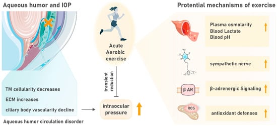
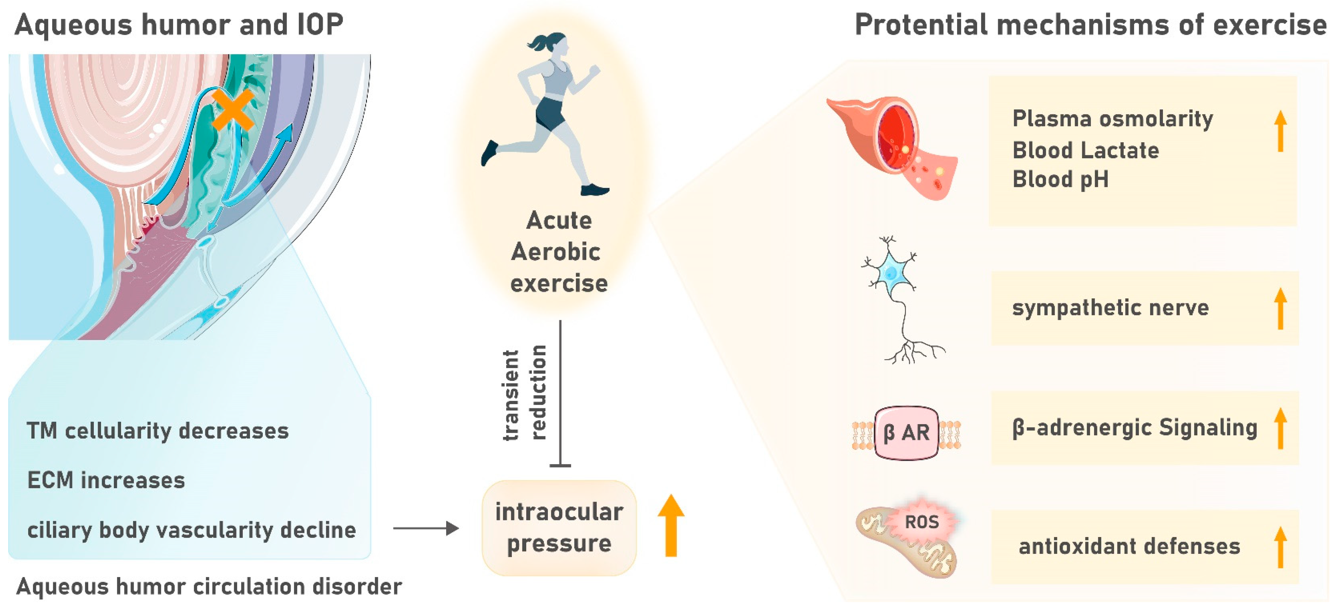
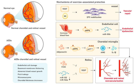
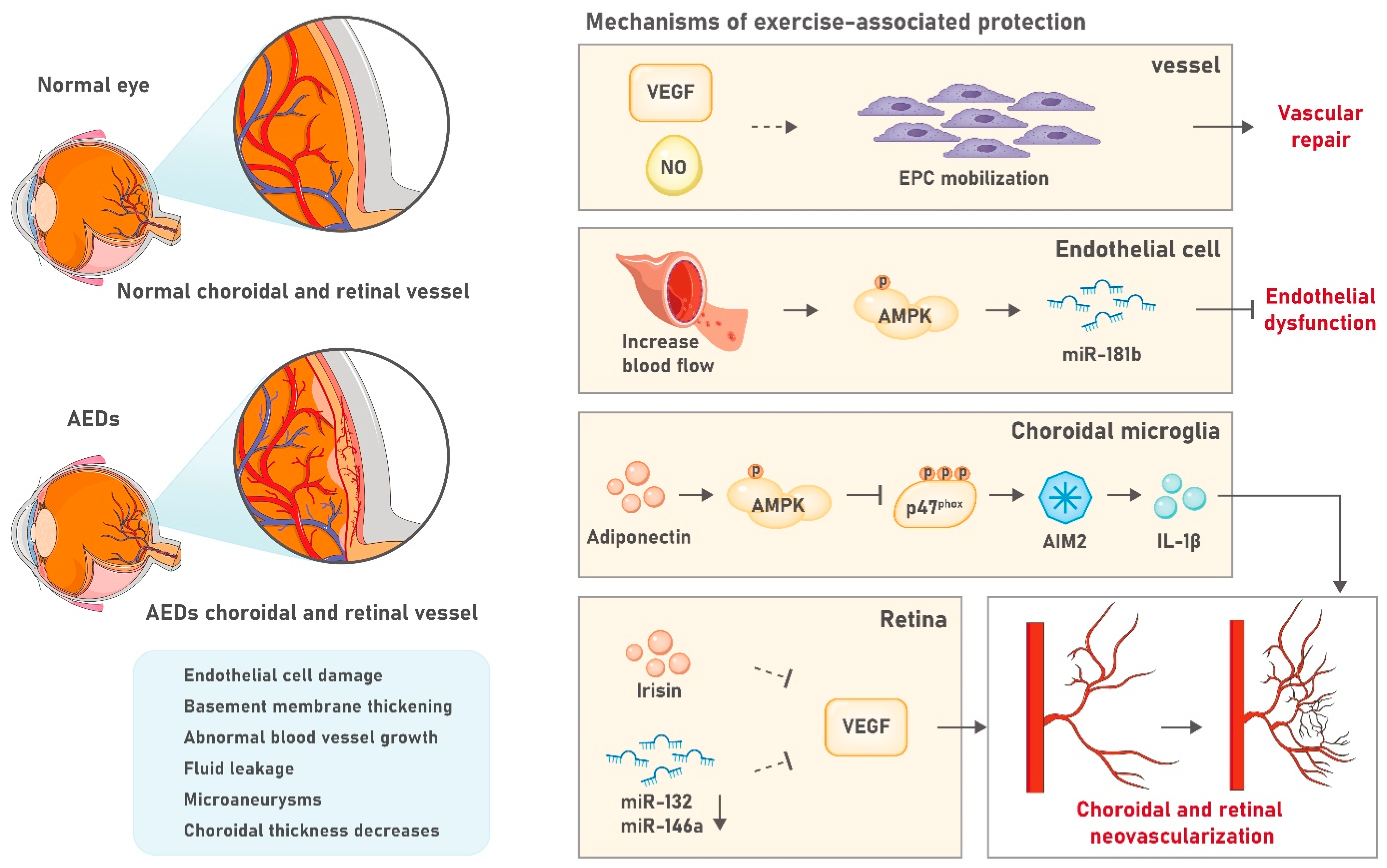
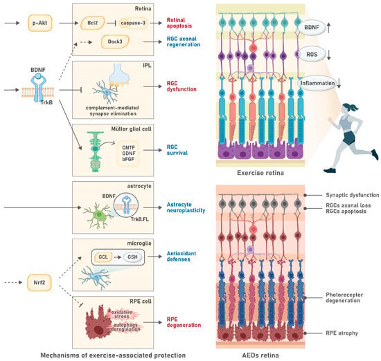
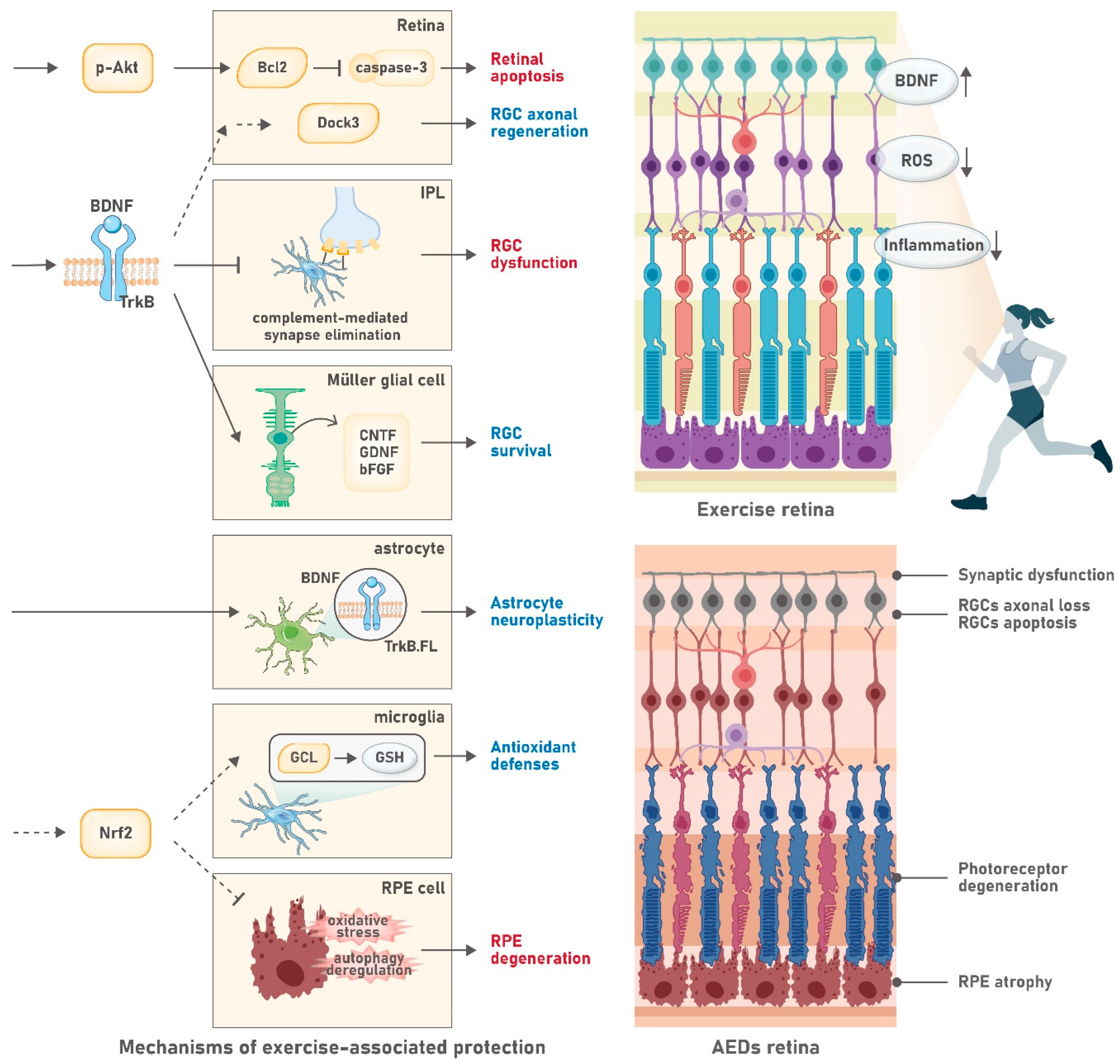

Disclaimer/Publisher’s Note: The statements, opinions and data contained in all publications are solely those of the individual author(s) and contributor(s) and not of MDPI and/or the editor(s). MDPI and/or the editor(s) disclaim responsibility for any injury to people or property resulting from any ideas, methods, instructions or products referred to in the content. |
© 2026 by the authors. Licensee MDPI, Basel, Switzerland. This article is an open access article distributed under the terms and conditions of the Creative Commons Attribution (CC BY) license.