


Abstract
Syndecan-2 (SDC-2) is a cell surface heparan sulfate proteoglycan whose transmembrane domain (TMD)-mediated dimerization regulates colon cancer cell behavior. In addition to functioning as a membrane receptor, SDC-2 undergoes matrix metalloproteinase-7 (MMP-7)-mediated ectodomain shedding, producing soluble fragments that promote angiogenesis and tumor progression. However, the mechanistic link between TMD-mediated dimerization and ectodomain shedding has remained unclear. Here, we show that different SDC family members exhibit distinct propensities for SDS-resistant dimer formation, which correlate with their susceptibility to MMP-7-mediated cleavage. Notably, the dimeric forms of both SDC-2 and SDC-4 were cleaved more rapidly by MMP-7, whereas dimerization-defective mutants of SDC-2 (S2GL) and SDC-4 (S4GL) displayed significantly reduced proteolytic cleavage in vitro. Conditioned media from cells expressing wild-type SDC-2 (HT29-S2W) contained elevated levels of shed SDC-2 and promoted increased migration of HT29 colon cancer cells and macrophages. In contrast, this effect was diminished in media from cells expressing the dimerization-defective S2GL mutant. Furthermore, a TMD mutant of SDC-2 identified in colon cancer patients showed impaired dimerization and decreased ectodomain shedding. Together, these findings demonstrate that TMD-mediated dimerization is required for MMP-7-dependent shedding of SDC-2 and drives tumor progression by enhancing cancer cell migration and immune cell recruitment.
1. Introduction
Syndecans (SDC-1–4) are transmembrane heparan sulfate proteoglycans involved in diverse cellular processes, including adhesion, migration, wound healing, and angiogenesis [1,2,3]. Their transmembrane domains (TMDs) contain a conserved GXXXG motif that promotes SDS-resistant dimerization, although the extent of this dimerization varies across family members [4,5]. Among them, SDC-2 exhibits particularly strong homodimerization through a conserved GXXXG motif together with a unique Phe167 residue [6] that enhances intermolecular interactions. This TMD-dependent dimerization is essential for SDC-2 function: replacing its TMD with that of SDC-4 markedly reduces SDS-resistant dimerization and impairs SDC-2-mediated cell spreading and migration [6], underscoring the critical role of the TMD in SDC-2 activity. In studies using chimeric constructs, substituting the wild-type SDC-2 TMD with a dimerization-defective mutant and fusing it to the cytoplasmic domain of platelet-derived growth factor receptor (PDGFR) resulted in decreased phosphorylation at Tyr579 and Tyr857. This reduction attenuated Src and PI3K activation and ultimately impaired cell migration [7]. These findings indicate that the dimeric state of the TMD dictates the configuration of the cytoplasmic domain and downstream intracellular signaling, thereby regulating key cellular functions.
Beyond its membrane-bound receptor role, SDC-2 also functions as a soluble ligand generated through shedding of its extracellular domain [8,9,10]. Shed SDC fragments are elevated in colon cancer [10] as well as in various inflammatory and fibrotic diseases [11,12,13,14,15,16,17], indicating a strong association with disease progression. This shedding is primarily mediated by matrix metalloproteinases, particularly MMP-7 [18,19]. In pancreatic cancer cells, fibroblast growth factor-2 (FGF-2) upregulates MMP-7 activity, thereby promoting SDC-1 shedding [20]. In colon cancer cell lines (HT29 and HCT116), interleukin-1α (IL-1α) increases SDC-2 shedding without altering its expression by inducing MMP-7 mRNA and protein and activating p38 and ERK MAPK pathways [19]. In vivo, chronic colitis enhances colocalization of SDC-2 and MMP-7 and further promotes shedding [11]. These observations highlight the importance of MMP-7-dependent SDC-2 shedding in tumor progression and inflammatory responses.
Shed SDC-2 also exerts functional effects within the tumor microenvironment. A non-cleavable SDC-2 mutant displays reduced tumorigenic potential [10], emphasizing the significance of ectodomain shedding. Both circulating shed SDC-2 fragments in patient sera [10] and a synthetic peptide corresponding to the active sequence of the SDC-2 extracellular domain [10,21] have been shown to promote tumor growth and metastasis in multiple colon cancer models. In an orthotopic mouse model, systemic administration of this synthetic peptide enhanced tumor growth and angiogenesis by stabilizing hypoxia-inducible factor 1-alpha (HIF-1α), upregulating vascular endothelial growth factor (VEGF), and promoting macrophage infiltration [21]. Shed SDC-2 also stimulates endothelial cell migration and tube formation in glioma models, and tumor-derived pro-angiogenic factors further induce SDC-2 shedding [22], demonstrating its role as a paracrine regulator of tumor-associated angiogenesis.
Studies of other transmembrane proteins suggest that dimerization may serve as a key regulatory step in the shedding process. For example, shedding of CD44 and pro-neuregulin-1 requires dimerization and cytoplasmic domain modifications to permit protease access [23]. Similarly, somatic angiotensin I-converting enzyme (ACE) forms dimers via carbohydrate-mediated interactions in model membrane systems [24], demonstrating that dimerization and glycosylation can influence proteolytic cleavage. Collectively, these studies indicate that dimerization is a critical determinant of transmembrane protein shedding. Despite the well-established importance of SDC-2 dimerization in receptor function, its potential role in MMP-7-mediated shedding remains unknown. Moreover, it is unclear whether the biological activities of shed SDC-2 are affected by its dimerization state. Therefore, the present study aims to determine whether TMD-mediated SDC-2 dimerization regulates MMP-7-dependent ectodomain shedding and to elucidate how this regulatory mechanism influences colon cancer progression.
2. Results
2.1. SDC Family Members Exhibit Dimerization-Dependent Susceptibilities to MMP-7-Mediated Proteolysis
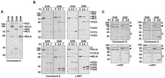
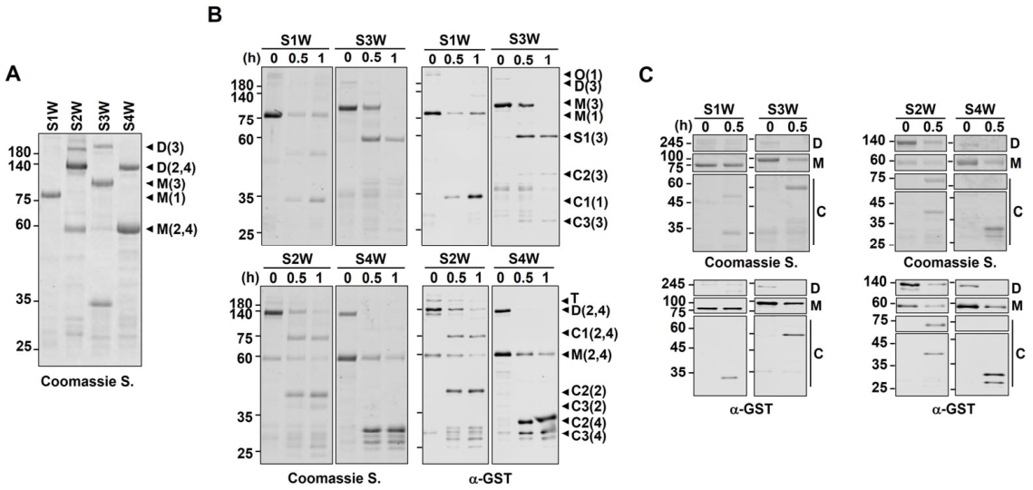
To investigate the relationship between dimerization and extracellular shedding of SDC, we first investigated the susceptibility of proteolytic cleavage by MMP-7 using recombinant SDC proteins. All four SDC isoforms were expressed as GST-tagged proteins, and their ability to form SDS-resistant dimers was evaluated (Figure 1A). In agreement with previous reports [4,5], SDC-2 exhibited the strongest TMD-mediated dimerization, followed by SDC-4. In contrast, SDC-1 and SDC-3 showed weaker dimer formation (Figure 1A). To determine whether these differences in dimerization influenced MMP-7-mediated proteolysis, GST-tagged SDCs immobilized on glutathione-agarose beads (mimicking membrane-anchored states) were incubated in vitro with purified MMP-7. Coomassie staining and GST immunoblotting revealed that proteolytic cleavage patterns were associated with the dimerization status of each SDC. By 0.5 h, the dimeric forms of SDC-1 and SDC-3 had largely disappeared, although their low initial abundance limited detailed comparisons. Notably, monomeric SDC-3 remained detectable at 0.5 h, while monomeric SDC-1 persisted up to 1 h (Figure 1B, top panel). For SDC-2 and SDC-4, which initially displayed abundant dimeric forms, the dimer bands decreased more rapidly than the monomeric bands by 1 h (Figure 1B, bottom panel). Quantitative analysis at 0.5 h confirmed that dimeric forms were more rapidly cleaved than monomeric ones across all other members, with a corresponding increase in cleaved fragment levels (Figure 1C). These results suggest that although the degree of dimerization varies among SDC isoforms—making direct quantitative comparisons difficult—the relative cleavage rates of dimeric versus monomeric forms reflect a dimerization-dependent susceptibility to MMP-7.
Figure 1.
SDC family members exhibit distinct susceptibilities to MMP-7-mediated proteolysis. (A) Equivalent amounts of bead-bound GST-tagged SDCs (S1W, S2W, S3W, and S4W) were separated by SDS-PAGE and visualized by Coomassie Brilliant Blue staining. T, Trimer; D, dimer; M, monomer. The number indicates each SDC member. N = 2. (B) GST-tagged SDCs were incubated with purified MMP-7 in vitro for the indicated time period, separated by SDS-PAGE, and analyzed by Coomassie Brilliant Blue staining (left panel) and anti-GST immunoblotting (right panel). C, cleaved SDC. N = 3. (C) After incubation with purified MMP-7 for the indicated time period, the dimeric and monomeric forms of each SDC were directly compared by SDS-PAGE to assess differential degradation. N = 3.
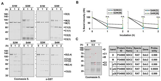
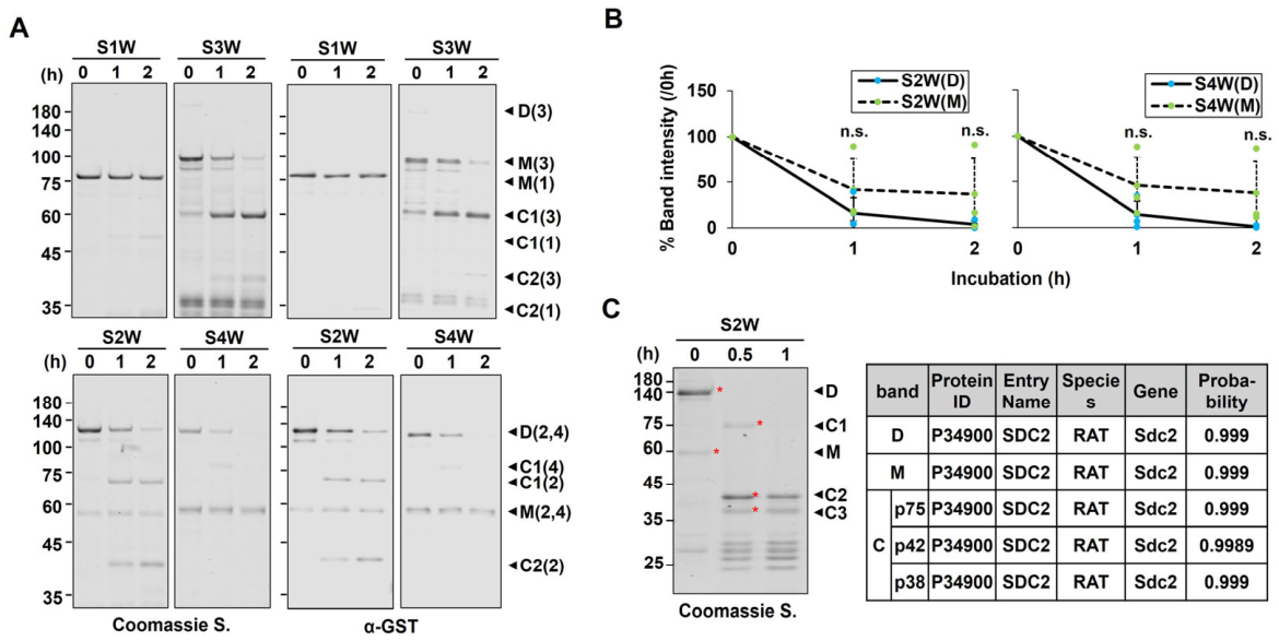
To assess whether similar trends could be observed using soluble SDCs (as opposed to bead-bound forms), purified GST-tagged SDCs were incubated with MMP-7 in solution (Figure 2). Under these conditions, SDC-1 showed no detectable dimer, and SDC-3 exhibited only a faint dimer band. Upon MMP-7 treatment, the dimeric form of SDC-3 was no longer detectable at 1 h, while the monomeric forms of both SDC-1 and SDC-3 remained visible up to 2 h. Despite the low levels of dimerization limiting direct comparisons, quantification of monomer bands showed a 22.2% decrease for SDC-1 and a 77.8% decrease for SDC-3 at 2 h relative to 0 h (Figure 2A, top panel), indicating that monomeric forms are more resistant to proteolytic shedding.
Figure 2.
Recombinant SDC family members exhibit differential susceptibility to MMP-7-mediated proteolysis in solution. (A) Equivalent amounts of purified GST-tagged SDCs were incubated with purified MMP-7 for the indicated time period. Samples were separated by SDS-PAGE and analyzed by Coomassie Brilliant Blue staining (left panel) and anti-GST immunoblotting (right panel). C, cleaved SDC. N = 3. (B) Quantitative analysis of proteolytic cleavage was performed using Image Studio Lite software Ver. 4.0. Data are presented as mean ± S.D. Statistical significance was determined using a two-tailed unpaired Student’s t-test, N = 3. n.s., not significant (S2W(D) vs. S2W(M)). (C) GST-tagged SDC-2 proteins were incubated with purified MMP-7 for the indicated time period. Specific bands were excised from the Coomassie-stained SDS-PAGE gel (left panel) and subjected to mass spectrometry analysis. The table summarizes the spectral counts for the identified proteins (right panel). D, dimer; M, monomer; C, Cleaved SDC. * Bands subjected to mass spectrometric analysis.
Conversely, SDC-2 and SDC-4 exhibited substantial loss of their dimeric forms by 2 h, while their monomeric forms remained comparatively stable (Figure 2A, bottom panel). Densitometric analysis showed that dimeric SDC-2 and SDC-4 levels were reduced by 83.8% and 85.7%, respectively, while their monomeric forms decreased by only 58.3% and 54.3% at the same time point (Figure 2B). To confirm that the observed cleavage fragments were derived from MMP-7-mediated cleavage of SDC-2, mass spectrometry was performed. The bands designated C1 (p75), C2 (p42), and C3 (p38), as well as the dimeric (D) and monomeric (M) forms, were all identified as fragments of rat SDC-2 (Figure 2C). Together, these findings demonstrate that MMP-7-induced proteolytic cleavage of SDCs is regulated by TMD-mediated dimerization. This regulatory mechanism is especially prominent in SDC-2 and SDC-4 and is consistently observed in both membrane-mimicking and soluble conditions.
2.2. TMD-Mediated Dimerization Regulates MMP-7-Induced Proteolytic Cleavage of the Extracellular Domains of SDC-2 and -4
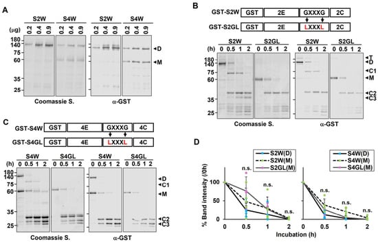
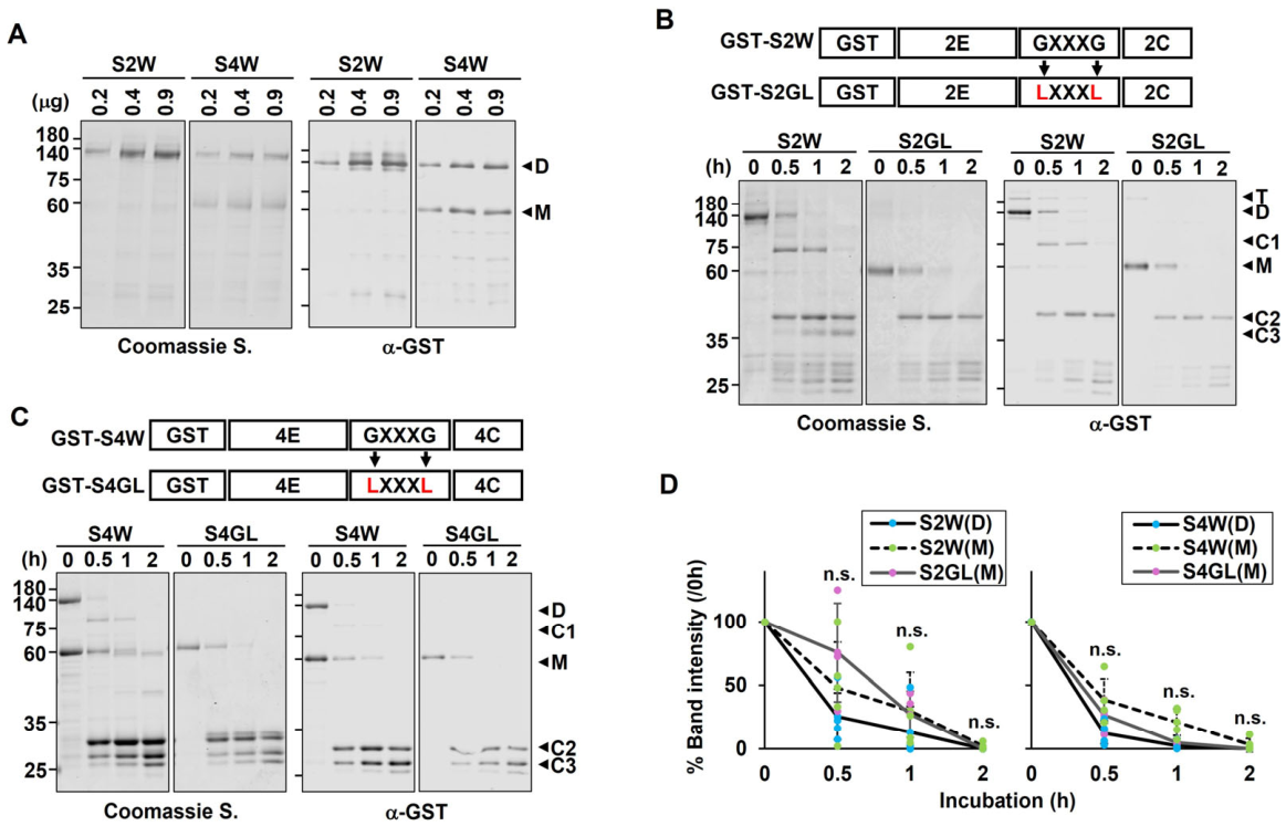
To further investigate the role of TMD-mediated dimerization in MMP-7-induced proteolytic cleavage of SDCs, we focused on SDC-2 and SDC-4, which consistently formed more abundant SDS-resistant dimers than other SDC isoforms. Despite varying protein concentrations, no concentration-dependent increase in dimer formation was observed for either SDC-2 or SDC-4 (Figure 3A), suggesting that TMD-mediated dimerization establishes a stable equilibrium between monomeric and dimeric forms that is independent of protein abundance. To assess the functional significance of dimerization in MMP-7-mediated proteolytic cleavage, we compared wild-type SDC-2 (S2W) and SDC-4 (S4W) with dimerization-defective mutants (S2GL and S4GL), in which the conserved GXXXG motif within the TMD was substituted with leucines [5,25]. In GST immunoblots, the dimeric form of S2W was completely cleaved after 1 h of incubation with MMP-7, whereas the S2GL mutant retained a detectable monomeric band at the same time point (Figure 3B). A similar trend was observed for SDC-4: the dimeric form of S4W was nearly undetectable by 0.5 h, while a strong monomeric band remained in the S4GL mutant (Figure 3C). Densitometric analysis showed that the dimeric band of S2W decreased by 74.6% at 0.5 h, while S2GL exhibited only a 24.2% reduction—indicating that the dimeric S2W was cleaved approximately three times faster than its dimerization-defective counterpart. Additionally, the monomeric form of S2W was cleaved more slowly, showing a 51.9% reduction compared to the dimeric form (Figure 3D, left panel). Similarly, S4W showed an 87.9% reduction in its dimeric form, while S4GL showed a 73.6% reduction. In contrast, the monomeric form of S4W showed only a 61.5% reduction over the same period (Figure 3D, right panel). These findings demonstrate that wild-type dimeric forms of SDC-2 and SDC-4 are significantly more susceptible to MMP-7-induced proteolytic cleavage than their dimerization-defective mutants.
Figure 3.
TMD-mediated dimerization regulates MMP-7-induced proteolytic cleavage of the extracellular domain of SDC-2 and -4. (A) The indicated amounts of purified SDC-2 (S2W) and SDC-4 (S4W) were incubated for 5 min, separated by SDS-PAGE, and then stained with Coomassie Brilliant Blue. N = 2. (B,C) Schematic representations of S2W and S4W and their dimerization-defective TMD mutants (S2GL and S4GL) are shown (top panel). Equivalent amounts of bead-bound GST-tagged S2W and S2GL proteins were incubated with purified MMP-7 for the indicated time period, separated by SDS-PAGE and analyzed by Coomassie Brilliant Blue staining (left panel) and anti-GST immunoblotting (right panel). N = 4. (D) Quantitative analysis of proteolytic cleavage was performed using Image Studio Lite software Ver. 4.0 based on quantification from four independent experiments. Data are presented as mean ± S.D. Statistical significance was determined using a two-tailed unpaired Student’s t-test, N = 4. n.s., not significant (S2W(D) vs. S2W(M) vs. S2GL(M) or S4W(D) vs. S4W(M) vs. S4GL(M)). T, trimer; D, dimer; M, monomer; C, Cleaved SDC.
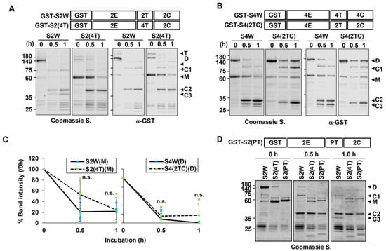
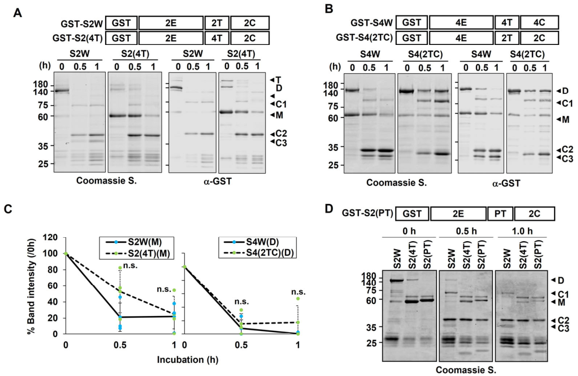
To further examine whether the strength of TMD-mediated dimerization affects the extent of SDC cleavage, we analyzed chimeric constructs in which the TMDs of SDC-2 and SDC-4 were reciprocally swapped (Figure 4). As reported previously [6], a chimera in which the TMD of SDC-2 was replaced with that of SDC-4 (S2(4T)) showed reduced dimerization compared to S2W (Figure 4A), whereas a chimera in which both the TMD and cytoplasmic domain of SDC-4 were replaced with those of SDC-2 (S4(2TC)) showed enhanced dimerization (Figure 4B). In both cases, MMP-7 treatment led to rapid cleavage of the dimeric forms (S2W, S2(4T), S4W, S4(2TC)). Interestingly, quantitative analysis of monomer band intensities at 0.5 h revealed that monomeric S2W was cleaved approximately 31.6% more rapidly than S2(4T), while the dimeric bands of S4W and S4(2TC) showed only a 6.6% difference in cleavage, indicating no substantial variation (Figure 4C). Consistently, another dimerization-defective mutant, S2(PT)—in which the TMD of SDC-2 was replaced with that of PDGFR, a domain known to promote ligand-dependent dimerization—existed exclusively as a monomer and exhibited markedly slower cleavage compared to the dimeric forms of S2W and S2(4T) (Figure 4D). Taken together, although a direct correlation between dimerization strength and overall proteolytic efficiency is difficult to establish, the consistent differences in cleavage rates between monomeric and dimeric forms within individual constructs strongly support the conclusion that TMD-mediated dimerization plays a critical regulatory role in MMP-7-induced ectodomain shedding of SDCs.
Figure 4.
TMD-mediated dimerization regulates MMP-7-induced proteolytic cleavage of SDC-2 and -4. (A,B,D) Schematic diagrams of S2W and S4W with their TMD and cytoplasmic domain substitution mutants (S2(4T), S4(2TC), and S2(PT)) are shown (top panel). Equivalent amounts of bead-bound GST-fused proteins were incubated with purified MMP-7 for the indicated time period, separated by SDS-PAGE and analyzed by Coomassie Brilliant Blue staining (left panel) and anti-GST immunoblotting (right panel). (C) Quantitative analysis of proteolytic cleavage was performed using Image Studio Lite software Ver. 4.0 based on quantification from three independent experiments. (A–C) Data are presented as mean ± S.D. Statistical significance was determined using a two-tailed unpaired Student’s t-test, N = 3. n.s., not significant (S2W(M) vs. S2(4T)(M) or S4W(D) vs. S4(2TC)(D)). (D) N = 1. T, trimer; D, dimer; M, monomer; C, Cleaved SDC.
2.3. TMD-Mediated Dimerization Promotes Ectodomain Shedding of SDC-2
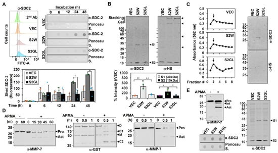
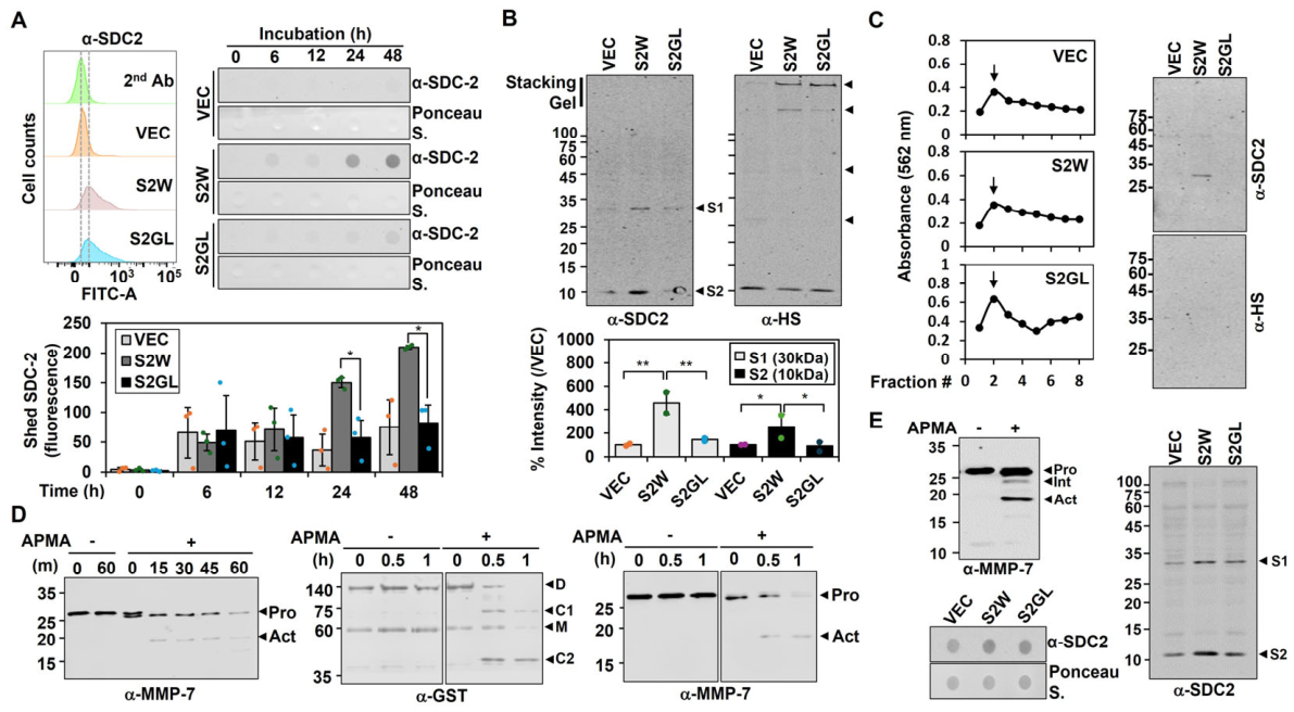
We next investigated whether TMD-mediated dimerization affects SDC-2 shedding at the cell surface. Shedding of SDC-2 was compared between cells expressing S2W and the dimerization-defective mutant S2GL. Consistent with our previous observation that SDC-2 can be proteolytically cleaved by MMP-7 in vitro, the level of shed SDC-2 progressively increased in the conditioned media of S2W-expressing cells between 12 and 48 h, whereas substantially less SDC-2 shedding was observed in S2GL-expressing cells, as determined by dot blotting with an anti-SDC-2 antibody (Figure 5A).
Figure 5.
MMP-7 regulates cell-surface SDC-2 shedding in a dimerization-dependent manner. (A) Cell-surface SDC-2 expression was analyzed by flow cytometry with an anti-SDC-2 antibody and IgG control (top left panel). N = 3. SDC-2 in conditioned media (CM) was detected by dot blotting (top right panel) and quantified from three independent experiments (bottom panel). Data are presented as mean ± S.D. Statistical significance was determined using a two-tailed unpaired Student’s t-test, * p < 0.05. N = 3 independent experiments. (B) Concentrated CM were analyzed by SDS-PAGE and Western blotting using anti-SDC-2 or anti-HS antibodies. Shed SDC-2 fragments are indicated by “S” (top panel). Quantification is shown for two independent experiments (bottom panel). Data are presented as mean ± S.D. Statistical significance was determined using a two-tailed unpaired Student’s t-test, *, p < 0.05; **, p < 0.01 (middle panels). N = 2 independent experiments. (C) CM were applied to a DEAE column, and bound proteins were eluted and analyzed by SDS-PAGE and Western blotting using anti-SDC-2 or anti-HS antibodies. (D,E) HT-29 cells were treated with IL-1α (1 ng/mL) for 24 h, and concentrated CM were incubated with 1 mM APMA. MMP-7 activation was assessed by Western blotting ((D), left panel; (E), top left panel). Purified GST-S2W (500 ng) was incubated with APMA-pretreated CM for 15 min and analyzed by Western blotting using anti-GST (middle panel) and anti-MMP-7 ((D), right panel) antibodies. N = 2–3. APMA-pretreated concentrated CM ((E), left top panel) was incubated with the indicated HT-29 cells for 2 h. Culture media were collected and analyzed by dot blotting for SDC-2 ((E), bottom left panel). The collected culture media were then concentrated 100-fold and analyzed by Western blotting for SDC-2 ((E), right panel). N = 2.
Unexpectedly, although cell-surface SDC-2 is heavily modified with HS chains, which can impede its migration during electrophoresis, a shed form of SDC-2 was detected by SDS-PAGE followed by immunoblotting (Figure 5B). Notably, whereas high-molecular-weight SDC-2 species retained in the stacking gel were detected by anti-HS antibodies, the ~30 kDa shed SDC-2 fragment was not detected by the anti-HS antibody (Figure 5B). This finding suggests that a fraction of shed SDC-2 is released without HS chains or loses its HS modification upon shedding. Immunoblotting with the anti-SDC-2 antibody further confirmed that SDC-2 shedding was substantially greater in S2W-expressing cells than in S2GL-expressing cells (Figure 5B). Consistently, DEAE-Sepharose purification of conditioned media revealed higher levels of shed SDC-2 in media from S2W-expressing cells than from S2GL-expressing cells, while the shed SDC-2 fraction was not detected by the anti-HS antibody (Figure 5C). These results indicate that TMD-mediated dimerization promotes SDC-2 ectodomain shedding.
Previous studies have shown that IL-1α treatment upregulates both the mRNA expression and secretion of MMP-7 [19]. To determine whether MMP-7 mediates SDC-2 ectodomain shedding, we prepared conditioned media enriched in cell-derived MMP-7, predominantly in its pro-form, from IL-1α-treated HT29 cells overexpressing SDC-2. The pro-form of MMP-7 in the conditioned media was subsequently activated by treatment with 1 mM 4-aminophenylmercuric acetate (APMA), a well-established MMP activator. Immunoblot analysis showed that pro-MMP-7 remained largely inactive for up to 60 min in the absence of APMA. In contrast, APMA treatment induced a marked decrease in pro-MMP-7 within 15 min, accompanied by the appearance of the active form of MMP-7 (Figure 5D), confirming efficient activation of cell-derived MMP-7 in the conditioned media. Incubation of GST-S2W with active, cell-derived MMP-7 resulted in complete cleavage of the dimeric GST-S2W species in vitro, as indicated by the disappearance of the corresponding dimeric bands concomitant with the appearance of active MMP-7 (Figure 5D). These results confirmed that cell-derived MMP-7 possesses proteolytic activity toward SDC-2. Importantly, when S2W- and S2GL-expressing cells were exposed to equivalent amounts of MMP-7-containing conditioned media, both dot blot and immunoblot analyses demonstrated substantially greater proteolytic cleavage of S2W than of S2GL at the cell surface (Figure 5E). Thus, TMD-mediated dimerization increases the susceptibility of SDC-2 to MMP-7-mediated proteolytic cleavage.
Taken together, these findings demonstrate that MMP-7 promotes SDC-2 ectodomain shedding at the cell surface and further indicate that TMD-mediated dimerization enhances the susceptibility of SDC-2 to MMP-7-dependent proteolysis. Therefore, dimerization appears to function not only as a mechanism that promotes SDC-2 clustering but also as a determinant of its accessibility to extracellular proteases, thereby facilitating ectodomain shedding.
2.4. Dimerization Influences Shed SDC-2-Mediated Cellular Functions
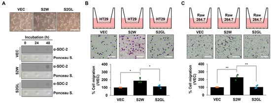
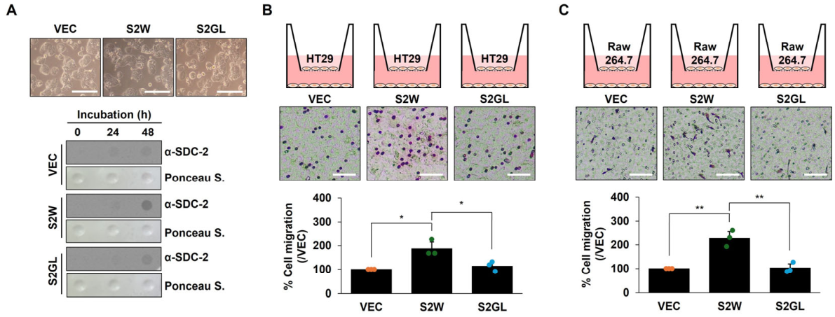
Previous studies have demonstrated that shed SDC-2 enhances primary tumor growth and metastasis by promoting the migratory activity of colon cancer cells and macrophages, thereby facilitating angiogenesis [10,21]. These findings suggest that differences in TMD-mediated dimerization may contribute to the regulation of shed SDC-2-driven cellular functions relevant to tumor progression. To test this hypothesis, either HT-29 colon cancer cells or Raw 264.7 macrophage-like cells were incubated with conditioned media containing varying levels of shed SDC-2, and their migratory responses were subsequently assessed (Figure 6). As expected, after 48 h, S2W-expressing HT-29 cells produced the highest levels of shed SDC-2 in the culture media, whereas S2GL-expressing cells produced substantially lower levels (Figure 6A). Strikingly, conditioned media from S2W-expressing cells induced approximately a two-fold increase in the migration of both HT-29 (Figure 6B) and Raw 264.7 cells (Figure 6C), compared to media from vector control (VEC) or S2GL-expressing cells. Together, these findings indicate that TMD-mediated dimerization of SDC-2 is required for efficient ectodomain shedding and also enhances its downstream functional effects, including migration-promoting activity in cancer cells and macrophages, which are closely associated with tumor progression and angiogenesis [10,21].
Figure 6.
Dimerization-dependent ectodomain shedding of SDC-2 influences the migration of colon cancer cells and macrophages. (A) Cell morphology at the beginning of incubation in serum-free media for the indicated times (top panel). CM collected at the corresponding time points were analyzed by dot blotting using an anti-SDC-2 antibody (bottom panel). (B) Schematic overview of the transwell migration assay (upper panel). After transfected HT29 cells (VEC, S2W and S2GL; 1 × 10⁵ cells/well) were pre-incubated separately in the lower chamber for 24 h, HT29 cells (5 × 10⁵ cells/well) were seeded into the upper chamber. Migration was assessed. Quantitative analysis was performed using Image J software Fiji based on quantification from three independent experiments. Data are presented as mean ± S.D. Statistical significance was determined using a two-tailed unpaired Student’s t-test, *, p < 0.05 (middle and bottom panels). N = 3 independent experiments. (C) Schematic overview of the transwell migration assay (top panel). After transfection, HT29 cells were pre-incubated separately in the lower chamber for 24 h, RAW 264.7 cells (2 × 10⁵ cells/well) were seeded into the upper chamber. Migration was assessed. Quantitative analysis was performed using Image J software Fiji based on quantification from three independent experiments. Data are presented as mean ± S.D. Statistical significance was determined using a two-tailed unpaired Student’s t-test, **, p < 0.01 (middle and bottom panels). N = 3 independent experiments. Scale bar, 100 μm.
2.5. SDC-2 Transmembrane Mutations in Colorectal Cancer Reduce Dimerization and Proteolytic Cleavage of the Extracellular Domain
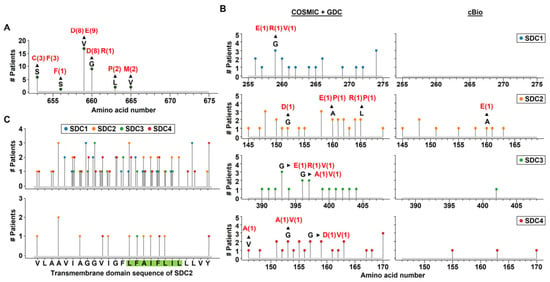
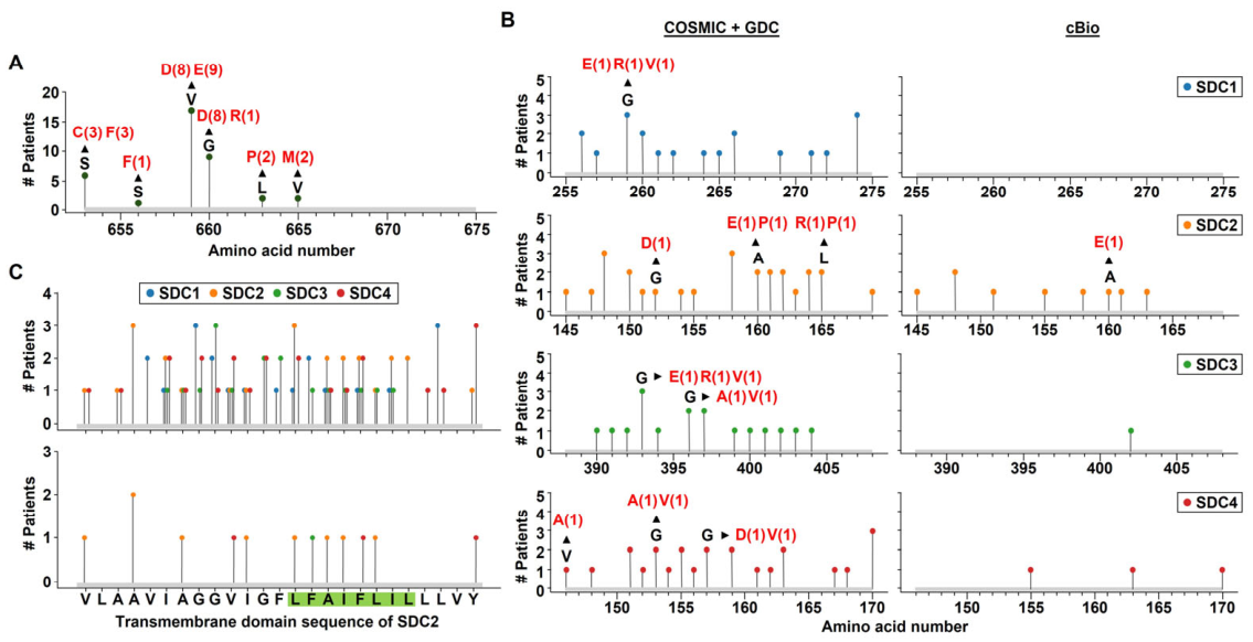
To evaluate the significance of TMD-mediated dimerization, we analyzed mutations in SDC-1–4 using COSMIC, Genomic Data Commons (GDC) Data Portal, and cBioPortal for Cancer Genomics (cBioPortal) datasets, with human epidermal growth factor receptor 2 (HER2) included as a dimerization-dependent reference receptor (Figure 7). HER2 mutations clustered around two key TMD motifs, and substitutions of hydrophobic residues with polar or charged residues (e.g., V659E, G660D) (Figure 7A) have previously been shown to modulate dimer stability [26], underscoring the functional impact of TMD mutations on receptor activity in cancer. Across SDCs, COSMIC and GDC revealed numerous TMD mutations—including E/D/R substitutions (Figure 7B, left)—whereas cBioPortal reported fewer events, with SDC-2 being the only isoform harboring E substitutions (Figure 7B, right). Mapping these mutations showed hotspots near conserved motifs such as the GXXXG and FA motifs and conserved L residues, with SDC-2 exhibiting the highest mutation frequency (Figure 7C).
Figure 7.
Mutation in the TMD of HER2 and SDC-2 in cancer patients. (A) The indicated point mutations of HER2 found in cancer patients were selected from the cBioPortal. (B) The indicated point mutations of SDCs found in cancer patients were selected from the COSMIC and GDC data portal or cBioPortal. (C) The indicated point mutations of SDCs found in cancer patients were selected from the COSMIC and GDC data portal (top panel) or cBioPortal (bottom panel). Each arrow shows the amino acid substitution at the corresponding site and the frequency of occurrence.
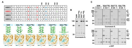
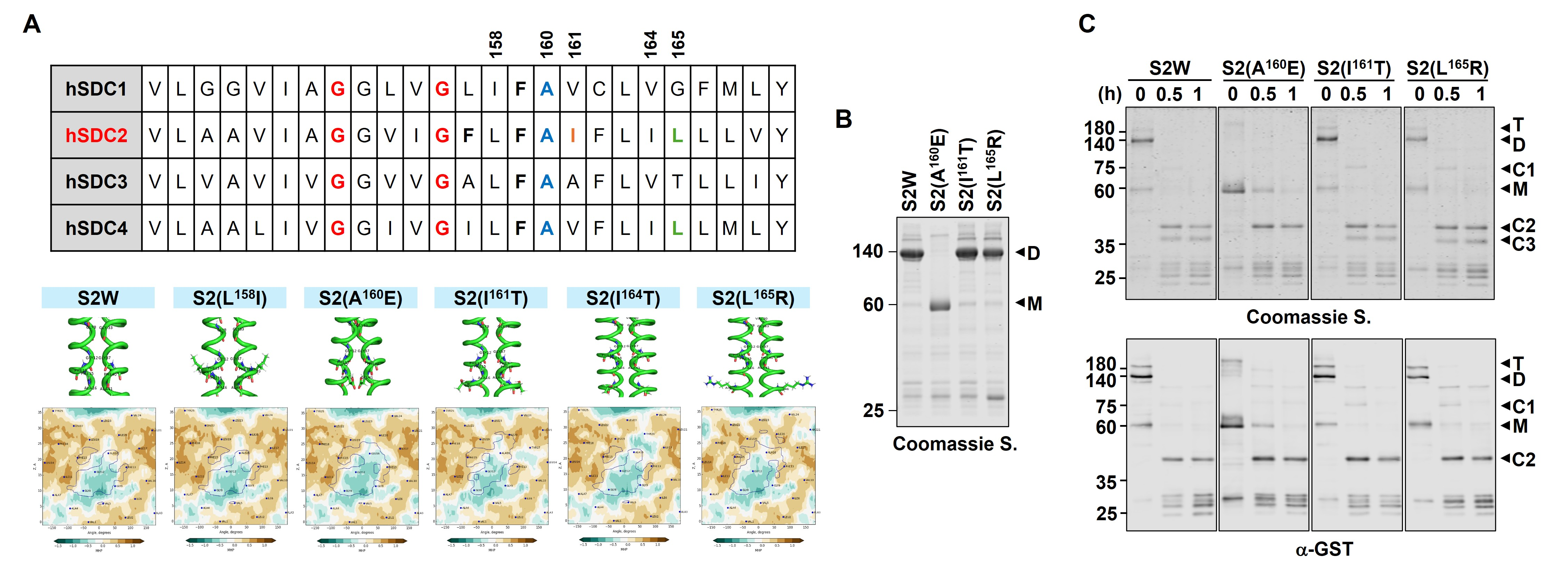
In colorectal cancer patients, several SDC-2 TMD mutations were found near critical dimerization elements. Five variants (L158I, A160E, I161T, I164T, L165R) were selected for further analysis, as they replaced hydrophobic residues with polar or charged amino acids (Figure 8). Using the Preddimer program, we found through molecular hydrophobicity potential (MHP) analyses that S2(A160E), S2(I161T), and S2(L165R) substantially disrupt the predicted dimer interfaces (Figure 8A). Among these, S2(A160E) completely lost SDS-resistant dimer formation (Figure 8B), and its monomeric form showed markedly reduced proteolytic cleavage by MMP-7 (Figure 8C), consistent with diminished shedding due to impaired dimerization. In contrast, S2(I161T) and S2(L165R) retained SDS-resistant dimer formation (Figure 8B) and underwent MMP-7-mediated cleavage at levels comparable to wild-type SDC-2 (Figure 8C). Overall, SDC-2 TMD mutations that alter hydrophobicity at key dimerization sites variably disrupt dimer formation and affect MMP-7-mediated proteolytic cleavage, potentially influencing cancer-related functions.
Figure 8.
Mutation in the TMD of SDC-2 in colorectal cancer patients shows reduced dimerization and proteolytic cleavage of the extracellular domain. (A) Schematic representations of the TMD amino acid sequences of human SDC-1, -2, -3, and -4. The indicated point mutations found in colon cancer patients were selected from the COSMIC and GDC data portal database (upper panel). Dimerization models were generated using the Preddimer and visualized using PyMOL program (Ver. 3.1.6.1), and MHP between wild-type and mutant SDC-2 was compared to predict changes in dimerization propensity (bottom panel). (B) Equal amounts of bead-bound GST-tagged S2W, S2(A160E), S2(I161T), and S2(L165R) were separated by SDS-PAGE and stained with Coomassie Brilliant Blue. N = 3. (C) Equal amounts of bead-bound GST-tagged wild-type and mutants were incubated with pro-MMP-7, separated by SDS-PAGE, stained with Coomassie Brilliant Blue (top panel), and detected with an anti-GST antibody (bottom panel). N = 3.
3. Discussion
Although TMD-mediated dimerization is known to be essential for various cellular functions of SDC-2, its relationship with ectodomain shedding has remained poorly understood. In this study, we identify a previously uncharacterized mechanistic link between SDC-2 dimerization and MMP-7-dependent cleavage, revealing that dimerization is an important determinant of efficient proteolytic processing and its associated tumor-promoting activity. We found that dimeric SDC-2 is significantly more susceptible to MMP-7-mediated shedding than its monomeric form. This was demonstrated both in vitro and in cell-based assays, in which dimerization-defective mutants such as S2GL showed reduced cleavage (Figure 1, Figure 2, Figure 3, Figure 4 and Figure 5). In HT-29 colon cancer cells, wild-type SDC-2 underwent time-dependent shedding, whereas the dimerization-defective mutant remained largely uncleaved, indicating that dimerization is required for the susceptibility of SDC-2 to MMP-7-mediated cleavage under cellular conditions.
Functionally, shed SDC-2 from wild-type cells enhanced colon cancer cell migration and macrophage chemotaxis, whereas conditioned media from cells expressing dimerization-defective mutants failed to elicit these effects (Figure 6). These findings suggest that shed SDC-2 can function as a paracrine factor that modulates both tumor and immune cell behavior, thereby contributing to a pro-tumorigenic microenvironment. Thus, TMD-mediated dimerization may play a dual role in SDC-2 biology by regulating not only its membrane organization but also the generation and, consequently, the biological activity of its shed form.
Interestingly, the shed SDC-2 species detected in conditioned media was not detectable with the anti-HS antibody (Figure 5), despite the extensive HS modification of cell-surface SDC-2. This finding suggests that ectodomain shedding may generate an HS-deficient pool of soluble SDC-2, either through selective shedding of non-HS-modified molecules or through loss of HS chains during or following proteolytic cleavage. Given the established role of HS chains in mediating ligand interactions and signaling, the differential HS status of membrane-bound and shed SDC-2 may have functional implications for the activity of soluble SDC-2 in the extracellular environment. Whether HS loss occurs before, during, or after proteolytic shedding, and whether this modification alters the biological activity of soluble SDC-2, remain important questions for future investigation.
To assess the potential clinical relevance of this mechanism, we analyzed colorectal cancer patient datasets and identified naturally occurring mutations in the TMD of SDC-2, particularly near the conserved GXXXG motif (Figure 7). Notably, the S2(A160E) mutation, which introduces a negatively charged residue into the hydrophobic TMD, disrupted dimerization and decreased susceptibility to proteolytic cleavage (Figure 8), similar to the engineered S2GL mutant. These findings highlight the structural complexity of SDC-2 processing and underscore the need for high-resolution structural studies to define the precise dimerization and cleavage interfaces. Moreover, our data reveal a notable distinction from the conventional paradigm in which receptor dimerization promotes oncogenic signaling, as exemplified by HER2. Although dimerization is generally associated with receptor activation, certain cancer-associated mutations, such as S2(A160E), may instead disrupt dimerization while simultaneously reducing SDC-2 shedding. This observation raises the possibility that, in specific tumor contexts, altered dimerization could influence the balance between membrane-bound and soluble SDC-2 and thereby affect its biological functions. Further studies will be required to determine whether such mutations confer a selective advantage and whether they alter the functional properties of membrane-bound or shed SDC-2.
The therapeutic implications are significant. Inhibiting SDC-2 dimerization could reduce shedding and downstream signaling. Prior studies have shown that Fibulin-7 (FBLN7) binds the GXXXG motif in SDC-4 and blocks dimerization and signaling [27]. Given the conservation of this motif in SDC-2, FBLN7 or similar peptide-based inhibitors may represent a promising strategy to modulate SDC-2 activity in cancer and other diseases where its shedding is elevated, such as colitis, glioma, and keloids [11,22,28]. Despite these findings, our study has several limitations. First, although our biochemical experiments demonstrate direct cleavage of SDC-2 by MMP-7, the involvement of other proteases in cellular SDC-2 shedding cannot be excluded. IL-1α stimulation and APMA treatment may also affect other proteases. Thus, selective MMP-7 inhibition or genetic depletion is needed to establish its specific role in SDC-2 shedding. Second, although increased SDC-2 shedding appears to promote cell migration and macrophage chemotaxis, other soluble factors in the conditioned media may also contribute. Further studies using SDC-2-neutralizing antibodies or immunodepletion are required to clarify the specific role of shed SDC-2. Third, the SDS-resistant dimerization observed with GST-tagged SDC-2 TMD constructs in vitro may not fully reflect SDC-2 oligomerization in native membranes, which could be influenced by other membrane-associated factors and the lipid environment. Finally, in vivo studies using animal models are needed to validate the role of dimerization-dependent shedding in tumor growth and metastasis.
In conclusion, our study supports a model in which TMD-mediated dimerization of SDC-2 is required for MMP-7-mediated cleavage, leading to the release of soluble extracellular domains that promote tumor progression through paracrine signaling. These findings provide new mechanistic insight into proteoglycan shedding and identify the dimerization–shedding axis as a potential therapeutic target. Future studies should define the structural basis of this interaction, elucidate its integration with signaling pathways, and explore strategies to inhibit SDC-2 shedding to limit tumor aggressiveness and chronic inflammation.
4. Materials and Methods
4.1. Materials
A monoclonal antibody recognizing SDC-2 across mouse, rat, and human species was generated by AdipoGen Inc. (Incheon, Korea) using the Fc-fused SDC2 extracellular domain [29]. A monoclonal anti-MMP-7 antibody (Catalog no. MAB9071), IL-1α (Catalog no. 200-LA), and human recombinant MMP-7 proenzyme (Catalog no. 907-MP) were purchased from R&D Systems (Minneapolis, MN, USA). A monoclonal anti-GST antibody (Catalog no. sc-374171) was purchased from Santa Cruz Biotechnology (Santa Cruz, CA, USA). A monoclonal anti-Heparan Sulfate (HS) antibody (Catalog no. 370255-S) was purchased from Amsbio (Abingdon, UK).
4.2. Cell Culture and Generation of Stable Cell Lines
Human colon adenocarcinoma HT29 cells were obtained from the ATCC and cultured in McCoy’s 5A medium (Hyclone, Logan, UT, USA) supplemented with 10% FBS (Hyclone, Logan, UT, USA) and 0.1% gentamycin (Sigma-Aldrich, St. Louis, MO, USA). Cells were maintained at 37 °C in a humidified incubator with 5% CO2. RAW 264.7 murine macrophage cell line was obtained from ATCC and maintained in Dulbecco’s Modified Eagle’s Medium (DMEM/HIGH GLUCOSE; Hyclone, Logan, UT, USA) supplemented with 5% FBS and 0.1% gentamicin. For the generation of stable cell lines expressing SDC-2 or the SDC-2 mutant, HT29 cells (1 × 10⁵ cells) were transfected with 1 μg of pcDNA3.1 (VEC), pcDNA3. 1-FLAG-SDC2 (S2W), or pcDNA3.1-S2GL (S2GL) using Viva Magic reagent (Vivagen, Gyeonggi-do, Korea), according to the manufacturer’s instructions. Transfected cells were selected for 4 weeks in McCoy’s 5A medium containing 400 μg/mL G418 (EMD Biosciences, San Diego, CA, USA). Individual stable clones were isolated by picking individual colonies.
4.3. Flow Cytometry
HT29 cells (VEC, S2W, S2GL) were washed with PBS, then detached using 1 mM EDTA and 5% FBS in PBS. The cells were collected by centrifugation and resuspended in PBS and incubated with specific antibodies with 10% FBS in PBS at 4 °C overnight. The cells were washed with PBS with 0.05% Tween-20, which is followed by incubation with FITC-conjugated anti-mouse IgG (Thermo Fisher Scientific, Waltham, MA, USA) in 10% FBS-containing PBS for 1 h at 25 °C. The expression level of SDC-2 at the cell surface was evaluated by counting 1 × 10⁴ cells with a BD FACS LSRFortessa (BD Biosciences, Franklin Lakes, NJ, USA).
4.4. Expression and Purification of Recombinant Proteins
cDNAs of rat SDC-1, -2, -3, and -4 encoding full-length proteins were synthesized by polymerase chain reaction (PCR) and subcloned into the GST expression vector pGEX-5X-1 (Amersham Biosciences, Piscataway, NJ, USA). These constructs were used to transform Escherichia coli DH5α and BL21. The expression of GST fusion proteins was induced by incubation with 1 mM isopropyl-D-thiogalactopyranoside for 4 h at 37 °C. The fusion proteins were purified with glutathione-agarose beads (Macherey-nagel, Düren, Germany) as described previously [25].
4.5. Proteolytic Cleavage of SDCs
Glutathione agarose bead-bound GST-tagged proteins or purified GST-tagged proteins (500 ng) were incubated with human recombinant pro-MMP-7 (1 μg/mL) in reaction buffer containing 20 mM Tris-HCl (pH 7.4), 150 mM NaCl, 5 mM CaCl2, and 0.001% Brij-35 at 37 °C for the indicated times. To examine the proteolytic activity of cell-derived MMP-7, CM from HT-29 cells was collected and centrifuged at 1000 rpm for 3 min to remove cells, followed by centrifugation at 12,000 rpm for 10–30 min at 4 °C to remove cellular debris. The CM was concentrated 100-fold using Amicon Ultra-4 centrifugal filters (Merck Millipore, Darmstadt, Germany). The concentrated CM containing MMP-7 (5 μL), with or without 1 mM APMA, was incubated with GST-tagged proteins (500 ng) in reaction buffer at 37 °C for the indicated times. The reaction products were analyzed by SDS-PAGE, followed by Coomassie Brilliant Blue staining and immunoblotting.
4.6. Mass Spectrometry Analysis
Protein samples were separated by SDS-PAGE and visualized by Coomassie Brilliant Blue staining. Bands of interest were excised from the gel and subjected to in-gel digestion followed by mass spectrometry analysis at the Digital Omics Research Center, Korea Basic Science Institute (KBSI, Cheongju, Korea). The acquired mass spectrometry data were searched against the Rattus norvegicus reference proteome (UniProt proteome ID: UP000002494). SDC-2 was identified based on peptides matching rat SDC-2 (UniProt accession number P34900).
4.7. DEAE-Sepharose Column Chromatography
CM from HT29 cells (15 mL/dish; 100 mL total) was collected and centrifuged at 1000 rpm for 3 min at room temperature, followed by 12,000 rpm for 10 min at 4 °C to remove cells and debris. DEAE-Sepharose Fast Flow beads (GE Healthcare, Chicago, IL, USA; 400 μL) were packed into a 10 mL Poly-Prep chromatography column (Bio-Rad, Hercules, CA, USA), washed with 30 mL PBS, and equilibrated with PBS. NaCl and EDTA were added to the conditioned media to a final concentration of 0.25 M and 20 mM, respectively. The media were then loaded onto the column and allowed to pass through the beads by gravity flow at 4 °C. The column was washed with 30 mL of 0.25 M NaCl in 50 mM Tris-HCl (pH 7.5), and bound proteins were eluted with elution buffer (5 M urea, 2 M NaCl, 0.5% Triton X-100, 20 mM EDTA, 50 mM Tris-HCl, pH 4.0) in 100-μL fractions. Protein concentrations were determined using a BCA protein assay by measuring absorbance at 562 nm with a microplate reader (SpectraMax M2, San Jose, CA, USA). Selected fractions were subsequently analyzed by SDS-PAGE, followed by immunoblotting.
4.8. Dot Blotting
HT29 cells (VEC, S2W, S2GL) were seeded on a 6-well plate (5 × 10⁵ cells/well) or 100 mm dish (15× 105 cells/dish) and incubated for 48 h at 37 °C, 5% CO2. After PBS and serum-free media (SFM) washing, CM was collected at the indicated time points and centrifuged at 14,000 rpm for 30 min at 4 °C to remove cellular debris. The supernatants were subjected to dot blotting. The proteins were subsequently transferred to 0.45-μm nitrocellulose blotting membranes (GE Healthcare, Chicago, IL, USA) and were incubated with specific antibodies. Protein detection was performed using an Odyssey CLx imager (LI-COR Biosciences, Lincoln, NE, USA), and analysis was performed with the Image Studio Lite software Ver. 4.0 (LI-COR Biosciences, Lincoln, NE, USA).
4.9. Immunoblotting
Purified proteins (500 ng) and concentrated CM samples (100-fold, 5 μL) were denatured in sample buffer and separated by 8 and 10 or 12 and 15% SDS-PAGE. Following electrophoresis, the proteins were transferred onto 0.45-μm nitrocellulose membranes and incubated with the appropriate primary antibodies. Protein signals were detected using an Odyssey CLx imaging system (LI-COR Biosciences, Lincoln, NE, USA), and densitometric analysis was performed using Image Studio Lite software Ver. 4.0 (LI-COR Biosciences, Lincoln, NE, USA).
4.10. Transwell Migration Assay
HT 29 Cells (VEC, S2W, S2GL) were seeded (1 × 10⁵ cells/well) in McCoy’s 5A medium containing 10% FBS were loaded into each well of 24-well plate, and the plate was incubated for 24 h at 37 °C in a 5% CO2 incubator. To quantify migrated cells, each membrane of Transwell plates (8-μm pore size; Costar, Corning, NY, USA) was coated with gelatin B (10 μg/mL) in PBS. Gelatin-coated membranes were dried at room temperature for 1 h, and transwell plates were assembled into a 24-well plate. Cells (HT29, 5 × 10⁵ cells/well; RAW 264.7, 2 × 10⁵ cells/well) in McCoy’s 5A medium containing 2% FBS or DMEM medium containing 5% FBS were loaded into each upper chamber, and the plate was incubated for 24 h at 37 °C in a 5% CO2 incubator. The cells that had migrated to the lower surface of the membrane were stained with 50% hematoxylin solution (Glentham Life Sciences, Corsham, UK) and 0.5% eosin (Sigma-Aldrich, St. Louis, MO, USA), and the image was acquired with a Leica DM1000 LED microscope (Leica Microsystems, Wetzlar, Germany), and analysis was performed with the Image J software Fiji (version 1.54g; National Institutes of Health, Bethesda, MD, USA).
4.11. Database Mining and Structural Prediction
Point mutations in the TMDs of HER2 and SDCs were retrieved from the cBioPortal [30], COSMIC, and the GDC Data Portal. Among these, SDC-2 TMD mutations were modeled using the PREDDIMER server (https://preddimer.nmr.ru/), and MHPs were analyzed using the same server to assess changes in helix interaction potential. The predicted TMD dimer structures were visulized using PyMOL (version 3.1.6.1; Schrodinger, LLC).
4.12. Statistical Analysis
All data are presented as the mean ± S.D. Differences between groups were tested for statistical significance using Student’s t-test and considered significant at **, p < 0.01; *, p < 0.05.
Author Contributions
Writing—original draft, Y.J. and E.-S.O.; Writing—review and editing, Y.J., J.H. and E.-S.O.; Project administration, Y.J. and E.-S.O.; Conceptualization, Y.J. and E.-S.O.; Formal analysis, Y.J., J.H., H.Y., S.C. and Y.U.; Investigation, Y.J., J.H., H.Y., S.C. and Y.U.; Data curation, Y.J., J.H., H.Y., S.C. and Y.U.; Visualization, Y.J. and J.H.; Validation, Y.J., J.H. and Y.U.; Supervision, E.-S.O.; Funding acquisition, E.-S.O. All authors have read and agreed to the published version of the manuscript.
Funding
This research was supported by the Basic Science Research Program through the National Research Foundation of Korea (NRF) grant funded by the Korean government (MSIP) (RS-2023-00208377, RS-2024-00469143) to E.-S.O.
Institutional Review Board Statement
Not applicable.
Informed Consent Statement
Not applicable.
Data Availability Statement
All the data generated in this study can be shared upon request.
Conflicts of Interest
The authors declare no conflict of interest.
References
- Berdiaki, A.; Neagu, M.; Tzanakakis, P.; Spyridaki, I.; Perez, S.; Nikitovic, D. Extracellular Matrix Components and Mechanosensing Pathways in Health and Disease. Biomolecules 2024, 14, 1186. [Google Scholar] [CrossRef] [Scilit]
- Hagen, M.W.; Setiawan, N.J.; Woodruff, K.A.; Termini, C.M. Syndecans in hematopoietic cells and their niches. Am. J. Physiol. Cell Physiol. 2024, 327, C372–C378, Correction in Am. J. Physiol. Cell Physiol. 2024, 327, C867–C867. https://doi.org/10.1152/ajpcell.00326.2024_COR. [Google Scholar] [CrossRef] [Scilit]
- Gopal, S.; Arokiasamy, S.; Pataki, C.; Whiteford, J.R.; Couchman, J.R. Syndecan receptors: Pericellular regulators in development and inflammatory disease. Open Biol. 2021, 11, 200377. [Google Scholar] [CrossRef] [Scilit]
- Dews, I.C.; Mackenzie, K.R. Transmembrane domains of the syndecan family of growth factor coreceptors display a hierarchy of homotypic and heterotypic interactions. Proc. Natl. Acad. Sci. USA 2007, 104, 20782–20787. [Google Scholar] [CrossRef] [Scilit]
- Choi, Y.; Kwon, M.J.; Lim, Y.; Yun, J.H.; Lee, W.; Oh, E.S. Trans-regulation of Syndecan Functions by Hetero-oligomerization. J. Biol. Chem. 2015, 290, 16943–16953. [Google Scholar] [CrossRef] [Scilit]
- Kwon, M.J.; Choi, Y.; Yun, J.H.; Lee, W.; Han, I.O.; Oh, E.S. A unique phenylalanine in the transmembrane domain strengthens homodimerization of the syndecan-2 transmembrane domain and functionally regulates syndecan-2. J. Biol. Chem. 2015, 290, 5772–5782. [Google Scholar] [CrossRef] [Scilit]
- Hwang, J.; Jang, B.; Kim, A.; Lee, Y.; Lee, J.; Kim, C.; Kim, J.; Moon, K.M.; Kim, K.; Wagle, R.; et al. Syndecan Transmembrane Domain Specifically Regulates Downstream Signaling Events of the Transmembrane Receptor Cytoplasmic Domain. Int. J. Mol. Sci. 2021, 22, 7918. [Google Scholar] [CrossRef] [Scilit]
- Choi, S.; Lee, H.; Choi, J.R.; Oh, E.S. Shedding; towards a new paradigm of syndecan function in cancer. BMB Rep. 2010, 43, 305–310. [Google Scholar] [CrossRef] [Scilit]
- Lichtenthaler, S.F.; Lemberg, M.K.; Fluhrer, R. Proteolytic ectodomain shedding of membrane proteins in mammals-hardware, concepts, and recent developments. EMBO J. 2018, 37, e99456. [Google Scholar] [CrossRef] [Scilit]
- Choi, S.; Choi, Y.; Jun, E.; Kim, I.S.; Kim, S.E.; Jung, S.A.; Oh, E.S. Shed syndecan-2 enhances tumorigenic activities of colon cancer cells. Oncotarget 2015, 6, 3874–3886. [Google Scholar] [CrossRef] [Scilit]
- Hong, H.; Song, H.K.; Jang, B.; Park, E.; Han, D.S.; Kim, S.E.; Oh, E.S. Colocalization with MMP-7 in the Distal Colon is Crucial for Syndecan-2 Shedding in Dextran Sulfate Sodium-Induced Colitis Mice. J. Inflamm. Res. 2021, 14, 4987–5000. [Google Scholar] [CrossRef] [Scilit]
- Floer, M.; Clausen, M.; Meister, T.; Vollenberg, R.; Bettenworth, D.; Tepasse, P.R. Soluble syndecan-1 as marker of intestinal inflammation: A preliminary study and evaluation of a new panel of biomarkers for non-invasive prediction of active ulcerative colitis. Adv. Clin. Exp. Med. 2021, 30, 655–660. [Google Scholar] [CrossRef] [Scilit]
- Steppan, J.; Hofer, S.; Funke, B.; Brenner, T.; Henrich, M.; Martin, E.; Weitz, J.; Hofmann, U.; Weigand, M.A. Sepsis and major abdominal surgery lead to flaking of the endothelial glycocalix. J. Surg. Res. 2011, 165, 136–141. [Google Scholar] [CrossRef] [Scilit]
- Sanchez, C.; Lambert, C.; Dubuc, J.E.; Bertrand, J.; Pap, T.; Henrotin, Y. Syndecan-4 Is Increased in Osteoarthritic Knee, but Not Hip or Shoulder, Articular Hypertrophic Chondrocytes. Cartilage 2021, 13, 862S–871S. [Google Scholar] [CrossRef] [Scilit]
- Bollmann, M.; Pinno, K.; Ehnold, L.I.; Martens, N.; Martson, A.; Pap, T.; Starke, C.; Lohmann, C.H.; Bertrand, J. MMP-9 mediated Syndecan-4 shedding correlates with osteoarthritis severity. Osteoarthr. Cartil. 2021, 29, 280–289. [Google Scholar] [CrossRef] [Scilit]
- Kliment, C.R.; Englert, J.M.; Gochuico, B.R.; Yu, G.; Kaminski, N.; Rosas, I.; Oury, T.D. Oxidative stress alters syndecan-1 distribution in lungs with pulmonary fibrosis. J. Biol. Chem. 2009, 284, 3537–3545. [Google Scholar] [CrossRef] [Scilit]
- Kliment, C.R.; Oury, T.D. Extracellular superoxide dismutase protects cardiovascular syndecan-1 from oxidative shedding. Free Radic. Biol. Med. 2011, 50, 1075–1080. [Google Scholar] [CrossRef] [Scilit]
- Manon-Jensen, T.; Multhaupt, H.A.; Couchman, J.R. Mapping of matrix metalloproteinase cleavage sites on syndecan-1 and syndecan-4 ectodomains. FEBS J. 2013, 280, 2320–2331. [Google Scholar] [CrossRef] [Scilit]
- Kwon, M.J.; Hong, E.; Choi, Y.; Kang, D.H.; Oh, E.S. Interleukin-1alpha promotes extracellular shedding of syndecan-2 via induction of matrix metalloproteinase-7 expression. Biochem. Biophys. Res. Commun. 2014, 446, 487–492. [Google Scholar] [CrossRef] [Scilit]
- Ding, K.; Lopez-Burks, M.; Sanchez-Duran, J.A.; Korc, M.; Lander, A.D. Growth factor-induced shedding of syndecan-1 confers glypican-1 dependence on mitogenic responses of cancer cells. J. Cell Biol. 2005, 171, 729–738. [Google Scholar] [CrossRef] [Scilit]
- Jang, B.; Song, H.K.; Hwang, J.; Lee, S.; Park, E.; Oh, A.; Hwang, E.S.; Sung, J.Y.; Kim, Y.N.; Park, K.; et al. Shed syndecan-2 enhances colon cancer progression by increasing cooperative angiogenesis in the tumor microenvironment. Matrix Biol. 2022, 107, 40–58. [Google Scholar] [CrossRef] [Scilit]
- Fears, C.Y.; Gladson, C.L.; Woods, A. Syndecan-2 is expressed in the microvasculature of gliomas and regulates angiogenic processes in microvascular endothelial cells. J. Biol. Chem. 2006, 281, 14533–14536. [Google Scholar] [CrossRef] [Scilit]
- Hartmann, M.; Parra, L.M.; Ruschel, A.; Lindner, C.; Morrison, H.; Herrlich, A.; Herrlich, P. Inside-out Regulation of Ectodomain Cleavage of Cluster-of-Differentiation-44 (CD44) and of Neuregulin-1 Requires Substrate Dimerization. J. Biol. Chem. 2015, 290, 17041–17054. [Google Scholar] [CrossRef] [Scilit]
- Kost, O.A.; Balyasnikova, I.V.; Chemodanova, E.E.; Nikolskaya, I.I.; Albrecht, R.F., 2nd; Danilov, S.M. Epitope-dependent blocking of the angiotensin-converting enzyme dimerization by monoclonal antibodies to the N-terminal domain of ACE: Possible link of ACE dimerization and shedding from the cell surface. Biochemistry 2003, 42, 6965–6976. [Google Scholar] [CrossRef] [Scilit]
- Choi, S.; Lee, E.; Kwon, S.; Park, H.; Yi, J.Y.; Kim, S.; Han, I.O.; Yun, Y.; Oh, E.S. Transmembrane domain-induced oligomerization is crucial for the functions of syndecan-2 and syndecan-4. J. Biol. Chem. 2005, 280, 42573–42579. [Google Scholar] [CrossRef] [Scilit]
- Ou, S.I.; Schrock, A.B.; Bocharov, E.V.; Klempner, S.J.; Haddad, C.K.; Steinecker, G.; Johnson, M.; Gitlitz, B.J.; Chung, J.; Campregher, P.V.; et al. HER2 Transmembrane Domain (TMD) Mutations (V659/G660) That Stabilize Homo- and Heterodimerization Are Rare Oncogenic Drivers in Lung Adenocarcinoma That Respond to Afatinib. J. Thorac. Oncol. 2017, 12, 446–457. [Google Scholar] [CrossRef] [Scilit]
- Yao, G.; Zheng, X.; Hu, Y.; Zhao, Y.; Kong, B.; Ti, Y.; Bu, P.L. FBLN7 mediates vascular smooth muscle cell phenotype switching and vascular remodeling in hypertension. Theranostics 2024, 14, 7569–7588. [Google Scholar] [CrossRef] [Scilit]
- Mukhopadhyay, A.; Wong, M.Y.; Chan, S.Y.; Do, D.V.; Khoo, A.; Ong, C.T.; Cheong, H.H.; Lim, I.J.; Phan, T.T. Syndecan-2 and decorin: Proteoglycans with a difference--implications in keloid pathogenesis. J. Trauma Inj. Infect. Crit. Care 2010, 68, 999–1008. [Google Scholar] [CrossRef] [Scilit]
- Ryu, H.Y.; Lee, J.; Yang, S.; Park, H.; Choi, S.; Jung, K.C.; Lee, S.T.; Seong, J.K.; Han, I.O.; Oh, E.S. Syndecan-2 functions as a docking receptor for pro-matrix metalloproteinase-7 in human colon cancer cells. J. Biol. Chem. 2009, 284, 35692–35701. [Google Scholar] [CrossRef] [Scilit]
- de Bruijn, I.; Kundra, R.; Mastrogiacomo, B.; Tran, T.N.; Sikina, L.; Mazor, T.; Li, X.; Ochoa, A.; Zhao, G.; Lai, B.; et al. Analysis and Visualization of Longitudinal Genomic and Clinical Data from the AACR Project GENIE Biopharma Collaborative in cBioPortal. Cancer Res. 2023, 83, 3861–3867. [Google Scholar] [CrossRef] [Scilit]
Disclaimer/Publisher’s Note: The statements, opinions and data contained in all publications are solely those of the individual author(s) and contributor(s) and not of MDPI and/or the editor(s). MDPI and/or the editor(s) disclaim responsibility for any injury to people or property resulting from any ideas, methods, instructions or products referred to in the content. |
© 2026 by the authors. Licensee MDPI, Basel, Switzerland. This article is an open access article distributed under the terms and conditions of the Creative Commons Attribution (CC BY) license.