


Abstract
Serotonin receptor 5-HT7 and tropomyosin receptor kinase B (TrkB) are both key regulators of neuroplasticity implicated in the pathophysiology and treatment of depression. Although functional crosstalk between these receptors has been suggested, the underlying molecular mechanism remains unknown. Here, we investigated the physical and functional interaction between 5-HT7 and TrkB receptors at behavioral, biochemical, and molecular levels. Using males of the antidepressant-sensitive cataleptic (ASC) mouse model of depressive-like behavior, we demonstrated that repeated (9 days) activation of 5-HT7 receptors with the selective agonist LP-211 produced antidepressant-like effects in the sucrose preference test. These behavioral effects were dependent on TrkB signaling, as it was prevented by pretreatment with the TrkB antagonist ANA12. Both acute and repeated 5-HT7 receptor activation increased TrkB phosphorylation in the mouse brain, although this long-term effect was brain region- and amino acid residue-specific. To determine if these functional interactions were mediated by a direct physical association, we used co-immunoprecipitation and FRET assays. We found that 5-HT7 and TrkB form heteroreceptor complexes in vitro in neuroblastoma cells and in vivo in the mouse frontal cortex, hippocampus, and midbrain. Collectively, our findings support an oligomerization between 5-HT7 and TrkB receptors and suggest that this association may contribute to the antidepressant-like effects of 5-HT7 receptor activation.
1. Introduction
Major depressive disorder is associated with dysfunction of neural circuits involved in emotional regulation and impaired neural plasticity [1]. Chronic stress and other pathological factors can disrupt synaptic connectivity and neuronal function within these circuits, whereas effective antidepressant treatments promote adaptive changes in neuronal and synaptic plasticity [2]. These findings have established restoration of neural plasticity as an important component of antidepressant action [1,2].
Among the molecular systems regulating neural plasticity, brain-derived neurotrophic factor (BDNF) and its receptor tropomyosin receptor kinase B (TrkB) have attracted particular attention. BDNF–TrkB signaling regulates neuronal survival, dendritic remodeling, synaptic transmission, and long-term synaptic plasticity, and its dysregulation has been implicated in depression [3,4,5]. Importantly, TrkB is expressed in serotonergic neurons of the dorsal raphe nucleus (DRN), as supported by its mRNA detection in serotonergic cell bodies and by reduced midbrain TrkB levels following serotonergic lesions or Tph2-Cre-mediated TrkB deletion [3,6]. Moreover, TrkB is increasingly recognized not only as the principal receptor mediating BDNF signaling, but also as a molecular convergence point for pharmacological regulation of neural plasticity. Notably, Casarotto et al. demonstrated that structurally and pharmacologically diverse antidepressants, including conventional antidepressants and the rapid-acting antidepressant ketamine, can directly bind to the transmembrane domain of TrkB and potentiate BDNF-dependent signaling [7]. Disruption of the TrkB antidepressant-binding site impaired antidepressant-induced plasticity and behavioral responses, providing direct evidence that TrkB can serve as a molecular target of antidepressant drug action [7]. Subsequent work further showed that direct TrkB binding contributes to the plasticity-promoting and antidepressant-like effects of psychedelics [8], supporting the broader concept of TrkB as a pharmacologically relevant integrator of signals controlling neural plasticity [9].
An important mechanism through which TrkB may integrate signals from other neurotransmitter systems is functional crosstalk with G protein-coupled receptors (GPCRs). GPCR–RTK crosstalk is a well-established phenomenon in which activation of one receptor system can modulate the activity of the other through intracellular signaling pathways or receptor transactivation [10]. Importantly, such functional crosstalk does not necessarily require direct physical association between the receptors. However, in some cases, GPCRs and RTKs can form receptor complexes that enable signaling interactions at the receptor level, providing an additional mechanism for integrating extracellular signals [11,12,13,14,15]. Such GPCR–RTK heteroreceptor complexes have been described in the brain and implicated in the regulation of neuronal survival, synaptic transmission, and plasticity [16].
Serotonergic GPCRs provide a particularly relevant context for this type of receptor-level interaction, given the well-established role of serotonin signaling in the regulation of mood and neural plasticity. In our previous study, we demonstrated that 5-HT2A receptors form heteroreceptor complexes with TrkB in neuronal systems and that this interaction modulates TrkB phosphorylation and downstream signaling [14]. These findings provided direct evidence that a serotonergic GPCR can interact with TrkB at the receptor level and suggested that such heteroreceptor complexes may represent a mechanism linking serotonergic and neurotrophin signaling. However, whether similar receptor-level interactions occur between TrkB and other serotonin receptor subtypes remains largely unexplored.
The serotonin 5-HT7 receptor is a particularly interesting candidate in this regard. The 5-HT7 receptor is widely expressed in the CNS, particularly in the thalamus, hypothalamus, hippocampus, prefrontal cortex, amygdala, and raphe nuclei, consistent with its involvement in mood regulation, cognition, and circadian rhythms [17]. It primarily couples to Gs proteins, activating adenylyl cyclase and the cAMP/PKA pathway, and can also signal through G12 proteins to regulate Rho GTPases, cytoskeletal remodeling, and neuronal morphology [17]. 5-HT7 receptors are involved in the regulation of neuronal development, synaptic function, dendritic morphology, and several forms of neural plasticity [18]. Pharmacological modulation of 5-HT7 receptors has also been associated with antidepressant-like effects in a broad range of preclinical models, although the behavioral consequences of 5-HT7 receptor activation appear to depend on the ligand, duration of treatment, brain region, and experimental context [19,20]. Importantly, previous studies have demonstrated functional coupling between 5-HT7 receptors and TrkB [21]: activation of 5-HT7 receptors was shown to increase TrkB expression in primary cortical cultures and promote TrkB phosphorylation in neuron-like cells, indicating that 5-HT7 signaling can modulate TrkB-dependent pathways [21]. However, the molecular mechanism underlying this functional crosstalk remained unclear.
The antidepressant-sensitive catalepsy (ASC) mouse line was established by selective breeding for high susceptibility to catalepsy in the offspring of CBA mice, which are prone to catalepsy [22,23], and AKR mice, which are resistant to it [24]. In addition to cataleptic freezing, which is attenuated by chronic treatment with classical antidepressants such as imipramine and fluoxetine [25,26], ASC mice exhibit increased immobility in the forced swim and tail suspension tests and reduced locomotor activity compared with their parental strains [27]. They also display increased anxiety-like behavior [28]. At the neurobiological level, ASC mice exhibit alterations in serotonergic neurotransmission and neuroanatomical abnormalities, along with reduced BDNF expression [29,30,31]. Importantly, intracerebroventricular administration of recombinant BDNF produces an antidepressant-like effect in this mouse line [32,33]. These features make the ASC mice a useful model for investigating how serotonergic signaling and BDNF–TrkB-related mechanisms contribute to depressive-like behavior and the effects of antidepressant compounds.
It remains unknown whether 5-HT7 and TrkB can physically associate to form a heteroreceptor complex. Establishing such an interaction would provide a potential molecular mechanism linking serotonergic signaling to TrkB-dependent plasticity and could help explain how 5-HT7 receptor activation influences antidepressant-related processes. In the present study, we therefore investigated the relationship between 5-HT7 and TrkB receptors at behavioral, biochemical, and molecular levels. Using the ASC mice, we examined the antidepressant-like effects of the selective 5-HT7 receptor agonist LP-211 and their dependence on TrkB signaling using the TrkB antagonist ANA12. We further assessed the effects of 5-HT7 receptor activation on TrkB phosphorylation in the hippocampus, frontal cortex and midbrain and tested the physical interaction of 5-HT7 and TrkB receptors using co-immunoprecipitation and FRET. Together, these approaches allowed us to determine whether functional 5-HT7–TrkB crosstalk is accompanied by a formation of protein-protein complexes between these two receptors.
2. Results
2.1. Acute 5-HT7 Receptor Activation Increases TrkB Phosphorylation in the Mouse Brain
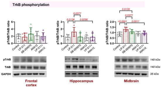
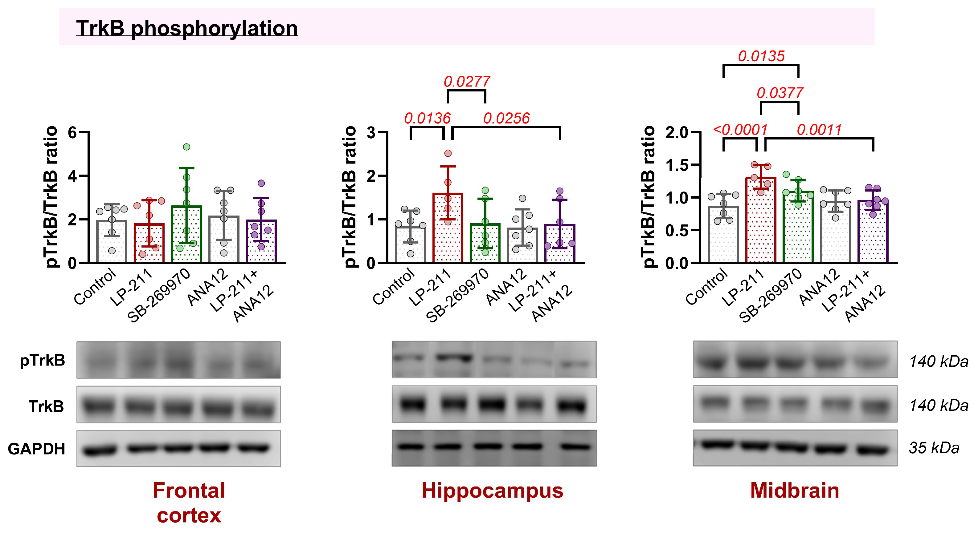
To assess the functional relationship between 5-HT7 and TrkB receptors, we examined the effects of acute administration of the selective 5-HT7 receptor agonist LP-211, the 5-HT7 receptor antagonist SB-269970, the TrkB antagonist ANA12, as well as a combination of LP-211 and ANA12 on TrkB phosphorylation in the brain of C57Bl/6 mice. TrkB phosphorylation was analyzed by SDS-PAGE and Western blot in membrane fractions isolated from the frontal cortex, hippocampus, and midbrain (Figure 1).
Figure 1.
Acute effects of 5-HT7 and TrkB receptor modulation on TrkB phosphorylation in different brain regions of C57Bl/6 mice. The pTrkB/TrkB ratio was analyzed by SDS-PAGE and Western blot (WB) in membrane fractions isolated from the frontal cortex (FC), hippocampus (HC) and midbrain (MB) following acute treatment with LP-211, SB-269970, ANA12, or LP-211 administered after ANA12 pretreatment. Representative WBs are shown. Data are presented as mean ± SD. (one-way ANOVA). Significant between-group differences are indicated on the graphs.
No significant treatment-related changes in the pTrkB/TrkB ratio were detected in the frontal cortex (F(4,30) = 0.50, p = 0.73; Figure 1).
In the hippocampus, although the overall effect of treatment did not reach statistical significance (F(4,26) = 2.42, p = 0.07), pairwise comparison revealed that LP-211 increased TrkB phosphorylation compared with the control group (p = 0.014), whereas the pTrkB/TrkB ratio was lower in the LP-211 + ANA12 group than in the LP-211 group (p = 0.026).
In the midbrain, TrkB phosphorylation was also significantly affected by treatment (F(4,28) = 6.39, p = 0.001; Figure 1). The pTrkB/TrkB ratio was markedly increased by LP-211 (p < 0.0001) and SB-269970 (p = 0.014) compared with the control group. This increase was attenuated by ANA12 pretreatment (LP-211 vs. LP-211 + ANA12, p = 0.0011). Furthermore, the pTrkB/TrkB ratio was lower in the SB-269970 group (p = 0.038) compared with the LP-211 group.
These findings indicate that the most pronounced changes in TrkB phosphorylation were associated with 5-HT7 receptor activation by LP-211. In contrast, 5-HT7 receptor blockade with SB-269970 did not produce comparable effects. Accordingly, subsequent experiments were focused on 5-HT7 receptor activation by LP-211.
2.2. Behavioral Effects of Repeated LP-211 Administration and Co-Administration of ANA12 in ASC Mice
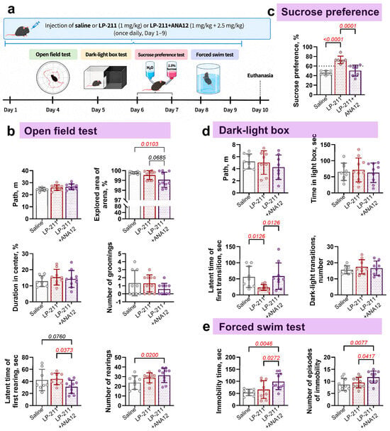
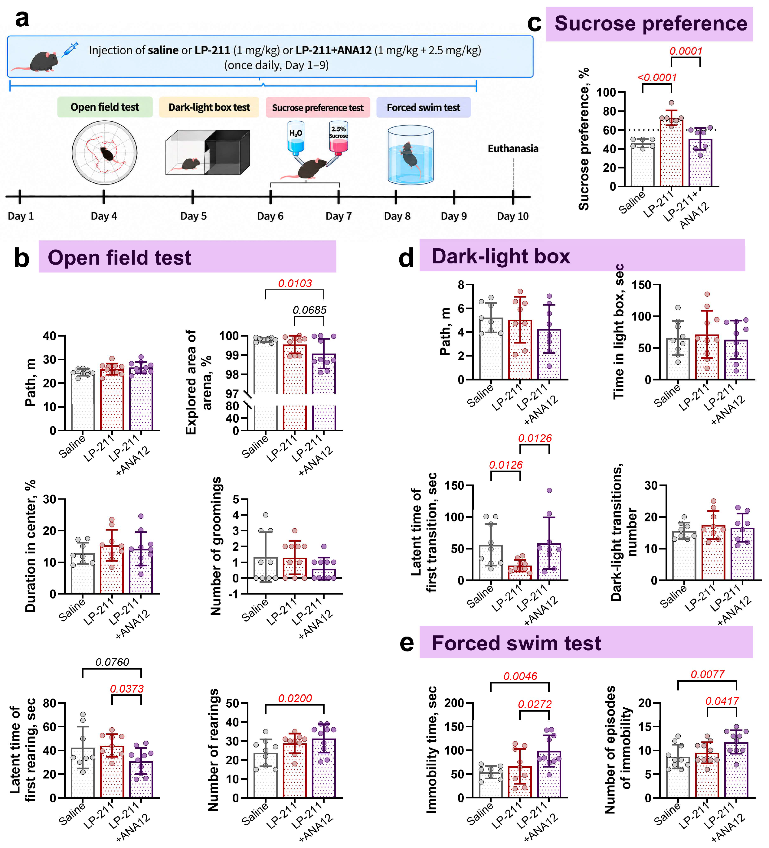
To assess the effects of prolonged 5-HT7 receptor activation on behavior, ASC mice were subjected to a battery of behavioral tests starting on day 4 of 9 days of drug administration (Figure 2a). In the open field test, the total distance traveled did not differ significantly among the three experimental groups (F(2,25) = 2.109, p = 0.1425; Figure 2b), indicating that the treatments did not produce a major alteration in general locomotor activity. However, mice pretreated with ANA12 and subsequently treated with LP-211 explored a smaller area of the arena than control animals (F(2,24) = 4.131, p = 0.0103; Figure 2b), with a trend toward a reduction compared with the LP-211 group. Also, LP-211 with ANA12 administration significantly increased the number of rearings compared with saline (F(2,25) = 3.156; p = 0.0200; Figure 2b). The latency to the first rearing did not differ significantly between the saline and LP-211 groups. However, it was significantly shorter in the LP-211 + ANA12 group than in the LP-211 group (p = 0.0373), with a trend toward a decrease compared with saline (p = 0.0760; Figure 2b). Other open-field parameters, including time spent in the center and grooming, did not differ significantly between groups, providing no evidence of pronounced alterations in exploratory and stereotyped behavior.
Figure 2.
Behavioral effects of repeated administration of the 5-HT7 receptor agonist LP-211 alone and in combination with the TrkB antagonist ANA12 in ASC mice. (a) Schematic representation of the experimental design. The following parameters were measured: (b) in the open field test: total distance traveled (path), explored area of the arena (in % of the total area of the arena), time spent in the center, number of rearings, latency to the first rearing, and number of grooming episodes; (c) in the sucrose preference test: sucrose preference; (d) in the dark–light box test: total distance traveled, time spent in the light compartment, latency to the first transition, and number of transitions between compartments; (e) in the forced swim test: time spent immobile and number of episodes of immobility. Bars represent mean ± SD (6 ≤ n ≤ 10) (one-way ANOVA). Significant between-group differences are indicated on the graphs.
Consistent with these findings, the dark–light box test revealed no significant differences among groups in total distance traveled, time spent in the light compartment, or number of transitions. However, LP-211 treatment significantly reduced the latency to the first transition compared with both control and LP-211 + ANA12 groups (F(2,24) = 3.748; Figure 2d), consistent with a moderate anxiolytic-like effect of LP-211 on transition latency, which was not observed following co-administration with ANA12.
The antidepressant-like effects of LP-211 were further examined using the forced swim and sucrose preference tests. In the forced swim test, the LP-211 + ANA12 group showed significantly increased total immobility time (F(2,24) = 5.413, p = 0.0115) and number of immobility episodes (F(2,27) = 4.477, p = 0.0209) compared with both the saline-treated control and LP-211 groups (Figure 2e). In the sucrose preference test, ASC mice displayed a baseline preference of approximately 40%, substantially below the ~65% preference reported as the limit for non-depressed mice in the literature [34]. Prolonged LP-211 treatment significantly increased sucrose preference (F(2,17) = 19.19, p < 0.0001), whereas this increase was not observed following co-administration of ANA12 (Figure 2c). Overall, repeated LP-211 administration produced an antidepressant-like effect in the sucrose preference test and a more limited effect on transition latency in the dark–light box test, while co-administration with the TrkB antagonist prevents these effects.
2.3. TrkB Phosphorylation Was Altered After Prolonged 5-HT7 Receptor Activation in a Brain Region-Specific Manner
Using Western blot analysis, we examined the protein levels of 5-HT7 and TrkB receptors and the phosphorylation of TrkB at three amino acid residues (Y516, Y706, Y817) in the membrane fractions of the frontal cortex, hippocampus, and midbrain of ASC mice.
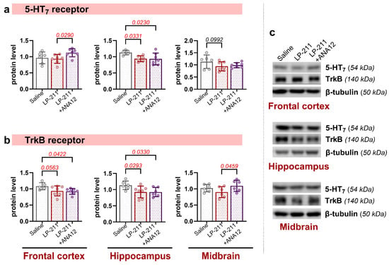
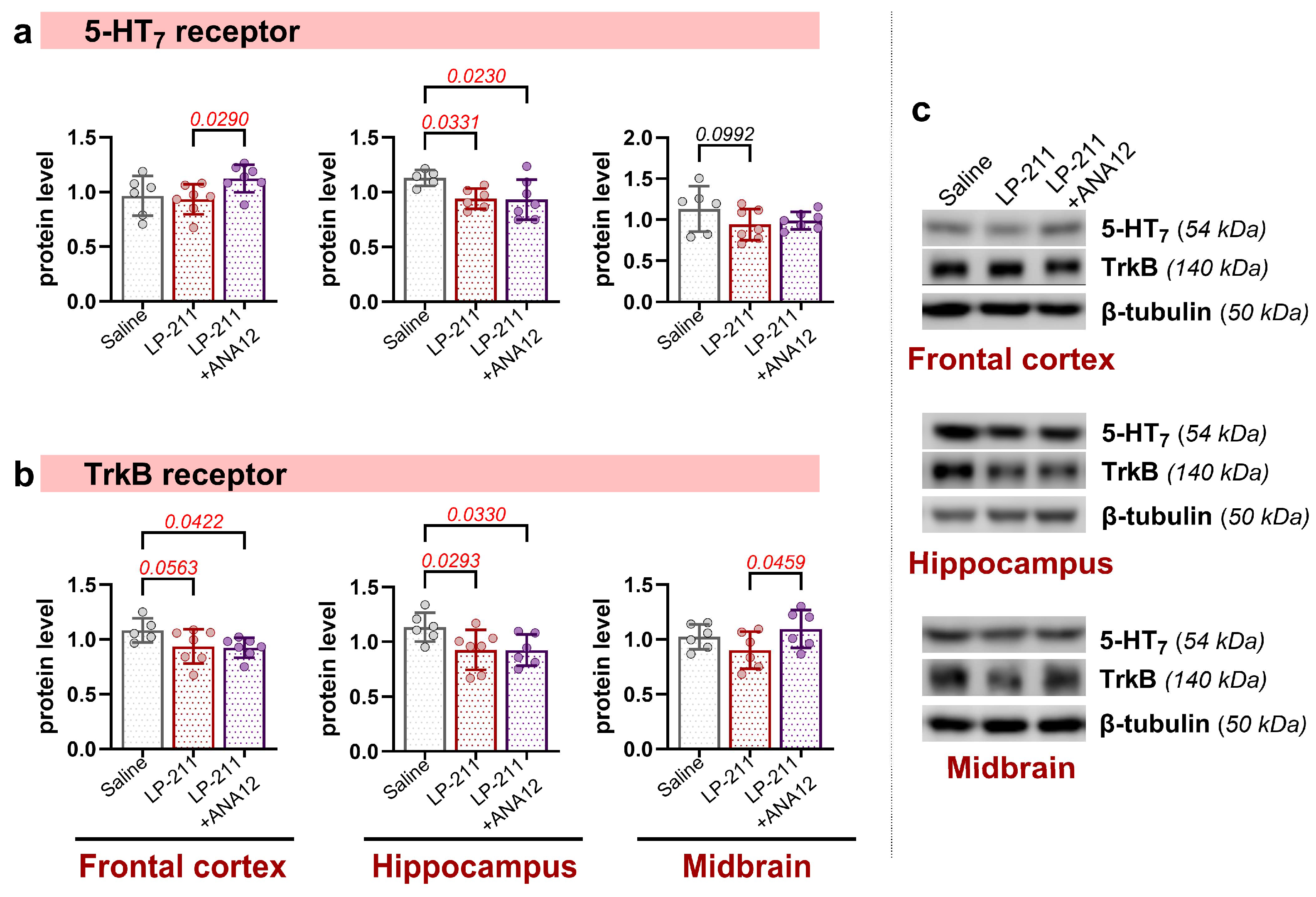
Prolonged administration of the selective 5-HT7 receptor agonist LP-211 produced a region-specific change in 5-HT7 receptor protein levels (Figure 3a). In the hippocampus, both LP-211 and LP-211 + ANA12 significantly reduced 5-HT7 receptor levels (F(2,15) = 3.824, p = 0.0455), whereas no significant differences between groups were detected in the midbrain (F(2,17) = 1.615, p = 0.2280). An effect of pretreatment with ANA12 in the frontal cortex (F(2,17) = 3.216, p = 0.0654) was found.
Figure 3.
Brain region-specific changes in 5-HT7 and TrkB receptor protein levels following prolonged 5-HT7 receptor activation. Protein levels of 5-HT7 (a) and TrkB (b) receptors were analyzed by SDS-PAGE and Western blot (WB) in membrane fractions isolated from the frontal cortex, hippocampus, and midbrain of ASC mice following prolonged treatment with LP-211 alone or in combination with ANA12. β-tubulin was used as a loading control. Representative WBs are shown (c). Data are presented as mean ± SD. (5 ≤ n ≤ 7) (one-way ANOVA). Significant between-group differences are indicated on the graphs.
TrkB receptor levels also differed across brain regions and treatment groups (Figure 3b). In the frontal cortex and hippocampus, TrkB protein levels were significantly lower in both LP-211-treated groups than in the control group (F(2,16) = 2.886, p = 0.0851 for cortex and F(2,16) = 3.681, p = 0.0484 for hippocampus). In the midbrain, TrkB levels differed between the LP-211 and LP-211 + ANA12 groups, with higher levels observed following TrkB antagonist treatment (F(2,15) = 2.425, p = 0.0459).
It is well established that the Y706 residue in the TrkB activation loop is required for full catalytic activity and efficient trans-autophosphorylation of other tyrosines, including Y515 and Y816 [35]. Importantly, Y515 in the juxtamembrane region serves as the main docking site for Shc, coupling TrkB to Ras–MAPK and PI3K–Akt [36], whereas Y816 in the C-terminal tail provides the high-affinity docking site for PLCγ [37] and drives PLCγ-dependent signaling critical for synaptic plasticity and certain forms of learning.
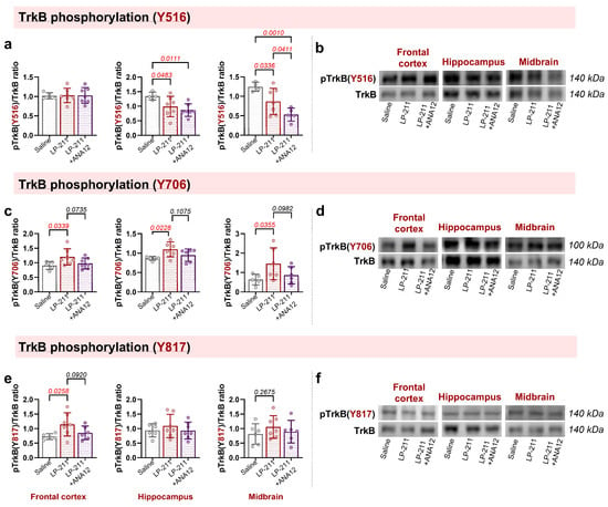
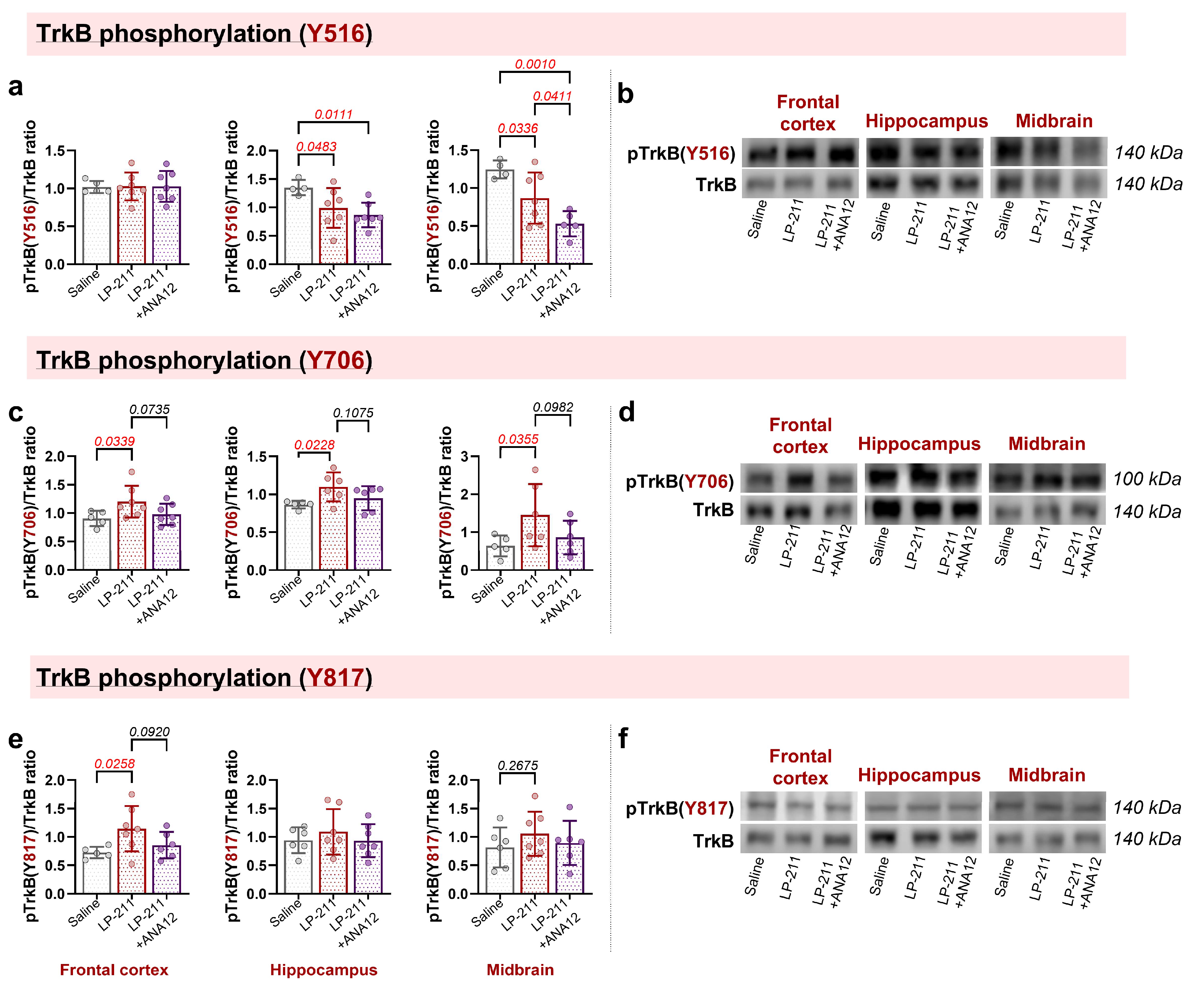
Thus, we next examined whether prolonged 5-HT7 receptor activation altered TrkB phosphorylation at three residues, Y516, Y817, and Y706 (Figure 4a,c,e). At Y516, no significant differences were detected in the frontal cortex (F(2,17) = 1.557, p = 0.2393). In contrast, LP-211 significantly reduced TrkB Y516 phosphorylation in the hippocampus (F(2,15) = 4.266, p = 0.0341) and midbrain (F(2,13) = 8.825, p = 0.0038). In the midbrain, the LP-211 + ANA12 group demonstrated a stronger reduction (Figure 4a) in Y516 phosphorylation, indicating an additive effect of the TrkB antagonist.
Figure 4.
Brain-region-specific changes in TrkB phosphorylation following prolonged 5-HT7 receptor activation. ASC mice were treated with LP-211 alone or in combination with ANA12. TrkB phosphorylation at Y516 (a,b), Y706 (c,d), and Y817 (e,f) and total TrkB were analyzed by SDS-PAGE and Western blot (WB) in membrane fractions isolated from the frontal cortex, hippocampus and midbrain. Representative WBs are shown (b,d,f). Data are presented as mean ± SD. (4 ≤ n ≤ 7) (one-way ANOVA). Significant between-group differences are indicated on the graphs.
A different pattern was observed for TrkB Y706 phosphorylation (Figure 4c). LP-211 increased TrkB Y706 phosphorylation in the frontal cortex (F (2,15) = 3.155, p = 0.0339) hippocampus (F(2,14) = 3.432, p = 0.0228) and midbrain (F(2,14) = 2.991, p = 0.0355) compared with the control group. In all brain structures, the increase in Y706 phosphorylation observed following LP-211 administration was not detected in animals pretreated with ANA12.
TrkB Y817 phosphorylation was increased only in the frontal cortex of animals treated with LP-211 alone (F (2,15) = 3.376, p = 0.0258).
2.4. 5-HT7 and TrkB Receptors Form Heterodimers in Neuroblastoma N1E-115 Cells and in the Mouse Brain
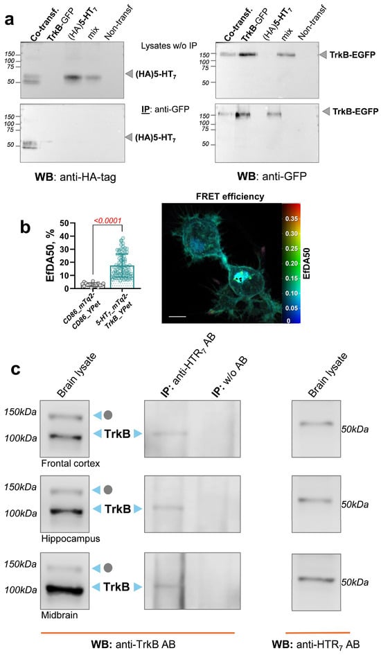
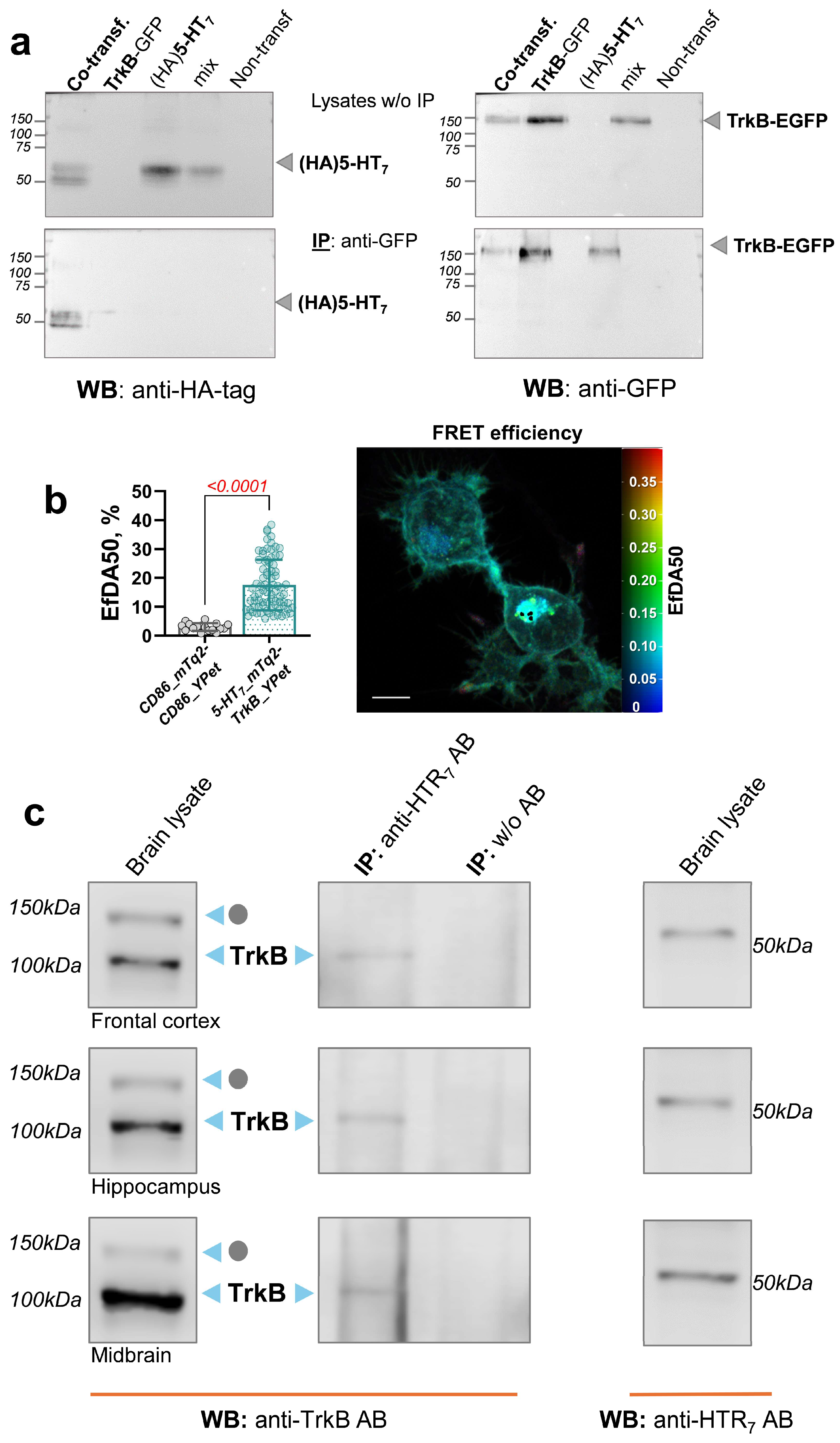
To determine whether the effects of 5-HT7 receptor activation on TrkB phosphorylation could result from a physical interaction between these receptors, we examined the formation of 5-HT7–TrkB receptor complexes in vitro. Co-immunoprecipitation experiments were performed using neuroblastoma N1E-115 cells co-expressing HA-tagged 5-HT7 and GFP-tagged TrkB. Following immunoprecipitation with an antibody against the HA tag, GFP-tagged TrkB was detected only in cells co-expressing both HA-5-HT7 and GFP-TrkB (Figure 5a). To control for potential artificial protein aggregation during sample preparation, cells expressing only one receptor type (either HA-5-HT7 or GFP-TrkB) were mixed before lysis and analyzed in parallel. Although both receptors were readily detected, no co-immunoprecipitation was observed under these conditions (Figure 5a), supporting the specificity of the 5-HT7–TrkB heterooligomerization.
Figure 5.
Interaction between receptors 5-HT7 and TrkB in neuroblastoma N1E-115 cells. (a) Co-immunoprecipitation of recombinant HA-tagged 5-HT7 and GFP (mEGFP)-tagged TrkB. Using a mixture of cells expressing each individual protein (mix) or cells that co-expressed both proteins (co-transfection), we performed immunoprecipitation (IP) with anti-GFP antibody, followed by Western blot (WB) analysis with anti-GFP (right) and anti-HA (left) antibodies. Top: Expression of proteins before IP (Lysate w/o IP). Bottom: Expression of proteins after IP. (b) Specific interaction between 5-HT7-mTurquoise2 and TrkB-YPet. Cells co-expressing 5-HT7-mTurquoise2 and TrkB-YPet were analyzed using the lux-FRET method after confocal microscopy. Apparent FRET efficiency (EfDA) and a representative cell are shown. Scale bar: 20 µm. (c) Co-immunoprecipitation between 5-HT7 and TrkB receptors. Whole-brain homogenates were prepared from different brain areas and subjected to IP with anti-5-HT7 receptor antibody, followed by WB analysis with anti-TrkB and anti-5-HT7 antibodies. AB: antibody; IP, immunoprecipitation; WB, Western blot; grey dots marks glycosylated TrkB. Results are representative of at least three independent experiments. Data are presented as mean ± SD (one-way ANOVA). Significant between-group differences are indicated on the graphs.
Because co-immunoprecipitation requires protein solubilization and concentration, which may promote artificial protein aggregation [38], we next examined the 5-HT7–TrkB interaction in living cells using a Förster resonance energy transfer (FRET)-based approach. The apparent FRET efficiency (EfDA) between mTurquoise2-labeled 5-HT7 (5-HT7-mTurquoise2, donor) and YPet-labeled TrkB (TrkB-YPet, acceptor) was measured in living N1E-115 cells using the linear unmixing FRET (lux-FRET) method [39] combined with confocal microscopy (Figure 5b). The lux-FRET analysis revealed high apparent FRET efficiency (EfDA = 17.55%) between 5-HT7-mTurquoise2 and TrkB-YPet. In contrast, cells expressing a monomeric fluorophore-tagged CD86 protein [40] yielded significantly lower EfDA values (Figure 5b). Together, the co-immunoprecipitation and FRET data support a heterodimerization of 5-HT7 and TrkB receptors in living cells.
To demonstrate the presence of oligomerization between 5-HT7 and TrkB receptors in vivo, we then performed a co-immunoprecipitation assay using brain tissue lysates from mice and identified 5-HT7–TrkB oligomeric complexes in samples from the frontal cortex, hippocampus, and midbrain (Figure 5c).
3. Discussion
In the present study, we provide evidence that 5-HT7 receptor activation can influence TrkB signaling and that this interaction may contribute to the behavioral changes of 5-HT7 receptor stimulation in ASC mice, a strain selected for a genetically defined depressive-like phenotype. Repeated administration of the selective 5-HT7 receptor agonist LP-211 increased sucrose preference and reduced the latency to the first transition in the dark–light box test; these effects were not observed when LP-211 was co-administered with the TrkB antagonist ANA12. Consistent with these behavioral findings, LP-211 increased TrkB phosphorylation in the hippocampus, midbrain and frontal cortex, whereas ANA12 pretreatment reduced the LP-211-induced increase in TrkB phosphorylation, indicating that TrkB signaling is functionally involved in the effects of 5-HT7 receptor activation. Importantly, co-immunoprecipitation experiments demonstrated an interaction between 5-HT7 and TrkB receptors not only in a heterologous cellular system but also in brain tissue. This finding was further supported by FRET measurements in living cells, which provided evidence for a physical interaction between these two receptors.
In our experiments, repeated administration of the selective 5-HT7 receptor agonist LP-211 to ASC mice increased sucrose preference and demonstrated a modest anxiolytic-like effect in the dark–light box test, as reflected by a decreased latency to the first transition from the dark to the light compartment. This result is consistent with studies that indicated anxiolytic-like effects of repeated LP-211 treatment [41,42].
Existing studies and reviews have predominantly reported antidepressant-like effects of 5-HT7 receptor antagonism [19,20,43] or genetic receptor deletion [44], whereas evidence for beneficial effects of selective 5-HT7 receptor activation has remained comparatively limited. In line with this, the literature data indicated that TrkB receptor blockade does reduce the time of immobility in the FST [45,46,47]. In our study, however, the LP-211 + ANA12 group showed increased immobility time and number of immobility episodes compared with both the saline-treated control and LP-211 groups. The absence of an ANA12-only group, however, prevents us from distinguishing the independent behavioral effects of TrkB antagonism from its effects in combination with LP-211. Other studies have also reported context-dependent effects of LP-211. For example, it was recently shown that LP-211 alone did not significantly enhance sucrose preference in rats with neuropathic pain [48]. Likewise, acute high-dose LP-211 (10 mg/kg) reduced sucrose preference in naive mice but did not worsen behavior in already anhedonic animals [49]. However, in our study, repeated low-dose LP-211 treatment reduced the severity of anhedonia, which is in good agreement with our previous findings showing that AAV-mediated overexpression of 5-HT7 receptors in the midbrain raphe nuclei area produced an antidepressant-like effect in ASC mice [50]. Thus, two independent approaches to enhancing 5-HT7 receptor signaling—pharmacological activation and increased receptor availability—were associated with antidepressant-like effects in the same mouse model. In their systematic review, Gottlieb et al. [19] highlighted the heterogeneity of experimental models and interventions, underscoring the difficulty of generalizing the behavioral effects of 5-HT7 modulation across experimental conditions. This context is consistent with the framework proposed by Fukuyama et al. [20], who suggested that 5-HT7 inhibition may be beneficial when dysfunctional transmission occurs within mature, structurally intact networks, whereas 5-HT7 activation may instead facilitate neurotransmission when network function is compromised by developmental abnormalities or loss of neural circuitry through modulation of intracellular signaling. The ASC phenotype, which arises from long-term selection and is accompanied by constitutive alterations in the serotonergic system [29,30], neuroanatomy [30] and BDNF expression [31], may therefore represent a biological context in which 5-HT7 activation produces effects distinct from those observed in other depression models.
A functional relationship between 5-HT7 and TrkB receptors was first described by Samarajeewa et al. [21], who showed that 5-HT7 receptor activation promotes TrkB receptor phosphorylation in several cellular models, including primary cortical cultures, SH-SY5Y cells, and RGC-5 cells. In particular, acute stimulation with the 5-HT7 agonist LP-12 produced a rapid increase in TrkB phosphorylation, which the authors interpreted as 5-HT7 receptor-mediated transactivation of TrkB, whereas prolonged stimulation also increased the expression of full-length TrkB and its basal phosphorylation at Y816 [21]. Our findings extend these observations to the intact mouse brain, demonstrating that acute administration of the selective 5-HT7 agonist LP-211 increased TrkB phosphorylation in the hippocampus and midbrain, whereas the TrkB antagonist ANA12 attenuated this response. Moreover, the absence of a corresponding increase in total receptor levels argues against a nonspecific effect on TrkB abundance and supports regulation at the level of receptor phosphorylation.
Our findings further suggest that the effects of prolonged 5-HT7 receptor activation on TrkB signaling are more complex than a simple increase in receptor activity. Whereas acute LP-211 administration increased pTrkB levels in the midbrain, prolonged treatment produced a more selective and region-specific pattern of TrkB phosphorylation. This distinction may be important for understanding how sustained 5-HT7 receptor activation influences TrkB-dependent signaling. TrkB engages several major intracellular pathways, including Ras/MAPK, PI3K/Akt, and PLCγ-dependent signaling, while the relative contribution of these pathways depends on cellular context and the spatiotemporal pattern of receptor activation [35,51]. Importantly, phosphorylation of individual intracellular tyrosine residues contributes to the recruitment and activation of distinct signaling components. In particular, phosphorylation of Y516 and Y817 provides docking sites for downstream effectors, whereas phosphorylation within the activation loop, including Y706, is closely associated with regulation of receptor kinase activity [35]. In our experiments, prolonged LP-211 treatment increased Y706 phosphorylation in the frontal cortex, hippocampus and midbrain, accompanied by reduced Y516 phosphorylation and increased Y817 phosphorylation only in the frontal cortex. It should be noted that the level of 5-HT7 receptor protein was lower in the experimental groups, specifically in the hippocampus and midbrain; this may indicate greater sensitivity of these receptor populations to LP-211, leading to enhanced ligand binding and subsequent receptor internalization from the membrane, and thus suggests downregulation resulting from their chronic activation [52]. Together with the region-specific changes in total TrkB protein, these findings suggest that sustained 5-HT7 receptor activation may modify the pattern of TrkB signaling rather than simply increasing its overall activity.
The functional coupling between 5-HT7 and TrkB receptors may involve their physical association within receptor complexes. Our co-immunoprecipitation experiments demonstrated an association between 5-HT7 and TrkB receptors in the frontal cortex, hippocampus, and midbrain, while FRET experiments performed in living cells provided independent evidence for the oligomerization between the two receptors. Together, these methods validate the presence of the complexes in native tissue while avoiding the limitations of cell lysis. Similar physical interactions have been described for other GPCR–RTK pairs such as A2AR-FGFR1 [11], 5-HT1A-FGFR1 [12,13,15], A2AR-TrkB [53] and 5-HT2A-TrkB [14]. Together with our findings, these observations support a model in which physical association within a receptor complex may provide a structural framework through which activation of a GPCR can influence the conformation, phosphorylation, and signaling properties of an associated RTK.
Whether the functional effects of TrkB oligomerization depend on the identity of the interacting serotonin receptor remains an important question. Our previous study of 5-HT2A–TrkB heteroreceptors provides an informative comparison. We demonstrated that 5-HT2A and TrkB physically interact in vitro and in vivo, and that this association was accompanied by reduced basal TrkB autophosphorylation and attenuated TrkB activation by the agonist 7,8-DHF [14]. Consistent with these findings, pharmacological blockade of 5-HT2A receptors increased basal TrkB phosphorylation in the frontal cortex and hippocampus in vivo [34]. In contrast, the present study indicates that activation of 5-HT7 is associated with increased TrkB phosphorylation, suggesting that physical interaction with TrkB does not necessarily produce a uniform functional effect across serotonin receptor subtypes. Rather, the type and activation state of the associated GPCR may shape the functional output of the receptor complex, potentially by influencing the conformation and signaling properties of TrkB. Such receptor-specific regulation may provide a mechanism through which serotonergic signaling can differentially engage TrkB-dependent pathways involved in neuronal plasticity. However, although our findings support a physical association between 5-HT7 and TrkB receptors and indicate functional coupling between these receptor systems, whether complex formation directly mediates the effects of 5-HT7 activation remains to be established.
Together, our complementary findings suggest that the oligomerization between 5-HT7 and TrkB may provide a molecular basis for their functional coupling and contribute to the regulation of TrkB signaling following 5-HT7 receptor activation.
4. Limitations
Several limitations should be considered when interpreting the present findings. Although our co-immunoprecipitation data support an association between 5-HT7 and TrkB receptors in brain tissue, they do not establish the existence, stoichiometry, or functional significance of a defined heterodimeric complex in vivo. FRET measurements provide complementary evidence of close molecular proximity between the receptors in living cells, but they do not establish that the same arrangement occurs in brain tissue. Most importantly, the present experiments do not determine whether physical association between 5-HT7 and TrkB is necessary for the observed changes in TrkB phosphorylation or the behavioral effects of LP-211. Addressing this question will require selective disruption of the receptor interaction while preserving the intrinsic functions of both receptors. Likewise, although the effects of LP-211 on TrkB phosphorylation and the attenuation of its behavioral effects by ANA12 support the involvement of TrkB signaling, the downstream pathways through which this signaling contributes to the behavioral effects of 5-HT7 activation remain to be determined. This issue is particularly relevant given the region- and phosphorylation site-specific changes in TrkB phosphorylation observed after prolonged LP-211 treatment, the functional consequences of which cannot be inferred from phosphorylation measurements alone. Another limitation concerns the use of the ASC mouse line, which exhibits a depressive-like behavioral phenotype and some neurobiological alterations, including changes in serotonergic neurotransmission and reduced BDNF expression. These features may influence the response to 5-HT7 receptor modulation and limit the generalizability of our behavioral findings to other animal models. In particular, differences in the genetic and neurobiological backgrounds of the models used may contribute to the apparent discrepancy between the antidepressant-like effects of 5-HT7 agonists observed here and the effects of 5-HT7 antagonists reported in other studies. However, whether these differences account for the divergent behavioral outcomes remains to be established. Future studies combining selective manipulation of the 5-HT7–TrkB interaction with analysis of downstream signaling pathways, as well as comparisons across animal models with distinct genetic and neurobiological backgrounds, will therefore be important for defining how receptor complex formation contributes to the regulation of TrkB-dependent neuroplasticity and behavior.
5. Materials and Methods
5.1. Animals and Drugs
Adult (P60) male C57BL/6J mice (10–11 weeks old, 23–26 g, Jackson Laboratory, Bar Harbor, ME, USA, RRID: IMSR_JAX:000664) were used for acute treatment with LP-211, SB-269970 or ANA12 and for co-immunoprecipitation experiments. Adult (P60) male ASC mice, which were bred by long-term selection for a predisposition to catalepsy in the Laboratory of behavioral neurogenomics at the Institute of Cytology and Genetics of the Siberian Branch of the Russian Academy of Sciences (10–11 weeks old, 23–26 g (Novosibirsk, Russia, RRID: MGI:6200645)), were used for repeated (9 days) treatment with LP-211 and ANA12.
Genetically predisposed to depressive-like behavior ASC/Icg (ASC, antidepressant sensitive catalepsy) strain was created in the Laboratory of Behavioral Neurogenomics, Institute of Cytology and Genetics SB RAS as the result of prolonged selection of hybrids between catalepsy-prone CBA/Lac and catalepsy-resistant AKR/J strains for high predisposition to catalepsy [22,23]. Since female sex hormones can substantially modulate TrkB phosphorylation in rodent forebrain neurons across the estrous cycle, we used male mice to avoid the influence of confounding factors [54,55,56,57]. The mice were housed under standard laboratory conditions, 3–4 animals per cage, in the natural light–dark cycle (16 h light and 8 h dark with gradual transitions between the light and dark phases to simulate dusk and dawn) with free access to water and feed. All efforts were made to minimize animal suffering. Animals were monitored for signs of ill health or distress throughout the experimental procedures.
Animals were randomly allocated to the experimental groups. However, the order of behavioral testing was not fully randomized, as animals were tested group-wise rather than in a randomized sequence across experimental groups. The experimenter conducting the animal procedures and behavioral testing was blinded to group allocation.
LP-211 (N-(4-cyanophenylmethyl)-4-(2-diphenyl)-1-piperazinehexanamide), a selective competitive full agonist of the 5-HT7 receptor (EC50 = 44 nM for the human receptor), was dissolved in DMSO and diluted with saline. Its selectivity over the key 5-HT1A and 5-HT2A receptors exceeds 200-fold in functional assays [52,58], and the compound rapidly enters the brain, reaching concentrations comparable to those in plasma [52]. SB-269970 is a well-characterized selective 5-HT7 receptor antagonist [59,60]. ANA12(N-[2-[[(hexahydro-2-oxo-1H-azepin-3-yl)amino]carbonyl]phenyl]-benzo[b]thiophene-2-carboxamide) is a selective TrkB inhibitor/antagonist [45]. ANA12 binds TrkB directly and specifically, blocking its downstream signaling without altering TrkA or TrkC function; it is not degraded before entering the brain, where its active concentrations persist from 30 min to 6 h after administration.
In the acute experiment, the selective 5-HT7 receptor agonist LP-211 (Sigma Aldrich, St. Louis, MO, USA), the selective 5-HT7 receptor antagonist SB-269970 (Sigma Aldrich) and the TrkB antagonist ANA12 (Sigma Aldrich) were used. All doses were selected based on published data. Animals were divided into five groups. Animals that received ANA12 (2.5 mg/kg; i.p.), SB-269970 (10 mg/kg; i.p.), or physiological saline (0.9% NaCl; i.p.) were euthanized 30 min after administration. Animals that received LP-211 (10 mg/kg; i.p.) were euthanized 1 h after administration. In the LP-211+ANA12 group, animals received LP-211 (10 mg/kg; i.p.) 30 min after pretreatment with ANA12 (2.5 mg/kg) and were euthanized 1 h later.
To examine how repeated 5-HT7 receptor activation affects ASC mice behavior, LP-211 was administered at 1 mg/kg. Since some of the downstream effects of 5-HT7 activation may be mediated through TrkB receptors, a separate group of animals received ANA12 at 2.5 mg/kg 30 min before LP-211 injection. Control animals received DMSO in saline. All drugs were injected intraperitoneally for 9 days, and the doses were selected based on published data. One day after the final behavioral test, the animals were euthanized by decapitation. The brain was removed and placed on ice, after which the brain structures (frontal cortex, hippocampus (dorsal and ventral), midbrain) were dissected. The resulting samples were frozen in liquid nitrogen and stored at −80 °C until further procedures.
5.2. Cell Culture and Transfection
Mouse N1E-115 neuroblastoma cells from the American Type Culture Collection (ATCC, cat. # CRL-2263, Manassas, VA, USA, RRID: CVCL_0451) were grown at 37 °C and 5% CO2 in DMEM (Gibco,Thermo Fisher Scientific, Grand Island, NY, USA) that contained 10% fetal bovine serum (Gibco) and penicillin/streptomycin (100 U/mL, Gibco). Transient transfection was performed with Lipofectamine 2000 (Invitrogen, Thermo Fisher Scientific, Grand Island, NY, USA).
5.3. Recombinant-DNA Procedures
Murine 5-HT7 cDNA was cloned into the pcDNA3.1(+) donor vector (Invitrogen, Carlsbad, CA, USA) carrying an HA-tag. TrkB_mEGFP was kindly gifted by Ryohei Yasuda (Addgene plasmid # 83952; http://n2t.net/addgene:83952; accessed on 13 July 2019; RRID: Addgene_83952). Additionally, 5-HT7 was cloned into a pcDNA3.1(+) vector carrying mTurquoise2 (5-HT7-mTurquoise2), and TrkB_mEGFP was used to generate TrkB-YPet by replacing mEGFP with YPet.
5.4. Co-Immunoprecipitation
Co-immunoprecipitation in lysates of N1E-115 cells that co-expressed HA-tagged 5-HT7 and GFP-tagged TrkB was performed as described previously [61] with an antibody against GFP (1:250; GeneTex, Irvine, CA, USA, cat. # GTX26556, RRID: AB_371421). Immunoblotting was carried out using a horseradish peroxidase (HRP)-conjugated anti-GFP (1:1000; LSBio (LifeSpan, Seattle, WA, USA), cat. # LS-C50850-500, RRID: AB_1220053) or HRP-conjugated anti-HA tag (1:250; Roche, Basel, Switzerland, cat. # 12013819001, RRID: AB_390917) antibody.
Co-immunoprecipitation from hippocampal, cortical, and midbrain homogenates was performed according to the protocol described elsewhere [62]. Briefly, brain samples isolated from adult (P90) C57BL/6J mice were homogenized, and membrane fractions were prepared by differential centrifugation. The lysates were incubated with a rabbit polyclonal antibody against 5-HT7 (1:50; Abcam, Cambridge, UK, cat. # ab128892, RRID: AB_11139629) followed by incubation with Protein A-Sepharose and western blotting with a goat anti-TrkB antibody (1:1000; R&D Systems, Minneapolis, MN, USA, cat. # AF1494, RRID: AB_2155264).
5.5. Linear Unmixing FRET Analysis
These measurements were performed on live N1E-115 neuroblastoma cells as described elsewhere [39]. 5-HT7-mTurquoise2 and TrkB-YPet served as the donor and acceptor, respectively. As a negative control, cells were co-transfected with mTurquoise2-tagged CD86 and YPet-tagged CD86. After 16 h, the cells were imaged with a Zeiss LSM 780 microscope (Zeiss, Göttingen, Germany) equipped with a C-Apochromat 40×/1.2 W Korr water immersion objective via excitation of the fluorescent proteins at 440 and 514 nm according to the protocol. For image analysis and evaluation, custom-written MATLAB (version R2019a, MathWorks, Natick, MA, USA) scripts were employed. We calculated predicted apparent FRET efficiency as EfDA = ½ EfD/(1 − xD) = ½ EfA/xD, assuming a standard dimerization model [62,63].
5.6. Western Blotting
To isolate the membrane protein, specimens were homogenized in a buffer containing 300 mM sucrose, 10 mM Tris-HCl, pH 7.2, 1 mM EDTA, 5 mM β-mercaptoethanol, with the addition of 1 mM Na3VO4, 2 mM phenylmethylsulfonyl fluoride, and a protease and phosphatase inhibitor cocktail. The mixture was centrifuged at 500× g for 15 min at 4 °C, and the supernatant was collected and centrifuged at 20,000× g at 4 °C for an hour. The precipitate was resuspended in a homogenizing buffer. Protein concentration was estimated spectrophotometrically using the Pierce BCA Protein Assay Kit (Thermo Fisher Scientific Inc., Waltham, MA, USA) and a NanoDrop 2000C spectrophotometer (Thermo Scientific Inc., Waltham, MA, USA), followed by adjustment of samples to equal concentrations with 2× Laemmli sample buffer. After denaturation by boiling for 10 min at 95 °C, the cell extracts (20 μg of protein per lane) were resolved on 10% SDS-PAGE and blotted onto a nitrocellulose membrane (Bio-Rad Laboratories, Hercules, CA, USA). The membrane was incubated with a primary antibody [an anti-TrkB (1:1000; R&D Systems, cat. # AF1494, RRID: AB_2155264), anti-pTrkB (Tyr 516) (1:500; Affinity Biosciences, Cincinnati, OH, USA, cat. # AF3462, RRID: AB_2834900), anti-pTrkB (Tyr 706) (1:500; Affinity Biosciences, cat. # AF3461, RRID: AB_2834899), anti-pTrkB (Tyr 817) (1:1000; Affinity Biosciences, cat. # AF8389, RRID: AB_2840450) or anti-5-HT7 antibody (1:1000; Abcam, Cambridge, UK, cat. # ab128892, RRID: AB_11139629)] at 4 °C overnight, then the membrane was washed in Tris-buffered saline supplemented with 0.05% of Tween 20 (TBS-T) and incubated with an HRP-conjugated secondary antibody. After protein detection, the blot was stripped and then re-probed with an anti-β-tubulin antibody (1:20,000; Abcam, cat. # ab6046, RRID: AB_2210370) as a loading control. Immunoreactive bands were detected by means of the Clarity Western ECL Substrate (Bio-Rad Laboratories, Hercules, CA, USA). Protein bands were documented on a C-DiGit Blot Scanner (LI-COR, Lincoln, NE, USA) and quantified in the Image Studio software (LI-COR Image Studio Software, version 5.2, RRID:SCR_015795, Lincoln, NE, USA). Target protein levels were assessed in chemiluminescence relative units and normalized to β-tubulin chemiluminescence relative units.
5.7. Behavioral Testing
5.7.1. Open Field Test
To measure locomotor activity and exploratory behavior, the open-field paradigm was employed. The setup consisted of a circular arena (60 cm diameter) enclosed by a plastic wall 25 cm in height. Illumination was provided from beneath the floor by two 12 W halogen lamps placed 40 cm below it; light was passed through a mat and a semitransparent floor [64]. Each mouse was released near the wall, and its movements were registered for 5 min using a digital camera (Sony, Tokyo, Japan). The arena was thoroughly cleaned after every trial. Video records were analyzed frame by frame with the original EthoStudio software (Novosibirsk, Russia) [65]. Total distance moved, explored arena area, and time spent in the center were calculated automatically. Rearings, taken as an index of exploratory behavior, and groomings, as a measure of stereotypy, were counted manually.
5.7.2. Dark–Light Box Test
This test was used to examine anxiety-related behavior. The setup consisted of a white plastic box (45 × 21 × 26 cm) divided into two sections by an opaque wall containing a 7 × 7 cm opening. The dark section was enclosed, whereas the light section had a translucent plastic floor illuminated from below by two halogen bulbs (12 W each). Each animal was initially placed in the dark section, with its head oriented toward the opening. During the 5 min trial, the following measures were taken: number of entries into the light section, latency of the first entry into the light section, total time spent in the light section, and the total traveled distance.
5.7.3. Forced Swim Test
The forced swim test is a standard paradigm for assessing depression-like behavior. In this test, each mouse was placed into a clear glass cylinder (15 × 30 cm) filled with water at 25 °C. After 2 min of adaptation, EthoStudio software was used to record total immobility time and the latency to the first immobility episode over the following 4 min [66].
5.7.4. Sucrose Preference Test
The sucrose preference test was performed according to the protocol described by Strekalova and Steinbusch (2010) [67], with the following modification: because the animals were housed in OptiMice cages and tested in a separate test cage with a distinct configuration, the positions of the drinking bottles in the test cage differed from those in the home cage. This prevented the development of a side preference that could confound the test, so we did not perform the one-week habituation to two bottles as described in the original protocol. To reduce neophobic reactions to the sucrose solution, mice were pre-exposed to 2.5% sucrose solution for 2 h in a one-bottle paradigm one day before the first sucrose preference test (baseline measurement). For the sucrose preference test, mice were given free access to two drinking bottles containing either water or 2.5% sucrose solution for 8 h during the dark (active) phase of the light cycle. To minimize the confounding effect of side preference, the positions of the bottles were switched in the middle of the testing period (after 4 h). No food or water deprivation was applied prior to the test, and no stressors were administered for at least 12 h before testing. The consumption of water and sucrose solution was determined by weighing the bottles before and after the test. Sucrose preference was calculated as the percentage of sucrose solution consumed from the total volume of liquid drunk, according to the formula:
Sucrose Preference = [V(sucrose solution)/(V(sucrose solution) + V(water))] × 100%
A sucrose preference level at or below 65% was used as the criterion for anhedonia, as previously validated [34].
5.8. Statistical Analysis
The data are presented as the mean ± standard deviation (SD). Dixon’s Q test was used to identify and remove outliers in the dataset. Gaussian distribution was evaluated using the Shapiro–Wilk normality test. The mean values were compared using one-way ANOVA followed by Fisher’s post hoc test. An unpaired t-test with Welch’s correction was applied to assess FRET efficiency. In the Results section, N values are presented after the Dixon test.
Author Contributions
T.I. conceived the study. T.I., A.T. and V.N. designed the experiments. T.I., A.T., D.E., M.S. and V.N. conducted the experiments. T.I. analyzed the data. V.N. contributed reagents, materials, and analytical tools. A.T. and T. I. wrote the manuscript. V.N. edited the manuscript. All authors have read and agreed to the published version of the manuscript.
Funding
This research was supported by the Russian Science Foundation grant 25-25-00238 to Tatiana V. Ilchibaeva. The funder had no role in study design, data collection and analysis or preparation of the manuscript.
Institutional Review Board Statement
All animal procedures were conducted in accordance with international guidelines for biomedical research (European Communities Council Directive 86/609/EEC) and were regulated by standard operating procedures developed following GOST 33044-2014 (Principles of Good Laboratory Practice) and bioethical norms. The protocol was approved by the Ethics Committee of the Institute of Cytology and Genetics SB RAS (approval No. 180, dated 28 August 2024 and approval No. 209, dated 30 January 2025).
Informed Consent Statement
Not applicable.
Data Availability Statement
The original contributions presented in this study are included in the article. Further inquiries can be directed to the corresponding authors.
Acknowledgments
We thank Andre Zeug and Evgeni Ponimaskin from the Hannover Medical School (Hannover, Germany) for the opportunity to perform the lux-FRET analysis and excellent technical support. Experiments on mice were conducted at the ICG SB RAS, the Multi-Access Center of Animal Genetic Resources (unique identifier RFMEFI62119X0023) and compensated by the basic-research project FWNR-2026-0028. During the preparation of this manuscript, the authors used ChatGPT (GPT-5.6) for language checking and editing to improve the clarity and readability of the text. The authors have reviewed and edited the output and take full responsibility for the content of this publication.
Conflicts of Interest
The authors declare no conflicts of interest.
Abbreviations
The following abbreviations are used in this manuscript:
| 5-HT | 5-hydroxytryptamine |
| 5-HT1A | 5-hydroxytryptamine 1A receptor |
| 5-HT2A | 5-hydroxytryptamine 2A receptor |
| 5-HT7 | 5-hydroxytryptamine 7 receptor |
| ANOVA | analysis of variance |
| ASC | antidepressant-sensitive catalepsy |
| ATCC | American Type Culture Collection |
| BCA | bicinchoninic acid |
| BDNF | brain-derived neurotrophic factor |
| CD86 | cluster of differentiation 86 |
| cDNA | complementary DNA |
| DMEM | Dulbecco’s Modified Eagle Medium |
| DMSO | dimethyl sulfoxide |
| ECL | enhanced chemiluminescence |
| EDTA | ethylenediaminetetraacetic acid |
| FRET | Förster resonance energy transfer |
| GFP | green fluorescent protein |
| GPCR | G protein-coupled receptor |
| HA | hemagglutinin |
| HRP | horseradish peroxidase |
| i.p. | intraperitoneal |
| IP | immunoprecipitation |
| lux-FRET | linear unmixing Förster resonance energy transfer |
| mEGFP | monomeric enhanced green fluorescent protein |
| RRID | Research Resource Identification |
| RTK | receptor tyrosine kinase |
| SD | standard deviation |
| TBS-T | Tris-buffered saline supplemented with Tween 20 |
| TrkB | tropomyosin receptor kinase B |
| WB | Western blot |
References
- Malhi, G.S.; Mann, J.J. Depression. Lancet 2018, 392, 2299–2312. [Google Scholar] [CrossRef] [Scilit] [PubMed]
- Duman, R.S.; Aghajanian, G.K.; Sanacora, G.; Krystal, J.H. Synaptic Plasticity and Depression: New Insights from Stress and Rapid-Acting Antidepressants. Nat. Med. 2016, 22, 238–249. [Google Scholar] [CrossRef] [Scilit] [PubMed]
- Adachi, M.; Autry, A.E.; Mahgoub, M.; Suzuki, K.; Monteggia, L.M. TrkB Signaling in Dorsal Raphe Nucleus Is Essential for Antidepressant Efficacy and Normal Aggression Behavior. Neuropsychopharmacology 2017, 42, 886–894. [Google Scholar] [CrossRef] [Scilit] [PubMed]
- Castrén, E.; Monteggia, L.M. Brain-Derived Neurotrophic Factor Signaling in Depression and Antidepressant Action. Biol. Psychiatry 2021, 90, 128–136. [Google Scholar] [CrossRef] [Scilit] [PubMed]
- Casarotto, P.; Umemori, J.; Castrén, E. BDNF Receptor TrkB as the Mediator of the Antidepressant Drug Action. Front. Mol. Neurosci. 2022, 15, 1032224. [Google Scholar] [CrossRef] [Scilit] [PubMed]
- Sahu, M.P.; Pazos-Boubeta, Y.; Steinzeig, A.; Kaurinkoski, K.; Palmisano, M.; Borowecki, O.; Piepponen, T.P.; Castrén, E. Depletion of TrkB Receptors From Adult Serotonergic Neurons Increases Brain Serotonin Levels, Enhances Energy Metabolism and Impairs Learning and Memory. Front. Mol. Neurosci. 2021, 14, 616178. [Google Scholar] [CrossRef] [Scilit] [PubMed]
- Casarotto, P.C.; Girych, M.; Fred, S.M.; Kovaleva, V.; Moliner, R.; Enkavi, G.; Biojone, C.; Cannarozzo, C.; Sahu, M.P.; Kaurinkoski, K.; et al. Antidepressant Drugs Act by Directly Binding to TRKB Neurotrophin Receptors. Cell 2021, 184, 1299–1313.e19. [Google Scholar] [CrossRef] [Scilit] [PubMed]
- Moliner, R.; Girych, M.; Brunello, C.A.; Kovaleva, V.; Biojone, C.; Enkavi, G.; Antenucci, L.; Kot, E.F.; Goncharuk, S.A.; Kaurinkoski, K.; et al. Psychedelics Promote Plasticity by Directly Binding to BDNF Receptor TrkB. Nat. Neurosci. 2023, 26, 1032–1041. [Google Scholar] [CrossRef] [Scilit] [PubMed]
- Brunello, C.A.; Cannarozzo, C.; Castrén, E. Rethinking the Role of TRKB in the Action of Antidepressants and Psychedelics. Trends Neurosci. 2024, 47, 865–874. [Google Scholar] [CrossRef] [Scilit] [PubMed]
- Di Liberto, V.; Mudò, G.; Belluardo, N. Crosstalk between Receptor Tyrosine Kinases (RTKs) and G Protein-Coupled Receptors (GPCR) in the Brain: Focus on Heteroreceptor Complexes and Related Functional Neurotrophic Effects. Neuropharmacology 2019, 152, 67–77. [Google Scholar] [CrossRef] [Scilit] [PubMed]
- Flajolet, M.; Wang, Z.; Futter, M.; Shen, W.; Nuangchamnong, N.; Bendor, J.; Wallach, I.; Nairn, A.C.; Surmeier, D.J.; Greengard, P. FGF Acts as a Co-Transmitter through Adenosine A(2A) Receptor to Regulate Synaptic Plasticity. Nat. Neurosci. 2008, 11, 1402–1409. [Google Scholar] [CrossRef] [Scilit] [PubMed]
- Borroto-Escuela, D.O.; Romero-Fernandez, W.; Mudó, G.; Pérez-Alea, M.; Ciruela, F.; Tarakanov, A.O.; Narvaez, M.; Di Liberto, V.; Agnati, L.F.; Belluardo, N.; et al. Fibroblast Growth Factor Receptor 1- 5-Hydroxytryptamine 1A Heteroreceptor Complexes and Their Enhancement of Hippocampal Plasticity. Biol. Psychiatry 2012, 71, 84–91, Erratum in Biol. Psychiatry 2012, 72, 164. [Google Scholar] [CrossRef] [Scilit] [PubMed]
- Borroto-Escuela, D.O.; Corrales, F.; Narvaez, M.; Oflijan, J.; Agnati, L.F.; Palkovits, M.; Fuxe, K. Dynamic Modulation of FGFR1-5-HT1A Heteroreceptor Complexes. Agonist Treatment Enhances Participation of FGFR1 and 5-HT1A Homodimers and Recruitment of β-Arrestin2. Biochem. Biophys. Res. Commun. 2013, 441, 387–392. [Google Scholar] [CrossRef] [Scilit] [PubMed]
- Ilchibaeva, T.; Tsybko, A.; Zeug, A.; Müller, F.E.; Guseva, D.; Bischoff, S.; Ponimaskin, E.; Naumenko, V. Serotonin Receptor 5-HT2A Regulates TrkB Receptor Function in Heteroreceptor Complexes. Cells 2022, 11, 2384. [Google Scholar] [CrossRef] [Scilit] [PubMed]
- Ambrogini, P.; Lattanzi, D.; Pagliarini, M.; Di Palma, M.; Sartini, S.; Cuppini, R.; Fuxe, K.; Borroto-Escuela, D.O. 5HT1AR-FGFR1 Heteroreceptor Complexes Differently Modulate GIRK Currents in the Dorsal Hippocampus and the Dorsal Raphe Serotonin Nucleus of Control Rats and of a Genetic Rat Model of Depression. Int. J. Mol. Sci. 2023, 24, 7467. [Google Scholar] [CrossRef] [Scilit] [PubMed]
- Mirchandani-Duque, M.; Choucri, M.; Hernández-Mondragón, J.C.; Crespo-Ramírez, M.; Pérez-Olives, C.; Ferraro, L.; Franco, R.; Pérez de la Mora, M.; Fuxe, K.; Borroto-Escuela, D.O. Membrane Heteroreceptor Complexes as Second-Order Protein Modulators: A Novel Integrative Mechanism through Allosteric Receptor-Receptor Interactions. Membranes 2024, 14, 96. [Google Scholar] [CrossRef] [Scilit] [PubMed]
- Guseva, D.; Wirth, A.; Ponimaskin, E. Cellular Mechanisms of the 5-HT7 Receptor-Mediated Signaling. Front. Behav. Neurosci. 2014, 8, 306. [Google Scholar] [CrossRef] [Scilit] [PubMed]
- Crispino, M.; Volpicelli, F.; Perrone-Capano, C. Role of the Serotonin Receptor 7 in Brain Plasticity: From Development to Disease. Int. J. Mol. Sci. 2020, 21, 505. [Google Scholar] [CrossRef] [Scilit] [PubMed]
- Gottlieb, N.; Li, T.-Y.; Young, A.H.; Stokes, P.R. The 5-HT7 Receptor System as a Treatment Target for Mood and Anxiety Disorders: A Systematic Review. J. Psychopharmacol. 2023, 37, 1167–1181. [Google Scholar] [CrossRef] [Scilit] [PubMed]
- Fukuyama, K.; Motomura, E.; Okada, M. Therapeutic Potential and Limitation of Serotonin Type 7 Receptor Modulation. Int. J. Mol. Sci. 2023, 24, 2070. [Google Scholar] [CrossRef] [Scilit] [PubMed]
- Samarajeewa, A.; Goldemann, L.; Vasefi, M.S.; Ahmed, N.; Gondora, N.; Khanderia, C.; Mielke, J.G.; Beazely, M.A. 5-HT7 Receptor Activation Promotes an Increase in TrkB Receptor Expression and Phosphorylation. Front. Behav. Neurosci. 2014, 8, 391. [Google Scholar] [CrossRef] [Scilit] [PubMed]
- Kondaurova, E.M.; Bazovkina, D.V.; Kulikov, A.V.; Popova, N.K. Selective Breeding for Catalepsy Changes the Distribution of Microsatellite D13Mit76 Alleles Linked to the 5-HT1A Serotonin Receptor Gene in Mice. Genes Brain Behav. 2006, 5, 596–601. [Google Scholar] [CrossRef] [Scilit] [PubMed]
- Kulikov, A.V.; Bazovkina, D.V.; Kondaurova, E.M.; Popova, N.K. Genetic Structure of Hereditary Catalepsy in Mice. Genes Brain Behav. 2008, 7, 506–512. [Google Scholar] [CrossRef] [Scilit] [PubMed]
- Kulikov, A.V.; Kozlachkova, E.Y.; Maslova, G.B.; Popova, N.K. Inheritance of Predisposition to Catalepsy in Mice. Behav. Genet. 1993, 23, 379–384. [Google Scholar] [CrossRef] [Scilit] [PubMed]
- Tikhonova, M.A.; Lebedeva, V.V.; Kulikov, A.V.; Bazovkina, D.V.; Popova, N.K. Effect of Imipramine on the Behavior and Cerebral 5-HT1A Serotonin Receptors in Mice Genetically Predisposed to Catalepsy. Bull. Exp. Biol. Med. 2006, 141, 48–50. [Google Scholar] [CrossRef] [Scilit] [PubMed]
- Tikhonova, M.A.; Alperina, E.L.; Tolstikova, T.G.; Bazovkina, D.V.; Di, V.Y.; Idova, G.V.; Kulikov, A.V.; Popova, N.K. Effects of Chronic Fluoxetine Treatment on Catalepsy and the Immune Response in Mice with a Genetic Predisposition to Freezing Reactions: The Roles of Types 1A and 2A Serotonin Receptors and the Tph2 and SERT Genes. Neurosci. Behav. Physiol. 2010, 40, 521–527. [Google Scholar] [CrossRef] [Scilit] [PubMed]
- Bazovkina, D.V.; Kulikov, A.V.; Kondaurova, E.M.; Popova, N.K. Selection for the Predisposition to Catalepsy Enhances Depressive-like Traits in Mice. Genetika 2005, 41, 1222–1228. [Google Scholar] [CrossRef] [Scilit]
- Naumenko, V.S.; Bazovkina, D.V.; Semenova, A.A.; Tsybko, A.S.; Il’chibaeva, T.V.; Kondaurova, E.M.; Popova, N.K. Effect of Glial Cell Line-Derived Neurotrophic Factor on Behavior and Key Members of the Brain Serotonin System in Mouse Strains Genetically Predisposed to Behavioral Disorders. J. Neurosci. Res. 2013, 91, 1628–1638. [Google Scholar] [CrossRef] [Scilit] [PubMed]
- Naumenko, V.S.; Bazovkina, D.V.; Kondaurova, E.M.; Zubkov, E.A.; Kulikov, A.V. The Role of 5-HT2A Receptor and 5-HT2A/5-HT 1A Receptor Interaction in the Suppression of Catalepsy. Genes Brain Behav. 2010, 9, 519–524. [Google Scholar] [CrossRef] [Scilit] [PubMed]
- Tikhonova, M.A.; Kulikov, A.V.; Bazovkina, D.V.; Kulikova, E.A.; Tsybko, A.S.; Bazhenova, E.Y.; Naumenko, V.S.; Akulov, A.E.; Moshkin, M.P.; Popova, N.K. Hereditary Catalepsy in Mice Is Associated with the Brain Dysmorphology and Altered Stress Response. Behav. Brain Res. 2013, 243, 53–60. [Google Scholar] [CrossRef] [Scilit] [PubMed]
- Alsalloum, M.; Ilchibaeva, T.; Tsybko, A.; Eremin, D.; Naumenko, V. A Truncated Receptor TrkB Isoform (TrkB.T1) in Mechanisms of Genetically Determined Depressive-like Behavior of Mice. Biomedicines 2023, 11, 2573. [Google Scholar] [CrossRef] [Scilit] [PubMed]
- Tikhonova, M.A.; Kulikov, A.V.; Bazovkina, D.V.; Morozova, M.V.; Naumenko, V.S.; Popova, N.K. Antidepressant-like Effects of Central BDNF Administration in Mice of Antidepressant Sensitive Catalepsy (ASC) Strain. Chin. J. Physiol. 2012, 55, 284–293. [Google Scholar] [CrossRef] [Scilit] [PubMed]
- Naumenko, V.S.; Kondaurova, E.M.; Bazovkina, D.V.; Tsybko, A.S.; Tikhonova, M.A.; Kulikov, A.V.; Popova, N.K. Effect of Brain-Derived Neurotrophic Factor on Behavior and Key Members of the Brain Serotonin System in Genetically Predisposed to Behavioral Disorders Mouse Strains. Neuroscience 2012, 214, 59–67. [Google Scholar] [CrossRef] [Scilit] [PubMed]
- Strekalova, T.; Spanagel, R.; Bartsch, D.; Henn, F.A.; Gass, P. Stress-Induced Anhedonia in Mice Is Associated with Deficits in Forced Swimming and Exploration. Neuropsychopharmacology 2004, 29, 2007–2017. [Google Scholar] [CrossRef] [Scilit] [PubMed]
- Enkavi, G. Molecular Determinants of Signal Transduction in Tropomyosin Receptor Kinases. FEBS Open Bio 2026, 16, 252–267. [Google Scholar] [CrossRef] [Scilit] [PubMed]
- Musumeci, G.; Sciarretta, C.; Rodríguez-Moreno, A.; Al Banchaabouchi, M.; Negrete-Díaz, V.; Costanzi, M.; Berno, V.; Egorov, A.V.; von Bohlen Und Halbach, O.; Cestari, V.; et al. TrkB Modulates Fear Learning and Amygdalar Synaptic Plasticity by Specific Docking Sites. J. Neurosci. 2009, 29, 10131–10143. [Google Scholar] [CrossRef] [Scilit] [PubMed]
- Gruart, A.; Sciarretta, C.; Valenzuela-Harrington, M.; Delgado-García, J.M.; Minichiello, L. Mutation at the TrkB PLC{gamma}-Docking Site Affects Hippocampal LTP and Associative Learning in Conscious Mice. Learn. Mem. 2007, 14, 54–62. [Google Scholar] [CrossRef] [Scilit] [PubMed]
- Guo, H.; An, S.; Ward, R.; Yang, Y.; Liu, Y.; Guo, X.-X.; Hao, Q.; Xu, T.-R. Methods Used to Study the Oligomeric Structure of G-Protein-Coupled Receptors. Biosci. Rep. 2017, 37, BSR20160547. [Google Scholar] [CrossRef] [Scilit] [PubMed]
- Wlodarczyk, J.; Woehler, A.; Kobe, F.; Ponimaskin, E.; Zeug, A.; Neher, E. Analysis of FRET Signals in the Presence of Free Donors and Acceptors. Biophys. J. 2008, 94, 986–1000. [Google Scholar] [CrossRef] [Scilit] [PubMed]
- Dorsch, S.; Klotz, K.-N.; Engelhardt, S.; Lohse, M.J.; Bünemann, M. Analysis of Receptor Oligomerization by FRAP Microscopy. Nat. Methods 2009, 6, 225–230. [Google Scholar] [CrossRef] [Scilit] [PubMed]
- Adriani, W.; Travaglini, D.; Lacivita, E.; Saso, L.; Leopoldo, M.; Laviola, G. Modulatory Effects of Two Novel Agonists for Serotonin Receptor 7 on Emotion, Motivation and Circadian Rhythm Profiles in Mice. Neuropharmacology 2012, 62, 833–842. [Google Scholar] [CrossRef] [Scilit] [PubMed]
- Ruocco, L.A.; Romano, E.; Treno, C.; Lacivita, E.; Arra, C.; Gironi-Carnevale, U.A.; Travaglini, D.; Leopoldo, M.; Laviola, G.; Sadile, A.G.; et al. Emotional and Risk Seeking Behavior after Prepuberal Subchronic or Adult Acute Stimulation of 5-HT7-Rs in Naples High Excitability Rats. Synapse 2014, 68, 159–167. [Google Scholar] [CrossRef] [Scilit] [PubMed]
- Dhar, A.K.; Keswani, R.; Anitha, K.; Pathak, G.; Nigade, A.; Bhatt, S. Potential Role of 5-HT(7) Receptor Antagonists in the Treatment of Depression: Pharmacological and Bioengineering Perspectives. Prog. Neuro-Psychopharmacol. Biol. Psychiatry 2026, 149, 111915. [Google Scholar] [CrossRef] [Scilit] [PubMed]
- Guscott, M.; Bristow, L.J.; Hadingham, K.; Rosahl, T.W.; Beer, M.S.; Stanton, J.A.; Bromidge, F.; Owens, A.P.; Huscroft, I.; Myers, J.; et al. Genetic Knockout and Pharmacological Blockade Studies of the 5-HT7 Receptor Suggest Therapeutic Potential in Depression. Neuropharmacology 2005, 48, 492–502. [Google Scholar] [CrossRef] [Scilit] [PubMed]
- Cazorla, M.; Prémont, J.; Mann, A.; Girard, N.; Kellendonk, C.; Rognan, D. Identification of a Low-Molecular Weight TrkB Antagonist with Anxiolytic and Antidepressant Activity in Mice. J. Clin. Investig. 2011, 121, 1846–1857. [Google Scholar] [CrossRef] [Scilit] [PubMed]
- Zhang, J.; Yao, W.; Dong, C.; Yang, C.; Ren, Q.; Ma, M.; Han, M.; Hashimoto, K. Comparison of Ketamine, 7,8-Dihydroxyflavone, and ANA12 Antidepressant Effects in the Social Defeat Stress Model of Depression. Psychopharmacology 2015, 232, 4325–4335. [Google Scholar] [CrossRef] [Scilit] [PubMed]
- Liang, X.; Ding, Y.; Zhu, X.; Qiu, J.; Shen, X.; Xiong, Y.; Zhou, J.; Liang, X.; Xie, W. Suprachiasmatic Nucleus Dysfunction Induces Anxiety- and Depression-like Behaviors via Activating the BDNF-TrkB Pathway of the Striatum. Transl. Psychiatry 2025, 15, 92. [Google Scholar] [CrossRef] [Scilit] [PubMed]
- Goura, V.; Jayarajan, P.; Kishore, A.; Nirogi, R. 5-Hydroxytryptamine 7 Receptor Agonist LP-211 in Combination with Gabapentin Ameliorates Neuropathic Pain Comorbidities Associated with Mechanical Allodynia in Diabetic and Nerve-Ligated Rats. Behav. Pharmacol. 2025, 36, 535–556. [Google Scholar] [CrossRef] [Scilit] [PubMed]
- Bijata, M.; Bączyńska, E.; Müller, F.E.; Bijata, K.; Masternak, J.; Krzystyniak, A.; Szewczyk, B.; Siwiec, M.; Antoniuk, S.; Roszkowska, M.; et al. Activation of the 5-HT7 Receptor and MMP-9 Signaling Module in the Hippocampal CA1 Region Is Necessary for the Development of Depressive-like Behavior. Cell Rep. 2022, 38, 110532. [Google Scholar] [CrossRef] [Scilit] [PubMed]
- Rodnyy, A.Y.; Kondaurova, E.M.; Bazovkina, D.V.; Kulikova, E.A.; Ilchibaeva, T.V.; Kovetskaya, A.I.; Baraboshkina, I.A.; Bazhenova, E.Y.; Popova, N.K.; Naumenko, V.S. Serotonin 5-HT7 Receptor Overexpression in the Raphe Nuclei Area Produces Antidepressive Effect and Affects Brain Serotonin System in Male Mice. J. Neurosci. Res. 2022, 100, 1506–1523. [Google Scholar] [CrossRef] [Scilit] [PubMed]
- Minichiello, L. TrkB Signalling Pathways in LTP and Learning. Nat. Rev. Neurosci. 2009, 10, 850–860. [Google Scholar] [CrossRef] [Scilit] [PubMed]
- Hedlund, P.B.; Leopoldo, M.; Caccia, S.; Sarkisyan, G.; Fracasso, C.; Martelli, G.; Lacivita, E.; Berardi, F.; Perrone, R. LP-211 Is a Brain Penetrant Selective Agonist for the Serotonin 5-HT(7) Receptor. Neurosci. Lett. 2010, 481, 12–16. [Google Scholar] [CrossRef] [Scilit] [PubMed]
- Di Palma, M.; Sartini, S.; Lattanzi, D.; Cuppini, R.; Pita-Rodriguez, M.; Diaz-Carmenate, Y.; Narvaez, M.; Fuxe, K.; Borroto-Escuela, D.O.; Ambrogini, P. Evidence for the Existence of A2AR-TrkB Heteroreceptor Complexes in the Dorsal Hippocampus of the Rat Brain: Potential Implications of A2AR and TrkB Interplay upon Ageing. Mech. Ageing Dev. 2020, 190, 111289. [Google Scholar] [CrossRef] [Scilit] [PubMed]
- Spencer-Segal, J.L.; Waters, E.M.; Bath, K.G.; Chao, M.V.; McEwen, B.S.; Milner, T.A. Distribution of Phosphorylated TrkB Receptor in the Mouse Hippocampal Formation Depends on Sex and Estrous Cycle Stage. J. Neurosci. 2011, 31, 6780–6790. [Google Scholar] [CrossRef] [Scilit] [PubMed]
- Spencer-Segal, J.L.; Tsuda, M.C.; Mattei, L.; Waters, E.M.; Romeo, R.D.; Milner, T.A.; McEwen, B.S.; Ogawa, S. Estradiol Acts via Estrogen Receptors Alpha and Beta on Pathways Important for Synaptic Plasticity in the Mouse Hippocampal Formation. Neuroscience 2012, 202, 131–146. [Google Scholar] [CrossRef] [Scilit] [PubMed]
- Wang, W.; Kantorovich, S.; Babayan, A.H.; Hou, B.; Gall, C.M.; Lynch, G. Estrogen’s Effects on Excitatory Synaptic Transmission Entail Integrin and TrkB Transactivation and Depend Upon Β1-Integrin Function. Neuropsychopharmacology 2016, 41, 2723–2732. [Google Scholar] [CrossRef] [Scilit] [PubMed]
- Chan, C.B.; Ye, K. Sex Differences in Brain-Derived Neurotrophic Factor Signaling and Functions. J. Neurosci. Res. 2017, 95, 328–335, Erratum in J. Neurosci. Res. 2020, 98, 404. [Google Scholar] [CrossRef] [Scilit] [PubMed]
- Leopoldo, M.; Lacivita, E.; De Giorgio, P.; Fracasso, C.; Guzzetti, S.; Caccia, S.; Contino, M.; Colabufo, N.A.; Berardi, F.; Perrone, R. Structural Modifications of N-(1,2,3,4-Tetrahydronaphthalen-1-Yl)-4-Aryl-1-Piperazinehexanamides: Influence on Lipophilicity and 5-HT7 Receptor Activity. Part III. J. Med. Chem. 2008, 51, 5813–5822. [Google Scholar] [CrossRef] [Scilit] [PubMed]
- Hagan, J.J.; Price, G.W.; Jeffrey, P.; Deeks, N.J.; Stean, T.; Piper, D.; Smith, M.I.; Upton, N.; Medhurst, A.D.; Middlemiss, D.N.; et al. Characterization of SB-269970-A, a Selective 5-HT(7) Receptor Antagonist. Br. J. Pharmacol. 2000, 130, 539–548. [Google Scholar] [CrossRef] [Scilit] [PubMed]
- Siwiec, M.; Hess, G. The Use of Serotonin Type 7 Receptor Antagonists as a Pharmacological Intervention in Chronic Stress. Insights from Animal Studies. Int. J. Biochem. Cell Biol. 2024, 175, 106647. [Google Scholar] [CrossRef] [Scilit] [PubMed]
- Bijata, M.; Labus, J.; Guseva, D.; Stawarski, M.; Butzlaff, M.; Dzwonek, J.; Schneeberg, J.; Böhm, K.; Michaluk, P.; Rusakov, D.A.; et al. Synaptic Remodeling Depends on Signaling between Serotonin Receptors and the Extracellular Matrix. Cell Rep. 2017, 19, 1767–1782. [Google Scholar] [CrossRef] [Scilit] [PubMed]
- Renner, U.; Zeug, A.; Woehler, A.; Niebert, M.; Dityatev, A.; Dityateva, G.; Gorinski, N.; Guseva, D.; Abdel-Galil, D.; Fröhlich, M.; et al. Heterodimerization of Serotonin Receptors 5-HT1A and 5-HT7 Differentially Regulates Receptor Signalling and Trafficking. J. Cell Sci. 2012, 125, 2486–2499. [Google Scholar] [CrossRef] [Scilit] [PubMed]
- Prasad, S.; Zeug, A.; Ponimaskin, E. Analysis of Receptor-Receptor Interaction by Combined Application of FRET and Microscopy. Methods Cell Biol. 2013, 117, 243–265. [Google Scholar] [CrossRef] [Scilit] [PubMed]
- Kulikov, A.V.; Tikhonova, M.A.; Kulikov, V.A. Automated Measurement of Spatial Preference in the Open Field Test with Transmitted Lighting. J. Neurosci. Methods 2008, 170, 345–351. [Google Scholar] [CrossRef] [Scilit] [PubMed]
- Khotskin, N.V.; Plyusnina, A.V.; Kulikova, E.A.; Bazhenova, E.Y.; Fursenko, D.V.; Sorokin, I.E.; Kolotygin, I.; Mormede, P.; Terenina, E.E.; Shevelev, O.B.; et al. On Association of the Lethal Yellow (A(Y)) Mutation in the Agouti Gene with the Alterations in Mouse Brain and Behavior. Behav. Brain Res. 2019, 359, 446–456. [Google Scholar] [CrossRef] [Scilit] [PubMed]
- Kulikov, A.V.; Morozova, M.V.; Kulikov, V.A.; Kirichuk, V.S.; Popova, N.K. Automated Analysis of Antidepressants’ Effect in the Forced Swim Test. J. Neurosci. Methods 2010, 191, 26–31. [Google Scholar] [CrossRef] [Scilit] [PubMed]
- Strekalova, T.; Steinbusch, H.W.M. Measuring Behavior in Mice with Chronic Stress Depression Paradigm. Prog. Neuro-Psychopharmacol. Biol. Psychiatry 2010, 34, 348–361. [Google Scholar] [CrossRef] [Scilit] [PubMed]
Disclaimer/Publisher’s Note: The statements, opinions and data contained in all publications are solely those of the individual author(s) and contributor(s) and not of MDPI and/or the editor(s). MDPI and/or the editor(s) disclaim responsibility for any injury to people or property resulting from any ideas, methods, instructions or products referred to in the content. |
© 2026 by the authors. Licensee MDPI, Basel, Switzerland. This article is an open access article distributed under the terms and conditions of the Creative Commons Attribution (CC BY) license.