


Abstract
The endoplasmic reticulum (ER) must coordinate protein entry, maturation and removal to sustain plant growth and stress adaptation. Although unfolded protein response (UPR) signaling and quality control pathways are well characterized, their activation alone does not reveal which process limits ER function. Here, we propose a Load, Capacity and Clearance (LCC) framework that organizes these mechanisms by their effects on protein flux. The Load module regulates protein influx, Capacity supports productive folding, maturation, assembly and export, and Clearance removes misfolded proteins and selected portions of the ER. We examine transcriptional remodeling, translational regulation, regulated RNA decay, ER-associated degradation and selective autophagy, distinguishing mechanisms demonstrated during plant ER stress from those established in other stress responses. How these processes are coordinated, and how much each contributes to ER recovery, remain to be quantified. We outline testable predictions and experimental approaches to determine which processes limit recovery, how the timing of influx restoration affects recovery, and whether combined measurements of influx, processing and clearance explain or predict stress outcomes better than UPR markers alone. This framework may guide strategies to improve plant stress resilience and recombinant protein production.
1. Introduction
The endoplasmic reticulum (ER) supports the synthesis, folding, maturation and quality control of secretory and membrane proteins. Environmental stresses and developmental demands can disturb these processes, leading to the accumulation of unfolded or misfolded proteins and activation of the unfolded protein response (UPR) [1,2,3]. Seed maturation, reproductive development and defense responses provide useful examples of how plants cope with changes in the demand for protein secretion. During these processes, the ER must meet changing demands for protein folding and maturation, even when its capacity to carry out these functions is limited [4,5,6]. Understanding how plants accommodate these demands requires attention to both the proteins entering the ER and the fate of material already present.
Studies of the plant UPR have identified transcriptional programs regulated mainly by inositol-requiring enzyme 1 (IRE1) and the basic leucine zipper (bZIP) transcription factor bZIP60, as well as by bZIP28. The transcription factor bZIP17 also participates in this response, although its role varies with the physiological and stress conditions [7,8,9]. RNA turnover, translational regulation, ER-associated degradation (ERAD) and autophagy, including selective autophagic ER turnover (ER-phagy), extend this response beyond transcription [10,11,12,13,14,15]. The remaining challenge is to determine how these mechanisms affect functional outcomes. For example, increased expression of UPR target genes alone does not demonstrate recovery of protein folding, maturation or secretion. Likewise, changes in total cellular protein synthesis do not directly indicate the amount of protein entering the ER.
We propose a Load, Capacity and Clearance (LCC) framework to organize this evidence by its effects on protein flux (Figure 1). Load describes regulation of protein entry, including changes in transcript availability and translation. Capacity describes the ability to support productive folding, maturation, assembly and export. Clearance describes removal of persistent proteins or ER material through degradation. These functions share regulators and can operate concurrently. In particular, UPR signaling can increase both productive processing capacity and the availability of clearance machinery [3,15]. The framework asks which function limits adaptation under a given condition and whether changing that function improves recovery.
Figure 1.
Functional organization of plant endoplasmic reticulum (ER) proteostasis. The Load, Capacity and Clearance (LCC) framework relates regulation of protein entry to productive processing and removal of persistent material. Load includes translational regulation and regulated IRE1-dependent decay (RIDD) of selected transcripts. RIDD acts on RNA and can promote ER-stress-induced autophagy, but it does not directly remove ER proteins. Capacity includes folding, maturation, glycoprotein quality control, trafficking and membrane organization, supported by unfolded protein response (UPR) signaling. Clearance includes ER-associated degradation (ERAD), the ubiquitin-proteasome system (UPS) and autophagic turnover, including ER-phagy. Selective translation as a means of controlling ER influx remains proposed. IRE1, inositol-requiring enzyme 1. Solid arrows denote established processes or defined fluxes, whereas dashed arrows denote proposed, unresolved or condition-dependent links. Glycan structures are shown schematically. Blue circles, green circles, blue squares, red triangles and yellow stars represent glucose, mannose, N-acetylglucosamine, fucose and xylose, respectively.
Protein influx and protein load represent distinct aspects of ER protein homeostasis. Protein influx refers to the rate at which proteins enter the ER lumen or are inserted into the ER membrane, whereas protein load refers to the amount of protein requiring ER processing at a given time. Thus, the Load module regulates influx, whereas protein load is an accumulated state of the system. Processing capacity is the ability to process proteins under specified conditions, whereas throughput is the amount actually processed per unit time. Clearance capacity and actual clearance flux should likewise be distinguished. We use proteostatic burden qualitatively to describe the demand that unresolved ER material places on processing and clearance. The term does not denote a measured ratio, a common steady state or a validated composite score. These distinctions organize the proposed framework, but they are not interchangeable molecular measurements. Folding and export also represent different steps, so their rates must be interpreted in relation to the substrate and compartment measured.
The following sections begin with Capacity because transcriptional UPR signaling provides the best-established entry point into plant ER stress biology. We then examine Load and Clearance before considering their coordination. Our focus is on identifying the process that limits recovery by distinguishing pathway activation from measured protein influx, productive processing and degradation. We formulate experimental comparisons that test the consequences of relieving different limitations, changing the timing of influx restoration, and combining flux measurements with conventional UPR markers. Mechanisms demonstrated during plant ER stress are distinguished from precedents established in other stresses or organisms. The LCC framework is intended for quantitative testing and does not presume that its variables or causal relationships have already been measured together in plants. Key plant genes, their species and locus identifiers, and their functional assignments within the LCC framework are summarized in Table 1.
Table 1.
Key plant genes and their assigned LCC functions.
2. Capacity and Productive ER Processing
2.1. Stress Sensing and Activation of the Plant UPR
IRE1 is a highly conserved ER stress transducer. Arabidopsis encodes two canonical paralogs, IRE1A and IRE1B, each containing an ER-luminal domain, a single transmembrane segment, and cytosolic kinase and endoribonuclease domains [19]. Both paralogs catalyze the unconventional splicing of bZIP60 mRNA, generating a transcript that encodes a soluble and transcriptionally active form of bZIP60 that induces many UPR genes (Figure 2) [10,20]. Arabidopsis also encodes the structurally distinct IRE1C, which lacks the canonical ER-luminal sensor domain. Although IRE1C makes little detectable contribution to the conventional chemically induced UPR under the conditions examined, genetic studies indicate a developmental role when IRE1A and IRE1B function is compromised, particularly in male gametophyte development [21,22]. Although the downstream ribonuclease (RNase) functions of IRE1A and IRE1B are well characterized, how these proteins are activated in vivo remains unclear. Genetic studies in Arabidopsis have established the functional importance of the kinase and RNase domains of IRE1B [23], but how changes in the ER lumen or membrane lead to IRE1 activation remains unresolved. Models based mainly on yeast and metazoan studies suggest that IRE1 activation involves dimerization or higher-order assembly, trans-autophosphorylation, and associated conformational changes in activation of the RNase domain [24,25]. Dissociation of the ER chaperone binding protein (BiP), direct binding to unfolded proteins, and changes in ER membrane properties have also been proposed as activation signals in non-plant systems [26,27,28,29]. In Arabidopsis, glycerol treatment, which is associated with increased membrane lipid saturation, activates regulated IRE1-dependent decay (RIDD) even when IRE1B lacks its ER-luminal sensor domain. This finding supports the possibility that IRE1B can respond to membrane changes independently of this domain [21]. However, whether plant IRE1 interacts directly with BiP or recognizes unfolded proteins remains unclear. The molecular events that link changes in ER membrane properties to IRE1 activation also remain to be determined [3].
Figure 2.
Unfolded protein response (UPR) signaling and endoplasmic reticulum (ER) processing. Inositol-requiring enzyme 1 (IRE1) removes a 23-nucleotide intron from bZIP60 mRNA, and RNA ligation generates the transcript encoding active bZIP60s. Proteolytic activation releases transcriptional domains of membrane-tethered basic leucine zipper (bZIP) factors. Site-2 protease (S2P) is required for bZIP28 activation, whereas Site-1 protease is dispensable and the upstream protease remains unidentified. Site-1 protease contributes to bZIP17 processing. The partly overlapping transcriptional programs can increase processing capacity and the machinery for ER-associated degradation (ERAD), but gene induction alone does not demonstrate increased throughput. bZIP60u/s, unspliced/spliced bZIP60; nt, nucleotide; BiP, binding protein; PDI, protein disulfide isomerase; CNX, calnexin; CRT, calreticulin. Solid arrows denote established processes or defined fluxes, whereas dashed arrows denote proposed, unresolved or condition-dependent relationships.
A complementary ER-to-nucleus signaling pathway involves bZIP28 and bZIP17, although the activation mechanism is better characterized for bZIP28. Under non-stress conditions, BiP binds to the lumen-facing C-terminal region of bZIP28 and helps retain it in the ER. ER stress promotes BiP dissociation and coat protein complex II (COPII)-dependent movement of bZIP28 from the ER to the Golgi, where proteolytic processing releases its cytosolic N-terminal transcription-factor domain for nuclear translocation [8,30,31,32]. The sequence of proteolytic events leading to this activation remains unclear. Mutation of a putative Site-1 protease (S1P) recognition sequence impairs bZIP28 processing and function, whereas genetic evidence shows that Site-2 protease (S2P) is required but S1P itself is dispensable [33,34]. These observations suggest that one or more unidentified proteases cleave bZIP28 before S2P, although the identity and subcellular location of this upstream cleavage remain unknown. bZIP28 is therefore best described as undergoing Golgi-associated proteolytic activation that requires S2P rather than the classical sequential S1P-S2P cleavage model. bZIP17 undergoes a similar stress-induced proteolytic activation and nuclear translocation process, particularly under salt stress. Salt exposure promotes processing of ER-localized bZIP17 and nuclear accumulation of its N-terminal transcriptional domain, with S1P directly involved in this processing, but its ER retention, stress sensing, and trafficking remain less extensively characterized than those of bZIP28 (Figure 2) [7,35].
2.2. Transcriptional Outputs of IRE1 and bZIP60
Upon ER stress, the endoribonuclease domain of IRE1 cleaves bZIP60 mRNA at two sites, removing a 23-nucleotide intron [10,20]. The Arabidopsis transfer RNA (tRNA) ligase 1 (RLG1) can ligate the cleaved RNA fragments, generating a frameshifted transcript in which the C-terminal region containing the transmembrane domain is replaced by a shorter, splicing-specific region termed open reading frame 2 (ORF2) [20,36]. The resulting soluble bZIP60 protein, designated bZIP60s, accumulates in the nucleus and activates UPR-responsive genes. Recent evidence shows that ORF2 contains a conserved nuclear localization signal and contributes to both the nuclear accumulation and full transcriptional activity of bZIP60s [37]. Thus, unconventional splicing not only removes the membrane-anchoring region but also generates a C-terminal sequence that supports the full activity of the resulting transcription factor (Figure 2).
The transcriptional output of this branch includes ER chaperones, folding machinery and other components of the secretory pathway [3,10,20]. These outputs can expand processing capacity, but their abundance is not a direct measure of substrate throughput. IRE1 also regulates RNA decay and protein synthesis, and those functions are considered in the Load module below.
The IRE1-bZIP60 branch is activated by chemical ER stress as well as environmental and immune-related signals. bZIP60 splicing is induced by tunicamycin, dithiothreitol, and heat, whereas genetic evidence also links this pathway to salicylic acid- and pathogen-associated responses [4,10,20]. The relative contributions of IRE1A and IRE1B vary with the stress condition, with IRE1B playing a predominant role in bZIP60 processing during tunicamycin-induced ER stress and IRE1A contributing more strongly to pathogen- and salicylic-acid-induced bZIP60 splicing [4]. The broader UPR network also supports reproductive thermotolerance, although genetic evidence points to the combined roles of bZIP28 and bZIP60 rather than the IRE1-bZIP60 branch alone [5].
2.3. Contributions of bZIP28 and bZIP17
Activated bZIP28 regulates ER stress-responsive transcription by binding to ER stress response elements and interacting with nuclear factor Y (NF-Y) proteins. bZIP28 binds to the bZIP-recognition region of ER stress response element I (ERSE-I) and forms transcriptional complexes with NF-YA4, NF-YB3, and NF-YC2 that regulate UPR-responsive genes, including BiP3 and NF-YC2 [38]. Genetic and transcriptomic analyses further show considerable overlap between bZIP28 and bZIP60 in the canonical chemically induced UPR, whereas bZIP17 and bZIP28 have partly redundant roles in maintaining ER homeostasis, growth-associated transcription, and normal root elongation [9].
bZIP17 is particularly well characterized in salt-stress responses. Salt stress induces bZIP17 processing and nuclear accumulation, and expression of its activated form enhances salt tolerance [7,35]. The resulting transcriptional program differs in part from the canonical chemically induced UPR in that bZIP17 contributes to salt-induced BiP3 expression but does not produce the same broad induction of calnexin, calreticulin, and protein disulfide isomerase genes observed following dithiothreitol treatment [39]. bZIP17 also contributes to reproductive thermotolerance. Combined transcriptomic analysis of heat-stressed flowers and chromatin immunoprecipitation sequencing (ChIP-seq) of heat-stressed seedlings identified candidate direct bZIP17 targets linked to floral heat responses [40].
The partly overlapping outputs of these transcription factors allow ER remodeling to differ among tissues and stresses [9,41]. For the Capacity module, the relevant question is how these transcriptional differences alter folding, maturation or export. Target-gene induction should therefore be interpreted alongside measurements of substrate processing rather than used as a substitute for them (Figure 2).
2.4. From Remodeling to Processing Throughput
Productive ER processing requires coordinated folding, post-translational modification, assembly and trafficking. Molecular chaperones and folding catalysts operate within a system that also depends on glycoprotein quality control, redox conditions, membrane organization and export machinery [2,42]. A change in one component may benefit selected substrates while leaving another processing step limiting. The effect of increasing chaperone abundance, for example, should be assessed through substrate maturation and export as well as protein accumulation. This is a prediction of the framework, not a universal outcome of chaperone manipulation.
The separation of capacity from throughput is important here. Low throughput can reflect restricted influx even when the machinery retains substantial capacity, whereas high throughput can coexist with increasing burden if incoming demand rises faster. Measurements should therefore resolve both supply and processing. For a defined reporter, the appearance of a mature form, its movement to a downstream compartment and its degradation provide different information. Reporter abundance alone combines these processes and cannot identify the limiting step.
UPR-dependent induction of ERAD machinery increases the potential for Clearance [15]. Although such induction is part of transcriptional remodeling, removal of the substrate is assigned to Clearance. This illustrates why UPR signaling should not be equated exclusively with productive processing. Transcriptional changes must also be followed by protein synthesis, localization and assembly before additional machinery becomes functional. Direct regulation of existing machinery or protein production may operate sooner, but the timing of these contributions during plant ER stress remains to be measured in the same tissue.
3. Load and Regulation of Protein Influx
The Load module concerns the quantity and composition of proteins entering the ER. Changes in total translation can influence this input, but only measurements that identify ER-destined proteins can establish the effect on ER influx. Genome-wide analyses under hypoxia and immune stimulation show that plant stress responses alter transcript-specific translation independently of changes in mRNA abundance [43,44]. These studies establish mechanisms of translational regulation while leaving their quantitative contribution to ER entry unresolved. Four forms of regulation are considered below (Figure 3).
Figure 3.
Translational regulation and RNA turnover with potential effects on endoplasmic reticulum (ER) influx. GENERAL CONTROL NONDEREPRESSIBLE 2 (GCN2) phosphorylates eukaryotic translation initiation factor 2α (eIF2α) at Ser56, but its effects on bulk translation vary with stress conditions, and its contribution during ER stress remains unresolved. SUPPRESSOR OF PHYA-105 1 (SPA1) phosphorylates the eIF2α C-terminal region during photomorphogenesis. Upstream open reading frames (uORFs) mediate selective translation, exemplified by TL1-BINDING FACTOR 1 (TBF1) in immunity. ALCOHOL DEHYDROGENASE 1 (ADH1) and CALMODULIN-LIKE 37/38 (CML37/38) show selective translational regulation during submergence, but their uORF involvement is unestablished. Inositol-requiring enzyme 1 (IRE1) influences synthesis through an unresolved route, and regulated IRE1-dependent decay (RIDD) changes transcript availability and links RNA regulation to autophagy. Effects on actual ER entry remain unquantified. ROS stands for reactive oxygen species, and ORF stands for open reading frame. Solid arrows denote established processes or defined fluxes, whereas dashed arrows denote proposed, unresolved or condition-dependent relationships.
3.1. IRE1 and Protein Synthesis During ER Stress
Evidence from Arabidopsis links IRE1A and IRE1B to the maintenance of protein synthesis during ER stress. Loss of both proteins reduces synthesis, and chemical chaperones restore it more effectively than suppression of GENERAL CONTROL NONDEREPRESSIBLE 2 (GCN2) expression or expression of a non-phosphorylatable variant of the alpha subunit of eukaryotic translation initiation factor 2 (eIF2α). The response is also not fully explained by bZIP60 signaling [14]. These observations place IRE1 within the regulation of translation during ER stress without establishing a direct molecular route from IRE1 to the translation machinery.
The direction of this effect matters. Maintenance of protein synthesis by IRE1 is not evidence that IRE1 simply suppresses incoming protein load. Improved folding conditions could support continued synthesis, while defects in ER homeostasis could inhibit it indirectly. Resolving these possibilities will require measurements of translation and ER entry alongside substrate processing. Within the LCC framework, IRE1 is therefore treated as a regulator that can influence the balance between supply and processing, not as a dedicated attenuation switch.
No molecular intermediate that directly relays an IRE1 activation state to the plant translation-initiation machinery has been identified by the studies discussed here. In Arabidopsis, the protein-synthesis phenotype of ire1a ire1b is not fully explained by bZIP60 or the tested GCN2-eIF2α interventions, whereas chemical chaperones improve synthesis [14]. One possible indirect connection is that IRE1-dependent induction of folding machinery improves ER conditions that permit continued synthesis, but this interpretation has not been established as the causal relay [10,14]. RIDD provides an experimentally supported connection to RNA availability [11], but the magnitude and selectivity of its effects on protein synthesis and ER entry remain unresolved. GCN2 and SUPPRESSOR OF PHYA-105 1 (SPA1) are established translation regulators, but the cited studies do not establish either as a direct molecular intermediate linking IRE1 activation to the plant translation-initiation machinery during ER stress [14,45,46].
3.2. GCN2 and Phosphorylation of eIF2α
Phosphorylation of the translation initiation factor eIF2α is a conserved mechanism of translational regulation during cellular stress, but its effects on global protein synthesis vary considerably across stress conditions in plants compared with the canonical yeast and metazoan systems [47,48]. In Arabidopsis, GCN2 is the only identified kinase that phosphorylates the conserved regulatory residue Ser56 of eIF2α, which corresponds functionally to Ser51 in mammals [45,49]. Importantly, a separate phosphorylation mechanism has recently been identified in which SPA1 phosphorylates the C-terminal region of plant eIF2α and promotes translation during photomorphogenesis [46].
Plant GCN2 responds to a wide range of environmental and metabolic stresses. Its activation has been associated with amino-acid or purine limitation, wounding, herbicide exposure, UV-B irradiation, excess light, cold, salinity, submergence, and chloroplast-derived reactive oxygen species [45,49,50,51,52]. However, GCN2 activation does not necessarily lead to uniform repression of translation. Amino-acid limitation and submergence are associated with GCN2-dependent reductions in overall protein synthesis or polysome loading, whereas cold- and salt-induced GCN2 activation occurs without a major reduction in global ribosome loading under the conditions examined [45,49,50,51]. Under herbicide-induced amino acid limitation, GCN2 alters ribosome loading on specific groups of transcripts, suggesting that it can regulate translation selectively rather than simply repressing translation globally [45].
Plants lack a confirmed ortholog of the metazoan protein kinase R-like ER kinase (PERK). GCN2 responds to several metabolic and environmental signals and should not be assumed to perform the same sensing role. Tunicamycin did not detectably activate GCN2 in dark-acclimated Arabidopsis seedlings when treatment was performed in the dark [45]. Together with the IRE1-associated findings above, this observation argues against explaining translational regulation during plant ER stress solely through GCN2-dependent eIF2α phosphorylation [14,53]. GCN2 could nevertheless change the demand placed on the ER by altering production of particular proteins. Demonstrating this effect requires identification of ER-destined transcripts whose translation depends on GCN2, followed by measurement of the resulting protein entry. A decrease in bulk translation or polysome loading alone is insufficient. Stress type, tissue and chloroplast status should be retained as experimental variables because they influence GCN2 activation and its translational consequences [45,51].
3.3. Selective Translation and the Composition of Influx
Global translational attenuation can reduce overall protein synthesis, but broad inhibition would also limit the production of proteins required for stress adaptation and recovery. Plants can instead shift translational activity among different transcripts, maintaining or enhancing the translation of selected mRNAs while reducing the synthesis of others [43,44]. A major mechanism that contributes to this selectivity involves upstream open reading frames (uORFs) in the 5′ untranslated region. Translation of a uORF often reduces expression of the downstream main coding sequence, for example, by causing ribosome stalling or limiting access of scanning ribosomes to the main start codon. However, leaky scanning and reinitiation can allow translation of the main coding sequence. The regulatory outcome depends on uORF sequence and position, the sequence surrounding the initiation codon, transcript structure, and cellular conditions. Stress, nutrient availability, and hormonal signals can change these regulatory effects, allowing uORFs to link transcript-specific protein production to changes in cellular conditions [54,55].
Experimentally characterized examples from other plant stress responses illustrate how such mechanisms can redirect translation toward selected transcripts. TL1-BINDING FACTOR 1 (TBF1) illustrates this regulation in plant immunity. Its transcript contains two uORFs that repress the downstream coding sequence under non-induced conditions, whereas immune activation relieves this repression while also inducing transcription [56]. This example establishes a precedent for stress-responsive selective translation, but TBF1 is not evidence of selective ER influx during ER stress. Preferential synthesis of a transcription factor such as TBF1 can support adaptation indirectly, whereas a direct change in influx requires altered synthesis and entry of secretory, membrane or ER-resident proteins. A complementary example of selective translational regulation comes from Arabidopsis under submergence. GCN2 and the ethylene signaling component ETHYLENE INSENSITIVE 2 (EIN2) contribute to global translational repression while promoting the translation of selected mRNAs. Under submergence in the light, enhanced translation of ALCOHOL DEHYDROGENASE 1 (ADH1), CALMODULIN-LIKE 37 (CML37) and CML38 mRNAs involves GCN2 and phosphorylation of eIF2α at Ser56 [50]. These findings support selective translational regulation during submergence but do not establish that this regulation is mediated by uORFs or alters protein influx into the ER (Figure 3).
Genome-wide ribosome profiling shows that selective regulation extends far beyond individual transcripts. Hypoxia alters ribosome occupancy and translational efficiency across numerous Arabidopsis mRNAs, whereas immune stimulation produces extensive translational changes that cannot be predicted solely from transcript abundance [43,44]. Reporter analyses further show that conserved peptide uORFs can regulate translation in response to heat or water limitation, illustrating that some uORFs can respond to environmental signals [57]. Comparable genome-wide identification of transcripts preferentially translated during plant ER stress remains limited, however, and the factors that determine their selection remain unclear.
Combined transcriptome and translatome analyses could identify candidates, but ribosome occupancy must be interpreted with care because it does not by itself establish completed protein synthesis or translocation. Follow-up measurements of nascent proteins and ER entry are needed to connect transcript selection to the Load module. RNA structure, RNA modification and initiation-factor activity may contribute alongside uORFs, but their involvement in ER-specific regulation remains a question for experimental testing.
3.4. RIDD and Transcript Availability
Plants can regulate future protein production by altering mRNA abundance. Translational attenuation changes the use of existing transcripts, whereas regulated RNA degradation reduces the pool of transcripts available for subsequent translation. These mechanisms therefore provide complementary control over the rate and composition of protein production. Regulated IRE1-dependent decay (RIDD) was initially identified in Drosophila as IRE1-dependent degradation of selected mRNAs encoding secretory-pathway proteins and was subsequently demonstrated in plants [11,58]. In Arabidopsis, loss of both IRE1A and IRE1B prevents the normal stress-induced degradation of selected ER-associated mRNAs encoding secretory-pathway proteins. This response occurs independently of bZIP60 and has been observed following tunicamycin or dithiothreitol treatment and during heat stress [11].
RIDD reduces the availability of selected transcripts for translation. Its substrates include functional mRNAs, so this activity is not restricted to disposal of damaged RNA. Degradation of mRNAs encoding secretory-pathway proteins may lower future protein production, although the extent of the resulting reduction in ER influx has not been quantified [11]. RIDD is placed here within Load regulation because its immediate effect concerns RNA supply, not removal of existing ER proteins.
RIDD also connects Load regulation with autophagy. The RNase activity of IRE1B, but neither its kinase activity nor bZIP60, is required for ER stress-induced autophagy under the conditions examined. Transcripts encoding β-glucosidase 21 (BGLU21), ROSY1, an MD2-related lipid-recognition (ML) protein, and pathogenesis-related protein 14 (PR-14) are sensitive to RIDD, and their products suppress autophagic activation when overexpressed [12]. Their degradation provides a mechanism by which an RNA-level response can promote downstream clearance. This does not establish that all ER-phagy pathways depend on RIDD.
The range and selectivity of plant RIDD nevertheless remain incompletely defined. Relatively few endogenous Arabidopsis substrates have been functionally validated, and the sequence, structural, and subcellular features that determine their recognition remain unclear [3,11,12,59]. Recent evidence from wheat (Triticum aestivum) shows that its IRE1 homolog, TaIRE1, mediates TabZIP60 mRNA splicing and directly cleaves microRNA172 (miR172) precursors, thereby contributing to heat-stress tolerance, suggesting that plant IRE1 substrates may extend beyond conventional ER-associated protein-coding mRNAs [16]. Whether this activity represents a broadly conserved RIDD-like mechanism in plants remains to be established. It is also unknown how RIDD substrate selection varies across tissues and stress conditions or to what extent RIDD reduces protein influx into the ER in vivo. Its classification as a Load-regulating mechanism should therefore be regarded as a testable functional interpretation rather than a directly quantified effect.
4. Clearance of Persistent ER Material
When proteins fail to fold or assemble correctly and persist in the ER, reducing protein influx or increasing folding capacity alone may be insufficient to restore ER homeostasis. Clearance pathways remove these proteins and other ER-associated material destined for degradation. ERAD couples substrate recognition to extraction from the ER and proteasomal degradation, whereas autophagy delivers proteins and portions of the ER to the vacuole for degradation. These pathways overlap in substrate range and can operate concurrently [2,13,60,61]. Their contributions are considered together because substrate properties and the availability of functional degradation machinery can influence pathway engagement (Figure 4).
Figure 4.
Removal of persistent endoplasmic reticulum (ER) material. ER-associated degradation (ERAD) couples substrate recognition and ubiquitination to retrotranslocation, CELL DIVISION CYCLE 48 (CDC48)-dependent extraction and proteasomal degradation through the ubiquitin-proteasome system (UPS). OS9, HRD3 and HRD1 illustrate glycoprotein ERAD, not universal substrate recognition. Autophagy delivers cargo to the vacuole, and ER-phagy is selective autophagy of ER portions and associated cargo. The separate panels distinguish cargo contexts within autophagy. The C53-UFL1-DDRGK1 complex illustrates ER turnover linked to cotranslational stress. UFL1, UFM1-specific ligase 1; UFM1, ubiquitin-fold modifier 1; DDRGK1, DDRGK domain-containing protein 1; ATG8, autophagy-related protein 8; Ub, ubiquitin. Extractability, aggregation and membrane association can influence pathway engagement, but a universal threshold for routing between ERAD and autophagy has not been established in plants. Solid arrows denote established processes or defined fluxes, whereas dashed arrows denote proposed, unresolved or condition-dependent relationships.
4.1. ERAD and Proteasomal Degradation
ERAD involves coordinated processes of substrate recognition, removal from folding and maturation pathways, retrotranslocation or membrane extraction, ubiquitination, and proteasomal degradation. A central challenge is distinguishing terminally misfolded proteins from folding intermediates that remain capable of maturation. For glycoprotein substrates, N-glycan remodeling contributes to this selection. In Arabidopsis, the α-mannosidases MNS4 and MNS5 generate mannose-trimmed N-glycan structures associated with glycoprotein ERAD, whereas the EMS-mutagenized bri1 suppressor (EBS) proteins EBS6/OS9 and EBS5/HRD3 cooperate in the recognition and delivery of selected substrates to HRD1-containing machinery [62,63,64,65]. These glycan-dependent processes help direct persistently misfolded glycoproteins toward degradation but should not be interpreted as a universal recognition mechanism for all plant ERAD substrates.
Following substrate recognition, ERAD engages ER membrane-associated complexes centered on the E3 ubiquitin ligases HRD1A and HRD1B, together with accessory factors including EBS5/HRD3 and EBS6/OS9. Genetic analyses of misfolded receptor variants established partly redundant roles of HRD1A and HRD1B and essential contributions of EBS5/HRD3 and EBS6/OS9 to glycoprotein ERAD in Arabidopsis [62,64,65,66,67,68]. Substrate ubiquitination, retrotranslocation, and membrane extraction are closely coordinated and may occur as linked steps rather than in a fixed sequence. Recent analyses of a soluble misfolded glycoprotein directly demonstrated HRD1-dependent ubiquitination and retrotranslocation, CELL DIVISION CYCLE 48 (CDC48)-dependent extraction, and subsequent degradation by the cytosolic 26S proteasome in plant cells [18]. Defects in these processes impair ER quality control and can disrupt plant growth, development, and stress adaptation (Figure 4) [69]. Plant ERAD also involves plant-specific components and ER-localized ubiquitin-conjugating enzymes. EBS7 and PAWH1/PAWH2 contribute to maintaining HRD1A protein stability [70], while the E2 ubiquitin-conjugating enzyme UBC32 contributes to degradation of the misfolded receptor bri1-9 [71]. These findings highlight the importance of ERAD component stability and ubiquitin-conjugating activity for substrate degradation. Accordingly, HRD1 transcript abundance alone should not be taken as a direct measure of functional ERAD capacity.
The cytosolic CDC48 ATPase complex supports extraction of ERAD substrates [18]. The 19S regulatory particle of the 26S proteasome recognizes ubiquitin conjugates, remodels or removes ubiquitin chains and uses adenosine triphosphate (ATP)-dependent activities to unfold and translocate substrates into the 20S proteolytic core [60,72]. Ubiquitination, retrotranslocation and extraction are coupled processes, but listing them does not imply an invariant sequence for every substrate. Defects in recognition may retain proteins in the ER, whereas impaired extraction or proteasome function can lead to ubiquitinated substrates or degradation intermediates.
UPR signaling contributes to this pathway by controlling the availability of ERAD components. In Arabidopsis, IRE1 is required for timely induction of HRD1B, EBS5/HRD3 and EBS6/OS9 during ER stress and supports clearance of the misfolded bri1-5 receptor [15]. This connects transcriptional remodeling with degradation, but induction of the machinery and actual substrate clearance are distinct readouts. Measuring both is necessary to determine whether more machinery relieves a functional limitation.
4.2. Autophagy During ER Stress
Autophagy complements ERAD and the ubiquitin-proteasome system (UPS) by expanding the range of cargo that can be removed. Here, autophagy refers primarily to macroautophagy, in which cytoplasmic material is enclosed within double-membrane autophagosomes and transported to the vacuole for degradation and recycling. Whereas proteasomal degradation requires substrates to be extracted, unfolded, and translocated into the proteolytic chamber of the 26S proteasome, macroautophagy can remove soluble proteins, protein assemblies, aggregates, organelles, and membrane-associated material [73,74]. Autophagy is therefore a central component of plant proteostasis during nutrient limitation, environmental stress, developmental transitions, senescence, and cellular damage, and mutations affecting core autophagy-related (ATG) proteins often impair stress adaptation.
Autophagy operates through both bulk and selective modes. Bulk autophagy promotes relatively nonselective recycling of cytoplasmic material, particularly during nutrient deprivation or severe cellular stress. Selective autophagy preferentially incorporates specific substrates into forming autophagosomes through cargo receptors or adaptors that interact with cargo and members of the autophagy-related protein 8 (ATG8) family proteins on the phagophore membrane [75,76,77]. This receptor-mediated process enables preferential removal of protein aggregates, damaged organelles, regulatory proteins, and other specific cellular components while limiting unnecessary degradation of functional material.
Autophagy is directly linked to plant ER stress responses. In Arabidopsis, tunicamycin and dithiothreitol stimulate autophagosome formation and the vacuolar delivery of ER-derived material, and this response requires IRE1B but does not require its canonical splicing target bZIP60 [78]. Expression of the misfolded ER proteins zeolin and CPY* is also sufficient to induce autophagy, while chemical chaperones or BiP overexpression suppress autophagy induced by the accumulation of unfolded proteins. Heat-induced autophagy is similarly influenced by IRE1B, although heat can activate additional stress pathways beyond ER proteotoxicity [79]. Subsequent work identified the RNase activity of IRE1B as an important regulator of this response and showed that RIDD degrades transcripts including BGLU21, ROSY1/ML, and PR-14, whose encoded products can suppress autophagic activation [12]. These findings link the accumulation of misfolded ER proteins to autophagy through an IRE1B-dependent mechanism that is distinct from canonical bZIP60-mediated transcription. They should not, however, be interpreted as demonstrating that all forms of ER-stress-induced autophagy or ER-phagy depend exclusively on IRE1B or RIDD.
4.3. Selective ER Turnover
One of the best-characterized plant ER-phagy pathways is mediated by C53, the plant ortholog of cyclin-dependent kinase 5 regulatory subunit-associated protein 3 (CDK5RAP3) and a soluble receptor that links ER-phagy to ribosome-associated quality control. C53 associates with UFM1-specific ligase 1 (UFL1), the E3 ligase for ubiquitin-fold modifier 1 (UFM1) conjugation (UFMylation), and its ER membrane adaptor DDRGK domain-containing protein 1 (DDRGK1) and binds ATG8 through non-canonical shuffled ATG8-interacting motifs (sAIMs). Ribosome stalling during cotranslational translocation activates this receptor complex and promotes selective autophagic clearance of ER-associated material during cotranslational stress [13]. Structural and biochemical analyses further support a regulatory model in which UFM1 and ATG8 bind the shuffled motifs of C53 with different preferences, thereby linking the activity of the UFMylation system to C53-ATG8 binding [80]. Loss of C53 or components of the UFMylation machinery increases sensitivity to ER stress, while disruption of UFL1 impairs ER remodeling and prolongs ER stress responses during salinity stress [13,81,82].
Plants possess additional ER-phagy systems that operate under different physiological and stress conditions. ATG8-INTERACTING PROTEIN 1 and 2 (ATI1 and ATI2) are plant-specific proteins that define a carbon-starvation-induced reticulophagy pathway and mediate autophagic turnover of the ER-associated MEMBRANE STEROID-BINDING PROTEIN 1 (MSBP1), and their established function is therefore distinct from C53-mediated responses to proteotoxic ER stress [83,84]. In Arabidopsis, ROOT HAIR DEFECTIVE3 (RHD3) interacts with ATG8 and contributes to selective ER turnover during ER stress, supporting its function as an ER-phagy receptor [85]. Arabidopsis SEC62 (AtSec62) has been implicated in ER-phagy under chemically induced ER stress, with evidence supporting its interaction with ATG8e through ATG8-interacting motifs (AIMs). Recovery phenotypes also suggest a role in recovery from transient ER stress [86]. However, its specific cargoes and the full extent of its function as an ER-phagy receptor remain unclear. In maize (Zea mays) endosperm, RETICULON 1 and 2 (Rtn1 and Rtn2) interact with ATG8 and regulate ER turnover and autophagic flux, supporting a possible receptor role for these reticulons in tissue-specific ER-phagy [17].
These examples establish that ER turnover varies with substrate, tissue and stress. C53 connects cotranslational stress to selective autophagy, whereas the ATI pathway illustrates a response to carbon starvation. They should not be combined into a single universal activation mechanism. Within the LCC framework, their common contribution is removal of selected ER-associated material, while their distinct triggers and cargo preferences determine when that contribution becomes important.
4.4. Determinants of Substrate Routing
Extractability, solubility, aggregation state and membrane topology can affect whether a substrate is processed through ERAD or autophagy. ERAD is suited to proteins that can be extracted, unfolded and delivered to the proteasome, whereas autophagy can accommodate assemblies and ER regions that are less accessible to this route. Physical size alone does not determine pathway choice, and some substrates can be handled by more than one mechanism [60,61,87].
An important distinction is between greater pathway activity and successful removal of burden. More autophagosomes can reflect increased formation or impaired downstream turnover, just as more ubiquitinated protein can reflect increased tagging or blocked degradation [60,73]. Substrate disappearance and delivery to the degradative compartment should therefore accompany pathway markers when assessing Clearance. A test of compensation between ERAD and autophagy should follow the same substrate in single and combined perturbations. Improved survival alone would not establish that the alternative pathway removed that substrate.
The plant studies discussed here do not establish a universal quantitative threshold separating ERAD from autophagy. Marginally misfolded receptor variants can already be ERAD clients, as illustrated by bri1-5 and bri1-9 [63,66]. C53, ATI1/ATI2 and maize reticulons support ER turnover in distinct stress, cargo and tissue contexts [13,17,83], but these findings do not demonstrate a single sensor that measures misfolding severity and switches between the two routes. Substrate recognition, extractability, membrane topology and receptor engagement provide a more evidence-based account of pathway engagement than a marginal-versus-severe division. To test redistribution between routes, a defined substrate could be followed under separate and combined ERAD and autophagy perturbations, with compartment-specific degradation or vacuolar-delivery assays and controls for changes in synthesis, ER entry and maturation. This is a proposed test of substrate-specific pathway use, not an established routing rule or aggregate-size cutoff.
5. Coordination and Testable Predictions
5.1. Shared Regulators and Functional Coupling
IRE1 provides an experimental point of connection among the modules, as bZIP60 splicing changes transcription, RIDD changes RNA availability and can promote autophagy, and IRE1 also influences protein synthesis and ERAD component induction [10,11,12,14,15]. These results establish multiple outputs of a shared regulator. They do not show that each output changes independently or quantify how much each contributes to recovery.
The consequences of this coordination also depend on the identity of the proteins being degraded. IRE1 supports ERAD component induction and degradation of the misfolded bri1-5 receptor [15]. Protein turnover can also affect adaptation by changing the abundance of UPR regulators. In an IRE1-proficient background, loss of the E3 ubiquitin ligase Phosphatase Type 2CA (PP2CA)-Interacting RING Finger Protein 1 (PIR1) stabilizes ABSCISIC ACID INSENSITIVE5 (ABI5) and enhances bZIP60 expression, whereas the partial loss-of-function pir1-3 allele partially suppresses ER-stress sensitivity in ire1a ire1b plants [88]. These findings distinguish degradation of misfolded ER clients from turnover of regulators that support the adaptive UPR. Increased UPS activity alone therefore does not establish improved Clearance of ER clients.
The LCC framework therefore favors experiments that separate regulatory activity from functional flux. Perturbing IRE1 can change several processes at once, making a phenotype difficult to assign to Load, Capacity or Clearance alone. Branch-selective perturbations, time courses and substrate-specific readouts would help distinguish primary changes from secondary consequences. More generally, a change in influx alters demand on processing and degradation, while clearance can release quality-control machinery occupied by persistent substrates. Such coupling is expected to make the outcome of an intervention dependent on the state of the other functions.
5.2. Timing of Adaptation and Recovery
Temporal coordination is thus best understood as dynamic adjustment rather than a fixed sequence of pathways. Transient stress may be managed in part by rapid modulation of protein influx and existing quality-control machinery, whereas prolonged or severe stress is more likely to require coordinated activity across all three modules. Successful recovery requires not only reduction of proteostatic burden but also appropriately timed restoration of protein synthesis and secretory activity. The distinct roles of bZIP28 and bZIP60 in different tissues during recovery from transient ER stress further emphasize the need to distinguish the recovery phase from initial UPR activation [41]. Sustained translational suppression would limit growth and secretory activity, whereas restoration of protein synthesis before processing and clearance capacities have recovered would be expected to cause proteostatic imbalance again. The timing of this adjustment varies with stress intensity and duration, tissue type, developmental stage, secretory demand, and metabolic state [3,41]. Direct time-resolved measurements of protein influx, processing throughput, and clearance within the same plant tissue remain limited, making the proposed temporal organization a testable prediction of the LCC framework rather than an established sequence.
The most informative temporal comparison is between restoration of protein production and recovery of processing and clearance. The framework predicts that the same increase in influx could have different consequences before and after recovery of those functions. This can be tested by varying the timing of a controlled increase in production of the same substrate at comparable, directly verified ER entry rates, while monitoring entry, maturation, degradation and renewed stress. The result should be evaluated alongside controls for direct effects of the intervention on growth and stress signaling.
5.3. Growth and Secretory Demand
Proteostasis responses both support and consume cellular resources. Producing additional folding and trafficking machinery requires biosynthesis, and prolonged suppression of translation can restrict growth. Degradation requires energy and can remove material with residual function. UPR regulators are also linked to root elongation, reproductive thermotolerance and recovery of growth after temporary stress [5,9,41,89]. These observations motivate analysis of physiological performance together with molecular readouts.
The relevant balance will differ among developing seeds, reproductive tissues, growing roots and defense-active cells. Secretory demand, tissue identity and metabolic state should therefore be specified rather than treated as background variation. Circadian regulation of UPR target gene expression by CIRCADIAN CLOCK ASSOCIATED 1 (CCA1) further underscores the need to control the timing of stress treatment and sample collection [90]. The framework predicts that patterns of influx, processing and clearance may explain some of this variation, but their explanatory value must be tested against established stress markers and other physiological variables.
5.4. Predictions and Criteria for Evaluating the Framework
Three predictions make the framework experimentally assessable (Table 2; Figure 5). First, the benefit of an intervention should depend on the process that limits recovery. Increasing productive processing capacity may provide limited benefit when substrate extraction or degradation remains limiting. Similarly, enhancing clearance may provide partial relief without restoring ER homeostasis if protein influx continues to exceed the combined productive processing and clearance fluxes. For example, under conditions with comparable UPR marker induction, improving folding capacity is predicted to preferentially benefit substrates limited by folding or maturation, whereas improving extraction or degradation is predicted to preferentially benefit substrates limited by removal. A factorial comparison of separate and combined perturbations can test these predictions, provided that the limiting steps are independently verified and secondary effects on protein production and stress signaling are controlled. Consistently equivalent benefits across conditions with independently verified differences in the limiting process would weaken this prediction.
Table 2.
Experimental tests of the LCC framework.
Figure 5.
Proposed states of endoplasmic reticulum (ER) protein flux. Influx is the rate of entry, whereas protein load is the amount requiring processing. Proteostatic burden is used qualitatively for the demand that unresolved ER material places on processing and clearance, but it is not a validated composite score. Capacity denotes processing ability, whereas throughput is actual productive processing per unit time. The illustrated states are conditional alternatives, rather than obligatory temporal phases. The framework predicts that the benefit of reducing influx depends on residual processing and clearance and that premature restoration of influx may delay recovery. These predictions require matched measurements of entry, productive processing and degradation. Solid arrows denote established processes or defined fluxes, whereas dashed arrows denote proposed, unresolved or condition dependent relationships.
Second, recovery should depend on the timing of influx restoration. Restoring protein production before processing and clearance recover is predicted to increase persistent burden or delay physiological recovery. If equivalent restoration of influx produces comparable outcomes across verified differences in processing and clearance, the proposed timing dependence would require revision. Timing experiments must control substrate identity and stress exposure so that differences are not simply caused by unequal damage.
Third, combined flux measurements should explain outcomes that UPR markers alone do not. Tissues or treatments with similar UPR activation may differ in recovery because their actual influx, maturation or clearance differ. This prediction can be evaluated by comparing the performance of models using conventional markers with models that also include measured fluxes. Validation should use independent conditions or samples, not only the observations used to fit the models. Failure to improve prediction would limit the added value of the framework for that setting, but it would not by itself invalidate the underlying UPR mechanisms.
Thresholds and nonlinear responses are additional hypotheses, not assumptions required for classification into LCC modules. Graded interventions could reveal whether clearance saturates or whether persistent burden is associated with abrupt physiological decline. However, harmful signaling or membrane dysfunction may also explain such responses [88]. These alternatives should be measured or controlled before attributing a threshold specifically to protein flux.
5.5. Measurements and Practical Limitations
Quantitative testing requires compatible measurements from the same defined tissue or cell population over time. Ribosome profiling identifies changes in transcript specific occupancy [43], while protein synthesis assays [14], substrate localization and ERAD assays [18], autophagic ER-delivery assays [78], and quantitative proteomics [91] provide complementary information. These studies support individual measurement approaches, not a validated integrated LCC assay. Ribosome occupancy does not directly measure ER entry, and protein abundance at a single time point cannot distinguish synthesis from turnover.
Measurements of ER influx should distinguish actual entry from membrane association alone. Depending on the substrate, short labeling pulses could be combined with ER-enriched fractions and readouts such as protease protection, signal-peptide cleavage or initial N-glycosylation, with controls for fraction purity, membrane integrity and rapid turnover. N-glycosylation is unsuitable as an entry readout during tunicamycin treatment. Cycloheximide chases measure turnover under inhibited protein synthesis, which also changes ER influx and prevents ongoing synthesis of processing and degradation machinery. When testing coupled LCC fluxes during ongoing protein synthesis, these assays should therefore be complemented by validated pulse-chase approaches that do not require global translation inhibition.
A proposed integration pipeline would begin with a defined ER substrate, tissue and stress-recovery treatment, with recovery assessed after stress withdrawal where appropriate [41]. Pilot experiments would establish feasible labeling, entry and fate assays and select sampling intervals for that substrate. Matched parallel samples would then be collected at shared treatment and recovery times. Short labeling pulses combined with independently validated entry readouts could estimate influx, and pulse-chase sampling with maturation, localization and degradation readouts could distinguish productive handling from persistence. Autophagic cargo would additionally require evidence of delivery to the vacuole and turnover, using suitable pathway controls [13,78]. UPR markers and physiological recovery would be measured in matched samples. Separate and combined perturbations of processing and degradation could then test a candidate limitation, while controlling their effects on synthesis and entry. Measurements would use compatible normalization and biological replication, and each assay would retain its own measured variable rather than being summed into a composite score. Ribosome profiling and proteomics could guide substrate selection without being required at every time point. This pipeline is an experimental proposal assembled from established approaches, not a procedure already validated for simultaneous measurement of all LCC variables in plants.
Assays should define the substrate, compartment, time interval and normalization used. An apparent increase in secretion could reflect increased production rather than improved processing, while faster disappearance of an ER reporter could reflect export rather than degradation. Separating these fates is necessary to avoid assigning the same change to several fluxes. Measurements of protein flux should distinguish between processing outcomes without counting successive steps undergone by the same protein as separate outcomes. For ER-resident proteins, successful processing should be assessed by their maturation or function rather than by their export from the ER. Processing capacity should be inferred through controlled changes in demand and substrate handling, not equated automatically with observed throughput. Reporter expression itself can impose additional load and therefore requires appropriate expression controls.
The differing response times of protein production, maturation, degradation and recovery make a shared steady state an assumption that requires testing. The primary studies cited here resolve particular aspects of these processes, including substrate retrotranslocation and degradation [18], autophagic delivery of ER material to the vacuole [78], and recovery after temporary ER stress [41]. They do not jointly calibrate a steady state proteostatic burden. We therefore do not infer such a quantity from UPR marker intensity, total protein abundance or an assumed relationship among the modules.
An initial experimental assessment should instead follow substrate persistence, productive maturation and pathway-dependent degradation over a defined stress-recovery time course, together with physiological recovery. Sampling intervals should be selected from pilot kinetics, rather than assigned a universal timescale. If a plateau is observed, continued production and turnover must be distinguished from little activity before interpreting it as a steady state. A stable amount of unresolved substrate alone would not demonstrate restored ER function. These comparisons would characterize the tested substrate and conditions, but they would not yet establish a general burden score.
A quantitative model would require experimentally constrained substrate states, rates and feedbacks, followed by validation against independent time courses. The cited plant studies do not provide these jointly measured parameters, so the present review does not specify a kinetic equation, rate constants or a steady-state burden estimator. Additional determinants, including energy status, membrane homeostasis and cell death signaling, may be needed if LCC measurements fail to explain recovery.
Mathematical models can help formalize the relationship among influx, accumulated load, productive processing and clearance once these measurements are available. The present review does not supply a fitted model or claim a validated scalar measure of proteostatic burden. If LCC variables fail to explain outcomes, additional determinants such as energy status, membrane homeostasis or cell-death signaling may be needed. The value of the framework lies in making these explanatory limits testable.
6. Conclusions and Perspectives
The LCC framework organizes plant ER proteostasis around protein entry, productive processing and removal of persistent material. Its main contribution is to pose an experimental question about which function limits adaptation and whether relieving that limitation improves recovery. Distinguishing capacity from throughput and influx from accumulated load is essential to answering this question. Shared regulators such as IRE1 provide biological connections among the modules, while the extent of their quantitative coupling remains unresolved.
This perspective also suggests strategies for plant biotechnology. Recombinant antibody accumulation in Arabidopsis seeds can activate the UPR, illustrating how product expression creates demands on ER processing [92]. Based on this framework, adjusting protein production to the host’s capacity for processing and degradation may increase the accumulation of correctly matured proteins, although the outcome will depend on the target protein and expression system [93,94]. The appropriate intervention will depend on substrate fate. A correctly matured product should be preserved. A slowly folding intermediate may benefit from folding support or moderated production, whereas a persistently misfolded substrate may require enhanced degradation. Increased degradation may therefore improve host proteostasis while reducing product accumulation. Product quality, yield and host recovery should be evaluated separately. Similar reasoning can guide stress-resilience studies by selecting interventions according to a measured limitation rather than UPR activation alone.
The immediate priority is to link molecular mechanisms to measurements of protein entry, maturation, export and degradation within the same biological system. These measurements should distinguish a partial reduction in ER stress from the restoration of ER homeostasis. The practical value of the LCC framework will depend on whether these measurements explain or predict recovery better than conventional UPR markers alone. Until this is demonstrated, the framework offers a way to organize and test hypotheses, with its assumptions and limitations clearly stated.
Author Contributions
Conceptualization, K.O.L., J.Y.Y. and K.S.K.; investigation (literature review and critical analysis), K.O.L., J.Y.Y. and K.S.K.; writing—original draft preparation, K.O.L.; writing—review and editing, K.O.L., J.Y.Y. and K.S.K.; visualization, J.Y.Y. and K.S.K. All authors have read and agreed to the published version of the manuscript.
Funding
This work was supported by the National Research Foundation of Korea (NRF), funded by the Korean government through the Ministry of Science and ICT (MSIT) and the Ministry of Education (MOE), under Grant Nos. RS-2026-25483130, RS-2026-25557247 and RS-2020-NR049590; the Research Resurgence under the Glocal University 30 Project at Gyeongsang National University in 2025 and 2026; the BK21 FOUR Program funded by the Ministry of Education, Republic of Korea; and the Regional Growth and Talent Development System (ANCHOR) Project supported by the Ministry of Education and Gyeongsangnam-do.
Data Availability Statement
No new data were created or analyzed in this review. Data sharing is not applicable to this article.
Acknowledgments
During the preparation of this manuscript, the authors used ChatGPT (OpenAI, various model versions including GPT-6.1 Sol) to assist with language editing of the manuscript. The authors reviewed and revised all AI-assisted content, verified the references, and take full responsibility for the content of this publication.
Conflicts of Interest
The authors declare no conflicts of interest.
References
- Howell, S.H. Endoplasmic Reticulum Stress Responses in Plants. Annu. Rev. Plant Biol. 2013, 64, 477–499. [Google Scholar] [CrossRef] [Scilit] [PubMed]
- Strasser, R. Protein Quality Control in the Endoplasmic Reticulum of Plants. Annu. Rev. Plant Biol. 2018, 69, 147–172. [Google Scholar] [CrossRef] [Scilit] [PubMed]
- Ko, D.K.; Brandizzi, F. Dynamics of ER stress-induced gene regulation in plants. Nat. Rev. Genet. 2024, 25, 513–525. [Google Scholar] [CrossRef] [Scilit] [PubMed]
- Moreno, A.A.; Mukhtar, M.S.; Blanco, F.; Boatwright, J.L.; Moreno, I.; Jordan, M.R.; Chen, Y.; Brandizzi, F.; Dong, X.; Orellana, A.; et al. IRE1/bZIP60-mediated unfolded protein response plays distinct roles in plant immunity and abiotic stress responses. PLoS ONE 2012, 7, e31944. [Google Scholar] [CrossRef] [Scilit] [PubMed]
- Zhang, S.S.; Yang, H.; Ding, L.; Song, Z.T.; Ma, H.; Chang, F.; Liu, J.X. Tissue-Specific Transcriptomics Reveals an Important Role of the Unfolded Protein Response in Maintaining Fertility upon Heat Stress in Arabidopsis. Plant Cell 2017, 29, 1007–1023. [Google Scholar] [CrossRef] [Scilit] [PubMed]
- Vitale, A.; Pedrazzini, E. StresSeed: The Unfolded Protein Response During Seed Development. Front. Plant Sci. 2022, 13, 869008. [Google Scholar] [CrossRef] [Scilit] [PubMed]
- Liu, J.X.; Srivastava, R.; Che, P.; Howell, S.H. Salt stress responses in Arabidopsis utilize a signal transduction pathway related to endoplasmic reticulum stress signaling. Plant J. Cell Mol. Biol. 2007, 51, 897–909. [Google Scholar] [CrossRef] [Scilit] [PubMed]
- Liu, J.X.; Srivastava, R.; Che, P.; Howell, S.H. An endoplasmic reticulum stress response in Arabidopsis is mediated by proteolytic processing and nuclear relocation of a membrane-associated transcription factor, bZIP28. Plant Cell 2007, 19, 4111–4119. [Google Scholar] [CrossRef] [Scilit] [PubMed]
- Kim, J.S.; Yamaguchi-Shinozaki, K.; Shinozaki, K. ER-Anchored Transcription Factors bZIP17 and bZIP28 Regulate Root Elongation. Plant Physiol. 2018, 176, 2221–2230. [Google Scholar] [CrossRef] [Scilit] [PubMed]
- Deng, Y.; Humbert, S.; Liu, J.X.; Srivastava, R.; Rothstein, S.J.; Howell, S.H. Heat induces the splicing by IRE1 of a mRNA encoding a transcription factor involved in the unfolded protein response in Arabidopsis. Proc. Natl. Acad. Sci. USA 2011, 108, 7247–7252. [Google Scholar] [CrossRef] [Scilit] [PubMed]
- Mishiba, K.; Nagashima, Y.; Suzuki, E.; Hayashi, N.; Ogata, Y.; Shimada, Y.; Koizumi, N. Defects in IRE1 enhance cell death and fail to degrade mRNAs encoding secretory pathway proteins in the Arabidopsis unfolded protein response. Proc. Natl. Acad. Sci. USA 2013, 110, 5713–5718. [Google Scholar] [CrossRef] [Scilit] [PubMed]
- Bao, Y.; Pu, Y.; Yu, X.; Gregory, B.D.; Srivastava, R.; Howell, S.H.; Bassham, D.C. IRE1B degrades RNAs encoding proteins that interfere with the induction of autophagy by ER stress in Arabidopsis thaliana. Autophagy 2018, 14, 1562–1573. [Google Scholar] [CrossRef] [Scilit] [PubMed]
- Stephani, M.; Picchianti, L.; Gajic, A.; Beveridge, R.; Skarwan, E.; Sanchez de Medina Hernandez, V.; Mohseni, A.; Clavel, M.; Zeng, Y.; Naumann, C.; et al. A cross-kingdom conserved ER-phagy receptor maintains endoplasmic reticulum homeostasis during stress. eLife 2020, 9, e58396. [Google Scholar] [CrossRef] [Scilit] [PubMed]
- Yoo, J.Y.; Ko, K.S.; Vu, B.N.; Lee, Y.E.; Choi, H.N.; Lee, Y.N.; Fanata, W.I.D.; Harmoko, R.; Lee, S.K.; Chung, W.S.; et al. IRE1 is implicated in protein synthesis regulation under ER stress conditions in plants. Plant Physiol. Biochem. 2024, 215, 108963. [Google Scholar] [CrossRef] [Scilit] [PubMed]
- Yoo, J.Y.; Choi, H.N.; Ko, K.S.; Fanata, W.I.D.; Harmoko, R.; Kim, S.H.; Hong, J.C.; Lee, S.K.; Chung, W.S.; Lee, K.O. IRE1 coordinates UPR and ERAD pathways to maintain protein homeostasis under ER stress in Arabidopsis. Plant Physiol. Biochem. 2025, 228, 110321. [Google Scholar] [CrossRef] [Scilit] [PubMed]
- Li, H.; Qin, Z.; Geng, X.; Cao, J.; Yuan, X.; Peng, H.; Yao, Y.; Hu, Z.; Guo, W.; Zhang, Y.; et al. TaIRE1-mediated unconventional splicing of the TabZIP60 mRNA and the miR172 precursor regulates heat stress tolerance in wheat. J. Integr. Plant Biol. 2025, 67, 2388–2400. [Google Scholar] [CrossRef] [Scilit] [PubMed]
- Zhang, X.; Ding, X.; Marshall, R.S.; Paez-Valencia, J.; Lacey, P.; Vierstra, R.D.; Otegui, M.S. Reticulon proteins modulate autophagy of the endoplasmic reticulum in maize endosperm. eLife 2020, 9, e51918. [Google Scholar] [CrossRef] [Scilit] [PubMed]
- Schoberer, J.; Vavra, U.; Shin, Y.J.; Grunwald-Gruber, C.; Strasser, R. Elucidation of the late steps in the glycan-dependent ERAD of soluble misfolded glycoproteins. Plant J. Cell Mol. Biol. 2025, 121, e17185. [Google Scholar] [CrossRef] [Scilit] [PubMed]
- Koizumi, N.; Martinez, I.M.; Kimata, Y.; Kohno, K.; Sano, H.; Chrispeels, M.J. Molecular characterization of two Arabidopsis Ire1 homologs, endoplasmic reticulum-located transmembrane protein kinases. Plant Physiol. 2001, 127, 949–962. [Google Scholar] [CrossRef]
- Nagashima, Y.; Mishiba, K.; Suzuki, E.; Shimada, Y.; Iwata, Y.; Koizumi, N. Arabidopsis IRE1 catalyses unconventional splicing of bZIP60 mRNA to produce the active transcription factor. Sci. Rep. 2011, 1, 29. [Google Scholar] [CrossRef] [Scilit] [PubMed]
- Mishiba, K.-i.; Iwata, Y.; Mochizuki, T.; Matsumura, A.; Nishioka, N.; Hirata, R.; Koizumi, N. Unfolded protein-independent IRE1 activation contributes to multifaceted developmental processes in Arabidopsis. Life Sci. Alliance 2019, 2, e201900459. [Google Scholar] [CrossRef] [Scilit] [PubMed]
- Pu, Y.; Ruberti, C.; Angelos, E.R.; Brandizzi, F. AtIRE1C, an unconventional isoform of the UPR master regulator AtIRE1, is functionally associated with AtIRE1B in Arabidopsis gametogenesis. Plant Direct 2019, 3, e00187. [Google Scholar] [CrossRef] [Scilit] [PubMed]
- Deng, Y.; Srivastava, R.; Howell, S.H. Protein kinase and ribonuclease domains of IRE1 confer stress tolerance, vegetative growth, and reproductive development in Arabidopsis. Proc. Natl. Acad. Sci. USA 2013, 110, 19633–19638. [Google Scholar] [CrossRef] [Scilit] [PubMed]
- Korennykh, A.V.; Egea, P.F.; Korostelev, A.A.; Finer-Moore, J.; Zhang, C.; Shokat, K.M.; Stroud, R.M.; Walter, P. The unfolded protein response signals through high-order assembly of Ire1. Nature 2009, 457, 687–693. [Google Scholar] [CrossRef] [Scilit] [PubMed]
- Langlais, T.; Pelizzari-Raymundo, D.; Mahdizadeh, S.J.; Gouault, N.; Carreaux, F.; Chevet, E.; Eriksson, L.A.; Guillory, X. Structural and molecular bases to IRE1 activity modulation. Biochem. J. 2021, 478, 2953–2975. [Google Scholar] [CrossRef] [Scilit] [PubMed]
- Pincus, D.; Chevalier, M.W.; Aragon, T.; van Anken, E.; Vidal, S.E.; El-Samad, H.; Walter, P. BiP binding to the ER-stress sensor Ire1 tunes the homeostatic behavior of the unfolded protein response. PLoS Biol. 2010, 8, e1000415. [Google Scholar] [CrossRef] [Scilit] [PubMed]
- Gardner, B.M.; Walter, P. Unfolded proteins are Ire1-activating ligands that directly induce the unfolded protein response. Science 2011, 333, 1891–1894. [Google Scholar] [CrossRef] [Scilit] [PubMed]
- Promlek, T.; Ishiwata-Kimata, Y.; Shido, M.; Sakuramoto, M.; Kohno, K.; Kimata, Y. Membrane aberrancy and unfolded proteins activate the endoplasmic reticulum stress sensor Ire1 in different ways. Mol. Biol. Cell 2011, 22, 3520–3532. [Google Scholar] [CrossRef] [Scilit] [PubMed]
- Volmer, R.; van der Ploeg, K.; Ron, D. Membrane lipid saturation activates endoplasmic reticulum unfolded protein response transducers through their transmembrane domains. Proc. Natl. Acad. Sci. USA 2013, 110, 4628–4633. [Google Scholar] [CrossRef] [Scilit] [PubMed]
- Srivastava, R.; Chen, Y.; Deng, Y.; Brandizzi, F.; Howell, S.H. Elements proximal to and within the transmembrane domain mediate the organelle-to-organelle movement of bZIP28 under ER stress conditions. Plant J. Cell Mol. Biol. 2012, 70, 1033–1042. [Google Scholar] [CrossRef] [Scilit] [PubMed]
- Srivastava, R.; Deng, Y.; Shah, S.; Rao, A.G.; Howell, S.H. BINDING PROTEIN Is a Master Regulator of the Endoplasmic Reticulum Stress Sensor/Transducer bZIP28 in Arabidopsis. Plant Cell 2013, 25, 1416–1429. [Google Scholar] [CrossRef] [Scilit] [PubMed]
- Srivastava, R.; Deng, Y.; Howell, S.H. Stress sensing in plants by an ER stress sensor/transducer, bZIP28. Front. Plant Sci. 2014, 5, 59. [Google Scholar] [CrossRef] [Scilit] [PubMed]
- Sun, L.; Zhang, S.S.; Lu, S.J.; Liu, J.X. Site-1 protease cleavage site is important for the ER stress-induced activation of membrane-associated transcription factor bZIP28 in Arabidopsis. Sci. China Life Sci. 2015, 58, 270–275. [Google Scholar] [CrossRef] [Scilit] [PubMed]
- Iwata, Y.; Ashida, M.; Hasegawa, C.; Tabara, K.; Mishiba, K.I.; Koizumi, N. Activation of the Arabidopsis membrane-bound transcription factor bZIP28 is mediated by site-2 protease, but not site-1 protease. Plant J. Cell Mol. Biol. 2017, 91, 408–415. [Google Scholar] [CrossRef] [Scilit] [PubMed]
- Liu, J.X.; Srivastava, R.; Howell, S.H. Stress-induced expression of an activated form of AtbZIP17 provides protection from salt stress in Arabidopsis. Plant Cell Environ. 2008, 31, 1735–1743. [Google Scholar] [CrossRef] [Scilit] [PubMed]
- Nagashima, Y.; Iwata, Y.; Mishiba, K.; Koizumi, N. Arabidopsis tRNA ligase completes the cytoplasmic splicing of bZIP60 mRNA in the unfolded protein response. Biochem. Biophys. Res. Commun. 2016, 470, 941–946. [Google Scholar] [CrossRef] [Scilit] [PubMed]
- Iwata, Y.; Mizoguchi, H.; Koizumi, N. Enhancement of bZIP60 function through C-terminal region translated after splicing in Arabidopsis. Plant Biotechnol. 2025, 42, 383–388. [Google Scholar] [CrossRef] [Scilit] [PubMed]
- Liu, J.X.; Howell, S.H. bZIP28 and NF-Y transcription factors are activated by ER stress and assemble into a transcriptional complex to regulate stress response genes in Arabidopsis. Plant Cell 2010, 22, 782–796. [Google Scholar] [CrossRef] [Scilit] [PubMed]
- Henriquez-Valencia, C.; Moreno, A.A.; Sandoval-Ibañez, O.; Mitina, I.; Blanco-Herrera, F.; Cifuentes-Esquivel, N.; Orellana, A. bZIP17 and bZIP60 Regulate the Expression of BiP3 and Other Salt Stress Responsive Genes in an UPR-Independent Manner in Arabidopsis thaliana. J. Cell. Biochem. 2015, 116, 1638–1645. [Google Scholar] [CrossRef] [Scilit] [PubMed]
- Gao, J.; Wang, M.J.; Wang, J.J.; Lu, H.P.; Liu, J.X. bZIP17 regulates heat stress tolerance at reproductive stage in Arabidopsis. aBIOTECH 2022, 3, 1–11. [Google Scholar] [CrossRef] [Scilit] [PubMed]
- Ruberti, C.; Lai, Y.; Brandizzi, F. Recovery from temporary endoplasmic reticulum stress in plants relies on the tissue-specific and largely independent roles of bZIP28 and bZIP60, as well as an antagonizing function of BAX-Inhibitor 1 upon the pro-adaptive signaling mediated by bZIP28. Plant J. Cell Mol. Biol. 2018, 93, 155–165. [Google Scholar] [CrossRef] [Scilit] [PubMed]
- Pastor-Cantizano, N.; Ko, D.K.; Angelos, E.; Pu, Y.; Brandizzi, F. Functional Diversification of ER Stress Responses in Arabidopsis. Trends Biochem. Sci. 2020, 45, 123–136. [Google Scholar] [CrossRef] [Scilit] [PubMed]
- Juntawong, P.; Girke, T.; Bazin, J.; Bailey-Serres, J. Translational dynamics revealed by genome-wide profiling of ribosome footprints in Arabidopsis. Proc. Natl. Acad. Sci. USA 2014, 111, E203–E212. [Google Scholar] [CrossRef] [Scilit] [PubMed]
- Xu, G.; Greene, G.H.; Yoo, H.; Liu, L.; Marques, J.; Motley, J.; Dong, X. Global translational reprogramming is a fundamental layer of immune regulation in plants. Nature 2017, 545, 487–490. [Google Scholar] [CrossRef] [Scilit] [PubMed]
- Lokdarshi, A.; Guan, J.; Urquidi Camacho, R.A.; Cho, S.K.; Morgan, P.W.; Leonard, M.; Shimono, M.; Day, B.; von Arnim, A.G. Light Activates the Translational Regulatory Kinase GCN2 via Reactive Oxygen Species Emanating from the Chloroplast. Plant Cell 2020, 32, 1161–1178. [Google Scholar] [CrossRef] [Scilit] [PubMed]
- Chang, H.H.; Huang, L.C.; Browning, K.S.; Huq, E.; Cheng, M.C. The phosphorylation of carboxyl-terminal eIF2alpha by SPA kinases contributes to enhanced translation efficiency during photomorphogenesis. Nat. Commun. 2024, 15, 3467. [Google Scholar] [CrossRef] [Scilit] [PubMed]
- Walter, P.; Ron, D. The unfolded protein response: From stress pathway to homeostatic regulation. Science 2011, 334, 1081–1086. [Google Scholar] [CrossRef] [Scilit] [PubMed]
- Immanuel, T.M.; Greenwood, D.R.; MacDiarmid, R.M. A critical review of translation initiation factor eIF2alpha kinases in plants—Regulating protein synthesis during stress. Funct. Plant Biol. 2012, 39, 717–735. [Google Scholar] [CrossRef] [Scilit] [PubMed]
- Lageix, S.; Lanet, E.; Pouch-Pelissier, M.N.; Espagnol, M.C.; Robaglia, C.; Deragon, J.M.; Pelissier, T. Arabidopsis eIF2alpha kinase GCN2 is essential for growth in stress conditions and is activated by wounding. BMC Plant Biol. 2008, 8, 134. [Google Scholar] [CrossRef] [Scilit] [PubMed]
- Cho, H.Y.; Chou, M.Y.; Ho, H.Y.; Chen, W.C.; Shih, M.C. Ethylene modulates translation dynamics in Arabidopsis under submergence via GCN2 and EIN2. Sci. Adv. 2022, 8, eabm7863. [Google Scholar] [CrossRef] [Scilit] [PubMed]
- Lokdarshi, A.; Morgan, P.W.; Franks, M.; Emert, Z.; Emanuel, C.; von Arnim, A.G. Light-Dependent Activation of the GCN2 Kinase Under Cold and Salt Stress Is Mediated by the Photosynthetic Status of the Chloroplast. Front. Plant Sci. 2020, 11, 431. [Google Scholar] [CrossRef] [Scilit] [PubMed]
- Llabata, P.; Richter, J.; Faus, I.; Slominska-Durdasiak, K.; Zeh, L.H.; Gadea, J.; Hauser, M.T. Involvement of the eIF2alpha Kinase GCN2 in UV-B Responses. Front. Plant Sci. 2019, 10, 1492. [Google Scholar] [CrossRef] [Scilit] [PubMed]
- Howell, S.H. Evolution of the unfolded protein response in plants. Plant Cell Environ. 2021, 44, 2625–2635. [Google Scholar] [CrossRef] [Scilit] [PubMed]
- von Arnim, A.G.; Jia, Q.; Vaughn, J.N. Regulation of plant translation by upstream open reading frames. Plant Sci. 2014, 214, 1–12. [Google Scholar] [CrossRef] [Scilit] [PubMed]
- Merchante, C.; Stepanova, A.N.; Alonso, J.M. Translation regulation in plants: An interesting past, an exciting present and a promising future. Plant J. Cell Mol. Biol. 2017, 90, 628–653, Erratum in Plant J. Cell Mol. Biol. 2019, 98, 1157. https://doi.org/10.1111/tpj.14359. [Google Scholar] [CrossRef] [Scilit] [PubMed]
- Pajerowska-Mukhtar, K.M.; Wang, W.; Tada, Y.; Oka, N.; Tucker, C.L.; Fonseca, J.P.; Dong, X. The HSF-like transcription factor TBF1 is a major molecular switch for plant growth-to-defense transition. Curr. Biol. 2012, 22, 103–112. [Google Scholar] [CrossRef] [Scilit] [PubMed]
- Causier, B.; Hopes, T.; McKay, M.; Paling, Z.; Davies, B. Plants utilise ancient conserved peptide upstream open reading frames in stress-responsive translational regulation. Plant Cell Environ. 2022, 45, 1229–1241. [Google Scholar] [CrossRef] [Scilit] [PubMed]
- Hollien, J.; Weissman, J.S. Decay of endoplasmic reticulum-localized mRNAs during the unfolded protein response. Science 2006, 313, 104–107. [Google Scholar] [CrossRef] [Scilit] [PubMed]
- Meng, Z.; Zheng, S.; Brandizzi, F.; Liu, Y.; Li, C.; Ai, C.; Wang, Y.; Qi, J.; Zhang, X. Unfolding Plant Defence: Endoplasmic Reticulum Stress Signalling at the Plant-Pathogen Interface. Plant Biotechnol. J. 2026, 24, 4799–4822. [Google Scholar] [CrossRef] [Scilit] [PubMed]
- Christianson, J.C.; Jarosch, E.; Sommer, T. Mechanisms of substrate processing during ER-associated protein degradation. Nat. Rev. Mol. Cell Biol. 2023, 24, 777–796. [Google Scholar] [CrossRef] [Scilit] [PubMed]
- Chen, Q.; Wu, Y.; Yu, F.; Xie, Q. Coordinative regulation of ERAD and selective autophagy in plants. Essays Biochem. 2022, 66, 179–188. [Google Scholar] [CrossRef] [Scilit] [PubMed]
- Hüttner, S.; Veit, C.; Schoberer, J.; Grass, J.; Strasser, R. Unraveling the function of Arabidopsis thaliana OS9 in the endoplasmic reticulum-associated degradation of glycoproteins. Plant Mol. Biol. 2012, 79, 21–33. [Google Scholar] [CrossRef] [Scilit] [PubMed]
- Hüttner, S.; Veit, C.; Vavra, U.; Schoberer, J.; Liebminger, E.; Maresch, D.; Grass, J.; Altmann, F.; Mach, L.; Strasser, R. Arabidopsis Class I α-Mannosidases MNS4 and MNS5 Are Involved in Endoplasmic Reticulum-Associated Degradation of Misfolded Glycoproteins. Plant Cell 2014, 26, 1712–1728. [Google Scholar] [CrossRef] [Scilit] [PubMed]
- Su, W.; Liu, Y.D.; Xia, Y.; Hong, Z.; Li, J.M. The Arabidopsis Homolog of the Mammalian OS-9 Protein Plays a Key Role in the Endoplasmic Reticulum-Associated Degradation of Misfolded Receptor-Like Kinases. Mol. Plant 2012, 5, 929–940. [Google Scholar] [CrossRef] [Scilit] [PubMed]
- Hüttner, S.; Strasser, R. Endoplasmic reticulum-associated degradation of glycoproteins in plants. Front. Plant Sci. 2012, 3, 67. [Google Scholar] [CrossRef] [Scilit] [PubMed]
- Su, W.; Liu, Y.; Xia, Y.; Hong, Z.; Li, J. Conserved endoplasmic reticulum-associated degradation system to eliminate mutated receptor-like kinases in Arabidopsis. Proc. Natl. Acad. Sci. USA 2011, 108, 870–875. [Google Scholar] [CrossRef] [Scilit] [PubMed]
- Liu, L.; Cui, F.; Li, Q.; Yin, B.; Zhang, H.; Lin, B.; Wu, Y.; Xia, R.; Tang, S.; Xie, Q. The endoplasmic reticulum-associated degradation is necessary for plant salt tolerance. Cell Res. 2011, 21, 957–969. [Google Scholar] [CrossRef] [Scilit] [PubMed]
- Wu, J.; Huang, P.; Li, J. The Hrd1-Mediated ERAD Pathway in Plants: Conserved Principles and Plant-Specific Innovations. Int. J. Mol. Sci. 2026, 27, 1801. [Google Scholar] [CrossRef] [Scilit] [PubMed]
- Liu, Y.; Li, J. Endoplasmic reticulum-mediated protein quality control in Arabidopsis. Front. Plant Sci. 2014, 5, 162. [Google Scholar] [CrossRef] [Scilit] [PubMed]
- Lin, L.; Zhang, C.; Chen, Y.; Wang, Y.; Wang, D.; Liu, X.; Wang, M.; Mao, J.; Zhang, J.; Xing, W.; et al. PAWH1 and PAWH2 are plant-specific components of an Arabidopsis endoplasmic reticulum-associated degradation complex. Nat. Commun. 2019, 10, 3492. [Google Scholar] [CrossRef] [Scilit] [PubMed]
- Cui, F.; Liu, L.; Zhao, Q.; Zhang, Z.; Li, Q.; Lin, B.; Wu, Y.; Tang, S.; Xie, Q. Arabidopsis ubiquitin conjugase UBC32 is an ERAD component that functions in brassinosteroid-mediated salt stress tolerance. Plant Cell 2012, 24, 233–244. [Google Scholar] [CrossRef] [Scilit] [PubMed]
- Smalle, J.; Vierstra, R.D. The ubiquitin 26S proteasome proteolytic pathway. Annu. Rev. Plant Biol. 2004, 55, 555–590. [Google Scholar] [CrossRef] [Scilit] [PubMed]
- Marshall, R.S.; Vierstra, R.D. Autophagy: The Master of Bulk and Selective Recycling. Annu. Rev. Plant Biol. 2018, 69, 173–208. [Google Scholar] [CrossRef] [Scilit] [PubMed]
- Minina, E.A.; Moschou, P.N.; Vetukuri, R.R.; Sanchez-Vera, V.; Cardoso, C.; Liu, Q.; Elander, P.H.; Dalman, K.; Beganovic, M.; Lindberg Yilmaz, J.; et al. Transcriptional stimulation of rate-limiting components of the autophagic pathway improves plant fitness. J. Exp. Bot. 2018, 69, 1415–1432. [Google Scholar] [CrossRef] [Scilit] [PubMed]
- Bu, F.; Yang, M.; Guo, X.; Huang, W.; Chen, L. Multiple Functions of ATG8 Family Proteins in Plant Autophagy. Front. Cell Dev. Biol. 2020, 8, 466. [Google Scholar] [CrossRef] [Scilit] [PubMed]
- Johansen, T.; Lamark, T. Selective Autophagy: ATG8 Family Proteins, LIR Motifs and Cargo Receptors. J. Mol. Biol. 2020, 432, 80–103. [Google Scholar] [CrossRef] [Scilit] [PubMed]
- Luo, S.; Li, X.; Zhang, Y.; Fu, Y.; Fan, B.; Zhu, C.; Chen, Z. Cargo Recognition and Function of Selective Autophagy Receptors in Plants. Int. J. Mol. Sci. 2021, 22, 1013. [Google Scholar] [CrossRef] [Scilit] [PubMed]
- Liu, Y.; Burgos, J.S.; Deng, Y.; Srivastava, R.; Howell, S.H.; Bassham, D.C. Degradation of the endoplasmic reticulum by autophagy during endoplasmic reticulum stress in Arabidopsis. Plant Cell 2012, 24, 4635–4651. [Google Scholar] [CrossRef] [Scilit] [PubMed]
- Yang, X.; Srivastava, R.; Howell, S.H.; Bassham, D.C. Activation of autophagy by unfolded proteins during endoplasmic reticulum stress. Plant J. Cell Mol. Biol. 2016, 85, 83–95. [Google Scholar] [CrossRef] [Scilit] [PubMed]
- Picchianti, L.; Sanchez de Medina Hernandez, V.; Zhan, N.; Irwin, N.A.; Groh, R.; Stephani, M.; Hornegger, H.; Beveridge, R.; Sawa-Makarska, J.; Lendl, T.; et al. Shuffled ATG8 interacting motifs form an ancestral bridge between UFMylation and autophagy. EMBO J. 2023, 42, e112053. [Google Scholar] [CrossRef] [Scilit] [PubMed]
- Stephani, M.; Picchianti, L.; Dagdas, Y. C53 is a cross-kingdom conserved reticulophagy receptor that bridges the gap betweenselective autophagy and ribosome stalling at the endoplasmic reticulum. Autophagy 2021, 17, 586–587. [Google Scholar] [CrossRef] [Scilit] [PubMed]
- Li, B.; Niu, F.; Zeng, Y.; Tse, M.K.; Deng, C.; Hong, L.; Gao, S.; Lo, S.W.; Cao, W.; Huang, S.; et al. Ufmylation reconciles salt stress-induced unfolded protein responses via ER-phagy in Arabidopsis. Proc. Natl. Acad. Sci. USA 2023, 120, e2208351120. [Google Scholar] [CrossRef] [Scilit] [PubMed]
- Wu, J.; Michaeli, S.; Picchianti, L.; Dagdas, Y.; Galili, G.; Peled-Zehavi, H. ATI1 (ATG8-interacting protein 1) and ATI2 define a plant starvation-induced reticulophagy pathway and serve as MSBP1/MAPR5 cargo receptors. Autophagy 2021, 17, 3375–3388. [Google Scholar] [CrossRef] [Scilit] [PubMed]
- Honig, A.; Avin-Wittenberg, T.; Ufaz, S.; Galili, G. A new type of compartment, defined by plant-specific Atg8-interacting proteins, is induced upon exposure of Arabidopsis plants to carbon starvation. Plant Cell 2012, 24, 288–303. [Google Scholar] [CrossRef] [Scilit] [PubMed]
- Sun, J.; Wang, W.; Zheng, H. ROOT HAIR DEFECTIVE3 Is a Receptor for Selective Autophagy of the Endoplasmic Reticulum in Arabidopsis. Front. Plant Sci. 2022, 13, 817251. [Google Scholar] [CrossRef] [Scilit] [PubMed]
- Hu, S.; Ye, H.; Cui, Y.; Jiang, L. AtSec62 is critical for plant development and is involved in ER-phagy in Arabidopsis thaliana. J. Integr. Plant Biol. 2020, 62, 181–200. [Google Scholar] [CrossRef] [Scilit] [PubMed]
- Mochida, K.; Nakatogawa, H. ER-phagy: Selective autophagy of the endoplasmic reticulum. EMBO Rep. 2022, 23, e55192. [Google Scholar] [CrossRef] [Scilit] [PubMed]
- Ko, D.K.; Kim, J.Y.; Thibault, E.A.; Brandizzi, F. An IRE1-proteasome system signalling cohort controls cell fate determination in unresolved proteotoxic stress of the plant endoplasmic reticulum. Nat. Plants 2023, 9, 1333–1346. [Google Scholar] [CrossRef] [Scilit] [PubMed]
- Ko, D.K.; Brandizzi, F. Advanced genomics identifies growth effectors for proteotoxic ER stress recovery in Arabidopsis thaliana. Commun. Biol. 2022, 5, 16. [Google Scholar] [CrossRef] [Scilit] [PubMed]
- Ahn, G.; Jung, I.J.; Shin, G.I.; Jeong, S.Y.; Ji, M.G.; Huh, J.S.; Hwang, J.W.; Kim, J.; Cha, J.Y.; Lee, S.Y.; et al. The core morning clock component CCA1 enhances UPR target gene expression to facilitate ER stress recovery. Plant Commun. 2025, 6, 101284. [Google Scholar] [CrossRef] [Scilit] [PubMed]
- Lyu, Y.S.; Shao, Y.J.; Yang, Z.T.; Liu, J.X. Quantitative Proteomic Analysis of ER Stress Response Reveals both Common and Specific Features in Two Contrasting Ecotypes of Arabidopsis thaliana. Int. J. Mol. Sci. 2020, 21, 9741. [Google Scholar] [CrossRef] [Scilit] [PubMed]
- De Wilde, K.; De Buck, S.; Vanneste, K.; Depicker, A. Recombinant antibody production in Arabidopsis seeds triggers an unfolded protein response. Plant Physiol. 2013, 161, 1021–1033. [Google Scholar] [CrossRef] [Scilit] [PubMed]
- Schillberg, S.; Finnern, R. Plant molecular farming for the production of valuable proteins—Critical evaluation of achievements and future challenges. J. Plant Physiol. 2021, 258–259, 153359. [Google Scholar] [CrossRef] [Scilit] [PubMed]
- Rybicki, E.P. Plant molecular farming for pharmaceuticals: The state of the art. npj Sci. Plants 2026, 2, 22. [Google Scholar] [CrossRef] [Scilit]
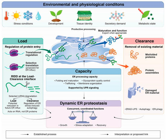
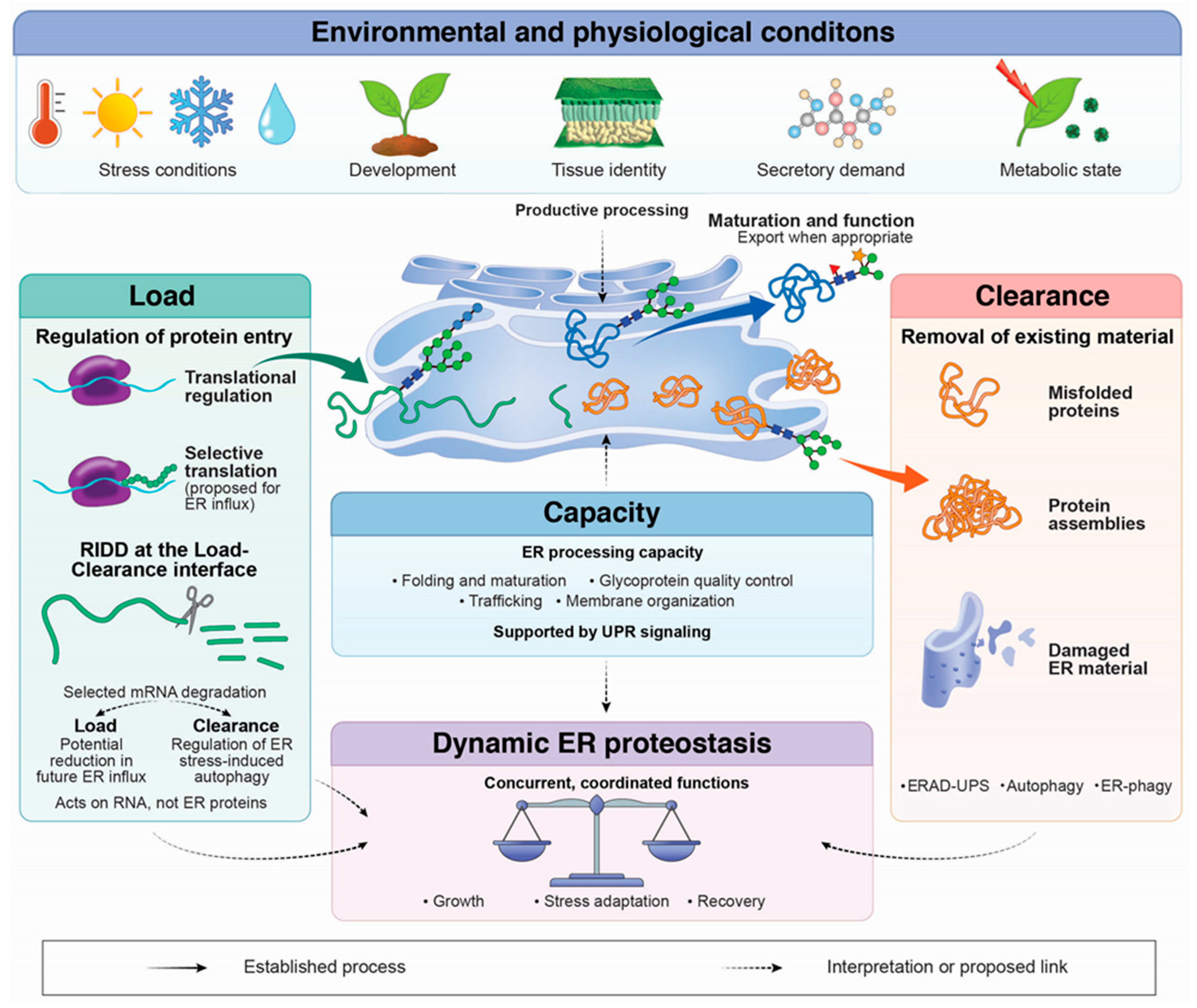
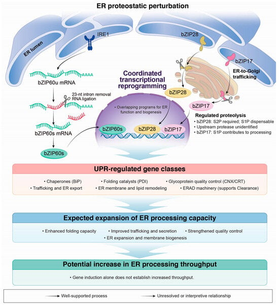
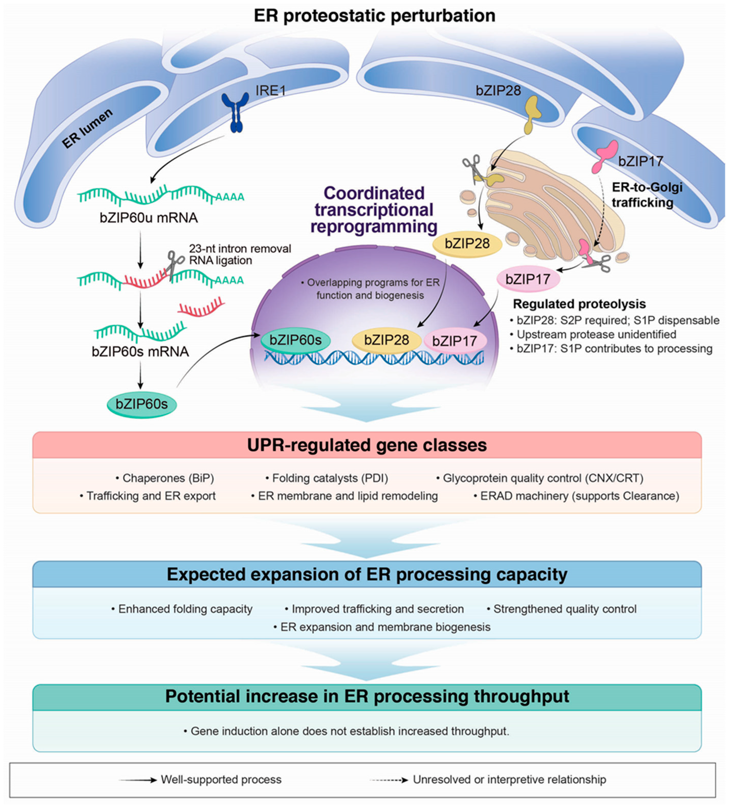
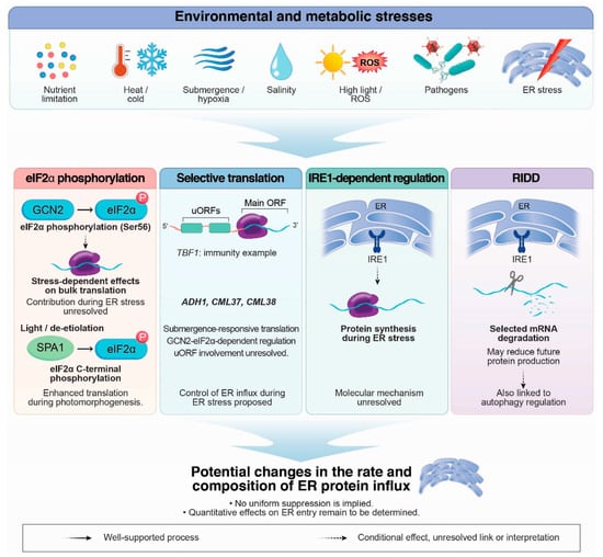
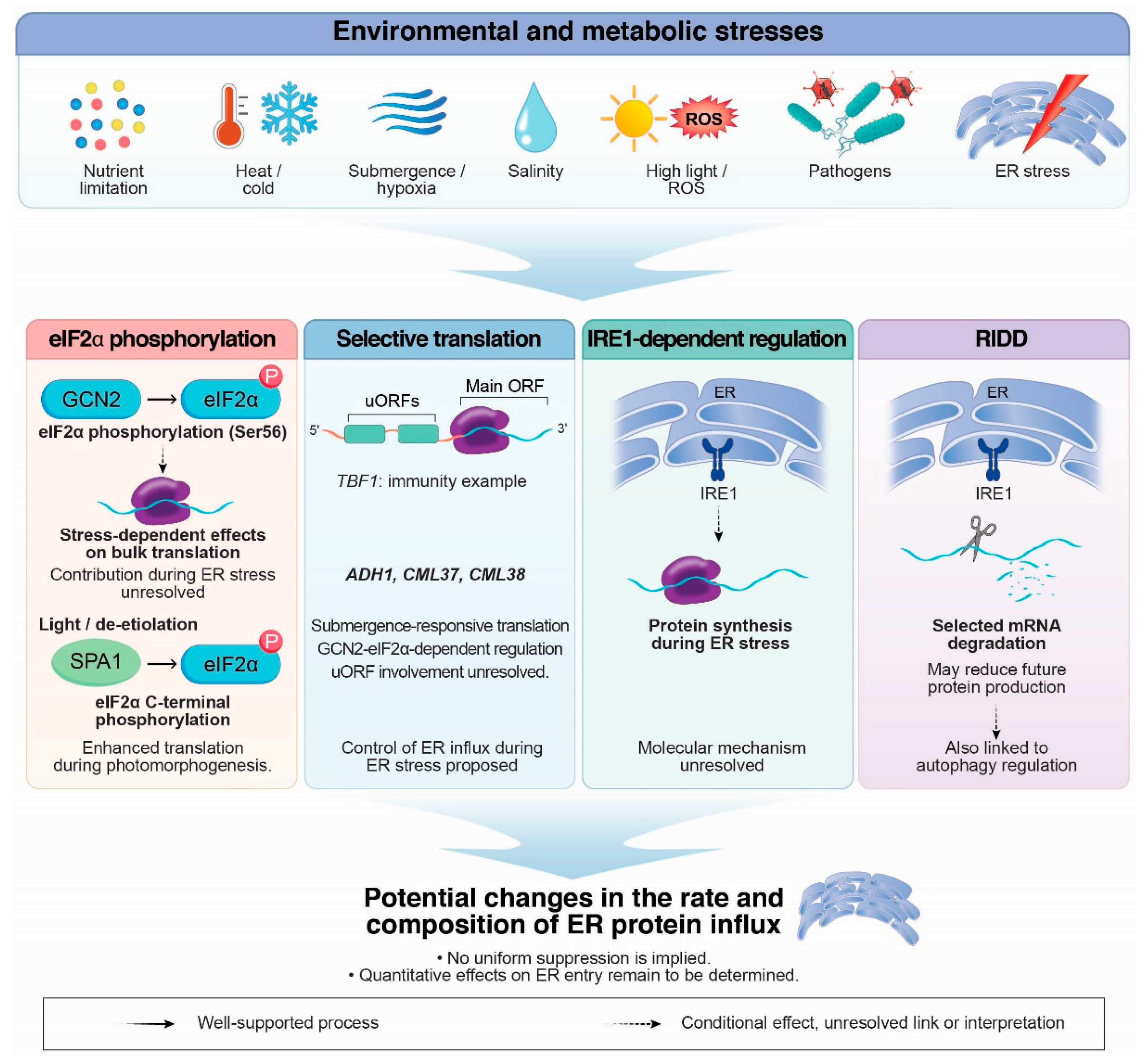
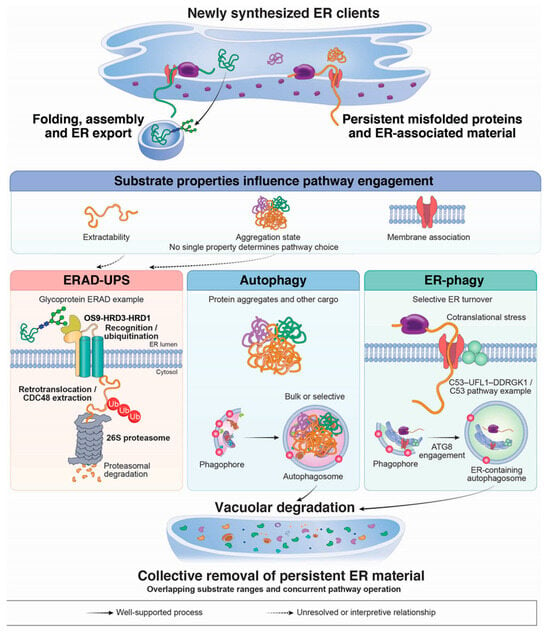
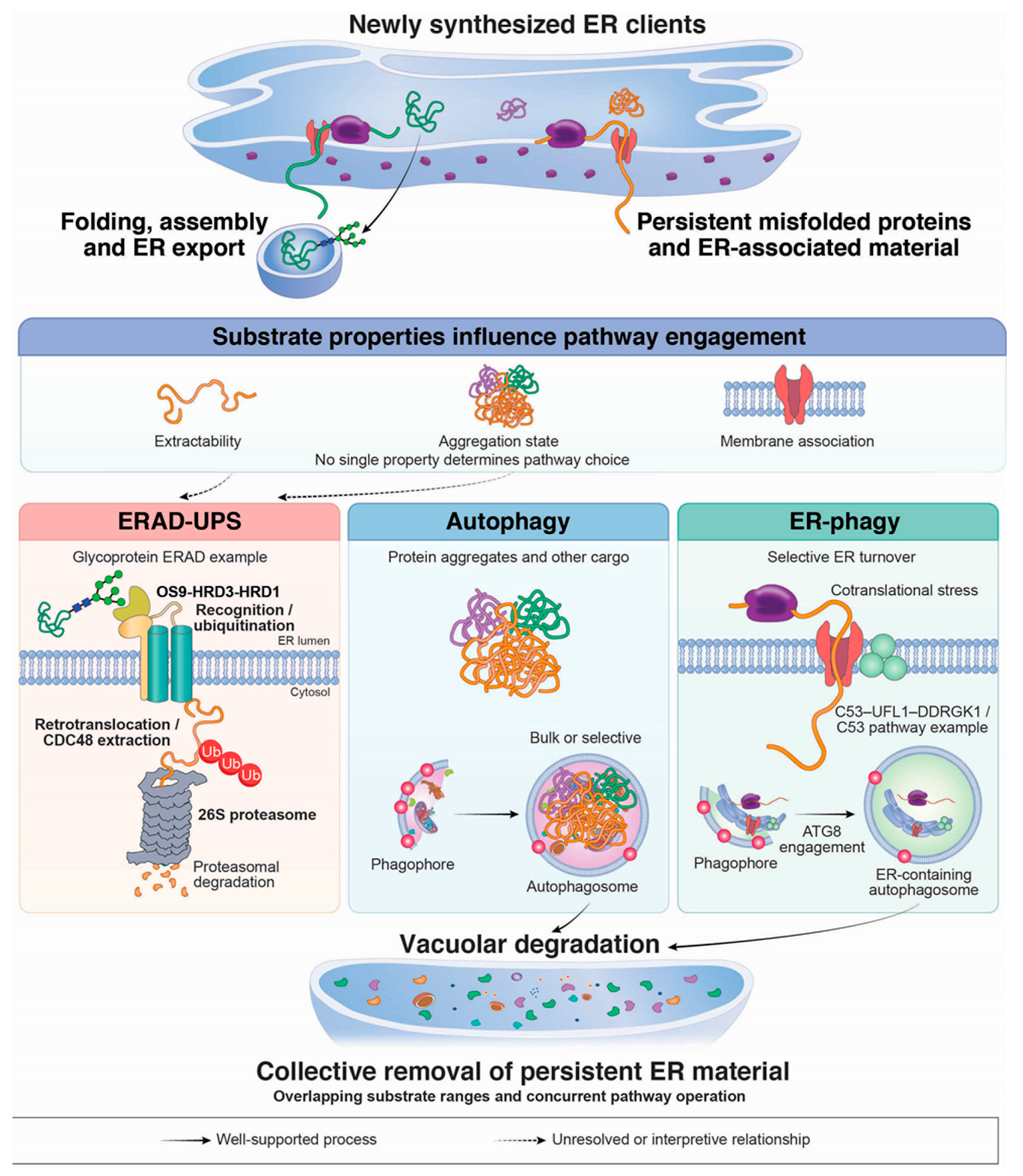
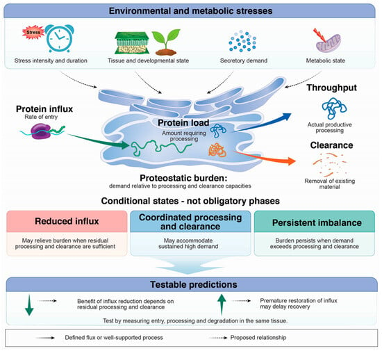
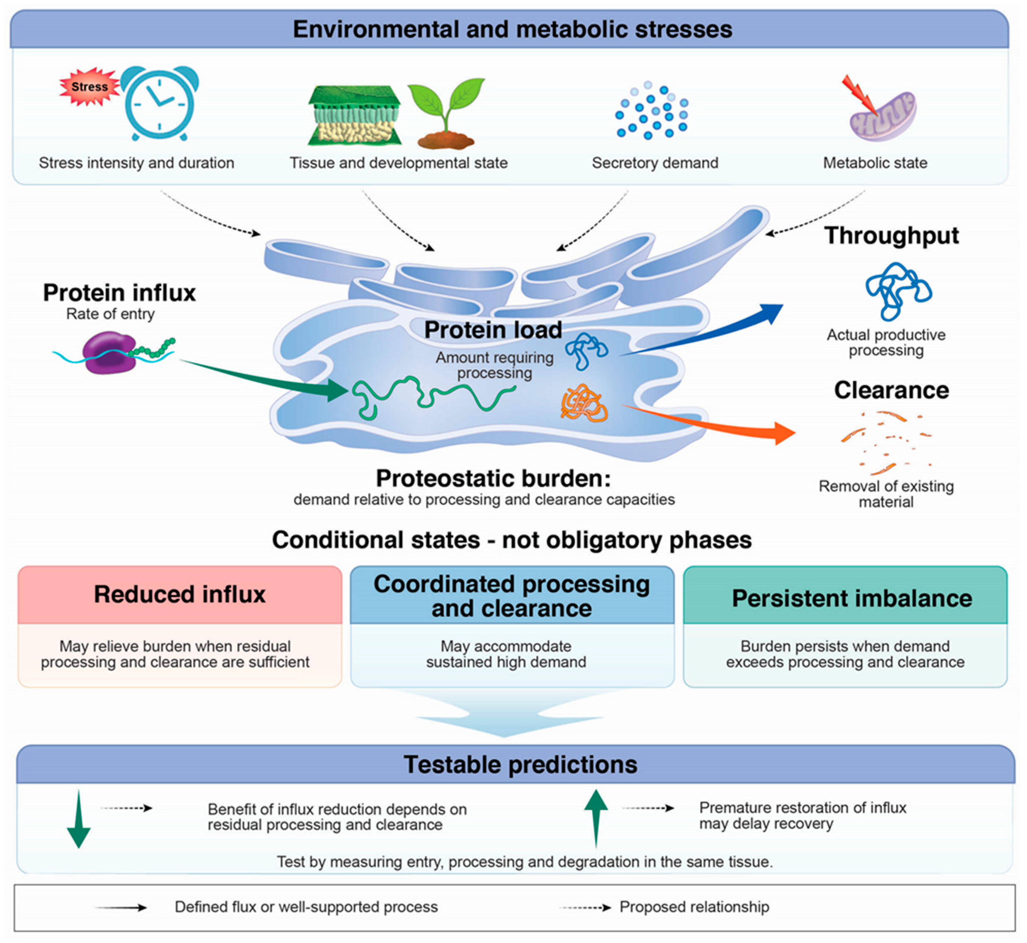

Disclaimer/Publisher’s Note: The statements, opinions and data contained in all publications are solely those of the individual author(s) and contributor(s) and not of MDPI and/or the editor(s). MDPI and/or the editor(s) disclaim responsibility for any injury to people or property resulting from any ideas, methods, instructions or products referred to in the content. |
© 2026 by the authors. Licensee MDPI, Basel, Switzerland. This article is an open access article distributed under the terms and conditions of the Creative Commons Attribution (CC BY) license.