


Abstract
Amyotrophic lateral sclerosis (ALS) is a fatal neurodegenerative disorder whose genetic architecture and molecular mechanisms remain incompletely understood, particularly in sporadic disease. To investigate whether genetically regulated DNA methylation may help interpret ALS susceptibility, we conducted a methylome-wide association study of genetically predicted blood methylation using the PrediXcan framework. CpG-specific models from the ARIES and Understanding Society cohorts were applied to ALS genome-wide association study (GWAS) summary statistics from 27,205 cases and 110,881 controls of European ancestry. Using S-PrediXcan, we evaluated 192,378 unique CpG sites. At nominal p < 0.05, 3741 CpGs were associated with ALS risk using ARIES models and 13,127 using Understanding Society models. After Bonferroni correction, 25 CpGs across eight genomic regions remained significant, including signals near established ALS and ALS–frontotemporal dementia loci (C9orf72, TBK1, SCFD1, and MOB3B). Three CpGs mapped to WHAMM at 15q25.2, a region not previously implicated by ALS GWAS. Predicted methylation was positively associated with risk at 18 CpGs and inversely associated at seven. Complementary whole-blood transcriptome-wide association analysis identified 11 significant genes, including C9orf72. Together, these findings prioritize regulatory candidates for functional investigation without establishing that methylation changes cause ALS.
1. Introduction
Amyotrophic lateral sclerosis (ALS) is a fatal neurodegenerative disorder characterized by progressive motor neuron loss, muscle weakness, paralysis, and respiratory failure, with a median survival of approximately 2.5 years from symptom onset [1]. Diagnosis remains challenging because early symptoms often overlap with those of other conditions [2], and patients may not encounter a neurologist until relatively late in the diagnostic journey [3,4].
ALS is clinically heterogeneous, with upper and lower motor neuron dysfunction affecting bulbar, cervical, thoracic, or lumbar regions and progressively impairing limb movement, swallowing, speech, and respiratory function [2,5]. Cognitive and behavioral changes may also arise early in the disease course [6,7] and are recognized in approximately 35–50% of individuals with ALS [8,9]. This combination of rapid progression and substantial clinical heterogeneity underscores the need to better understand the biological mechanisms that influence ALS susceptibility and disease expression.
Approximately 10% of ALS cases are classified as familial, whereas the remaining 90% are considered sporadic [10]. An underlying Mendelian mutation can be identified in approximately 50–85% of familial ALS cases [11] and in up to 10–20% of apparently sporadic cases [12,13,14]. Thus, although known pathogenic variants explain a substantial proportion of familial disease, the genetic architecture underlying ALS susceptibility remains incompletely understood, particularly among sporadic cases.
Large-scale genetic studies have substantially advanced the identification of ALS susceptibility loci. The largest ALS genome-wide association study (GWAS) to date included 29,612 individuals with ALS and 122,656 controls and identified multiple loci associated with disease risk [15]. However, GWAS signals alone often provide limited information about the molecular mechanisms through which associated variants influence disease susceptibility, particularly because many risk variants occur in noncoding regions. Integrative approaches that connect inherited genetic variation with intermediate molecular traits may therefore help translate statistical associations into biologically interpretable signals [16].
PrediXcan was developed to address this challenge by estimating genetically regulated gene expression from genotype data using prediction models trained in reference transcriptomic datasets and subsequently testing predicted expression for association with phenotypes [16]. S-PrediXcan extends this framework to GWAS summary statistics, allowing gene-level associations to be estimated without access to individual-level genotype and phenotype data [17]. PrediXcan and S-PrediXcan have shown high concordance, supporting the use of summary-statistic approaches for large-scale molecular association studies [17].
Although initially developed for transcriptome-wide association studies (TWAS), the same framework can be extended to other genetically regulated molecular traits. DNA methylation at many CpG sites is influenced by inherited genetic variation [18,19,20,21], exhibits a significant single-nucleotide polymorphism (SNP)-based heritable component [21,22,23], and is associated with specific genetic variants through methylation quantitative trait loci [19,24,25,26]. These properties make a genetically regulated component of DNA methylation predictable from genotype.
Building on this concept, Fryett et al. adapted the PrediXcan framework to model genetically predicted DNA methylation and test its association with complex traits [27]. Using matched genotype and methylation data from the Accessible Resource for Integrated Epigenomics Studies (ARIES) [28] and the Understanding Society study [29], they developed prediction models for CpG sites whose methylation levels could be reliably estimated from nearby genetic variation [27]. This approach provides an opportunity to investigate whether the genetically regulated component of DNA methylation is associated with complex disease risk without requiring methylation measurements in the GWAS population itself.
Despite the availability of these methylation prediction models, the relationship between genetically predicted blood DNA methylation and ALS susceptibility has not, to our knowledge, been systematically evaluated at scale. Consequently, it remains unclear which CpG sites show genetically predicted methylation associations with ALS risk and whether these associations can help prioritize candidate regulatory signals beyond conventional variant-level GWAS findings.
Here, we conducted a methylome-wide association study (MWAS) by applying these established ARIES and Understanding Society methylation prediction models to ALS GWAS summary statistics from 27,205 cases and 110,881 controls of European ancestry [15] using S-PrediXcan [17]. We tested whether genetically predicted blood DNA methylation at individual CpG sites was associated with ALS susceptibility and complemented these analyses with TWAS using GTEx v8 whole-blood gene expression prediction models [30]. By integrating ALS genetic association data with genetically regulated methylation and gene expression, our goal was not to identify diagnostic biomarkers or establish causal methylation changes, but to prioritize CpG sites, genes, and genomic regions that may help explain how inherited genetic variation influences ALS risk. This strategy adds a regulatory layer to conventional GWAS interpretation and may help refine broad genetic association signals into biologically testable candidates for functional investigation.
2. Results
2.1. Genetically Predicted Blood DNA Methylation Identifies 25 CpGs Associated with ALS Risk
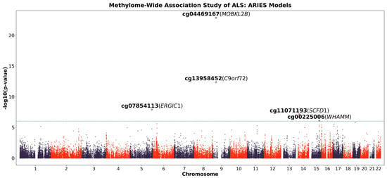
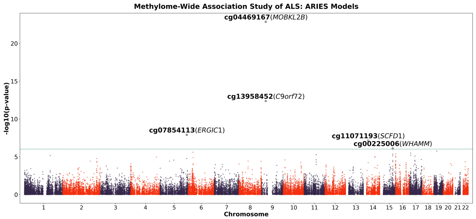
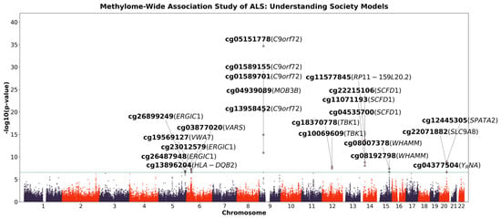
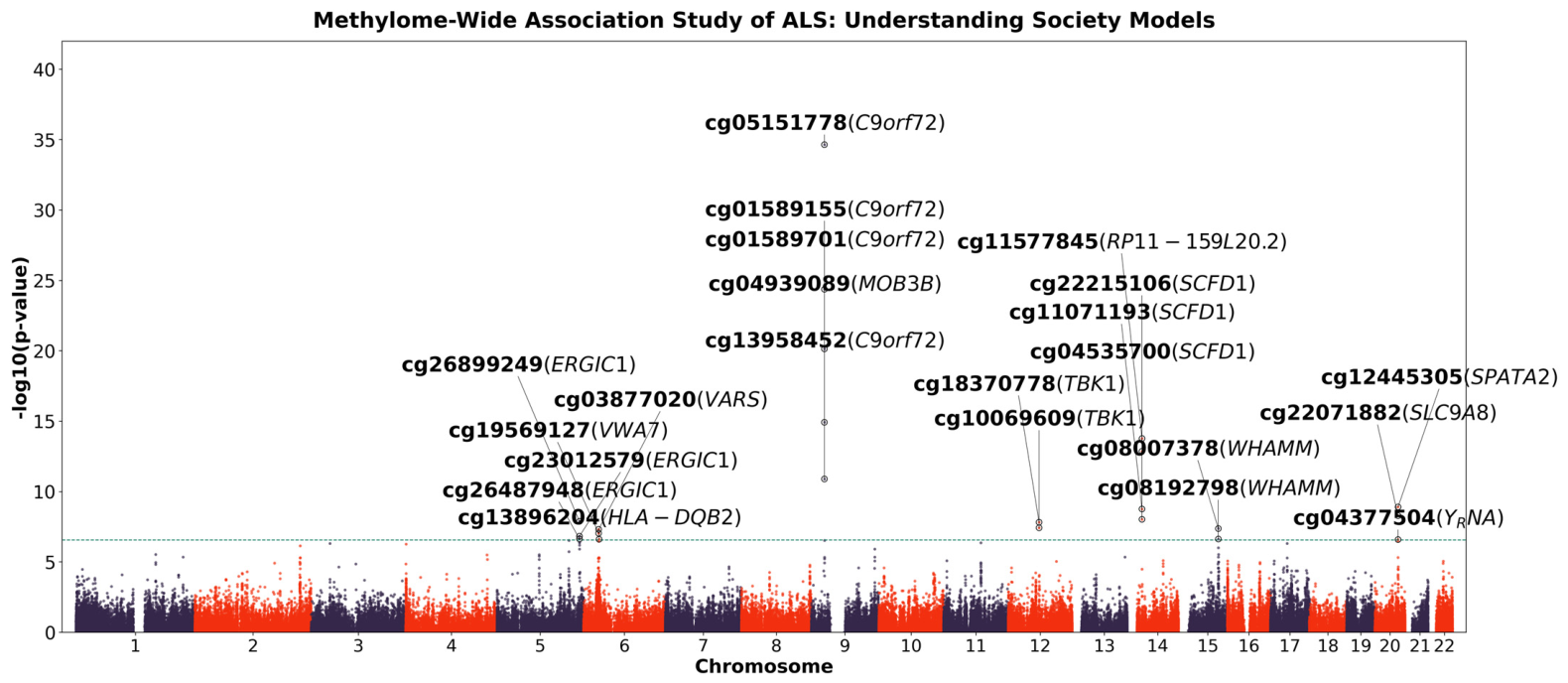
After applying the prediction performance criteria from Fryett et al., 52,391 methylation prediction models from ARIES [28] and 179,965 from Understanding Society [29] were available for analysis, representing 193,315 unique CpG sites (Figure 1 and Figure 2). Following quality control (R ≥ 0.1) and alignment with SNPs available in the ALS GWAS summary statistics, 52,088 ARIES CpGs and 179,215 Understanding Society CpGs were retained. Across both datasets, 192,378 unique CpGs were tested, including 38,925 CpGs represented in both prediction resources. At a nominal threshold of p < 0.05, 3741 CpGs were associated with ALS risk using the ARIES models and 13,127 using the Understanding Society models. After Bonferroni correction, 25 CpGs across eight genomic regions remained significantly associated with ALS risk (Table 1; Figure 1 and Figure 2). Five were identified using ARIES models and 22 using Understanding Society models, with two CpGs (cg13958452, cg11071193) significant in both datasets. Predicted methylation was positively associated with ALS risk at 18 of the 25 sites and inversely associated at seven. Notably, three significant CpGs (cg00225006, cg08007378, and cg08192798) mapped to WHAMM at 15q25.2, and were located more than 1 Mb away from any previously reported ALS GWAS risk variants. Two of them (cg00225006 and cg08007378) showed concordant positive effect directions in either the ARIES or Understanding Society models.
Figure 1.
Manhattan plot of the association results from the ALS methylome-wide association study for ARIES models. The x-axis represents the genomic position of each CpG site, and the y-axis represents the −log10-transformed p-value of the association. Each dot represents a specific CpG site. Alternating purple and red colors distinguish adjacent chromosomes. The green line indicates the Bonferroni-corrected significance threshold (p-value = 9.60 × 10⁻⁷; 0.05/52,088). CpG sites that reached the Bonferroni-corrected threshold and their nearby genes are annotated.
Figure 2.
Manhattan plot of ALS methylome-wide association results using Understanding Society models. The x-axis represents the genomic position of each CpG site, and the y-axis represents the −log10-transformed p-value of the association. Each dot represents a specific CpG site. The green line indicates the Bonferroni-corrected significance threshold (p-value = 2.79 × 10⁻⁷; 0.05/179,215). CpG sites that reached the Bonferroni-corrected threshold and their nearby genes are annotated.
Table 1.
Twenty-five CpG sites significantly associated with ALS risk.
2.2. Genetically Predicted Blood Gene Expression Identifies 11 Genes Associated with ALS Risk
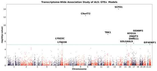
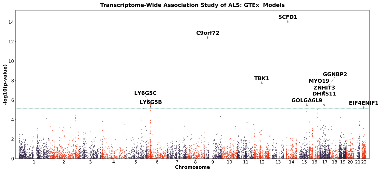
Using GTEx v8 whole-blood gene expression prediction models [16,17,30] developed from RNA-sequencing data from 670 whole-blood samples [31], we performed TWAS analysis. We tested the genetically predicted expression level of 7224 genes for the association with ALS risk (Figure 3). Eleven genes were significant after Bonferroni correction (p < 6.92 × 10⁻⁶).
Figure 3.
Manhattan plot of the association results from the ALS transcriptome-wide association study. The x-axis represents the genomic position of each gene, and the y-axis represents the −log10-transformed p-value of the association. Each dot represents a gene. The green line indicates the Bonferroni-corrected significance threshold (p-value = 6.92 × 10⁻⁶; 0.05/7224). Genes that reached the Bonferroni-corrected threshold are annotated.
Comparison of the TWAS and methylation-based association results revealed several loci represented in both analyses. At the C9orf72 locus, cg13958452 was associated with ALS risk across both the ARIES and Understanding Society methylation models, while genetically predicted C9orf72 expression was also associated with ALS risk in TWAS. Similarly, cg11071193, mapped near SCFD1, was significant across both MWAS models, while SCFD1 was identified in the TWAS analysis. Additional MWAS-associated CpGs mapped near C9orf72, TBK1, and SCFD1, genes that were also identified in the TWAS. These overlapping associations prioritize these loci for further investigation but do not establish corresponding regulatory relationships between DNA methylation and gene expression.
2.3. MWAS-Identified CpGs Overlap with Neurodegenerative Disease GWAS Loci
To evaluate whether MWAS-identified CpGs colocalized with previously reported genetic associations, we examined their proximity to genome-wide significant variants reported in the GWAS Catalog [32] for ALS, frontotemporal dementia (FTD), and Alzheimer’s disease (AD). CpGs located within 50 kb of a reported GWAS variant were considered proximal. Overall, 20 CpGs were located near 19 unique GWAS variants. Of these variants, 11 were associated with ALS, five with FTD, and four with AD, with one variant associated with both ALS and FTD. Thirteen CpGs were located within 50 kb of more than one GWAS variant, with the strongest clustering observed at the 9p21.2, 12q14.2, and 14q12 regions (Table 2).
Table 2.
MWAS-identified CpGs located near reported ALS and other neurodegenerative disease GWAS variants.
3. Discussion
This is the first large-scale study to evaluate associations between genetically predicted blood DNA methylation and ALS risk using the PrediXcan framework. Across methylation prediction models developed from two independent reference datasets, we identified 25 CpG sites associated with ALS risk. Three of these (cg00225006, cg08007378, and cg08192798) mapped to a novel locus at 15q25.2 that has not previously been implicated in ALS by GWAS. Complementary TWAS identified an association between genetically predicted C9orf72 expression and ALS risk, alongside significant methylation associations in both datasets at this locus (cg13958452). These findings support convergent genetically regulated molecular associations, though their underlying regulatory relationship remains unresolved. The PrediXcan framework offers an approach to one of the central challenges in ALS genetics: translating GWAS signals into biologically interpretable mechanisms, particularly when associated variants lie in noncoding regions.
However, a PrediXcan-based MWAS does not directly measure DNA methylation and cannot establish that methylation changes cause ALS. Rather, it tests whether the genetically regulated component of methylation predicted from models is associated with disease risk. The models used in this study were trained using the ARIES cohort (450K array) and the Understanding Society cohort (EPIC array). Partially due to differences in the underlying datasets, these models had different CpG coverages, with 52,088 CpGs tested using the ARIES models and 179,215 using the Understanding Society models. Only 38,925 CpGs were shared between the model sets [27]. For shared CpGs, differences in SNP selection, model weights, and prediction accuracy may affect association statistics. Accordingly, limited overlap does not by itself establish conflicting results, but it highlights the need to evaluate cross-model consistency and independently validate associations supported by a single resource. Our results should be viewed as genetic evidence linking predicted methylation variation to ALS susceptibility and as a means of prioritizing loci for subsequent functional and biological investigation, with particular consideration of the two CpGs that reached Bonferroni-corrected significance in both prediction resources.
The first category of findings includes genes or loci with prior evidence from ALS or ALS-FTD genetic and molecular studies. Among these, two ALS-associated CpGs, cg18370778 and cg10069609, were annotated to TBK1. Whether through haploinsufficiency, reduced kinase activity, or disrupted protein–protein interactions, TBK1 mutations appear to converge on a shared pathogenic pathway involving impaired autophagic clearance, proteostatic dysfunction, and accelerated neurodegeneration in ALS and FTD [33].
Three additional ALS risk-associated CpGs (cg11071193, cg22215106, and cg04535700) mapped to SCFD1. Previous studies have shown that SCFD1 expression quantitative trait loci are significantly associated with increased ALS risk and that SCFD1 expression is altered in post-mortem ALS tissue, where it correlates with disease duration [34]. SCFD1 is a key component of a large transcriptional network linked to ALS-related pathways and genes enriched for schizophrenia risk [34], a condition known to be genetically correlated with ALS [35].
Notably, cg11577845, annotated to RP11-159L20.2, was associated with ALS risk. Although RP11-159L20.2 itself has not been directly implicated in ALS, this CpG lies within the same genomic locus as the SCFD1-annotated CpGs, raising the possibility that these signals reflect regulatory variation within a shared ALS-relevant region.
Several other findings mapped to loci with established or emerging ALS-related evidence, with particularly strong biological support at the C9orf72 locus. Multiple ALS risk-associated CpGs were located near C9orf72, including cg01589155 and cg13958452. GGGGCC repeat expansions in C9orf72 are a major genetic cause of both ALS and FTD [36,37], with proposed pathogenic mechanisms including loss of C9orf72 function, toxic RNA foci, and accumulation of dipeptide repeat proteins [38]. Of particular interest, cg01589155 was previously associated with survival in the epigenome-wide association study by Hop et al. [39], suggesting that methylation-related variation in this locus may influence not only ALS susceptibility but also disease progression. In addition, Ramesh et al. [40] reported significant hypermethylation of cg01589155, cg05151778, and cg01589701 at the C9orf72 locus in whole blood from patients with FTD carrying a pathogenic C9orf72 repeat expansion (n = 27), compared with patients with FTD without the expansion (n = 250), linking methylation at these sites to expansion status within FTD. Evidence for cg13958452 was also consistent across both the ARIES and Understanding Society methylation models, and C9orf72 was also significantly associated with ALS risk in the TWAS analysis. The consistency of these signals across methylation and gene expression models strengthens the evidence linking molecular variation at the C9orf72 locus to ALS susceptibility.
In addition, two ALS-associated CpGs, cg04939089 and cg04469167, were annotated to MOB3B, a gene located within the shared ALS–FTD risk locus at 9p21.2. In a multitrait GWAS of FTD and ALS, MOB3B emerged as a key candidate gene at this locus, with genetic effects contributing to susceptibility across both diseases [41]. Bayesian fine-mapping and colocalization further supported a shared causal signal at 9p21.2 (PP4 = 0.764), strengthening evidence that MOB3B may contribute to the common genetic architecture of ALS and FTD [41].
Support for previously implicated loci was also observed at SLC9A8/SPATA2, where cg22071882 mapped to SLC9A8 and cg12445305 mapped to SPATA2. This finding is consistent with prior GWAS evidence linking the SLC9A8/SPATA2 region to ALS susceptibility [15]. In addition, cg13896204 was annotated to HLA-DQB2, an immune-related gene previously reported to be upregulated in the anterior horn of the spinal cord in sporadic ALS [42]. Together, these findings provide epigenetic support for several loci already implicated in ALS through genetic association, shared ALS–FTD susceptibility, or disease-related molecular alterations.
Several findings also pointed to pathways involved in intracellular trafficking, ER stress, and protein homeostasis. Four ALS-associated CpGs mapped to ERGIC1, which encodes a membrane-bound protein localized to the endoplasmic reticulum (ER)–Golgi intermediate compartment and involved in membrane trafficking and selective cargo transport between the ER and Golgi complex [43]. Disruption of ER–Golgi transport has been reported as a shared pathogenic mechanism across SOD1-, TDP-43-, and FUS-associated ALS [44], while ER stress has also been implicated more broadly in ALS pathogenesis [45].
A separate signal, cg03877020, mapped to VARS and was positively associated with ALS risk. VARS1 encodes valine-tRNA ligase and has been identified among translational proteins showing increased affinity for cytoplasmic TDP-43 [46]. Given that nuclear depletion and cytoplasmic accumulation of TDP-43 are central pathological features of ALS, this observation raises a potential connection between VARS1 and TDP-43-associated cellular dysfunction. However, the available evidence is derived from protein interaction studies in cell lines and does not establish that VARS1 colocalizes with TDP-43, aggregates, or contributes directly to ALS pathogenesis.
Additional candidate loci were supported by prior molecular evidence in ALS. The ALS risk-associated CpG cg19569127 mapped to VWA7, a gene previously identified among 44 transcripts that were significantly differentially expressed across two independent ALS post-mortem motor cortex RNA-seq datasets, KCL BrainBank and TargetALS [47]. VWA7 was downregulated in ALS relative to controls in both cohorts, with log2 fold changes of −0.24 and −0.47, respectively (adjusted p = 0.0284 in both datasets), and was included in an extracellular matrix-related transcriptional signature shared across the two cohorts [47].
Three additional CpGs (cg00225006, cg08007378, and cg08192798) mapped to WHAMM within 15q25.2, a region not previously implicated in ALS through GWAS. Although this represents a potentially novel ALS-associated locus, WHAMM has previously been proposed as a cell-type-specific candidate gene with possible relevance to ALS pathogenesis [48]. Another ALS-associated signal, cg04377504, mapped to Y RNA. Supporting the potential relevance of this finding, Y RNA abundance has been reported to differ significantly between individuals with ALS and controls in both plasma and serum [49]. These observations highlight additional loci and molecular features that warrant further investigation but currently have more limited evidence linking them directly to ALS biology.
The proximity of several MWAS-identified CpGs to previously reported ALS and FTD GWAS variants indicates that a subset of the methylation signals falls within regions already implicated in neurodegenerative disease. However, genomic proximity alone does not establish that these variants regulate the corresponding CpGs or that the associations reflect the same underlying causal signal. Further analyses incorporating mQTL data, linkage disequilibrium structure, and colocalization will be needed to determine whether the genetic and methylation associations share common regulatory mechanisms.
Several limitations should be considered when interpreting these findings. First, the methylation prediction models were derived from blood rather than brain or spinal cord tissue. Although blood-based models may capture systemic components of ALS biology, they may not reflect genetically regulated methylation patterns in motor neurons, glial cells, or other central nervous system cell types. Development and application of tissue- and cell-type-specific prediction models will therefore be important for determining whether the associations identified here are also relevant within disease-affected tissues. Second, the direction of an MWAS association should not be interpreted as indicating an activating or repressive regulatory effect. The functional consequences of DNA methylation depend on the genomic location and regulatory context of the CpG site. Consequently, the directions of the MWAS and TWAS associations cannot be assumed to represent directly comparable regulatory effects. Finally, the analyses were restricted to individuals of European ancestry, which may limit the generalizability of the findings. Future studies incorporating larger and more ancestrally diverse populations will be needed to assess the robustness and transferability of these associations.
In summary, this study provides a systematic evaluation of genetically predicted blood DNA methylation in relation to ALS risk. By integrating methylation prediction models with large-scale ALS genetic data, we identified signals spanning established ALS genes and loci, regions linked to ALS-relevant biological pathways, and less characterized candidate loci that may warrant further investigation. These results illustrate how PrediXcan-based MWAS can complement conventional GWAS by helping prioritize genetically regulated molecular signals within or near associated regions, particularly where the functional consequences of underlying genetic variation remain unclear. Importantly, these associations should not be interpreted as evidence that altered DNA methylation itself causes ALS, but rather as a framework for prioritizing loci and molecular mechanisms for subsequent functional validation.
4. Materials and Methods
4.1. Study Population
We used ALS GWAS summary statistics from 27,205 cases and 110,881 controls of European ancestry [15], representing the largest ALS GWAS dataset available to date. The GWAS combined individual-level genotype data from 117 cohorts into six strata matched by genotyping platform, with standard quality control procedures applied prior to association analysis [15].
4.2. DNA Methylation Prediction Models
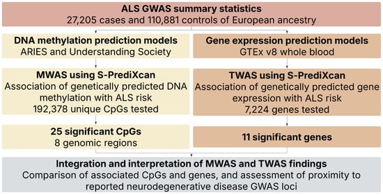
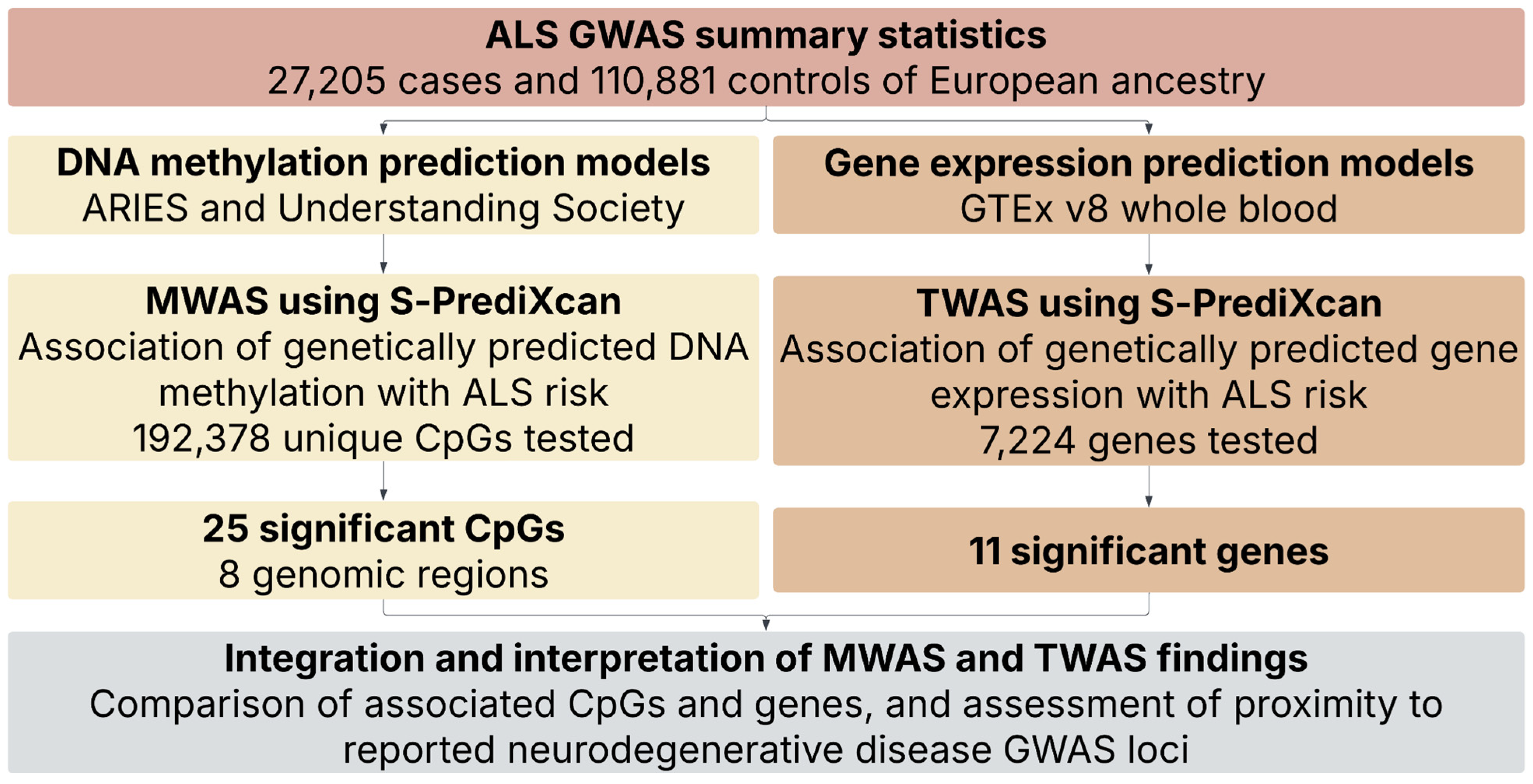
We used previously developed CpG-specific DNA methylation prediction models from Fryett et al. [27] for association analyses with ALS risk (Figure 4). Model development has been described in detail previously [27]. Briefly, the models were trained using samples with matched genotype and DNA methylation data from the ARIES [28] and Understanding Society [29] cohorts. Following quality control, 841 ARIES mother samples and 1120 Understanding Society samples were available for model development.
Figure 4.
Study design flowchart.
Three penalized regression approaches were evaluated: ridge regression [50], LASSO [51], and elastic net [52] with α = 0.5. For each approach, the regularization parameter λ was selected using 10-fold cross-validation to minimize the mean squared error between predicted and observed methylation among models containing at least one SNP. Prediction performance was assessed using the correlation (R) between predicted and observed methylation levels, and elastic net with α = 0.5 was selected as the optimal modeling approach.
Five cis-window sizes surrounding each CpG (250 kb, 500 kb, 1 Mb, 2 Mb, and 3 Mb) were subsequently evaluated, with the optimal window selected separately for each CpG site. Final models were trained using all available samples within each cohort and externally validated in the other cohort. Only models achieving a validation correlation of R ≥ 0.1 were retained for downstream analyses.
4.3. Gene Expression Prediction Models
We used GTEx v8 elastic net transcriptome prediction models developed by Barbeira et al. [16,17,30]. These models were trained using matched GTEx v8 RNA-seq and genotype data from individuals of European ancestry and were restricted to genes annotated in GENCODE v26 as protein-coding, long noncoding RNA, or pseudogenes [53].
For each gene–tissue pair, adjusted gene expression was modeled as a function of nearby genetic variation using elastic net regression. Candidate variants were restricted to HapMap 3 CEU SNPs with minor allele frequency > 0.01 located within 1 Mb upstream of the transcription start site or 1 Mb downstream of the transcription end site. Models were fitted using glmnet [54] with α = 0.5, and the penalty parameter was selected by 10-fold cross-validation. Models were retained when the mean Pearson correlation between predicted and observed expression across the 10 folds exceeded 0.1 and the nested cross-validated correlation test reached p < 0.05.
4.4. Association Analysis of Genetically Predicted DNA Methylation Levels with ALS Risk
DNA methylation prediction models were included in downstream association analyses if they met predefined performance criteria of R ≥ 0.1. Each retained CpG-specific prediction model was applied to the ALS GWAS summary statistics [15] using S-PrediXcan [17], following the previously described implementation [55]. For each CpG site, S-PrediXcan generated a Z-score representing the association between genetically predicted DNA methylation level and ALS risk. Positive Z-scores indicate that higher genetically predicted methylation is associated with increased ALS risk, whereas negative Z-scores indicate an inverse association. The S-PrediXcan association statistic was approximated as described by Barbeira et al. [17]:
In this equation, wsm represents the weight assigned to SNP s in the prediction model for DNA methylation at CpG m. The term and represent the estimated effect size and corresponding standard error, respectively, for the association of SNP (s) with ALS risk in the GWAS summary statistics. The terms and represent the estimated standard deviations of SNP (s) and the genetically predicted methylation level at CpG site (m), respectively. Correlations among SNPs included in each prediction model were accounted for in the analysis. Statistical significance was determined using Bonferroni correction for the number of CpG sites tested, with a Bonferroni-corrected p < 0.05 considered statistically significant.
4.5. Association Analysis of Genetically Predicted Gene Expression with ALS Risk
To complement the methylome-wide association analysis, we performed a transcriptome-wide association study (TWAS) to evaluate associations between genetically regulated whole-blood gene expression and ALS risk. Publicly available GTEx v8 whole-blood gene expression prediction models were obtained from PredictDB [56]. These models use elastic net regression to predict gene expression from nearby genetic variants [16,17,30] and were applied to the same ALS GWAS summary statistics used for the methylation analyses [15].
S-PrediXcan [17] was used to estimate the association between genetically predicted expression of each gene and ALS risk. For each gene, the analysis generated a Z-score and corresponding p-value representing the direction and statistical evidence of the association. Positive Z-scores indicate that higher genetically predicted gene expression is associated with increased ALS risk, whereas negative Z-scores indicate an inverse association. Genes were evaluated when a GTEx v8 whole-blood prediction model was available and sufficient model SNPs overlapped with the ALS GWAS data. Statistical significance was determined using Bonferroni correction for the number of genes tested, with a Bonferroni-corrected p < 0.05 considered statistically significant. All software and data sources used are shown in Table 3.
Table 3.
Data sources, prediction models, and analytical tools used in this study.
5. Conclusions
In this first large-scale PrediXcan study of genetically predicted blood DNA methylation and ALS risk, we identified 25 CpG sites associated with ALS, including three at the 15q25.2 locus, a region not previously implicated by ALS GWAS. Complementary TWAS also identified an association at C9orf72, supporting convergent molecular associations without establishing a shared regulatory mechanism or a causal relationship between methylation and gene expression.
The broader impact of these findings is that they add an epigenetic regulatory dimension to the interpretation of ALS genetic risk. Rather than identifying additional risk variants alone, this approach helps prioritize specific CpG sites, genes, and genomic regions through which inherited genetic variation may influence disease susceptibility. In this way, PrediXcan-based MWAS can help refine broad GWAS loci into more biologically interpretable candidates, highlight regulatory mechanisms that may otherwise be missed by variant-level analyses, and identify targets for functional validation. This is particularly relevant in ALS, where many associated variants lie in noncoding regions and their biological consequences remain poorly understood. By linking genetic variation to predicted methylation and integrating these signals with gene expression analyses, our study provides a complementary strategy for moving from genetic association toward mechanistic understanding of ALS susceptibility.
Author Contributions
Conceptualization, T.Z.; methodology, T.Z.; formal analysis, T.Z.; writing—original draft preparation, T.Z.; writing—review and editing, T.Z., M.M.H., G.W. and V.V.B. All authors contributed to the interpretation of the results. All authors have read and agreed to the published version of the manuscript.
Funding
Research reported in this publication was supported by a gift fund from the Live Like Lou Foundation and the National Center for Advancing Translational Sciences of the National Institutes of Health under Award Number TL1TR002244. The content is solely the responsibility of the authors and does not necessarily represent the official views of the National Institutes of Health or the Live Like Lou Foundation.
Institutional Review Board Statement
Not applicable. This study used publicly available, deidentified summary-level data and did not involve the enrollment of participants or analysis of individual-level data by the authors. Ethics approval and informed consent for the original studies are described in the respective source publications.
Informed Consent Statement
Not applicable.
Data Availability Statement
The data presented in this study are available on request from the corresponding author due to each developer’s instructions.
Acknowledgments
The authors gratefully acknowledge the investigators and participants whose contributions made the publicly available datasets used in this study possible.
Conflicts of Interest
The authors declare that they have no competing interests.
Abbreviations
| ALS | amyotrophic lateral sclerosis |
| ARIES | Accessible Resource for Integrated Epigenomic Studies |
| CpG | cytosine–phosphate–guanine |
| ER | endoplasmic reticulum |
| FTD | frontotemporal dementia |
| GTEx | Genotype-Tissue Expression |
| GWAS | genome-wide association study |
| MWAS | methylome-wide association study |
| RNA-seq | RNA sequencing |
| SNP | single-nucleotide polymorphism |
| TWAS | transcriptome-wide association study |
References
- Vasta, R.; De Mattei, F.; Tafaro, S.; Canosa, A.; Manera, U.; Grassano, M.; Palumbo, F.; Cabras, S.; Matteoni, E.; Di Pede, F.; et al. Changes to Average Survival of Patients with Amyotrophic Lateral Sclerosis (1995–2018): Results from the Piemonte and Valle d’Aosta Registry. Neurology 2025, 104, e213467. [Google Scholar] [PubMed]
- Feldman, E.L.; Goutman, S.A.; Petri, S.; Mazzini, L.; Savelieff, M.G.; Shaw, P.J.; Sobue, G. Amyotrophic Lateral Sclerosis. Lancet 2022, 400, 1363–1380. [Google Scholar] [CrossRef] [Scilit] [PubMed]
- Williams, J.R.; Fitzhenry, D.; Grant, L.; Martyn, D.; Kerr, D.A. Diagnosis Pathway for Patients with Amyotrophic Lateral Sclerosis: Retrospective Analysis of the US Medicare Longitudinal Claims Database. BMC Neurol. 2013, 13, 160. [Google Scholar] [CrossRef] [Scilit] [PubMed]
- Falcão de Campos, C.; Gromicho, M.; Uysal, H.; Grosskreutz, J.; Kuzma-Kozakiewicz, M.; Oliveira Santos, M.; Pinto, S.; Petri, S.; Swash, M.; de Carvalho, M. Delayed Diagnosis and Diagnostic Pathway of ALS Patients in Portugal: Where Can We Improve? Front. Neurol. 2021, 12, 761355. [Google Scholar] [CrossRef] [Scilit] [PubMed]
- Goutman, S.A.; Hardiman, O.; Al-Chalabi, A.; Chió, A.; Savelieff, M.G.; Kiernan, M.C.; Feldman, E.L. Recent Advances in the Diagnosis and Prognosis of Amyotrophic Lateral Sclerosis. Lancet Neurol. 2022, 21, 480–493. [Google Scholar] [CrossRef] [Scilit] [PubMed]
- Beeldman, E.; Govaarts, R.; de Visser, M.; Klein Twennaar, M.; van der Kooi, A.J.; van den Berg, L.H.; Veldink, J.H.; Pijnenburg, Y.A.L.; de Haan, R.J.; Schmand, B.A.; et al. Progression of Cognitive and Behavioural Impairment in Early Amyotrophic Lateral Sclerosis. J. Neurol. Neurosurg. Psychiatry 2020, 91, 779–780. [Google Scholar] [CrossRef] [Scilit] [PubMed]
- Pender, N.; Pinto-Grau, M.; Hardiman, O. Cognitive and Behavioural Impairment in Amyotrophic Lateral Sclerosis. Curr. Opin. Neurol. 2020, 33, 649–654. [Google Scholar] [CrossRef] [Scilit] [PubMed]
- Crockford, C.; Newton, J.; Lonergan, K.; Chiwera, T.; Booth, T.; Chandran, S.; Colville, S.; Heverin, M.; Mays, I.; Pal, S.; et al. ALS-Specific Cognitive and Behavior Changes Associated with Advancing Disease Stage in ALS. Neurology 2018, 91, e1370–e1380. [Google Scholar] [CrossRef] [Scilit] [PubMed]
- Bersano, E.; Sarnelli, M.F.; Solara, V.; Iazzolino, B.; Peotta, L.; De Marchi, F.; Facchin, A.; Moglia, C.; Canosa, A.; Calvo, A.; et al. Decline of Cognitive and Behavioral Functions in Amyotrophic Lateral Sclerosis: A Longitudinal Study. Amyotroph. Lateral Scler. Front. Degener. 2020, 21, 373–379. [Google Scholar] [CrossRef] [Scilit] [PubMed]
- Renton, A.E.; Chiò, A.; Traynor, B.J. State of Play in Amyotrophic Lateral Sclerosis Genetics. Nat. Neurosci. 2014, 17, 17–23. [Google Scholar] [CrossRef] [Scilit] [PubMed]
- Müller, K.; Brenner, D.; Weydt, P.; Meyer, T.; Grehl, T.; Petri, S.; Grosskreutz, J.; Schuster, J.; Volk, A.E.; Borck, G.; et al. Comprehensive Analysis of the Mutation Spectrum in 301 German ALS Families. J. Neurol. Neurosurg. Psychiatry 2018, 89, 817–827. [Google Scholar] [CrossRef] [Scilit] [PubMed]
- Van Daele, S.H.; Moisse, M.; van Vugt, J.J.F.A.; Zwamborn, R.A.J.; van der Spek, R.; van Rheenen, W.; Van Eijk, K.; Kenna, K.; Corcia, P.; Vourc’h, P.; et al. Genetic Variability in Sporadic Amyotrophic Lateral Sclerosis. Brain 2023, 146, 3760–3769. [Google Scholar] [CrossRef] [Scilit] [PubMed]
- Ruf, W.P.; Boros, M.; Freischmidt, A.; Brenner, D.; Grozdanov, V.; de Meirelles, J.; Meyer, T.; Grehl, T.; Petri, S.; Grosskreutz, J.; et al. Spectrum and Frequency of Genetic Variants in Sporadic Amyotrophic Lateral Sclerosis. Brain Commun. 2023, 5, fcad152. [Google Scholar] [CrossRef] [Scilit] [PubMed]
- Shepheard, S.R.; Parker, M.D.; Cooper-Knock, J.; Verber, N.S.; Tuddenham, L.; Heath, P.; Beauchamp, N.; Place, E.; Sollars, E.S.A.; Turner, M.R.; et al. Value of Systematic Genetic Screening of Patients with Amyotrophic Lateral Sclerosis. J. Neurol. Neurosurg. Psychiatry 2021, 92, 510–518. [Google Scholar] [CrossRef] [Scilit]
- van Rheenen, W.; van der Spek, R.A.A.; Bakker, M.K.; van Vugt, J.J.F.A.; Hop, P.J.; Zwamborn, R.A.J.; de Klein, N.; Westra, H.-J.; Bakker, O.B.; Deelen, P.; et al. Common and Rare Variant Association Analyses in Amyotrophic Lateral Sclerosis Identify 15 Risk Loci with Distinct Genetic Architectures and Neuron-Specific Biology. Nat. Genet. 2021, 53, 1636–1648, Correction in Nat. Genet. 2022, 54, 361.. [Google Scholar] [CrossRef] [Scilit] [PubMed]
- Gamazon, E.R.; Wheeler, H.E.; Shah, K.P.; Mozaffari, S.V.; Aquino-Michaels, K.; Carroll, R.J.; Eyler, A.E.; Denny, J.C.; GTEx Consortium; Nicolae, D.L.; et al. A Gene-Based Association Method for Mapping Traits Using Reference Transcriptome Data. Nat. Genet. 2015, 47, 1091–1098. [Google Scholar] [CrossRef] [Scilit] [PubMed]
- Barbeira, A.N.; Dickinson, S.P.; Bonazzola, R.; Zheng, J.; Wheeler, H.E.; Torres, J.M.; Torstenson, E.S.; Shah, K.P.; Garcia, T.; Edwards, T.L.; et al. Exploring the Phenotypic Consequences of Tissue Specific Gene Expression Variation Inferred from GWAS Summary Statistics. Nat. Commun. 2018, 9, 1825. [Google Scholar] [CrossRef] [Scilit] [PubMed]
- Bell, J.T.; Tsai, P.-C.; Yang, T.-P.; Pidsley, R.; Nisbet, J.; Glass, D.; Mangino, M.; Zhai, G.; Zhang, F.; Valdes, A.; et al. Epigenome-Wide Scans Identify Differentially Methylated Regions for Age and Age-Related Phenotypes in a Healthy Ageing Population. PLoS Genet. 2012, 8, e1002629. [Google Scholar] [CrossRef] [Scilit] [PubMed]
- Grundberg, E.; Meduri, E.; Sandling, J.K.; Hedman, Å.K.; Keildson, S.; Buil, A.; Busche, S.; Yuan, W.; Nisbet, J.; Sekowska, M.; et al. Global Analysis of DNA Methylation Variation in Adipose Tissue from Twins Reveals Links to Disease-Associated Variants in Distal Regulatory Elements. Am. J. Hum. Genet. 2013, 93, 1158. [Google Scholar] [CrossRef] [Scilit]
- Hannon, E.; Knox, O.; Sugden, K.; Burrage, J.; Wong, C.C.Y.; Belsky, D.W.; Corcoran, D.L.; Arseneault, L.; Moffitt, T.E.; Caspi, A.; et al. Characterizing Genetic and Environmental Influences on Variable DNA Methylation Using Monozygotic and Dizygotic Twins. PLoS Genet. 2018, 14, e1007544. [Google Scholar] [CrossRef] [Scilit] [PubMed]
- van Dongen, J.; Nivard, M.G.; Willemsen, G.; Hottenga, J.-J.; Helmer, Q.; Dolan, C.V.; Ehli, E.A.; Davies, G.E.; van Iterson, M.; Breeze, C.E.; et al. Genetic and Environmental Influences Interact with Age and Sex in Shaping the Human Methylome. Nat. Commun. 2016, 7, 11115. [Google Scholar] [CrossRef] [Scilit] [PubMed]
- Quon, G.; Lippert, C.; Heckerman, D.; Listgarten, J. Patterns of Methylation Heritability in a Genome-Wide Analysis of Four Brain Regions. Nucleic Acids Res. 2013, 41, 2095–2104. [Google Scholar] [CrossRef] [Scilit] [PubMed]
- Rowlatt, A.; Hernández-Suárez, G.; Sanabria-Salas, M.C.; Serrano-López, M.; Rawlik, K.; Hernandez-Illan, E.; Alenda, C.; Castillejo, A.; Soto, J.L.; Haley, C.S.; et al. The Heritability and Patterns of DNA Methylation in Normal Human Colorectum. Hum. Mol. Genet. 2016, 25, 2600–2611. [Google Scholar] [CrossRef] [Scilit] [PubMed]
- Gaunt, T.R.; Shihab, H.A.; Hemani, G.; Min, J.L.; Woodward, G.; Lyttleton, O.; Zheng, J.; Duggirala, A.; McArdle, W.L.; Ho, K.; et al. Systematic Identification of Genetic Influences on Methylation across the Human Life Course. Genome Biol. 2016, 17, 61. [Google Scholar] [CrossRef] [Scilit] [PubMed]
- Richardson, T.G.; Shihab, H.A.; Hemani, G.; Zheng, J.; Hannon, E.; Mill, J.; Carnero-Montoro, E.; Bell, J.T.; Lyttleton, O.; McArdle, W.L.; et al. Collapsed Methylation Quantitative Trait Loci Analysis for Low Frequency and Rare Variants. Hum. Mol. Genet. 2016, 25, 4339–4349. [Google Scholar] [CrossRef] [Scilit] [PubMed]
- Volkov, P.; Olsson, A.H.; Gillberg, L.; Jørgensen, S.W.; Brøns, C.; Eriksson, K.-F.; Groop, L.; Jansson, P.-A.; Nilsson, E.; Rönn, T.; et al. A Genome-Wide mQTL Analysis in Human Adipose Tissue Identifies Genetic Variants Associated with DNA Methylation, Gene Expression and Metabolic Traits. PLoS ONE 2016, 11, e0157776. [Google Scholar] [CrossRef] [Scilit] [PubMed]
- Fryett, J.J.; Morris, A.P.; Cordell, H.J. Investigating the Prediction of CpG Methylation Levels from SNP Genotype Data to Help Elucidate Relationships between Methylation, Gene Expression and Complex Traits. Genet. Epidemiol. 2022, 46, 629–643. [Google Scholar] [CrossRef] [Scilit] [PubMed]
- Relton, C.L.; Gaunt, T.; McArdle, W.; Ho, K.; Duggirala, A.; Shihab, H.; Woodward, G.; Lyttleton, O.; Evans, D.M.; Reik, W.; et al. Data Resource Profile: Accessible Resource for Integrated Epigenomic Studies (ARIES). Int. J. Epidemiol. 2015, 44, 1181–1190. [Google Scholar] [CrossRef] [Scilit] [PubMed]
- Hannon, E.; Gorrie-Stone, T.J.; Smart, M.C.; Burrage, J.; Hughes, A.; Bao, Y.; Kumari, M.; Schalkwyk, L.C.; Mill, J. Leveraging DNA-Methylation Quantitative-Trait Loci to Characterize the Relationship between Methylomic Variation, Gene Expression, and Complex Traits. Am. J. Hum. Genet. 2018, 103, 654–665. [Google Scholar] [CrossRef] [Scilit] [PubMed]
- Barbeira, A.N.; Bonazzola, R.; Gamazon, E.R.; Liang, Y.; Park, Y.; Kim-Hellmuth, S.; Wang, G.; Jiang, Z.; Zhou, D.; Hormozdiari, F.; et al. Exploiting the GTEx Resources to Decipher the Mechanisms at GWAS Loci. Genome Biol. 2021, 22, 49. [Google Scholar] [CrossRef] [Scilit]
- GTEx Consortium. The GTEx Consortium Atlas of Genetic Regulatory Effects across Human Tissues. Science 2020, 369, 1318–1330. [Google Scholar] [CrossRef] [Scilit] [PubMed]
- Cerezo, M.; Sollis, E.; Ji, Y.; Lewis, E.; Abid, A.; Bircan, K.O.; Hall, P.; Hayhurst, J.; John, S.; Mosaku, A.; et al. The NHGRI-EBI GWAS Catalog: Standards for Reusability, Sustainability and Diversity. Nucleic Acids Res. 2025, 53, D998–D1005. [Google Scholar] [CrossRef] [Scilit] [PubMed]
- Manganelli, F.; Perfetto, C.; Carletta, O.; Gerbino, V. TBK1 Mutations in Amyotrophic Lateral Sclerosis and Frontotemporal Dementia: Mechanistic Insights into Impaired Autophagy and Proteostatic Failure. Cells 2026, 15, 477. [Google Scholar] [CrossRef] [Scilit] [PubMed]
- Iacoangeli, A.; Fogh, I.; Selvackadunco, S.; Topp, S.D.; Shatunov, A.; van Rheenen, W.; Al-Khleifat, A.; Opie-Martin, S.; Ratti, A.; Calvo, A.; et al. SCFD1 Expression Quantitative Trait Loci in Amyotrophic Lateral Sclerosis Are Differentially Expressed. Brain Commun. 2021, 3, fcab236. [Google Scholar] [CrossRef] [Scilit] [PubMed]
- McLaughlin, R.L.; Schijven, D.; van Rheenen, W.; van Eijk, K.R.; O’Brien, M.; Kahn, R.S.; Ophoff, R.A.; Goris, A.; Bradley, D.G.; Al-Chalabi, A.; et al. Genetic Correlation between Amyotrophic Lateral Sclerosis and Schizophrenia. Nat. Commun. 2017, 8, 14774. [Google Scholar] [CrossRef] [Scilit] [PubMed]
- DeJesus-Hernandez, M.; Mackenzie, I.R.; Boeve, B.F.; Boxer, A.L.; Baker, M.; Rutherford, N.J.; Nicholson, A.M.; Finch, N.A.; Flynn, H.; Adamson, J.; et al. Expanded GGGGCC Hexanucleotide Repeat in Noncoding Region of C9ORF72 Causes Chromosome 9p-Linked FTD and ALS. Neuron 2011, 72, 245–256. [Google Scholar] [CrossRef] [Scilit] [PubMed]
- Renton, A.E.; Majounie, E.; Waite, A.; Simón-Sánchez, J.; Rollinson, S.; Gibbs, J.R.; Schymick, J.C.; Laaksovirta, H.; van Swieten, J.C.; Myllykangas, L.; et al. A Hexanucleotide Repeat Expansion in C9ORF72 Is the Cause of Chromosome 9p21-Linked ALS-FTD. Neuron 2011, 72, 257–268. [Google Scholar] [CrossRef] [Scilit]
- Chong, Z.Z.; Souayah, N. Targeting Gene C9orf72 Pathogenesis for Amyotrophic Lateral Sclerosis. Int. J. Mol. Sci. 2025, 26, 4276. [Google Scholar] [CrossRef] [Scilit] [PubMed]
- Hop, P.J.; Zwamborn, R.A.J.; Hannon, E.; Shireby, G.L.; Nabais, M.F.; Walker, E.M.; van Rheenen, W.; van Vugt, J.J.F.A.; Dekker, A.M.; Westeneng, H.-J.; et al. Genome-Wide Study of DNA Methylation Shows Alterations in Metabolic, Inflammatory, and Cholesterol Pathways in ALS. Sci. Transl. Med. 2022, 14, eabj0264. [Google Scholar] [CrossRef] [Scilit]
- Ramesh, N.; Evans, A.; Wojta, K.; Yang, Z.; Boks, M.P.; Kahn, R.S.; de Boer, S.C.M.; van der Lee, S.J.; Pijnenburg, Y.A.L.; Reus, L.M.; et al. Accurate DNA Methylation Predictor for C9orf72 Repeat Expansion Alleles in the Pathogenic Range. HGG Adv. 2026, 7, 100522. [Google Scholar] [CrossRef] [Scilit] [PubMed]
- Chen, K.; Gao, T.; Liu, Y.; Zhu, K.; Wang, T.; Zeng, P. Identifying Risk Loci for FTD and Shared Genetic Component with ALS: A Large-Scale Multitrait Association Analysis. Neurobiol. Aging 2024, 134, 28–39. [Google Scholar] [CrossRef] [Scilit] [PubMed]
- Andrés-Benito, P.; Moreno, J.; Aso, E.; Povedano, M.; Ferrer, I. Amyotrophic Lateral Sclerosis, Gene Deregulation in the Anterior Horn of the Spinal Cord and Frontal Cortex Area 8: Implications in Frontotemporal Lobar Degeneration. Aging 2017, 9, 823–851. [Google Scholar] [CrossRef] [Scilit] [PubMed]
- Breuza, L.; Halbeisen, R.; Jenö, P.; Otte, S.; Barlowe, C.; Hong, W.; Hauri, H.-P. Proteomics of Endoplasmic Reticulum-Golgi Intermediate Compartment (ERGIC) Membranes from Brefeldin A-Treated HepG2 Cells Identifies ERGIC-32, a New Cycling Protein That Interacts with Human Erv46. J. Biol. Chem. 2004, 279, 47242–47253. [Google Scholar] [CrossRef] [Scilit] [PubMed]
- Soo, K.Y.; Halloran, M.; Sundaramoorthy, V.; Parakh, S.; Toth, R.P.; Southam, K.A.; McLean, C.A.; Lock, P.; King, A.; Farg, M.A.; et al. Rab1-Dependent ER-Golgi Transport Dysfunction Is a Common Pathogenic Mechanism in SOD1, TDP-43 and FUS-Associated ALS. Acta Neuropathol. 2015, 130, 679–697. [Google Scholar] [CrossRef] [Scilit] [PubMed]
- Zhao, C.; Liao, Y.; Rahaman, A.; Kumar, V. Towards Understanding the Relationship between ER Stress and Unfolded Protein Response in Amyotrophic Lateral Sclerosis. Front. Aging Neurosci. 2022, 14, 892518. [Google Scholar] [CrossRef] [Scilit] [PubMed]
- Cheng, F.; Chapman, T.; Venturato, J.; Davidson, J.M.; Polido, S.A.; Rosa-Fernandes, L.; San Gil, R.; Suddull, H.J.; Zhang, S.; Macaslam, C.Y.; et al. Proteomics Analysis of the TDP-43 Interactome in Cellular Models of ALS Pathogenesis. J. Neurochem. 2025, 169, e70079. [Google Scholar] [CrossRef] [Scilit] [PubMed]
- Kabiljo, R.; Marriott, H.; Hunt, G.P.; Pfaff, A.L.; Al Khleifat, A.; Adey, B.; Jones, A.; Troakes, C.; Quinn, J.P.; Dobson, R.J.B.; et al. Transcriptomics Analyses of ALS Post-Mortem Motor Cortex Highlight Alteration and Potential Biomarkers in the Neuropeptide Signalling Pathway. medRxiv 2023. [Google Scholar] [CrossRef] [Scilit]
- Liu, W.; Venugopal, S.; Majid, S.; Ahn, I.S.; Diamante, G.; Hong, J.; Yang, X.; Chandler, S.H. Single-Cell RNA-Seq Analysis of the Brainstem of Mutant SOD1 Mice Reveals Perturbed Cell Types and Pathways of Amyotrophic Lateral Sclerosis. Neurobiol. Dis. 2020, 141, 104877. [Google Scholar] [CrossRef] [Scilit] [PubMed]
- Loher, P.; Londin, E.; Ilieva, H.; Pasinelli, P.; Rigoutsos, I. Re-Analyses of Samples from Amyotrophic Lateral Sclerosis Patients and Controls Identify Many Novel Small RNAs with Diagnostic and Prognostic Potential. Mol. Neurobiol. 2025, 62, 8135–8149. [Google Scholar] [CrossRef] [Scilit] [PubMed]
- Hoerl, A.E.; Kennard, R.W. Ridge Regression: Biased Estimation for Nonorthogonal Problems. Technometrics 2000, 42, 80. [Google Scholar] [CrossRef]
- Tibshirani, R. Regression Shrinkage and Selection via the Lasso. J. R. Stat. Soc. Ser. B Stat. Methodol. 1996, 58, 267–288. [Google Scholar] [CrossRef] [Scilit]
- Zou, H.; Hastie, T. Addendum: Regularization and Variable Selection via the Elastic Net. J. R. Stat. Soc. Ser. B Stat. Methodol. 2005, 67, 768. [Google Scholar] [CrossRef] [Scilit]
- Barbeira, A.N.; Melia, O.J.; Liang, Y.; Bonazzola, R.; Wang, G.; Wheeler, H.E.; Aguet, F.; Ardlie, K.G.; Wen, X.; Im, H.K. Fine-Mapping and QTL Tissue-Sharing Information Improves the Reliability of Causal Gene Identification. Genet. Epidemiol. 2020, 44, 854–867. [Google Scholar] [CrossRef] [Scilit] [PubMed]
- Friedman, J.; Hastie, T.; Tibshirani, R. Regularization Paths for Generalized Linear Models via Coordinate Descent. J. Stat. Softw. 2010, 33, 1–22. [Google Scholar] [CrossRef] [Scilit]
- Jia, G.; Ping, J.; Shu, X.; Yang, Y.; Cai, Q.; Kweon, S.-S.; Choi, J.-Y.; Kubo, M.; Park, S.K.; Bolla, M.K.; et al. Genome- and Transcriptome-Wide Association Studies of 386,000 Asian and European-Ancestry Women Provide New Insights into Breast Cancer Genetics. Am. J. Hum. Genet. 2022, 109, 2185–2195. [Google Scholar] [CrossRef] [Scilit] [PubMed]
- PredictDB Team. GTEx v8 Models on eQTL and sQTL. 2021. Available online: https://predictdb.org/post/2021/07/21/gtex-v8-models-on-eqtl-and-sqtl/ (accessed on 28 August 2025).

Disclaimer/Publisher’s Note: The statements, opinions and data contained in all publications are solely those of the individual author(s) and contributor(s) and not of MDPI and/or the editor(s). MDPI and/or the editor(s) disclaim responsibility for any injury to people or property resulting from any ideas, methods, instructions or products referred to in the content. |
© 2026 by the authors. Licensee MDPI, Basel, Switzerland. This article is an open access article distributed under the terms and conditions of the Creative Commons Attribution (CC BY) license.