


Abstract
Intervertebral disc degeneration (IVDD) develops through interacting mechanical, matrix, nutritional, inflammatory and age-related processes. Mitochondrial reactive oxygen species (mtROS) and mitophagy participate in some of these contexts, but current evidence does not place mitophagy failure at the start of every degenerative trajectory. Most mechanistic studies have been performed in nucleus pulposus (NP) cells. In these models, mechanical overload, inflammatory mediators and oxidant stress can depolarize mitochondria, disturb fission–fusion balance and increase mtROS. Mitophagy can limit injury when damaged cargo is selected and delivered through a competent lysosomal pathway; accumulation of pathway markers during blocked flux, or mitochondrial clearance that is not matched by renewal, need not be protective. The most extensive mechanistic literature concerns apoptosis and senescence. Evidence linking mitochondrial quality control to pyroptosis and ferroptosis is growing but remains model-dependent, whereas direct evidence for necroptosis is comparatively sparse. We use a capacity-matching framework to organize these findings around damage burden, cargo selection, lysosomal completion and mitochondrial renewal within the regional metabolic constraints of the disc. This is an interpretive framework rather than a newly identified pathway. Its purpose is to define testable conditions under which mitophagy is likely to be protective, insufficient or maladaptive, while preserving the distinction between NP-derived evidence and the more limited evidence from annulus fibrosus and cartilage endplate cells.
1. Introduction
Intervertebral disc degeneration involves progressive proteoglycan loss, disruption of the collagen network, dehydration, annulus fibrosus (AF) fissures, cartilage endplate (CEP) sclerosis or calcification, and declining cellularity. These changes impair load distribution and accompany several painful spinal disorders, although structural degeneration does not necessarily produce pain. The three disc regions have distinct but interdependent functions. The gelatinous NP resists compression, the fibrous AF contains the NP and bears tensile load, and the CEP regulates solute exchange with the vertebral circulation. A mechanistic account of IVDD must therefore connect cellular stress with matrix turnover, nutrient transport and communication among these regions.
Mitophagy dysfunction should not be interpreted as a necessary starting point for IVDD. Cartilage-endplate calcification can restrict solute transport, annulus fibrosus fissuring can compromise nucleus pulposus containment, matrix abnormalities can weaken load bearing, and age-related proteoglycan depletion can reduce hydration even when mitochondrial clearance remains competent [1,2,3,4,5,6,7,8,9,10,11,12,13,14,15,16]. These processes may secondarily impose mitochondrial stress, but they establish that mitophagy is a context-dependent modifier among several interacting mechanisms rather than a universal initiating event.
Mitochondrial dysfunction connects these features of the disc microenvironment with changes in cell fate and matrix metabolism. Mature NP cells rely mainly on glycolysis, but their mitochondria remain important for redox signaling, calcium buffering, metabolite exchange and innate immune responses. Degenerative human disc tissue and experimental IVDD models show loss of membrane potential, permeability-transition-pore opening, abnormal fission or fusion, reduced ATP production and increased mtROS [1,2,3,4,5,6,7]. Mitochondria are both a source and a target of these oxidants. Low ROS levels contribute to adaptive signaling, whereas sustained production damages mitochondrial proteins, lipids and DNA. Injury to the respiratory chain can then generate further ROS and prolong the stress response.
Mitophagy selectively delivers damaged or superfluous mitochondria to lysosomes. In the canonical damage-sensing pathway, loss of mitochondrial membrane potential stabilizes PINK1 on the outer membrane. PINK1 then recruits Parkin, promotes ubiquitination of mitochondrial proteins and engages autophagy receptors. Hypoxia-responsive and membrane-associated receptors, including BNIP3, BNIP3L/NIX, FUNDC1, OPTN and PHB2, provide additional routes to mitochondrial sequestration. In the chronically hypoxic, acidic and nutrient-limited disc, these pathways contribute to normal metabolic adaptation as well as stress responses [17].
mtROS production and mitophagy influence each other. Mitochondrial damage can initiate quality control, and completed mitophagy removes organelles that would otherwise continue to generate ROS. IVDD studies, however, do not support the assumption that greater pathway activation is always beneficial. Compression increases PINK1 and Parkin but can block mitophagosome–lysosome fusion [18]. Loss of NLRX1 produces widespread mitochondrial fragmentation and compensatory PINK1–Parkin activity associated with senescence [19]. BNIP3 deficiency increases mitochondrial delivery through alternative receptors without restoring mitochondrial morphology or NP-cell metabolism [17]. High compression and BNIP3-linked excessive autophagy have also been associated with senescent or apoptotic phenotypes [20,21]. Protein abundance and autophagosome number alone therefore cannot show whether mitochondrial clearance is complete, selective or protective. This review does not propose a new mitochondrial pathway or a universal theory of IVDD. Instead, it examines mitochondria-involved disease contexts by integrating damage burden, cargo selection, lysosomal completion and mitochondrial replacement within the metabolic and mechanical constraints of the disc.
2. Mitochondrial Redox Homeostasis and Mitophagy in the Intervertebral Disc
2.1. A Specialized Redox Environment
The healthy adult disc contains few blood vessels, and most nutrients reach its cells by diffusion across the CEP. Oxygen and glucose concentrations decline toward the NP, whereas lactate accumulates and lowers extracellular pH. NP cells are consequently adapted to low oxygen tension through HIF-dependent transcription and glycolytic metabolism. This adaptation does not eliminate mitochondrial function. Networked mitochondria are present in NP cells, and their morphology and metabolic contribution are actively regulated under hypoxia [17,22]. Mitochondria also provide signals that couple energy state to matrix synthesis, inflammatory responses and survival.
Redox homeostasis reflects a balance among ROS production, antioxidant buffering, repair and removal of damaged macromolecules or organelles. Electron leakage from respiratory-chain complexes, altered NADH/NAD+ balance, calcium overload, permeability transition and excessive fission can all increase mitochondrial superoxide. Superoxide is converted to hydrogen peroxide, which can diffuse and signal more broadly. Antioxidant systems—including superoxide dismutase, glutathione, thioredoxin and Nrf2-dependent enzymes—normally constrain this signal. SIRT3 further supports mitochondrial redox fitness by deacetylating mitochondrial metabolic and antioxidant proteins. SIRT3 expression is reduced in more severely degenerated human NP tissues, while SIRT3 loss increases TBHP-induced mtROS, membrane-potential loss, senescence and apoptosis [23]. HIF-1α can also restrain mitochondrial oxidative stress in disc cells, consistent with the idea that hypoxic adaptation is part of normal redox control rather than simply a pathological response [24].
2.2. Mitophagy as One Arm of Mitochondrial Quality Control
Mitophagy operates within a larger quality-control network comprising mitochondrial proteostasis, biogenesis, fission, fusion and lysosomal degradation. Mildly damaged mitochondria may be repaired or fused with healthier organelles. Fission can segregate a damaged segment, which is then selected for removal, while mitochondrial biogenesis replaces lost capacity. Productive mitophagy, therefore, requires more than initiation; damaged cargo must be recognized, enclosed, delivered to an acidified lysosome and degraded, after which the mitochondrial network must recover.
PINK1–Parkin is the most frequently examined pathway in IVDD. Parkin loss aggravates mitochondrial depolarization, mtROS accumulation and apoptosis in stressed NP cells, whereas Parkin-dependent clearance is associated with preserved matrix metabolism [25,26,27,28]. Downstream cargo recognition also matters. TBK1-dependent phosphorylation of p62 at Ser403 promotes selective autophagy, and TBK1 overexpression protects NP cells from TNF-α-induced apoptosis and senescence in a Parkin- and autophagy-dependent manner [29]. Mitofusins further connect network configuration with cargo selection, although Mfn2-associated autophagy and Mfn1-associated quality-control phenotypes should not be treated as interchangeable [30,31].
Receptor-mediated pathways add context specificity. OPTN increases mitolysosome formation and limits mtROS, senescence and matrix catabolism in oxidant-stressed NP cells [32]. FUNDC1 expression can be transcriptionally enhanced by a circFUNDC1–CDK9 complex, promoting mitochondrial clearance in human NP cells and explants [33]. BNIP3 is especially relevant under hypoxia, but its role extends beyond acting as an LC3-binding receptor: BNIP3 helps maintain mitochondrial morphology, glycolytic capacity and ATP production [17]. PHB2, located in the inner mitochondrial membrane, has recently been linked to removal of damaged mitochondria and suppression of necroptosis [34]. These pathways partly overlap, and compensation among receptors means that loss of one receptor does not necessarily reduce total mitophagy.
Available evidence does not show that NP cells use all mitophagy pathways equally. PINK1–Parkin is the most frequently examined route after depolarization caused by compression, oxidants or inflammatory cytokines [18,25,26,27,28,29]. BNIP3 appears particularly relevant to chronic hypoxic and metabolic adaptation [17,22], whereas FUNDC1 and OPTN have mainly been studied under oxidant or inflammatory stress [32,33]. Evidence for PHB2 is currently confined to a selected necroptosis model [34]. These routes can overlap or compensate for one another, and no comparative study establishes a single obligatory pathway across healthy, early-degenerate and advanced NP cells. Apparent pathway preference should therefore be interpreted according to the stressor, cell state and flux assay used.
2.3. Disc Region, Cell State and Metabolic Context
Disc regions differ in oxygen supply, substrate use and mechanical demand. Direct measurements in human disc cells found higher oxygen consumption in AF than NP cells, and AF cells can oxidize lactate released by NP cells, supporting region-dependent metabolic coupling [35,36]. The CEP regulates solute transport into the disc; oxidative injury, ferroptosis and calcification in endplate chondrocytes can therefore affect both local cell survival and nutrient delivery to the inner disc [8,9,10,11,12,13]. These regional differences make it inappropriate to treat a response measured in isolated NP cells as a mechanism operating uniformly throughout the disc.
The evidence base remains NP-dominant, but AF evidence is not absent. Oxidative-stress-induced ferroptotic changes have been reported in both AF and NP cells [37], and an AF-repair study linked engineered-exosome treatment to restoration of mitophagy and matrix homeostasis in AF models [38]. Such studies broaden the regional evidence without establishing that AF and NP cells use identical mitochondrial programs. CEP work is concentrated on oxidative injury, ferritinophagy or ferroptosis, Parkin-associated mitophagy and calcification [8,9,10,11,12,13]. Because head-to-head comparisons of NP, AF and CEP cells are uncommon, the review reports the tissue source of each finding and does not assign a dominant death pathway to a disc region.
Cell state further shifts the balance between repair and disposal. NP-derived progenitors, mature NP cells and senescent cells differ in proliferative demand, mitochondrial mass and lysosomal reserve. Restoration of SIRT1/FOXO3-associated flux lowers ROS and apoptosis in NP-derived mesenchymal stem cells [39,40,41,42,43]. The optimal turnover rate for a progenitor, however, may not apply to a terminally differentiated or already senescent cell.
Metabolic and ionic conditions further shape this response. NLRX1 interacts with SLC39A7 to regulate mitochondrial Zn2+ trafficking, OPA1 processing, selective fission and mitophagy. Loss of NLRX1 causes network collapse, widespread fragmentation and compensatory PINK1–Parkin activation [19]. BNIP3 deficiency alters substrate use, reduces glycolytic capacity and ATP production, and causes early degenerative changes despite increased clearance through alternative receptors [17]. Acidic pH, nutrient deprivation, advanced glycation products and disturbed mitochondria–ER contacts also alter redox load and the capacity to recover after turnover [44,45,46,47,48,49,50]. ATF5- and Sesn2-associated mitochondrial unfolded-protein responses provide an upstream repair mechanism that cooperates with mitophagy when proteostasis is insufficient [51,52]. Mitophagy activity therefore cannot be interpreted independently of the metabolic state of the disc cell or the replacement capacity of the remaining mitochondrial network.
2.4. Mitochondrial-Derived Vesicles as a Complementary Quality-Control Route
Mitochondrial-derived vesicles (MDVs) provide a selective response to spatially restricted mitochondrial damage without requiring elimination of the entire organelle. These single- or double-membrane carriers can enrich oxidized mitochondrial proteins and deliver selected cargo to endolysosomal compartments through routes that are partly independent of ATG5 and LC3. Specific MDV populations are regulated by PINK1–Parkin and use Syntaxin-17-dependent fusion machinery [53,54,55,56]. MDVs may therefore operate upstream of organelle-scale mitophagy by excising damaged subdomains while preserving the remaining mitochondrial network. In non-disc models, increased MDV trafficking can also partly compensate for impaired LC3-mediated mitophagy [57]. Mitochondrial cargo can additionally enter extracellular vesicle pathways, although intracellular, lysosome-directed MDVs and extracellular mitochondria-derived vesicles represent functionally distinct destinations [58,59].
Evidence from degenerative human and experimental NP cells now links MDV-related transport to IVDD. Cytoplasmic mislocalization of the RNA-binding protein TDP43 was accompanied by mitochondrial accumulation of the protein, loss of mitochondrial membrane potential and cellular senescence. TDP43 was also enriched in extracellular vesicle fractions released by degenerative or IL-1β-stimulated NP cells. Depletion of TOMM22-positive vesicles reduced this cargo, supporting a mitochondrial origin. The vesicles induced inflammatory and senescent phenotypes in recipient NP cells, whereas genetic or pharmacological suppression of TDP43 reduced mitochondrial dysfunction, senescence and disc degeneration [60]. In this setting, vesicular transport may export mitochondrial stress rather than confining quality control to intracellular disposal.
The intracellular and extracellular routes require different experimental evidence. Intracellular lysosome-directed MDVs are defined by mitochondrial budding, enrichment of selected mitochondrial cargo and delivery to LAMP1-positive endolysosomal compartments without extracellular release [53,54,55,56]. Extracellular vesicles containing mitochondrial material require isolation from conditioned medium, particle and topology characterization, demonstration of mitochondrial cargo and assessment of established extracellular-vesicle markers and contaminants. TOMM22, TOMM20 or VDAC1 support mitochondrial origin but do not, by themselves, establish vesicle biogenesis or distinguish an extracellular MDV from another extracellular vesicle carrying mitochondrial proteins [58,59,60]. The IVDD study therefore supports extracellular transfer of mitochondria-associated TDP43 from degenerative NP cells, whereas the STX17/SNAP29/VAMP7-dependent lysosomal route is drawn from foundational non-disc studies.
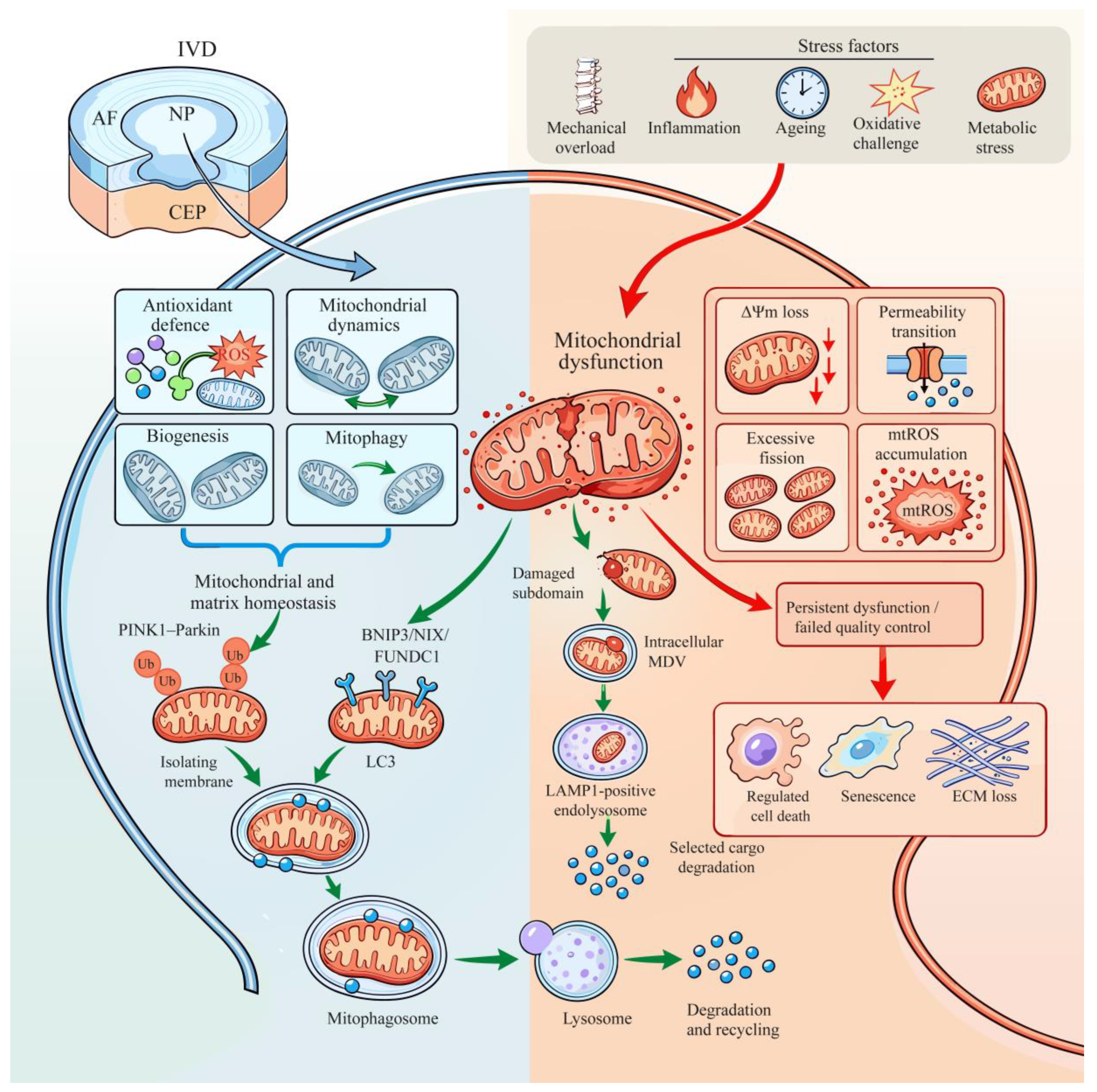
MDV formation may therefore have different consequences within the disc. Packaging damaged mitochondrial constituents could preserve functional mitochondrial mass in the donor cell, whereas extracellular release may expose neighboring cells to harmful cargo. It is not yet known whether TDP43-containing vesicles share their biogenesis with lysosome-directed MDVs, increase when mitophagic flux is saturated or selectively carry oxidized mitochondrial components. MDVs are best regarded as a complementary branch of mitochondrial quality control rather than a substitute for mitophagy. Their effect in IVDD is likely to depend on cargo identity, intracellular or extracellular destination, uptake by recipient cells and clearance from the disc [53,54,55,56,57,58,59,60].These mitochondrial quality-control routes and their potential outcomes are summarized in Figure 1.
Figure 1.
Mitochondrial quality control in nucleus pulposus cells. The disc inset identifies the NP cell represented in the main panel. IVDD-associated stressors can cause mitochondrial membrane-potential loss, permeability transition, excessive fission and mtROS accumulation. Antioxidant defense, mitochondrial dynamics, biogenesis and mitophagy contribute to mitochondrial and matrix homeostasis. Damaged mitochondria can enter PINK1–Parkin- or receptor-mediated mitophagy. Smaller intracellular MDVs may deliver selected cargo to LAMP1-positive endolysosomes; this lysosome-directed route is established mainly outside the disc field. Persistent mitochondrial dysfunction can contribute to regulated cell death, senescence and ECM loss, but the scheme does not imply that mitophagy failure is required for all IVDD. AF, annulus fibrosus; BNIP3, BCL2-interacting protein 3; CEP, cartilage endplate; ECM, extracellular matrix; FUNDC1, FUN14 domain-containing protein 1; IVD, intervertebral disc; LAMP1, lysosome-associated membrane glycoprotein 1; LC3, microtubule-associated protein 1 light chain 3; MDV, mitochondrial-derived vesicle; mtROS, mitochondrial reactive oxygen species; NIX, BCL2-interacting protein 3-like; NP, nucleus pulposus; PINK1, PTEN-induced kinase 1; ROS, reactive oxygen species; Ub, ubiquitin; ΔΨm, mitochondrial membrane potential. Green arrows indicate mitochondrial clearance or homeostatic recovery, whereas red arrows indicate persistent mitochondrial injury and degenerative outcomes; blue elements denote the broader mitochondrial quality-control network.
3. Stress-Dependent Coupling of mtROS Production and Mitophagy in IVDD
3.1. Mechanical Loading and Mitochondrial Damage
Mechanical loading is necessary for disc health, but excessive magnitude, duration or frequency can exceed cellular adaptation. In a bovine whole-disc one-strike model, a single high-energy deformation rapidly decreased mitochondrial membrane potential and produced an early, sustained rise in ROS. MitoQ markedly reduced this signal, supporting a mitochondrial origin, and also attenuated apoptosis- and necroptosis-associated changes and subsequent matrix damage [61]. Human-cell and CEP studies likewise connect matrix stiffness or excessive compression to Piezo1 signaling, mitochondrial fission and disturbed autophagy/mitophagy [62,63,64,65,66,67,68,69]. Although acute organ injury does not reproduce chronic human IVDD, this temporal sequence places mitochondrial redox disruption near the beginning of the degenerative response.
Sustained compression shows why increased mtROS and mitophagy initiation may coexist with continuing injury. In human NP cells, high compression increases mitochondrial permeability transition, depolarization and fission, decreases fusion, and recruits PINK1 and Parkin. Lysosomal dysfunction nevertheless prevents mitophagosome–lysosome fusion, leaving damaged mitochondria in place despite the rise in initiation markers [18]. High-magnitude compression also suppresses SIRT1 and induces ROS, mitochondrial dysfunction and senescence; protection by SIRT1 requires PINK1 [70]. HIF1A-, MIF- and SIRT3-linked programs preserve autophagic or mitochondrial adaptation under compression, whereas loss of these reserves favors apoptosis and oxidative injury [71,72,73]. One study, however, found that reducing PINK1–Parkin activity attenuated p53/p21-dependent senescence [20]. More recent work links an adaptive mechanical response to ITGB1/vimentin-dependent PINK1–Parkin activity [74]. Differences in loading protocol, cell source, exposure time and flux measurement probably explain part of this disagreement. The same pathway may indicate effective clearance at one stage and overwhelmed or maladaptive turnover at another.
Mechanical transduction also determines which mitochondrial material becomes available for clearance. In CEP cells, Piezo1-driven Ca2+/CaMKII/Drp1 signaling promotes excessive fission, mitochondrial dysfunction, inflammation, apoptosis and senescence [75]. ERK1/2-dependent Drp1 activation similarly fragments mitochondria and is associated with apoptosis and pyroptosis in oxidant-stressed NP cells [76]. Neither study directly measured mitophagic flux. They nevertheless show how fission may either isolate damaged segments for disposal or fragment the network too extensively for quality control to remain effective.
3.2. Inflammation, Oxidative Challenge and Ageing-Associated Stress
Inflammatory cytokines, such as TNF-α and IL-1β, increase ROS, impair mitochondrial function and stimulate matrix-catabolic pathways. TNF-α-induced Parkin-dependent mitophagy is initially protective; silencing Parkin increases mtROS, membrane-potential loss and apoptosis, whereas enhancing the pathway supports survival [25]. TBK1 and OPTN provide additional links between inflammatory stress, selective autophagy and mitochondrial cargo handling [29,32]. However, inflammation can also damage lysosomes and autophagosome maturation, converting increased initiation into incomplete turnover.
Hydrogen peroxide and TBHP are widely used to model oxidative stress. They reproducibly induce mitochondrial depolarization, mtROS, apoptosis, senescence and matrix degradation. These models allow causal testing of Nrf2–SIRT3, DJ-1–AKT–HK2–Parkin, the ATF5-dependent mitochondrial unfolded-protein response and FUNDC1 [33,51,77,78,79,80,81,82,83]. High-glucose/AGE conditions, circadian disruption and ASIC1a activation provide metabolically distinct routes to mitochondrial redox failure [84,85,86]. Bolus oxidants are nevertheless broad and are often applied under normoxic, nutrient-rich culture conditions. They establish pathway competence but cannot reproduce the sequence operating over decades of human degeneration.
Aging imposes a slower and more persistent mitochondrial stress. Progerin accumulation in NP cells impairs mitochondrial dynamics and promotes apoptosis and senescence [87]. Declining proteostasis, lysosomal acidity and autophagic flux allow damaged mitochondria to accumulate, while senescent cells expose neighboring cells to cytokines and oxidants [88]. EGR1 suppression and engineered restoration of AMPK–FOXO1a-associated mitophagy reduce mitochondrial injury and senescence in recent models [89,90]. The response also changes over time. DJ-1, AKT and HK2 support Parkin recruitment during early stress, but prolonged stress depletes DJ-1 and permits mitochondrial apoptosis [78]. Oxidative injury and mitochondrial clearance therefore become progressively uncoupled as adaptive reserves decline.
4. Bidirectional Interactions Between mtROS and Mitophagy
4.1. mtROS and Depolarization Initiate Mitochondrial Surveillance
Oxidative damage to respiratory complexes, cardiolipin and mitochondrial DNA lowers membrane potential and increases permeability transition. Depolarization blocks normal PINK1 import and degradation, allowing PINK1 to accumulate on the outer membrane and recruit Parkin. Oxidative and hypoxic signaling can also activate receptor-mediated pathways. In disc cells, early Parkin induction, DJ-1/HK2 recruitment, BNIP3 translocation and FUNDC1 activation are consistent with mtROS-containing stress acting upstream of mitochondrial selection [17,22,25,33,78].
mtROS should nevertheless not be treated as a single quantitative trigger. The location, duration and molecular target of ROS matter. A transient oxidant signal may initiate antioxidant transcription and selective clearance, whereas widespread lipid and protein oxidation can disable autophagy proteins, lysosomal membranes or the cytoskeleton required for cargo transport. ROS may therefore stimulate mitophagy initiation while simultaneously reducing the probability that flux will be completed.
4.2. Completed Mitophagy Limits mtROS and Danger Signaling
Direct manipulation of mitophagy supports its role in mitochondrial redox control. Parkin silencing increases mtROS and apoptosis during inflammatory or oxidative stress [25,26]. OPTN overexpression increases mitolysosome formation and reduces MitoSOX fluorescence and senescence, whereas OPTN deficiency has the opposite effects [32]. Nrf2–SIRT3 activation improves mitochondrial clearance, antioxidant capacity and survival; genetic interference or autophagy inhibition weakens these benefits [77]. Taurine, urolithin A, hyaluronic acid, platelet lysate and stromal-cell extracellular vesicles also reduce mitochondrial injury when flux is restored [91,92,93,94,95]. Because these interventions have several biological targets, their efficacy cannot by itself establish a single mitophagy-dependent mechanism.
Removing damaged mitochondria also restricts secondary danger signals. Persistent permeability transition can release mitochondrial DNA into the cytosol, where it activates TLR9 or cGAS–STING signaling. Humanin preserves mitochondrial homeostasis, lowers mtROS and mtDNA leakage and suppresses mtDNA–TLR9–NF-κB signaling during ferroptotic stress [96]. Taurine has been reported to reduce cytosolic mtDNA and enhance autophagic degradation of STING, thereby suppressing TBK1–IRF3/NLRP3 signaling in NP progenitor cells [97]. These studies do not directly establish selective mitophagy as the only mechanism, but they connect mitochondrial redox control and organelle clearance with sterile inflammation [98].
4.3. Why Increased Mitophagy Markers May Coexist with Degeneration
Increased pathway markers do not necessarily indicate increased mitophagic flux. PINK1, Parkin, LC3-II, p62 and autophagosomes may accumulate because initiation has risen, degradation is blocked or damaged cargo exceeds clearance capacity. Compression-induced lysosomal failure provides the clearest IVDD example: PINK1–Parkin recruitment increases while mitophagosomes fail to fuse with functional lysosomes [18]. Curcumin restores general autophagic flux by improving autophagosome–lysosome fusion and lysosomal function, with corresponding reductions in mitochondrial injury, apoptosis, senescence and ECM loss [99]. The study did not isolate mitochondria-specific cargo throughout, but it shows why the terminal degradative step must be measured.
Cargo selectivity is another determinant of outcome. NLRX1 supports fission of damaged mitochondrial segments. Without NLRX1, widespread fragmentation generates abundant cargo and compensatory PINK1–Parkin activity associated with severe senescence [19]. BNIP3 deletion increases mitochondrial delivery through alternative receptors but does not restore morphology or metabolism [17]. In other settings, excessive BNIP3-linked activity has been associated with YAP1-dependent pyroptosis or senescence, apoptosis and mTOR-sensitive disc injury [21,100,101]. NDUFA4L2 perturbation likewise shows that reduced mitochondrial turnover can be protective in a defined redox context [102]. Turnover may therefore become harmful when cargo selection is poor, clearance exceeds biogenesis or mitophagy forms part of a broader pro-death program.
Taken together, these studies support a capacity-matching interpretation for IVDD models in which mitochondrial stress is demonstrable. The component processes—damage sensing, selective disposal, lysosomal degradation and mitochondrial biogenesis—are established parts of mitochondrial quality control. The contribution of the present framework is its joint interpretation under the unusual metabolic and mechanical conditions of the disc, not the proposal of a new molecular pathway. It predicts that increasing PINK1–Parkin initiation will not rescue compressed cells if mitophagosome–lysosome fusion remains blocked; that accelerating clearance may be detrimental when mitochondrial replacement and ATP reserve are inadequate; and that the same intervention may differ among NP, AF and CEP cells because their stress exposure and metabolic reserve differ. These predictions require direct testing and should not be read as evidence that mitophagy failure is necessary or sufficient for IVDD.
This integration yields three testable predictions. First, increasing PINK1–Parkin initiation should not protect compressed cells when lysosomal fusion remains defective. Second, accelerating clearance should become maladaptive when biogenesis and ATP reserve cannot replace the removed organelles. Third, an intervention should produce different outcomes in NP, AF and CEP cells, or at early and late disease stages, when their stress burden and mitochondrial reserve differ. These predictions distinguish the framework from a generic assumption that more mitophagy is beneficial and require flux and functional measurements rather than pathway-protein abundance alone.The distinction between completed and blocked mitophagic flux, together with MDV-related cargo handling, is illustrated in Figure 2. IVDD-associated stressors and the corresponding mitochondrial responses are summarized in Table 1.
Figure 2.
Mitophagic flux, MDV-related cargo handling and the capacity-matching framework. (A) Depolarized mitochondria may undergo PINK1–Parkin- or receptor-mediated mitophagy or generate vesicles with different destinations. One IVDD study identified TDP43-containing, TOMM22-positive extracellular vesicles of mitochondrial origin that were taken up by neighboring NP cells and promoted inflammatory and senescent phenotypes [60]. The intracellular STX17/SNAP29/VAMP7-dependent route to LAMP1-positive endolysosomes is based mainly on non-disc studies [53,54,55,56]. (B) Completed mitophagic flux requires lysosomal fusion and cargo degradation; accumulation of intermediates can instead indicate blocked flux. (C) The authors’ framework relates damage burden to cargo selectivity, lysosomal capacity, mitochondrial renewal and regional metabolic context. The three illustrated outcomes are hypotheses for experimental testing, not established stages of IVDD. AF, annulus fibrosus; BNIP3, BCL2-interacting protein 3; CEP, cartilage endplate; FUNDC1, FUN14 domain-containing protein 1; IVDD, intervertebral disc degeneration; LAMP1, lysosome-associated membrane glycoprotein 1; MDV, mitochondrial-derived vesicle; MIRO1/2, mitochondrial Rho GTPase 1/2; mtDNA, mitochondrial DNA; mtROS, mitochondrial reactive oxygen species; NIX, BCL2-interacting protein 3-like; NP, nucleus pulposus; PINK1, PTEN-induced kinase 1; SNAP29, synaptosomal-associated protein 29; STX17, syntaxin 17; TDP43, TAR DNA-binding protein 43; TOMM22, translocase of outer mitochondrial membrane 22; VAMP7, vesicle-associated membrane protein 7. Arrows indicate the direction of cargo handling or pathway progression; green checks denote completed clearance, whereas red crosses denote blocked or incomplete steps.
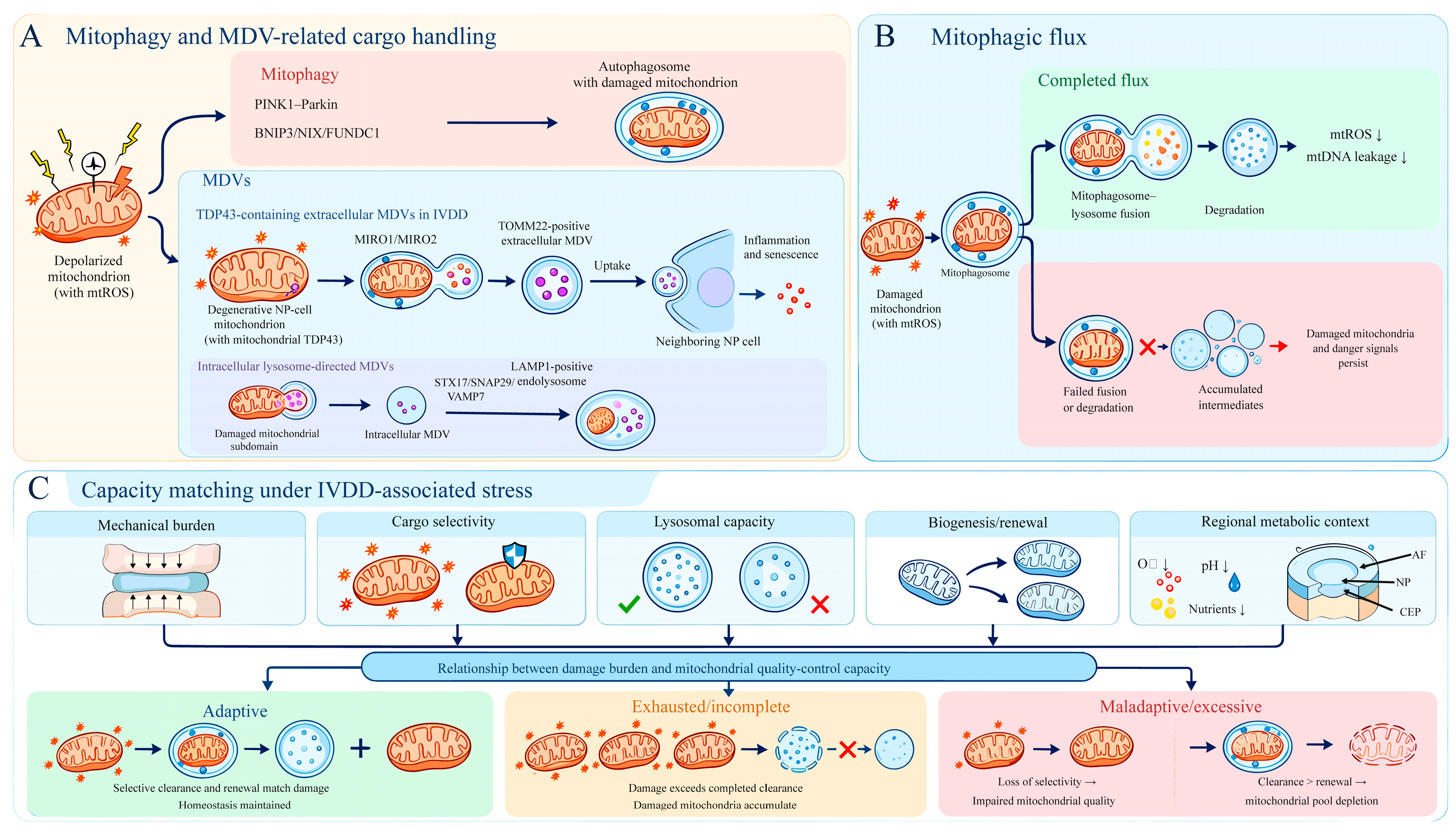
Table 1.
IVDD-associated stressors and mitochondrial quality-control responses.
5. mtROS, Mitophagy and Regulated Cell Death
5.1. Apoptosis
Among the regulated cell-death programs considered here, apoptosis has the strongest causal support. Mitochondrial depolarization, permeability transition, Bax/Bcl-2 imbalance, cytochrome-c release and caspase activation recur in AF, NP and progenitor-cell models [16,108,109]. Parkin knockdown aggravates mtROS accumulation, mitochondrial dysfunction and apoptosis [25,26]. Melatonin-induced clearance reduces TBHP-induced apoptosis, and interference with Parkin or mitophagy weakens this effect [110]. The miR-182-5p/SIRT1–circERCC2 axis provides further genetic evidence connecting mitochondrial turnover with apoptosis [111].
Several adaptive pathways modify this relationship [83]. SIRT1 requires PINK1 to limit compression-induced mitochondrial dysfunction and senescence [70]. Nrf2–SIRT3 improves antioxidant defense, autophagic flux and resistance to apoptosis [77]. ATF5-dependent mitochondrial proteostasis cooperates with PINK1-mediated clearance [51], whereas DJ-1–AKT–HK2 promotes early Parkin recruitment but loses efficacy during prolonged stress [78]. MANF–MFN2 signaling also connects mitochondrial fusion and mitophagy with apoptosis, although the evidence is currently limited to cultured human NP cells [112]. Across these models, competent mitochondrial quality control consistently raises the threshold for intrinsic apoptosis.
Mitophagy is unlikely to be the sole mediator of these effects. Most compounds that enhance mitochondrial clearance also alter redox, inflammatory or metabolic pathways. Reversal by cyclosporin A, chloroquine or 3-methyladenine indicates autophagy dependence but not mitochondrial selectivity, because each inhibitor has broader actions. Genetic perturbation, flux reporters, and direct measurement of mitochondrial cargo provide stronger evidence. Even with these limitations, bidirectional manipulation of Parkin and several upstream pathways makes apoptosis the best-supported death outcome jointly associated with mtROS and mitophagy.
5.2. Pyroptosis and Inflammasome-Associated Death
mtROS, oxidized mitochondrial components and leaked mtDNA can activate NLRP3. In primary human NP cells, reducing mtROS suppresses NLRP3 signaling and improves matrix metabolism [81,98,103,104,113,114]. SIRT1-, CREG1- and calreticulin-associated PINK1–Parkin activity has likewise been linked to less inflammasome activation or pyroptosis [105,115,116]. These findings support a mitochondrial damage–inflammasome connection, but inflammasome activation alone is not equivalent to pyroptotic death.
Attribution to pyroptosis requires caspase-1 activation, gasdermin cleavage, membrane permeabilization and pathway-specific rescue. ERK1/2–Drp1-dependent fragmentation activates both pyroptotic and apoptotic markers, and Drp1 inhibition improves both outcomes [76]. A20, PTPN22 and taurine connect improved mitochondrial turnover with reduced gasdermin- or caspase-1-associated injury, whereas PRDM1 provides a transcriptional route to CASP1 activation [117,118,119,120]. Melatonin-mediated Parkin activity has also been associated with lower inflammasome signaling in a radiculopathy model [121]. Removal of damaged mitochondria may therefore raise the pyroptotic threshold, but this evidence is newer and less independently replicated than the evidence for apoptosis.
5.3. Ferroptosis
Ferroptosis is driven by iron-dependent phospholipid peroxidation and failure of glutathione–GPX4 defense. mtROS can promote lipid oxidation, while mitochondrial metabolism, iron handling and membrane state influence susceptibility. In CEP chondrocytes, Nrf2/HO-1 activation increases Parkin-associated mitophagy and reduces oxidative dysfunction, ferroptosis and calcification [8,9,122]. Changes in mitochondrial redox state and clearance at the endplate may therefore affect nutrient supply to the entire disc as well as CEP-cell survival.
Several recent NP-cell studies provide more direct evidence [68,69,123,124]. Loss of VMP1 impairs PINK1–Parkin activity, worsens mitochondrial dysfunction and increases ferroptosis-related changes [125]. FBXO2 combines mitochondrial clearance with LCN2 degradation, thereby limiting iron overload, glutathione depletion and lipid peroxidation [126]. HIF-1α/BNIP3 and FBXW7 studies also place mitophagy upstream of ferroptosis resistance. ACSL1, NOX4–LRRC8A and metallothionein-2A identify lipid-metabolic, inflammatory and antioxidant pathways that may converge on mitochondrial redox stress [127,128,129,130,131]. Mitophagy may thus reduce one source of oxidative pressure without correcting every component of ferroptosis.
Humanin reduces erastin-induced ferroptosis and preserves membrane potential. It also limits permeability transition, mtROS, mtDNA leakage and HSP27-sensitive NF-κB and JAK2/STAT3 signaling [96]. Mitochondrial homeostasis is clearly involved, but mitophagic flux was not the principal causal variable. TBHP creates a related interpretive problem because it is used as both an oxidative-stress and a ferroptosis inducer, despite activating apoptosis and other injury programs. Ferroptosis should therefore be assigned using lipid peroxidation and iron measurements, GPX4 or glutathione changes, characteristic rescue and exclusion of competing death modes. Evidence that mtROS and mitophagy jointly regulate ferroptosis is increasing, but the precise relationship remains incompletely defined.
5.4. Necroptosis
Necroptosis is mediated by RIPK1–RIPK3–MLKL signaling and produces inflammatory membrane rupture. Acute one-strike loading increases necroptosis-associated changes in whole bovine discs, and MitoQ or necrostatin-1 partially reduces cell loss and matrix injury [61]. This places mtROS upstream of a necroptotic component but does not itself establish mitophagy control.
The clearest evidence connecting mitophagy with necroptosis comes from PHB2 manipulation. In human NP cells exposed to TNF-α/SM-164/Z-VAD-FMK, PHB2 loss worsens mitochondrial dysfunction and RIPK1–RIPK3–MLKL signaling. PHB2 overexpression restores autophagosome–lysosome activity, limits necroptosis and preserves matrix proteins [34]. Compression-induced death of NP progenitors provides related evidence, although broad HSP90 inhibition and mixed apoptosis or necroptosis limit pathway specificity [109]. Recent reviews accordingly regard necroptosis as a less developed branch of IVDD research than apoptosis or pyroptosis [132].
5.5. Senescence and Mixed Cell-Fate States
Senescence is not regulated cell death, but it is a major alternative fate after mitochondrial stress. Senescent NP cells remain in the tissue and release inflammatory and matrix-catabolic factors, allowing degeneration to progress without immediate loss of cellularity. PINK1-, OPTN- and FUNDC1-associated clearance generally reduces ROS and senescence [32,33,67,70,82,106,107,133]. NLRX1 deficiency produces a different outcome: widespread fragmentation and compensatory mitophagy accompany a pronounced senescent phenotype [19]. Whether a damaged cell dies or persists in a senescent state may depend on residual ATP, lysosomal competence and the capacity to rebuild a functional mitochondrial network.
Markers of more than one cell-fate program are often detected in the same IVDD model, but this does not establish simultaneous execution or a fixed sequence of transitions. For example, A20-associated enhancement of mitophagy reduced both pyroptotic and apoptotic readouts in NP cells, and inhibition of pyroptosis also reduced apoptosis in that model [117]. This result supports pathway interaction, but it does not define a universal switch between pyroptosis and apoptosis. Similar caution applies when mtROS reduction improves several endpoints at once: a shared upstream lesion may influence multiple programs without determining which program executes death in each cell. The evidence base and principal limitations for each cell-fate programme are summarized in Table 2.
Table 2.
Evidence base and interpretive limits for mitochondrial involvement in cell-fate programs in IVDD.
5.6. Shared Upstream Determinants and Limits of Current Evidence
Hypoxia, ATP depletion, mitochondrial depolarization, lysosomal dysfunction, inflammasome priming, iron availability and lipid-peroxidation defense can alter susceptibility to apoptosis, pyroptosis, ferroptosis or senescence. The corresponding execution machinery is distinct: mitochondrial outer-membrane permeabilization and caspases support apoptosis; caspase-1 and gasdermin cleavage support pyroptosis; iron-dependent phospholipid peroxidation with impaired glutathione–GPX4 defense supports ferroptosis; and durable p16–RB or p53–p21 signaling supports senescence. IVDD studies have examined many of these nodes separately, but direct time-resolved comparisons under matched hypoxia or energy stress are rare. The available data therefore support shared upstream determinants and pathway crosstalk rather than a deterministic hierarchy of cell-fate switching [34,61,76,96,97,98,103,104,105,113,114,115,116,117,118,119,120,121,122,123,124,125,126,127,128,129,130,131,132].
Future experiments should combine time-resolved measurements of membrane potential, ATP, lysosomal function and mtROS with execution-specific assays and pathway-specific rescue. Cytochrome-c release and caspase activation, gasdermin cleavage and membrane permeabilization, lipid peroxidation and GPX4 status, phosphorylated MLKL, and durable senescence markers should be assessed in the same model. Single-cell or longitudinal approaches would be particularly useful for separating mixed populations from genuine transitions within individual cells. Until such evidence is available, the review treats the cell-fate programs as mechanistically connected but not interchangeable.
6. Consequences for Intervertebral Disc Homeostasis
Mitochondrial injury extends from the organelle to the whole disc through several connected processes. Loss of viable NP cells reduces aggrecan and collagen II synthesis. Senescent and inflammasome-active cells release cytokines and proteases that increase MMP and ADAMTS activity. mtROS and mitochondrial nucleic acids maintain NF-κB, NLRP3, TLR9 and cGAS–STING signaling, while macrophage–NP-cell crosstalk can amplify these responses [134]. Matrix turnover consequently shifts towards catabolism, reducing osmotic swelling and redistributing mechanical stress to the AF and CEP.
Surviving cells may also become dysfunctional before overt death. BNIP3 loss alters mitochondrial morphology, glycolytic capacity, ATP production and broader substrate use, and Bnip3-deficient mice show early NP degeneration despite compensatory mitophagy [17]. NLRX1 deficiency similarly uncouples selective fission from clearance and promotes metabolic collapse and senescence [19]. Mitophagy-dependent reprogramming of lactate metabolism and SPP1–integrin-mediated blockade of PINK1–Parkin further connect mitochondrial state to matrix phenotype and calcification [135,136]. Mitochondrial quality therefore determines whether living disc cells retain an appropriate metabolic and matrix-producing phenotype.
Endplate failure creates another route for tissue-level amplification. CEP-cell apoptosis, ferroptosis and calcification reduce permeability and restrict nutrient diffusion. NP and inner-AF cells are then exposed to greater glucose limitation, acidity and waste accumulation, which further compromise lysosomal and mitochondrial function. Nrf2/HO-1–Parkin signaling in CEP models may therefore preserve the central disc indirectly [8,9]. Direct experiments that trace these effects between disc regions remain rare.
Evidence that pathway manipulation changes disc structure comes from bovine organ culture and multiple needle-puncture, compression or instability models. Interventions that reduce mtROS, restore flux or enhance selected mitophagy pathways commonly preserve MRI signal, disc height, histological organization or matrix markers. Yet these models impose acute injury on young animals and do not reproduce decades of human ageing, endplate vascular change, immune exposure or pain biology. Human evidence is mainly cross-sectional, showing altered pathway regulators across degeneration grades. It supports disease relevance but cannot distinguish cause, compensation and consequence.
These cellular and tissue changes can form a reinforcing loop in experimental settings in which mitochondrial stress is prominent. Mechanical or nutritional stress may injure mitochondria; incomplete or maladaptive clearance can then sustain mtROS, cell loss, senescence and matrix catabolism, while structural deterioration further increases mechanical and metabolic stress. Other trajectories can begin with AF structural failure, CEP calcification, matrix defects or age-related proteoglycan loss without demonstrable mitophagy failure. Mitochondrial quality control may amplify or modify such trajectories after they begin. Mitochondria-directed treatment should therefore be matched to a documented mitochondrial defect rather than assumed to apply to all forms or stages of IVDD.
7. Current Potential Therapies
No intervention that jointly controls mtROS and mitophagic quality has been established as a disease-modifying treatment for human IVDD. All strategies discussed below remain preclinical.
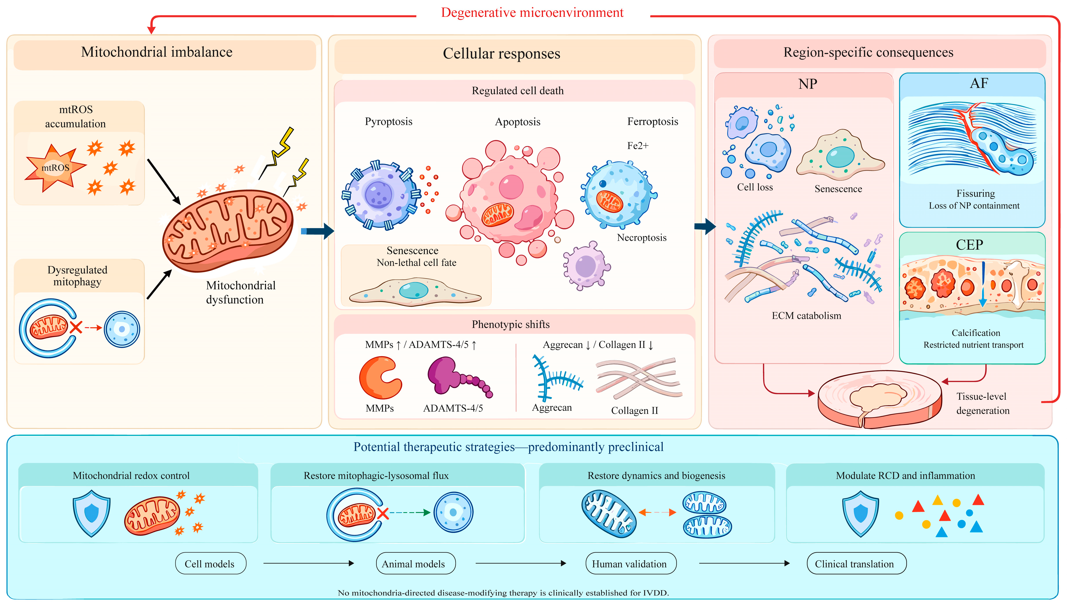
Therapeutic evidence should be separated into two categories. Relatively direct evidence requires pathway-specific genetic perturbation or rescue together with mitochondrial cargo or flux measurements. By contrast, many antioxidants, natural products and biomaterials are pleiotropic, and altered mitophagy is only one of several associated effects. The latter studies support therapeutic hypotheses but do not establish mitophagy as the causal mediator. No clinical trial has yet validated a mitochondria-directed or mitophagy-directed disease-modifying treatment for IVDD. The context-dependent links among mitochondrial imbalance, cell fate, regional disc pathology and potential intervention strategies are summarized in Figure 3.
Figure 3.
Context-dependent mitochondrial contributions to cell fate, regional disc pathology and potential intervention. mtROS accumulation and dysregulated mitophagy can contribute to mitochondrial dysfunction, cell-fate change and matrix catabolism. The relative sizes of the cell-fate illustrations represent the breadth of IVDD evidence reviewed, not biological prevalence or effect size: apoptosis has the broadest mechanistic literature; pyroptosis and ferroptosis have growing but model-dependent support; and direct mitophagy–necroptosis evidence is comparatively sparse. Senescence is shown separately because it is a non-lethal cell fate. The larger NP panel reflects the predominance of NP-derived studies; AF and CEP panels emphasize structural containment and nutrient-transport consequences without implying equivalent mitochondrial evidence. Candidate interventions are preclinical strategy classes, and the lower sequence indicates validation still required rather than completed clinical progression. ADAMTS, a disintegrin and metalloproteinase with thrombospondin motifs; AF, annulus fibrosus; CEP, cartilage endplate; ECM, extracellular matrix; IVDD, intervertebral disc degeneration; MMP, matrix metalloproteinase; mtROS, mitochondrial reactive oxygen species; NP, nucleus pulposus; RCD, regulated cell death. Solid arrows indicate proposed directional relationships, and the red return arrow denotes a feed-forward degenerative loop. Box colors are used for visual grouping and do not represent quantitative effect size.
Mitochondrial redox control offers the most direct way to reduce the initial oxidative burden [7,133,137,138,139]. MitoQ lowers mtROS and preserves mitochondrial function in compressed NP cells and one-strike whole-disc culture [18,61]. Honokiol activates AMPK–PGC-1α–SIRT3 signaling, improves antioxidant capacity and mitochondrial dynamics, and attenuates puncture-induced degeneration in rats [23]. Melatonin has been linked to both Parkin-dependent clearance and PGC-1α-mediated coordination of mitochondrial biogenesis, dynamics and mitophagy [110,140]. These agents have several targets, so their benefits cannot be attributed solely to mitophagy.
Lowering mtROS alone does not remove damaged mitochondria. Restoring lysosomal competence and downstream flux may provide a complementary approach [81,82,83,123,124]. MitoQ improves mitophagosome–lysosome fusion during compression [18], while curcumin restores general autophagic flux through AMPK–mTOR–ULK1 signaling and improved lysosomal function [99]. Hypoxia- or oxidative-stress-preconditioned stromal-cell exosomes deliver cargo that enhances mitochondrial clearance [141,142]. Salidroside similarly restores SIRT1/FOXO3-dependent flux in NP-derived progenitors [39]. Delivery, cargo consistency and long-term safety remain unresolved for these strategies.
Greater selectivity may be achieved by targeting mitochondrial dynamics, cargo recognition or receptor-mediated clearance. NLRX1 restoration improves selective fission and prevents compensatory overclearance [19]. Inhibition of excessive ERK1/2–Drp1 or Piezo1–CaMKII–Drp1 activity may reduce widespread fragmentation [75,76]. FGF21/SIRT1/FOXO3 signaling and modulation of OPTN, FUNDC1, BNIP3 or PHB2 are also plausible targets [143]. Their effects are likely to vary with cell type and stress because mitophagy receptors overlap in function and can participate in pro-death responses.
Regional biology also constrains therapeutic interpretation. NP-directed studies chiefly address redox burden, lysosomal completion and mitochondrial mass under hypoxia and acidity. AF studies include direct evidence for regional metabolism [35,36], oxidative-stress-associated ferroptosis [37] and an engineered-exosome repair strategy linked to mitophagy recovery [38], but this literature is much smaller than the NP literature and does not justify transferring NP targets directly to AF cells. CEP-directed strategies must consider calcification, ferroptosis and nutrient transport as well as mitochondrial redox control [8,9,10,11,12,13]. Delivery studies should therefore identify the intended disc region and demonstrate local exposure and response rather than relying only on whole-disc histology.
When mitochondrial recovery remains incomplete, inflammatory and cell-death pathways become additional therapeutic targets [68,69,113,114,122,123,124]. Inhibition of NLRP3, ferroptosis or RIPK1–RIPK3–MLKL signaling may preserve disc cells. Blocking death execution without restoring mitochondrial fitness, however, could leave metabolically impaired or senescent cells in the tissue. Combination treatment may therefore need to reduce mtROS, restore competent flux and restrain the cell-death pathway that remains active.
Clinical translation is limited by both biology and delivery. The avascular, dense and acidic disc restricts penetration, while drug retention varies with molecular properties and delivery vehicle [144]. Intradiscal puncture can also create a leakage path; through-puncture experiments show that leakage depends on puncture and injectate conditions, so retention cannot be inferred from injection alone [145]. Systemic mitochondrial targeting raises off-target concerns, whereas repeated local injection may damage AF containment. Biological dose is equally important; sustained ROS suppression may blunt redox signaling, and indiscriminate mitophagy enhancement may remove functional mitochondria from cells with limited renewal capacity.
Hydrogels and nanoparticles offer delivery solutions, not proof of clinical efficacy. Injectable hydrogels can prolong local residence and provide controlled release; nanoparticles can protect cargo or support mitochondrial and stimulus-responsive targeting [90,93,114,133,144,146,147]. An AF-repair microneedle/exosome system provides region-specific proof of concept for combining structural repair with modulation of mitophagy [38]. These platforms still require assessment of diffusion through the disc matrix, leakage, degradation products, sterilization, manufacturing consistency, long-term biocompatibility and performance under physiological loading.
Patient selection is equally important. A strategy designed for excessive mtROS may not correct lysosomal failure, and a mitophagy enhancer may be unsuitable when mitochondrial replacement is already limited. Future studies should define the dominant defect, the intended disc region and the degeneration stage before assigning therapeutic readiness. Table 3 therefore separates target-level mechanistic support from pleiotropic association and delivery proof-of-concept.
Table 3.
Preclinical therapeutic strategies targeting mitochondrial dysfunction and mitophagy in IVDD.
8. Conclusions
Current evidence does not support a simple sequence in which ROS cause injury and mitophagy provides protection, nor does it support mitophagy failure as a universal initiating cause of IVDD. Disc degeneration may progress through AF structural compromise, CEP calcification, matrix abnormalities and age-related proteoglycan loss without demonstrated failure of mitochondrial clearance. Within mitochondrial-stress-dominant contexts, however, a capacity-matching model helps explain why the same pathway can be protective, ineffective or maladaptive. Mismatch can arise from blocked lysosomal flux, non-selective fragmentation, clearance that exceeds biogenesis or failure of the remaining network to meet regional metabolic demand. Selective MDV transport may remove mitochondrial material before whole-organelle clearance becomes necessary, whereas TDP43-containing mitochondria-derived vesicles in NP cells may propagate stress between cells; their relationship to lysosome-directed MDVs remains unresolved [60].
The strength of the evidence differs among cell fates. Mitochondrial apoptosis and senescence are supported by human-cell studies, organ culture and in vivo models. Mechanistic evidence for pyroptosis and ferroptosis is growing, whereas necroptosis remains less developed and mixed execution states are often poorly resolved. Increased PINK1, Parkin, LC3 or mitochondrial turnover should therefore be interpreted only after cargo selectivity, lysosomal completion, mitochondrial replacement and the relevant execution pathway have been examined.
Progress depends on measuring mitochondrial clearance rather than inferring it from PINK1, Parkin, LC3 or autophagosome abundance. Tandem mCherry–GFP–LC3 reports general autophagosome maturation and is useful for detecting lysosomal blockade, but it does not identify mitochondrial cargo. Mitochondria-specific reporters, such as mito-QC or mt-Keima, together with lysosomal-inhibitor controls and colocalization of mitochondrial cargo with lysosomes, are more appropriate for mitophagic flux [22,148,149]. Flux measurements should be paired with mitochondrial mass, respiration or ATP production and indices of biogenesis so that clearance can be interpreted against replacement. Regional comparisons should use NP, AF and CEP cells under relevant oxygen, pH and loading conditions.
Longitudinal designs should connect these mitochondrial states to execution-specific cell-death assays and matrix outcomes. MDV studies should determine how intracellular and extracellular destinations are selected and whether vesicle release increases when mitophagic or lysosomal capacity is exceeded. The capacity-matching framework is therefore an IVDD-specific conceptual synthesis, not a newly discovered molecular pathway. Its value lies in defining measurable conditions that may explain why the same mitophagy-associated intervention is protective in one disc context but ineffective or harmful in another.
9. Literature Search Strategy
This work was designed as a structured narrative review rather than a systematic review or meta-analysis. PubMed/MEDLINE was searched for publications from 1 January 2012 to 11 September 2026 (inclusive). For articles published online ahead of issue assignment, the first online publication date was used to determine eligibility. The final date-limited query was rerun through the PubMed interface on 16 October 2026 and returned 1137 records; this rerun verified the search expression and record count and did not extend the prespecified evidence cutoff:
((“Intervertebral Disc Degeneration”[Mesh] OR “intervertebral disc degeneration”[tiab] OR IVDD[tiab] OR “degenerative disc disease”[tiab] OR “nucleus pulposus”[tiab] OR “annulus fibrosus”[tiab] OR “cartilage endplate”[tiab]) AND (“Mitophagy”[Mesh] OR mitophagy[tiab] OR “Mitochondria”[Mesh] OR mitochondria*[tiab] OR “mitochondrial dysfunction”[tiab] OR “Reactive Oxygen Species”[Mesh] OR “mitochondrial reactive oxygen species”[tiab] OR mtROS[tiab] OR “Oxidative Stress”[Mesh] OR “oxidative stress”[tiab] OR “mitochondrial-derived vesicle”[tiab] OR “mitochondrial-derived vesicles”[tiab] OR MDV[tiab] OR MDVs[tiab])) AND (“2012/01/01”[Date—Publication]: “2026/09/11”[Date—Publication]).
Section-specific searches added the terms “regulated cell death”, apoptosis, pyroptosis, ferroptosis, necroptosis, senescence, PINK1, Parkin, BNIP3, FUNDC1, OPTN, PHB2 and NLRX1. Google Scholar and reference lists were also screened. Earlier publications were retained only when needed to establish foundational AF mechanobiology or flux-reporter methods that could not be replaced by a later primary source; these citation-tracked exceptions are explicitly distinguished from the 15-year primary search window.
Eligible sources included English-language primary studies using human disc tissue, disc-derived cells, organ culture or animal IVDD models that examined mitochondrial redox stress, mitophagy, MDVs or regulated cell fate. Selected mechanistic studies outside the disc field were retained only to define MDV biology or a measurement method. Studies unrelated to disc biology, reports without relevant mitochondrial outcomes, conference abstracts, duplicate publications and non-English reports were excluded. Reviews were used for context but not as substitutes for primary evidence when original studies were available.
Source selection was iterative and was based on relevance to the review questions, model provenance and mechanistic informativeness. This was not a systematic review: duplicate independent screening, a PRISMA flow diagram, prospective protocol registration, formal risk-of-bias scoring and a complete exclusion ledger were not used. Accordingly, Table 2 provides descriptive information on the kinds of evidence and their principal limitations rather than a formal GRADE-style hierarchy. Earlier publications were retained only for foundational AF mechanobiology or flux-reporter methods that could not be replaced by later primary studies.
Author Contributions
K.C.: writing—original draft, conceptualization, and visualization; L.W.: writing—original draft, supervision, methodology, writing—review and editing, and project administration; H.C.: writing—original draft and investigation; Z.F.: writing—original draft, supervision, conceptualization, writing—review and editing, and funding acquisition. All authors have read and agreed to the published version of the manuscript.
Funding
This research was funded by the National Natural Science Foundation of China, grant number 81974351, and the National Key Research and Development Program of China, grant number 2025YFC2511604.
Data Availability Statement
No new data were created or analyzed in this study. Data sharing is not applicable to this article.
Conflicts of Interest
The authors declare no conflicts of interest.
References
- Song, Y.; Lu, S.; Geng, W.; Feng, X.; Luo, R.; Li, G.; Yang, C. Mitochondrial quality control in intervertebral disc degeneration. Exp. Mol. Med. 2021, 53, 1124–1133. [Google Scholar] [CrossRef] [Scilit]
- Wang, D.; Zheng, H.; Zhou, W.; Duan, Z.; Jiang, S.; Li, B.; Zheng, X.; Jiang, L. Mitochondrial Dysfunction in Oxidative Stress-Mediated Intervertebral Disc Degeneration. Orthop. Surg. 2022, 14, 1569–1582. [Google Scholar] [CrossRef] [Scilit]
- Song, C.; Xu, Y.; Peng, Q.; Chen, R.; Zhou, D.; Cheng, K.; Cai, W.; Liu, T.; Huang, C.; Fu, Z.; et al. Mitochondrial dysfunction: A new molecular mechanism of intervertebral disc degeneration. Inflamm. Res. 2023, 72, 2249–2260. [Google Scholar] [CrossRef] [Scilit]
- Zhang, C.; Peng, X.; Wang, F.; Xie, Z.; Chen, L.; Wu, X. Update on the Correlation Between Mitochondrial Dysfunction and Intervertebral Disk Degeneration. DNA Cell Biol. 2022, 41, 257–261. [Google Scholar] [CrossRef] [Scilit]
- Qiu, B.; Xie, X.; Xi, Y. Mitochondrial quality control: The real dawn of intervertebral disc degeneration? J. Transl. Med. 2024, 22, 1126. [Google Scholar] [CrossRef] [Scilit]
- Saberi, M.; Zhang, X.; Mobasheri, A. Targeting mitochondrial dysfunction with small molecules in intervertebral disc aging and degeneration. Geroscience 2021, 43, 517–537. [Google Scholar] [CrossRef] [Scilit]
- Nasto, L.; Robinson, A.; Ngo, K.; Clauson, C.; Dong, Q.; St Croix, C.; Sowa, G.; Pola, E.; Robbins, P.; Kang, J.; et al. Mitochondrial-derived reactive oxygen species (ROS) play a causal role in aging-related intervertebral disc degeneration. J. Orthop. Res. 2013, 31, 1150–1157. [Google Scholar] [CrossRef] [Scilit]
- Shao, Y.; Sun, L.; Yang, G.; Wang, W.; Liu, X.; Du, T.; Chen, F.; Jing, X.; Cui, X. Icariin protects vertebral endplate chondrocytes against apoptosis and degeneration via activating Nrf-2/HO-1 pathway. Front Pharmacol. 2022, 13, 937502. [Google Scholar] [CrossRef] [Scilit]
- Cui, X.; Liu, X.; Kong, P.; Du, T.; Li, T.; Yang, G.; Zhang, W.; Jing, X.; Wang, W. PTEN inhibitor VO-OHpic protects endplate chondrocytes against apoptosis and calcification via activating Nrf-2 signaling pathway. Aging 2023, 15, 2275–2292. [Google Scholar] [CrossRef] [Scilit]
- Kang, L.; Liu, S.; Li, J.; Tian, Y.; Xue, Y.; Liu, X. Parkin and Nrf2 prevent oxidative stress-induced apoptosis in intervertebral endplate chondrocytes via inducing mitophagy and anti-oxidant defenses. Life Sci. 2020, 243, 117244. [Google Scholar] [CrossRef] [Scilit]
- Wang, H.; Liu, X.; Yang, H.; Jing, X.; Wang, W.; Liu, X.; Zhang, B.; Liu, X.; Shao, Y.; Cui, X. Activation of the Nrf-2 pathway by pinocembrin safeguards vertebral endplate chondrocytes against apoptosis and degeneration caused by oxidative stress. Life Sci. 2023, 333, 122162. [Google Scholar] [CrossRef] [Scilit]
- Jing, X.; Wang, W.; He, X.; Liu, X.; Yang, X.; Su, C.; Shao, Y.; Ge, Z.; Wang, H.; Cui, X. HIF-2α/TFR1 mediated iron homeostasis disruption aggravates cartilage endplate degeneration through ferroptotic damage and mtDNA release: A new mechanism of intervertebral disc degeneration. J. Orthop. Transl. 2024, 46, 65–78. [Google Scholar] [CrossRef] [Scilit]
- Jiang, B.; Bai, C.; Pan, J.; Shen, B.; Li, L. Screening and identification of microRNAs mediating cartilage endplate degeneration in human intervertebral disks. Front Med. Lausanne 2024, 11, 1446294. [Google Scholar] [CrossRef] [Scilit]
- Feng, C.; Hu, Z.; Zhao, M.; Leng, C.; Li, G.; Yang, F.; Fan, X. Region-specific mitophagy in nucleus pulposus, annulus fibrosus, and cartilage endplate of intervertebral disc degeneration: Mechanisms and therapeutic strategies. Front Pharmacol. 2025, 16, 1579507. [Google Scholar] [CrossRef] [Scilit]
- Xu, W.; Yang, R.; Zheng, H.; Yu, W.; Zheng, X.; Li, B.; Jiang, S.; Jiang, L. PGC-1α acts as an mediator of Sirtuin2 to protect annulus fibrosus from apoptosis induced by oxidative stress through restraining mitophagy. Int. J. Biol. Macromol. 2019, 136, 1007–1017. [Google Scholar] [CrossRef] [Scilit]
- Rannou, F.; Lee, T.; Zhou, R.; Chin, J.; Lotz, J.; Mayoux-Benhamou, M.; Barbet, J.; Chevrot, A.; Shyy, J. Intervertebral disc degeneration: The role of the mitochondrial pathway in annulus fibrosus cell apoptosis induced by overload. Am. J. Pathol. 2004, 164, 915–924. [Google Scholar] [CrossRef] [Scilit]
- Madhu, V.; Hernandez-Meadows, M.; Boneski, P.; Qiu, Y.; Guntur, A.; Kurland, I.; Barve, R.; Risbud, M. The mitophagy receptor BNIP3 is critical for the regulation of metabolic homeostasis and mitochondrial function in the nucleus pulposus cells of the intervertebral disc. Autophagy 2023, 19, 1821–1843. [Google Scholar] [CrossRef] [Scilit]
- Kang, L.; Liu, S.; Li, J.; Tian, Y.; Xue, Y.; Liu, X. The mitochondria-targeted anti-oxidant MitoQ protects against intervertebral disc degeneration by ameliorating mitochondrial dysfunction and redox imbalance. Cell Prolif. 2020, 53, e12779. [Google Scholar] [CrossRef] [Scilit]
- Song, Y.; Liang, H.; Li, G.; Ma, L.; Zhu, D.; Zhang, W.; Tong, B.; Li, S.; Gao, Y.; Wu, X.; et al. The NLRX1-SLC39A7 complex orchestrates mitochondrial dynamics and mitophagy to rejuvenate intervertebral disc by modulating mitochondrial Zn2+ trafficking. Autophagy 2024, 20, 809–829. [Google Scholar] [CrossRef] [Scilit]
- Huang, D.; Peng, Y.; Li, Z.; Chen, S.; Deng, X.; Shao, Z.; Ma, K. Compression-induced senescence of nucleus pulposus cells by promoting mitophagy activation via the PINK1/PARKIN pathway. J. Cell Mol. Med. 2020, 24, 5850–5864. [Google Scholar] [CrossRef] [Scilit]
- Luo, J.; Yu, T.; Hu, P.; Liu, J. Hsa_circ_0101645 contributes to excessive autophagy and apoptosis in intervertebral disc degeneration by acting as a miR-1304-5p sponge modulating BNIP3 expression. Noncoding RNA Res. 2026, 17, 76–89. [Google Scholar] [CrossRef] [Scilit]
- Madhu, V.; Boneski, P.; Silagi, E.; Qiu, Y.; Kurland, I.; Guntur, A.; Shapiro, I.; Risbud, M. Hypoxic Regulation of Mitochondrial Metabolism and Mitophagy in Nucleus Pulposus Cells Is Dependent on HIF-1α-BNIP3 Axis. J. Bone Min. Res. 2020, 35, 1504–1524. [Google Scholar] [CrossRef] [Scilit]
- Wang, J.; Nisar, M.; Huang, C.; Pan, X.; Lin, D.; Zheng, G.; Jin, H.; Chen, D.; Tian, N.; Huang, Q.; et al. Small molecule natural compound agonist of SIRT3 as a therapeutic target for the treatment of intervertebral disc degeneration. Exp. Mol. Med. 2018, 50, 1–14. [Google Scholar] [CrossRef] [Scilit]
- Yang, W.; Jia, C.; Liu, L.; Fu, Y.; Wu, Y.; Liu, Z.; Yu, R.; Ma, X.; Gong, A.; Liu, F.; et al. Hypoxia-Inducible Factor-1α Protects Against Intervertebral Disc Degeneration Through Antagonizing Mitochondrial Oxidative Stress. Inflammation 2023, 46, 270–284. [Google Scholar] [CrossRef] [Scilit]
- Zhang, Z.; Xu, T.; Chen, J.; Shao, Z.; Wang, K.; Yan, Y.; Wu, C.; Lin, J.; Wang, H.; Gao, W.; et al. Parkin-mediated mitophagy as a potential therapeutic target for intervertebral disc degeneration. Cell Death Dis. 2018, 9, 980, Correction in Cell Death Dis. 2021, 12, 915. [Google Scholar] [CrossRef] [Scilit]
- Lan, T.; Zheng, Y.; Li, N.; Chen, X.; Shen, Z.; Yan, B. CRISPR/dCas9-Mediated Parkin Inhibition Impairs Mitophagy and Aggravates Apoptosis of Rat Nucleus Pulposus Cells Under Oxidative Stress. Front Mol. Biosci. 2021, 8, 674632. [Google Scholar] [CrossRef] [Scilit]
- Sun, K.; Jing, X.; Guo, J.; Yao, X.; Guo, F. Mitophagy in degenerative joint diseases. Autophagy 2021, 17, 2082–2092. [Google Scholar] [CrossRef] [Scilit]
- Lin, J.; Zheng, X.; Zhang, Z.; Zhuge, J.; Shao, Z.; Huang, C.; Jin, J.; Chen, X.; Chen, Y.; Wu, Y.; et al. Inhibition of LRRK2 restores parkin-mediated mitophagy and attenuates intervertebral disc degeneration. Osteoarthr. Cartil. 2021, 29, 579–591. [Google Scholar] [CrossRef] [Scilit]
- Hu, S.; Chen, L.; Al Mamun, A.; Ni, L.; Gao, W.; Lin, Y.; Jin, H.; Zhang, X.; Wang, X. The therapeutic effect of TBK1 in intervertebral disc degeneration via coordinating selective autophagy and autophagic functions. J. Adv. Res. 2021, 30, 1–13. [Google Scholar] [CrossRef] [Scilit]
- Chen, Y.; Lin, J.; Chen, J.; Huang, C.; Zhang, Z.; Wang, J.; Wang, K.; Wang, X. Mfn2 is involved in intervertebral disc degeneration through autophagy modulation. Osteoarthr. Cartil. 2020, 28, 363–374. [Google Scholar] [CrossRef] [Scilit]
- Shin, J.; Kang, Y.; Kim, H.; Moon, S.; Suk, K.; Park, S.; Lee, B.; Kwon, J. Mitofusin 1 in mitochondrial quality control and anti-inflammatory responses in nucleus pulposus cells during disc degeneration. Sci. Rep. 2025, 15, 38530. [Google Scholar] [CrossRef] [Scilit]
- Hu, Z.; Wang, Y.; Gao, X.; Zhang, Y.; Liu, C.; Zhai, Y.; Chang, X.; Li, H.; Li, Y.; Lou, J.; et al. Optineurin-mediated mitophagy as a potential therapeutic target for intervertebral disc degeneration. Front Pharmacol. 2022, 13, 893307. [Google Scholar] [CrossRef] [Scilit]
- Gu, T.; He, Y.; Zhou, J.; Qiu, X.; Yang, W.; Zhu, Q.; Liang, Y.; Zheng, Y.; Yik, J.H.N.; Haudenschild, D.R.; et al. CircFUNDC1 interacts with CDK9 to promote mitophagy in nucleus pulposus cells under oxidative stress and ameliorates intervertebral disc degeneration. Cell Death Dis. 2025, 16, 94. [Google Scholar] [CrossRef] [Scilit]
- Zhu, Z.; Liang, Y.; Liu, X.; Kong, F.; Sun, K.; Xu, X.; Shi, J. PHB2 mitigates intervertebral disc degeneration by modulating mitophagy to inhibit necroptosis in nucleus pulposus cells. iScience 2026, 29, 114365. [Google Scholar] [CrossRef] [Scilit]
- Cisewski, S.E.; Wu, Y.; Damon, B.J.; Sachs, B.L.; Kern, M.J.; Yao, H. Comparison of Oxygen Consumption Rates of Nondegenerate and Degenerate Human Intervertebral Disc Cells. Spine 2018, 43, E60–E67. [Google Scholar] [CrossRef] [Scilit]
- Wang, D.; Hartman, R.; Han, C.; Zhou, C.M.; Couch, B.; Malkamaki, M.; Roginskaya, V.; Van Houten, B.; Mullett, S.J.; Wendell, S.G.; et al. Lactate oxidative phosphorylation by annulus fibrosus cells: Evidence for lactate-dependent metabolic symbiosis in intervertebral discs. Arthritis Res. Ther. 2021, 23, 145. [Google Scholar] [CrossRef] [Scilit]
- Yang, R.Z.; Xu, W.N.; Zheng, H.L.; Zheng, X.F.; Li, B.; Jiang, L.S.; Jiang, S.D. Involvement of oxidative stress-induced annulus fibrosus cell and nucleus pulposus cell ferroptosis in intervertebral disc degeneration pathogenesis. J. Cell Physiol. 2021, 236, 2725–2739. [Google Scholar] [CrossRef] [Scilit]
- Hu, S.; Zhu, M.; Xing, H.; Xue, Y.; Li, J.; Wang, Z.; Zhu, Z.; Fang, M.; Li, Z.; Xu, J.; et al. Thread-structural microneedles loaded with engineered exosomes for annulus fibrosus repair by regulating mitophagy recovery and extracellular matrix homeostasis. Bioact. Mater. 2024, 37, 1–13. [Google Scholar] [CrossRef] [Scilit]
- Li, Z.; Wu, Y.; Hua, B.; Sun, H.; Wu, H.; Zai, S.; Liu, C.; Zhang, Y.; Li, Z.; You, X.; et al. Awakening endogenous repair: Salidroside boosts mitophagy in NPMSCs via SIRT1/FOXO3 to combat intervertebral disc degeneration. Stem Cell Res. Ther. 2026, 17, 237. [Google Scholar] [CrossRef] [Scilit]
- Huang, Z.; Wang, Z.; Cheng, X.; Huang, Z.; Han, Y.; Cui, Y.; Liu, B.; Tian, W. Melatonin alleviates oxidative stress-induced injury to nucleus pulposus-derived mesenchymal stem cells through activating PI3K/Akt pathway. J. Orthop. Transl. 2023, 43, 66–84. [Google Scholar] [CrossRef] [Scilit]
- Xie, T.; Pan, R.; Huang, W.; Dong, S.; Wu, S.; Ye, Y. Myricetin alleviates H2O2-induced senescence and apoptosis in rat nucleus pulposus-derived mesenchymal stem cells. Folia Histochem Cytobiol. 2023, 61, 98–108. [Google Scholar] [CrossRef] [Scilit]
- Wang, J.; Zhu, L.; Shi, P.; Wang, P.; Dai, Y.; Wang, Y.; Lu, X.; Cheng, X.; Feng, X.; Zhang, L. 1,25(OH)2D3 Mitigates Oxidative Stress-Induced Damage to Nucleus Pulposus-Derived Mesenchymal Stem Cells through PI3K/Akt Pathway. Oxid. Med. Cell Longev. 2022, 2022, 1427110. [Google Scholar] [CrossRef] [Scilit]
- Zhang, Y.; Yang, S.; You, X.; Li, Z.; Chen, L.; Dai, R.; Sun, H.; Zhang, L. CircSPG21 ameliorates oxidative stress-induced senescence in nucleus pulposus-derived mesenchymal stem cells and mitigates intervertebral disc degeneration through the miR-217/SIRT1 axis and mitophagy. Stem Cell Res. Ther. 2025, 16, 49, Correction in Stem Cell Res. Ther. 2025, 16, 198. [Google Scholar] [CrossRef] [Scilit]
- Hua, W.; Xie, L.; Dong, C.; Yang, G.; Chi, S.; Xu, Z.; Yang, C.; Wang, H.; Wu, X. Procyanidin C1 ameliorates acidic pH stress induced nucleus pulposus degeneration through SIRT3/FOXO3-mediated mitochondrial dynamics. J. Transl. Med. 2024, 22, 1071, Correction in J. Transl. Med. 2025, 23, 1290. [Google Scholar] [CrossRef] [Scilit]
- Liu, J.; Yuan, C.; Pu, L.; Wang, J. Nutrient deprivation induces apoptosis of nucleus pulposus cells via activation of the BNIP3/AIF signalling pathway. Mol. Med. Rep. 2017, 16, 7253–7260. [Google Scholar] [CrossRef] [Scilit][Green Version]
- Song, Y.; Li, S.; Geng, W.; Luo, R.; Liu, W.; Tu, J.; Wang, K.; Kang, L.; Yin, H.; Wu, X.; et al. Sirtuin 3-dependent mitochondrial redox homeostasis protects against AGEs-induced intervertebral disc degeneration. Redox Biol. 2018, 19, 339–353. [Google Scholar] [CrossRef] [Scilit]
- Song, Y.; Wang, Y.; Zhang, Y.; Geng, W.; Liu, W.; Gao, Y.; Li, S.; Wang, K.; Wu, X.; Kang, L.; et al. Advanced glycation end products regulate anabolic and catabolic activities via NLRP3-inflammasome activation in human nucleus pulposus cells. J. Cell Mol. Med. 2017, 21, 1373–1387. [Google Scholar] [CrossRef] [Scilit]
- Liang, Q.; Zhang, J.; Yan, J.; Guo, W.; Zhao, J.; Lin, L.; Li, S.; Feng, D.; Liao, B. S-palmitoylation regulates the function of the mitochondria-associated endoplasmic reticulum membrane to alleviate the senescence of nucleus pulposus cells. PLoS ONE 2026, 21, e0348801. [Google Scholar] [CrossRef] [Scilit]
- Kang, L.; Wang, J.; Zhao, C.; Li, Q.; Zhang, Z.; Zhang, H.; Jia, C.; Zhou, L.; Wang, Y.; Chen, Y.; et al. PACS-2 Mitigates NPSC Apoptosis and Intervertebral Disc Degeneration by Preserving MAM Integrity via the SP1/LRRK2/Mfn2 Axis. Adv. Sci. Weinh. 2026, 13, e11781. [Google Scholar] [CrossRef] [Scilit]
- Song, Y.; Geng, W.; Zhu, D.; Liang, H.; Du, Z.; Tong, B.; Wang, K.; Li, S.; Gao, Y.; Feng, X.; et al. SYNJ2BP ameliorates intervertebral disc degeneration by facilitating mitochondria-associated endoplasmic reticulum membrane formation and mitochondrial Zn2+ homeostasis. Free Radic. Biol. Med. 2024, 212, 220–233. [Google Scholar] [CrossRef] [Scilit]
- Xu, W.; Zheng, H.; Yang, R.; Sun, Y.; Peng, B.; Liu, C.; Song, J.; Jiang, S.; Zhu, L. The mitochondrial UPR induced by ATF5 attenuates intervertebral disc degeneration via cooperating with mitophagy. Cell Biol. Toxicol. 2024, 40, 16. [Google Scholar] [CrossRef] [Scilit]
- Xu, W.; Liu, C.; Zheng, H.; Xu, H.; Yang, R.; Jiang, S.; Zhu, L. Sesn2 Serves as a Regulator between Mitochondrial Unfolded Protein Response and Mitophagy in Intervertebral Disc Degeneration. Int. J. Biol. Sci. 2023, 19, 571–592. [Google Scholar] [CrossRef] [Scilit]
- Soubannier, V.; Rippstein, P.; Kaufman, B.; Shoubridge, E.; McBride, H. Reconstitution of mitochondria derived vesicle formation demonstrates selective enrichment of oxidized cargo. PLoS ONE 2012, 7, e52830. [Google Scholar] [CrossRef] [Scilit]
- Soubannier, V.; McLelland, G.; Zunino, R.; Braschi, E.; Rippstein, P.; Fon, E.; McBride, H. A vesicular transport pathway shuttles cargo from mitochondria to lysosomes. Curr. Biol. 2012, 22, 135–141. [Google Scholar] [CrossRef] [Scilit]
- McLelland, G.; Soubannier, V.; Chen, C.; McBride, H.; Fon, E. Parkin and PINK1 function in a vesicular trafficking pathway regulating mitochondrial quality control. EMBO J. 2014, 33, 282–295. [Google Scholar] [CrossRef] [Scilit]
- McLelland, G.L.; Lee, S.A.; McBride, H.M.; Fon, E.A. Syntaxin-17 delivers PINK1/parkin-dependent mitochondrial vesicles to the endolysosomal system. J. Cell Biol. 2016, 214, 275–291. [Google Scholar] [CrossRef] [Scilit]
- Towers, C.; Wodetzki, D.; Thorburn, J.; Smith, K.; Caino, M.; Thorburn, A. Mitochondrial-derived vesicles compensate for loss of LC3-mediated mitophagy. Dev. Cell 2021, 56, 2029–2042.e5. [Google Scholar] [CrossRef] [Scilit]
- Todkar, K.; Chikhi, L.; Desjardins, V.; El-Mortada, F.; Pépin, G.; Germain, M. Selective packaging of mitochondrial proteins into extracellular vesicles prevents the release of mitochondrial DAMPs. Nat. Commun. 2021, 12, 1971. [Google Scholar] [CrossRef] [Scilit]
- Howard, M.; Erickson, J.; Cuba, Z.; Kim, S.; Zhou, W.; Gade, P.; Carter, R.; Mitchell, K.; Branscome, H.; Siddhi, D.; et al. A secretory form of Parkin-independent mitophagy contributes to the repertoire of extracellular vesicles released into the tumour interstitial fluid in vivo. J. Extracell. Vesicles 2022, 11, e12244. [Google Scholar] [CrossRef] [Scilit]
- Liao, Z.; Zhu, D.; Ou, Z.; Zhao, M.; Zhao, K.; Tong, B.; Yang, C.; Li, S. TDP43 cytoplasmic mislocalization initiates mitochondrial dysfunction and intercellular senescence propagation in intervertebral disc degeneration. Exp. Mol. Med. 2026, 58, 1464–1478. [Google Scholar] [CrossRef] [Scilit]
- Li, B.; Liu, X.; Gao, M.; Zhang, F.; Chen, X.; He, Z.; Wang, J.; Tian, W.; Chen, D.; Zhou, Z.; et al. Programmed NP Cell Death Induced by Mitochondrial ROS in a One-Strike Loading Disc Degeneration Organ Culture Model. Oxid. Med. Cell Longev. 2021, 2021, 5608133. [Google Scholar] [CrossRef] [Scilit]
- Ke, W.; Wang, B.; Liao, Z.; Song, Y.; Li, G.; Ma, L.; Wang, K.; Li, S.; Hua, W.; Yang, C. Matrix stiffness induces Drp1-mediated mitochondrial fission through Piezo1 mechanotransduction in human intervertebral disc degeneration. J. Transl. Med. 2023, 21, 711. [Google Scholar] [CrossRef] [Scilit]
- Zheng, Q.; Wang, C.; Shao, S.; Wu, M.; Dou, Q.; Wang, Q.; Sun, L. Intermittent cyclic mechanical compression promotes endplate chondrocytes degeneration by disturbing Nrf2/PINK1 signaling pathway-dependent mitophagy. Hum. Cell 2023, 36, 1978–1990. [Google Scholar] [CrossRef] [Scilit]
- Shi, S.; Kang, X.; Zhou, Z.; He, Z.; Zheng, S.; He, S. Excessive mechanical stress-induced intervertebral disc degeneration is related to Piezo1 overexpression triggering the imbalance of autophagy/apoptosis in human nucleus pulpous. Arthritis Res. Ther. 2022, 24, 119. [Google Scholar] [CrossRef] [Scilit]
- Cao, T.; Peng, F.; Wang, W.; Jing, X.; Chen, F.; Sun, M.; Ma, J.; Yang, W.; Xu, Z.; Xie, Z.; et al. Compressive stress induces cartilage endplate degeneration through the Piezo1/YAP-TEAD/NLRP3 axis. Life Sci. 2026, 388, 124195. [Google Scholar] [CrossRef] [Scilit]
- Xue, S.; Soleimani Samarkhazan, H. Cyclic tensile loading regulates nucleus pulposus cell autophagy through mitochondrial dynamics: Molecular mechanisms and implications. J. Transl. Med. 2026, 24, 481. [Google Scholar] [CrossRef] [Scilit]
- Li, P.; Hou, G.; Zhang, R.; Gan, Y.; Xu, Y.; Song, L.; Zhou, Q. High-magnitude compression accelerates the premature senescence of nucleus pulposus cells via the p38 MAPK-ROS pathway. Arthritis Res. Ther. 2017, 19, 209. [Google Scholar] [CrossRef] [Scilit]
- Xiang, Z.; Zhang, P.; Jia, C.; Xu, R.; Cao, D.; Xu, Z.; Lu, T.; Liu, J.; Wang, X.; Qiu, C.; et al. Piezo1 channel exaggerates ferroptosis of nucleus pulposus cells by mediating mechanical stress-induced iron influx. Bone Res. 2024, 12, 20. [Google Scholar] [CrossRef] [Scilit]
- Jia, C.; Xiang, Z.; Zhang, P.; Liu, L.; Zhu, X.; Yu, R.; Liu, Z.; Wang, S.; Liu, K.; Wang, Z.; et al. Selenium-SelK-GPX4 axis protects nucleus pulposus cells against mechanical overloading-induced ferroptosis and attenuates senescence of intervertebral disc. Cell Mol. Life Sci. 2024, 81, 49. [Google Scholar] [CrossRef] [Scilit]
- Wang, Y.; Wang, H.; Zhuo, Y.; Hu, Y.; Zhang, Z.; Ye, J.; Liu, L.; Luo, L.; Zhao, C.; Zhou, Q.; et al. SIRT1 alleviates high-magnitude compression-induced senescence in nucleus pulposus cells via PINK1-dependent mitophagy. Aging 2020, 12, 16126–16141. [Google Scholar] [CrossRef] [Scilit]
- He, R.; Wang, Z.; Cui, M.; Liu, S.; Wu, W.; Chen, M.; Wu, Y.; Qu, Y.; Lin, H.; Chen, S.; et al. HIF1A Alleviates compression-induced apoptosis of nucleus pulposus derived stem cells via upregulating autophagy. Autophagy 2021, 17, 3338–3360. [Google Scholar] [CrossRef] [Scilit]
- Wang, Y.; Hu, Y.; Wang, H.; Liu, N.; Luo, L.; Zhao, C.; Zhou, D.; Tong, H.; Li, P.; Zhou, Q. Deficiency of MIF Accentuates Overloaded Compression-Induced Nucleus Pulposus Cell Oxidative Damage via Depressing Mitophagy. Oxid. Med. Cell Longev. 2021, 2021, 6192498. [Google Scholar] [CrossRef] [Scilit]
- Hu, B.; Wang, P.; Zhang, S.; Liu, W.; Lv, X.; Shi, D.; Zhao, L.; Liu, H.; Wang, B.; Chen, S.; et al. HSP70 attenuates compression-induced apoptosis of nucleus pulposus cells by suppressing mitochondrial fission via upregulating the expression of SIRT3. Exp. Mol. Med. 2022, 54, 309–323. [Google Scholar] [CrossRef] [Scilit]
- Liu, X.; Yang, F.; Duan, Y.; Xiao, H.; Cao, Z.; Wang, Z.; Zhang, H.; Kang, X. Itgb1-Mediated Stabilization of Vimentin Alleviates Excessive Mechanical Stress-Induced Nucleus Pulposus Cell Pyroptosis and Intervertebral Disc Degeneration via PINK1-Parkin-Dependent Mitophagy. Cell Prolif. 2026, 59, e70256. [Google Scholar] [CrossRef] [Scilit]
- Lin, Z.; Xu, G.; Lu, X.; Wang, H.; Lu, F.; Xia, X.; Song, J.; Jiang, J.; Ma, X.; Zou, F. Piezo1 exacerbates inflammation-induced cartilaginous endplate degeneration by activating mitochondrial fission via the Ca2+/CaMKII/Drp1 axis. Aging Cell 2025, 24, e14440. [Google Scholar] [CrossRef] [Scilit]
- Su, S.; Wu, X.; Li, B.; Zhang, F.; Zhang, K.; Wang, H.; Lin, Y.; Chen, J. Inhibition of ERK1/2 mediated activation of Drp1 alleviates intervertebral disc degeneration via suppressing pyroptosis and apoptosis in nucleus pulposus cells. J. Orthop. Transl. 2025, 51, 163–175. [Google Scholar] [CrossRef] [Scilit]
- Hu, S.; Zhang, C.; Qian, T.; Bai, Y.; Chen, L.; Chen, J.; Huang, C.; Xie, C.; Wang, X.; Jin, H. Promoting Nrf2/Sirt3-Dependent Mitophagy Suppresses Apoptosis in Nucleus Pulposus Cells and Protects against Intervertebral Disc Degeneration. Oxid. Med. Cell Longev. 2021, 2021, 6694964. [Google Scholar] [CrossRef] [Scilit]
- Lin, J.; Wang, L.; Wu, Y.; Xiang, Q.; Zhao, Y.; Zheng, X.; Jiang, S.; Sun, Z.; Fan, D.; Li, W. Involvement of DJ-1 in the pathogenesis of intervertebral disc degeneration via hexokinase 2-mediated mitophagy. Exp. Mol. Med. 2024, 56, 747–759. [Google Scholar] [CrossRef] [Scilit]
- Wang, J.; Xia, Z.; Su, Z.; Xue, H.; Huang, C.; Su, M. Knockdown of microRNA-96-5p resists oxidative stress-induced apoptosis in nucleus pulposus cells. Am. J. Transl. Res. 2023, 15, 4912–4921. [Google Scholar]
- Chen, W.; Li, D.; Chen, L.; Fei, J.; Bian, M.; Zeng, Q.; Jiang, Z.; Wu, J. RTA 408 attenuates TBHP-Induced apoptosis in nucleus pulposus cells via Nrf2/ARE and NF-κB signaling pathways: In vitro and in vivo evidence for mitigating rats’ intervertebral disc degeneration. Arthritis Res. Ther. 2025, 27, 128. [Google Scholar] [CrossRef] [Scilit]
- Tu, Y.; Ren, J.; Fang, W.; Zhou, C.; Zhao, B.; Hua, T.; Chen, Y.; Chen, Z.; Feng, Y.; Jin, H.; et al. Daphnetin-mediated mitophagy alleviates intervertebral disc degeneration via the Nrf2/PINK1 pathway. Acta Biochim Biophys. Sin. Shanghai 2025, 57, 927–940. [Google Scholar] [CrossRef] [Scilit]
- Chen, W.; Huang, W.; Chen, L.; Chen, Y.; Hu, Z.; Wu, J.; Zhang, Q.; Ren, Y.; Cui, P.; He, D. FOXO3 counteracts oxidative stress-induced nucleus pulposus cell senescence via BNIP3-mediated mitophagy in intervertebral disc degeneration. Free Radic. Biol. Med. 2026, 255, 884–896. [Google Scholar] [CrossRef] [Scilit]
- Wu, O.; Jin, Y.; Weng, S.; Zhang, Z.; Tu, K.; Sun, J.; Chen, L.; Chen, Q.; Chen, Z.; Jones, M.; et al. METTL3 regulates α-KG-dependent mitophagy and apoptosis in nucleus pulposus cells through the MALAT1/miR-23c/IDH1 axis. Cell Rep. 2026, 45, 116793. [Google Scholar] [CrossRef] [Scilit]
- Wu, D.; Huang, W.; Zhang, J.; He, L.; Chen, S.; Zhu, S.; Sang, Y.; Liu, K.; Hou, G.; Chen, B.; et al. Downregulation of VEGFA accelerates AGEs-mediated nucleus pulposus degeneration through inhibiting protective mitophagy in high glucose environments. Int. J. Biol. Macromol. 2024, 262, 129950. [Google Scholar] [CrossRef] [Scilit]
- Sang, P.; Ma, Y.; Zhang, X.; Chen, B.; He, F.; Shen, N.; Zhao, J. BMAL1 attenuates intervertebral disc degeneration by activating the SIRT1/PGC-1α pathway: Evidence from vitro studies. Sci. Rep. 2025, 15, 9651. [Google Scholar] [CrossRef] [Scilit]
- Zhang, Z.; Kang, L.; Zhou, L.; Wang, Y.; Jia, C.; Zhao, C.; Zhang, B.; Wang, J.; Zhang, H.; Zhang, R.; et al. ASIC1a Promotes nucleus pulposus derived stem cells apoptosis through modulation of SIRT3-dependent mitochondrial redox homeostasis in intervertebral disc degeneration. Redox Rep. 2025, 30, 2504120. [Google Scholar] [CrossRef] [Scilit]
- Xu, X.; Wang, D.; Zheng, C.; Gao, B.; Fan, J.; Cheng, P.; Liu, B.; Yang, L.; Luo, Z. Progerin accumulation in nucleus pulposus cells impairs mitochondrial function and induces intervertebral disc degeneration and therapeutic effects of sulforaphane. Theranostics 2019, 9, 2252–2267. [Google Scholar] [CrossRef] [Scilit]
- Yang, Y.; Li, H.; Zuo, J.; Lei, F. Mechanistic Interactions Driving Nucleus Pulposus Cell Senescence in Intervertebral Disc Degeneration: A Multi-Axial Perspective of Mechanical, Immune, and Metabolic Pathways. JOR Spine 2025, 8, e70089. [Google Scholar] [CrossRef] [Scilit]
- Wu, Z.; Wang, K.; Chen, Y.; Song, W.; Liu, Y.; Zhou, K.; Mao, P.; Ma, Z.; Zhang, H. Knocking down EGR1 inhibits nucleus pulposus cell senescence and mitochondrial damage through activation of PINK1-Parkin dependent mitophagy, thereby delaying intervertebral disc degeneration. Free Radic. Biol. Med. 2024, 224, 9–22. [Google Scholar] [CrossRef] [Scilit]
- Wu, W.; Cheng, Z.; Shi, P.; Gao, H.; Chen, Y.; Zhang, A.; Chen, X.; Wu, W.; Zhang, Y. Ultrasound-responsive piezoelectric hydrogel attenuates oxidative stress-induced nucleus pulposus senescence via AMPK-FOXO1a-mediated mitophagy for intervertebral disc regeneration. Biomaterials 2026, 328, 123828. [Google Scholar] [CrossRef] [Scilit]
- Lin, S.; Li, T.; Zhang, B.; Wang, P. Taurine rescues intervertebral disc degeneration by activating mitophagy through the PINK1/Parkin pathway. Biochem Biophys. Res. Commun. 2024, 739, 150587. [Google Scholar] [CrossRef] [Scilit]
- Lin, J.; Zhuge, J.; Zheng, X.; Wu, Y.; Zhang, Z.; Xu, T.; Meftah, Z.; Xu, H.; Wu, Y.; Tian, N.; et al. Urolithin A-induced mitophagy suppresses apoptosis and attenuates intervertebral disc degeneration via the AMPK signaling pathway. Free Radic. Biol. Med. 2020, 150, 109–119. [Google Scholar] [CrossRef] [Scilit]
- Zhang, F.; Wang, S.; Gao, M.; Li, B.; He, Z.; Tang, T.; Zhu, Z.; Liu, S.; Zhou, Z. Hyaluronic acid ameliorates intervertebral disc degeneration via promoting mitophagy activation. Front. Bioeng. Biotechnol. 2022, 10, 1057429. [Google Scholar] [CrossRef] [Scilit]
- Ding, Z.; Du, W.; Huang, J.; Han, J.; Bai, J.; Yang, G.; Zhang, Y.; Ding, Y. Allogeneic platelet lysate activates the SIRT1-PINK1/Parkin pathway: A promising approach for improving mitochondrial function in an in vitro model of intervertebral disc degeneration. Int. Immunopharmacol. 2025, 144, 113700. [Google Scholar] [CrossRef] [Scilit]
- Jia, S.; Yang, T.; Gao, S.; Bai, L.; Zhu, Z.; Zhao, S.; Wang, Y.; Liang, X.; Li, Y.; Gao, L.; et al. Exosomes from umbilical cord mesenchymal stem cells ameliorate intervertebral disc degeneration via repairing mitochondrial dysfunction. J. Orthop. Transl. 2024, 46, 103–115. [Google Scholar] [CrossRef] [Scilit]
- Zhu, D.; Wang, Z.; Li, Y.; Chen, S.; Kang, X. Humanin reduces nucleus pulposus cells ferroptosis to alleviate intervertebral disc degeneration: An in vitro and in vivo study. J. Orthop. Transl. 2025, 50, 274–294. [Google Scholar] [CrossRef] [Scilit]
- Zai, S.; Wu, H.; You, X.; Liu, C.; Li, Z.; Hua, B.; Hou, C.; Li, Z.; Zhang, L. Calming sterile inflammation in intervertebral disc degeneration: Taurine disrupts mitochondria-cGAS-STING signaling via autophagy enhancement. Precis Clin. Med. 2026, 9, bag013. [Google Scholar] [CrossRef] [Scilit]
- Lu, P.; Zheng, H.; Meng, H.; Liu, C.; Duan, L.; Zhang, J.; Zhang, Z.; Gao, J.; Zhang, Y.; Sun, T. Mitochondrial DNA induces nucleus pulposus cell pyroptosis via the TLR9-NF-κB-NLRP3 axis. J. Transl. Med. 2023, 21, 389. [Google Scholar] [CrossRef] [Scilit]
- Kang, L.; Xiang, Q.; Zhan, S.; Song, Y.; Wang, K.; Zhao, K.; Li, S.; Shao, Z.; Yang, C.; Zhang, Y. Restoration of Autophagic Flux Rescues Oxidative Damage and Mitochondrial Dysfunction to Protect against Intervertebral Disc Degeneration. Oxid. Med. Cell Longev. 2019, 2019, 7810320. [Google Scholar] [CrossRef] [Scilit]
- Peng, X.; Ji, H.; Gao, J.; Hong, S.; Zhang, T.; Yang, G.; Wu, X.; Gao, Y.; Wang, K. YAP1 exacerbates pyroptosis and senescence in nucleus pulposus cells by promoting BNIP3-mediated mitophagy. Int. Immunopharmacol. 2024, 143, 113434. [Google Scholar] [CrossRef] [Scilit]
- Peng, X.; Zhang, C.; Gao, J.; Wang, F.; Bao, J.; Zhou, Z.; Sun, R.; Ji, H.; Cabral, V.; Wu, X. A20 ameliorates disc degeneration by suppressing mTOR/BNIP3 axis-mediated mitophagy. Genes Genom. 2023, 45, 657–671. [Google Scholar] [CrossRef] [Scilit]
- Xu, W.; Zheng, H.; Yang, R.; Liu, T.; Yu, W.; Zheng, X.; Li, B.; Jiang, S.; Jiang, L. Mitochondrial NDUFA4L2 attenuates the apoptosis of nucleus pulposus cells induced by oxidative stress via the inhibition of mitophagy. Exp. Mol. Med. 2019, 51, 1–16. [Google Scholar] [CrossRef] [Scilit]
- Wang, Z.; Zhang, P.; Zhao, Y.; Yu, F.; Wang, S.; Liu, K.; Cheng, X.; Shi, J.; He, Q.; Xia, Y.; et al. Scutellarin Protects Against Mitochondrial Reactive Oxygen Species-Dependent NLRP3 Inflammasome Activation to Attenuate Intervertebral Disc Degeneration. Front. Bioeng. Biotechnol. 2022, 10, 883118. [Google Scholar] [CrossRef] [Scilit]
- Zhao, Y.; Qiu, C.; Wang, W.; Peng, J.; Cheng, X.; Shangguan, Y.; Xu, M.; Li, J.; Qu, R.; Chen, X.; et al. Cortistatin protects against intervertebral disc degeneration through targeting mitochondrial ROS-dependent NLRP3 inflammasome activation. Theranostics 2020, 10, 7015–7033. [Google Scholar] [CrossRef] [Scilit]
- Ma, Z.; Tang, P.; Dong, W.; Lu, Y.; Tan, B.; Zhou, N.; Hao, J.; Shen, J.; Hu, Z. SIRT1 alleviates IL-1β induced nucleus pulposus cells pyroptosis via mitophagy in intervertebral disc degeneration. Int. Immunopharmacol. 2022, 107, 108671. [Google Scholar] [CrossRef] [Scilit]
- Wang, Y.; Shen, J.; Chen, Y.; Liu, H.; Zhou, H.; Bai, Z.; Hu, Z.; Guo, X. PINK1 protects against oxidative stress induced senescence of human nucleus pulposus cells via regulating mitophagy. Biochem. Biophys. Res. Commun. 2018, 504, 406–414. [Google Scholar] [CrossRef] [Scilit]
- Feng, C.; Zhang, Y.; Yang, M.; Lan, M.; Liu, H.; Huang, B.; Zhou, Y. Oxygen-Sensing Nox4 Generates Genotoxic ROS to Induce Premature Senescence of Nucleus Pulposus Cells through MAPK and NF-κB Pathways. Oxid. Med. Cell Longev. 2017, 2017, 7426458. [Google Scholar] [CrossRef] [Scilit]
- Lin, X.; Zheng, Z.; Zhang, Q.; Zhang, Z.; An, Y. Expression of miR-195 and its target gene Bcl-2 in human intervertebral disc degeneration and their effects on nucleus pulposus cell apoptosis. J. Orthop. Surg. Res. 2021, 16, 412. [Google Scholar] [CrossRef] [Scilit]
- Hu, B.; Zhang, S.; Liu, W.; Wang, P.; Chen, S.; Lv, X.; Shi, D.; Ma, K.; Wang, B.; Wu, Y.; et al. Inhibiting Heat Shock Protein 90 Protects Nucleus Pulposus-Derived Stem/Progenitor Cells From Compression-Induced Necroptosis and Apoptosis. Front. Cell Dev. Biol. 2020, 8, 685. [Google Scholar] [CrossRef] [Scilit]
- Chen, Y.; Wu, Y.; Shi, H.; Wang, J.; Zheng, Z.; Chen, J.; Chen, X.; Zhang, Z.; Xu, D.; Wang, X.; et al. Melatonin ameliorates intervertebral disc degeneration via the potential mechanisms of mitophagy induction and apoptosis inhibition. J. Cell Mol. Med. 2019, 23, 2136–2148. [Google Scholar] [CrossRef] [Scilit]
- Xie, L.; Huang, W.; Fang, Z.; Ding, F.; Zou, F.; Ma, X.; Tao, J.; Guo, J.; Xia, X.; Wang, H.; et al. CircERCC2 ameliorated intervertebral disc degeneration by regulating mitophagy and apoptosis through miR-182-5p/SIRT1 axis. Cell Death Dis. 2019, 10, 751. [Google Scholar] [CrossRef] [Scilit]
- Ma, L.; Meng, X.; Abudurexiti, T.; Liu, Y.; Gao, J.; Sheng, W. MANF overexpression ameliorates oxidative stress-induced apoptosis of human nucleus pulposus cells by facilitating mitophagy through promoting MFN2 expression. Sci. Rep. 2025, 15, 476. [Google Scholar] [CrossRef] [Scilit]
- Zhang, Y.; Liu, S.; Dong, S.; Song, G.; Wang, K.; Zhou, Z.; Yang, G.; Yang, X.; Peng, S.; Zhang, Y.; et al. Pterostilbene mitigates intervertebral disc degeneration via dual regulation of mitophagy and NLRP3 ubiquitination to inhibit nucleus pulposus cell pyroptosis. Phytomedicine 2026, 159, 158459. [Google Scholar] [CrossRef] [Scilit]
- Ma, M.; Zhang, C.; Zhong, Z.; Wang, Y.; He, X.; Zhu, D.; Qian, Z.; Yu, B.; Kang, X. SiRNA incorporated in slow-release injectable hydrogel continuously silences DDIT4 and regulates nucleus pulposus cell pyroptosis through the ROS/TXNIP/NLRP3 axis to alleviate intervertebral disc degeneration. Bone Jt. Res. 2024, 13, 247–260. [Google Scholar] [CrossRef] [Scilit]
- Zhang, Y.; Xing, D.; Liu, Y.; Sha, S.; Xiao, Y.; Liu, Z.; Yin, Q.; Gao, Z.; Liu, W. CREG1 attenuates intervertebral disc degeneration by alleviating nucleus pulposus cell pyroptosis via the PINK1/Parkin-related mitophagy pathway. Int. Immunopharmacol. 2025, 147, 113974. [Google Scholar] [CrossRef] [Scilit]
- Song, Z.; Gao, Y.; Yu, J.; Zhang, Y.; Xuan, W.; Rong, Y.; Zhang, Y.; Gao, Z. Calreticulin attenuates intervertebral disc degeneration by suppressing NLRP3 inflammasome activation through PINK1/Parkin-mediated mitophagy. Int. Immunopharmacol. 2026, 169, 115992. [Google Scholar] [CrossRef] [Scilit]
- Peng, X.; Zhang, C.; Zhou, Z.; Wang, K.; Gao, J.; Qian, Z.; Bao, J.; Ji, H.; Cabral, V.; Wu, X. A20 attenuates pyroptosis and apoptosis in nucleus pulposus cells via promoting mitophagy and stabilizing mitochondrial dynamics. Inflamm. Res. 2022, 71, 695–710. [Google Scholar] [CrossRef] [Scilit]
- Liang, H.; Yang, S.; Huang, Y.; Zhu, Y.; Wu, Q.; Wu, Z.; Li, S.; Shi, Y.; Chen, Z.; Jin, H.; et al. PTPN22 as a therapeutic target in intervertebral disc degeneration: Modulating mitophagy and pyroptosis through the PI3K/AKT/mTOR axis. J. Adv. Res. 2026, 80, 775–789. [Google Scholar] [CrossRef] [Scilit]
- Ou, Z.; Wei, J.; Tong, B.; Lei, J.; Liang, H.; Zhu, D.; Wang, H.; Zhou, X.; Wu, D.; Xu, H.; et al. Taurine can restrict MtdsRNA mediated pyroptosis by enhancing mitophagy in nucleus pulposus cells. Cell Commun. Signal 2025, 23, 423. [Google Scholar] [CrossRef] [Scilit]
- Yu, C.; Li, J.; Kuang, W.; Ni, S.; Cao, Y.; Duan, Y. PRDM1 promotes nucleus pulposus cell pyroptosis leading to intervertebral disc degeneration via activating CASP1 transcription. Cell Biol. Toxicol. 2024, 40, 89. [Google Scholar] [CrossRef] [Scilit]
- Xie, L.; Zhao, Z.; Chen, Z.; Ma, X.; Xia, X.; Wang, H.; Zheng, C.; Jiang, J. Melatonin Alleviates Radiculopathy Against Apoptosis and NLRP3 Inflammasomes Via the Parkin-Mediated Mitophagy Pathway. Spine 2021, 46, E859–E868. [Google Scholar] [CrossRef] [Scilit]
- Ma, Z.; Lu, H.; Feng, X.; Du, T.; Li, J.; Zhang, Q.; Gu, X.; Shao, Y.; Jing, X.; Su, C. Nrf2 protects against cartilage endplate degeneration through inhibiting NCOA4-mediated ferritinophagy. Int. J. Mol. Med. 2024, 53, 15. [Google Scholar] [CrossRef] [Scilit]
- Liu, S.; Zhao, J.; Dong, S.; Zhang, Y.; Zhao, Y.; Ran, N.; Yang, G.; Peng, C.; Yang, X.; Song, G.; et al. Scutellarein Attenuates Intervertebral Disc Degeneration by Promoting PINK1/Parkin-Mediated Mitophagy to Alleviate Inflammation, Ferroptosis, and Mitochondrial Dysfunction in Nucleus Pulposus Cells. Phytother Res. 2026, 40, 2986–3008. [Google Scholar] [CrossRef] [Scilit]
- Dou, H.; Chen, Y.; Chen, J.; Zhu, Y.; Yang, S.; Yan, Y.; Lin, Y.; Hu, S. Lycium barbarum alleviates oxidative stress-induced ferroptosis and enhances mitophagy in intervertebral disc degeneration. Cell Signal 2025, 136, 112114. [Google Scholar] [CrossRef] [Scilit]
- Zhang, Y.; Gao, Y.; Liu, S.; Yang, G.; Rong, Y.; Wu, D.; Gao, Z. VMP1 attenuates ferroptosis and mitochondrial dysfunction in nucleus pulposus cells through the PINK1/Parkin-mediated mitophagy pathway. J. Orthop. Surg. Res. 2025, 20, 630. [Google Scholar] [CrossRef] [Scilit]
- Wu, T.; Wang, Y.; Shen, B.; Guo, K.; Zhu, Z.; Liang, Y.; Zeng, J.; Wu, D. FBXO2 Alleviates Intervertebral Disc Degeneration via Dual Mechanisms: Activating PINK1-Parkin Mitophagy and Ubiquitinating LCN2 to Suppress Ferroptosis. Adv. Sci. Weinh. 2025, 12, e06150. [Google Scholar] [CrossRef] [Scilit]
- Wang, H.; Zhou, Z.; Wu, T.; Fan, Z.; Jin, Z.; Cao, Y.; Huangfu, C.; Wang, Y.; Liu, X.; Liu, D. Deferoxamine improves intervertebral disc degeneration by activating HIF-1α/BNIP3-mediated mitophagy and inhibiting ferroptosis. Int. Immunopharmacol. 2025, 166, 115583. [Google Scholar] [CrossRef] [Scilit]
- Lu, X.; Lin, Z.; Li, D.; Gong, Z.; Ma, T.; Wu, J.; Xiao, W.; Xu, C.; Guan, Y.; Yang, S.; et al. A novel mechanism of FBXW7 in combating intervertebral disc degeneration: Mitigating ferroptosis in nucleus pulposus cells through the regulation of mitophagy. Int. Immunopharmacol. 2025, 155, 114668. [Google Scholar] [CrossRef] [Scilit]
- Zhou, Y.; Wang, K.; Ren, M.; Wang, J.; Wang, F.; Zhuang, B.; Chen, L.; Zhang, Z.; Wang, C. Identification and functional validation of ACSL1 as a biomarker regulating ferroptosis in nucleus pulposus cell. Biosci. Rep. 2025, 45, 215–231. [Google Scholar] [CrossRef] [Scilit]
- Zhang, F.; Cui, D.; Wang, Z.; Li, Y.; Wang, K.; Lu, H.; Yu, H.; Jiao, W.; Cui, X. NOX4 Regulates NLRP3 by Inhibiting the Ubiquitination of LRRC8A to Promote Ferroptosis in Nucleus Pulposus Cells. Inflammation 2025, 48, 3111–3129. [Google Scholar] [CrossRef] [Scilit]
- Cai, H.; Zheng, H.; Chen, Q.; Song, S.; Yang, B.; Wang, Y.; Deng, H.; Mardan, M.; Lu, Z.; Chen, P.; et al. The protective up-regulation of metallothionein-2A in intervertebral disc degeneration inhibits nucleus pulposus cell ferroptosis through activation of the PI3K/AKT/mTOR pathway. Cell Death Discov. 2026, 12, 111. [Google Scholar] [CrossRef] [Scilit]
- Zhang, X.; Liu, Z.; Chen, Y.; Bai, F.; Zhang, Y.; Li, M.; Yan, S.; Luo, Z.; Ding, M. Novel Regulatory Cell Death in Intervertebral Disc Degeneration. J. Cell Physiol. 2026, 241, e70180. [Google Scholar] [CrossRef] [Scilit]
- Bu, W.; Shi, Y.; Huang, X.; Wu, S.; Jiang, L.; Pan, C.; Li, D.; Xu, Z.; Wang, H.; Chen, H.; et al. Rescue of nucleus pulposus cells from an oxidative stress microenvironment via glutathione-derived carbon dots to alleviate intervertebral disc degeneration. J. Nanobiotechnol. 2024, 22, 412. [Google Scholar] [CrossRef] [Scilit]
- Kong, X.; Hu, R.; Zhang, P.; Li, Q.; Song, K.; Gu, H.; Zheng, J.; Liu, S.; Liu, L.; Jin, L.; et al. Modulating Macrophage-Nucleus Pulposus Cell Crosstalk via Sequential Drug Delivery Attenuates Disc Degeneration. Adv. Sci. Weinh. 2025, 12, e07178. [Google Scholar] [CrossRef] [Scilit]
- Wang, K.; Wu, X.; Li, H.; Xu, X.; Cheng, F.; Chen, J.; Zhou, H.; Xu, J.; Yu, C.; Li, Y.; et al. Mitophagy Reprograms Lactate Metabolism to Suppress THBS1 via H3K18la Reduction, Alleviating Intervertebral Disc Degeneration. Res. Wash. C 2025, 8, 0957. [Google Scholar] [CrossRef] [Scilit]
- Gu, H.; Li, Q.; Liu, Z.; Li, Y.; Liu, K.; Kong, X.; Zhang, Y.; Meng, Q.; Song, K.; Xie, Q.; et al. SPP1-ITGα5/β1 Accelerates Calcification of Nucleus Pulposus Cells by Inhibiting Mitophagy via Ubiquitin-Dependent PINK1/PARKIN Pathway Blockade. Adv. Sci. Weinh. 2025, 12, e2411162. [Google Scholar] [CrossRef] [Scilit]
- He, W.; Tian, X.; Zhou, Q.; Lv, J.; Li, Y.; Jin, C.; Liu, H.; Yang, H.; Xu, Y.; He, F.; et al. Selenium nanoparticles ameliorate lumbar disc degeneration by restoring GPX1-mediated redox homeostasis and mitochondrial function of nucleus pulposus cells. J. Nanobiotechnol. 2024, 22, 634. [Google Scholar] [CrossRef] [Scilit]
- Seol, D.; Coleman, M.; Martin, J.; Song, I.; Jaidev, L.; Salem, A.; Lim, T. Targeting oxidative stress with amobarbital to prevent intervertebral disc degeneration: Part I. in vitro and ex vivo studies. Spine J. 2021, 21, 1021–1030. [Google Scholar] [CrossRef] [Scilit]
- Tian, Y.; Duan, J.; Cao, Y.; Zhou, H.; Diwan, A.; Tu, J. Bardoxolone Methyl Ameliorates Compression-Induced Oxidative Stress Damage of Nucleus Pulposus Cells and Intervertebral Disc Degeneration Ex Vivo. Front. Bioeng. Biotechnol. 2021, 9, 814040. [Google Scholar] [CrossRef] [Scilit]
- Lan, T.; Yang, W.; Yan, B.; Guo, W.; Zhang, Y. Melatonin attenuates intervertebral disc degeneration by restoring mitochondrial homeostasis through PGC-1α signaling pathway. Cell Mol. Life Sci. 2025, 82, 330. [Google Scholar] [CrossRef] [Scilit]
- Jin, Y.; Wu, O.; Chen, Q.; Chen, L.; Zhang, Z.; Tian, H.; Zhou, H.; Zhang, K.; Gao, J.; Wang, X.; et al. Hypoxia-Preconditioned BMSC-Derived Exosomes Induce Mitophagy via the BNIP3-ANAX2 Axis to Alleviate Intervertebral Disc Degeneration. Adv. Sci. Weinh. 2024, 11, e2404275. [Google Scholar] [CrossRef] [Scilit]
- Teng, Y.; Wu, T.; Wu, Y.; Ge, J.; Chen, R.; Sun, X.; Zhao, L.; Zhong, X.; Yan, Q.; Zhang, Q.; et al. Oxidative stress-preconditioned exosomes target BMF to restore mitophagy for alleviating intervertebral disc degeneration. Autophagy 2026, 22, 1–20. [Google Scholar] [CrossRef] [Scilit]
- Wu, Z.; Ran, R.; Xie, Q.; Zhang, C.; Chen, Y.; Cheng, P.; Wang, K.; Zhang, H. FGF21-Mediated Upregulation of SIRT1 Delays Intervertebral Disc Degeneration by Promoting PINK1/Parkin Dependent Mitophagy Through Deacetylation of FOXO3. Aging Cell 2026, 25, e70449. [Google Scholar] [CrossRef] [Scilit]
- Rudnik-Jansen, I.; Du, J.; Karssemakers-Degen, N.; Tellegen, A.R.; Wadhwani, P.; Zuncheddu, D.; Meij, B.P.; Thies, J.; Emans, P.; Öner, F.C.; et al. Drug retention after intradiscal administration. Drug Deliv. 2024, 31, 2415579. [Google Scholar] [CrossRef] [Scilit]
- Varden, L.J.; Turner, E.J.; Coon, A.T.; Michalek, A.J. Establishing a through-puncture model for assessing post-injection leakage in the intervertebral disc. Eur. Spine J. 2022, 31, 865–873. [Google Scholar] [CrossRef] [Scilit]
- Chen, Q.; Qian, Q.; Xu, H.; Zhou, H.; Chen, L.; Shao, N.; Zhang, K.; Chen, T.; Tian, H.; Zhang, Z.; et al. Mitochondrial-Targeted Metal-Phenolic Nanoparticles to Attenuate Intervertebral Disc Degeneration: Alleviating Oxidative Stress and Mitochondrial Dysfunction. ACS Nano 2024, 18, 8885–8905. [Google Scholar] [CrossRef] [Scilit]
- Gao, H.; Wu, W.; Chen, X.; Shi, P.; Zhang, A.; Wu, W.; Yu, Z.; Yang, C.; Cheng, Z.; Zhang, Y. A sono-piezoelectric scaffold prompts disc regeneration by activating Ca2+/CaMKII/Parkin-mediated mitophagy. Biomaterials 2026, 334, 124316. [Google Scholar] [CrossRef] [Scilit]
- Katayama, H.; Kogure, T.; Mizushima, N.; Yoshimori, T.; Miyawaki, A. A sensitive and quantitative technique for detecting autophagic events based on lysosomal delivery. Chem. Biol. 2011, 18, 1042–1052. [Google Scholar] [CrossRef] [Scilit]
- McWilliams, T.G.; Prescott, A.R.; Allen, G.F.; Tamjar, J.; Munson, M.J.; Thomson, C.; Muqit, M.M.K.; Ganley, I.G. Mito-QC illuminates mitophagy and mitochondrial architecture in vivo. J. Cell Biol. 2016, 214, 333–345. [Google Scholar] [CrossRef] [Scilit]
Disclaimer/Publisher’s Note: The statements, opinions and data contained in all publications are solely those of the individual author(s) and contributor(s) and not of MDPI and/or the editor(s). MDPI and/or the editor(s) disclaim responsibility for any injury to people or property resulting from any ideas, methods, instructions or products referred to in the content. |
© 2026 by the authors. Licensee MDPI, Basel, Switzerland. This article is an open access article distributed under the terms and conditions of the Creative Commons Attribution (CC BY) license.