


Abstract
The convergence of ligand-based and structure-based computational strategies provides a robust foundation for hit optimization in anticancer drug discovery. We integrated ligand-based SAR with structure-based covalent docking to guide the optimization of haloacetamide-derived di(hetero)arylmethanes. SAR revealed that the 2-haloacetamide warhead is essential, while the di(hetero)aryl scaffold modulates activity, leading to submicromolar bis(pyrazolyl)methane hits. Rather than seeking to establish a definitive molecular target, we used GAPDH and GSTO1 as experimentally motivated target hypotheses to derive structural constraints for covalent scaffold design. Covalent docking was used to prioritize these hypotheses and guide the design of phosphorylated diarylmethane derivatives, leading to the identification of highly cytotoxic bromoacetamide compound, comparable to doxorubicin. In PANC-1 cells, this lead decreased LDH activity and increased PDH activity, suggesting a shift in cellular metabolism from glycolysis toward oxidative phosphorylation. The results demonstrate the utility of target-hypothesis-guided design under conditions in which molecular target identity remains unresolved.
1. Introduction
Structure- and ligand-based drug design are the two general types of computer-aided drug design approaches in existence [1,2]. A structure-based approach implies prior knowledge of the drug target and employs molecular docking to predict novel candidate compounds [3,4]. Though well-known and industry-recognized, this approach is not without obstacles. The most evident limitation is the need for an experimentally elucidated or predicted three-dimensional structure of the target, which is not always available. Several other challenges exist, ranging from the adequacy of the 3D structures of the target and the ligand to the accuracy of the scoring function [3,5,6]. On the other hand, the ligand-based approach is the most frequent choice when the target information is not available [7,8]. Although an attractive alternative to a structure-based approach, it requires a library of structurally diverse compounds sharing the same scaffold, which may be difficult to synthesize. In addition to this, the entire spectrum of activity should be covered to obtain a reliable model, which also often causes complications. While the integration of ligand- and structure-based approaches may be a promising way to guide the search for novel hits, reports of such combined strategies are still rare [9,10].
Recently, we have reported the synthesis and evaluation of in vitro cytotoxicity against cancer and normal human cell lines for a series of dibenzoxanthene and di(hetero)arylmethane derivatives [11]. Among these, compound 1 (Figure 1) combined excellent efficacy with an IC50 of 1.7 µM in the HuTu-80 cancer cell line and a high selectivity index of 73, significantly outperforming the reference drug Doxorubicin. The obtained results suggest that the mechanism of cytotoxic action for the hit compound 1 is associated with the induction of apoptosis via the intrinsic mitochondrial pathway. It was also shown to induce a metabolic crisis through inhibition of glycolysis in human ovarian teratocarcinoma PA-1 cells. These encouraging preliminary results prompted us to further explore the chemical space of di(hetero)arylmethanes to reveal key structural features responsible for their activity and to identify more potent hit compounds.
Figure 1.
General workflow for ligand-based SAR analysis, formulation of candidate target hypotheses, and target-hypothesis-guided design of novel hit compounds.
Figure 1 shows our library design alongside the general workflow of the present study. The amide fragment and the di(hetero)aryl moiety were chosen as the main structural features of hit compound 1, connected by a (poly)methylene spacer. Systematic modifications of these structural elements, followed by cytotoxicity evaluation, allowed us to clarify the contribution of each fragment to the overall activity, constituting the “ligand-based” part of the work. The data obtained were subsequently used in the “structure-based” part of the study to formulate plausible molecular target hypotheses through careful examination of the literature and to evaluate these hypotheses using covalent docking. The objective was not to establish a definitive molecular target for the haloacetamide derivatives, but rather to determine whether experimentally motivated target hypotheses could be incorporated into the design process and used as structural constraints to guide the exploration of previously untested chemical space. Glyceraldehyde 3-phosphate dehydrogenase (GAPDH) and glutathione S-transferase omega-1 (GSTO1) were considered as candidate targets based on their biological relevance, reported susceptibility to covalent modification, and compatibility with the electrophilic haloacetamide warhead. Covalent docking was therefore used to evaluate these hypotheses at the structural level and to derive design principles for novel diarylmethane scaffolds. The resulting predictions were used to design and select plausible novel hits, which were subsequently synthesized and evaluated experimentally, providing a prospective test of whether the target hypotheses could successfully guide scaffold design.
2. Results and Discussion
2.1. Library Synthesis
The syntheses of di(hetero)arylmethane derivatives were carried out following the method previously reported by us (Scheme 1; see the Supplementary Materials, P. S2 for experimental details) [11,12]. Starting from commercially available 3,3-diethoxypropan-1-amine 2a, the amide derivatives 2b–h were obtained via reaction with the respective acyl chlorides. The treatment of amine 2a with ethyl trifluoroacetate afforded the trifluoroacetic acid derivative 2g.
Scheme 1.
Synthesis of a di(hetero)arylmethane library. (i) Et3N (1 eq), CH2Cl2, −5—−10 °C, 2 h (for 2d–f Et3N (5 eq), C6H6, rt); (ii) CF3COOEt (7 eq), −30 °C, 12 h; (iii) CF3COOH (2 mL), CHCl3 (10 mL), rt, 48 h; (iv) CF3COOH (3 mL), 70 °C, 8–30 h; (v) ClCH2C(O)Cl (3.33 eq), 100 °C, 23 h; (vi) CF3COOH (3 mL), 70 °C, 24 h.
With the required starting acetals 2 in hand, we carried out the synthesis of the desired compounds 1 and 3a–f through direct condensation of the acetal with 4-hydroxy-6-methylpyran-2-one in the presence of TFA as a catalyst. The target compounds were obtained in moderate to good yields (41–72%) by simply stirring the reagents in chloroform at room temperature for 48 h.
However, for bis(pyrazol-3-one) derivatives 4b,c, these reaction conditions proved inappropriate, and no desired product was observed at room temperature regardless of the solvent, catalyst employed, or its amount. Only prolonged refluxing of acetal 2b with pyrazol-5-ones bearing 4-chlorophenyl and 4-methylphenyl substituents at the nitrogen atom in neat trifluoroacetic acid afforded compounds 4b and 4c in 70% and 24% yield, respectively. Notably, other pyrazol-5-one derivatives failed to react even under these harsh conditions. We attribute this failure to the lower nucleophilicity of pyrazol-3-ones, which makes them less prone to electrophilic attack by the intermediate oxonium cation. Not only that, but the protonation of the carbonyl group of pyrazol-3-ones in a strongly acidic medium, similar to that documented for imidazolin-2-ones [13], may render them even less reactive. Thus, the low reactivity of pyrazol-3-ones prevented us from further expanding the scope of bis(pyrazol-3-one)methanes. A similar reactivity problem was encountered in the acid-catalyzed condensation of isatin and N-(2-chloroacetyl)isatin 2i with phenols and electron-rich heterocycles. Only highly nucleophilic sesamol afforded the desired compounds 5a and 5b upon refluxing the reagents in neat trifluoroacetic acid; all other tested compounds appeared to be unreactive. Although these synthetic difficulties prevented us from expanding the library of di(hetero)arylmethanes, we considered it sufficient for the intended preliminary cytotoxicity testing.
2.2. Activity Screening and SAR Analysis
The in vitro cytotoxicity of newly synthesized di(hetero)arylmethanes was evaluated against the human cancer cell lines HuTu-80 (duodenal adenocarcinoma) and M-HeLa (cervical carcinoma), as well as the normal cell lines WI-38 (fetal lung fibroblast) and Chang liver cells, using the MTT assay after a 48 h treatment. Obtained IC50 values (the concentration of the test compound that causes the death of 50% of cells in the experimental population) are summarized in Table 1, with the IC50 value of doxorubicin shown for comparison (see Supplementary Materials, P. S24 for details). To get a bigger picture of the influence of a di(hetero)aryl moiety on the activity of compounds under study, we also included IC50 values for the previously obtained [11] di(hetero)arylmethanes 4d–4g in the analysis.
Table 1.
Influence of amide and di(hetero)aryl moieties on the cytotoxicity (IC50, µM) of di(hetero)arylmethane derivatives a.
We started our SAR investigation by analyzing the effect of the amide moiety on the activity of di(hetero)arylmethanes. For this, the IC50 values of the compounds 1 and 3a–g, which share the common bis(2-oxopyran-3-yl)methane fragment but feature varying amide moieties, were compared. Replacement of the 2-chloroacetamide moiety by acetamide (compound 3b), benzamide (compound 3c), trichloro- and trifluoroacetamide (compounds 3d and 3f, respectively) resulted in a considerable decrease in cytotoxicity against M-HeLa cells compared to hit compound 1 (IC50 = 56.2± 4.7 µM vs. 77–91 µM). In case of the HuTu-80 cell line, the increase in IC50 values was much more drastic, rising from 1.9 ± 0.2 µM to 76–106 µM; thus, the activity of modified di(hetero)arylmethanes was 40–50-fold lower than that of the hit compound 1. The same effect was observed when the amide fragment was completely removed (compound 3a). Interestingly, the cytotoxicity of amine 3a did not differ significantly from that of amide derivatives 3b–d and 3f, indicating that the amide moiety itself is unlikely to be responsible for the activity of the hit compound 1. The replacement of the chlorine atom in compound 1 by a triphenylphosphonium group (compound 3g) resulted in a complete loss of cytotoxicity, suggesting that the chloromethylene group is essential for the di(hetero)arylmethanes to be active. Finally, the tris(chloromethylene)methyl group also rendered compound 3e inactive. This implies that it is not the amide fragment or the chloromethylene group alone, but a chloroacetamide substituent as a whole that accounts for the activity of the di(hetero)arylmethane 1. Notably, the cytotoxicity of the above compounds against WI38 and Chang liver cells did not change markedly upon changing the amide substituent. Thus, the 2-chloroacetamide moiety has little effect on the activity against normal cells, while being crucial for the activity against cancer cell lines.
The evaluation of the relationship between the structure of the di(hetero)aryl moiety and the cytotoxicity proved to be much more complex. Replacing the 4-hydroxy-6-methylpyran-2-one fragment in compound 1 with a structurally similar 4-hydroxychromen-2-one moiety (compound 4d) resulted in a subtle decrease in cytotoxicity against M-HeLa cells (IC50 56.2 ± 4.7 µM vs. 65.3 ± 5.4 µM). At the same time, the activity against HuTu-80 cells showed a 25-fold reduction, with IC50 values increasing from 1.9 ± 0.2 μM to 52.1 ± 4.2 μM. Switching from 4-hydroxychromen-2-one (compound 4d) to 4-hydroxy-1-methylquinolin-2-one (compound 4a) yielded a considerably more potent derivative, albeit with lower activity than the parent compound 1. The bis(6-hydroxybenzo[d][1,3]dioxol-5-yl)methane derivative 4e and dibenzoxanthene derivatives 4f,g also appeared to be less active than the hit compound. Nonetheless, the overall decrease in cytotoxicity was less pronounced in this series of compounds compared to the compounds 3a–g. This is consistent with our hypothesis about the key role of 2-chloroacetamide moiety in the cytotoxic activity of the di(hetero)arylmethane derivatives. Notably, compound 4a outperformed the hit compound 1 in cytotoxicity against the M-HeLa cell line (IC50 20.4 ± 1.7 µM vs. 56.2 ± 4.7 µM). Furthermore, bis(3-oxo-2-arylpyrazol-4-yl)methane derivatives 4b,c were the most potent among the tested compounds, exhibiting ca 3-5-fold greater cytotoxicity against both M-HeLa and HuTu-80 cell lines compared to compound 1. Interestingly, virtually no dependence of cytotoxicity against both cancer and normal cells on the aryl substituent at the nitrogen atom was observed. We have also tested compounds 5a,b, which possess a conformationally rigid isatin moiety instead of a di(methylene) spacer. Only moderate cytotoxicity against cancer cell lines was observed, thereby indicating the importance of both flexible di(methylene) and 2-chloroacetamide moieties.
Thus, the modification of the di(hetero)aryl scaffold enabled the identification of two novel hit compounds 4b,c with improved cytotoxic properties. Despite this, no evident correlation between the di(hetero)aryl moiety and cytotoxicity of the studied compounds could be drawn from the data in hand. In principle, several approaches exist, such as ligand-based pharmacophore modeling [14,15] and machine learning methods [16,17], which can be used to relate the structure of a compound to its biological activity. However, these approaches require a sufficiently large compound library, which was inaccessible for us due to several difficulties associated with its synthesis (vide supra).
2.3. Candidate Target Hypotheses and Covalent Docking
In light of the above, we opted to employ molecular docking methods in the hope of revealing the role of the di(hetero)aryl fragment in the cytotoxicity of the compounds under study. We hypothesized that the 2-chloroacetamide moiety in di(hetero)arylmethanes may act by alkylating a biological target. The well-known ability of the chlorine atom in 2-chloroacetamides to readily undergo nucleophilic substitution upon action of S- or N-nucleophiles also supports this hypothesis. Thus, we initiated a thorough literature analysis in search of possible targets for such alkylation. As mentioned before, the hit compound 1 was shown to inhibit glycolysis in human ovarian teratocarcinoma PA-1 cells [11]. Taking this into account, we limited our search to targets that are (a) related to the glycolytic pathway, (b) known to have covalent inhibitors, and/or (c) covalently inhibited by compounds structurally related to 2-chloroacetamides.
GAPDH is a key glycolytic enzyme that catalyzes the conversion of glyceraldehyde 3-phosphate to 1,3-bisphosphoglycerate coupled with NAD+ reduction to NADH. Several covalent GAPDH inhibitors have been reported [18], with α-halomethyl carbonyl compounds being particularly prominent [19,20,21,22,23,24,25,26]. These compounds are structurally related to our di(hetero)arylmethanes (Figure 2A) and inhibit GAPDH through SN2 alkylation of the catalytic cysteine, consistent with our proposed mechanism; notably, they also exhibit significant anticancer activity, supporting GAPDH as a plausible candidate target.
Figure 2.
Known covalent GAPDH inhibitors bearing an α-halomethylcarbonyl scaffold [19,20,21,22,23,24,25,26] (A), covalent GSTO1 inhibitors [27,28,29] (B), and PKM2 modulators acting through irreversible cysteine modification [30,31,32,33,34,35,36] (C); the relevant functional groups are highlighted in red.
We next considered the M2 splice form of pyruvate kinase (PKM2), which catalyzes the rate-limiting conversion of phosphoenolpyruvate to pyruvate. Several covalent PKM2 modulators have been reported (Figure 2C), most of which irreversibly modify cysteine residues through Michael addition to electrophilic alkenes or alkynes. Micheliolide irreversibly induces PKM2 tetramerization through covalent modification of Cys424 [30]. The pentacyclic triterpenoid celastrol has also been shown to covalently inhibit PKM2 [31,32]. Its anticancer activity is well established [33,34], although not directly attributed to PKM2 inhibition. Synthetic PKM2 inhibitors bearing propiolyl [35] or organoarsenous [36] electrophilic groups have also been reported, further supporting PKM2 as a viable candidate target.
Finally, N-aryl-2-chloroacetamides structurally related to our 2-chloroacetamide-derived di(hetero)arylmethanes have been reported as covalent GSTO1 inhibitors [27,28] (Figure 2B). GSTO1 has also been identified as a covalent target of piperlongumine, a naturally occurring compound found in long pepper [29]. In both cases, GSTO1 inhibition proceeds through alkylation of the active-site cysteine via either SN2 substitution of the 2-chloroacetamide or Michael addition to the activated double bond. Although GSTO1 is not a glycolytic enzyme, it is highly expressed in aggressive cancer cells [37,38,39] and has been implicated in chemotherapeutic resistance [40,41]. These findings prompted us to consider GSTO1 as an additional plausible candidate target.
With these candidate target hypotheses formulated, we performed covalent docking studies on a series of di(hetero)arylmethanes to evaluate the influence of di(hetero)aryl moieties on cytotoxicity against the HuTu-80 and M-HeLa cell lines. The purpose of the docking analysis was not to establish the cellular target identity, but to convert a mechanistically plausible target hypothesis into structural constraints for compound design. The ligands for docking were selected from the synthesized library (compounds 4a–c and 5b), as well as from compounds previously reported by us (compounds 1 and 4d–g) [11]. The choice of compounds was driven by the presence of the 2-chloroacetamide moiety and their cytotoxicity against cancer cell lines (IC50 < 100 µM). For the compounds 4b,c, possible tautomers were also accounted for (see Supplementary Materials, P. S15). Additionally, the starting acetal 2b, which lacks any (hetero)aryl groups, was included in the docking studies as a completely inactive (IC50 > 100 µM for all tested cell lines) reference compound (Figure 3). Geometries of the ligands were optimized by the semiempirical GFN2-xTB method [42] using the ORCA 6.0.0 program [43,44]. The optimized structures were further prepared for docking using the Meeko 0.6.1 python package [45].
Figure 3.
Compounds included in covalent molecular docking studies.
X-ray structures of a tetramer of PKM2 (PDB code 3GR4), GAPDH (PDB code 6M61) and GSTO1 (PDB code 4YQU) [28] were obtained from the Protein Data Bank. Receptors were prepared for covalent docking by first removing all solvent and ligand molecules, adding hydrogen atoms and charges using ChimeraX 1.11.1 software [46], and processing them with the Meeko 0.6.1 python package. Based on the literature data [18,28,29], the Cys152 and Cys32 residues in the catalytic sites were chosen as the binding sites for GAPDH and GSTO1, respectively. For GAPDH, docking was performed both with NAD+ removed from the binding site and with NAD+ retained in its native position (hereafter denoted as GAPDH/NAD). In the case of PKM2, several cysteine residues have been reported to undergo covalent modification. The propiolyl-based inhibitor binds to the Cys317 and Cys326 residues near the substrate-binding site of PKM2 [35]. On the contrary, the organoarsenous ligands modify the Cys474 residue located near the allosteric pocket of PKM2 [36]. The Cys31 [32] and Cys424 [31] residues have been reported as attachment sites for celastrol, with the latter also serving as the binding site for micheliolide [30]. Thus, all of these were considered in docking studies. The covalent docking was performed with the AutoDock-GPU 1.6 program [47]. All docking runs were carried out in triplicate to confirm the reproducibility of the predicted binding poses and energies; the mean docking score from the three runs was used in subsequent analysis.
To assess the relationship between predicted binding affinity and activity, docking scores were correlated with cytotoxicity values for 10 compounds across 16 combinations, comprising the GAPDH and GAPDH/NAD, GSTO1, and five PKM2 cysteine sites, evaluated against M-HeLa and HuTu-80 cells. Several strong positive correlations were observed before correction; however, given the number of comparisons, statistical significance was assessed using the Bonferroni correction. After correction, three correlations remained significant: GAPDH/NAD and GAPDH with M-HeLa cytotoxicity (r = 0.834, p(Bonferroni) = 0.0437; r = 0.872, p(Bonferroni) = 0.0160, respectively) and GSTO1 with M-HeLa cytotoxicity (r = 0.862, p(Bonferroni) = 0.0213). None of the PKM2 correlations and none of the GAPDH or GSTO1 correlations with HuTu-80 cytotoxicity remained significant after correction. The three correlations that remained significant after Bonferroni correction were subsequently subjected to leave-one-out sensitivity analysis using Cook’s Distance, with 4/N as the critical threshold. No influential observations were identified, as all data points remained below the corresponding threshold in each analysis. These results were therefore used as a statistical basis for prioritizing GAPDH and GSTO1 as candidate target hypotheses for subsequent structure-based design, rather than as evidence of direct target involvement. The complete correlation and multiple-testing analysis is provided in the Supporting Information, P. S8.
We next analyzed the highest-ranked docking poses of the most potent compounds 4b and 4c covalently bound to Cys152 of GAPDH using PLIP v2.4.0 [48] for protein-ligand interaction analysis. Both compounds exhibited nearly identical binding modes, consistent with their highly similar cytotoxicity profiles across the tested cell lines (Figure 4A). The amide carbonyl oxygen atom forms a hydrogen bond with the amide group of Asn316, while the Cys152 residue forms an additional hydrogen bond with the nitrogen atom of one of the pyrazolyl moieties. The two aryl substituents occupy two distinct hydrophobic interaction sites. One aryl substituent establishes hydrophobic interactions with the side chain of Thr103, whereas the other interacts with the side chains of Thr177 and Thr211. In addition, the pyrazolyl moiety forms a hydrophobic contact with Tyr320, reinforcing the first interaction site. For the sake of comparison, the top-ranking pose of the moderately active compound 4f is depicted in Figure 4B. Only a hydrogen bond with the Asn316 residue is observed in this case, making the binding less effective. Moreover, unlike compounds 4b and 4c, compound 4f engages only one of the two hydrophobic interaction sites, involving Tyr320, Ala123, and Ile14. This suboptimal binding mode leads to a lower docking score, consistent with the lower activity of compound 4f.
Figure 4.
Highest-ranked covalent docking poses (AutoDock-GPU 1.6): compounds 4b (yellow) and 4c (magenta) bound to Cys152 of GAPDH (A) and Cys32 of GSTO1 (C), and compound 4f bound to Cys152 of GAPDH (B) and Cys32 of GSTO1 (D). Key hydrogen bonds and hydrophobic interactions are highlighted.
A similar analysis was performed for the covalent docking to the Cys32 residue of GSTO1. As with GAPDH, the highest-ranking poses for compounds 4b and 4c were nearly identical (Figure 4C). The two arylpyrazolyl moieties interact with Arg132, Leu230, Tyr229, Trp222, Phe225, Leu226, and Phe34, Pro33, and Val127 residues. In addition, one of the arylpyrazolyl fragments forms π-π stacking and π-cation interactions with Tyr229 and Lys57, respectively. The dibenzoxanthene derivative 4f also engages in hydrophobic interactions with Arg132, Leu226, Leu230, Ile131, Trp222, Phe225, Pro33, Phe34, and Pro124, as well as π-π stacking with Tyr229 (Figure 4D). However, it lacks π-cation interactions. Consequently, its docking score is lower than those of the bis(pyrazolyl) derivatives 4b,c, which aligns with its reduced cytotoxicity.
The data above suggest that the pyrazolyl fragments in bis(3-oxo-2-(aryl)pyrazol-4-yl)methanes 4b and 4c are responsible for forming hydrogen bonds with GAPDH residues, while the aryl substituents enhance binding both to GAPDH and GSTO1 via hydrophobic and π-stacking interactions. These findings indicate that the di(hetero)aryl moiety plays a crucial role in optimal fitting of the ligand to the binding site. We note that docking studies and observed correlations do not establish or experimentally validate the proposed targets. Rather, they were used to prioritize the candidate target hypotheses and provided a practical structural framework for guiding the rational design of novel scaffolds.
2.4. Target-Hypothesis-Guided Design, Synthesis, and Evaluation of Novel Compounds
Guided by the above considerations, we proposed a design for novel compounds I and II with potentially enhanced activity (Figure 5A). The proposed design incorporates a haloacetamide fragment as the reactive warhead for covalent binding to the cysteine residue. Optimal binding and proper orientation of the warhead are ensured by the (hetero)aryl moiety, which features hydrogen bond donor and/or acceptor sites and is connected to a hydrophobic aryl substituent either directly or via a linker. In compounds I, the warhead and the affinity anchor are connected via a conformationally flexible dimethylene spacer, chosen on the basis of the data obtained in the present study and our previous results [11]. The design of the compounds II was inspired by the covalent GSTO1 inhibitors reported by Cravatt [27] and Neamati [28] (see Figure 2B). We hypothesized that the (hetero)aryl fragment may serve both as the binding motif that provides non-covalent affinity and as a rigid linker for the haloacetamide moiety. Finally, the substituent adjacent to the (hetero)aryl group—which may be a second (hetero)aryl fragment—introduces additional structural diversity and enables fine-tuning of ligand properties (e.g., solubility, lipophilicity, bioavailability, etc.).
Figure 5.
General design of potentially active haloacetamide derivatives A) and novel diarylmethanes used in covalent molecular docking studies B).
We subsequently evaluated how the proposed scaffold modifications affected the predicted covalent binding of the newly designed ligands within the binding sites defined by the leading target hypotheses. Diarylmethane derivatives 6, 7, and 8, bearing aryl sulfide, aryl sulfone, and arylamine substituents, respectively, were selected as representatives of type I compounds (Figure 5B). To evaluate the influence of aryl substituents on the docking score, we used unsubstituted phenyl, 4-chlorophenyl, and 4-methylphenyl moieties. We also selected the phosphorylated diarylmethanes 9 to represent type II compounds. This choice was guided by their synthetic accessibility [49] and known activity against human cancer cells [50,51,52], with the expectation that incorporation of an additional haloacetamide warhead could enhance cytotoxic activity. The effects of substitution at the phenolic moiety, phosphorus atom, and amino group were investigated by varying the alkyl, aryl, alkoxy, aryloxy, and amide substituents (see Supplementary Materials, P. S21, Figure S16). The designed compounds were modeled and prepared for covalent docking as described above. For compound 9, both enantiomers were considered. Docking was performed against the Cys152 residue of GAPDH and the Cys32 residue of GSTO1, as these candidate targets showed the highest correlation between experimental cytotoxicity and docking scores.
All diarylmethanes 6–8 outperformed the hit compounds 1 and 4b,c in terms of docking score, exhibiting scores below −13.70, whereas those of the hit compounds were above –12.9 for both GAPDH and GSTO1. Notably, the structurally related diarylmethane 4e, bearing two sesamol units, exhibited docking scores above −9.8 (see Supplementary Materials, P. S20, Table S2). This observation is in full agreement with our hypothesis that hydrophobic aryl substituents are important for achieving optimal binding. At the same time, no clear trends were observed for the influence of the p-substituent on the aryl fragment on binding affinity (Figure 6). On the contrary, the nature of the linker affected the docking score to some extent, with the sulfonyl group being the most favorable, and sulfur and nitrogen linkers being slightly less effective.
Figure 6.
Observed trends in the influence of substituents on the docking scores of diarylmethanes 6–8 and phosphorylated diarylmethanes 9.1–9.3; varied fragments are highlighted in red.
Having established the binding properties of diarylmethanes 6–8, we subsequently focused on the phosphorylated diarylmethanes 9 to further explore the effects of scaffold modification. Since these compounds possess a stereocenter and thus two enantiomers are possible, both were modeled to assess whether the configuration of the stereocenter affects the predicted binding score. The S- and R-enantiomers of all compounds 9 exhibited similar docking scores, with differences of ca 0.02–1.15 units for GAPDH and GSTO1. Given that the standard error of the AutoDock score is 2–2.5 kcal/mol, these differences were considered negligible and a mean value was used in subsequent analysis as the approximation of the binding affinity of the racemic mixture. Variation in the substituents at the phenolic moiety demonstrated that increasing steric bulk enhances the predicted binding affinity of the compounds 9.1a–c for both candidate targets (Supplementary Materials, P. S21, Table S3). Introduction of substituents at the nitrogen atom also increased the predicted binding affinity of the modeled compounds 9.2, following the order H < methyl < acetyl < 2-bromoacetyl < benzoyl < phenyl. Substituents at the phosphorus atom appeared to have the largest effect on the predicted binding affinity. The docking scores for the compounds 9.3 spanned ca 4.4–4.6 units, compared with ca 2.7–3.1 units for variations in the substituents at the nitrogen atom (compounds 9.2) and ca 2.0–2.1 unit at the phenolic moiety (compounds 9.1). The phenoxy group afforded the highest affinity, followed by phenyl and the propyloxy and iso-propyloxy groups. Further shortening of the alkoxy chain to ethoxy and methoxy progressively weakened the predicted binding. The methyl group produced the highest (least favorable) docking score.
To gain further insight into the combined effects of substituents at the phosphorus and nitrogen atoms on the docking score, we modeled an additional series of phosphorylated diarylmethanes bearing a 4-hydroxy-3,5-di-tert-butyl moiety and varying substituents at both positions (compounds 9.4–9.6, Supplementary Materials, P. S23, Figure S17 and Table S4). The same trends observed for the parent series were reproduced, suggesting that the effects of the substituents are additive and largely independent of the core scaffold. Phosphorus substituents exerted the strongest influence, with phenoxy derivatives affording the most favorable docking scores (9.4d; mean score for both enantiomers, −15.02 for GAPDH and −14.14 for GSTO1), followed by propyloxy/iso-propyloxy, ethoxy, and methoxy derivatives (9.4a; −11.57/−10.51 for GAPDH/GSTO1), corresponding to a range of ca 3.5 score units. Nitrogen substitution produced a smaller but consistent effect of ca 1.5–2.5 score units. Among these derivatives, the methyl-substituted compound (9.4e) gave the least favorable scores (−12.85/−11.79 for GAPDH/GSTO1), whereas the bromoacetyl (9.5c) and benzoyl (9.5b) derivatives gave the most favorable scores (−14.39/−13.18 and −14.86/−14.15 for GAPDH/GSTO1, respectively). Compared with the reference compounds 4b and 4c (−12.92/−12.48 and −12.94/−12.54 for GAPDH/GSTO1, respectively), most compounds in the 9.4–9.6 series showed more favorable docking scores, with the best combinations, such as 9.6d, yielding scores below −16 kcal/mol for both plausible targets.
Based on these structural predictions, compounds 9.5c,d,f and 9.6c,e, bearing alkoxy and phenoxy substituents at the phosphorus atom, respectively, were selected for prospective experimental evaluation of the target-hypothesis-guided design. Importantly, these compounds were selected and synthesized on the basis of the predicted structural properties of the candidate-target complexes, before their cytotoxicity was determined. Their subsequent biological evaluation therefore provided a prospective test of whether the target-hypothesis-derived structural criteria could successfully guide the discovery of potent compounds.
The synthetic route was planned around late-stage acylation of intermediates 12 to install the terminal amide groups (Scheme 2). Although benzoyl substituent on the nitrogen atom afforded more favorable docking scores, we opted for haloacetyl moieties to avoid potential selectivity issues during the acylation step. We also introduced both chloroacetamide and the more reactive bromoacetamide moieties to evaluate the effect of this modification on the activity of the resulting compounds.
Scheme 2.
Synthesis of the selected potential hit compounds. (i) HP(O)(OR)2: R = Me (90–110 °C, 30 min); R = Ph (90–110 °C, 16 h); R = Et (Na, toluene, 110 °C, 1.5 h); (ii) K3[Fe(CN)6], 2N KOH, PhH/H2O, rt, 3 h; (iii) dioxane, rt, 6h; (iv) K2CO3 (8 equiv.), DCM, rt, 3 h.
Because the presence of two haloacetamide fragments gives rise to two possible binding modes, we performed additional docking studies. Interestingly, the regioisomers 9.5c′d′,f′, and 9.6c′,e′, in which the candidate target is engaged through the haloacetamide group ortho to the benzylic phosphonate, consistently showed more favorable docking scores than their para-binding counterparts 9.5c,d,f, and 9.6c,e, with the difference being particularly pronounced for GSTO1.
With this in hand, the selected compounds were synthesized via phosphorylated quinone methides 11 [53], which are readily accessible from 2,6-di-tert-butyl-4-((dimethylamino)methyl)phenol 10 and dialkyl H-phosphonates. Reaction of 11 with m-phenylenediamine afforded the key intermediates 12 [54], which were subsequently acylated with haloacetyl halides to furnish the target compounds in 68–75% yield.
We next evaluated the cytotoxicity of the synthesized racemic compounds against a panel of cancer and normal cell lines (Table 2; see Supplementary Materials, p. S26, Table S5 for full data). The chloroacetamide derivatives 9.5f and 9.6e were more active against M-HeLa cells than compound 1, but remained less potent than compounds 4b,c. By contrast, the bromoacetamide analogues 9.5c,d and 9.6c displayed ca 24–60-fold higher activity against M-HeLa cells, although their activity against HuTu-80 cells was somewhat lower. Nevertheless, compounds 9.5c,d and 9.6c showed cytotoxicity in the same range as doxorubicin against both cancer cell lines, with 9.5d exhibiting the highest potency. The increased activity of the bromoacetamides was accompanied by greater effects on normal cells, indicating a concomitant reduction in the separation between antitumor activity and general cytotoxicity.
Table 2.
Cytotoxicity (IC50, µM) of synthesized compounds 9.5c–f and 9.6c,e against cancer and normal cell lines a.
The pronounced activity of the bromoacetamide derivatives is consistent with their greater electrophilic reactivity and with the proposed covalent mode of action. At the same time, the prospective compounds differ from the single-warhead compounds used to establish the docking–cytotoxicity relationships by incorporating two haloacetamide warheads. Importantly, these compounds were docked considering both covalent attachment sites, thereby allowing the potential contribution of either warhead to the predicted binding modes to be assessed. Thus, the observed potency is likely to reflect the combined influence of the scaffold features identified through the structure-based analysis and the intrinsic properties of the prospective molecules, including warhead number and electrophilicity. The latter factors may also increase nonspecific protein modification and therefore preclude assigning the enhanced cytotoxicity to any single structural feature. This consideration is particularly relevant to 9.5d, which showed high potency against M-HeLa cells (IC50 = 0.9 µM) but only a limited difference in activity toward WI-38 cells (IC50 = 1.7 µM). Taken together, the cytotoxicity data are consistent with the proposed design rationale and support further exploration of these scaffolds, while the relative contributions of scaffold architecture, warhead multiplicity, and electrophilicity remain to be established experimentally.
2.5. Additional Biological Studies of the Hit Compound 9.5d
To complement the in silico target hypotheses and to characterize the functional metabolic consequences of treatment, we evaluated the impact of compound 9.5d on lactate dehydrogenase (LDH) and pyruvate dehydrogenase (PDH) in PANC-1 pancreatic ductal adenocarcinoma cells. LDH activity ensures NAD+ regeneration and completes anaerobic glycolysis, and its activity correlates with tumor invasiveness and metastasis [55]. In contrast, the mitochondrial PDH complex catalyzes the irreversible oxidative decarboxylation of pyruvate, directing it into the tricarboxylic acid (TCA) cycle. Suppression of LDH activity and activation of PDH are associated with reduced tumor potential and reversal of the Warburg effect [56], making both enzymes relevant phenotypic reporters of glycolytic perturbation.
As shown in Figure 7a, incubation of PANC-1 pancreatic ductal adenocarcinoma cells with the reference flavonoid quercetin (QC) at a concentration of 20 µM for 48 h resulted in a 26% decrease in enzyme activity. This is consistent with previously published data, which provided the rationale for selecting this concentration [55]. In turn, the compound 9.5d at a concentration of ca 0.9 µM, corresponding to 1/2 of the IC50 previously established by us in the MTT assay on this cell line, led to an 18% decrease in LDH activity. No less important results were obtained when evaluating the effect of 9.5d on the pyruvate dehydrogenase complex. To validate the experimental system, a classical inhibitor of pyruvate dehydrogenase kinases—sodium dichloroacetate (DCA)—was used as a positive control. As shown in Figure 7b, incubation of PANC-1 cells with DCA at a concentration of 1 mM (which corresponds to the range of its therapeutic concentrations in vitro) resulted in a 39.3% increase in PDH enzymatic activity relative to the control. In turn, the 9.5d at 0.9 µM demonstrated a comparable stimulatory effect, increasing PDH activity to virtually the same extent as a millimolar dose of DCA.
Figure 7.
Effect of compound 9.5d at a concentration of 1 µM on (a) lactate dehydrogenase (LDH) and (b) pyruvate dehydrogenase (PDH) activities in PANC-1 cells. Cells were incubated with the compounds for 48 h. Results are expressed as a percentage of the control (1% DMSO): Mean ± SEM (n = 3). Statistical significance was determined by one-way analysis of variance (ANOVA) followed by Dunnett’s post hoc test. * p < 0.05, ** p < 0.01, *** p < 0.001, **** p < 0.0001 compared to the control.
Taken together, the data obtained indicate that compound 9.5d contributes to a decrease in lactate dehydrogenase activity and an increase in pyruvate dehydrogenase activity. This may reflect a redistribution of pyruvate towards oxidative metabolism. A similar metabolic profile is characteristic of compounds that can limit the production of glycolytic energy, contributing to oxidative stress and mitochondrial dysfunction, which can subsequently trigger apoptosis.
An important limitation of the present study is that the molecular targets responsible for the cellular activity of the compounds were not experimentally established. Accordingly, GAPDH and GSTO1 should be regarded as candidate targets rather than definitive cellular targets. The purpose of incorporating these proteins into the study was not to resolve target identity, but to test whether structurally plausible covalent target hypotheses could provide useful guidance for compound design. The prospective activity of the newly designed scaffolds supports the utility of this approach, while direct target identification remains an important subject for future investigation.
3. Materials and Methods
3.1. General
The 1H and 13C NMR spectra were recorded on a Bruker Avance 600 spectrometer (Bruker, Billerica, MA, USA) (operating frequency 600 MHz and 150 MHz, respectively), Bruker Avance 500 spectrometer (operating frequency 500 MHz and 126 MHz, respectively) (Bruker, Billerica, MA, USA) and Bruker Avance 400 spectrometer (operating frequency 400 MHz and 101 MHz, respectively) (Bruker, Billerica, MA, USA) with respect to the residual proton signals of deuterated solvents (DMSO-d6, CDCl3). The 31P NMR spectra were recorded on Bruker Avance 600 and Bruker Avance 400 spectrometers (operating frequencies of 243 MHz and 162 MHz, respectively) with 85% H3PO4 as the external standard. The IR spectra were recorded on a Vector 22 Fourier spectrometer by Bruker (Billerica, MA, USA) in the range of 400–4000 cm−1 from KBr pellets. The melting points were determined in glass capillaries on a Stuart SMP 10 instrument (Stuart Scientific Co. Ltd., Stone, UK). The halogen content was determined by the Schöniger method.
The high-resolution mass spectra (HRMS) with electrospray ionization (ESI) were obtained using an ESI-QTOF Impact II mass spectrometer with an Elute UHPLC system (Bruker Daltonik GmbH, Bremen, Germany). The column YMC-Triart C18 (50 × 2.0 mm; 3 μm) was used. The column thermostat temperature was set at 40 °C and the autosampler temperature at 12 °C. Elution solvents Milli-Q water (A) + 0.1% formic acid and HPLC-grade acetonitrile+ 0.1% formic acid (B) were used, and the elution gradient was the following: 0 min at 5% B, 3 min at 95% B, 4 min at 95% B, 4.1 min at 5% B, 6 min at 5% B with a flow rate of 0.3 mL/min. The injection volume was 2 μL. Measurements were made in positive mode in the range m/z 50–1900. The ESI source conditions were as follows: capillary voltage 4500 V, desolvation temperature 220 °C, drying gas nitrogen at a flow rate of 6 L/min. The samples were prepared in HPLC-grade acetonitrile to a concentration of 0.005 mg/mL. The sodium iodide solution in Milli-Q water (0.2 mg/mL) was used as a calibrant. The Hystar (Bruker Daltonik GmbH, Bremen, Germany, version 6.0) and the otofControl (Bruker Daltonik GmbH, Bremen, Germany, version 5.2) programs were used to control the chromatograph and mass spectrometer. Data processing was performed by DataAnalysis software (Bruker Daltonik GmbH, Bremen, Germany, version 5.3).
All commercially available reagents were used as received for the reactions without any purification. All solvents were purified and dried according to standard procedures. Dialkyl/diphenylphosphonates 12 were synthesized according to the previously reported procedure [54].
3.2. Synthesis of Starting Compounds
3.2.1. Synthesis of Acetals 2b,c
A solution of 3,3-diethoxypropan-1-amine (2.00 g, 1.41 mL, 17.70 mmol, 1 equiv.) and triethylamine (1.80 g, 2.45 mL, 17.70 mmol, 1 equiv.) in anhydrous dichloromethane (15 mL) was cooled to between −5 and −10 °C. A solution of the corresponding acid chloride (1.0 equiv.) in anhydrous dichloromethane (10 mL) was added dropwise with stirring. After complete addition, the reaction mixture was stirred between −5 and −10 °C for 2 h and then allowed to warm to room temperature. The precipitate was removed by filtration, and the filtrate was concentrated under reduced pressure. The residue was dissolved in chloroform (20 mL) and washed with distilled water (5 × 4 mL). The organic layer was dried over anhydrous Na2SO4, filtered, and concentrated under reduced pressure to afford the target compounds 2b or 2c.
2-Chloro-N-(3,3-diethoxypropyl)acetamide (2b). Yellow oil; 2.85 g (72%). 1H NMR (500 MHz, CDCl3) δ 7.33 (s, 1H), 4.54 (t, J = 4.9 Hz, 1H), 3.97 (s, 2H), 3.65 (dq, J = 9.3, 7.0 Hz, 2H), 3.47 (dq, J = 9.4, 7.0 Hz, 2H), 3.41–3.30 (m, 2H), 1.84–1.75 (m, 2H), 1.18 (t, J = 7.1 Hz, 6H). 13C{1H} NMR (126 MHz, CDCl3) δ 160.1, 97.0, 56.6, 37.0, 30.1, 26.9, 9.6. IR (liquid, cm−1) ν 1059, 1127, 1538, 1667, 1721, 2883, 2976, 3306. MS (ESI) m/z: calcd 223.1; found 224.4 [M + H]+. Anal. Calcd for C9H18ClNO3: C, 48.32; H, 8.11; Cl, 15.85; N, 6.26. Found: C, 48.29; H, 8.13; Cl, 15.87; N, 6.24.
N-(3,3-diethoxypropyl)acetamide (2c). Yellow oil; 2.25 g (67%). 1H NMR (500 MHz, CDCl3) δ 6.35 (s, 1H), 4.50 (t, J = 5.2 Hz, 1H), 3.62 (dq, J = 9.4, 7.0 Hz, 2H), 3.45 (dq, J = 9.4, 7.0 Hz, 2H), 3.28 (q, J = 5.8 Hz, 2H), 1.89 (s, 3H), 1.81–1.70 (m, 2H), 1.16 (t, J = 7.0 Hz, 6H). 13C{1H} NMR (126 MHz, CDCl3) δ 164.4, 96.7, 56.3, 29.9, 27.3, 17.5, 9.6. IR (liquid, cm−1) ν 1064, 1373, 1435, 3305. MS (ESI) m/z: calcd 189.1; found 189.9 [M + H]+. Anal. Calcd for C9H19NO3: C, 57.12; H, 10.12; N, 7.40. Found: C, 57.10; H, 10.14; N, 7.42.
3.2.2. Synthesis of Acetals 2d–f
To a stirred solution of 3,3-diethoxypropan-1-amine (1.00 g, 0.75 mL, 6.80 mmol, 1 equiv.) and triethylamine (3.44 g, 4.68 mL, 33.96 mmol, 5.0 equiv) in anhydrous benzene (20 mL), a solution of the corresponding acid chloride (1.0 equiv) in anhydrous benzene (10 mL) was added dropwise. The reaction mixture was stirred at room temperature for 2 h. The precipitate was removed by filtration, and the filtrate was concentrated under reduced pressure. The residue was dissolved in chloroform (20 mL), washed with distilled water (5 × 4 mL), dried over anhydrous Na2SO4, filtered, and concentrated under reduced pressure to afford acetals 2d-f.
N-(3,3-diethoxypropyl)benzamide (2d). Yellow oil; 0.85 g (47%). 1H NMR (400 MHz, CDCl3) δ 7.81–7.73 (m, 2H), 7.51–7.32 (m, 3H), 7.11 (s, 1H), 4.64 (t, J = 5.0 Hz, 1H), 3.71 (dq, J = 9.4, 7.1 Hz, 2H), 3.62–3.54 (m, 2H), 3.58–3.46 (m, 2H), 1.94 (ddd, J = 7.8, 6.2, 4.9 Hz, 2H), 1.22 (t, J = 7.1 Hz, 6H). 13C{1H} NMR (126 MHz, CDCl3) δ 167.2, 134.7, 131.2, 128.4, 126.8, 102.7, 62.1, 35.9, 32.8, 15.3. IR (liquid, cm−1) ν 1491, 1544, 1638, 3082, 3295. MS (ESI) m/z: calcd 251.2; found 252.4 [M + H]+. Anal. Calcd for C14H21NO3: C, 66.91; H, 8.42; N, 5.57. Found: C, 66.90; H, 8.43; N, 5.59.
2,2,2-trichloro-N-(3,3-diethoxypropyl)acetamide (2e). Yellow oil; 0.65 g (32%). 1H NMR (400 MHz, CDCl3) δ 7.75 (s, 1H), 4.63 (t, J = 4.7 Hz, 1H), 3.72 (dq, J = 9.4, 7.0 Hz, 2H), 3.58–3.47 (m, 4H), 1.97–1.86 (m, 2H), 1.23 (t, J = 7.0 Hz, 6H). 13C{1H} NMR (126 MHz, CDCl3) δ 161.6, 102.7, 92.8, 62.6, 37.2, 32.1, 15.3. IR (liquid, cm−1) ν 1097, 3324. MS (ESI) m/z: calcd 291.0; found 292.2 [M + H]+. Anal. Calcd for C9H16Cl3NO3: C, 36.95; H, 5.51; Cl, 36.35; N, 4.79. Found: C, 36.98; H, 5.50; Cl, 36.32; N, 4.80.
3-chloro-2,2-bis(chloromethyl)-N-(3,3-diethoxypropyl)propenamide (2f). Yellow oil; 1.31 g (57%). 1H NMR (600 MHz, CDCl3) δ 4.60 (t, J = 5.0 Hz, 1H), 3.86 (s, 6H), 3.70 (dq, J = 9.5, 7.1 Hz, 2H), 3.52 (dq, J = 9.5, 7.0 Hz, 2H), 3.45 (q, J = 5.4 Hz, 2H), 1.88–1.83 (m, 2H), 1.24 (t, J = 7.1 Hz, 6H). 13C{1H} NMR (126 MHz, CDCl3) δ 162.9, 96.5, 56.6, 48.6, 38.7, 30.6, 27.4, 9.9. MS (ESI) m/z: calcd 337.1; found 338.0 [M + H]+. Anal. Calcd for C12H22Cl3NO3: C, 43.07; H, 6.63; Cl, 31.78; N, 4.19. Found: C, 43.10; H, 6.60; Cl, 31.77; N, 4.20.
3.2.3. Synthesis of N-(3,3-Diethoxypropyl)-2,2,2-trifluoroacetamide (2g, Procedure Is Adopted from [57])
Ethyl trifluoroacetate (4.40 g, 3.68 mL, 31.0 mmol, 4.5 equiv.) was added dropwise to neat 3,3-diethoxypropan-1-amine (1.00 g, 0.75 mL, 6.80 mmol, 1 equiv.) cooled to –30 °C. The reaction mixture was stirred at −30 °C for 12 h. The solvent was then removed under reduced pressure to afford the corresponding trifluoroacetamide 2g as a yellow oil. Yield 1.43 g (82%). 1H NMR (400 MHz, CDCl3) δ 4.58 (t, J = 4.6 Hz, 1H), 3.72–3.63 (m, 2H), 3.53–3.42 (m, 4H), 1.90–1.81 (m, 2H), 1.21–1.16 (t, J = 7.1 Hz, 6H). 13C{1H} NMR (126 MHz, CDCl3) δ 157.0 (q, J = 36.9 Hz), 116.0 (q, J = 287.3 Hz), 102.6, 62.8, 35.7, 31.9, 15.0. MS (ESI) m/z: calcd 243.1; found 244.3 [M + H]+. Anal. Calcd for C9H16F3NO3: C, 44.44; H, 6.63; N, 5.76. Found: C, 44.47; H, 6.61; N, 5.74.
3.2.4. Synthesis of 1-(2-Chloroacetyl)indoline-2,3-dione (2i, Procedure Is Adopted from [58])
A mixture of indoline-2,3-dione (3.00 g, 20.39 mmol, 1.0 equiv) and chloroacetyl chloride (10.00 g, 88.54 mmol, 7.00 mL, 3.3 equiv) was refluxed for 23 h. The reaction mixture was then concentrated under reduced pressure. The residue was triturated with diethyl ether, and the resulting solid was collected by filtration through a sintered-glass funnel, washed with diethyl ether (25 mL), and dried under reduced pressure to afford the title compound as an orange solid. Yield 3.84 g (85%); m.p. 210–212 °C. 1H NMR (600 MHz, DMSO-d6) δ 11.25 (s, 1H), 8.15 (dd, J = 8.7, 1.2 Hz, 1H), 7.73–7.65 (m, 2H), 7.33 (td, J = 7.6, 1.1 Hz, 1H), 4.37 (s, 2H). The characterization data is in accordance with previously reported [59].
3.3. Synthesis of Compounds 3, 4 and 5
3.3.1. Synthesis of Compounds 1 and 3
To a solution of the corresponding acetal (6.71 mmol, 1.0 equiv) in chloroform (10 mL) were added 4-hydroxy-6-methyl-2H-pyran-2-one (1.69 g, 13.41 mmol, 2.0 equiv) and trifluoroacetic acid (2 mL). The reaction mixture was stirred at room temperature for 48 h and then concentrated under reduced pressure. The residue was thoroughly washed with diethyl ether (4 × 20 mL). The resulting solid was collected by filtration through a sintered-glass funnel and dried under reduced pressure to afford target compounds 3a–g.
N-(3,3-bis(4-hydroxy-6-methyl-2-oxo-2H-pyran-3-yl)propyl)-2-chloroacetamide (1). Yellow solid; 1.80 g (70%); m.p. 130–132 °C. 1H NMR (400 MHz, DMSO-d6) δ 8.09 (t, J = 5.5 Hz, 1H), 6.15–5.81 (m, 2H), 4.41 (t, J = 8.1 Hz, 1H), 3.99 (s, 2H), 3.03–2.87 (m, 2H), 2.08 (t, J = 7.8 Hz, 2H). The characterization data is in accordance with previously reported [11].
3,3-bis(4-hydroxy-6-methyl-2-oxo-2H-pyran-3-yl)propan-1-aminium trifluoroacetate (3a). Yellow solid; 1.09 g (50%); m.p. 118–121 °C. 1H NMR (400 MHz, DMSO-d6) δ 7.71 (d, J = 7.0 Hz, 3H), 6.04–5.88 (m, 2H), 4.30 (t, J = 8.1 Hz, 1H), 2.66 (dt, J = 10.8, 5.6 Hz, 2H), 2.25 (td, J = 12.1, 10.1, 6.3 Hz, 2H), 2.11 (s, 6H). 13C{1H} NMR (101 MHz, DMSO-d6) δ 167.3, 165.0, 158.5 (q, J = 32.6), 116.8 (q, J = 297.0), 101.4, 101.1, 88.1, 38.4, 28.7, 28.6, 19.1. HRMS (ESI) m/z: [M-CF3COO−]+ calcd for C15H18NO6 308.1129; found 308.1129. Anal. Calcd for C17H18F3NO8: C, 48.46; H, 4.31; N, 3.32. Found: C, 48.47; H, 4.33; N, 3.30.
N-(3,3-bis(4-hydroxy-6-methyl-2-oxo-2H-pyran-3-yl)propyl)acetamide (3b). Light yellow solid; 0.85 g (65%); m.p. 182–186 °C. 1H NMR (400 MHz, DMSO-d6) δ 7.69 (t, J = 4.5 Hz, 1H), 5.98 (d, J = 1.1 Hz, 2H), 4.42 (t, J = 8.1 Hz, 1H), 2.88 (q, J = 7.6, 6.9 Hz, 2H), 2.19–2.09 (s, 6H), 2.10–1.97 (m, 2H), 1.74 (s, 3H). 13C{1H} NMR (126 MHz, DMSO-d6) δ 168.8, 167.1, 165.7, 160.4, 102.2, 101.1, 37.8, 29.8, 28.1, 22.6, 19.0. HRMS (ESI) m/z: [M + H]+ calcd for C17H20NO7 350.1234; found 350.1234; [M + Na]+ calcd for C17H19NO7Na 372.1054; found 372.1054. Anal. Calcd for C17H19NO7: C, 58.45; H, 5.48; N, 4.01. Found: C, 58.43; H, 5.50; N, 4.04.
N-(3,3-bis(4-hydroxy-6-methyl-2-oxo-2H-pyran-3-yl)propyl)benzamide (3c). Light yellow solid; 0.54 g (57%); m.p. 181–188 °C. 1H NMR (600 MHz, DMSO-d6) δ 8.36 (t, J = 5.4 Hz, 1H), 7.82–7.76 (m, 2H), 7.50 (td, J = 7.3, 1.4 Hz, 1H), 7.44 (td, J = 7.5, 1.5 Hz, 2H), 5.99 (s, 2H), 4.57–4.46 (m, 1H), 3.14 (dt, J = 8.4, 6.2 Hz, 2H), 2.24–2.18 (m, 2H), 2.14 (s, 6H). 13C{1H} NMR (126 MHz, DMSO-d6) δ 167.2, 165.8, 160.4, 134.8, 130.9, 128.1, 127.1, 102.3, 101.2, 64.9, 38.4, 29.6, 28.0, 19.0. HRMS (ESI) m/z: [M + H]+ calcd for C22H22NO7 412.1391; found 412.1389; [M + Na]+ calcd for C22H21NO7Na 434.1210; found 434.1209. Anal. Calcd for C22H21NO7: C, 64.23; H, 5.15; N, 3.40. Found: C, 64.21; H, 5.17; N, 3.37.
N-(3,3-bis(4-hydroxy-6-methyl-2-oxo-2H-pyran-3-yl)propyl)-2,2,2-trichloroacetamide (3d). Beige solid; 0.77 g (72%); m.p. 126–129 °C. 1H NMR (600 MHz, DMSO-d6) δ 8.90 (t, J = 5.6 Hz, 1H), 5.97 (s, 2H), 4.40 (t, J = 8.1 Hz, 1H), 3.11–3.02 (q, J = 6.8 Hz, 2H), 2.19–2.15 (m, 2H), 2.13 (s, 6H). 13C{1H} NMR (126 MHz, DMSO-d6) δ 165.5, 161.2, 160.3, 128.3, 102.0, 101.1, 92.9, 30.7, 29.2, 28.1, 19.0. HRMS (ESI) m/z: [M + H]+ calcd for C17H17Cl3NO7 452.0065; found 452.0064; [M + Na]+ calcd for C17H16Cl3NO7Na 473.9885; found 473.9882. Anal. Calcd for C17H16Cl3NO7: C, 45.11; H, 3.56; Cl, 23.49; N, 3.09. Found: C, 45.14; H, 3.53; Cl, 23.51; N, 3.07.
N-(3,3-bis(4-hydroxy-6-methyl-2-oxo-2H-pyran-3-yl)propyl)-3-chloro-2,2-bis(chloromethyl)propanamide (3e). Beige solid; 0.33 g (54%); m.p. 110–111 °C. 1H NMR (500 MHz, DMSO-d6) δ 8.04 (t, J = 5.5 Hz, 1H), 6.00 (d, J = 1.1 Hz, 2H), 4.45 (t, J = 8.1 Hz, 1H), 3.85 (s, 6H), 2.98 (ddt, J = 7.9, 5.4, 3.3 Hz, 2H), 2.17–2.14 (m, 6H), 2.14–2.08 (m, 2H). 13C{1H} NMR (126 MHz, DMSO-d6) δ 167.4, 167.1, 165.6, 160.4, 102.0, 101.0, 53.2, 44.0, 38.6, 29.5, 28.1, 19.0. HRMS (ESI) m/z: [M + H]+ calcd for C20H23Cl3NO7 494.0535; found 494.0531; [M + Na]+ calcd for C20H22Cl3NO7Na 516.0354; found 516.0350. Anal. Calcd for C20H22Cl3NO7: C, 48.55; H, 4.48; Cl, 21.50; N, 2.83. Found: C, 48.53; H, 4.49; Cl, 21.48; N, 2.85.
N-(3,3-bis(4-hydroxy-6-methyl-2-oxo-2H-pyran-3-yl)propyl)-2,2,2-trifluoroacetamide (3f). Beige solid; 0.34 g (41%); m.p. 107–111 °C. 1H NMR (400 MHz, DMSO-d6) δ 9.28 (t, J = 5.6 Hz, 1H), 5.97 (s, 2H), 4.41 (t, J = 8.1 Hz, 1H), 3.07 (q, J = 6.6 Hz, 2H), 2.22–2.15 (m, 2H), 2.14 (s, 6H). 13C{1H} NMR (101 MHz, DMSO-d6) δ 167.05, 165.40, 160.31, 155.9 (q, J = 35.7 Hz), 115.9 (q, J = 288.3 Hz), 101.86, 101.00, 38.34, 28.88, 28.07, 19.01. HRMS (ESI) m/z: [M + H]+ calcd for C17H17F3NO7 404.0952; found 404.0952; [M + Na]+ calcd for C17H16F3NO7Na 426.0771; found 426.0770. Anal. Calcd for C17H16F3NO7: C, 50.63; H, 4.00; F, 14.13; N, 3.47. Found: C, 50.65; H, 4.01; F, 14.11; N, 3.48.
3.3.2. Synthesis of Compounds 4
A mixture of acetal 2b (0.40 g, 1.79 mmol, 1.0 equiv) and the corresponding heterocyclic compound (3.59 mmol, 2.0 equiv) in trifluoroacetic acid (3 mL) was refluxed for 8–30 h. The reaction mixture was then concentrated under reduced pressure. The residue was thoroughly washed with diethyl ether (4 × 20 mL). The resulting solid was collected by filtration through a sintered-glass funnel and dried under reduced pressure to afford the desired products 4a–c.
N-(3,3-bis(4-hydroxy-1-methyl-2-oxo-1,2-dihydroquinolin-3-yl)propyl)-2-chloroacetamide (4a). Beige solid; 0.19 g (22%); m.p. 203–207 °C. 1H NMR (400 MHz, DMSO-d6) δ 8.22–8.12 (m, 1H), 8.06 (dd, J = 8.1, 1.5 Hz, 2H), 7.73–7.61 (m, 4H), 7.38 (t, J = 7.4 Hz, 2H), 4.78 (t, J = 7.7 Hz, 1H), 3.93 (s, 2H), 3.72 (s, 6H), 3.02 (q, J = 6.7 Hz, 2H), 2.56 (q, J = 7.7 Hz, 2H). 13C{1H} NMR (126 MHz, DMSO-d6) δ 165.7, 161.0, 159.9, 137.9, 131.3, 123.4, 122.7, 115.2, 110.9, 97.9, 42.5, 38.3, 30.8, 30.1, 27.9. HRMS (ESI) m/z: [M + H]+ calcd for C25H25ClN3O5 482.1477; found 482.1474; [M + Na]+ calcd for C25H24ClN3O5Na 504.1297; found 504.1294. Anal. Calcd for C25H24ClN3O5: C, 62.31; H, 5.02; Cl, 7.36; N, 8.72. Found: C, 62.30; H, 5.05; Cl, 7.37; N, 8.74.
N-(3,3-bis(5-methyl-3-oxo-2-(p-tolyl)-2,3-dihydro-1H-pyrazol-4-yl)propyl)-2-chloroacetamide (4b). Yellow solid; 0.63 g (70%); m.p. 171–172 °C. 1H NMR (500 MHz, DMSO-d6) δ 8.24 (t, J = 5.4 Hz, 1H), 7.56 (d, J = 8.5 Hz, 4H), 7.28–7.23 (m, 4H), 4.04 (s, 2H), 3.58 (t, J = 7.9 Hz, 1H), 3.05 (q, J = 6.5 Hz, 2H), 2.32 (s, 6H), 2.24 (s, 6H), 2.00–1.92 (m, 2H). 13C{1H} NMR (126 MHz, DMSO-d6) δ 165.9, 157.3, 145.0, 135.5, 134.1, 129.4, 121.0, 105.1, 42.7, 37.7, 30.7, 25.7, 20.5, 11.1. HRMS (ESI) m/z: [M + H]+ calcd for C27H31ClN5O3 508.2110; found 508.2108; [M + Na]+ calcd for C27H30ClN5O3Na 530.1929; found 530.1926. Anal. Calcd for C27H30ClN5O3: C, 63.84; H, 5.95; Cl, 6.98; N, 13.79. Found: C, 63.85; H, 5.93; Cl, 6.97; N, 13.80.
N-(3,3-bis(2-(4-chlorophenyl)-5-methyl-3-oxo-2,3-dihydro-1H-pyrazol-4-yl)propyl)-2-chloroacetamide (4c). Beige solid; 0.24 g (24%); m.p. 204–209 °C. 1H NMR (400 MHz, DMSO-d6) δ 8.19 (t, J = 5.4 Hz, 1H), 7.85–7.65 (m, 4H), 7.51 (d, J = 8.9 Hz, 4H), 4.03 (s, 2H), 3.55 (t, J = 7.9 Hz, 1H), 3.04 (q, J = 6.5 Hz, 2H), 2.22 (s, 6H), 2.16 (q, J = 7.2 Hz, 2H). 13C{1H} NMR (126 MHz, DMSO-d6) δ 165.9, 157.4, 135.9, 129.6, 128.9, 121.8, 105.3, 42.6, 37.7, 30.6, 25.8, 11.4 HRMS (ESI) m/z: [M + H]+ calcd for C25H25Cl3N5O3 548.1017; found 548.1018; [M + Na]+ calcd for C25H24Cl3N5O3Na 570.0837; found 570.0838. Anal. Calcd for C25H24Cl3N5O3: C, 54.71; H, 4.41; Cl, 19.38; N, 12.76. Found: C, 54.72; H, 4.43; Cl, 19.36; N, 12.75.
3.3.3. Synthesis of Compounds 5
A mixture of indoline-2,3-dione (0.10 g, 0.68 mmol, 1.0 equiv) or 1-(2-chloroacetyl)indoline-2,3-dione (0.30 g, 1.35 mmol, 1.0 equiv) and benzo[d][1,3]dioxol-5-ol (0.19 g, 1.36 mmol, 2.0 equiv. or 0.37 g, 2.71 mmol, 2.0 equiv., respectively) in trifluoroacetic acid (3 mL) was refluxed for 24 h. The reaction mixture was then concentrated under reduced pressure. The residue was sequentially washed with water, benzene, and diethyl ether. The resulting solid was collected by filtration through a sintered-glass funnel and dried under reduced pressure to afford the desired products 5a or 5b.
3,3-bis(6-hydroxybenzo[d][1,3]dioxol-5-yl)indolin-2-one (5a). Beige solid; 0.04 g (16%); m.p. 227–231 °C. 1H NMR (500 MHz, DMSO-d6) δ 10.93 (s, 1H), 9.98 (s, 1H), 9.35 (s, 1H), 7.26 (td, J = 7.7, 1.3 Hz, 1H), 7.03 (td, J = 7.5, 1.1 Hz, 1H), 6.96 (d, J = 7.7 Hz, 1H), 6.89 (d, J = 7.3 Hz, 1H), 6.54 (s, 1H), 6.30 (s, 1H), 6.23 (d, J = 11.6 Hz, 2H), 5.96 (s, 1H), 5.90 (s, 3H). 13C{1H} NMR (126 MHz, DMSO-d6) δ 183.5, 152.6, 149.4, 147.8, 146.8, 141.7, 140.4, 139.6, 132.1, 128.2, 125.8, 122.0, 117.2, 110.1, 107.9, 101.2, 100.9, 97.7, 59.3. HRMS (ESI) m/z: [M + H]+ calcd for C22H16NO7 406.0921; found 406.0920; [M + Na]+ calcd for C22H15NO7Na 428.0741; found 428.0738. Anal. Calcd for C22H15NO7: C, 65.19; H, 3.73; N, 3.46. Found: C, 65.21; H, 3.72; N, 3.45.
1-(2-chloroacetyl)-3,3-bis(6-hydroxybenzo[d][1,3]dioxol-5-yl)indolin-2-one (5b). Brown solid; 0.38 g (59%); m.p. 184–186 °C. 1H NMR (500 MHz, DMSO-d6) δ 9.91 (s, 1H), 9.77 (s, 1H), 7.73 (d, J = 8.0 Hz, 1H), 7.47 (t, J = 7.7 Hz, 1H), 7.36 (s, 2H), 7.23 (t, J = 7.7 Hz, 1H), 7.10 (s, 1H), 6.98 (d, J = 8.0 Hz, 1H), 6.66 (s, 1H), 6.36 (s, 1H), 6.16 (s, 1H), 6.09 (s, 1H), 6.03 (s, 1H), 5.88 (s, 2H), 4.12 (d, J = 14.9 Hz, 1H), 3.72 (d, J = 15.0 Hz, 1H). 13C{1H} NMR (126 MHz, DMSO-d6) δ 179.3, 163.8, 148.4, 147.7, 147.4, 146.5, 144.4, 140.1, 135.8, 129.9, 129.1, 128.3, 127.5, 125.9, 120.5, 116.2, 106.2, 106.0, 101.7, 101.0, 97.8, 94.0, 57.7, 42.5. HRMS (ESI) m/z: [M + H]+ calcd for C24H17ClNO8 482.0637; found 482.0636; [M + Na]+ calcd for C24H16ClNO8Na 504.0457; found 504.0450. Anal. Calcd for C24H16ClNO8: C, 59.83; H, 3.35; N, 2.91; Cl, 7.36. Found: C, 59.85; H, 3.33; N, 2.90; Cl, 7.37.
3.3.4. Synthesis of Compounds 9.5c–f and 9.6c,e
To a solution of the corresponding phosphonate 12 (1 mmol) in dichloromethane (5 mL), a solution of the corresponding 2-haloacyl halide (8 mmol) in dichloromethane (1 mL) was added dropwise. Potassium carbonate (8 mmol) was then added, and the reaction mixture was stirred at room temperature for 2–3 h. The resulting inorganic salts were removed by filtration, and the solvent was evaporated under reduced pressure. Distilled water was added to the resulting oil, and the mixture was allowed to stand for 3 days. The precipitated solid was collected by filtration, washed with water, and dried in a vacuum desiccator over P2O5 (4 h, 23 °C, 15 mm Hg) to give the target compounds.
Diethyl[(2,4-bis(2-bromoacetamido)phenyl)(3,5-di-tert-butyl-4-hydroxyphenyl)methyl]phosphonate (9.5c). White solid; 73%. M.p. 110–111 °C. IR (KBr, cm−1) ν 562, 771, 1025, 1049, 1210, 1607, 1680, 3409, 3627. 1H NMR (600 MHz, DMSO-d6) δ 1.02 (t, J = 7.0 Hz, 3H), 1.03 (t, J = 7.0 Hz, 3H), 1.32 (s, 18H), 3.73 (m, 1H), 3.80 (m, 1H), 3.85 (m, 2H), 4.02 (s, 2H), 4.05 (q, J = 26.1 Hz, 2H), 4.76 (d, J = 26.3 Hz, 1H), 6.82 (s, 1H), 7.19 (s, 2H), 7.46 (dd, J = 9.0, 1.9 Hz, 1H), 7.68 (s, 1H), 7.73 (d, J = 8.0 Hz, 1H), 9.84 (s, 1H), 10.42 (s, 1H). 13C{1H} NMR (101 MHz, DMSO-d6) δ 16.7 (d, J = 5.1 Hz), 16.8 (d, J = 6.1 Hz), 29.7, 29.9, 30.8, 34.9, 46.8 (d, J = 138.4 Hz), 63.9 (d, J = 6.1 Hz), 64.0 (d, J = 7.1 Hz), 117.7, 118.3, 125.4 (d, J = 4.0 Hz), 126.4 (d, J = 6.1 Hz), 126.7, 126.7, 131.9 (d, J = 8.1 Hz), 136.0 (d, J = 7.2 Hz), 136.7, 137.6, 153.6, 164.9, 165.9. 31P{1H} NMR (243 MHz, DMSO-d6) δ 26.3. Anal. Calcd for C29H41Br2N2O6P: C, 55.77; H, 6.62; Br, 12.79; N, 4.49; P, 4.96. Found: C, 55.75; H, 6.64; Br, 12.78; N, 4.50; P, 4.97. HRMS (ESI) m/z: calcd for [M + H]+ C29H41Br2N2O6P 703.1142; found 703.1145.
Dimethyl[(2,4-bis(2-bromoacetamido)phenyl)(3,5-di-tert-butyl-4-hydroxyphenyl)methyl]phosphonate (9.5d). White solid; 70%. M.p. 146–167 °C. IR (KBr, cm−1) ν 565, 768, 1035, 1055, 1212, 1608, 1684, 3268, 3632. 1H NMR (600 MHz, DMSO-d6) δ 1.41 (s, 18H), 3.59 (d, J = 10.7 Hz, 3H), 3.69 (d, J = 10.8 Hz, 3H), 3.86 (s, 2H), 3.96 (s, 2H), 4.61 (d, J = 26.6 Hz, 1H), 4.78 (s, 1H), 7.19 (s, 2H), 7.34 (d, J = 8.5 Hz, 1H), 7.67 (s, 1H), 7.72 (d, J = 8.5 Hz, 1H), 8.50 (s, 1H), 9.70 (s, 1H). 13C{1H} NMR (101 MHz, DMSO-d6) δ 29.7, 29.8, 30.8, 34.9, 42.2 (d, J = 140.4 Hz), 54.2 (d, J = 9.1 Hz), 54.3 (d, J = 9.1 Hz), 117.8, 118.3, 125.3, 126.2, 126.6 (d, J = 6.8 Hz), 132.1 (d, J = 2.0 Hz), 136.2 (d, J = 8.1 Hz), 136.8, 137.7, 153.7, 164.7, 165.6. 31P{1H} NMR (243 MHz, DMSO-d6) δ 29.1. Anal. Calcd for C27H37Br2N2O6P: C, 47.95; H, 5.51; Br, 23.63; N, 4.14; P, 4.58. Found: C, 47.95; H, 5.50; Br, 23.64; N, 4.17; P, 4.60. HRMS (ESI) m/z: [M + Na]+ calcd for C27H37Br2N2NaO6P 697.0654; found 697.0652; [M + H]+ calcd for C27H38Br2N2O6P 675.0829; found 675.0831.
Dimethyl[(2,4-bis(2-chloroacetamido)phenyl)(3,5-di-tert-butyl-4-hydroxyphenyl)methyl]phosphonate (9.5f). White solid; 75%. M.p. 166–168 °C. IR (KBr, cm−1) ν 629, 768, 1032, 1049, 1238, 1604, 1686, 3397, 3582. 1H NMR (600 MHz, DMSO-d6) δ 1.34 (s, 18H), 3.49 (d, J = 10.3 Hz, 3H), 3.50 (d, J = 10.3 Hz, 3H), 4.23 (s, 4H), 4.81 (d, J = 25.9 Hz, 1H), 6.87 (s, 1H), 7.20 (s, 2H), 7.48 (d, J = 9.1 Hz, 1H), 7.70 (s, 1H), 7.73 (d, J = 8.0 Hz, 1H), 9.82 (s, 1H), 9.96 (s, 1H). 13C{1H} NMR (151 MHz, DMSO-d6) δ 30.8, 35.0, 42.2 (d, J = 141.3 Hz), 43.4, 43.4, 53.3 (d, J = 6.5 Hz), 53.5 (d, J = 6.5 Hz), 117.5, 117.4, 126.1 (d, J = 6.8 Hz), 127.2, 127.9, 130.4, 136.0 (d, J = 10.5 Hz), 137.9, 139.4, 153.1, 165.2, 165.6. 31P{1H} NMR (243 MHz, DMSO-d6) δ 29.5. Anal. Calcd for C27H37Cl2N2O6P: C, 55.20; H, 6.35; Cl, 12.07; N, 4.77; P, 5.27. Found: C, 55.35; H, 6.36; Cl, 12.10; N, 4.78; P, 5.28. HRMS (MALDI-TOF/TOF) m/z: calcd for [M + H]+ C27H38Cl2N2O6P 587.1839; found 587.1842.
Diphenyl[(2,4-bis(2-bromoacetamido)phenyl)(3,5-di-tert-butyl-4-hydroxyphenyl)methyl]phosphonate (9.6c). White solid; 68% yield; m.p. 93–94 °C. IR (KBr, cm−1) ν 584, 764, 938, 1239, 1598, 1679, 3269, 3628. 1H NMR (600 MHz, DMSO-d6) δ 1.29 (s, 18H), 4.01 (s, 2H), 4.10 (q, J = 26.1 Hz, 2H), 5.25 (d, J = 27.3 Hz, 1H), 6.59 (d, J = 8.1 Hz, 2H), 6.96 (d, J = 6.9 Hz, 2H), 7.08 (t, J = 7.4 Hz, 1H), 7.15 (t, J = 7.4 Hz, 1H), 7.18 (t, J = 7.5 Hz, 1H), 7.30 (m, 4H), 7.50 (d, J = 10.0 Hz, 1H), 7.72 (s, 1H), 7.90 (d, J = 8.9 Hz, 1H), 9.97 (s, 1H), 10.47 (s, 1H). 13C{1H} NMR (101 MHz, DMSO-d6) δ 29.7, 29.8, 30.7, 34.9, 46.4 (d, J = 138.4 Hz), 117.9, 118.6, 120.8 (d, J = 4.0 Hz), 121.1 (d, J = 4.0 Hz), 124.4 (d, J = 5.1 Hz), 125.7 (d, J = 5.1 Hz), 125.8, 126.0, 127.2, 127.3, 130.2, 131.7 (d, J = 5.1 Hz), 135.9 (d, J = 8.1 Hz), 137.0, 138.0, 150.4 (d, J = 9.1 Hz), 150.7 (d, J = 9.1 Hz), 154.1, 164.8, 165.6. 31P{1H} NMR (243 MHz, DMSO-d6) δ 19.8. HRMS (ESI) m/z: calcd for [M + H]+ C37H42Br2N2O6P 799.1142; found 799.1144. Anal. Calcd for C37H41Br2N2O6P: C, 55.51; H, 5.16; Br, 19.96; N, 3.50; P, 3.87. Found: C, 55.52; H, 5.14; Br, 19.98; N, 3.51; P, 3.89.
Diphenyl[(2,4-bis(2-chloroacetamido)phenyl)(3,5-di-tert-butyl-4-hydroxyphenyl)methyl]phosphonate (9.6e). White solid; 70%. M.p. 88–90 °C. IR (KBr, cm−1) ν 629, 766, 939, 1239, 1594, 1686, 3390, 3630. 1H NMR (500 MHz, DMSO-d6) δ 1.31 (s, 18H), 4.25, 4.27, 4.30 (s, 4H), 5.28 (d, J = 27.0 Hz, 1H), 6.61 (d, J = 8.0 Hz, 2H), 6.96 (d, J = 7.7 Hz, 2H), 7.10 (t, J = 7.4 Hz, 1H), 7.20 (m, 3H), 7.30 (m, 4H), 7.53 (d, J = 10.0 Hz, 1H), 7.76 (s, 1H), 7.94 (d, J = 8.7 Hz, 1H), 9.92 (s, 1H), 10.43 (s, 1H). 13C{1H} NMR (126 MHz, DMSO-d6) δ 30.6, 34.9, 43.2 (d, J = 140.3 Hz), 43.4, 43.9, 117.6, 118.1, 120.4 (d, J = 3.7 Hz), 120.9 (d, J = 3.8 Hz), 125.3, 125.6, 125.7 (d, J = 5.3 Hz), 127.1, 129.9, 130.0, 130.0, 136.2 (d, J = 12.6 Hz), 138.2, 139.6, 150.3 (d, J = 9.6 Hz), 150.7 (d, J = 9.4 Hz), 153.7, 165.2, 165.7. 31P{1H} NMR (162 MHz, DMSO-d6) δ 19.8. Anal. Calcd for C37H41Cl2N2O6P: C, 62.45; H, 5.81; Cl, 9.96; N, 3.94; P, 4.35. Found: C, 62.23; H, 5.80; Cl, 9.93; N, 3.93; P, 4.34. HRMS (ESI) m/z: calcd for [M + H]+ C37H42Cl2N2O6P 711.2152; found 711.2155.
3.4. Docking Studies
For the docking, a tetramer of PKM2 (PDB code 3GR4), GAPDH (PDB code 6M61) and a dimer of GSTO1 (PDB code 4YQU) [28] were obtained from the Protein Data Bank. Receptors were prepared for covalent docking by first removing all solvent and ligand molecules (except for the NAD+ molecule for GAPDH; see below), adding hydrogen atoms and charges using ChimeraX 1.11.1 software [46], and processing them with the Meeko 0.6.1 python package [45]. For the covalent docking to the Cys317, Cys326, and Cys474 residues of PKM2, only subunit A of the tetramer was used based on the spatial positions of these residues. For the Cys31 and Cys424 residues, subunits A/B and A/C were considered, respectively, due to their proximity to these residues. For the GAPDH (Cys152 residue), only the O subunit of the tetramer was used for the docking. The docking was performed both with the NAD+ removed from the binding site and with NAD+ retained in its native position. For the docking to the Cys32 residue of GSTO1, only subunit A was used.
The following box dimensions (points_x points_y points_z, grid spacing (in Å)) and coordinates of center (x, y, z (in Å)) were used in docking: PKM2, residue Cys317 (box: 74 74 70, 0.375; center: −7.304 4.252 16.082); PKM2, residue Cys326 (box: 72 74 72, 0.375; center: −5.281 9.462 5.545); PKM2, residue Cys474 (box: 72 74 70, 0.375; center: −22.239 −16.233 1.186); PKM2, residue Cys31 (box: 72 70 76, 0.375; center: 18.994 −0.338 4.908); PKM2, residue Cys424 (box: 74 70 76, 0.375; center: 4.394 −16.740 5.446); GAPDH, residue Cys152 (box: 94 94 96, 0.375; center: 48.185 27.445 16.743); GSTO1, residue Cys32 (box: 88 90 82, 0.375; center: 19.102 −9.945 0.776).
Ligands for covalent docking were prepared by first converting their SMILES representations to 3D coordinates (XYZ format) using the Open Babel 3.1.1 program [60], followed by geometry optimization with the semiempirical GFN2-xTB method [42] as implemented in ORCA 6.0.0 [43,44]. The optimized structures were further prepared for docking using the Meeko 0.6.1 python package [45]. For compounds 9.1–9.6, which possess a stereocenter, both (R)- and (S)-enantiomers were modeled to assess whether the stereochemical configuration substantially affected the predicted binding affinity.
Covalent docking was performed using AutoDock-GPU 1.6 [47] with the default settings and the number of runs set to 100 (--nruns 100). The affinity grid maps required by AutoDock-GPU were generated using AutoGrid 4.2.7 utility [61]. The generated poses were clustered using the built-in AutoDock-GPU 1.6 clustering tool based on the RMSD (2.0 Å cutoff by default) to identify the most populated clusters and their lowest-energy conformations. All docking runs were carried out in triplicate to confirm the reproducibility of the predicted binding poses and energies; the mean docking score from the three runs was used in subsequent analysis. PLIP v2.4.0 [48] was used for protein-ligand interaction analysis.
3.5. Biological Studies
3.5.1. Evaluation of the Cytotoxicity of Compounds
The cytotoxic effect on cells was determined using the colorimetric method of cell proliferation-the MTT test. NADP-H-dependent cellular oxidoreductase enzymes can, under certain conditions, reflect the number of viable cells. These enzymes are able to reduce the tetrazolium dye (MTT)–3-(4,5-dimethylthiazol-2-yl)-2,5-diphenyl-tetrazolium bromide to insoluble blue-violet formazan, which crystallizes inside the cell. The amount of formazan formed is proportional to the number of cells with active metabolism. Cells were seeded on a 96-well Nunc plate at a concentration of 5 × 10³ cells per well in a volume of 100 μL of medium and cultured in a CO2 incubator at 37 °C until a monolayer was formed. Then the nutrient medium was removed, and 100 µL of solutions of the test drug in the given dilutions were added to the wells, which were prepared directly in the nutrient medium with the addition of 5% DMSO to improve solubility. In the first well of a 96-well plate, the DMSO concentration was 1.25%. The plate also contained control wells with the same solvent content (1.25%), in which cell viability was ≥98%. After 48 h of incubation of the cells with the tested compounds, the nutrient medium was removed from the plates and 100 µL of the nutrient medium without serum with MTT at a concentration of 0.5 mg/mL was added and incubated for 4 h at 37 °C. Formazan crystals were added 100 µL of DMSO to each well. Optical density was recorded at 540 nm on an Invitrologic microplate reader (Medico-Biological Union, Novosibirsk, Russia). The experiments for all compounds were repeated three times. The compounds were tested in the concentration range of 1–100 µM. The IC50 values were calculated using the online calculator Quest Graph™ IC50 Calculator (AAT Bioquest, Inc., Pleasanton, CA, USA; Version 2026, https://www.aatbio.com/tools/ic50-calculator, accessed on 21 September 2026).
3.5.2. Determination of Lactate Dehydrogenase Activity
Lactate dehydrogenase (LDH) activity in PANC-1 cells was determined using the commercial Lactate Dehydrogenase (LDH) Activity Assay Kit (E-BC-K046-S, Elabscience, Wuhan, China) according to the manufacturer’s protocol. The method is based on the colorimetric detection of pyruvate formed in the LDH-catalyzed reaction, yielding a colored product whose intensity is proportional to the enzyme activity.
The optical density of the samples was recorded at 450 nm using an AIFR-01 Uniplan plate spectrophotometer, Pikon (Moscow, Russia). Each experiment was performed in triplicate.
3.5.3. Determination of Pyruvate Dehydrogenase Activity
Pyruvate dehydrogenase (PDH) activity was determined in PANC-1 cell lysates using the PDH Activity Assay Kit (E-BC-K650-M, ElabScience, Wuhan, China). The principle of the method is based on recording the rate of reduction of 2,6-dichlorophenolindophenol (DCPIP), which is coupled to the PDH-catalyzed oxidative decarboxylation of pyruvate. During the reaction, pyruvate is converted to acetyl-coenzyme A, and the reducing equivalents generated through oxidation reduce DCPIP via a system of intermediate carriers.
The optical density of the samples was recorded at 605 nm using an AIFR-01 Uniplan plate spectrophotometer, Pikon (Moscow, Russia). Each experiment was performed in triplicate.
4. Conclusions
In conclusion, we successfully combined ligand-based SAR with structure-based covalent docking to develop a target-hypothesis-guided strategy for exploring haloacetamide-derived di(hetero)arylmethanes. GAPDH and GSTO1 were treated as candidate molecular targets based on prior biological and chemical evidence, rather than as experimentally established targets. Covalent docking identified binding-site features and compound-protein interaction patterns that were associated with cytotoxic potency and were subsequently used as structural constraints for scaffold design. Importantly, these calculations were not intended to establish target identity; their purpose was to determine whether candidate target hypotheses could provide actionable information for prospective molecular design. The subsequent synthesis and biological evaluation of the designed phosphorylated diarylmethanes with haloacetamide warheads provided a prospective test of this strategy and resulted in highly potent bromoacetamide derivatives (IC50 = 0.9 µM against M-HeLa and 4.5 µM against HuTu-80), rivaling the reference drug doxorubicin. The present data do not establish GAPDH or GSTO1 as the exclusive or primary cellular targets of the compounds, and alternative protein targets cannot be excluded. Nevertheless, these findings demonstrate that covalent target hypotheses can serve as practical design constraints for the discovery of novel scaffolds even when definitive molecular target identification is not yet available, providing a complementary strategy for covalent drug-discovery programs operating under incomplete target information.
Supplementary Materials
The following supporting information can be downloaded at: https://www.mdpi.com/article/10.3390/ijms27219559/s1 [62,63,64,65].
Author Contributions
Conceptualization, A.S.G.; investigation (synthesis), K.O.S., H.B.T.N., A.M.S., E.A.C. and E.M.G.; investigation (biology), A.P.L., A.S.S., A.D.V., and Y.R.A.; writing—original draft preparation, A.S.G.; writing—review and editing, A.S.G.; supervision (synthesis), A.V.S.; supervision (biology), M.E.N.; project administration, A.R.B., M.A.P. and O.G.S. All authors have read and agreed to the published version of the manuscript.
Funding
This work was funded by the Russian Science Foundation (grant no. 25-73-31002, https://rscf.ru/project/25-73-31002/).
Institutional Review Board Statement
Not applicable.
Informed Consent Statement
Not applicable.
Data Availability Statement
The original contributions presented in this study are included in the article/Supplementary Material. Further inquiries can be directed to the corresponding author.
Acknowledgments
The authors are grateful to the Assigned Spectral-Analytical Center of the FRC Kazan Scientific Center of RAS for technical assistance in research.
Conflicts of Interest
The authors declare no conflicts of interest.
References
- Yu, W.; MacKerell, A.D. Computer-Aided Drug Design Methods. In Antibiotics: Methods and Protocols; Springer: New York, NY, USA, 2017; pp. 85–106. [Google Scholar]
- Rudrapal, M.; Egbuna, C. (Eds.) Computer Aided Drug Design (CADD): From Ligand-Based Methods to Structure-Based Approaches, 1st ed.; Elsevier Science: Amsterdam, The Netherlands, 2022; ISBN 9780323914338. [Google Scholar]
- Wei, H.; McCammon, J.A. Structure and Dynamics in Drug Discovery. npj Drug Discov. 2024, 1, 1. [Google Scholar] [CrossRef] [Scilit] [PubMed]
- Batool, M.; Ahmad, B.; Choi, S. A Structure-Based Drug Discovery Paradigm. Int. J. Mol. Sci. 2019, 20, 2783. [Google Scholar] [CrossRef] [Scilit] [PubMed]
- Pala, D.; Clark, D.E. Caught between a ROCK and a Hard Place: Current Challenges in Structure-Based Drug Design. Drug Discov. Today 2024, 29, 104106. [Google Scholar] [CrossRef] [Scilit] [PubMed]
- Ross, G.A.; Morris, G.M.; Biggin, P.C. One Size Does Not Fit All: The Limits of Structure-Based Models in Drug Discovery. J. Chem. Theory Comput. 2013, 9, 4266–4274. [Google Scholar] [CrossRef] [Scilit] [PubMed]
- Acharya, C.; Coop, A.; E. Polli, J.; D. MacKerell, A. Recent Advances in Ligand-Based Drug Design: Relevance and Utility of the Conformationally Sampled Pharmacophore Approach. Curr. Comput. Aided-Drug Des. 2011, 7, 10–22. [Google Scholar] [CrossRef] [Scilit] [PubMed]
- Yadav, V.; Reang, J.; Vinita; Tonk, R.K. Ligand-Based Drug Design (LBDD). In Computer Aided Drug Design (CADD): From Ligand-Based Methods to Structure-Based Approaches; Elsevier: Amsterdam, The Netherlands, 2022; pp. 57–99. [Google Scholar]
- Tan, L.; Geppert, H.; Sisay, M.T.; Gütschow, M.; Bajorath, J. Integrating Structure- and Ligand-Based Virtual Screening: Comparison of Individual, Parallel, and Fused Molecular Docking and Similarity Search Calculations on Multiple Targets. ChemMedChem 2008, 3, 1566–1571. [Google Scholar] [CrossRef] [Scilit] [PubMed]
- Swann, S.L.; Brown, S.P.; Muchmore, S.W.; Patel, H.; Merta, P.; Locklear, J.; Hajduk, P.J. A Unified, Probabilistic Framework for Structure- and Ligand-Based Virtual Screening. J. Med. Chem. 2011, 54, 1223–1232. [Google Scholar] [CrossRef] [Scilit] [PubMed]
- Smolobochkin, A.; Niyazova, D.; Gazizov, A.; Syzdykbayev, M.; Voloshina, A.; Amerhanova, S.; Lyubina, A.; Neganova, M.; Aleksandrova, Y.; Babaeva, O.; et al. Discovery of Di(Het)Arylmethane and Dibenzoxanthene Derivatives as Potential Anticancer Agents. Int. J. Mol. Sci. 2024, 25, 6724. [Google Scholar] [CrossRef] [Scilit] [PubMed]
- Gazizov, A.S.; Burilov, A.R.; Pudovik, M.A. Reactions of Polyhydric Phenols with Nitrogen-Containing Acetals in the Synthesis of Polyphenols and Heterocyclic Compounds. Russ. Chem. Bull. 2016, 65, 2143–2150. [Google Scholar] [CrossRef] [Scilit]
- Gazizov, A.S.; Kuznetsova, E.A.; Kamaletdinov, A.Z.; Smolobochkin, A.V.; Lodochnikova, O.A.; Gerasimova, D.P.; Burilov, A.R.; Pudovik, M.A. The “Cobra Effect” in the Imidazolinone Series: How a Donor Can Disable the Nucleophilicity. Org. Chem. Front. 2023, 10, 4550–4558. [Google Scholar] [CrossRef] [Scilit]
- Maji, S.; Pattanayak, S.K.; Sen, A.; Badavath, V.N. Pharmacophore Modeling in Drug Design. In Computer Aided Drug Design (CADD): From Ligand-Based Methods to Structure-Based Approaches; Elsevier: Amsterdam, The Netherlands, 2022; pp. 157–179. [Google Scholar]
- Momin, Y.; Beloshe, V. Pharmacophore Modeling in Drug Design. In Advances in Pharmacology; Academic Press: Cambridge, MA, USA, 2025; pp. 313–324. [Google Scholar]
- Vamathevan, J.; Clark, D.; Czodrowski, P.; Dunham, I.; Ferran, E.; Lee, G.; Li, B.; Madabhushi, A.; Shah, P.; Spitzer, M.; et al. Applications of Machine Learning in Drug Discovery and Development. Nat. Rev. Drug Discov. 2019, 18, 463–477. [Google Scholar] [CrossRef] [Scilit] [PubMed]
- Dara, S.; Dhamercherla, S.; Jadav, S.S.; Babu, C.M.; Ahsan, M.J. Machine Learning in Drug Discovery: A Review. Artif. Intell. Rev. 2022, 55, 1947–1999. [Google Scholar] [CrossRef] [Scilit] [PubMed]
- Galbiati, A.; Zana, A.; Conti, P. Covalent Inhibitors of GAPDH: From Unspecific Warheads to Selective Compounds. Eur. J. Med. Chem. 2020, 207, 112740. [Google Scholar] [CrossRef] [Scilit] [PubMed]
- Chen, T.C.; Yu, J.; Nouri Nigjeh, E.; Wang, W.; Myint, P.T.; Zandi, E.; Hofman, F.M.; Schönthal, A.H. A Perillyl Alcohol-Conjugated Analog of 3-Bromopyruvate without Cellular Uptake Dependency on Monocarboxylate Transporter 1 and with Activity in 3-BP-Resistant Tumor Cells. Cancer Lett. 2017, 400, 161–174. [Google Scholar] [CrossRef] [Scilit] [PubMed]
- Tang, Z.; Yuan, S.; Hu, Y.; Zhang, H.; Wu, W.; Zeng, Z.; Yang, J.; Yun, J.; Xu, R.; Huang, P. Over-Expression of GAPDH in Human Colorectal Carcinoma as a Preferred Target of 3-Bromopyruvate Propyl Ester. J. Bioenerg. Biomembr. 2012, 44, 117–125. [Google Scholar] [CrossRef] [Scilit] [PubMed]
- Ganapathy-Kanniappan, S.; Geschwind, J.-F.H.; Kunjithapatham, R.; Buijs, M.; Syed, L.H.; Rao, P.P.; Ota, S.; Kwak, B.K.; Loffroy, R.; Vali, M. 3-Bromopyruvate Induces Endoplasmic Reticulum Stress, Overcomes Autophagy and Causes Apoptosis in Human HCC Cell Lines. Anticancer Res. 2010, 30, 923–935. [Google Scholar] [PubMed]
- Ko, Y.H.; Verhoeven, H.A.; Lee, M.J.; Corbin, D.J.; Vogl, T.J.; Pedersen, P.L. A Translational Study “Case Report” on the Small Molecule “Energy Blocker” 3-Bromopyruvate (3BP) as a Potent Anticancer Agent: From Bench Side to Bedside. J. Bioenerg. Biomembr. 2012, 44, 163–170. [Google Scholar] [CrossRef] [Scilit] [PubMed]
- Li, Y.K.; Boggaram, J.; Byers, L.D. Alkylation of Glyceraldehyde-3-Phosphate Dehydrogenase with Haloacetylphosphonates. An Unusual PH-Dependence. Biochem. J. 1991, 275, 767–773. [Google Scholar] [CrossRef] [Scilit] [PubMed]
- McKee, R.W.; Wong, W.; Landman, M. Effects of Iodoacetate on Glycolysis and Respiration in Ehrlich-Lettré Ascites Carcinoma Cells. Biochim. Biophys. Acta Enzymol. Biol. Oxid. 1965, 105, 410–423. [Google Scholar] [CrossRef] [Scilit] [PubMed]
- Yamaguchi, M.; Tsuchiya, Y.; Hishinuma, K.; Chikuma, T.; Hojo, H. Conformational Change of Glyceraldehyde-3-Phosphate Dehydrogenase Induced by Acetylleucine Chloromethyl Ketone Is Followed by Unique Enzymatic Degradation. Biol. Pharm. Bull. 2003, 26, 1648–1651. [Google Scholar] [CrossRef] [Scilit] [PubMed][Green Version]
- Yamaguchi, M.; Tsuchiya, Y.; Chikuma, T.; Hojo, H. Degradation of Glyceraldehyde-3-Phosphate Dehydrogenase Induced by Acetylleucine Chloromethyl Ketone in U937 Cells. Biochem. Pharmacol. 2002, 63, 1857–1862. [Google Scholar] [CrossRef] [Scilit] [PubMed]
- Tsuboi, K.; Bachovchin, D.A.; Speers, A.E.; Spicer, T.P.; Fernandez-Vega, V.; Hodder, P.; Rosen, H.; Cravatt, B.F. Potent and Selective Inhibitors of Glutathione S-Transferase Omega 1 That Impair Cancer Drug Resistance. J. Am. Chem. Soc. 2011, 133, 16605–16616. [Google Scholar] [CrossRef] [Scilit] [PubMed]
- Ramkumar, K.; Samanta, S.; Kyani, A.; Yang, S.; Tamura, S.; Ziemke, E.; Stuckey, J.A.; Li, S.; Chinnaswamy, K.; Otake, H.; et al. Mechanistic Evaluation and Transcriptional Signature of a Glutathione S-Transferase Omega 1 Inhibitor. Nat. Commun. 2016, 7, 13084. [Google Scholar] [CrossRef] [Scilit] [PubMed]
- Li, L.; Zhao, Y.; Cao, R.; Li, L.; Cai, G.; Li, J.; Qi, X.; Chen, S.; Zhang, Z. Activity-Based Protein Profiling Reveals GSTO1 as the Covalent Target of Piperlongumine and a Promising Target for Combination Therapy for Cancer. Chem. Commun. 2019, 55, 4407–4410. [Google Scholar] [CrossRef] [Scilit] [PubMed]
- Li, J.; Li, S.; Guo, J.; Li, Q.; Long, J.; Ma, C.; Ding, Y.; Yan, C.; Li, L.; Wu, Z.; et al. Natural Product Micheliolide (MCL) Irreversibly Activates Pyruvate Kinase M2 and Suppresses Leukemia. J. Med. Chem. 2018, 61, 4155–4164. [Google Scholar] [CrossRef] [Scilit] [PubMed]
- Luo, P.; Zhang, Q.; Zhong, T.-Y.; Chen, J.-Y.; Zhang, J.-Z.; Tian, Y.; Zheng, L.-H.; Yang, F.; Dai, L.-Y.; Zou, C.; et al. Celastrol Mitigates Inflammation in Sepsis by Inhibiting the PKM2-Dependent Warburg Effect. Mil. Med. Res. 2022, 9, 22. [Google Scholar] [CrossRef] [Scilit] [PubMed]
- Fan, N.; Zhang, X.; Zhao, W.; Zhao, J.; Luo, D.; Sun, Y.; Li, D.; Zhao, C.; Wang, Y.; Zhang, H.; et al. Covalent Inhibition of Pyruvate Kinase M2 Reprograms Metabolic and Inflammatory Pathways in Hepatic Macrophages against Non-Alcoholic Fatty Liver Disease. Int. J. Biol. Sci. 2022, 18, 5260–5275. [Google Scholar] [CrossRef] [Scilit] [PubMed]
- Wang, C.; Dai, S.; Zhao, X.; Zhang, Y.; Gong, L.; Fu, K.; Ma, C.; Peng, C.; Li, Y. Celastrol as an Emerging Anticancer Agent: Current Status, Challenges and Therapeutic Strategies. Biomed. Pharmacother. 2023, 163, 114882. [Google Scholar] [CrossRef] [Scilit] [PubMed]
- Shi, J.; Li, J.; Xu, Z.; Chen, L.; Luo, R.; Zhang, C.; Gao, F.; Zhang, J.; Fu, C. Celastrol: A Review of Useful Strategies Overcoming Its Limitation in Anticancer Application. Front. Pharmacol. 2020, 11, 558741. [Google Scholar] [CrossRef] [Scilit] [PubMed]
- Hsieh, I.-S.; Gopula, B.; Chou, C.-C.; Wu, H.-Y.; Chang, G.-D.; Wu, W.-J.; Chang, C.-S.; Chu, P.-C.; Chen, C.S. Development of Novel Irreversible Pyruvate Kinase M2 Inhibitors. J. Med. Chem. 2019, 62, 8497–8510. [Google Scholar] [CrossRef] [Scilit] [PubMed]
- Wang, J.; Zhou, S.; Cheng, Y.; Cheng, L.; Qin, Y.; Zhang, Z.; Bi, A.; Xiang, H.; He, X.; Tian, X.; et al. Selective Covalent Targeting of Pyruvate Kinase M2 Using Arsenous Warheads. J. Med. Chem. 2023, 66, 2608–2621. [Google Scholar] [CrossRef] [Scilit] [PubMed]
- Adam, G.C.; Burbaum, J.; Kozarich, J.W.; Patricelli, M.P.; Cravatt, B.F. Mapping Enzyme Active Sites in Complex Proteomes. J. Am. Chem. Soc. 2004, 126, 1363–1368. [Google Scholar] [CrossRef] [Scilit] [PubMed]
- Board, P.G.; Coggan, M.; Cappello, J.; Zhou, H.; Oakley, A.J.; Anders, M.W. S-(4-Nitrophenacyl)Glutathione Is a Specific Substrate for Glutathione Transferase Omega 1-1. Anal. Biochem. 2008, 374, 25–30. [Google Scholar] [CrossRef] [Scilit] [PubMed]
- Adam, G.C.; Sorensen, E.J.; Cravatt, B.F. Proteomic Profiling of Mechanistically Distinct Enzyme Classes Using a Common Chemotype. Nat. Biotechnol. 2002, 20, 805–809. [Google Scholar] [CrossRef] [Scilit] [PubMed]
- Yan, X.; Pan, L.; Yuan, Y.; Lang, J.; Mao, N. Identification of Platinum-Resistance Associated Proteins through Proteomic Analysis of Human Ovarian Cancer Cells and Their Platinum-Resistant Sublines. J. Proteome Res. 2007, 6, 772–780. [Google Scholar] [CrossRef] [Scilit] [PubMed]
- Piaggi, S.; Raggi, C.; Corti, A.; Pitzalis, E.; Mascherpa, M.C.; Saviozzi, M.; Pompella, A.; Casini, A.F. Glutathione Transferase Omega 1-1 (GSTO1-1) Plays an Anti-Apoptotic Role in Cell Resistance to Cisplatin Toxicity. Carcinogenesis 2010, 31, 804–811. [Google Scholar] [CrossRef] [Scilit] [PubMed]
- Bannwarth, C.; Ehlert, S.; Grimme, S. GFN2-XTB—An Accurate and Broadly Parametrized Self-Consistent Tight-Binding Quantum Chemical Method with Multipole Electrostatics and Density-Dependent Dispersion Contributions. J. Chem. Theory Comput. 2019, 15, 1652–1671. [Google Scholar] [CrossRef] [Scilit] [PubMed]
- Neese, F. Software Update: The ORCA Program System—Version 5.0. WIREs Comput. Mol. Sci. 2022, 12, e1606. [Google Scholar] [CrossRef] [Scilit]
- Neese, F.; Wennmohs, F.; Becker, U.; Riplinger, C. The ORCA Quantum Chemistry Program Package. J. Chem. Phys. 2020, 152, 224108. [Google Scholar] [CrossRef] [Scilit] [PubMed]
- Meeko 0.6.1. Available online: https://github.com/forlilab/meeko (accessed on 20 October 2025).
- Meng, E.C.; Goddard, T.D.; Pettersen, E.F.; Couch, G.S.; Pearson, Z.J.; Morris, J.H.; Ferrin, T.E. UCSF ChimeraX: Tools for Structure Building and Analysis. Protein Sci. 2023, 32, e4792. [Google Scholar] [CrossRef] [Scilit] [PubMed]
- Santos-Martins, D.; Solis-Vasquez, L.; Tillack, A.F.; Sanner, M.F.; Koch, A.; Forli, S. Accelerating A uto D ock 4 with GPUs and Gradient-Based Local Search. J. Chem. Theory Comput. 2021, 17, 1060–1073. [Google Scholar] [CrossRef] [Scilit] [PubMed]
- Adasme, M.F.; Linnemann, K.L.; Bolz, S.N.; Kaiser, F.; Salentin, S.; Haupt, V.J.; Schroeder, M. PLIP 2021: Expanding the Scope of the Protein–Ligand Interaction Profiler to DNA and RNA. Nucleic Acids Res. 2021, 49, W530–W534. [Google Scholar] [CrossRef] [Scilit] [PubMed]
- Zhang, B.; Liu, L.; Mao, S.; Zhou, M.; Wang, H.; Li, L. Base-Catalyzed 1,6-Hydrophosphonylation of p-Quinone Methides with Diphenylphosphane Oxide/Phosphites. Eur. J. Org. Chem. 2019, 2019, 3898–3907. [Google Scholar] [CrossRef] [Scilit]
- Gibadullina, E.M.; Nguyen, T.H.B.; Nguyen, T.T.; Strelnik, A.G.; Voloshina, A.D.; Lyubina, A.P.; Amerhanova, S.K.; Burilov, A.R. Synthesis of New P-Quinone Methide Containing Morpholine Fragment: Access to (Diarylmethyl)Phosphonamidates with Antitumor Activity. Mendeleev Commun. 2023, 33, 234–236. [Google Scholar] [CrossRef] [Scilit]
- Gibadullina, E.; Nguyen, T.T.; Strelnik, A.; Sapunova, A.; Voloshina, A.; Sudakov, I.; Vyshtakalyuk, A.; Voronina, J.; Pudovik, M.; Burilov, A. New 2,6-Diaminopyridines Containing a Sterically Hindered Benzylphosphonate Moiety in the Aromatic Core as Potential Antioxidant and Anti-Cancer Drugs. Eur. J. Med. Chem. 2019, 184, 111735. [Google Scholar] [CrossRef] [Scilit] [PubMed]
- Vidyakina, A.A.; Kim, M.D.; Silonov, S.A.; Galkin, E.S.; Kryukova, M.A.; Gibadullina, E.M.; Shakirov, A.M.; Voloshina, A.D.; Chugunova, E.A.; Burilov, A.R.; et al. Cooperative Organelle Targeting from a Single Molecular Module: Sterically Hindered Phenol–Diaminopyridine–Functionalized Cycloalkynes for Mitochondria–Endoplasmic Reticulum Localization. Chem. Commun. 2026, 62, 12152–12156. [Google Scholar] [CrossRef] [Scilit] [PubMed]
- Shaekhov, T.R.; Gibadullina, E.M.; Voronina, Y.K.; Syakaev, V.V.; Sharafutdinova, D.R.; Burilov, A.R.; Pudovik, M.A. Synthesis of Novel Phosphorus-Containing Sterically Hindered Phenols by the Reaction of Diphenyl (3,5-Di-Tert-Butyl-4-Oxocyclohexa-2,5-Dienylidene)Methylphosphonate with Phenols. Russ. Chem. Bull. 2011, 60, 1999–2002. [Google Scholar] [CrossRef] [Scilit]
- Gibadullina, E.M.; Shaekhov, T.R.; Badrtdinov, A.K.; Burilov, A.R. α-Phosphorylated 2,6-Di-Tert-Butyl-4-Methylidene-2,5-Cyclohexadienones in the Reactions with Meta-Phenylenediamine. Russ. Chem. Bull. 2014, 63, 1455–1456. [Google Scholar] [CrossRef] [Scilit]
- Sharma, D.; Singh, M.; Rani, R. Role of LDH in Tumor Glycolysis: Regulation of LDHA by Small Molecules for Cancer Therapeutics. Semin. Cancer Biol. 2022, 87, 184–195. [Google Scholar] [CrossRef] [Scilit] [PubMed]
- Zhang, Z.; Deng, X.; Liu, Y.; Liu, Y.; Sun, L.; Chen, F. PKM2, Function and Expression and Regulation. Cell Biosci. 2019, 9, 52, Correction in Cell Biosci. 2019, 9, 59. [Google Scholar] [CrossRef] [Scilit] [PubMed]
- Page, P.; Blonski, C.; Périé, J. An Improved Chemical and Enzymatic Synthesis of New Fructose Derivatives for Import Studies by the Glucose Transporter in Parasites. Tetrahedron 1996, 52, 1557–1572. [Google Scholar] [CrossRef] [Scilit]
- Huntress, E.H.; Bornstein, J. A New Synthesis of 2,4-Dihydroxyquinoline. J. Am. Chem. Soc. 1949, 71, 745–746. [Google Scholar] [CrossRef] [Scilit] [PubMed]
- Ismail, M.M.; Kamel, M.M.; Mohamed, L.W.; Faggal, S.I. Synthesis of New Indole Derivatives Structurally Related to Donepezil and Their Biological Evaluation as Acetylcholinesterase Inhibitors. Molecules 2012, 17, 4811–4823. [Google Scholar] [CrossRef] [Scilit] [PubMed]
- O’Boyle, N.M.; Banck, M.; James, C.A.; Morley, C.; Vandermeersch, T.; Hutchison, G.R. Open Babel: An Open Chemical Toolbox. J. Cheminform. 2011, 3, 33. [Google Scholar] [CrossRef] [Scilit] [PubMed]
- AutoGrid. Available online: https://github.com/ccsb-scripps/autogrid (accessed on 6 August 2025).
- Yan, Y.; Zang, X.; Jamieson, C.S.; Lin, H.-C.; Houk, K.N.; Zhou, J.; Tang, Y. Biosynthesis of the fungal glyceraldehyde-3-phosphate dehydrogenase inhibitor heptelidic acid and mechanism of self-resistance. Chem. Sci. 2020, 11, 9554–9562. [Google Scholar] [CrossRef] [Scilit]
- Feeney, J.; Newman, G.A.; Pauwels, P.J.S. A study of pyrazolin-5-one tautomerism. Part IV. Natural abundance carbon-13 nuclear magnetic resonance spectra. J. Chem. Soc. C Org. 1970, 1842. [Google Scholar] [CrossRef] [Scilit]
- Bell, S.J.; Mazzola, E.P.; Dinovi, M.J.; Reynolds, W.F.; Nielsen, K.W. 13 C NMR investigation of tautomeric and acid‐base equilibria of pyrazolone T and three analogs. J. Heterocycl. Chem. 1991, 28, 641–645. [Google Scholar] [CrossRef] [Scilit]
- Tanaka, M.; Sugimura, N.; Fujisawa, A.; Yamamoto, Y. Stabilizers of edaravone aqueous solution and their action mechanisms. 1. Sodium bisulfite. J. Clin. Biochem. Nutr. 2017, 61, 159–163. [Google Scholar] [CrossRef] [Scilit][Green Version]
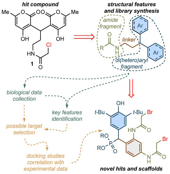
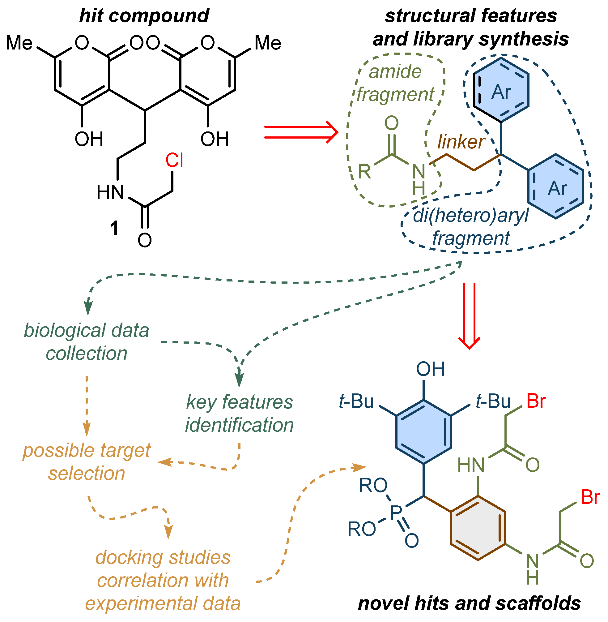
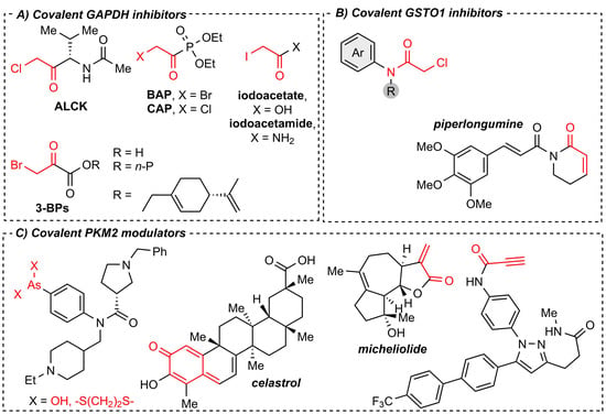
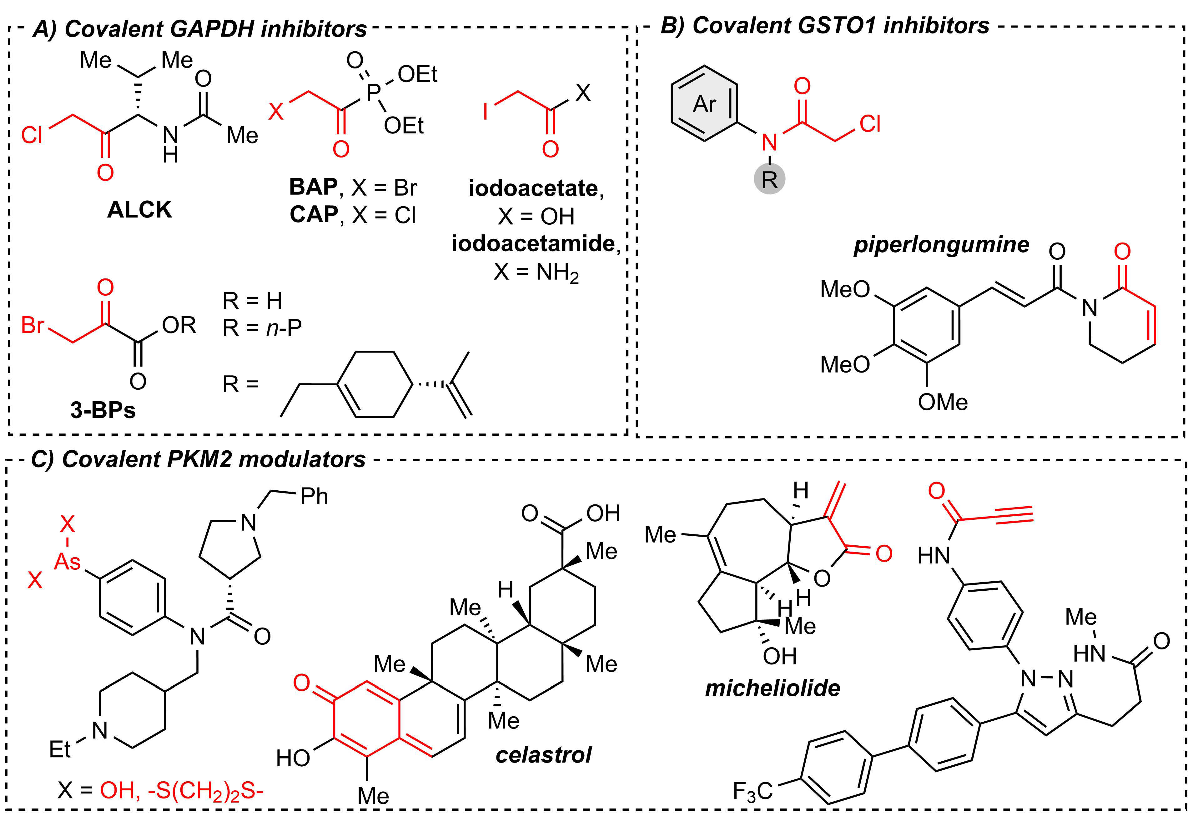
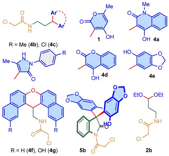
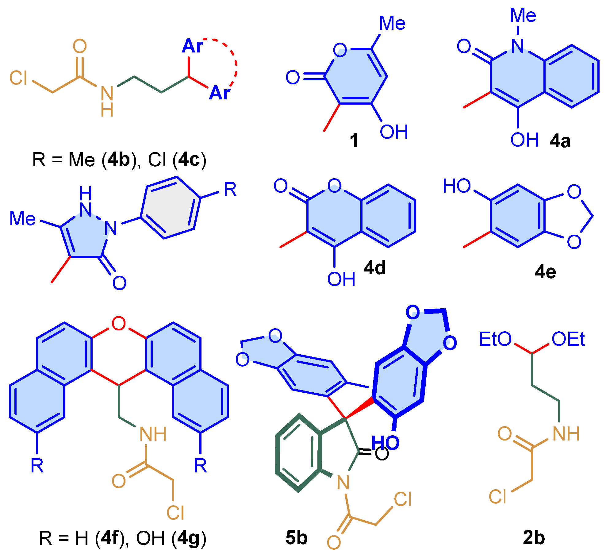
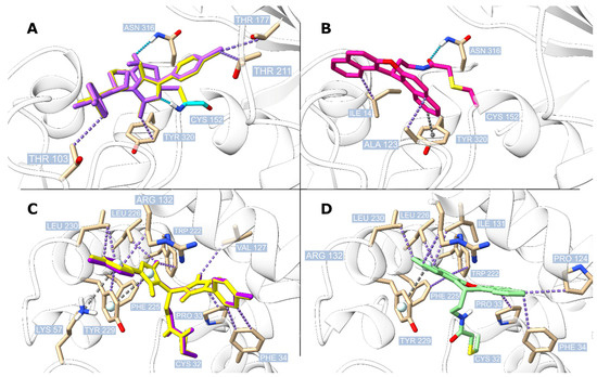
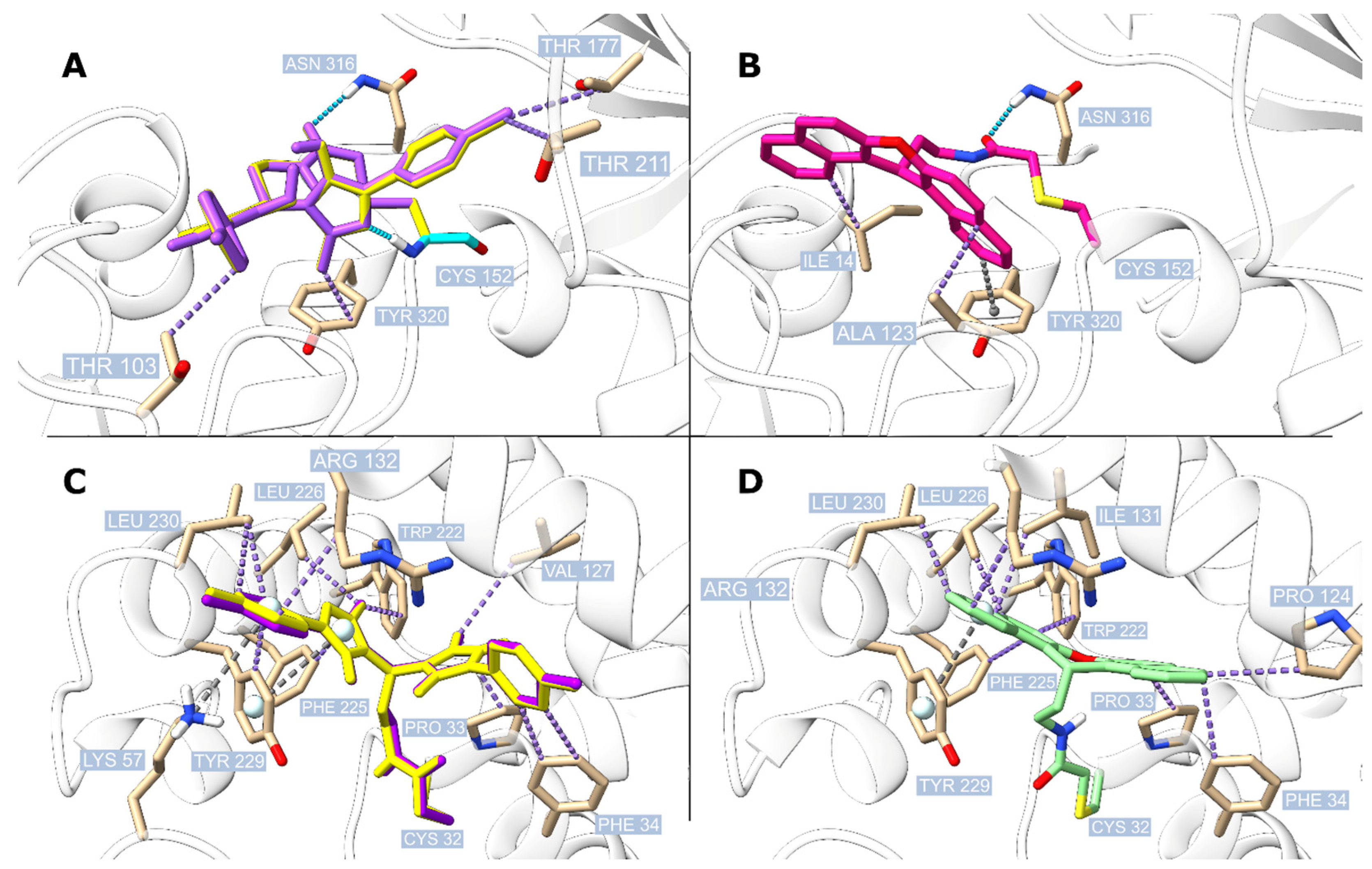
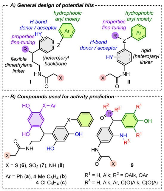
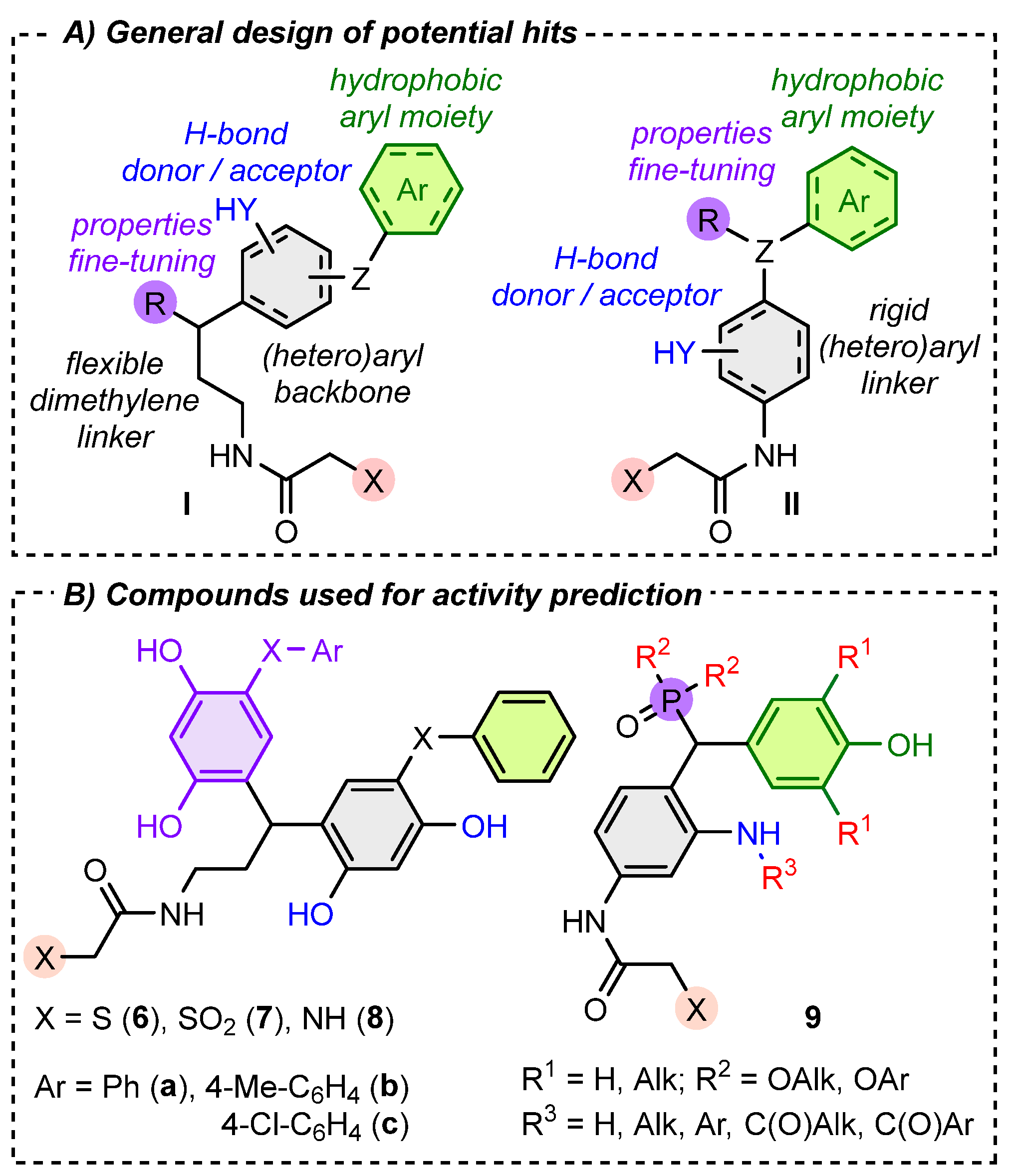
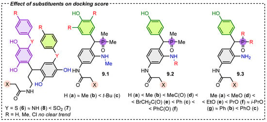
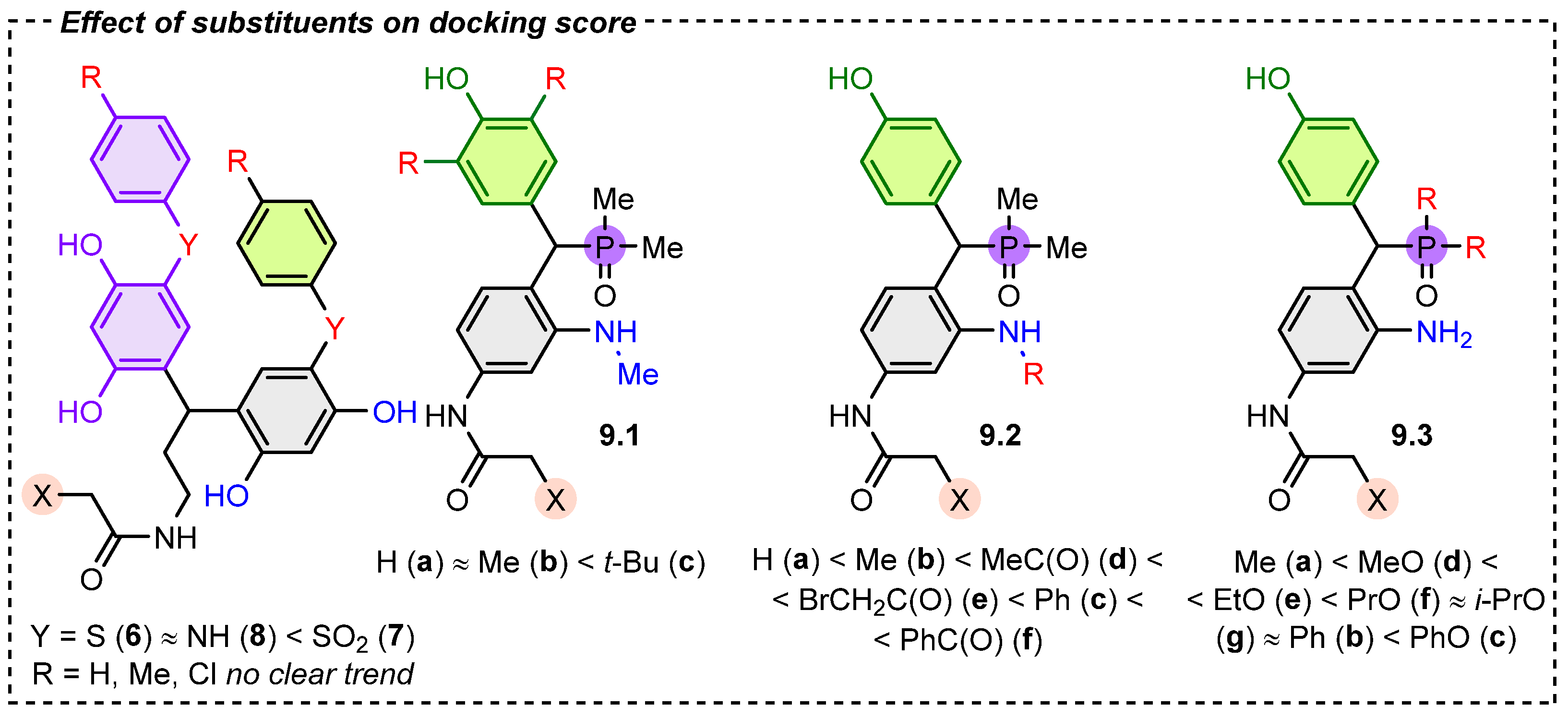
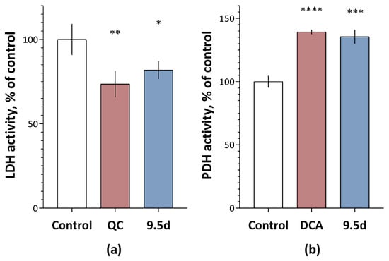
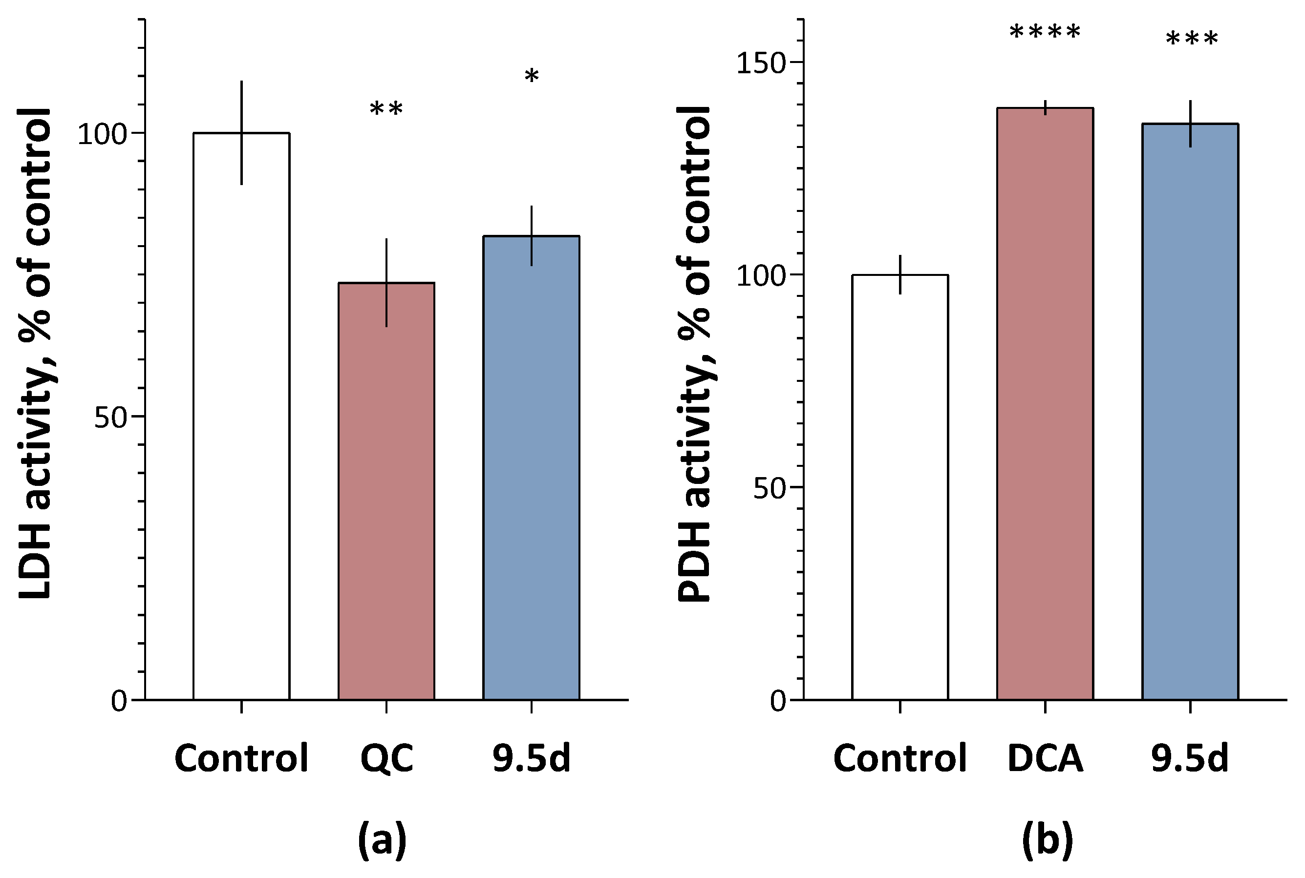
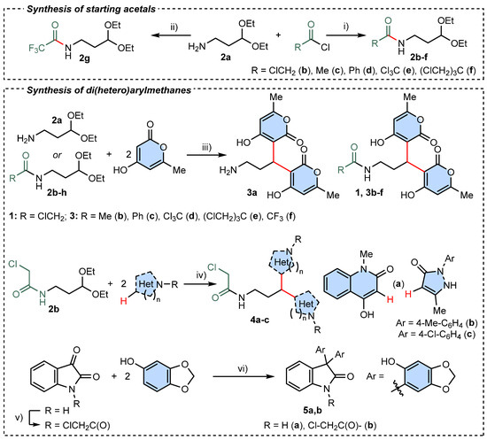
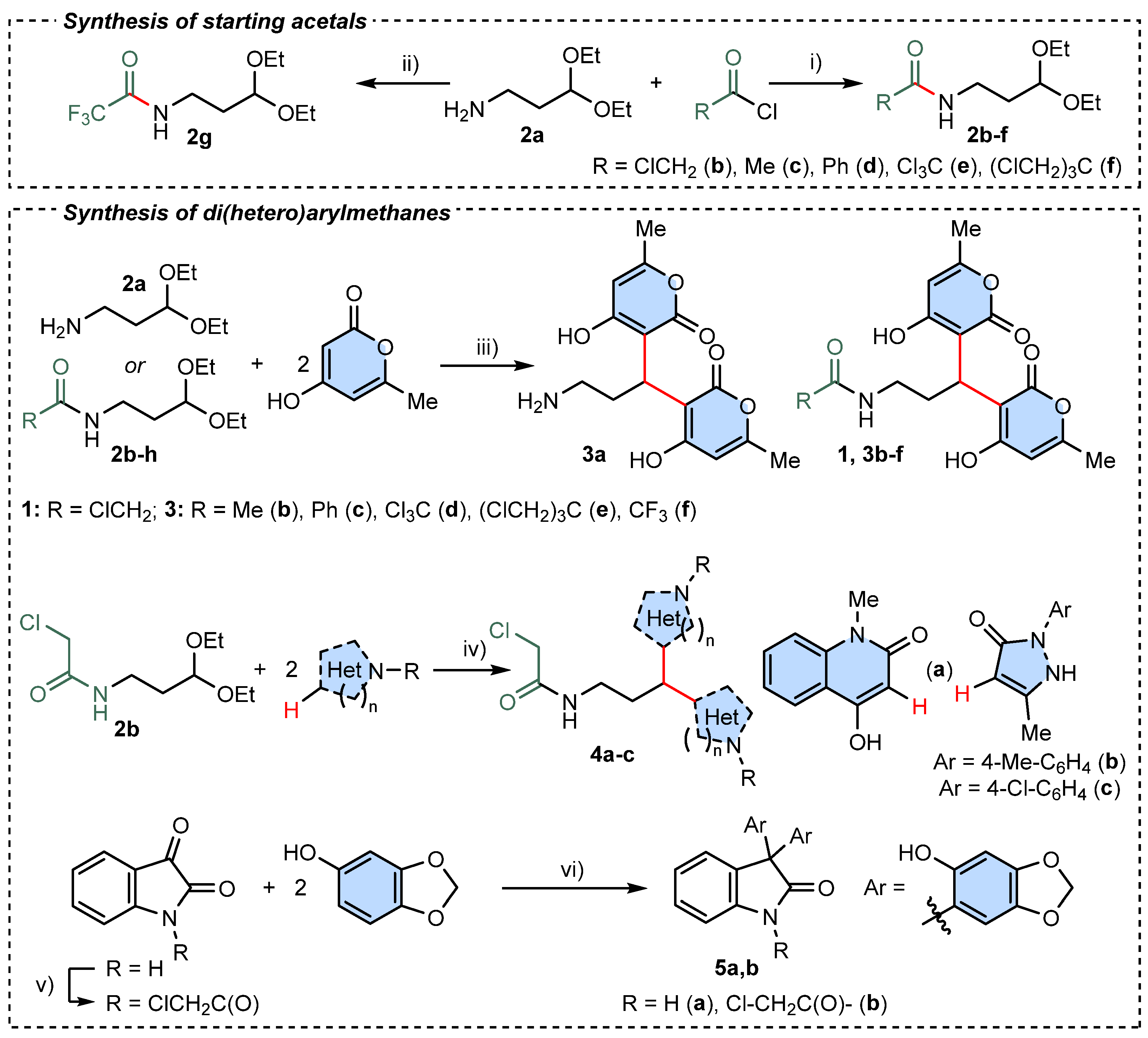
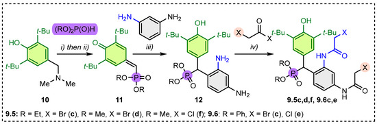
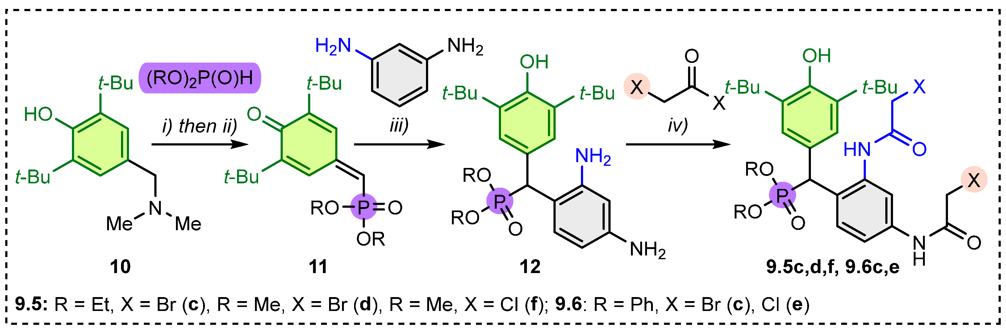

Disclaimer/Publisher’s Note: The statements, opinions and data contained in all publications are solely those of the individual author(s) and contributor(s) and not of MDPI and/or the editor(s). MDPI and/or the editor(s) disclaim responsibility for any injury to people or property resulting from any ideas, methods, instructions or products referred to in the content. |
© 2026 by the authors. Licensee MDPI, Basel, Switzerland. This article is an open access article distributed under the terms and conditions of the Creative Commons Attribution (CC BY) license.