


Abstract
Ovarian cancer (OC) is the most lethal gynecological malignancy, largely because non-specific symptoms contribute to advanced-stage diagnosis and poor prognosis, underscoring the need for improved non-invasive biomarkers. Circulating microRNAs (c-miRNAs) have emerged as promising candidates for OC detection, risk stratification, prognosis, and treatment monitoring. This systematic review, prospectively registered in PROSPERO (CRD420251230898), evaluated the diagnostic and prognostic performance of c-miRNAs and miRNA-based multivariate signatures. MEDLINE/PubMed, Scopus, and CENTRAL were searched from inception to 30 May 2026. Eligible studies assessed c-miRNAs in serum, plasma, whole blood, or extracellular vesicle fractions from women with confirmed or suspected OC including studies using prediagnostic samples from women subsequently diagnosed with OC. Records were screened, data extracted, and risk of bias evaluated according to the type of result using QUADAS-2 for diagnostic accuracy, QUIPS for prognostic-factor associations, and PROBAST for multivariable prediction models. Of 697 records identified, 91 publications met the inclusion criteria. Most studies used case–control or observational designs and analysed serum or plasma samples. Single c-miRNAs generally showed moderate diagnostic accuracy, whereas multivariate miRNA panels and multimodal models incorporating CA-125 or HE4 generally reported higher AUC values, including in some early-stage cohorts. Frequently reported candidates included members of the miR-200 family, miR-21, miR-1246, miR-125b, miR-145, miR-205, and miR-130a. Prognostic studies associated c-miRNAs with survival, recurrence, treatment response, and chemoresistance, although findings were more heterogeneous than diagnostic findings. Risk-of-bias concerns were common, particularly for patient selection in diagnostic studies, confounding in prognostic-factor analyses, and statistical analysis in prediction-model studies. Overall, c-miRNAs represent promising non-invasive biomarkers for OC, particularly when integrated into multivariate or multimodal models. However, methodological heterogeneity, small cohort sizes, limited external validation, and inconsistent reporting currently restrict clinical translation. Future studies should prioritize standardized pre-analytical and analytical workflows, adequately powered prospective cohorts, and independent multicenter validation.
1. Introduction
Ovarian cancer (OC) is one of the most lethal gynecological malignancies, with approximately 42% of patients surviving five years after diagnosis, largely because most cases are detected at an advanced stage [1]. Circulating CA-125 and human epididymis protein 4 (HE4) are established serum biomarkers for OC; however, their sensitivity is limited in early-stage disease, with CA-125 being elevated in only approximately 50% of patients with stage I OC [1]. Moreover, their diagnostic performance varies across populations, stages, assay thresholds, and comparator groups [2,3,4,5,6]. These limitations have prompted the search for novel, non-invasive biomarkers that may improve early detection and risk stratification.
Circulating microRNAs (c-miRNAs) are small non-coding RNAs, generally approximately 20–25 nucleotides long, that are detectable in blood and other biofluids. Their stability in circulation and disease-associated expression profiles have prompted their investigation as minimally invasive biomarkers for OC [1,7,8,9]. Dysregulated circulating miRNAs reported in OC include members of the miR-200 family, miR-21, miR-1246, and miR-150-5p [9,10,11]. However, the direction and statistical significance of these associations have not always been reproduced across cohorts or analytical approaches. For example, an EV-based study identified preliminary differences in miR-21-5p and miR-150-5p, but these findings were not confirmed during technical replication and validation [7]. Circulating miRNA profiles have also been associated with adverse clinicopathological features and prognosis [9,12]. In one case–control study, higher total plasma cf-miRNA concentrations were associated with distant metastasis and poorer overall survival and remained independently associated with overall survival in multivariable analysis [9]. Individual circulating miRNAs have also been associated with tumour characteristics [11], while other extracellular miRNA profiles have been associated with progression-free and overall survival [12].
Multivariable circulating-miRNA panels have demonstrated promising diagnostic performance, and some studies have reported greater accuracy for miRNA panels than for individual miRNAs [8,13]. Nevertheless, performance estimates vary substantially among cohorts and study designs. Kumar et al. reported an AUC of 0.952, with 90.5% sensitivity and 100% specificity, for a three-miRNA serum panel distinguishing patients with stage I–II epithelial ovarian cancer from healthy controls [13]. In contrast, the seven-miRNA panel developed by Gahlawat et al. achieved an AUC of 0.81 for early-stage ovarian cancer, which increased to 0.93 when the panel was combined with CA-125 [9]. Yokoi et al. reported an AUC of 1.00, 99% sensitivity, and 100% specificity for a ten-miRNA model in an independent validation cohort; the model classified 95.1% of stage I cases as positive, although a separate stage I-specific AUC was not reported [8]. In another independent validation set, a two-miRNA classifier achieved an AUC of 0.789 for stage I–II disease, whereas integration of the miRNAs with CA-125 and HE4 increased the AUC to 1.00, with 88.9% sensitivity and 100% specificity [2]. The OCaMIR signature achieved stage I AUCs of 0.90 and 0.86 in two retrospective serum cohorts and 0.81 in a prospective serum cohort; its AUC for all-stage disease in the prospective cohort was 0.92 [14]. Although these findings demonstrate the potential of multianalyte miRNA-based models, their clinical applicability requires confirmation through larger, independently conducted prospective studies.
Despite these promising findings, the clinical translation of circulating miRNA biomarkers remains limited. Previous systematic evidence has identified small study populations, inconsistent normalization strategies, differences in patient selection and control groups, and substantial methodological heterogeneity as major barriers to reproducibility and cross-study comparison [1]. Standardization of sample collection, extracellular-vesicle isolation, miRNA quantification, normalization, and analytical reporting is therefore required, particularly for extracellular-vesicle-based biomarkers [1,15]. Previous systematic reviews and meta-analyses have primarily examined the diagnostic performance of circulating miRNAs [1,16] or focused specifically on exosomal miRNAs [17]. Consequently, an updated synthesis encompassing diagnostic, prognostic, treatment-response, longitudinal-monitoring, and multivariable biomarker studies is warranted. Conducted within the framework of the DISARM project, a Horizon Europe co-funded initiative focused on improving ovarian cancer risk assessment and early detection, this systematic review evaluates the diagnostic and prognostic performance of circulating miRNAs and multivariable biomarker signatures. It also examines their integration with established protein biomarkers, including CA-125 and HE4, and critically assesses the methodological and validation requirements necessary for their translation into clinical practice.
2. Results
2.1. Study Selection Results
The literature search identified a total of 697 records, including 376 from Scopus, 311 from MEDLINE/PubMed, and 10 from CENTRAL. After removing 45 duplicate records, 652 reports were screened for eligibility based on their titles and abstracts. Of these, 544 records were excluded. The primary reasons for exclusion at this stage were: studies not focused on OC (n = 180), studies not investigating c-miRNAs (n = 140), studies evaluating non-miRNA biomarkers (n = 90), non-human studies (n = 60), review articles/editorials/conference abstracts (n = 50), and irrelevant topics (n = 24). A total of 108 reports were sought for retrieval. Two reports could not be obtained, leaving 106 reports for full-text assessment. Of these, 15 reports were excluded: ineligible publication type or companion report (n = 4), ineligible biospecimen (n = 3), non-miRNA biomarker (n = 2), technical or bioinformatic study (n = 5), and clinical outcome outside the selected scope (n = 1). A referenced list of all reports excluded following full-text assessment, together with the primary reason for exclusion, is provided in Supplementary Table S6, which separately identifies the two reports not retrieved. Ultimately, 91 publications met all eligibility criteria and were included in the systematic review. The complete study selection process is summarized in the PRISMA flow diagram (Figure 1). Reassessment during revision added 18 eligible publications among the reports originally sought [18,19,20,21,22,23,24,25,26,27,28,29,30,31,32,33,34,35].
Figure 1.
PRISMA flow diagram of study selection process. The diagram outlines the identification, screening, eligibility assessment, and inclusion of studies evaluating c-miRNAs as diagnostic and/or prognostic biomarkers in OC. A total of 697 records were retrieved from database searches, with 45 duplicates removed prior to screening. After title and abstract screening (n = 652), 544 records were excluded. Of 108 reports sought, two were not retrieved; 106 underwent full-text assessment and 15 were excluded. A final total of 91 publications were included in the systematic review.
2.2. Study Characteristics
A total of 91 publications were included in this systematic review. Their principal clinical and methodological characteristics are summarized in Figure 2A–H. Cohort-overlap assessment identified probable reuse among Kumar [13,36], Wang [37,38], Meng [25,39,40], and Zuberi [19,41,42] publications; the Zuberi reports had matching cohort sizes and stage distributions from the same institution. Ruan [27] reanalysed Fukagawa’s serum dataset [43]. Overlap remained unconfirmed between Márton [44] and Penyige [45], among the Istanbul reports [21,33,35,46], and between the Timofeeva reports [31,32]. These reports were retained for their distinct biomarkers, biospecimen fractions, or analyses, but were not treated as independent validation. Counts represent publications, not necessarily independent cohorts.
Figure 2.
Clinical and methodological characteristics of the 91 publications included in the systematic review. (A) Clinical purpose of the included studies, classified as diagnostic, prognostic, or combined diagnostic-and-prognostic investigations. (B) Distribution of studies by FIGO stage within the study populations. (C) Geographic distribution of the included studies. (D) Histological subtypes investigated, including mixed-histology OC cohorts, serous-predominant disease, high-grade serous OC, ovarian clear cell carcinoma, and other or unspecified histologies. (E) Study designs used across the included literature. (F) Distribution of studies according to cohort or sample size. (G) Main technological platforms used for circulating miRNA profiling and validation. (H) Biospecimen sources used for miRNA analysis, classified as serum, plasma, both serum and plasma, whole blood, or blood with an unspecified fraction. Numbers shown within each chart represent the number of studies in each category.
As shown in Figure 2A, 50 publications primarily investigated diagnostic applications of circulating miRNAs, 14 focused on prognostic or predictive outcomes, and 27 evaluated both diagnostic and prognostic applications. This distribution demonstrates that most of the available literature has concentrated on the potential use of circulating miRNAs for OC detection, although a substantial proportion of studies also examined their association with survival, recurrence, treatment response, or other clinical outcomes.
The distribution of disease stages is presented in Figure 2B. Most studies enrolled patients across mixed FIGO stages I–IV (n = 68). Six publications focused mainly on advanced-stage disease, whereas only two (n = 2) studies specifically concentrated on early-stage OC. Disease stage was not reported or was insufficiently described in fifteen publications. This predominance of mixed-stage and advanced-stage cohorts should be considered when interpreting reported diagnostic performance, particularly the applicability of candidate biomarkers to early disease detection.
The geographical distribution of the included studies is illustrated in Figure 2C. Most investigations were conducted in Asia (n = 55), followed by Europe (n = 20), North America (n = 6), Africa (n = 3), and Oceania (n = 2). Five studies involved multinational or multicontinental cohorts. The strong geographical concentration in Asia may limit the generalizability of some biomarker signatures, particularly where external validation across ethnically and geographically diverse populations was not performed.
As shown in Figure 2D, mixed-histology OC cohorts represented the largest category (n = 37). Twenty-two publications included serous-focused or serous-predominant cohorts, while 14 specifically investigated high-grade serous OC. Two studies focused exclusively on ovarian clear cell carcinoma, five investigated other specified histologies, and eleven did not clearly report histological subtype. The predominance of mixed epithelial and serous cohorts reflects the clinical frequency of epithelial and high-grade serous OCs but provides comparatively limited evidence for rarer histological subtypes.
The methodological designs of the included studies are summarized in Figure 2E. Case–control biomarker studies were the most common design (n = 45), followed by observational biomarker studies (n = 20). Eleven publications used distinct discovery and validation phases, five were prospective cohort or trial-based biomarker investigations, five were retrospective biomarker or dataset-based studies, and five were experimental or functional biomarker studies. The predominance of case–control designs may contribute to inflated estimates of diagnostic performance because patients with established OC are often compared with clearly defined healthy controls rather than clinically representative populations presenting with adnexal masses. Günel and Suryawanshi were classified as case–control studies because independence of discovery and confirmation samples was unclear or absent [21,22].
Study population sizes varied considerably, as shown in Figure 2F. Sixty-seven publications included fewer than 200 participants or samples, sixteen publications included between 200 and 499 participants, and eight involved at least 500 participants, samples, or multicentre validation datasets. The large proportion of relatively small studies indicates that many reported biomarker findings remain exploratory and require confirmation in adequately powered prospective cohorts.
The primary analytical platforms are summarized in Figure 2G: qRT-PCR (56 publications), NGS (16), hybridization microarray (15), NanoString (2), digital PCR (1), and FirePlex (1). PCR-based screening arrays were classified as qRT-PCR. The classification of each study, including whether it was a discovery or validation study, is shown in Supplementary Table S3.
The clinical blood matrices are summarized in Figure 2H: serum (58 publications), plasma (30), both serum and plasma (1), whole blood (1), and blood with an unspecified fraction (1). These mutually exclusive categories describe the clinical blood specimen; ancillary non-blood experiments do not create additional categories. Gumusoglu-Acar et al. [46] was coded as plasma because samples were collected in EDTA tubes, although the source inconsistently also uses “serum”. The same EDTA-based classification was applied to Günel et al. [35].
2.3. Diagnostic Performance of c-miRNAs in OC
The diagnostic performance of c-miRNAs in OC was evaluated across multiple studies using predominantly case–control and multi-cohort designs. The results in Table 1 show varying but generally moderate-to-good diagnostic accuracy.
Overall, single miRNA biomarkers demonstrated moderate diagnostic performance, with AUC values typically ranging from approximately 0.70 to 0.85. For example, miR-193a-5p and miR-205 showed modest discrimination between OC and controls, with reported AUCs of 0.708 and 0.715, respectively [3,47], while members of the miR-200 family and miR-1246 achieved comparatively higher accuracy with AUC values up to 0.89 for miR-1246 [48]. Serum miR-1246 demonstrated an AUC of 0.89, with 87% sensitivity and 77% specificity, for differentiating patients with high-grade serous ovarian carcinoma from healthy controls and was evaluated using separate training and validation cohorts [48]. These estimates should not be compared directly because the studies differed in histological composition, specimen type, comparator selection, analytical methods, and validation design. Although some individual c-miRNAs demonstrated promising diagnostic performance, many studies used modest sample sizes and lacked independent external validation. Larger prospective studies employing standardized methods and clinically relevant comparator groups are therefore required before individual c-miRNAs can be implemented in clinical practice.
Multivariable miRNA signatures often demonstrated higher apparent diagnostic performance than individual miRNAs, and several studies reported improved AUCs when multiple miRNAs were combined within the same cohort [13,36,49,50]. Across the included studies, several multi-miRNA signatures achieved AUCs above 0.85 [14,49,51,52,53], while selected miRNA or multianalyte models reported AUCs of approximately 0.95 or higher [4,8,11,13,36,50]. However, these estimates were obtained using different patient populations, comparator groups, analytical platforms, and validation strategies and therefore should not be compared directly. Notably, the ten-miRNA model developed by Yokoi et al. achieved an AUC of 1.00, with 99% sensitivity and 100% specificity, in an independent validation cohort [8]. Other computationally derived models also demonstrated high apparent diagnostic accuracy [52,54,55]. The modelling approaches employed across the included studies included logistic regression [13,36,50,55], random forests, artificial neural networks, and XGBoost [55], as well as neural-network classifiers [52] and relative-expression-ordering classifiers [54]. Collectively, these findings indicate that multivariable miRNA signatures are promising diagnostic tools; however, differences in model development, internal and external validation, and comparator selection limit direct comparisons and preclude firm conclusions regarding their relative clinical performance.
Combining circulating miRNAs with established protein biomarkers, particularly CA-125 and HE4, frequently improved diagnostic performance within individual studies. In an independent validation cohort, a model combining miR-320b and miR-141-3p with CA-125 and HE4 achieved an AUC of 0.989, compared with 0.723 for the miRNA-only classifier; among patients with stage I–II disease, the corresponding AUCs were 1.00 and 0.789, respectively [2]. Similarly, a model combining miR-193a-5p, CA-125, and HE4 achieved an AUC of 0.996 for distinguishing epithelial ovarian cancer from healthy controls, although it was evaluated in a single cohort without independent validation [3]. A multimodal model incorporating serum miRNAs, protein biomarkers, and clinical variables achieved an AUC of 0.95 (95% CI: 0.90–0.98), with 92% sensitivity at 80% specificity, in an external validation cohort [4].
A subset of studies specifically evaluated early-stage OC detection, which remains a major unmet clinical need. Reported sensitivities for early-stage disease varied across studies and models. A 14-miRNA neural-network model achieved 73% sensitivity and 91% specificity in its validation subset [51], whereas a miRNA–protein model achieved 88.9% sensitivity and 100% specificity [2]. The multimodal model developed by Webber et al. achieved 80% sensitivity for early-stage cancers in external validation [4]. A combination of plasma exosomal miR-223 and CA-125 distinguished stage I–II epithelial ovarian cancer from healthy controls with an AUC of 0.944 (95% CI: 0.899–0.990), 97.8% sensitivity, and 88.5% specificity [56]. These estimates were derived using different comparator groups and validation approaches and therefore are not directly comparable.
Extracellular-vesicle-derived miRNA signatures also demonstrated promising diagnostic performance. The eight-miRNA OCEM signature achieved an AUC of 0.9762 in the plasma training set and 0.9375 in the internal plasma validation set for distinguishing OC from benign disease [53]. Although miR-1246 and miR-1290 were elevated in early-stage samples, the study did not report a separate early-stage-specific AUC for the OCEM signature [53].
Substantial heterogeneity was evident across the included studies in study design, cohort size, specimen source, comparator composition, analytical platform, and validation strategy. Some exploratory studies did not report complete diagnostic performance measures. For example, Penyige et al. identified differentially expressed plasma miRNAs but did not report ROC AUC, sensitivity, or specificity [45]. In contrast, the Bayesian analysis by Wiczling et al. reported limited cross-validated discrimination, with AUCs of 0.65 for three selected individual miRNAs (reported credible intervals spanning 0.62–0.67), an AUC of 0.65 for a three-miRNA combination, and an AUC of 0.72 when a larger miRNA subset was evaluated [57].
Overall, the descriptive findings suggest that multi-miRNA and multimodal models frequently achieved higher apparent diagnostic accuracy than individual miRNAs, including in some early-stage analyses. However, no quantitative meta-analysis or formal cross-study comparison of single-miRNA and multivariable models was performed, and the heterogeneity of the study populations, comparator groups, analytical methods, and validation designs limits conclusions regarding their relative performance.
Table 1.
Summary of diagnostic-focused c-miRNA studies included in the systematic review.
2.4. Prognostic Value of c-miRNAs
The prognostic relevance of c-miRNAs in OC was evaluated across a range of observational and cohort-based studies with varying clinical contexts and patient populations. As shown in Table 2, c-miRNAs were associated with key clinical outcomes, including OS, PFS, treatment response, and disease recurrence.
Several individual circulating miRNAs were associated with clinical outcomes. Higher serum miR-183 expression was associated with shorter OS and remained independently associated with OS in a multivariable Cox model [69]. Conversely, lower miR-148a expression was associated with shorter OS and remained an independent adverse prognostic factor after adjustment for clinicopathological variables [70]. Higher serum miR-21 and lower serum miR-145 levels were observed among patients with poorer chemotherapy response and among those who died within three years; however, this study evaluated three-year vital status using logistic regression rather than time-to-event survival analysis [71]. Higher serum miR-135a-3p expression was associated with longer PFS in an unadjusted Kaplan–Meier analysis, although an HR and 95% CI were not reported [43].
Evidence concerning multimiRNA prognostic signatures was more limited. Pal et al. reported that five circulating miRNAs—miR-27a, miR-182, miR-199a, miR-214, and miR-591—differed across chemotherapy-response groups and were associated with OS in unadjusted analyses [72]. However, the reported individual miRNA associations were not statistically significant in the multivariable Cox models; the adjusted covariates were not specified, and the signature was not evaluated in an independent validation cohort [72]. Therefore, the available evidence does not establish that multimiRNA signatures provide greater prognostic accuracy than individual circulating miRNAs.
Longitudinal measurements provided potentially useful information in some studies. Time-dependent plasma miR-200b levels were independently associated with PFS, whereas baseline miR-200b was not significantly associated with PFS, suggesting that serial measurements may provide information beyond a single baseline assessment [73]. In ovarian clear-cell carcinoma, serum miR-130a decreased following surgery and increased in all four patients who subsequently developed recurrence, sometimes before CA-125 increased [74]. However, the recurrence analysis was descriptive, involved only four recurrent cases, and did not report an HR, OR, or formal recurrence-prediction statistic.
Findings were not consistent across studies. In a first-line-treatment cohort, the kinetics of an 11-miRNA panel were heterogeneous and generally provided less effective treatment monitoring than CA-125, although selected miRNAs were associated with OS or PFS in univariable analyses [75]. Overall, c-miRNAs have potential as non-invasive prognostic and treatment-monitoring biomarkers in OC, particularly when evaluated longitudinally. Nevertheless, the evidence is limited by modest sample sizes, inconsistent outcome definitions, incomplete confounder adjustment, limited external validation, and the frequent reliance on univariable or exploratory analyses. Large, well-characterized prospective cohorts with prespecified models and independent validation are required before clinical utility can be established.
Table 2.
Prognostic and treatment-response studies of circulating miRNAs in ovarian cancer.
2.5. Dual Diagnostic and Prognostic Performance of Circulating miRNAs in OC
A subset of studies (n = 27) evaluated c-miRNAs with both diagnostic and prognostic relevance in OC (Table 3). These studies predominantly analysed serum or plasma samples across multiple FIGO stages and investigated both individual miRNAs and multi-miRNA signatures, with several incorporating established biomarkers such as CA-125.
Individual miRNAs demonstrated variable diagnostic performance, with reported AUCs generally ranging from approximately 0.70 to 0.93. Serum miR-145 distinguished patients with epithelial ovarian cancer from healthy controls with an AUC of 0.928 (95% CI: 0.86–0.95), whereas plasma miR-106a achieved an AUC of 0.879 (95% CI: 0.830–0.928) [41,79]. Serum miR-200b demonstrated an AUC of 0.743 when evaluated individually; its combination with CA-125 achieved 100% sensitivity and 93.1% specificity, although the AUC of the combined model was not reported [80]. Serum exosomal miR-484 achieved an AUC of 0.821, which increased to 0.912 when miR-484 was combined with CA-125 [81].
Selected multi-miRNA and multimodal models achieved high diagnostic performance, particularly when miRNAs were combined with CA-125 or evaluated using multivariable modelling. A seven-miRNA panel combined with CA-125 achieved an AUC of 0.97 overall and 0.93 for early-stage disease [9]. A model incorporating CA-125, miR-375, and miR-210 achieved an AUC of 0.984 [82], while a seven-miRNA small-extracellular-vesicle signature achieved AUCs of 0.913, 0.973, and 0.924 in its training, testing, and validation cohorts, respectively [83]. However, other multi-miRNA signatures in Table 3 demonstrated only moderate performance, and differences in cohort composition and validation design prevent direct comparisons with individual miRNAs.
Additional eligible reports provided heterogeneous evidence. Xu et al. reported a serum miR-21 survival association, but the reported ΔCt hazard-ratio direction was inconsistent with the narrative [18]. Zuberi et al. reported a serum miR-125b diagnostic AUC of 0.728 (95% CI 0.64–0.81), with no significant overall-survival association [19]. Zhou et al. evaluated a four-sEV-miRNA/CA-125 model for surgical residual disease, achieving an AUC of 0.903 (95% CI 0.846–0.945) in a separate validation set of 154 advanced-HGSOC patients [20]. This surgical-outcome AUC was excluded from the diagnostic comparison in Figure 8.
Günel et al. reported an AUC of 0.70 for detecting established recurrent EOC, without demonstrating prediction of future recurrence [21]. Suryawanshi et al. evaluated plasma classifiers for endometriosis-associated and serous OC; the expanded cohort included the discovery samples, so the findings do not constitute independent validation [22].
Several of these miRNAs were also associated with clinical outcomes. High serum miR-221 was reported as an independent adverse prognostic factor for OS [84], while higher exosomal miR-200b and miR-200c levels were associated with poorer OS, and higher miR-200c was additionally associated with shorter disease-free survival [39]. Lower miR-145 expression was associated with advanced disease and poorer survival, although an adjusted survival estimate was not reported [41]. Lower miR-125b expression was associated with adverse clinicopathological characteristics, poorer prognosis, and chemotherapy resistance [85]. Conversely, higher circulating miR-125b was associated with longer PFS and a favourable OS trend in another cohort [86]. Higher miR-26b expression was associated with a higher three-year survival rate, whereas lower miR-21 expression was associated with better survival; these findings were based on unadjusted survival comparisons without reported HRs [87].
Some studies also examined treatment-related or recurrence outcomes. A model combining miR-34a-5p with CA-125 was associated with the likelihood of optimal surgical cytoreduction, although this represented prediction of a surgical outcome rather than a time-to-event prognostic analysis [82]. Low serum exosomal miR-34a was associated with lymph-node metastasis and disease recurrence, but the study did not evaluate serial miRNA measurements for longitudinal monitoring [88].
Collectively, these findings suggest that c-miRNAs may have complementary diagnostic, prognostic, treatment-predictive, and recurrence-related applications in OC. The highest reported diagnostic AUCs were observed in selected multi-miRNA or multimodal models; however, this apparent advantage was not consistent across all studies and was not assessed through a formal pooled comparison. Furthermore, most prognostic findings lacked independent validation. To illustrate the overlap between the different clinical applications of c-miRNAs, a Venn diagram was constructed (Figure 3).
Figure 3.
Venn diagram of circulating microRNAs with diagnostic and prognostic relevance in OC. The diagram illustrates the overlap of circulating microRNAs identified across studies as having diagnostic and/or prognostic utility in OC. The left circle represents miRNAs reported primarily in diagnostic studies, while the right circle represents those associated with prognostic outcomes, including overall survival, progression-free survival, treatment response, or recurrence. The overlapping region highlights miRNAs with dual diagnostic and prognostic relevance. Notably, several miRNAs, including members of the miR-200 family, as well as miR-21, miR-125b, and miR-145, were consistently reported across both domains, suggesting potential as multifunctional biomarkers. This is a selected-marker overview, not an exhaustive inventory; surgical-outcome model components are reported separately in Table 2.
Table 3.
Summary of c-miRNAs with combined diagnostic and prognostic utility in OC.
2.6. Integrated Evidence Map of c-miRNA Biomarkers
An evidence gap map was created to show how the included publications were distributed across the main circulating miRNA biomarker categories and their clinical applications in OC (Figure 4). Study-level classifications used to generate the map are provided in Supplementary Table S3.
Figure 4.
Descriptive evidence map of circulating-miRNA research in ovarian cancer. Numbers indicate publications contributing an eligible result to each cell; blue bubble area is proportional to the count, and grey markers indicate zero. Publications can contribute to multiple cells. Counts represent coverage, not evidence strength, risk of bias or independent validation. Early-stage performance requires a stage-specific cancer-versus-noncancer analysis. Prediagnostic investigation includes a case-only time-to-diagnosis study. Operational definitions, supporting references and study-level qualifications are provided in Supplementary Table S3.
Most of the evidence focused on diagnostic applications, particularly studies evaluating individual or total circulating miRNAs, multi-miRNA combinations, and miRNA–protein combinations. Fewer studies specifically investigated diagnostic performance in early-stage OC, although multi-miRNA combinations were the most widely studied approach in this setting. Prognostic studies mainly focused on individual or total circulating miRNAs. In contrast, relatively few studies evaluated EV- or exosome-derived miRNAs, algorithm-based models, or combined miRNA–protein approaches for prognostic purposes. Clear evidence gaps were identified in serial monitoring for recurrence and in the analysis of samples collected before diagnosis. Studies related to treatment outcomes also focused mainly on individual circulating miRNAs.
Overall, the evidence was unevenly distributed across clinical applications. Longitudinal monitoring, prediagnostic evaluation, and validation in clinically relevant patient populations therefore remain important priorities for future research.
The technical landscape of the included studies reveals significant methodological variations across biofluid sources, analytical platforms, and data processing standards. These findings highlight a critical lack of standardization within the field, which may impede the clinical reproducibility of c-miRNA biomarkers in OC (Table 4).
Table 4.
Methodological characteristics of the studies included in the systematic review. Percentages use 91 publications.
2.7. Risk of Bias
Risk of bias was assessed at the level of the eligible result because several studies contributed diagnostic-accuracy, prognostic-factor, and/or prediction-model evidence and therefore appeared in more than one assessment. In total, 132 result-level assessments are tabulated. Domain judgments and supporting notes are provided in Supplementary Table S4.
QUADAS-2 was applied to 34 diagnostic-accuracy publications. Twenty-four were judged at high overall risk of bias and 10 at unclear risk; none was judged low overall. The principal concern was patient selection, particularly two-gate or case–control sampling with separately recruited cancer and control groups. Index-test conduct and flow and timing were frequently unclear because threshold prespecification, blinding, participant exclusions, or the interval between index and reference assessments were incompletely reported (Figure 5). Reports contributing expression comparisons without eligible diagnostic-accuracy estimates were retained in the qualitative synthesis but were not assigned QUADAS-2 assessments.
Figure 5.
Risk-of-bias assessment using QUADAS-2. Traffic-light plot presenting domain-level and overall risk-of-bias judgments, together with applicability concerns, for 34 diagnostic-accuracy publications [3,6,10,19,21,24,25,29,35,39,41,42,44,46,47,48,56,57,58,60,62,66,67,73,79,80,81,84,85,86,87,89,90,92]. Green indicates low risk or low applicability concern, yellow indicates unclear risk or concern, and red indicates high risk or high applicability concern. The Meng et al. (2016) reports are distinguished by their exosomal [39] and cell-free [25] miRNA measurements; the Zuberi et al. (2016) reports are distinguished by their miR-199a [42] and miR-125b [19] biomarkers.
QUIPS was applied to 33 prognostic-factor publications. Because QUIPS does not prescribe a single overall study-level rating, findings are reported by domain. High risk was most frequent for study confounding and statistical analysis and reporting (22/33 publications in each domain), whereas outcome measurement was judged low risk in 31 publications and moderate risk in two. Study participation, attrition, and prognostic-factor measurement were most commonly judged moderate risk because representativeness, missing data, normalization, blinding, or cut-point prespecification were incompletely reported (Figure 6).
Figure 6.
Risk-of-bias assessment using QUIPS. Traffic-light plot presenting domain-level judgments for 33 prognostic-factor assessments [9,12,18,19,23,30,39,40,41,42,43,69,70,71,72,73,75,76,77,79,80,81,82,83,84,85,86,87,88,89,92,93,95]. Green indicates low risk of bias, yellow indicates moderate risk, and red indicates high risk. The Zuberi et al. (2016) reports are distinguished by their miR-199a [42] and miR-125b [19] biomarkers.
PROBAST was applied to 65 multivariable diagnostic or prognostic model evaluations. Fifty-eight evaluations were judged at high overall risk of bias and seven at unclear risk; none was judged low overall. The Analysis domain was the main driver of high-risk judgments because of small development samples, data-driven predictor selection, limited internal or external validation, incomplete handling of missing data, absent calibration, and/or incomplete model specification. Statistical predictor selection was assigned to the Analysis domain and was not double-counted in the Predictors domain. Applicability was judged separately (Figure 7).
Figure 7.
PROBAST assessments of 65 multivariable models [2,3,4,5,6,8,9,10,11,12,13,14,20,22,25,29,31,32,36,37,38,39,40,47,49,50,51,52,53,54,55,56,57,58,59,65,68,71,78,80,81,82,83,85,87,92,93,95]. Green: low risk/concern; yellow: unclear; red: high. Risk of bias and applicability are assessed separately. Separate models from one publication appear as separate entries; rationales are provided in Supplementary Table S4. The Meng et al. (2016) reports are distinguished by their exosomal [39] and cell-free [25] miRNA measurements.
3. Discussion
This systematic review provides an updated and comprehensive synthesis of the current evidence on c-miRNAs and multivariate biomarker signatures in OC, integrating findings from 91 publications across diagnostic, prognostic, and multimodal applications. Overall, our findings suggest that c-miRNAs represent a promising class of non-invasive biomarkers, with multivariate and multimodal approaches generally showing higher reported AUC values than single miRNAs, particularly in early-stage disease detection and prognostic stratification.
3.1. Diagnostic Performance and Comparison with Existing Evidence
Our findings demonstrate that individual circulating miRNAs (c-miRNAs) generally exhibit moderate diagnostic accuracy, whereas multi-miRNA panels and multimodal biomarker signatures generally reported higher apparent performance, frequently exceeding AUC values of 0.90 [2,8,55]. These observations are consistent with previous systematic reviews and meta-analyses reporting superior diagnostic performance for miRNA panels compared with individual biomarkers [16]. However, the improved performance of multi-marker signatures is biologically plausible rather than merely additive. OC is a highly heterogeneous disease involving multiple dysregulated molecular pathways, including cell proliferation, apoptosis, angiogenesis, immune modulation, epithelial–mesenchymal transition, and metastasis. Individual miRNAs typically regulate only a subset of these processes, whereas combining multiple miRNAs allows a biomarker panel to capture a broader spectrum of tumour biology, thereby reducing the impact of biological variability between patients and improving diagnostic robustness.
Our review further indicates that integrating circulating miRNAs with established protein biomarkers, particularly CA-125 and HE4, produced some of the highest reported diagnostic-performance estimates. In an independent validation cohort, a model combining miR-320b and miR-141-3p with CA-125 and HE4 achieved an AUC of 0.989 overall and 1.00 for stage I–II disease [2]. A model combining miR-193a-5p with CA-125 and HE4 achieved an AUC of 0.996, although it was evaluated without independent validation [3]. Other studies also reported improved diagnostic performance after combining circulating miRNAs with CA-125 and/or HE4 [5,6,47]. These findings suggest that miRNAs and protein biomarkers may provide complementary discriminatory information. However, the included studies did not directly establish the biological mechanisms responsible for the observed improvements. Multimodal models may partly compensate for the limited sensitivity of CA-125 in early-stage disease and its imperfect specificity when distinguishing OC from benign gynecological conditions [1,96]. Nevertheless, the very high performance estimates should be interpreted cautiously because several models were developed in relatively small case–control cohorts and lacked independent external validation.
Compared with earlier systematic reviews and meta-analyses [1,16], our review includes more recent studies employing high-throughput miRNA profiling and multivariable computational methods to develop diagnostic models [8,36,54,55,65]. These methods included logistic regression, neural networks, relative-expression-ordering classifiers, random forests, artificial neural networks, XGBoost, and other machine-learning approaches. Although several models reported very high apparent discrimination, with some AUCs approaching 1.00 [8,36,55], these findings should be interpreted cautiously. Some studies used relatively small or highly selected case–control cohorts and compared patients with OC against healthy controls, potentially creating favourable conditions for inflated estimates of diagnostic performance. Machine-learning and other multivariable models developed using limited samples and numerous candidate predictors are particularly vulnerable to overfitting, whereby a model captures cohort-specific patterns rather than reproducible disease-associated signals [97]. This concern is consistent with our PROBAST assessment, in which 58 of 65 model evaluations were judged to have a high overall risk of bias. Furthermore, independent external validation was uncommon, limiting confidence that the reported performance would generalize to broader clinical populations, including patients with benign adnexal disease. Exceptionally high AUC values may therefore partly reflect model-development procedures, selected comparator groups, or other methodological characteristics rather than performance achievable in routine clinical practice.
A previous pooled analysis reported a sensitivity of 76%, specificity of 81%, and summary ROC AUC of 0.85 for circulating miRNAs in OC [16]. A more recent meta-analysis focused on exosomal miRNAs reported a pooled sensitivity of 82%, specificity of 81%, and summary ROC AUC of 0.88 [17]. Several multivariable models included in the present review reported study-specific AUCs above 0.90. However, these AUCs should not be directly compared numerically with pooled sensitivity or specificity estimates, and comparisons with summary ROC estimates remain descriptive because of differences in study populations, specimens, comparator groups, thresholds, and validation designs. The high performance reported by some recent models may reflect advances in biomarker discovery, sequencing technologies, and multivariable analytical methods, but it may also partly arise from study design and patient selection. In particular, case–control studies comparing patients with established OC against healthy controls may produce more favourable estimates than studies involving women with benign adnexal masses or indeterminate pelvic lesions. Such comparator-related spectrum and selection effects limit applicability to routine diagnostic populations [97,98]. Consequently, clinical utility cannot be established until these models are evaluated in prospective, multicentre studies involving representative screening or symptomatic referral populations.
A particular strength of this review is its detailed evaluation of early-stage OC detection, for which improvements in diagnostic performance could be clinically important. Although only two included studies primarily recruited patients with early-stage disease, several others reported stage-specific subgroup analyses. Yokoi et al. reported that their ten-miRNA model classified 95.1% of stage I cases as positive, although a separate stage I-specific AUC was not provided [8]. Webber et al. reported 80% sensitivity for early-stage cancers in an external validation cohort [4]. Yang et al. reported that plasma exosomal miR-223 combined with CA-125 distinguished stage I–II epithelial ovarian cancer from healthy controls with an AUC of 0.944 (95% CI: 0.899–0.990), 97.8% sensitivity, and 88.5% specificity [56]. These findings suggest that multianalyte approaches warrant further evaluation for early-stage detection. Nevertheless, early-stage sample sizes were frequently limited, mixed-stage cohorts predominated, and some analyses used healthy individuals rather than clinically relevant benign or symptomatic controls. These limitations reduce confidence in the generalizability of the reported performance and highlight the need for prospective validation in screening or symptomatic referral populations before clinical implementation.
Finally, the evidence map (Figure 4) highlights the concentration of research on diagnosis and the limited coverage of serial recurrence monitoring. Prospective validation and assessment of clinical utility remain priorities.
This imbalance suggests that future research should move beyond biomarker discovery toward prospective validation, assessment of clinical utility, and investigation of how circulating miRNAs can inform disease monitoring, treatment response, and patient prognosis.
The distribution of reported diagnostic AUCs according to biomarker-integration strategy is summarized descriptively in Figure 8. Study-level representative AUCs ranged from 0.540 to 0.933 for individual circulating miRNAs and from 0.685 to 1.00 for multi-miRNA panels or signatures. The substantial overlap between these categories indicates that multi-miRNA panels did not uniformly outperform individual miRNAs. In contrast, the representative AUCs included in the miRNA plus CA-125 and/or HE4 category ranged from 0.900 to 0.996, while those of the multimodal models ranged from 0.950 to 0.999. These distributions suggest a pattern of higher reported diagnostic performance with greater biomarker integration, particularly for models incorporating protein biomarkers or additional clinical variables. However, Figure 8 represents an unweighted descriptive summary rather than a meta-analysis: the plotted points are arithmetic midpoints of the observed minimum-to-maximum ranges and do not represent pooled or average effect estimates. Moreover, the categories differed in the contributing studies, populations, comparator groups, sample sizes, and validation strategies. Exceptionally high AUCs should therefore be interpreted cautiously because several were derived from selected case–control cohorts, used healthy controls, or lacked independent external validation, potentially leading to overestimation of performance in routine clinical populations.
Figure 8.
Reported diagnostic performance according to biomarker integration strategy. Diagnostic AUC values were grouped into four predefined categories: individual circulating miRNAs, multi-miRNA panels/signatures, miRNA models combined with CA-125 and/or HE4, and multimodal models incorporating miRNAs with additional protein or clinical variables. Each publication contributed a maximum of one study-level representative AUC per category. Validation or test-set estimates were prioritized over training or discovery estimates; when multiple eligible AUCs were reported within the same study and category, the median was used. The vertical bars represent the observed minimum-to-maximum range of the study-level representative AUCs within each category, and the plotted point represents the arithmetic midpoint of that range, calculated as (minimum + maximum)/2. No weighting by sample size, study quality, or precision was applied. The underlying raw AUC values and study-level calculations are provided in Supplementary Table S5.
3.2. Prognostic Value and Clinical Implications
Across the included studies, associations between circulating miRNAs and clinical outcomes were dependent on the specific miRNA, biospecimen, analytical method, and clinical context. Higher circulating miR-21 levels were associated with poorer chemotherapy response or survival in some cohorts [71,87], whereas lower miR-145 expression was associated with adverse outcomes [41,71]. Lower miR-125b expression was associated with poorer prognosis and chemotherapy resistance in one study [85], while another reported that higher miR-125b levels were associated with longer PFS and a favourable OS trend [86]. Findings for the miR-200 family were less consistent: higher exosomal miR-200b and miR-200c levels were associated with poorer survival in one study [39], whereas lower circulating miR-200c and miR-141 levels were associated with poorer outcomes in another [89]. These differences indicate that the prognostic direction of a miRNA should not be generalized across specimen types, patient populations, or analytical methods.
Longitudinal profiling and multivariable miRNA models may nevertheless provide clinically relevant prognostic information. Serial changes in plasma miR-200b were associated with PFS, whereas baseline miR-200b was not significantly prognostic [73]. Serum miR-130a decreased following surgery and increased in four patients who subsequently developed recurrence, sometimes before an increase in CA-125 [74]. Other studies examined miRNA kinetics during first-line treatment or after experimental therapy [75,76], while multivariable prognostic indices and treatment-specific miRNA models were evaluated in separate cohorts [12,77,78]. However, these approaches differed substantially in design, outcome definition, modelling strategy, and validation, and their incremental value over established clinical predictors remains uncertain.
Prognostic biomarker research was more heterogeneous than diagnostic research in the included literature. The investigated endpoints included OS, PFS, disease-free or recurrence-related outcomes, chemotherapy response, surgical outcome, and platinum resistance, which represent distinct clinical events and should not be considered interchangeable. Follow-up duration and the number of outcome events also varied considerably. Patient populations differed in disease stage, histological subtype, treatment regimen, and residual disease following surgery, while blood samples were collected at different time points, including before treatment, after surgery, during chemotherapy, and throughout follow-up. These differences limit direct comparisons among studies and are likely to contribute to the inconsistent prognostic findings.
Several studies evaluated serial changes in circulating miRNAs during treatment or follow-up [73,74,75,76]. These studies suggest that circulating miRNA levels may change during surgery, chemotherapy, disease progression, or recurrence and that serial measurements may provide information not captured by a single baseline sample. Nevertheless, the evidence was mixed. In the study by Robelin et al., the kinetics of 11 circulating miRNAs were heterogeneous and generally less informative for treatment monitoring than CA-125 [75]. Therefore, circulating miRNAs should currently be considered potential complementary biomarkers rather than replacements for CA-125.
The available evidence remains insufficient to support the routine clinical implementation of circulating miRNAs as prognostic biomarkers. Many studies used modest, single-centre cohorts, investigated different miRNA candidates, applied heterogeneous analytical methods, and lacked independent external validation. Several prognostic associations were based only on Kaplan–Meier or univariable analyses, while some multivariable studies did not clearly report their covariates or adjusted effect estimates [43,71,72,74,75]. Relatively few studies demonstrated that circulating miRNAs provided prognostic information independent of established variables such as disease stage, histological subtype, residual disease, and treatment regimen [12,39,69,70,73,77,79,84]. Evidence that miRNAs add clinically meaningful prognostic value to existing clinicopathological models therefore remains limited.
Overall, circulating miRNAs show potential for prognostic stratification, treatment-response assessment, and longitudinal disease monitoring. Future research should prioritize large prospective multicentre cohorts, standardized outcome definitions, harmonized sampling and analytical protocols, prespecified prognostic models, adequate numbers of outcome events, serial specimen collection, and independent external validation. These studies should also evaluate whether adding circulating miRNAs to established clinical models improves patient classification and leads to better treatment or follow-up decisions.
3.3. Dual Biomarker Function and Biological Relevance
Several circulating miRNAs were investigated across both diagnostic and prognostic or treatment-response studies, particularly members of the miR-200 family [39,44,59,73,80,89], miR-21 [63,71,87,91], miR-125b [85,86,91], and miR-145 [41,71]. Their repeated identification across different clinical settings supports their further evaluation as candidate biomarkers. However, recurrence across observational studies does not by itself establish that these circulating miRNAs are causal regulators of ovarian cancer biology. Furthermore, the direction of some associations was not consistent across studies. For example, higher exosomal miR-200b and miR-200c levels were associated with poorer outcomes in one study [39], whereas lower circulating miR-200c and miR-141 levels were associated with poorer survival in another [89].
The overlap between diagnostic and prognostic associations is nevertheless biologically plausible. Members of the miR-200 family have been investigated in relation to epithelial–mesenchymal-transition-associated processes [44], while miR-21, miR-125b, and miR-145 have been associated with ovarian cancer development, clinicopathological characteristics, treatment response, and survival [41,71,85,86,87,91]. Because individual miRNAs can influence multiple cellular pathways, changes in their circulating concentrations may be associated with both the presence of disease and its subsequent clinical course. However, most studies included in this review were observational biomarker studies and did not directly establish the molecular mechanisms responsible for the reported clinical associations.
Circulating miRNA measurements may also provide information about changes occurring during treatment and follow-up. Serial plasma miR-200b measurements were associated with progression-free survival, whereas the baseline measurement alone was not significantly associated with this outcome [73]. Serum miR-130a decreased after surgery and increased in four patients who subsequently developed recurrence, sometimes before an increase in CA-125, although this finding was based on a very small number of recurrent cases [74]. Other longitudinal studies reported heterogeneous miRNA kinetics, and an 11-miRNA panel generally provided less effective treatment monitoring than CA-125 [75]; treatment-associated changes were also reported in a small pilot study of platinum-resistant disease [76]. These results support further investigation of serial miRNA measurements, but they do not yet establish their ability to detect minimal residual disease, emerging chemoresistance, or recurrence before radiological evidence.
Relatively few of these candidate biomarkers have undergone rigorous independent external validation. Differences in biospecimen type, collection and processing procedures, analytical platforms, normalization strategies, comparator groups, and patient populations remain important sources of variability [1,15]. In addition, because individual miRNAs can be dysregulated in multiple diseases and biological processes, their disease specificity may be inadequate when they are used in isolation. Their clinical value may therefore be greater when they are incorporated into carefully validated multimiRNA or multimodal models rather than used as standalone biomarkers [1,16].
Overall, the repeated investigation of the miR-200 family, miR-21, miR-125b, and miR-145 across diagnostic and prognostic settings identifies them as candidates for further validation. Nevertheless, circulating concentrations should not be assumed to represent active participation in ovarian cancer pathogenesis, because they may reflect tumour-cell release, extracellular-vesicle secretion, treatment effects, systemic responses, or changes in blood-cell composition. Future prospective studies should determine whether these candidates provide reproducible and incremental clinical value for diagnosis, prognostic stratification, treatment monitoring, and recurrence surveillance.
3.4. Emerging Trends: Exosomal miRNAs and Machine Learning Models
An important emerging trend identified in this review is the increasing investigation of extracellular-vesicle-derived miRNAs, including miRNAs described as exosomal, as ovarian cancer biomarkers. Encapsulation within extracellular vesicles may protect miRNAs from extracellular degradation and can enrich molecular signals associated with their cells of origin. However, the available evidence does not yet establish that EV-associated miRNAs consistently provide greater diagnostic specificity than total circulating miRNAs, particularly because few studies have performed direct comparisons using identical patient populations and analytical methods.
A recent systematic review identified 27 studies examining exosomal miRNAs for ovarian cancer detection, of which 16 cohorts contributed diagnostic data to the meta-analysis. The pooled sensitivity was 0.82 (95% CI: 0.76–0.86), the pooled specificity was 0.81 (95% CI: 0.75–0.86), and the area under the summary ROC curve was 0.88 [17]. Although these estimates indicate promising diagnostic performance, the authors emphasized the need for methodological standardization and prospective validation.
Individual EV-miRNA studies also reported encouraging findings. A serum small-EV model comprising seven miRNAs, including several members of the miR-200 family, achieved an AUC of 0.903, with 80% sensitivity and 100% specificity, for distinguishing stage I epithelial ovarian cancer from benign ovarian disease. In the same analysis, CA-125 achieved an AUC of 0.694, with 60% sensitivity and 78.8% specificity [83]. Separately, a combination of exosomal miR-200a, miR-200b, and miR-200c distinguished epithelial ovarian cancer from benign ovarian disease with an AUC of 0.925, 88% sensitivity, and 90% specificity [39]. However, that study did not directly compare the miRNA panel with CA-125 because CA-125 measurements were unavailable for the benign and healthy comparator groups [39].
Integrated profiling of ascites- and plasma-derived EVs has also been used to identify multimiRNA signatures. The eight-miRNA ovarian cancer extracellular-vesicle-miRNA (OCEM) signature developed by Wang et al. achieved an AUC of 0.9762 in the plasma training set and 0.9375 in the internal plasma validation set for distinguishing ovarian cancer from benign disease [53]. Although the signature was derived by identifying overlapping miRNAs in ascites and plasma, its reported plasma performance was based on internal rather than independent external validation. Therefore, its reproducibility in prospectively recruited clinical populations remains to be established.
High-throughput sequencing and comprehensive profiling platforms can facilitate the discovery of miRNA signatures that might not be identified through candidate-miRNA approaches. Nevertheless, their application is affected by sequencing cost, preanalytical and batch effects, analytical complexity, differences between platforms, and the risk of overfitting when large numbers of molecular features are evaluated in small cohorts. Consequently, signatures discovered through high-throughput profiling require confirmation using standardized targeted assays and independent clinical cohorts before implementation.
In parallel, machine-learning methods are becoming increasingly prominent in ovarian cancer miRNA-biomarker research. Yokoi et al. analysed 4046 serum samples that passed quality control and developed a ten-miRNA model that achieved an AUC of 1.00, with 99% sensitivity and 100% specificity, in an independent validation cohort [8]. The model classified 95.1% of stage I cases as positive, although the study did not report a separate stage I-specific AUC, sensitivity, or specificity [8].
Stawiski et al. subsequently evaluated a previously identified 14-miRNA signature using XGBoost in prediagnostic serum samples from 80 women who later developed ovarian cancer [65]. The model distinguished samples collected less than two years before diagnosis from samples collected 2–7 years before diagnosis, achieving a median test-set AUC of 0.771 across repeated holdout analyses. This result suggests that the miRNA profile changed as the time to diagnosis shortened; however, because the study did not include cancer-free controls in this analysis, it did not establish the ability of the signature to detect ovarian cancer prospectively in a screening population [65]. The investigators also reported that differences in sequencing platforms and batch effects prevented direct transfer and external validation of the previously developed model.
Machine-learning methods can evaluate complex and potentially nonlinear relationships among multiple biomarkers. Nevertheless, their performance can be overestimated when feature selection and model development are conducted in small or highly selected datasets. Robust evaluation therefore requires appropriate separation of training and testing data, prevention of information leakage, calibration assessment, transparent reporting of model development, comparison with conventional statistical approaches, and independent external validation in clinically representative populations [65,97].
Preliminary findings presented in a 2025 ASCO conference abstract described an automated high-throughput EV-based multivariate index assay with 92% sensitivity and 93% specificity for all-stage ovarian cancer and 90% sensitivity for stage I disease [99]. The assay was reported to outperform CA-125; however, these findings are currently available only as a conference abstract and should be considered preliminary until complete information concerning cohort selection, comparator composition, model development, statistical comparisons, and independent validation is available in a full peer-reviewed publication.
Overall, EV-associated miRNA profiling and machine-learning-based analysis represent promising directions for ovarian cancer biomarker development. However, they should not yet be characterized as established improvements over conventional circulating-miRNA assays or clinical biomarkers. Their clinical value will depend on standardized EV isolation and miRNA measurement, transparent model development, independent external validation, and prospective evaluation in populations representative of real diagnostic practice.
3.5. Methodological Limitations and Risk of Bias
The result-specific assessment identified substantial but uneven methodological limitations. Among the 34 diagnostic-accuracy publications assessed with QUADAS-2, 24 were judged at high overall risk of bias and 10 at unclear risk. The principal concern was patient selection: two-gate or case–control sampling may exaggerate separation between cases and controls and thereby inflate diagnostic-accuracy estimates [98]. Incomplete reporting was judged unclear rather than high unless the available information supported a high-risk judgment. Heterogeneity in sample processing, analytical platforms, and normalization strategies further limits comparability across studies and complicates meta-analytic synthesis [17].
For prognostic-factor associations, QUIPS concerns were concentrated in study confounding and statistical analysis and reporting, with 22 of 33 publications judged high risk in each domain. These judgments reflected limited adjustment for relevant clinicopathological factors, small samples, data-derived cut points, and incomplete reporting. Under PROBAST, 58 of 65 model evaluations were judged at high overall risk of bias and seven at unclear risk, driven mainly by the Analysis domain. Consequently, apparently excellent AUCs and prognostic separation should be interpreted cautiously until the models undergo adequately powered prospective and external validation with complete model specification and calibration reporting [17,97].
These findings are consistent with previous systematic reviews, which highlight substantial heterogeneity, lack of standardization, and reproducibility challenges in miRNA biomarker research, underscoring the need for rigorous study design and transparent reporting [1,17].
3.6. Clinical Translation and Future Directions
While c-miRNAs demonstrate considerable potential as non-invasive biomarkers, clinical translation remains limited. A key barrier is the lack of large-scale prospective validation studies, particularly in real-world screening settings. In addition, the absence of standardized workflows for sample collection, processing, and data analysis continues to hinder reproducibility.
Recent efforts in the field are increasingly focused on large-scale, multicentered validation frameworks and the development of standardized operating procedures, aiming to improve reproducibility and facilitate clinical implementation. In parallel, there is a growing shift towards integrative, multi-analyte approaches that combine miRNA signatures with complementary biomarker modalities and clinical data, often supported by advanced computational and machine-learning techniques. Future research should therefore prioritize:
- Prospective, multicentered validation studies
- Standardization of pre-analytical and analytical protocols
- Integration of miRNA signatures with complementary biomarker modalities and clinical data
- Development of clinically applicable, cost-effective diagnostic platforms
This shift is also reflected in recent studies employing emerging bioassays, including exosomal miRNA profiling, multi-miRNA panels, multimodal models that combine miRNAs with CA-125 or HE4, and high-throughput sequencing supported by advanced bioinformatics pipelines.
3.7. Strengths and Limitations of the Present Review
This review has several strengths, including adherence to PRISMA guidelines, comprehensive database searches, dual independent screening and data extraction, and result-specific use of validated risk-of-bias tools: QUADAS-2, QUIPS, and PROBAST. Applying tools to the target result allowed a study contributing more than one type of evidence to be assessed appropriately. The inclusion of recent high-throughput and machine learning-based studies provides an updated perspective on this rapidly evolving field. The review also has limitations. Substantial clinical and methodological heterogeneity precluded quantitative meta-analysis; the review was restricted to published studies; and risk-of-bias judgments were sometimes limited by incomplete reporting in the primary reports. Two potentially relevant reports could not be retrieved; their eligibility could not be assessed (Supplementary Table S6).
4. Materials and Methods
4.1. Protocol and Reporting Standards
The Systematic Review was prospectively registered in PROSPERO (CRD420251230898, https://www.crd.york.ac.uk/prospero/ (accessed on 28 September 2026)). We performed and presented this review in accordance with the Preferred Reporting Items for Systematic Reviews and Meta-Analyses (PRISMA) guidelines [100].
4.2. Eligibility Criteria
Studies were considered eligible if they evaluated c-miRNAs or miRNA-based biomarker signatures in women with histologically confirmed ovarian cancer of any histological subtype, or with suspected ovarian malignancy subsequently verified by histopathology, including studies using prediagnostic samples from women subsequently diagnosed with OC. Acceptable biofluids included serum, plasma, whole blood, and exosome-derived fractions. Eligible study designs comprised case–control, cohort, cross-sectional, and biomarker discovery and validation studies.
Studies were excluded if they were restricted to animal or in vitro models, tissue-only miRNA analyses, or genetic variant investigations without measurement of c-miRNAs. We also excluded case reports, narrative or systematic reviews, and conference abstracts lacking sufficient extractable data. Studies evaluating circulating miRNAs as predictors of treatment response, surgical outcomes, or disease-course outcomes, including chemotherapy response, platinum resistance, progression, recurrence, or survival, were eligible. Studies reporting only treatment-induced changes in miRNA expression without an association with a clinical outcome were excluded. Studies predicting residual disease after cytoreductive surgery or informing surgical versus neoadjuvant treatment selection were eligible; these outcomes were analysed separately from cancer detection and survival. Additional exclusions applied to studies assessing miRNAs solely in non-blood biofluids (e.g., urine), and purely computational target/pathway predictions without analysis of clinical blood-miRNA measurements. Secondary analyses of patient-level serum, plasma or whole-blood miRNA datasets were eligible when they evaluated the clinical biomarker question; newly collected experimental samples were not required. Whole-blood measurements were eligible, whereas measurements restricted to purified blood-cell populations were outside the circulating-biomarker scope. This distinction between clinical-data reanalysis and mechanistic in silico prediction was clarified during revision. Studies primarily concerned with assay or technology development for miRNA detection or identification, rather than evaluation of circulating miRNAs as clinical biomarkers, were outside the review scope.
4.3. Search Strategy
A comprehensive search of MEDLINE/PubMed, Scopus, and CENTRAL was conducted from inception to 30 May 2026. The search combined MeSH terms and keywords related to OC, microRNAs, liquid biopsy, diagnosis, and prognosis. Reference lists of included studies and relevant reviews were screened to identify additional studies. Only studies in the English language were included in this systematic review.
4.4. Study Selection
Two reviewers (KA, MP) independently screened titles and abstracts using Rayyan [101], followed by full-text assessment of potentially eligible studies. Any disagreements were resolved through discussion and, when necessary, consultation with a third reviewer (CP). Duplicate records were identified and removed both manually and through Rayyan’s automated features. The study selection process is summarized in a PRISMA flow diagram (Figure 1). During manuscript revision, potential cohort overlap was assessed by comparing authors, institutions, recruitment periods, participant numbers, biospecimen types and cohort descriptions. Publications based on overlapping or potentially overlapping cohorts were linked and were not interpreted as independent validation.
4.5. Data Extraction
Data were extracted independently by the two reviewers (KA, MP) using a piloted and standardized data extraction form. The information collected included key study characteristics (author, year, country, and design), as well as study details such as sample size, control groups, FIGO stage, and histological subtype.
We also recorded methodological aspects of miRNA analysis, including the type of biofluid used (e.g., serum, plasma, whole blood, or exosomal fractions), the miRNA fraction analysed (cell-free, exosomal, or total circulating), and the analytical platforms employed (such as qRT-PCR, microarray, or next-generation sequencing). Where reported, normalization strategies were also documented. For Figure 2G, analytical-platform categories were mutually exclusive and based on the primary discovery or profiling platform. Studies using NGS or microarray for discovery followed by qRT-PCR validation were classified as NGS or microarray, respectively. PCR-based screening arrays were classified as qRT-PCR. NanoString, digital PCR and FirePlex were coded separately. Initial tissue-based candidate discovery was retained where it defined the discovery workflow; in Niemira et al., NanoString defined primary serum profiling and tissue RNA-seq was ancillary. The study-level assignments are included in Supplementary Table S3. Blood-matrix categories were based on the clinical blood fraction; ancillary non-blood specimens were recorded separately.
Details on the specific miRNAs or miRNA panels investigated were extracted, including whether studies focused on individual miRNAs or multivariate signatures, and whether these were evaluated alone or in combination with established protein biomarkers (e.g., CA-125, HE4). For diagnostic studies, we collected performance metrics such as area under the curve (AUC), sensitivity, specificity, and, where available, performance in early-stage disease. For prognostic studies, outcomes of interest included overall survival (OS), progression-free survival (PFS), treatment response, and recurrence, along with the corresponding statistical analyses (e.g., hazard ratios, Kaplan–Meier curves, and multivariable models).
Information on validation approaches (internal or external cohorts), the use of machine learning methods, and longitudinal or paired-sample designs was also extracted when available. Any disagreements between reviewers were resolved through discussion until consensus was reached.
4.6. Risk of Bias Assessment
Risk of bias was assessed according to the target result rather than by assigning one tool to each report. Diagnostic-accuracy results for individual or combined index tests were assessed with QUADAS-2 [98]; prognostic-factor association results were assessed with QUIPS [102]; and multivariable diagnostic or prognostic prediction-model results were assessed with PROBAST [97]. A study contributing more than one eligible result type could therefore be assessed with more than one tool, and distinct eligible model evaluations from the same report were assessed separately. ROBINS-I was not used because the review did not synthesize causal effects of non-randomized interventions. The original assessments were performed independently by two reviewers, with disagreements resolved by discussion and consensus. The authors verified the additional extractions and assessments for Xu, Zuberi, Zhou, Günel and Suryawanshi during revision. QUADAS-2 evaluates patient selection, the index test, the reference standard, and flow and timing, with applicability assessed for the first three domains. QUIPS evaluates study participation, attrition, prognostic-factor measurement, outcome measurement, confounding, and statistical analysis and reporting. PROBAST evaluates participants, predictors, outcome, and analysis, with applicability assessed separately. Unreported information was not assumed to indicate high risk; it was judged unclear in QUADAS-2 or PROBAST, or moderate in QUIPS, unless the reported methods supported another judgment. This approach represents an amendment to the original PROSPERO registration, which specified ROBINS-I. The amendment was made following peer-review feedback and methodological reconsideration and is documented in the PROSPERO record.
4.7. Data Synthesis
Owing to substantial clinical and methodological heterogeneity across the included studies, results were synthesized narratively rather than through meta-analysis. Studies were grouped according to their primary clinical application as diagnostic, prognostic, or combined diagnostic–prognostic investigations. Study characteristics and individual findings were tabulated, while the distribution of study designs, populations, analytical platforms, biospecimen sources, and risk-of-bias assessments was summarized visually.
For Figure 8, reported diagnostic AUC values were classified as individual miRNAs, multi-miRNA panels, miRNA+CA-125/HE4 models, or multimodal models. Each study contributed a maximum of one representative AUC per category; when multiple eligible AUCs were reported within the same category, their median was used. The minimum and maximum study-level AUCs defined the displayed range, while the plotted point represented the arithmetic midpoint of this range, calculated as (minimum + maximum)/2. No weighting was applied, and the figure should be interpreted as a descriptive summary rather than a meta-analytic estimate. The underlying AUC values are provided in Supplementary Table S5.
5. Conclusions
C-miRNAs, particularly when integrated into multimodal biomarker signatures, represent a promising approach for the detection and prognostic evaluation of OC. Among the candidates identified, members of the miR-200 family and miR-205 consistently appear across diagnostic, prognostic, and combined models, suggesting that they may form a core component of clinically relevant biomarker panels.
At the same time, the current body of evidence is characterized by important methodological limitations. Many studies rely on case–control designs and relatively small cohorts, which may lead to overestimation of diagnostic performance and limit the generalizability of findings. This is reflected in the frequent reporting of very high accuracy metrics in early-phase studies, which have yet to be confirmed in larger, prospectively designed cohorts.
An emerging trend in recent studies is the shift towards using exosomal or small extracellular vesicle (sEV)-derived miRNAs. These approaches may offer improved specificity and better reflect tumour biology, although further work is needed to determine their added value in clinical settings.
While much of the literature has focused on diagnostic applications, our findings also highlight the potential of specific miRNAs, such as miR-125b and miR-130a, for prognostic applications, including predicting recurrence and treatment response. These applications may ultimately be of greater clinical relevance, particularly in guiding patient management and therapeutic decision-making.
Overall, substantial heterogeneity in sample handling, analytical platforms, and normalization strategies remains a major challenge, limiting comparability across studies. Addressing these issues will require greater standardization, as well as well-designed prospective studies with appropriate clinical integration and external validation. Such steps are essential if miRNA-based biomarkers are to progress from promising research findings to tools that can be reliably implemented in clinical practice.
Supplementary Materials
The following supporting information can be downloaded at: https://www.mdpi.com/article/10.3390/ijms27198823/s1.
Author Contributions
Conceptualization, M.P., C.P.; methodology, K.A., M.P. and C.P.; software K.A. and M.P.; validation, K.A., M.P. and C.P.; formal analysis, K.A., M.P. and C.P.; investigation, K.A., M.P. and C.P.; resources, K.A., M.P. and C.P.; data curation, K.A., M.P., C.P., I.G., P.P. and F.V.; writing—original draft preparation, K.A., M.P. and C.P.; writing—review and editing, A.V., S.B., P.M.M., S.F., P.C., J.K. and D.D.; visualization, K.A., M.P. and A.V.; supervision, M.P. and C.P.; project administration, M.P. and C.P.; funding acquisition, M.P. All authors have read and agreed to the published version of the manuscript.
Funding
This work has received funding from the European Union’s Horizon Europe Health Program under grant agreement no. 101214318 (DISARM). Views and opinions expressed are, however, those of the author(s) only and do not necessarily reflect those of the European Union or the Health and Digital Executive Agency (HaDEA). Neither the European Union nor the granting authority can be held responsible for them. The funder(s) had no role in the design, conduct, interpretation, or reporting of this systematic review.
Institutional Review Board Statement
Not applicable.
Informed Consent Statement
Not applicable.
Data Availability Statement
No new primary data were generated. All study-level data extracted and analysed during this systematic review are provided in the article and its Supplementary Materials.
Acknowledgments
During the preparation of this manuscript, the author used ChatGPT (OpenAI, GPT-6 Astra) to improve the clarity, coherence, and grammar of this text. The authors have reviewed and edited the output and take full responsibility for the content of this publication.
Conflicts of Interest
The authors declare no conflicts of interest.
Abbreviations
The following abbreviations are used in this manuscript:
| AUC | area under the receiver operating characteristic curve |
| CA-125 | cancer antigen 125 |
| cf-miRNA | cell-free microRNA |
| cfRNA | cell-free RNA |
| CI | confidence interval |
| c-miRNA | circulating microRNA |
| DFS | disease-free survival |
| EOC | epithelial OC |
| EV | extracellular vesicle |
| FIGO | International Federation of Gynecology and Obstetrics |
| HE4 | human epididymis protein 4 |
| HGSOC | high-grade serous OC/carcinoma |
| HR | hazard ratio |
| IOTA | International Ovarian Tumour Analysis |
| miRNA | microRNA |
| NPV | negative predictive value |
| OC | ovarian cancer |
| OS | overall survival |
| PFS | progression-free survival |
| PPV | positive predictive value |
| ROC | receiver operating characteristic |
| ROMA | Risk of Ovarian Malignancy Algorithm |
| SEOC | serous epithelial ovarian cancer |
| sEV | small extracellular vesicle |
References
- Frisk, N.L.S.; Sørensen, A.E.; Pedersen, O.B.V.; Dalgaard, L.T. Circulating microRNAs for Early Diagnosis of Ovarian Cancer: A Systematic Review and Meta-Analysis. Biomolecules 2023, 13, 871. [Google Scholar] [CrossRef] [Scilit]
- Cirillo, P.D.R.; Margiotti, K.; Fabiani, M.; Barros-Filho, M.C.; Sparacino, D.; Cima, A.; Longo, S.A.; Cupellaro, M.; Mesoraca, A.; Giorlandino, C. Multi-Analytical Test Based on Serum miRNAs and Proteins Quantification for Ovarian Cancer Early Detection. PLoS ONE 2021, 16, e0255804. [Google Scholar] [CrossRef] [Scilit]
- Ren, X.; Zhang, H.; Cong, H.; Wang, X.; Ni, H.; Shen, X.; Ju, S. Diagnostic Model of Serum miR-193a-5p, HE4 and CA-125 Improves the Diagnostic Efficacy of Epithelium Ovarian Cancer. Pathol. Oncol. Res. 2018, 24, 739–744. [Google Scholar] [CrossRef] [Scilit]
- Webber, J.W.; Wollborn, L.; Mishra, S.; Vitonis, A.F.; Cramer, D.W.; Phan, R.T.; Pappas, T.C.; Stawiski, K.; Fendler, W.; Chowdhury, D.; et al. Serum miRNA Improves the Accuracy of a Multivariate Index Assay for Triage of an Adnexal Mass. Gynecol. Oncol. 2024, 190, 124–130. [Google Scholar] [CrossRef] [Scilit]
- Liang, J.; Yang, X.; Liu, L.; Qiao, L.; Peng, P.; Zhou, J. Combined Measurement of miRNA-183, HE4, and CA-125 Increases Diagnostic Efficiency for Ovarian Cancer. Eur. J. Gynaecol. Oncol. 2020, 41, 30–35. [Google Scholar] [CrossRef] [Scilit]
- Su, Y.Y.; Sun, L.; Guo, Z.R.; Li, J.C.; Bai, T.T.; Cai, X.X.; Li, W.H.; Zhu, Y.F. Upregulated Expression of Serum Exosomal miR-375 and miR-1307 Enhance the Diagnostic Power of CA-125 for Ovarian Cancer. J. Ovarian Res. 2019, 12, 6. [Google Scholar] [CrossRef] [Scilit]
- Frisk, N.L.S.; Jørgensen, M.M.; Bæk, R.; Atic, A.I.; Brodersen, T.R.; Ostrowski, S.R.; Larsen, M.H.; Posselt, D.; Høgdall, E.; Høgdall, C.; et al. Characterization of Small Extracellular Vesicles from Ovarian Cancer Patients and Pre-Diagnostic Patient Samples: Evidence from the Danish Blood Donor Study. PLoS ONE 2025, 20, e0323529. [Google Scholar] [CrossRef] [Scilit]
- Yokoi, A.; Matsuzaki, J.; Yamamoto, Y.; Yoneoka, Y.; Takahashi, K.; Shimizu, H.; Uehara, T.; Ishikawa, M.; Ikeda, S.; Sonoda, T.; et al. Integrated Extracellular microRNA Profiling for Ovarian Cancer Screening. Nat. Commun. 2018, 9, 4319. [Google Scholar] [CrossRef] [Scilit]
- Gahlawat, A.W.; Witte, T.; Haarhuis, L.; Schott, S. A Novel Circulating miRNA Panel for Non-Invasive Ovarian Cancer Diagnosis and Prognosis. Br. J. Cancer 2022, 127, 1550–1556. [Google Scholar] [CrossRef] [Scilit]
- Oliveira, D.N.P.; Carlsen, A.L.; Heegaard, N.H.H.; Prahm, K.P.; Christensen, I.J.; Høgdall, C.K.; Høgdall, E.V. Diagnostic Plasma miRNA-Profiles for Ovarian Cancer in Patients with Pelvic Mass. PLoS ONE 2019, 14, e0225249. [Google Scholar] [CrossRef] [Scilit]
- Niemira, M.; Erol, A.; Bielska, A.; Zeller, A.; Skwarska, A.; Chwialkowska, K.; Kuzmicki, M.; Szamatowicz, J.; Reszec, J.; Knapp, P.; et al. Identification of Serum miR-1246 and miR-150-5p as Novel Diagnostic Biomarkers for High-Grade Serous Ovarian Cancer. Sci. Rep. 2023, 13, 19287. [Google Scholar] [CrossRef] [Scilit]
- Yoshida, K.; Yokoi, A.; Matsuzaki, J.; Kato, T.; Ochiya, T.; Kajiyama, H.; Yamamoto, Y. Extracellular microRNA Profiling for Prognostic Prediction in Patients with High-grade Serous Ovarian Carcinoma. Cancer Sci. 2021, 112, 4977–4986. [Google Scholar] [CrossRef] [Scilit]
- Kumar, V.; Gupta, S.; Chaurasia, A.; Sachan, M. Evaluation of Diagnostic Potential of Epigenetically Deregulated MiRNAs in Epithelial Ovarian Cancer. Front. Oncol. 2021, 11, 681872. [Google Scholar] [CrossRef] [Scilit]
- Kandimalla, R.; Wang, W.; Yu, F.; Zhou, N.; Gao, F.; Spillman, M.; Moukova, L.; Slaby, O.; Salhia, B.; Zhou, S.; et al. OCaMIR—A Noninvasive, Diagnostic Signature for Early-Stage Ovarian Cancer: A Multi-Cohort Retrospective and Prospective Study. Clin. Cancer Res. 2021, 27, 4277–4286. [Google Scholar] [CrossRef] [Scilit]
- Witwer, K.W.; Buzás, E.I.; Bemis, L.T.; Bora, A.; Lässer, C.; Lötvall, J.; Nolte-‘t Hoen, E.N.; Piper, M.G.; Sivaraman, S.; Skog, J.; et al. Standardization of Sample Collection, Isolation and Analysis Methods in Extracellular Vesicle Research. J. Extracell. Vesicle 2013, 2, 20360. [Google Scholar] [CrossRef] [Scilit]
- Zhou, Q.; Zuo, M.-Z.; He, Z.; Li, H.-R.; Li, W. Identification of Circulating microRNAs as Diagnostic Biomarkers for Ovarian Cancer: A Pooled Analysis of Individual Studies. Int. J. Biol. Markers 2018, 33, 379–388. [Google Scholar] [CrossRef] [Scilit]
- Huang, Q.; Chen, J.; Huang, J. Exosomal microRNA Biomarkers in Ovarian Cancer Detection: Systematic Review and Meta-Analysis. Clin. Chim. Acta 2026, 584, 120771. [Google Scholar] [CrossRef] [Scilit]
- Xu, Y.-Z.; Xi, Q.-H.; Ge, W.-L.; Zhang, X.-Q. Identification of Serum MicroRNA-21 as a Biomarker for Early Detection and Prognosis in Human Epithelial Ovarian Cancer. Asian Pac. J. Cancer Prev. 2013, 14, 1057–1060. [Google Scholar] [CrossRef] [Scilit]
- Zuberi, M.; Khan, I.; Mir, R.; Gandhi, G.; Ray, P.C.; Saxena, A. Utility of Serum miR-125b as a Diagnostic and Prognostic Indicator and Its Alliance with a Panel of Tumor Suppressor Genes in Epithelial Ovarian Cancer. PLoS ONE 2016, 11, e0153902. [Google Scholar] [CrossRef] [Scilit]
- Zhou, X.; Liu, M.; Sun, L.; Cao, Y.; Tan, S.; Luo, G.; Liu, T.; Yao, Y.; Xiao, W.; Wan, Z.; et al. Circulating Small Extracellular Vesicles MicroRNAs plus CA-125 for Treatment Stratification in Advanced Ovarian Cancer. J. Transl. Med. 2023, 21, 927. [Google Scholar] [CrossRef] [Scilit]
- Günel, T.; Gumusoglu, E.; Dogan, B.; Ertem, F.B.; Hosseini, M.K.; Cevik, N.; Senol, T.; Topuz, S.; Aydinli, K. Potential Biomarker of Circulating hsa-miR-1273g-3p Level for Detection of Recurrent Epithelial Ovarian Cancer. Arch. Gynecol. Obstet. 2018, 298, 1173–1180. [Google Scholar] [CrossRef] [Scilit]
- Suryawanshi, S.; Vlad, A.M.; Lin, H.-M.; Mantia-Smaldone, G.; Laskey, R.; Lee, M.; Lin, Y.; Donnellan, N.; Klein-Patel, M.; Lee, T.; et al. Plasma MicroRNAs as Novel Biomarkers for Endometriosis and Endometriosis-Associated Ovarian Cancer. Clin. Cancer Res. 2013, 19, 1213–1224. [Google Scholar] [CrossRef] [Scilit]
- Shapira, I.; Oswald, M.; Lovecchio, J.; Khalili, H.; Menzin, A.; Whyte, J.; Dos Santos, L.; Liang, S.; Bhuiya, T.; Keogh, M.; et al. Circulating biomarkers for detection of ovarian cancer and predicting cancer outcomes. Br. J. Cancer 2014, 110, 976–983. [Google Scholar] [CrossRef] [Scilit]
- Kumari, P.; Sharma, I.; Saha, S.C.; Srinivasan, R.; Bhardwaj, P. Role of serum microRNAs as biomarkers for endometriosis, endometrioid carcinoma of ovary & endometrioid endometrial cancer. Indian J. Med. Res. 2022, 156, 516–523. [Google Scholar] [CrossRef] [Scilit]
- Meng, X.; Müller, V.; Milde-Langosch, K.; Trillsch, F.; Pantel, K.; Schwarzenbach, H. Circulating Cell-Free miR-373, miR-200a, miR-200b and miR-200c in Patients with Epithelial Ovarian Cancer. Adv. Exp. Med. Biol. 2016, 924, 3–8. [Google Scholar] [CrossRef] [Scilit]
- Xiong, J.; He, X.; Xu, Y.; Zhang, W.; Fu, F. MiR-200b is upregulated in plasma-derived exosomes and functions as an oncogene by promoting macrophage M2 polarization in ovarian cancer. J. Ovarian Res. 2021, 14, 74. [Google Scholar] [CrossRef] [Scilit]
- Ruan, L.; Xie, Y.; Liu, F.; Chen, X. Serum miR-1181 and miR-4314 associated with ovarian cancer: MiRNA microarray data analysis for a pilot study. Eur. J. Obstet. Gynecol. Reprod. Biol. 2018, 222, 31–38. [Google Scholar] [CrossRef] [Scilit]
- Ji, T.; Zheng, Z.G.; Wang, F.M.; Xu, L.J.; Li, L.F.; Cheng, Q.H.; Guo, J.F.; Ding, X.F. Differential microRNA Expression by Solexa Sequencing in the Sera of Ovarian Cancer Patients. Asian Pac. J. Cancer Prev. 2014, 15, 1739–1743. [Google Scholar] [CrossRef] [Scilit]
- Tian, L.; Xu, F.; Lu, Y.; Deng, Z.; Gao, Y.; Yang, J. Exploring a circulating circRNA and miRNA biomarker panel for early detection of ovarian cancer through multiple omics analysis. Sci. Rep. 2025, 15, 25809. [Google Scholar] [CrossRef] [Scilit]
- Sun, L.; Ke, M.; Yin, M.; Zeng, Y.; Ji, Y.; Hu, Y.; Fu, S.; Zhang, C. Extracellular vesicle-encapsulated microRNA-296-3p from cancer-associated fibroblasts promotes ovarian cancer development through regulation of the PTEN/AKT and SOCS6/STAT3 pathways. Cancer Sci. 2024, 115, 155–169. [Google Scholar] [CrossRef] [Scilit]
- Timofeeva, A.V.; Fedorov, I.S.; Asaturova, A.V.; Sannikova, M.V.; Tregubova, A.V.; Mayboroda, O.A.; Khabas, G.N.; Frankevich, V.E.; Sukhikh, G.T. Blood Plasma Small Non-Coding RNAs as Diagnostic Molecules for the Progesterone-Receptor-Negative Phenotype of Serous Ovarian Tumors. Int. J. Mol. Sci. 2023, 24, 12214. [Google Scholar] [CrossRef] [Scilit]
- Timofeeva, A.V.; Asaturova, A.V.; Sannikova, M.V.; Khabas, G.N.; Chagovets, V.V.; Fedorov, I.S.; Frankevich, V.E.; Sukhikh, G.T. Search for New Participants in the Pathogenesis of High-Grade Serous Ovarian Cancer with the Potential to Be Used as Diagnostic Molecules. Life 2022, 12, 2017. [Google Scholar] [CrossRef] [Scilit]
- Gumusoglu, E.; Gunel, T.; Hosseini, M.K.; Seymen, N.; Senol, T.; Sezerman, U.; Topuz, S.; Aydinli, K. The importance of dysregulated miRNAs on ovarian cysts and epithelial ovarian cancer. Eur. J. Gynaecol. Oncol. 2021, 42, 66–72. [Google Scholar] [CrossRef] [Scilit]
- An, Y.; Zhang, J.; Cheng, X.; Li, B.; Tian, Y.; Zhang, X.; Zhao, F. miR-454 suppresses the proliferation and invasion of ovarian cancer by targeting E2F6. Cancer Cell Int. 2020, 20, 237. [Google Scholar] [CrossRef] [Scilit]
- Gunel, T.; Dogan, B.; Gumusoglu, E.; Hosseini, M.K.; Topuz, S.; Aydinli, K. Regulation of HMGA2 and KRAS genes in epithelial ovarian cancer by miRNA hsa-let-7d-3p. J. Cancer Res. Ther. 2019, 15, 1321–1327. [Google Scholar] [CrossRef] [Scilit]
- Kumar, V.; Gupta, S.; Varma, K.; Chaurasia, A.; Sachan, M. Diagnostic Performance of microRNA-34a, Let-7f and microRNA-31 in Epithelial Ovarian Cancer Prediction. J. Gynecol. Oncol. 2022, 33, e49. [Google Scholar] [CrossRef] [Scilit]
- Wang, W.; Wu, L.; Li, C.; Zhou, X.; Liu, P.; Jia, X.; Chen, Y.; Zhu, W. Five Serum microRNAs for Detection and Predicting of Ovarian Cancer. Eur. J. Obstet. Gynecol. Reprod. Biol. X 2019, 3, 100017. [Google Scholar] [CrossRef] [Scilit]
- Wang, W.; Yin, Y.; Shan, X.; Zhou, X.; Liu, P.; Cao, Q.; Zhu, D.; Zhang, J.; Zhu, W. The Value of Plasma-Based MicroRNAs as Diagnostic Biomarkers for Ovarian Cancer. Am. J. Med. Sci. 2019, 358, 256–267. [Google Scholar] [CrossRef] [Scilit]
- Meng, X.; Müller, V.; Milde-Langosch, K.; Trillsch, F.; Pantel, K.; Schwarzenbach, H. Diagnostic and Prognostic Relevance of Circulating Exosomal miR-373, miR-200a, miR-200b and miR-200c in Patients with Epithelial Ovarian Cancer. Oncotarget 2016, 7, 16923–16935. [Google Scholar] [CrossRef] [Scilit]
- Meng, X.; Joosse, S.A.; Müller, V.; Trillsch, F.; Milde-Langosch, K.; Mahner, S.; Geffken, M.; Pantel, K.; Schwarzenbach, H. Diagnostic and Prognostic Potential of Serum miR-7, miR-16, miR-25, miR-93, miR-182, miR-376a and miR-429 in Ovarian Cancer Patients. Br. J. Cancer 2015, 113, 1358–1366. [Google Scholar] [CrossRef] [Scilit]
- Zuberi, M.; Mir, R.; Khan, I.; Javid, J.; Guru, S.A.; Bhat, M.; Sumi, M.P.; Ahmad, I.; Masroor, M.; Yadav, P.; et al. The Promising Signatures of Circulating microRNA-145 in Epithelial Ovarian Cancer Patients. MIRNA 2019, 9, 49–57. [Google Scholar] [CrossRef] [Scilit]
- Zuberi, M.; Khan, I.; Gandhi, G.; Ray, P.C.; Saxena, A. The Conglomeration of Diagnostic, Prognostic and Therapeutic Potential of Serum miR-199a and Its Association with Clinicopathological Features in Epithelial Ovarian Cancer. Tumor Biol. 2016, 37, 11259–11266. [Google Scholar] [CrossRef] [Scilit]
- Fukagawa, S.; Miyata, K.; Yotsumoto, F.; Kiyoshima, C.; Nam, S.O.; Anan, H.; Katsuda, T.; Miyahara, D.; Murata, M.; Yagi, H.; et al. MicroRNA-135a-3p as a Promising Biomarker and Nucleic Acid Therapeutic Agent for Ovarian Cancer. Cancer Sci. 2017, 108, 886–896. [Google Scholar] [CrossRef] [Scilit]
- Márton, É.; Lukács, J.; Penyige, A.; Janka, E.; Hegedüs, L.; Soltész, B.; Méhes, G.; Póka, R.; Nagy, B.; Szilágyi, M. Circulating Epithelial-Mesenchymal Transition-Associated miRNAs Are Promising Biomarkers in Ovarian Cancer. J. Biotechnol. 2019, 297, 58–65. [Google Scholar] [CrossRef] [Scilit]
- Penyige, A.; Márton, É.; Soltész, B.; Szilágyi-Bónizs, M.; Póka, R.; Lukács, J.; Széles, L.; Nagy, B. Circulating miRNA Profiling in Plasma Samples of Ovarian Cancer Patients. Int. J. Mol. Sci. 2019, 20, 4533. [Google Scholar] [CrossRef] [Scilit]
- Gumusoglu-Acar, E.; Gunel, T.; Hosseini, M.; Dogan, B.; Tekarslan, E.; Gurdamar, B.; Cevik, N.; Sezerman, U.; Topuz, S.; Aydinli, K. Metabolic Pathways of Potential miRNA Biomarkers Derived from Liquid Biopsy in Epithelial Ovarian Cancer. Oncol. Lett. 2023, 25, 142. [Google Scholar] [CrossRef] [Scilit]
- Zhu, Z.; Chen, Z.; Wang, M.; Zhang, M.; Chen, Y.; Yang, X.; Zhou, C.; Liu, Y.; Hong, L.; Zhang, L. Detection of Plasma Exosomal miRNA-205 as a Biomarker for Early Diagnosis and an Adjuvant Indicator of Ovarian Cancer Staging. J. Ovarian Res. 2022, 15, 27. [Google Scholar] [CrossRef] [Scilit]
- Todeschini, P.; Salviato, E.; Paracchini, L.; Ferracin, M.; Petrillo, M.; Zanotti, L.; Tognon, G.; Gambino, A.; Calura, E.; Caratti, G.; et al. Circulating miRNA Landscape Identifies miR-1246 as Promising Diagnostic Biomarker in High-Grade Serous Ovarian Carcinoma: A Validation across Two Independent Cohorts. Cancer Lett. 2017, 388, 320–327. [Google Scholar] [CrossRef] [Scilit]
- Choi, P.-W.; Bahrampour, A.; Ng, S.-K.; Liu, S.K.; Qiu, W.; Xie, F.; Kuo, W.P.; Kwong, J.; Hales, K.H.; Hales, D.B.; et al. Characterization of miR-200 Family Members as Blood Biomarkers for Human and Laying Hen Ovarian Cancer. Sci. Rep. 2020, 10, 20071. [Google Scholar] [CrossRef] [Scilit]
- Yokoi, A.; Yoshioka, Y.; Hirakawa, A.; Yamamoto, Y.; Ishikawa, M.; Ikeda, S.; Kato, T.; Niimi, K.; Kajiyama, H.; Kikkawa, F.; et al. A Combination of Circulating miRNAs for the Early Detection of Ovarian Cancer. Oncotarget 2017, 8, 89811–89823. [Google Scholar] [CrossRef] [Scilit]
- Gockley, A.; Pagacz, K.; Fiascone, S.; Stawiski, K.; Holub, N.; Hasselblatt, K.; Cramer, D.W.; Fendler, W.; Chowdhury, D.; Elias, K.M. A Translational Model to Improve Early Detection of Epithelial Ovarian Cancers. Front. Oncol. 2022, 12, 786154. [Google Scholar] [CrossRef] [Scilit]
- Elias, K.M.; Fendler, W.; Stawiski, K.; Fiascone, S.J.; Vitonis, A.F.; Berkowitz, R.S.; Frendl, G.; Konstantinopoulos, P.; Crum, C.P.; Kedzierska, M.; et al. Diagnostic Potential for a Serum miRNA Neural Network for Detection of Ovarian Cancer. eLife 2017, 6, e28932. [Google Scholar] [CrossRef] [Scilit]
- Wang, W.; Jo, H.; Park, S.; Kim, H.; Kim, S.I.; Han, Y.; Lee, J.; Seol, A.; Kim, J.; Lee, M.; et al. Integrated Analysis of Ascites and Plasma Extracellular Vesicles Identifies a miRNA-Based Diagnostic Signature in Ovarian Cancer. Cancer Lett. 2022, 542, 215735. [Google Scholar] [CrossRef] [Scilit]
- Hong, G.; Luo, F.; Chen, Z.; Ma, L.; Lin, G.; Wu, T.; Li, N.; Cai, H.; Hu, T.; Zhong, H.; et al. Predict Ovarian Cancer by Pairing Serum miRNAs: Construct of Single Sample Classifiers. Front. Med. 2022, 9, 923275. [Google Scholar] [CrossRef] [Scilit]
- Hamidi, F.; Gilani, N.; Belaghi, R.A.; Sarbakhsh, P.; Edgünlü, T.; Santaguida, P. Exploration of Potential miRNA Biomarkers and Prediction for Ovarian Cancer Using Artificial Intelligence. Front. Genet. 2021, 12, 724785. [Google Scholar] [CrossRef] [Scilit]
- Yang, L.; Yang, Z.; Liu, Z.; Qi, N.; Tao, L. Diagnostic Value of Plasma-Derived Exosomal miR-223 for Epithelial Ovarian Cancer. BMC Women’s Health 2024, 24, 150. [Google Scholar] [CrossRef] [Scilit]
- Wiczling, P.; Daghir-Wojtkowiak, E.; Kaliszan, R.; Markuszewski, M.J.; Limon, J.; Koczkowska, M.; Stukan, M.; Kuźniacka, A.; Ratajska, M. Bayesian Multilevel Model of Micro RNA Levels in Ovarian-Cancer and Healthy Subjects. PLoS ONE 2019, 14, e0221764. [Google Scholar] [CrossRef] [Scilit]
- Ali, F.T.; Soliman, R.M.; Hassan, N.S.; Ibrahim, A.M.; El-Gizawy, M.M.; Mandoh, A.A.Y.; Ibrahim, E.A. Sensitivity and Specificity of microRNA-204, CA-125, and CA19.9 as Biomarkers for Diagnosis of Ovarian Cancer. PLoS ONE 2022, 17, e0272308. [Google Scholar] [CrossRef] [Scilit]
- Kan, C.W.; Hahn, M.A.; Gard, G.B.; Maidens, J.; Huh, J.Y.; Marsh, D.J.; Howell, V.M. Elevated Levels of Circulating microRNA-200 Family Members Correlate with Serous Epithelial Ovarian Cancer. BMC Cancer 2012, 12, 627. [Google Scholar] [CrossRef] [Scilit]
- Khan, K.; Zafar, S.; Badshah, Y.; Ashraf, N.M.; Rafiq, M.; Danish, L.; Shabbir, M.; Trembley, J.H.; Afsar, T.; Almajwal, A.; et al. Cross Talk of Tumor Protein D52 (TPD52) with KLF9, PKCε, and MicroRNA 223 in Ovarian Cancer. J. Ovarian Res. 2023, 16, 202. [Google Scholar] [CrossRef] [Scilit]
- Langhe, R.; Norris, L.; Saadeh, F.A.; Blackshields, G.; Varley, R.; Harrison, A.; Gleeson, N.; Spillane, C.; Martin, C.; O’Donnell, D.M.; et al. A Novel Serum microRNA Panel to Discriminate Benign from Malignant Ovarian Disease. Cancer Lett. 2015, 356, 628–636. [Google Scholar] [CrossRef] [Scilit]
- Liu, J.; Yoo, J.; Ho, J.Y.; Jung, Y.; Lee, S.; Hur, S.Y.; Choi, Y.J. Plasma-Derived Exosomal miR-4732-5p Is a Promising Noninvasive Diagnostic Biomarker for Epithelial Ovarian Cancer. J. Ovarian Res. 2021, 14, 59. [Google Scholar] [CrossRef] [Scilit]
- Paliwal, N.; Vashist, M.; Chauhan, M. Evaluation of miR-22 and miR-21 as Diagnostic Biomarkers in Patients with Epithelial Ovarian Cancer. 3 Biotech 2020, 10, 142. [Google Scholar] [CrossRef] [Scilit]
- Ritter, A.; Hirschfeld, M.; Berner, K.; Jaeger, M.; Grundner-Culemann, F.; Schlosser, P.; Asberger, J.; Weiss, D.; Noethling, C.; Mayer, S.; et al. Discovery of Potential Serum and Urine-Based microRNA as Minimally-Invasive Biomarkers for Breast and Gynecological Cancer. Cancer Biomark. 2020, 27, 225–242. [Google Scholar] [CrossRef] [Scilit]
- Stawiski, K.; Fortner, R.T.; Pestarino, L.; Umu, S.U.; Kaaks, R.; Rounge, T.B.; Elias, K.M.; Fendler, W.; Langseth, H. Validation of miRNA Signatures for Ovarian Cancer Earlier Detection in the Pre-Diagnosis Setting Using Machine Learning Approaches. Front. Oncol. 2024, 14, 1389066. [Google Scholar] [CrossRef] [Scilit]
- Takamizawa, S.; Kojima, J.; Umezu, T.; Kuroda, M.; Hayashi, S.; Maruta, T.; Okamoto, A.; Nishi, H. miR-146a-5p and miR-191-5p as Novel Diagnostic Marker Candidates for Ovarian Clear Cell Carcinoma. Mol. Clin. Oncol. 2023, 20, 14. [Google Scholar] [CrossRef] [Scilit]
- Yoshimura, A.; Sawada, K.; Nakamura, K.; Kinose, Y.; Nakatsuka, E.; Kobayashi, M.; Miyamoto, M.; Ishida, K.; Matsumoto, Y.; Kodama, M.; et al. Exosomal miR-99a-5p Is Elevated in Sera of Ovarian Cancer Patients and Promotes Cancer Cell Invasion by Increasing Fibronectin and Vitronectin Expression in Neighboring Peritoneal Mesothelial Cells. BMC Cancer 2018, 18, 1065. [Google Scholar] [CrossRef] [Scilit]
- Zhang, A.; Hu, H. A Novel Blood-Based microRNA Diagnostic Model with High Accuracy for Multi-Cancer Early Detection. Cancers 2022, 14, 1450. [Google Scholar] [CrossRef] [Scilit]
- Chen, H.; Zhang, L.; Zhang, L.; Du, J.; Wang, H.; Wang, B. MicroRNA-183 Correlates Cancer Prognosis, Regulates Cancer Proliferation and Bufalin Sensitivity in Epithelial Ovarian Cancer. Am. J. Transl. Res. 2016, 8, 1748–1755. [Google Scholar]
- Gong, L.; Wang, C.; Gao, Y.; Wang, J. Decreased Expression of microRNA-148a Predicts Poor Prognosis in Ovarian Cancer and Associates with Tumor Growth and Metastasis. Biomed. Pharmacother. 2016, 83, 58–63. [Google Scholar] [CrossRef] [Scilit]
- Zhong, S.; Wu, W.; Liang, Y.; Wu, Z. Prognostic Significance and Therapeutic Outcome Prediction through Serum miR-21 and miR-145 in Ovarian Cancer. An. Acad. Bras. Ciênc. 2025, 97, e20240565. [Google Scholar] [CrossRef] [Scilit]
- Pal, R.; Choudhury, T.; Ghosh, M.; Vernakar, M.; Nath, P.; Nasare, V.D. A Signature of Circulating miRNAs Predicts the Prognosis and Therapeutic Outcome of Taxane/Platinum Regimen in Advanced Ovarian Carcinoma Patients. Clin. Transl. Oncol. 2024, 26, 1716–1724. [Google Scholar] [CrossRef] [Scilit]
- Kapetanakis, N.-I.; Uzan, C.; Jimenez-Pailhes, A.-S.; Gouy, S.; Bentivegna, E.; Morice, P.; Caron, O.; Gourzones-Dmitriev, C.; Le Teuff, G.; Busson, P. Plasma miR-200b in Ovarian Carcinoma Patients: Distinct Pattern of Pre/Post-Treatment Variation Compared to CA-125 and Potential for Prediction of Progression-Free Survival. Oncotarget 2015, 6, 36815–36824. [Google Scholar] [CrossRef] [Scilit]
- Chao, A.; Lai, C.-H.; Chen, H.-C.; Lin, C.-Y.; Tsai, C.-L.; Tang, Y.-H.; Huang, H.-J.; Lin, C.-T.; Chen, M.-Y.; Huang, K.-G.; et al. Serum microRNAs in Clear Cell Carcinoma of the Ovary. Taiwan. J. Obstet. Gynecol. 2014, 53, 536–541. [Google Scholar] [CrossRef] [Scilit]
- Robelin, P.; Tod, M.; Colomban, O.; Lachuer, J.; Ray-Coquard, I.; Rauglaudre, G.D.; Joly, F.; Chevalier-Place, A.; Combe, P.; Lortholary, A.; et al. Comparative Analysis of Predictive Values of the Kinetics of 11 Circulating miRNAs and of CA-125 in Ovarian Cancer during First Line Treatment (a GINECO Study). Gynecol. Oncol. 2020, 159, 256–263. [Google Scholar] [CrossRef] [Scilit]
- Benson, E.A.; Skaar, T.C.; Liu, Y.; Nephew, K.P.; Matei, D. Carboplatin with Decitabine Therapy, in Recurrent Platinum Resistant Ovarian Cancer, Alters Circulating miRNAs Concentrations: A Pilot Study. PLoS ONE 2015, 10, e0141279. [Google Scholar] [CrossRef] [Scilit]
- Halvorsen, A.R.; Kristensen, G.; Embleton, A.; Adusei, C.; Barretina-Ginesta, M.P.; Beale, P.; Helland, Å. Evaluation of Prognostic and Predictive Significance of Circulating MicroRNAs in Ovarian Cancer Patients. Dis. Markers 2017, 2017, 1–9. [Google Scholar] [CrossRef] [Scilit]
- Uehara, T.; Matsuzaki, J.; Yoshida, H.; Ogawa, Y.; Miura, J.; Fujimiya, H.; Yamamoto, Y.; Kawauchi, J.; Takizawa, S.; Yonemori, K.; et al. Potential Utility of Pretreatment Serum miRNAs for Optimal Treatment Selection in Advanced High-Grade Serous Ovarian Cancer. Jpn. J. Clin. Oncol. 2024, 54, 917–925. [Google Scholar] [CrossRef] [Scilit]
- Zhao, J.; Dong, X.; Liu, Q.-C.; Lu, Q. Expression of Plasma miR-106a in Epithelial Ovarian Cancer and Its Diagnostic and Prognostic Significance. Eur. J. Gynaecol. Oncol. 2018, 39, 769–772. [Google Scholar] [CrossRef]
- Helmy, D.; Mossallam, G.; Radwan, N.; Kamal, A.; Attia, I. Value of miR200b and Its Combination with Other Biochemical Markers in the Diagnosis of Epithelial Ovarian Cancer. Mol. Biol. Rep. 2025, 52, 35. [Google Scholar] [CrossRef] [Scilit]
- Zhang, W.; Su, X.; Li, S.; Liu, Z.; Wang, Q.; Zeng, H. Low Serum Exosomal miR-484 Expression Predicts Unfavorable Prognosis in Ovarian Cancer. Cancer Biomark. 2020, 27, 485–491. [Google Scholar] [CrossRef] [Scilit]
- Shah, J.S.; Gard, G.B.; Yang, J.; Maidens, J.; Valmadre, S.; Soon, P.S.; Marsh, D.J. Combining Serum microRNA and CA-125 as Prognostic Indicators of Preoperative Surgical Outcome in Women with High-Grade Serous Ovarian Cancer. Gynecol. Oncol. 2018, 148, 181–188. [Google Scholar] [CrossRef] [Scilit]
- Li, L.; Zhang, F.; Zhang, J.; Shi, X.; Wu, H.; Chao, X.; Ma, S.; Lang, J.; Wu, M.; Zhang, D.; et al. Identifying Serum Small Extracellular Vesicle MicroRNA as a Noninvasive Diagnostic and Prognostic Biomarker for Ovarian Cancer. ACS Nano 2023, 17, 19197–19210. [Google Scholar] [CrossRef] [Scilit]
- Hong, F.; Li, Y.; Xu, Y.; Zhu, L. Prognostic Significance of Serum microRNA-221 Expression in Human Epithelial Ovarian Cancer. J. Int. Med. Res. 2013, 41, 64–71. [Google Scholar] [CrossRef] [Scilit]
- Chen, Z.; Guo, X.; Sun, S.; Lu, C.; Wang, L. Serum miR-125b Levels Associated with Epithelial Ovarian Cancer (EOC) Development and Treatment Responses. Bioengineered 2020, 11, 311–317. [Google Scholar] [CrossRef] [Scilit]
- Zhu, T.; Gao, W.; Chen, X.; Zhang, Y.; Wu, M.; Zhang, P.; Wang, S. A Pilot Study of Circulating MicroRNA-125b as a Diagnostic and Prognostic Biomarker for Epithelial Ovarian Cancer. Int. J. Gynecol. Cancer 2017, 27, 3–10. [Google Scholar] [CrossRef] [Scilit]
- Song, K.-W.; Zhang, Q.-G.; Tan, W.-B.; Fang, Y.-N. Diagnostic Significance of Serum miR-26b and miR-21 Expressions in Ovarian Cancer and Their Associations with Clinicopathological Characteristics and Prognosis of Patients. Eur. Rev. Med. Pharmacol. Sci. 2020, 24, 1697–1703. [Google Scholar] [CrossRef] [Scilit]
- Maeda, K.; Sasaki, H.; Ueda, S.; Miyamoto, S.; Terada, S.; Konishi, H.; Kogata, Y.; Ashihara, K.; Fujiwara, S.; Tanaka, Y.; et al. Serum Exosomal microRNA-34a as a Potential Biomarker in Epithelial Ovarian Cancer. J. Ovarian Res. 2020, 13, 47. [Google Scholar] [CrossRef] [Scilit]
- Gao, Y.; Wu, J. MicroRNA-200c and microRNA-141 as Potential Diagnostic and Prognostic Biomarkers for Ovarian Cancer. Tumor Biol. 2015, 36, 4843–4850. [Google Scholar] [CrossRef] [Scilit]
- Guo, F.; Tian, J.; Lin, Y.; Jin, Y.; Wang, L.; Cui, M. Serum microRNA-92 Expression in Patients with Ovarian Epithelial Carcinoma. J. Int. Med. Res. 2013, 41, 1456–1461. [Google Scholar] [CrossRef] [Scilit]
- Habel, A.; Nassar, F.; Itani, M.; Bouaziz, H.; Hadj-Ahmed, M.; Msheik, Z.; Stayoussef, M.; Nasr, R.; Yacoubi-Loueslati, B. Mir-21 and Mir-125b as Theranostic Biomarkers for Epithelial Ovarian Cancer in Tunisian Women. Afr. Health Sci. 2023, 23, 256–264. [Google Scholar] [CrossRef] [Scilit]
- Minareci, Y.; Ak, N.; Sozen, H.; Tosun, O.A.; Kucukgergin, C.; Aydin, F.; Bingul, İ.; Salihoglu, M.Y.; Topuz, S. The Evaluation of miR-1181 and miR-4314 as Serum microRNA Biomarkers for Epithelial Ovarian Cancer Diagnosis and Prognosis. Mol. Biol. Rep. 2024, 51, 515. [Google Scholar] [CrossRef] [Scilit]
- Ranmale, S.; Kumar, P.; Tongaonkar, H.; Mehta, S.; Maniar, V.; Mania-Pramanik, J. Chemotherapy-Induced Alterations in miRNA Expression and Their Prognostic Implications in Ovarian Cancer. Front. Oncol. 2025, 15, 1580565. [Google Scholar] [CrossRef] [Scilit]
- Yunusova, N.; Dzhugashvili, E.; Yalovaya, A.; Kolomiets, L.; Shefer, A.; Grigor’eva, A.; Tupikin, A.; Kondakova, I.; Tamkovich, S. Comparative Analysis of Tumor-Associated microRNAs and Tetraspanines from Exosomes of Plasma and Ascitic Fluids of Ovarian Cancer Patients. Int. J. Mol. Sci. 2022, 24, 464. [Google Scholar] [CrossRef] [Scilit]
- Zheng, H.; Zhang, L.; Zhao, Y.; Yang, D.; Song, F.; Wen, Y.; Hao, Q.; Hu, Z.; Zhang, W.; Chen, K. Plasma miRNAs as Diagnostic and Prognostic Biomarkers for Ovarian Cancer. PLoS ONE 2013, 8, e77853. [Google Scholar] [CrossRef] [Scilit]
- Suri, A.; Perumal, V.; Ammalli, P.; Suryan, V.; Bansal, S.K. Diagnostic Measures Comparison for Ovarian Malignancy Risk in Epithelial Ovarian Cancer Patients: A Meta-Analysis. Sci. Rep. 2021, 11, 17308. [Google Scholar] [CrossRef] [Scilit]
- Wolff, R.F.; Moons, K.G.M.; Riley, R.D.; Whiting, P.F.; Westwood, M.; Collins, G.S.; Reitsma, J.B.; Kleijnen, J.; Mallett, S.; PROBAST Group. PROBAST: A Tool to Assess the Risk of Bias and Applicability of Prediction Model Studies. Ann. Intern. Med. 2019, 170, 51–58. [Google Scholar] [CrossRef] [Scilit]
- Whiting, P.F.; Rutjes, A.W.S.; Westwood, M.E.; Mallett, S.; Deeks, J.J.; Reitsma, J.B.; Leeflang, M.M.G.; Sterne, J.A.C.; Bossuyt, P.M.M.; QUADAS-2 Group. QUADAS-2: A Revised Tool for the Quality Assessment of Diagnostic Accuracy Studies. Ann. Intern. Med. 2011, 155, 529–536. [Google Scholar] [CrossRef] [Scilit]
- Salomon, C.; Lai, A.; Guanzon, D.; Sharma, S.; Scholz-Romero, K.; Razo, M.; Barnard, A.; Choolani, M.; Palma, C.; Khanabdali, R.; et al. Early detection of ovarian cancer: An accurate high-throughput extracellular vesicle test. J. Clin. Oncol. 2025, 43, 5582. [Google Scholar] [CrossRef] [Scilit]
- Page, M.J.; McKenzie, J.E.; Bossuyt, P.M.; Boutron, I.; Hoffmann, T.C.; Mulrow, C.D.; Shamseer, L.; Tetzlaff, J.M.; Akl, E.A.; Brennan, S.E.; et al. The PRISMA 2020 Statement: An Updated Guideline for Reporting Systematic Reviews. BMJ 2021, 372, n71. [Google Scholar] [CrossRef] [Scilit]
- Ouzzani, M.; Hammady, H.; Fedorowicz, Z.; Elmagarmid, A. Rayyan—A Web and Mobile App for Systematic Reviews. Syst. Rev. 2016, 5, 210. [Google Scholar] [CrossRef] [Scilit]
- Hayden, J.A.; van der Windt, D.A.; Cartwright, J.L.; Cote, P.; Bombardier, C. Assessing Bias in Studies of Prognostic Factors. Ann. Intern. Med. 2013, 158, 280–286. [Google Scholar] [CrossRef] [Scilit]
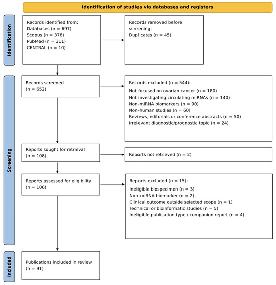
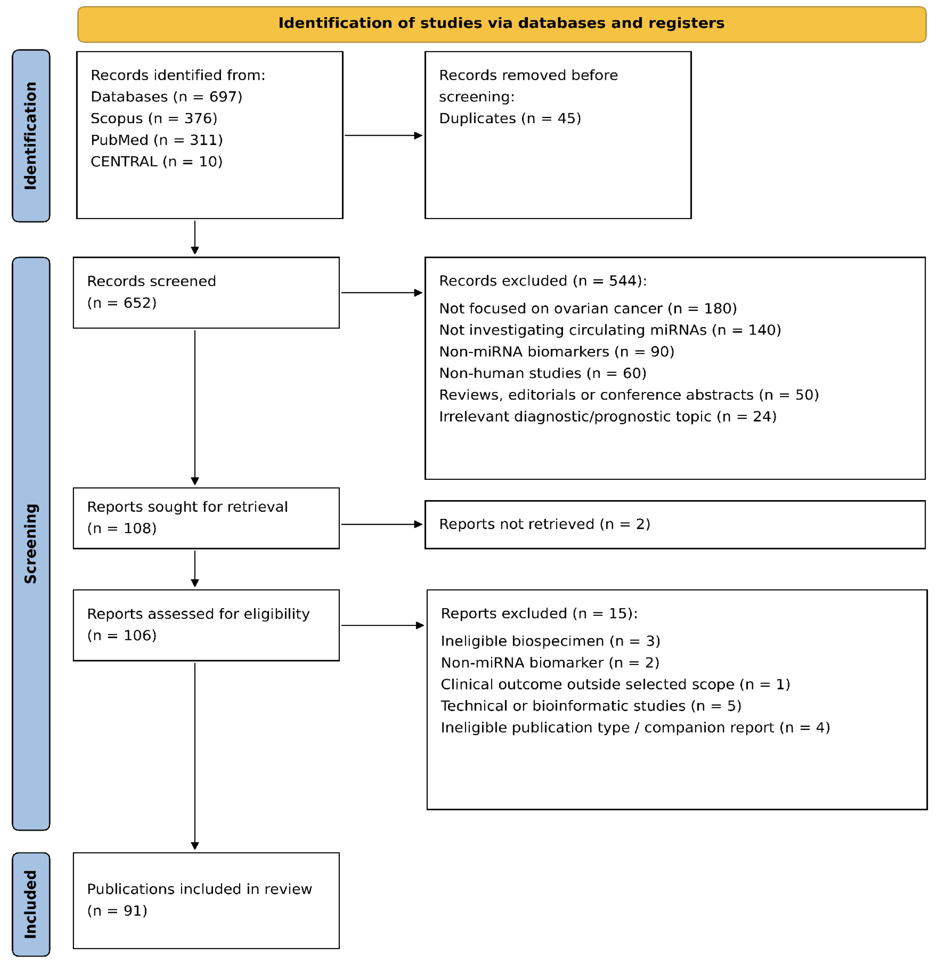
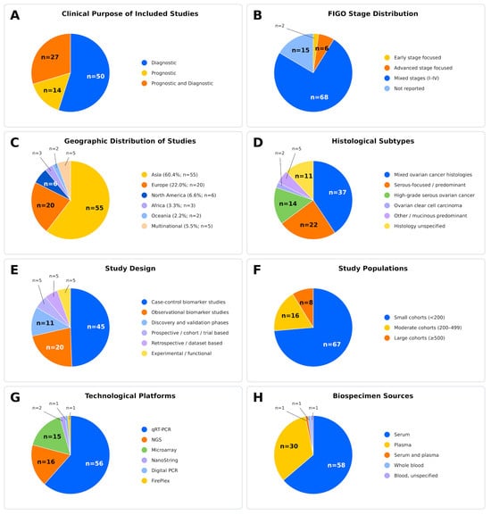
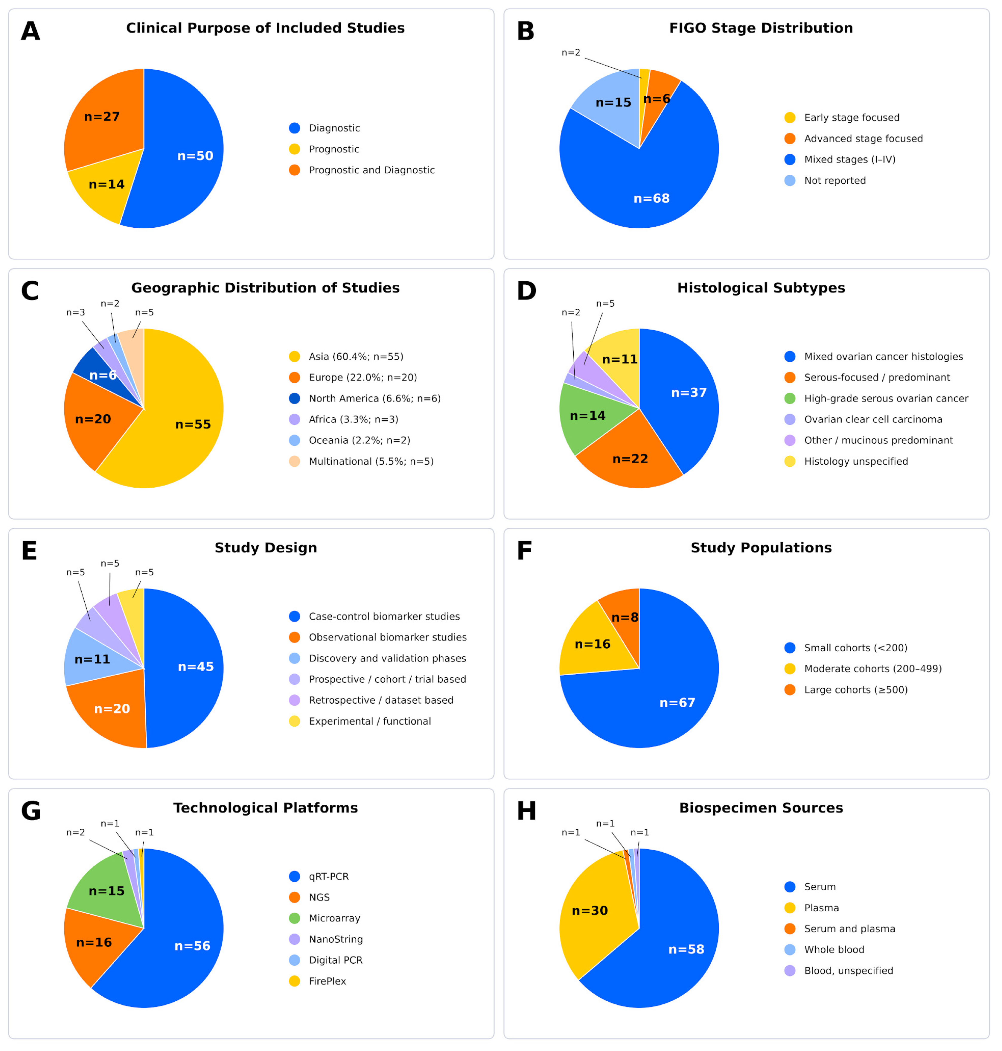
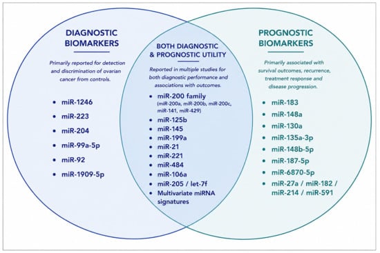
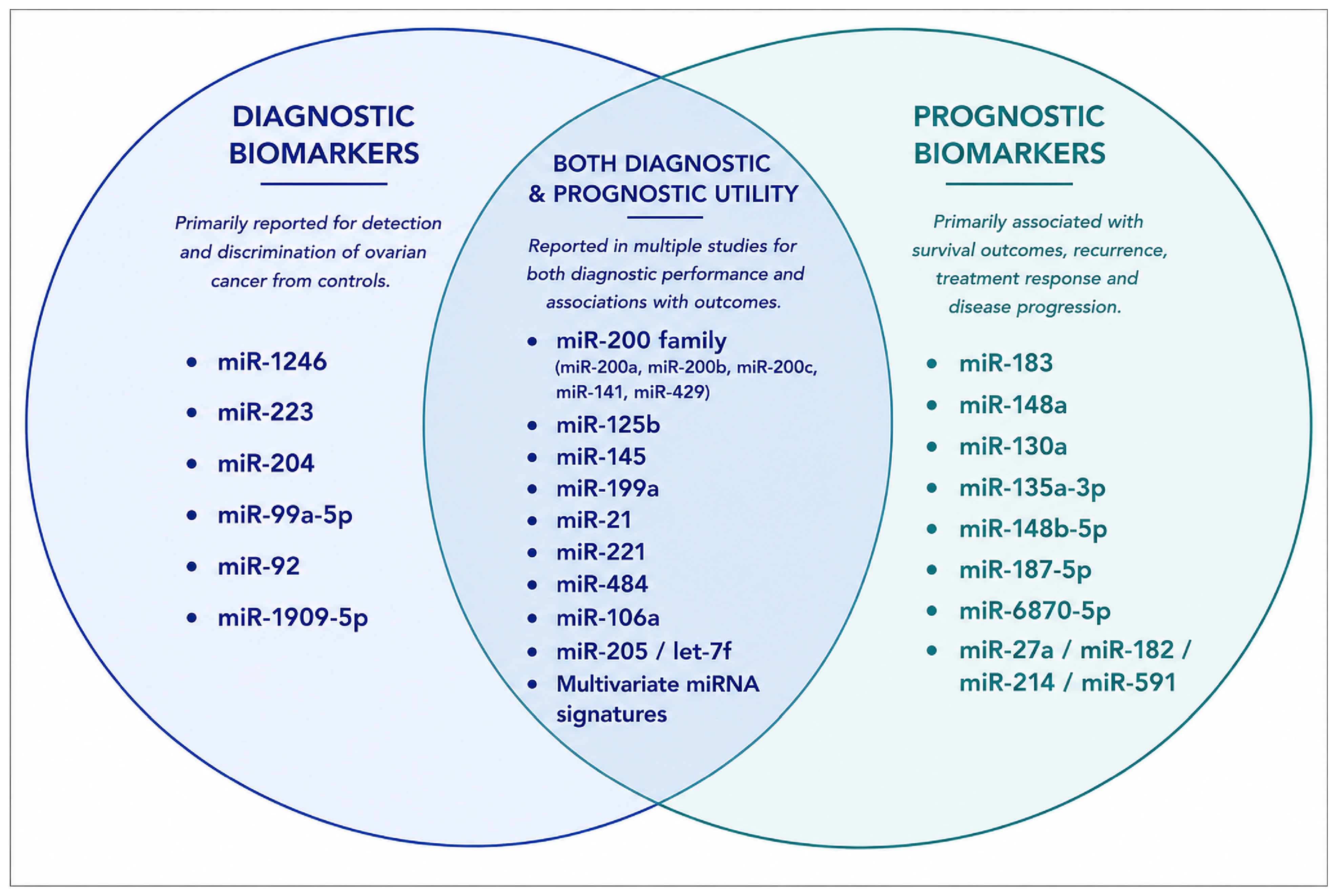
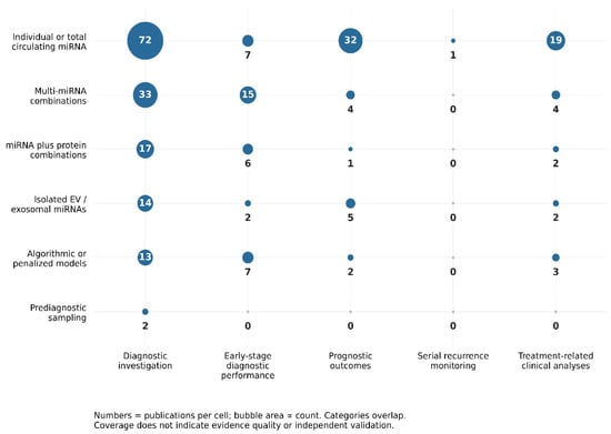
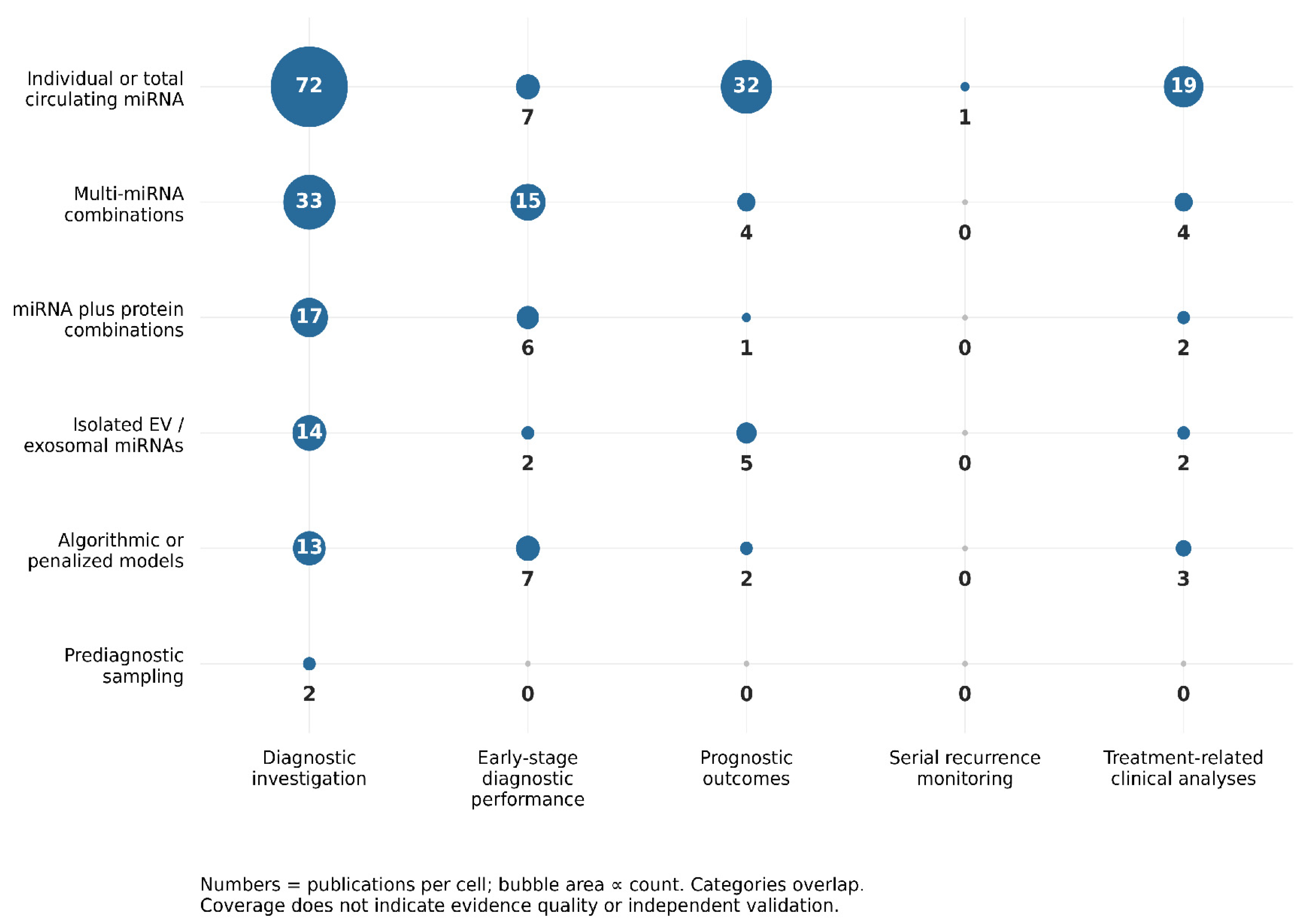
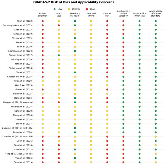
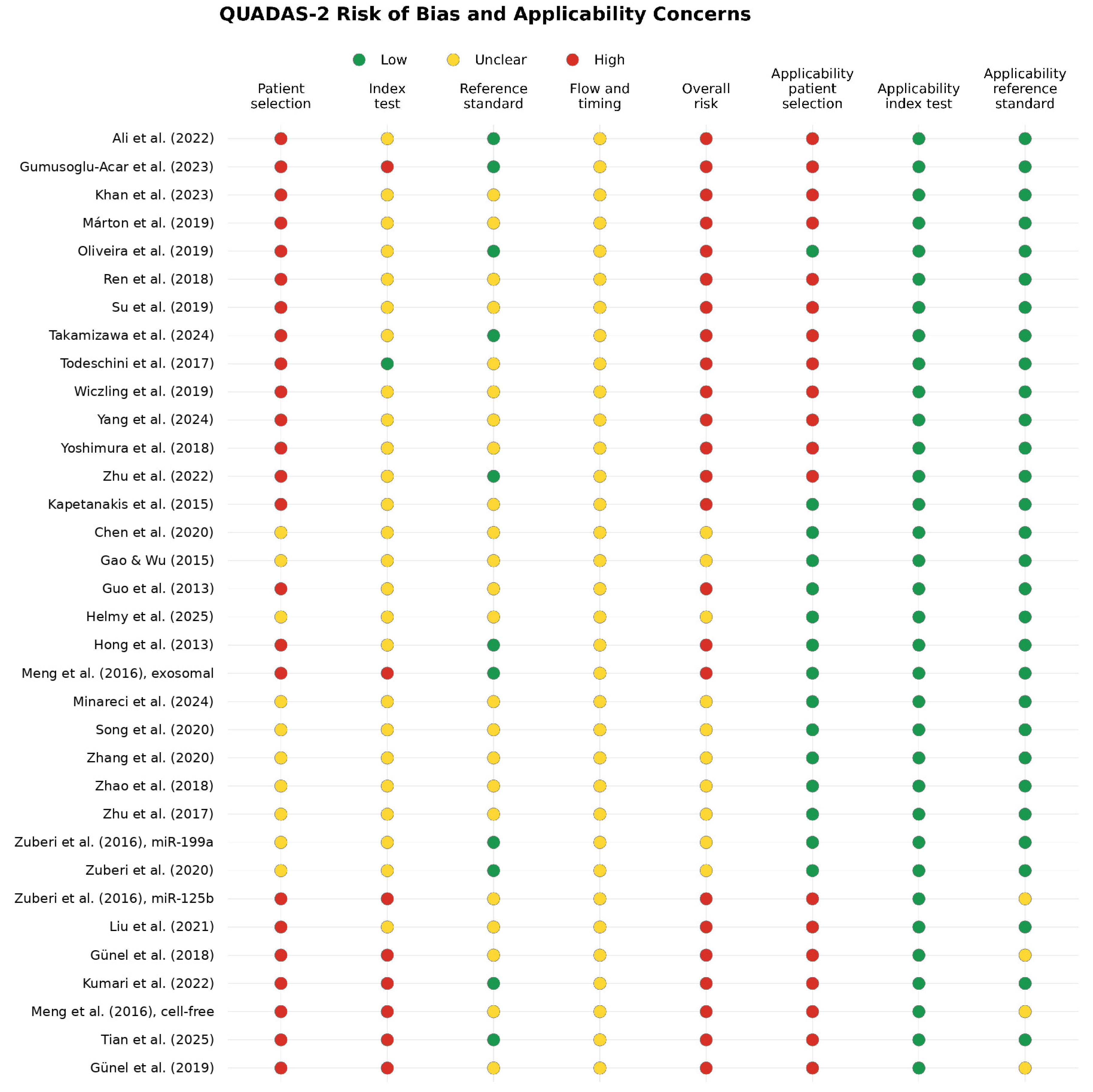
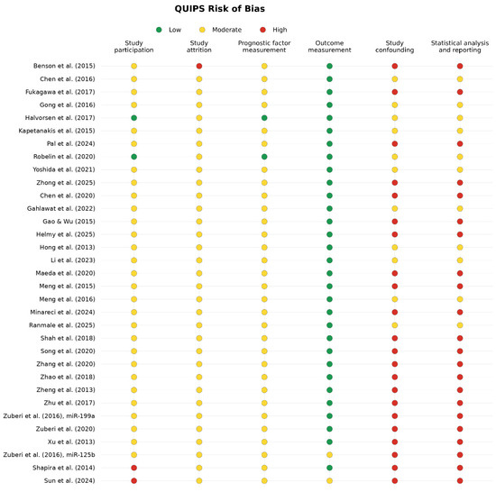
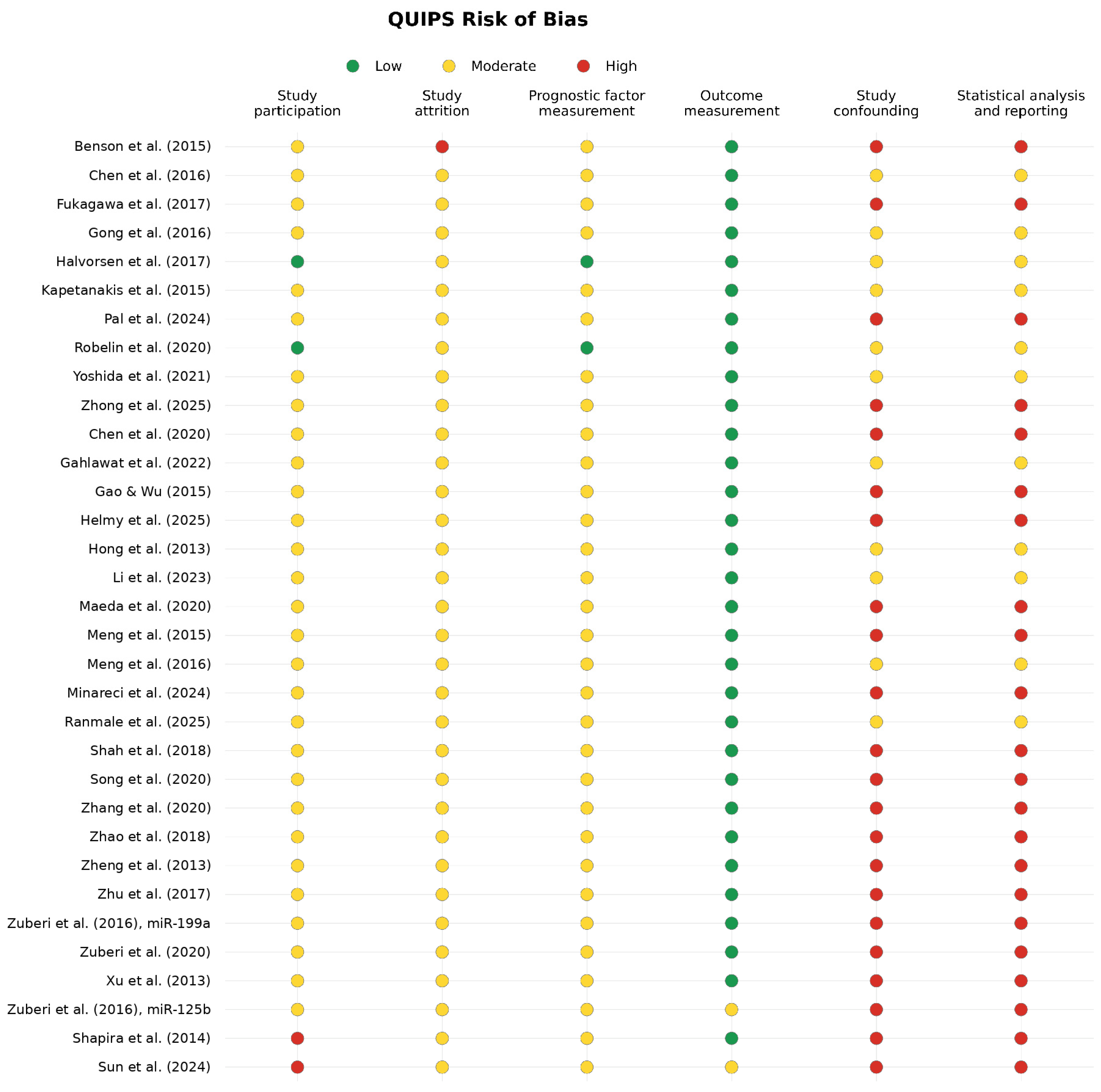
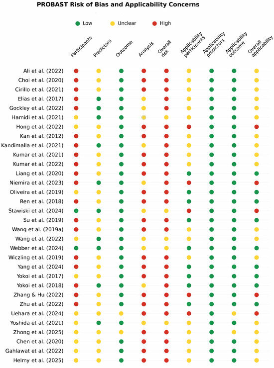
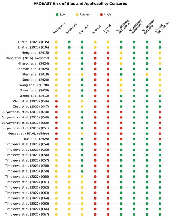
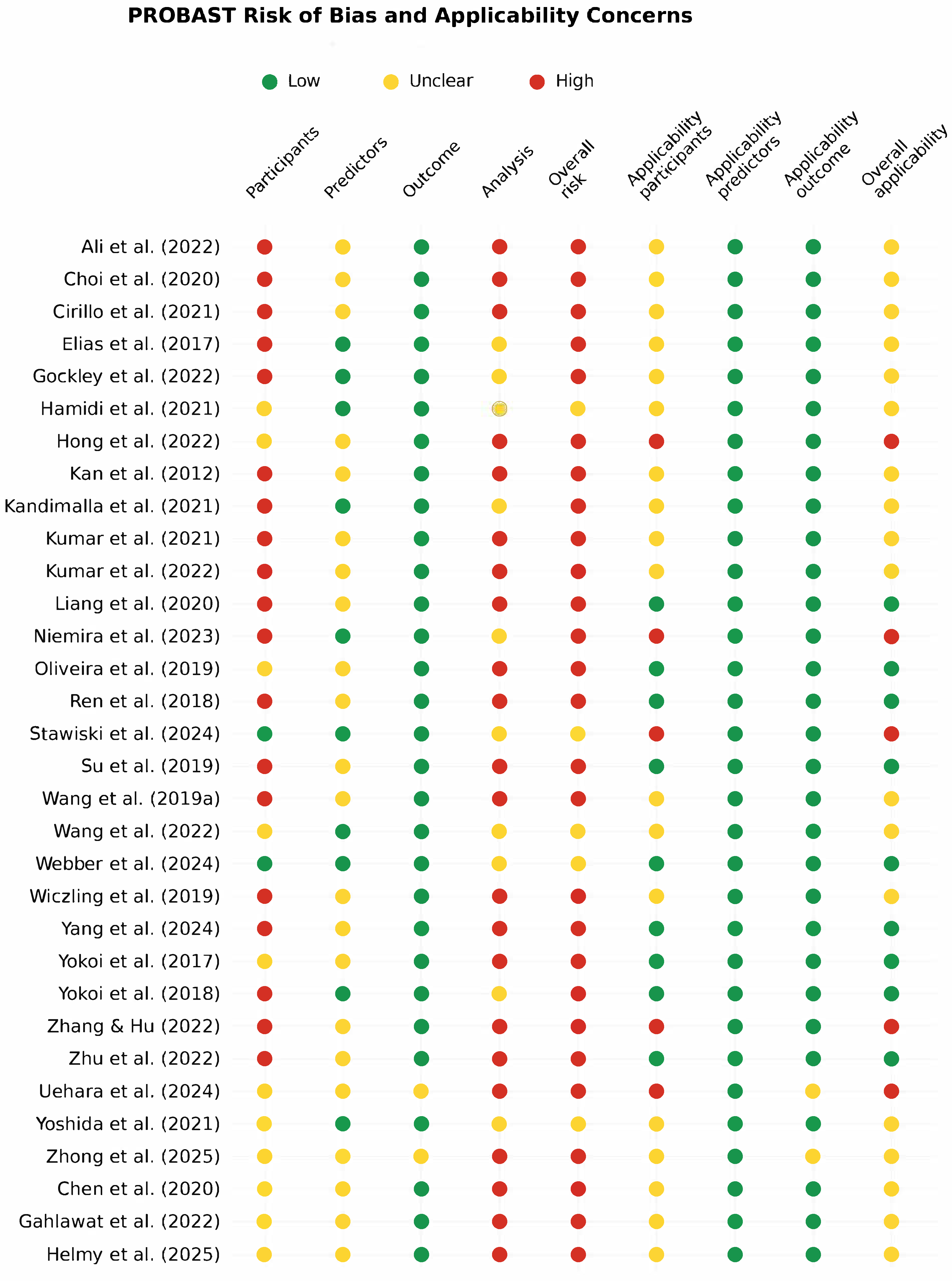
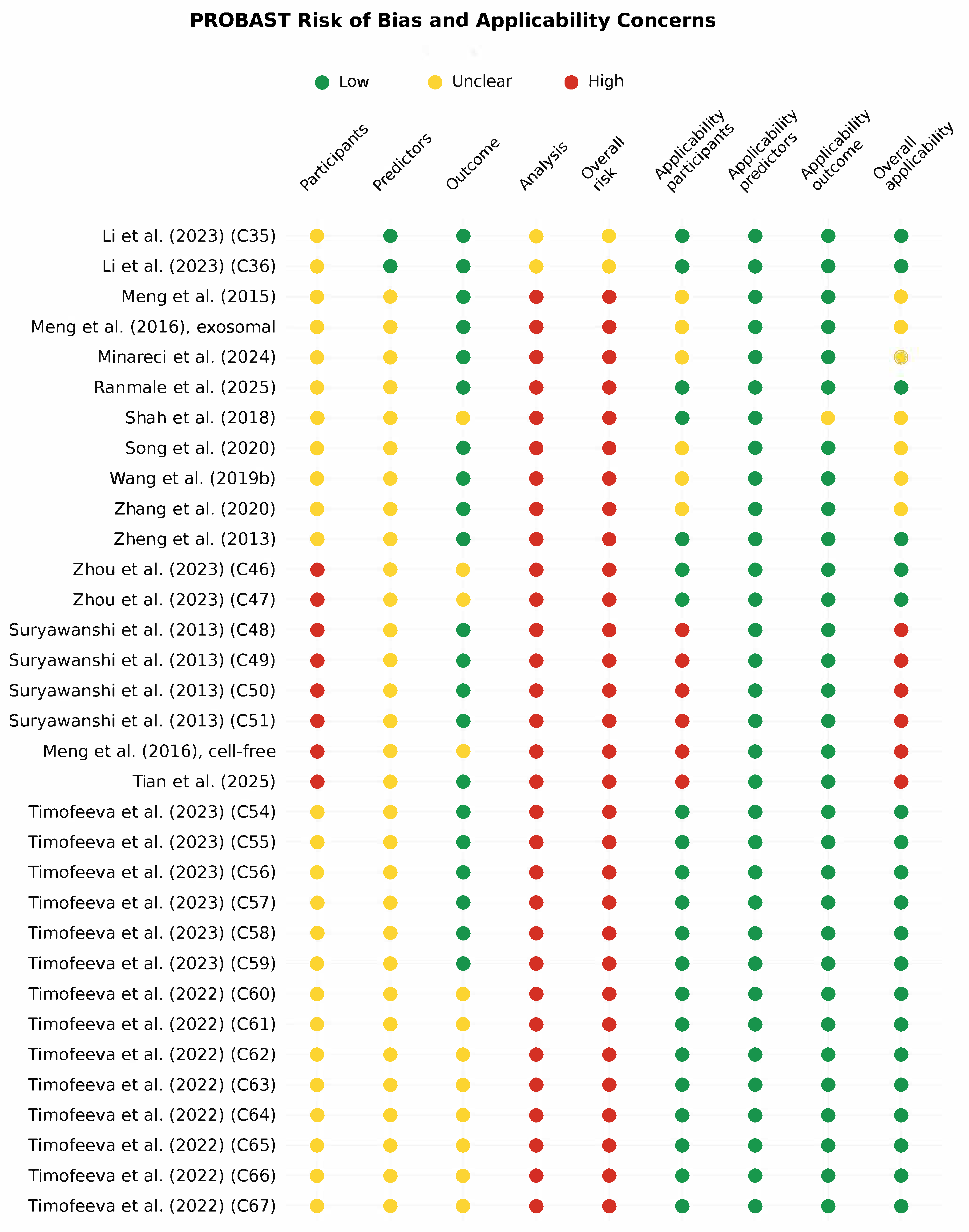
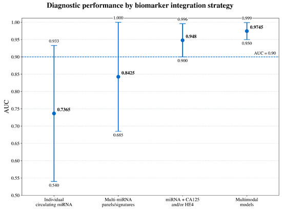
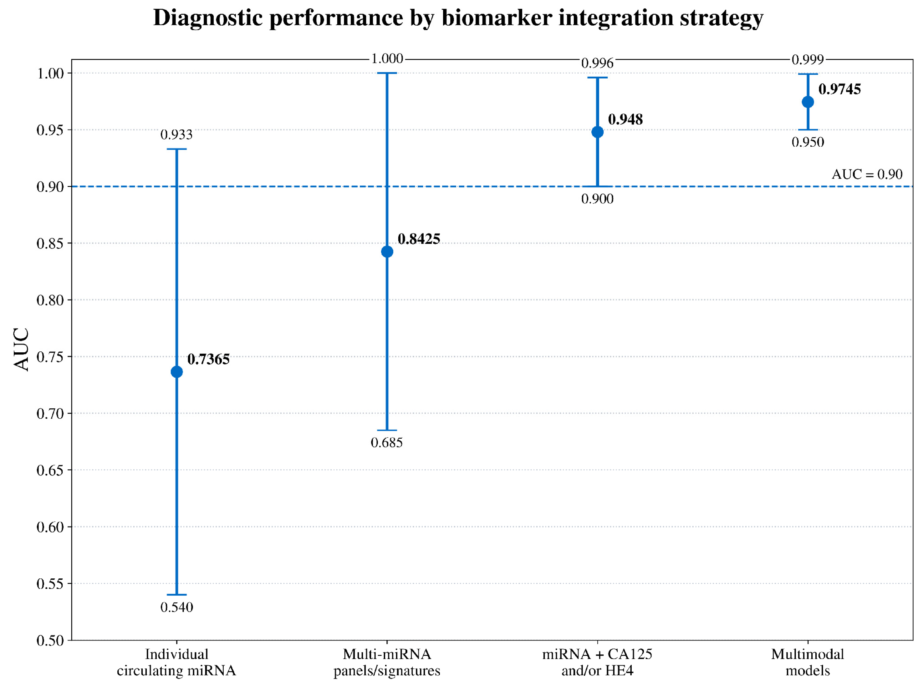

Disclaimer/Publisher’s Note: The statements, opinions and data contained in all publications are solely those of the individual author(s) and contributor(s) and not of MDPI and/or the editor(s). MDPI and/or the editor(s) disclaim responsibility for any injury to people or property resulting from any ideas, methods, instructions or products referred to in the content. |
© 2026 by the authors. Licensee MDPI, Basel, Switzerland. This article is an open access article distributed under the terms and conditions of the Creative Commons Attribution (CC BY) license.