


Abstract
Glioblastoma (GBM) is the most common and highly malignant primary brain tumor in adults. Even with advances in surgery, radiotherapy, and systemic therapies, patient outcomes remain poor due to extensive intratumoral heterogeneity and the development of therapeutic resistance. Circular RNAs (circRNAs) are a class of covalently closed RNA molecules that are highly abundant in the central nervous system and frequently dysregulated in cancer. While originally considered non-coding transcripts, current evidence demonstrates that a subset of circRNAs contain functional open reading frames and can undergo cap-independent translation to generate biologically active proteins and peptides. Recent studies have identified a growing number of translated circRNAs in GBM encoding proteins implicated in diverse processes such as receptor tyrosine kinase signaling, DNA repair, metabolism, stemness, invasion, and therapeutic resistance. These findings expand the coding potential of the GBM transcriptome and highlight an additional layer of post-transcriptional regulation. In this review, we summarize our current understanding of circRNA translation, discuss the biological functions of translated circRNAs in GBM, and examine their potential roles in tumor progression, therapeutic resistance, and clinical management. Additionally, we consider the opportunities and challenges associated with targeting translated circRNAs and their encoded proteins for the treatment of GBM.
1. Introduction
Glioblastoma (GBM) is a highly aggressive primary malignant brain tumor in adults and carries a poor prognosis despite maximal safe resection, radiotherapy, and temozolomide (TMZ) [1,2,3,4]. Nearly all tumors recur, reflecting extensive intratumoral heterogeneity and the ability of GBM cells to adapt to microenvironmental and therapeutic stress [2,3,4]. These adaptive responses include changes in signaling, metabolism, cell state, DNA-damage responses, and protein synthesis. Recent transcriptomic studies have shown that this complexity extends beyond conventional messenger RNAs to circular RNAs (circRNAs), endogenous transcripts generated by back-splicing in which a downstream splice donor is covalently joined to an upstream splice acceptor [5,6,7]. Their closed structure lacks free 5′ and 3′ ends, conferring resistance to exonuclease degradation and increased stability relative to many linear RNAs [5,6,7].
CircRNAs are particularly abundant in the central nervous system and display developmental, cell-type-specific, and activity-dependent regulation [8,9,10]. In glioma, altered circRNA expression has been linked to proliferation, invasion, stemness, angiogenesis, metabolism, and therapeutic resistance [10,11,12,13]. Although circRNAs were initially studied primarily as non-coding regulators of microRNAs, RNA-binding proteins, and transcription, a subset contains functional open reading frames and can be translated through cap-independent mechanisms [14,15,16,17]. IRES-dependent initiation and m6A-mediated ribosome recruitment are the best established mechanisms, while some circular templates can also support rolling-circle translation [14,15,16,17]. Thus, circRNAs can contribute to gene regulation at both the RNA and protein levels.
For clarity, throughout this review we use “translated circRNA” to refer to a circRNA for which translation is experimentally supported or specifically under consideration, and “circRNA-encoded protein” or “circRNA-encoded peptide” for the resulting translation product. We avoid using “protein-coding circRNA” as the default designation because many circRNAs may have dual functions, acting both as RNA regulatory molecules and as templates for translation.
Several translated circRNAs have been reported and functionally characterized in GBM. Tumor-suppressive examples include circFBXW7, circSHPRH, circAKT3, circLINC-PINT, circNTRK2, and circHEATR5B, whereas circHGF and circEGFR generate products with oncogenic activities [18,19,20,21,22,23,24,25,26]. Collectively, the reported proteins regulate pathways ranging from c-MET and EGFR signaling to c-Myc stability, AKT activation, transcriptional elongation, DNA repair, and glycolysis. These observations expand the coding potential of the GBM transcriptome and suggest that circRNA translation may contribute to molecular heterogeneity and stress adaptation, particularly when canonical cap-dependent translation is suppressed [13].
Several previous reviews have discussed the emerging capacity of circRNAs to encode proteins in glioma and GBM [27,28]. The present review extends these earlier syntheses by integrating the mechanisms that support circRNA translation with the experimental approaches used to establish translation and protein function and by critically examining the evidence for individual translated circRNAs characterized in GBM. Particular emphasis is placed on how circRNA-encoded proteins converge on signaling, metabolic, and stress-response pathways relevant to tumor progression and therapeutic response, as well as on the methodological limitations that currently constrain interpretation and clinical translation. Finally, we consider the potential clinical implications of translated circRNAs, including their development as biomarkers and therapeutic targets, and highlight key challenges and opportunities for future investigation in neuro-oncology.
2. Circular RNAs in GBM
2.1. CircRNA Biogenesis and Classification
CircRNAs are generated by back-splicing, producing a covalently closed RNA molecule lacking free termini (Figure 1A) [6,29,30]. Circularization can be promoted by intronic complementary sequences, particularly inverted Alu elements, that bring splice sites into close proximity. RNA-binding proteins, including QKI, MBL, FUS, and NF90/NF110, can further promote or inhibit back-splicing, providing regulated control over circRNA production [6,31,32,33,34]. CircRNAs are generally classified as exonic circRNAs, circular intronic RNAs, or exon-intron circRNAs (Figure 1B) [6,35,36]. Exonic circRNAs are predominantly cytoplasmic and represent the major class relevant to translation, whereas intron-containing forms are frequently nuclear and can influence transcription. The circular architecture also increases resistance to exonuclease-mediated decay and can prolong intracellular persistence. These properties may be especially relevant in GBM, where cells are exposed to hypoxia, nutrient limitation, oxidative stress, and therapy-induced changes in RNA metabolism [10,12]. However, direct evidence that the increased stability of circRNAs itself drives stress-adaptive phenotypes in GBM has not yet been established.
Figure 1.
Circular RNAs (circRNAs) in glioblastoma: biogenesis, expression, classification, and established non-coding functions. (A) CircRNAs are generated by back-splicing, aided by complementary intronic sequences and RNA-binding proteins. (B) Major classes include exonic circRNAs, circular intronic RNAs, and exon-intron circRNAs. (C) CircRNAs are abundant in the normal brain, especially in neurons; many are developmentally regulated and highly stable, and some exceed their cognate linear transcripts. (D) GBM-associated circRNAs may be upregulated or downregulated and have potential biomarker value. (E) Established non-coding functions include microRNA sequestration (ceRNA, competitive endogenous RNA activity), RNA-binding protein interactions, transcriptional regulation, and modulation of stemness, RNA stability, translation, and stress responses.
2.2. CircRNA Expression in the Normal Brain
The brain contains among the highest levels of circRNAs in mammalian tissues (Figure 1C) [8,9]. Many neuronal circRNAs are developmentally regulated, evolutionarily conserved, and enriched in mature neurons and synaptic compartments, with abundance often increasing during differentiation and aging [8,9,37]. Their accumulation likely reflects both regulated biogenesis and unusual stability. Physiological circRNA functions include microRNA sequestration, interaction with RNA-binding proteins, modulation of transcription, and, for a subset, protein synthesis [10,17]. Because neural cells naturally express a large and diverse circRNA repertoire, brain tumors arise in a tissue context already permissive for extensive circular RNA biology. This makes it important to distinguish cancer-specific changes from the elevated baseline abundance of circRNAs in normal brain [38,39].
2.3. Dysregulated CircRNAs in GBM
RNA-sequencing studies have demonstrated widespread circRNA dysregulation in GBM (Figure 1D) [11,40,41,42,43,44]. Both oncogenic and tumor-suppressive circRNAs have been reported, and expression patterns can vary with tumor grade, molecular state, and clinical outcome [11,12,41]. Dysregulated circRNAs can influence signaling, cell-cycle progression, invasion, stemness, and therapeutic response, and some are readily detected in extracellular vesicles and biological fluids, increasing interest in their biomarker potential [45]. Importantly, coding and non-coding activities are not mutually exclusive. A circular transcript may alter cellular behavior through RNA-mediated interactions while also serving as a template for a biologically active protein, complicating functional assignment but also expanding the regulatory potential of the transcript.
An important limitation of the existing literature is that several foundational studies predate the WHO CNS5 classification and analyzed cohorts described broadly as glioma or by histologic grade without IDH stratification. Under WHO CNS5, glioblastoma denotes an IDH-wildtype adult-type diffuse astrocytic glioma, whereas IDH-mutant grade 4 tumors are classified as astrocytoma, IDH-mutant, CNS WHO grade 4 [46]. Accordingly, throughout this review we use glioma or diffuse glioma when the primary study did not establish an IDH-wildtype glioblastoma diagnosis and reserve GBM for studies in which that designation is supported. This distinction is important because IDH-mutant and IDH-wildtype diffuse gliomas differ in molecular biology and clinical behavior, and circRNA expression or biomarker associations derived from mixed or historically classified cohorts may not generalize directly to contemporary IDH-wildtype GBM. Future studies should therefore report molecular classification and evaluate translated circRNAs within defined WHO CNS5 entities.
2.4. Established Non-Coding Functions of CircRNAs in GBM
The best-characterized non-coding circRNA function is microRNA sequestration, in which circular transcripts act as competing endogenous RNAs that reduce the availability of specific microRNAs for target repression [7,10]. CircRNAs can also bind RNA-binding proteins, alter protein localization or stability, serve as molecular scaffolds, and influence transcription (Figure 1E) [10,17,47]. In GBM, examples such as circNT5E, circHIPK3, and circPOSTN have been linked to oncogenic signaling, invasion, and stem-like phenotypes [48,49,50]. These RNA-mediated functions remain an important part of circRNA biology and provide context for the protein-coding field. The subsequent demonstration that some circRNAs encode functional proteins, however, established a mechanistically distinct layer of regulation and shifted attention from circRNA abundance alone to the question of which circular transcripts are actually translated in tumor cells.
3. Evidence for Circular RNA Translation
3.1. Discovery of Protein-Encoding Circular RNAs
CircRNAs were long considered non-coding because they lack the 5′ cap and poly(A) tail used by most eukaryotic mRNAs. Evidence for translation emerged when specific circRNAs were found in ribosome- and polysome-associated fractions and when proteins or peptides uniquely attributable to circular templates were subsequently detected (Table 1) [14,15,16]. CircZNF609 provided an early endogenous example, and genome-wide studies later identified numerous candidate translated circRNAs [14,15,16]. Particularly persuasive evidence comes from translation across the back-splice junction (BSJ), which can generate peptide sequences absent from the corresponding linear transcript. These findings established circRNAs as a potential source of previously unrecognized protein diversity and raised the possibility that their translation could be favored during cellular stress.
Table 1.
Mechanisms and experimental approaches used to identify and validate translated circRNAs, with relevance to glioblastoma.
3.2. Mechanisms of CircRNA Translation
3.2.1. IRES-Dependent Translation
IRES-dependent initiation is one of the principal mechanisms used by translated circRNAs. IRES elements recruit the translation machinery internally, bypassing the requirement for recognition of a 5′ cap [14,51,52]. Their activity is often influenced by IRES trans-acting factors (ITAFs), including hnRNP A1, PTBP1, La/SSB, PCBP2, and IGF2BP1, which can remodel RNA structure or facilitate recruitment of the 40S ribosomal subunit and initiation factors [53,54]. As functional IRESs are defined more reliably by activity rather than by primary sequence, bioinformatic prediction alone is insufficient; reporter assays, mutagenesis, ribosome profiling, and proteomic evidence remain important for validation [22,52,55].
This mechanism is particularly relevant to GBM because hypoxia, nutrient deprivation, oxidative stress, and anticancer therapy can suppress cap-dependent translation while favoring selective translation programs [56,57,58,59,60,61]. Several ITAFs are dysregulated in GBM, and oncogenic pathways such as EGFR, PI3K/AKT/mTOR, and RAS/MAPK can alter the abundance or activity of translation regulators [57,58,59,60,61]. Whether these pathways directly control specific circRNA IRESs remains incompletely defined, but the convergence of oncogenic signaling, stress, and cap-independent translation provides a plausible framework for selective circRNA protein production during tumor progression and treatment.
3.2.2. m6A-Mediated Translation
CircRNA translation can also be initiated by N6-methyladenosine (m6A). m6A residues provide internal recruitment signals that can support cap-independent translation, with YTHDF3 and initiation factors including eIF4G2/DAP5 and eIF3A participating in ribosome recruitment [16,62,63,64]. m6A deposition and removal are dynamically regulated by writer and eraser proteins, and circRNA translation through this pathway can increase under cellular stress [16,65]. This is relevant in GBM, where METTL3, ALKBH5, FTO, and YTH-domain proteins have been linked to stem-cell maintenance, metabolic adaptation, invasion, and treatment response [66,67,68,69,70]. m6A modification also affects circRNA localization, stability, and RNA-protein interactions, indicating that epitranscriptomic regulation may coordinate several aspects of circRNA biology [63,71].
3.2.3. Rolling Circle Translation
A feature unique to circular templates is rolling-circle translation. CircRNAs containing a continuous open reading frame without an in-frame stop codon can be traversed repeatedly by ribosomes, generating extended proteins composed of repeated peptide units [15,72,73]. Initiation generally still requires an internal recruitment mechanism such as an IRES or m6A, after which elongation can continue around the circular template. Endogenous examples remain less common than conventional circRNA translation, but the mechanism is biologically relevant in GBM because circEGFR has been reported to undergo rolling-circle translation to generate rtEGFR [25].
3.3. Experimental Approaches Used to Identify Translated CircRNAs
Establishing bona fide circRNA translation is technically demanding because most circRNAs arise from protein-coding genes and may share much of their sequence with linear mRNAs. Strong studies therefore combine orthogonal evidence demonstrating the circular transcript, ribosome engagement, production of a circRNA-specific protein product, and functional dependence on translation rather than on the RNA molecule alone [10,14,16,74]. No single assay is sufficient in every setting.
3.3.1. Ribosome Profiling
Ribosome profiling (Ribo-seq) identifies ribosome-protected RNA fragments and can reveal footprints spanning the BSJ, which strongly supports engagement of a circular template [15,73]. However, ribosome occupancy does not by itself prove productive translation, and low circRNA abundance can limit BSJ-spanning reads. Ribo-seq is therefore best viewed as a discovery and supporting tool rather than definitive proof.
3.3.2. Polysome Profiling
Polysome profiling provides complementary evidence by determining whether a circRNA associates with multiple translating ribosomes [75,76]. Redistribution after puromycin or initiation inhibitors can strengthen the interpretation, but polysome association alone remains insufficient because non-coding RNAs can associate with ribosomes without producing stable protein products.
3.3.3. Mass Spectrometry
Mass spectrometry can provide highly specific evidence when peptides spanning a circRNA-derived junction are detected [14,16,74]. Customized protein databases containing predicted circRNA open reading frames improve discovery, although low protein abundance, rapid turnover, and a limited number of unique peptides can reduce sensitivity [74,77,78]. Consequently, a negative proteomic result does not necessarily exclude translation.
3.3.4. Reporter Systems and Functional Validation
Reporter systems are useful for testing translational competence and dissecting IRES, m6A, or rolling-circle requirements [14,16,72,79,80]. Mutating start codons, IRES elements, m6A motifs, or continuous open reading frames can define mechanism, but artificial overexpression can create non-physiological translation. Reporter data should therefore be paired with endogenous validation.
3.3.5. Genetic and Molecular Validation
Genetic approaches provide critical separation of RNA and protein functions. BSJ-directed siRNAs or ASOs can selectively deplete circular transcripts, while disruption of intronic sequences required for circularization can reduce circRNA formation without necessarily eliminating host-gene transcription [7,10,79,80,81]. Rescue with wild-type versus translation-deficient circRNAs is particularly informative because it tests whether the phenotype requires the encoded protein [14].
3.3.6. Bioinformatic Prediction and Multi-Omics Integration
Computational resources such as circRNADb, riboCIRC, and TransCirc integrate open reading frames, IRES or m6A features, conservation, ribosome occupancy, and proteomic evidence to prioritize candidates [78,82,83,84]. Multi-omics integration can reduce false-positive assignments, but prediction cannot substitute for experimental confirmation. The most convincing designation of a translated circRNA therefore rests on convergent transcript-level, translation-level, protein-level, and functional evidence.
3.3.7. Common Artifacts and Safeguards
Several technical artifacts can mimic circRNA formation or translation and therefore require orthogonal validation. RNase R resistance enriches for circular species but is not, by itself, proof of circularity because susceptibility varies with RNA structure and experimental conditions. Likewise, reverse-transcriptase template switching and rolling-circle cDNA synthesis can generate apparent BSJ-containing products or concatemers, making confirmation by independent approaches important [6,21,85]. BSJ detection by RNA-seq or Ribo-seq is also sensitive to alignment algorithms, mapping parameters, read length, and sequencing depth; consequently, BSJ-spanning ribosome footprints should be interpreted as evidence of ribosome engagement rather than definitive proof of productive translation [85]. For mass spectrometry, the strongest evidence is a peptide unique to the circRNA-encoded product, ideally spanning a junction or otherwise absent from proteins produced by linear splice isoforms or alternative proteoforms. Database composition and false-discovery thresholds should be specified, and candidate peptides should be validated where feasible using synthetic standards, circRNA-selective depletion, or complementary immunodetection [85]. Together, these considerations favor convergent evidence from transcript-level, ribosome-level, protein-level, and genetic approaches rather than reliance on any single assay.
3.3.8. Contested and Unresolved Cases
Not all reported circRNA translation events have been independently confirmed, and several observations illustrate why candidate products should be interpreted cautiously. A prominent example is circZNF609, initially reported to associate with polysomes and produce a protein in a splicing-dependent, cap-independent manner. Subsequent analysis of commonly used circZNF609 overexpression constructs found that the detected protein products could persist when circRNA formation was disrupted and were most consistent with translation from linear or trans-spliced by-products; endogenous circZNF609-derived protein was not detected in that study [86]. More broadly, systematic testing of circRNA overexpression vectors has shown that cryptically spliced linear transcripts and concatemeric by-products can generate apparent translation products, including products that contain a back-splice-junction sequence and can therefore mimic rolling-circle translation [87]. Predicted open reading frames identified by sequence analysis, conservation, ribosome association, or isolated mass-spectrometric signals should therefore be regarded as candidates rather than established circRNA-encoded proteins unless circular origin and endogenous protein production are demonstrated independently. Apparent immunoreactivity also requires caution when antibodies can recognize shared sequences in canonical host-gene products or alternative proteoforms. These unresolved cases do not argue against circRNA translation as a biological phenomenon; rather, they underscore the need for orthogonal evidence, circRNA-selective genetic perturbation, translation-deficient rescue controls, and peptide or antibody reagents that distinguish the circular product from linear-transcript-derived proteins.
4. Protein-Encoding Circular RNAs in Glioblastoma
Only a small fraction of the thousands of circRNAs detected in GBM have been shown to encode functional proteins. Nevertheless, the best-supported examples already span receptor signaling, cell-cycle control, DNA repair, transcription, metabolism, stemness, and therapeutic response (Table 2; Figure 2). Their products can be either oncogenic or tumor suppressive, emphasizing that circRNA translation is not intrinsically pro-tumorigenic but instead expands the repertoire of regulatory proteins available to GBM cells.
Table 2.
Experimentally characterized translated circRNAs and their encoded proteins relevant to glioblastoma.
Figure 2.
Overview of translated circRNAs in glioblastoma. (A) CircRNAs can undergo cap-independent translation through IRES-dependent, m6A-mediated, or rolling-circle mechanisms. (B) Reported glioma/GBM examples include circHGF/C-HGF, circFBXW7/FBXW7-185aa, circSHPRH/SHPRH-146aa, circAKT3/AKT3-174aa, circLINC-PINT/PINT87aa, circNTRK2/NTRK2-243aa, circEGFR/rtEGFR, and circHEATR5B/HEATR5B-881aa. (C) Their products include both tumor-suppressive and oncogenic proteins. (D) Representative expression patterns distinguish upregulated oncogenic circRNAs from downregulated tumor-suppressive circRNAs. (E) These proteins regulate signaling, proliferation, invasion, stemness, metabolism, and therapeutic response. Arrows indicate translational progression, regulatory relationships, or expression changes as labeled; the dashed circular arrow denotes repeated ribosome transit during rolling-circle translation, and colored circular structures represent the indicated circRNAs.
4.1. circHGF
Discovery and Characterization
CircHGF is generated from the HGF locus and represents an oncogenic translated circRNA reported in GBM [23,88,89]. Expression studies showed increased circHGF in GBM relative to normal brain and an association with activation of the c-MET pathway. Saunders and colleagues demonstrated that circHGF encodes C-HGF, a novel HGF-related protein containing a unique junction-derived sequence that distinguishes it from the product of linear HGF mRNA [23]. Detection of this sequence supports translation from the circular template. Functionally, C-HGF stimulates c-MET signaling and downstream programs involved in GBM proliferation and survival. Silencing circHGF reduces growth, migration, and invasion, whereas increased circHGF promotes aggressive phenotypes; xenograft studies further supported a role in tumor growth [23]. The finding is notable because a circRNA-encoded product acts as a growth-factor-like extracellular signal rather than solely as an intracellular regulator. It therefore links non-canonical translation directly to a clinically relevant receptor tyrosine kinase pathway and suggests several possible points of intervention, including selective depletion of circHGF, inhibition of C-HGF, or blockade of c-MET signaling.
4.2. circFBXW7
CircFBXW7 was among the first translated circRNAs characterized in glioma and remains a prototypical tumor-suppressive example [18]. It encodes FBXW7-185aa, a 185-amino acid protein distinct from canonical FBXW7. The full-length FBXW7 pathway restrains several oncogenic proteins, and the circRNA-encoded product reinforces this tumor-suppressive network by antagonizing USP28-mediated stabilization of c-Myc [18]. Increased c-Myc degradation reduces expression of proliferative and metabolic programs required for tumor growth. CircFBXW7 is reduced in glioma specimens relative to non-neoplastic brain, while restoration of FBXW7-185aa suppresses proliferation and tumor growth in experimental models [18]. This study illustrates an important principle: a circRNA-encoded protein can influence a major cancer pathway through a mechanism distinct from, yet functionally complementary to, the canonical host-gene product.
4.3. circSHPRH
CircSHPRH encodes SHPRH-146aa, a 146-amino acid protein that supports the tumor-suppressive activity of full-length SHPRH [19]. SHPRH is an E3 ubiquitin ligase involved in genomic maintenance and DNA-damage responses. SHPRH-146aa protects the full-length protein from ubiquitin-mediated degradation, increasing its stability and thereby preserving tumor-suppressive function [19,90]. CircSHPRH expression is reduced in glioma specimens, and restoration of SHPRH-146aa inhibits tumor growth in vitro and in vivo [19]. The circSHPRH model is particularly relevant to treatment biology because genomic stability and DNA-repair capacity can influence sensitivity to radiation and alkylating agents. At the same time, the available data support a tumor-suppressive rather than a simple resistance-promoting role, illustrating why the biological effect of each translated circRNA must be evaluated individually. Mechanistically, SHPRH promotes K63-linked polyubiquitination of PCNA at stalled replication forks, supporting an error-free template-switching pathway of DNA-damage tolerance [90]. Thus, although SHPRH-146aa is tumor suppressive in the reported glioma models, stabilization of full-length SHPRH could also increase the capacity of tumor cells to tolerate particular forms of replication-blocking DNA damage. The net effect of circSHPRH/SHPRH-146aa on response to TMZ or radiation therefore cannot be predicted from its tumor-suppressive phenotype alone and requires direct experimental testing.
4.4. circAKT3
CircAKT3 produces AKT3-174aa, a tumor-suppressive protein with biological activity that contrasts with the conventional growth-promoting role of AKT signaling [20]. Translation was supported by back-splice-junction analysis, tagged expression constructs, and detection of circRNA-specific peptides. Mechanistically, AKT3-174aa interacts with phosphoinositide-dependent kinase-1 (PDK1) and interferes with phosphorylation required for maximal AKT activation, thereby restraining PI3K/AKT signaling [20]. Restoration of circAKT3 or AKT3-174aa reduces proliferation, clonogenic growth, migration, invasion, and orthotopic tumor growth, whereas depletion of circAKT3 increases AKT activity and malignant phenotypes. Clinical analyses further associated reduced circAKT3 abundance with higher-grade glioma and poorer outcome [20]. Because PI3K/AKT signaling is frequently activated in GBM through receptor tyrosine kinase alterations, PTEN loss, and other mechanisms, AKT3-174aa represents an endogenous brake on a central oncogenic pathway. It also illustrates how circularization can generate a protein whose function diverges substantially from the canonical product of the same genomic locus.
4.5. circLINC-PINT
CircLINC-PINT provides a different example because it arises from the LINC-PINT locus, previously characterized primarily as a non-coding RNA [91,92]. The circular transcript contains an open reading frame encoding the 87-amino acid peptide PINT87aa [21]. Translation was supported by circRNA-specific constructs, epitope tagging, mass-spectrometric peptide detection, and functional rescue experiments indicating that the biological activity depends on the encoded peptide. PINT87aa localizes to the nucleus and interacts with the RNA polymerase II-associated PAF1 complex, which regulates transcriptional elongation and chromatin-associated processes [21,93]. Through this interaction, PINT87aa suppresses elongation of oncogenic transcripts and restrains glioma growth. The finding expands the conceptual range of translated circRNAs by showing that they can produce nuclear regulatory peptides from loci not traditionally considered protein coding and can influence cancer through control of transcription rather than receptor or kinase signaling.
4.6. circNTRK2
CircNTRK2 encodes NTRK2-243aa, a tumor-suppressive protein whose function differs from that of the full-length TrkB receptor [24]. Rather than promoting neurotrophin signaling, NTRK2-243aa regulates tumor metabolism. NTRK2-243aa suppresses aerobic glycolysis by promoting PAX5 phosphorylation at Y102 and subsequent degradation, thereby reducing HK2 and PKM2 expression [24]. These metabolic changes are accompanied by decreased GBM proliferation, migration, and invasion, whereas circNTRK2 depletion produces the opposite phenotype. The study therefore connects circRNA translation to metabolic plasticity, an important feature of GBM cells growing in regions with fluctuating oxygen and nutrient availability. It also reinforces the principle that the function of a circRNA-encoded protein cannot be inferred simply from the canonical protein encoded by its host locus.
4.7. circEGFR
CircEGFR is distinctive because it undergoes rolling-circle translation to generate rtEGFR, a noncanonical EGFR-related protein [25,94,95,96]. A continuous open reading frame allows repeated traversal of the circular template, producing a product with sequences that distinguish it from full-length EGFR. Unlike circRNAs that encode a discrete protein of defined length, rolling-circle translation of circEGFR generates polymeric rtEGFR products containing repetitive EGFR-derived sequences rather than a single fixed-length protein. Translation and protein production were supported by circRNA-specific expression, ribosome association, peptide detection, and biochemical characterization [25]. rtEGFR promotes GBM proliferation, viability, motility, and tumor growth and sustains MAPK/ERK and PI3K/AKT signaling. Unlike canonical EGFR, whose activity is shaped by ligand availability, receptor internalization, and degradation, rtEGFR can sustain downstream signaling through a different regulatory context [25]. This has potential therapeutic significance because persistent signaling from a circRNA-derived EGFR product could reduce dependence on conventional receptor regulation and contribute to resistance to EGFR-directed strategies. CircEGFR therefore provides both a mechanistic example of endogenous rolling-circle translation in cancer and a direct connection between non-canonical translation and a dominant GBM oncogenic pathway.
4.8. circHEATR5B
CircHEATR5B encodes the relatively large HEATR5B-881aa protein and functions as a tumor suppressor in GBM [26]. Its size is notable because many early circRNA-encoded products were described as short microproteins, whereas HEATR5B-881aa illustrates that circular templates can encode much larger proteins. Translation was supported by circRNA-specific expression systems, ribosome association, and protein detection [26]. Restoration of circHEATR5B or HEATR5B-881aa inhibits proliferation, migration, clonogenic growth, and tumor formation, while circRNA depletion enhances malignant behavior. Mechanistically, HEATR5B-881aa suppresses glycolytic metabolism through the JMJD5/PKM2 axis [26,97,98,99,100]. PKM2 is a central regulator of glycolysis and also contributes to transcriptional programs that support cancer-cell adaptation. Together with NTRK2-243aa, HEATR5B-881aa indicates that translated circRNAs can shape metabolic state as well as classical signaling pathways, influencing how GBM cells adapt to the metabolically heterogeneous tumor microenvironment.
5. CircRNA-Derived Proteins as Regulators of GBM Biology
Accumulating evidence indicates that circRNA-encoded proteins regulate several core features of GBM biology (Figure 3). To avoid repeating the molecule-by-molecule descriptions in Section 4, this section integrates these findings by pathway node and biological consequence. A central theme is bidirectional regulation: translated circRNAs can either reinforce oncogenic networks or impose endogenous restraints on them, adding a layer of proteomic regulation to the genomic and transcriptional heterogeneity of GBM.
Figure 3.
Molecular pathways regulated by circRNA-encoded proteins in glioblastoma. (A) Signaling and transcriptional regulation: C-HGF activates c-MET signaling, AKT3-174aa restrains PI3K/AKT signaling, and PINT87aa inhibits oncogenic transcriptional elongation through the PAF1 complex. (B) FBXW7-185aa, SHPRH-146aa, AKT3-174aa, and NTRK2-243aa suppress proliferation or survival through distinct mechanisms. (C) C-HGF promotes invasion and migration, whereas AKT3-174aa restrains these phenotypes. (D) Potential or indirect effects on glioma stem-cell biology may occur through c-MET, c-Myc, and metabolic pathways; direct GSC-specific evidence remains limited. (E) SHPRH-146aa is directly linked to genomic maintenance by stabilizing full-length SHPRH; effects of other circRNA-encoded proteins on genomic stability remain less well established. Graphical symbols and arrows indicate the molecular components and regulatory relationships defined in the symbol key within the figure.
5.1. Receptor Tyrosine Kinase Input and Growth Signaling
C-HGF and rtEGFR act at the receptor tyrosine kinase input layer, reinforcing c-MET and EGFR signaling, respectively [23,25]. In contrast, AKT3-174aa restrains signaling downstream of receptor activation by interfering with PDK1-dependent AKT activation [20]. Viewed together, these proteins illustrate how translated circRNAs can modulate the same broad growth-signaling architecture at extracellular, receptor-associated, and intracellular nodes, contributing to heterogeneous pathway output and therapeutic response (Figure 3A).
5.2. Protein Stability and Transcriptional Control
Additional circRNA-encoded proteins act through protein stability and transcriptional control. FBXW7-185aa antagonizes USP28-dependent c-Myc stabilization, SHPRH-146aa stabilizes full-length SHPRH, and PINT87aa interacts with the PAF1 complex to restrain oncogenic transcriptional elongation [18,19,21]. These mechanisms connect circRNA translation to control of oncogenic protein abundance, genomic maintenance, and transcriptional output without requiring direct modulation of receptor signaling (Figure 3A,B).
The biological consequences of these pathway-level effects include altered proliferation, survival, and tumor growth. Importantly, the available examples include both oncogenic and tumor-suppressive products, indicating that the net contribution of circRNA translation depends on which translated circRNAs are expressed in a given cellular state rather than on translation from circular templates per se.
5.3. Metabolic Regulation
NTRK2-243aa and HEATR5B-881aa converge on glycolytic control through distinct mechanisms. NTRK2-243aa promotes PAX5 degradation and reduces HK2 and PKM2 expression, whereas HEATR5B-881aa suppresses the JMJD5/PKM2 axis [24,26]. Both reduce glycolytic activity and malignant phenotypes, linking circRNA translation to metabolic programs that support GBM growth and adaptation. Whether hypoxia, nutrient limitation, or therapy alters translation of these circRNAs remains an important unresolved question.
5.4. Regulation of Glioma Stem Cells
Glioma stem cells (GSCs) are implicated in tumor initiation, recurrence, and treatment resistance [101,102,103,104,105,106]. Direct studies of circRNA-encoded proteins in defined GSC states have not been systematically evaluated, but several validated proteins regulate pathways central to stem-cell maintenance. C-HGF, FBXW7-185aa, and NTRK2-243aa may influence stem-like phenotypes through c-MET signaling, c-Myc regulation, and metabolic control, respectively (Figure 3D) [18,23,24]. AKT3-174aa may also affect stem-cell-associated programs through its inhibitory effects on AKT signaling [20]. Determining whether specific circRNAs are selectively translated in therapy-persistent or stem-like states may clarify whether non-canonical translation contributes to cellular plasticity.
5.5. Regulation of Genomic Stability
CircSHPRH provides one of the clearest links between circRNA translation and genomic stability in GBM. Its encoded protein, SHPRH-146aa, protects full-length SHPRH from proteasomal degradation, thereby preserving the tumor-suppressive functions of the parental protein [19,90]. SHPRH participates in the cellular response to DNA damage, in part through regulation of proliferating cell nuclear antigen (PCNA) ubiquitination. Loss or reduced expression of circSHPRH and SHPRH-146aa could therefore compromise these protective mechanisms and favor the accumulation of genomic alterations that contribute to tumor evolution (Figure 3E). At present, however, comparable direct links between other GBM circRNA-encoded proteins and maintenance of genomic stability await further investigation.
5.6. Emerging Biological Functions of CircRNA-Derived Proteins
The currently characterized proteins likely represent only a small portion of the translated circRNA proteome. Their functions already include extracellular signaling, transcriptional regulation, control of protein stability, kinase modulation, and metabolic regulation, suggesting that additional biological activities will emerge as candidate proteins are validated. Functional studies should prioritize endogenous expression, physiological protein abundance, and separation of protein-dependent effects from non-coding actions of the same circular RNA. This distinction is essential because a circRNA may possess multiple functions that cannot be assigned from overexpression experiments alone.
6. CircRNA Translation and Therapeutic Resistance
Therapeutic resistance is the major barrier to durable control of GBM. Surgery, radiation, and TMZ impose strong selective pressures on a heterogeneous tumor in which stem-cell states, compensatory signaling, metabolic adaptation, DNA-damage responses, and translational reprogramming can support survival (Figure 4) [1,2,3,4,104,106,107,108,109,110,111]. CircRNA-derived proteins are particularly interesting in this context, as many are produced by cap-independent mechanisms that may remain active when conventional translation is abrogated [14,15,16]. This could allow selected proteins to be maintained under conditions that reduce global protein synthesis. Although mechanistic and pathway-level studies support a potential role for circRNA translation in treatment resistance, its broader contribution to therapeutic adaptation in GBM has yet to be fully established.
Figure 4.
Potential contribution of circRNA translation to therapeutic resistance in GBM. (A,B) Therapeutic and microenvironmental stresses can suppress cap-dependent translation. (C) IRES-dependent, m6A-mediated, and rolling-circle mechanisms may permit selective circRNA translation during stress. (D) Representative products include oncogenic C-HGF and rtEGFR and tumor-suppressive FBXW7-185aa, SHPRH-146aa, AKT3-174aa, PINT87aa, NTRK2-243aa, and HEATR5B-881aa. (E) Their effects are bidirectional: oncogenic products may reinforce signaling and survival, whereas tumor-suppressive products may restrain growth, oncogenic signaling, or metabolic adaptation. (F) The net effect on treatment sensitivity, persistence, recurrence, or progression is therefore expected to depend on the translated-protein repertoire, cellular context, and therapeutic stress. The contribution of circRNA translation to GBM treatment resistance has yet to be fully defined. Graphical symbols, colors, and arrows indicate the molecular components, circRNA-derived protein classes, and regulatory relationships defined in the symbol key within the figure.
6.1. Resistance to Temozolomide
TMZ resistance is multifactorial and involves MGMT-mediated repair of alkylation damage, alterations in DNA-damage responses, persistence of stem-like tumor-cell populations, metabolic adaptation, and activation of prosurvival signaling pathways [112,113,114,115]. CircRNA-derived proteins could intersect with several of these processes and thereby influence the cellular response to TMZ. SHPRH-146aa stabilizes full-length SHPRH and preserves its tumor-suppressive functions, including regulation of PCNA ubiquitination [19]. In contrast, the oncogenic products C-HGF and rtEGFR reinforce c-MET- and EGFR-associated signaling, respectively, providing potential mechanisms through which translated circRNAs could promote survival and proliferative recovery following therapeutic stress [23,25].
Tumor-suppressive circRNA-encoded proteins may exert opposing effects. AKT3-174aa limits AKT activation, whereas FBXW7-185aa promotes c-Myc degradation, thereby restraining two signaling programs closely associated with GBM cell growth and survival [18,20]. NTRK2-243aa and HEATR5B-881aa suppress glycolytic pathways through distinct mechanisms involving PAX5 and JMJD5/PKM2, respectively [24,26]. Because metabolic flexibility can support tumor-cell survival under therapeutic stress, loss of these tumor-suppressive products could favor cellular states better able to tolerate TMZ exposure. Thus, the effect of circRNA translation on TMZ response is unlikely to be uniformly pro- or anti-resistant, but may instead depend on the balance of oncogenic and tumor-suppressive proteins expressed within a particular tumor or cell state. Although these mechanistic relationships provide a rationale for linking circRNA translation to TMZ response, a direct causal role for most of these proteins in TMZ resistance has yet to be established. This distinction is important because effects on proliferation, survival signaling, or metabolism do not necessarily translate into altered TMZ sensitivity.
6.2. Radiation Resistance
Radiation resistance is likewise shaped by efficient DNA-damage repair, GSC enrichment, stress signaling, and changes in the tumor microenvironment [106,116,117,118]. CircSHPRH is particularly relevant because SHPRH-146aa stabilizes a protein involved in genomic maintenance [19,90], although the direction of any effect on treatment response cannot be inferred from this function alone. Because SHPRH-dependent PCNA polyubiquitination promotes lesion bypass at stalled replication forks, preservation of SHPRH could, in some contexts, enhance tolerance of therapy-induced DNA damage; conversely, loss of this genome-protective pathway may increase damage sensitivity while also promoting genomic instability. Direct studies of TMZ- and radiation-treated GBM models are therefore needed to define the net effect of SHPRH-146aa on therapeutic response. C-HGF/c-MET signaling may also support post-radiation survival through pro-growth and stemness pathways [23]. At present, however, it is unclear whether irradiation selectively increases or decreases translation of these circular RNAs. Time-resolved studies of circRNA abundance, ribosome engagement, and protein output after radiation would help distinguish effects on circRNA biogenesis from regulation of translation itself.
6.3. Resistance to Targeted Therapies
Resistance to targeted therapy is common in GBM because inhibition of a single pathway often triggers compensatory signaling or selection of alternative cellular states [119,120,121,122]. C-HGF and rtEGFR provide plausible alternative inputs into c-MET or EGFR-associated networks [23,25]. rtEGFR is especially notable because persistent signaling from a noncanonical EGFR-related product could reduce dependence on conventional receptor regulation. At the same time, tumor-suppressive circRNA products modulate downstream AKT, c-Myc, transcriptional, and metabolic nodes, illustrating how the translated circRNA repertoire might influence pathway dependence and drug sensitivity. Determining whether specific circRNA-encoded proteins are enriched after targeted inhibition could reveal adaptive mechanisms that are invisible to DNA-level profiling.
6.4. Adaptive Translational Programs in GBM
The strongest conceptual link between circRNA translation and resistance is translational plasticity. Cellular stress, nutrient limitation, and mTOR pathway inhibition can suppress eIF4F-dependent protein synthesis while allowing selective translation through IRES- or m6A-dependent mechanisms [13,16,56,123,124,125,126,127,128,129]. Because many circRNAs use these alternative initiation routes, their encoded proteins may continue to be produced [129,130,131]. A well-defined precedent in GBM comes from linear mRNAs: mTOR inhibition activates hnRNP A1-dependent IRES-mediated translation of cyclin D1 and c-MYC, allowing continued synthesis of these cell-cycle regulators despite suppression of cap-dependent translation [132]. In the same adaptive pathway, m6A modification of the cyclin D1 and c-MYC IRESs enhances hnRNP A1 binding and contributes to resistance to mTOR inhibition [133]. Independent work provides an additional GBM-specific example: Blau and colleagues showed that c-Jun is translated through a 5′-UTR IRES in GBM cells, and c-Jun translation persisted when cap-dependent translation was suppressed by PI3K inhibition or rapamycin [134]. Although these examples involve linear rather than circular RNAs, they demonstrate that GBM cells can engage alternative initiation mechanisms during therapeutic stress and provide a mechanistic precedent for testing whether translated circRNAs are regulated similarly. This provides a mechanistic rationale for studying circRNA translation during therapy and is particularly relevant to GBM, where regional hypoxia and nutrient deprivation coexist with treatment-induced stress. The key unanswered questions are which circRNAs are preferentially translated under these conditions, whether their protein output is quantitatively sufficient to alter treatment response, and whether disrupting specific translation events produces therapeutic benefit in orthotopic and patient-derived models.
7. Clinical Implications
Translated circRNAs have several features that could support clinical applications, including transcript stability, context-dependent expression, and the generation of peptide sequences not found in proteins encoded by linear mRNAs [10,17,27,28,41,74,133,134]. These properties create opportunities for biomarker development, liquid biopsy, selective RNA targeting, direct protein inhibition, and immunotherapy (Table 3). The same characteristics that make circRNAs biologically interesting, however, also create analytical challenges: many products are low abundance, circular and linear transcripts share extensive sequences, and tissue-specific expression must be distinguished from tumor specificity. Most applications therefore remain preclinical and require validation in large, molecularly annotated patient cohorts.
Table 3.
Potential clinical applications of translated circRNAs and their encoded proteins in glioblastoma.
7.1. Diagnostic and Prognostic Biomarkers
CircRNA expression patterns can differ between normal brain and GBM and may correlate with tumor grade, subtype, or outcome [27,41,135]. Several tumor-suppressive translated circRNAs are reduced in glioma, whereas oncogenic examples such as circHGF and circEGFR are increased in aggressive settings. The encoded proteins could provide an additional biomarker layer because junction-derived peptide sequences may be absent from the canonical proteome [74,77,129]. Antibodies or targeted mass-spectrometric assays recognizing these sequences could provide protein-level evidence of translation in clinical specimens. Such markers might complement established classifiers such as IDH status, MGMT promoter methylation, EGFR amplification, and TERT promoter mutation [46,136]. Clinical utility, however, will depend on assay reproducibility, sufficient abundance, and independent prognostic or predictive value beyond existing molecular features.
7.2. Liquid Biopsy Applications
The stability of circRNAs also makes them attractive for liquid biopsy. CircRNAs are detectable in extracellular vesicles, plasma, and cerebrospinal fluid, and GBM-derived extracellular vesicles can enter accessible biofluids [10,137,138,139]. Tumor-associated circRNAs could therefore provide minimally invasive markers of disease burden, treatment response, or recurrence. CircRNA-derived proteins may offer an additional level of specificity if junction-derived peptides can be detected reliably. Longitudinal measurement is particularly appealing because it could, in principle, capture dynamic changes during therapy. The major challenge is analytical sensitivity: many translated circRNA products are low abundance, and circulating signals must be distinguished from the high background of normal neural and systemic RNAs and proteins.
7.3. Therapeutic Targeting of Translated CircRNAs
The BSJ provides a natural sequence for transcript-selective intervention. Antisense oligonucleotides (ASOs), siRNAs, or RNA-targeting CRISPR-Cas13 approaches can in principle recognize the BSJ and deplete a circular transcript while sparing the corresponding linear mRNA [10,140]. This distinction is especially attractive when the host gene has essential physiological functions. For oncogenic circRNAs such as circHGF or circEGFR, selective RNA depletion could reduce production of the pathogenic protein without directly suppressing the canonical gene product. Since the circular transcript may also have non-coding functions, therapeutic effects would need to be assigned carefully. Major practical barriers include efficient delivery throughout infiltrative brain tumors, crossing or bypassing the blood–brain barrier, maintaining tumor selectivity, and limiting off-target effects.
7.4. CircRNA-Derived Proteins as Drug Targets
CircRNA-derived proteins may also be targeted directly. Junction-derived amino acid sequences can create molecular features absent from canonical proteins [14,15,16,74]. Extracellular products such as C-HGF may be comparatively accessible to antibodies, while intracellular products could be approached with small molecules, peptide inhibitors, or targeted protein degradation if suitable binding surfaces are identified [23,141]. Direct protein targeting would have the advantage of focusing on the functional output of translation, but feasibility will depend on protein abundance, structure, subcellular localization, and whether the product is required for tumor maintenance. For tumor-suppressive circRNA-encoded proteins, restoration, stabilization, or functional mimetics represent complementary strategies. These strategies are still largely conceptual and should be prioritized only after rigorous target validation.
7.5. CircRNA-Derived Neoantigens and Immunotherapy
Translation across BSJs can generate peptide sequences absent from the normal linear proteome, raising the possibility of circRNA-derived neoantigens [142,143,144,145,146,147]. Such peptides could expand the repertoire of tumor-specific targets for vaccines or other immunotherapies. However, immunological relevance cannot be inferred from translation alone: candidate peptides must be processed, presented by major histocompatibility complex molecules, and recognized by T cells in the tumor setting. Demonstrating natural presentation of circRNA-encoded peptides in GBM is therefore an important prerequisite for clinical development.
7.6. Challenges and Opportunities for Clinical Translation
Several barriers apply across these clinical applications. The field lacks standardized criteria for confirming endogenous circRNA translation, many encoded proteins are present at low abundance, and most reported molecules have not been validated across large independent cohorts [74,77]. Therapeutic approaches must additionally address delivery, tumor specificity, off-target effects, and intratumoral heterogeneity. Continued advances in RNA chemistry, delivery systems, proteomics, and genome editing may improve feasibility, but translated circRNAs should presently be regarded as promising experimental targets rather than established clinical biomarkers or therapies.
8. Future Directions and Outstanding Questions
The field has progressed from asking whether circRNAs can be translated to determining which translation events are biologically meaningful in human tumors. The most important next steps are therefore not simply to enlarge catalogs of predicted open reading frames but to establish endogenous protein production, define the mechanisms controlling translation in specific cellular states, and test functional relevance in physiologically appropriate GBM models. Rigorous validation using complementary approaches—including junction-specific proteomics and genetic strategies that distinguish circular from linear host transcripts—will be essential for separating genuine circRNA-encoded proteins from experimental artifacts.
An equally important question is when and where circRNA translation occurs within GBM. Hypoxia, nutrient deprivation, metabolic stress, and therapeutic exposure may alter cap-independent translation, but their effects on endogenous circRNA translation are not yet well defined. Single-cell, spatial, and proteogenomic approaches will help determine whether translated circRNAs are associated with particular tumor-cell states or microenvironmental niches. Ultimately, studies in patient-derived and orthotopic models will be needed to establish whether these proteins influence tumor heterogeneity, metabolic adaptation, or therapeutic response. The next phase of the field will therefore depend less on demonstrating that circRNA translation is possible and more on defining when it occurs, how it is regulated, and whether its products materially influence GBM biology.
The prevalence of circRNA translation in GBM remains unknown. Thousands of circRNAs are expressed, but only a small number have been assigned well-validated protein products [15,16,27,74]. Integrating long-read RNA sequencing with Ribo-seq, targeted proteomics, and BSJ computational pipelines should improve candidate discovery. However, the field must avoid equating predicted open reading frames or ribosome association with productive translation. Stringent orthogonal validation, ideally including detection of circRNA-specific peptides and genetic separation of circular and linear transcripts, will be necessary to define the true translated circRNA landscape.
A second priority is to distinguish functional drivers from incidental translation. The established proteins span oncogenic and tumor-suppressive activities, but it is unclear how many candidate products materially influence GBM fitness. Loss-of-function studies directed specifically at circular transcripts, combined with rescue using translation-competent and translation-deficient constructs, will be especially important for assigning protein-dependent functions. Endogenous tagging may further help establish localization and abundance. CircHGF illustrates the potential impact of this approach by showing that a circRNA can encode an extracellular activator of a major oncogenic receptor pathway [23].
The mechanisms controlling translation efficiency in GBM are also incompletely understood. IRES- and m6A-dependent initiation are established routes, yet the contribution of specific ITAFs, m6A regulators, oncogenic pathways, and microenvironmental stress remains to be defined for individual circRNAs [15,16,53,54]. Determining whether EGFR, c-MET, PI3K/AKT/mTOR, hypoxia, nutrient limitation, oxidative stress, or therapy selectively remodel the translated circRNA repertoire could reveal new links between signaling and translational adaptation. It will also be important to distinguish regulation of circRNA abundance from regulation of translation efficiency. A closely related question is whether circRNA translation causally contributes to therapeutic resistance. Stress-responsive IRES and m6A translation provide a plausible mechanism for maintaining selected proteins when cap-dependent synthesis is suppressed [13,16,128,148]. Future studies should therefore measure circRNA abundance, ribosome occupancy, and protein output dynamically before and after TMZ, radiation, and targeted therapy. Selective interruption of specific translation events in orthotopic and patient-derived models will be necessary to determine whether the mechanism is functionally important rather than simply correlated with cellular stress.
GSC biology is another important frontier. Several validated circRNA-encoded proteins regulate c-MET, AKT, c-Myc, and metabolic pathways implicated in stemness, but their expression and translation across GBM cell states are poorly defined [18,20,23,24]. Single-cell and spatial approaches coupled to protein-level validation may help determine whether specific translated circRNAs are enriched in GSCs, invasive-edge populations, hypoxic niches, or therapy-persistent states [149,150]. Such studies could also clarify whether circRNA translation contributes to transitions among cellular states rather than simply marking them. Therapeutic exploitation will require both target validation and delivery. BSJ-directed ASOs, siRNAs, and RNA-targeting CRISPR systems offer transcript selectivity, whereas unique peptide sequences may support antibody, small-molecule, degradation, or immunologic strategies [10,140,141,142,143,144,145,146,147]. The most attractive targets will be those with tumor-restricted expression, a demonstrable requirement for GBM maintenance, and a tractable molecular feature that can be targeted without disrupting the physiological host gene. For intracranial disease, delivery and intratumoral distribution will likely be decisive determinants of success.
Finally, translated circRNAs should be incorporated into broader models of GBM heterogeneity rather than studied in isolation. Combining circRNA profiling with genomics, epigenomics, proteomics, metabolomics, and spatial or single-cell analyses can identify cell-state-specific translational programs and reveal associations with treatment response [27,149]. Unlike static DNA alterations, circRNA translation can reflect dynamic post-transcriptional adaptation. Such integration could eventually add a functional layer to precision neuro-oncology, provided that candidate biomarkers or targets demonstrate reproducible value beyond existing molecular classifiers and can be measured reliably in patient material.
9. Conclusions
Translated circRNAs expand the functional output of the GBM transcriptome beyond conventional linear mRNAs. The best-supported examples produce both oncogenic and tumor-suppressive proteins that regulate receptor signaling, c-Myc and AKT activity, transcription, DNA repair, metabolism, and other features of malignant behavior [18,19,20,21,22,23,24,25,26]. Their diversity illustrates that circRNA translation is not a single pathway with a uniform consequence but a mechanism that can generate distinct regulatory proteins from otherwise familiar genomic loci.
Their reliance on cap-independent translation is particularly relevant to the stressed GBM microenvironment and raises the possibility that selected circRNA-encoded proteins persist during therapeutic challenge. At the same time, unique BSJs and junction-derived peptides create opportunities for selective detection and intervention. These possibilities are compelling, but direct evidence linking circRNA translation to treatment resistance and clinical outcome remains limited, and rigorous endogenous validation is essential.
The next phase of the field should therefore emphasize functional prioritization rather than catalog expansion. Defining which circRNAs are translated in patient tumors, how their translation is regulated across cell states and treatments, and which encoded proteins are required for tumor maintenance will determine whether this emerging biology can be translated into useful biomarkers or therapies. With improved multi-omics, proteomic, and genetic approaches, translated circRNAs may ultimately provide a new window into GBM heterogeneity and adaptive resistance.
Author Contributions
R.N.N.: writing—review and editing. J.F.G.: writing—review and editing. All authors have read and agreed to the published version of the manuscript.
Funding
This work was supported in part by NIH grant R01CA278783 and VA Merit Award I01BX002665 to J.F.G., and by the Jean Ferguson Endowment at UCLA.
Data Availability Statement
No new data were created or analyzed in this study. Data sharing is not applicable to this article.
Acknowledgments
We apologize to all authors whose data we were unable to refer to in this review owing to space restrictions. The authors acknowledge the use of ChatGPT (OpenAI) to assist in drafting, editing the text for clarity and readability, and developing conceptual figures and tables. The authors reviewed and edited the output to ensure factual accuracy and take full responsibility for the final content.
Conflicts of Interest
The authors declare no conflicts of interest.
References
- Stupp, R.; Mason, W.P.; van den Bent, M.J.; Weller, M.; Fisher, B.; Taphoorn, M.J.B.; Belanger, K.; Brandes, A.A.; Marosi, C.; Bogdahn, U.; et al. Radiotherapy plus Concomitant and Adjuvant Temozolomide for Glioblastoma. N. Engl. J. Med. 2005, 352, 987–996. [Google Scholar] [CrossRef] [Scilit] [PubMed]
- Wen, P.Y.; Weller, M.; Lee, E.Q.; Touat, M.; Khasraw, M.; Rahman, R.; Platten, M.; Lim, M.; Winkler, F.; Horbinski, C.; et al. Glioblastoma in Adults: A Society for Neuro-Oncology (SNO) and European Society of Neuro-Oncology (EANO) Consensus Review on Current Management and Future Directions. Neuro Oncol. 2025, 27, 2751–2788. [Google Scholar] [CrossRef] [Scilit] [PubMed]
- Tan, A.C.; Ashley, D.M.; López, G.Y.; Malinzak, M.; Friedman, H.S.; Khasraw, M. Management of Glioblastoma: State of the Art and Future Directions. CA Cancer J. Clin. 2020, 70, 299–312. [Google Scholar] [CrossRef] [Scilit] [PubMed]
- Price, M.; Ballard, C.A.P.; Benedetti, J.R.; Kruchko, C.; Barnholtz-Sloan, J.S.; Ostrom, Q.T. CBTRUS Statistical Report: Primary Brain and Other Central Nervous System Tumors Diagnosed in the United States in 2018–2022. Neuro Oncol. 2025, 27, iv1–iv66. [Google Scholar] [CrossRef] [Scilit]
- Salzman, J.; Gawad, C.; Wang, P.L.; Lacayo, N.; Brown, P.O. Circular RNAs Are the Predominant Transcript Isoform from Hundreds of Human Genes in Diverse Cell Types. PLoS ONE 2012, 7, e30733. [Google Scholar] [CrossRef] [Scilit] [PubMed]
- Jeck, W.R.; Sorrentino, J.A.; Wang, K.; Slevin, M.K.; Burd, C.E.; Liu, J.; Marzluff, W.F.; Sharpless, N.E. Circular RNAs Are Abundant, Conserved, and Associated with ALU Repeats. RNA 2013, 19, 141–157. [Google Scholar] [CrossRef] [Scilit] [PubMed]
- Memczak, S.; Jens, M.; Elefsinioti, A.; Torti, F.; Krueger, J.; Rybak, A.; Maier, L.; Mackowiak, S.D.; Gregersen, L.H.; Munschauer, M.; et al. Circular RNAs Are a Large Class of Animal RNAs with Regulatory Potency. Nature 2013, 495, 333–338. [Google Scholar] [CrossRef] [Scilit] [PubMed]
- Rybak-Wolf, A.; Stottmeister, C.; Glažar, P.; Jens, M.; Pino, N.; Giusti, S.; Hanan, M.; Behm, M.; Bartok, O.; Ashwal-Fluss, R.; et al. Circular RNAs in the Mammalian Brain Are Highly Abundant, Conserved, and Dynamically Expressed. Mol. Cell 2015, 58, 870–885. [Google Scholar] [CrossRef] [Scilit] [PubMed]
- You, X.; Vlatkovic, I.; Babic, A.; Will, T.; Epstein, I.; Tushev, G.; Akbalik, G.; Wang, M.; Glock, C.; Quedenau, C.; et al. Neural Circular RNAs Are Derived from Synaptic Genes and Regulated by Development and Plasticity. Nat. Neurosci. 2015, 18, 603–610. [Google Scholar] [CrossRef] [Scilit] [PubMed]
- Kristensen, L.S.; Andersen, M.S.; Stagsted, L.V.W.; Ebbesen, K.K.; Hansen, T.B.; Kjems, J. The Biogenesis, Biology and Characterization of Circular RNAs. Nat. Rev. Genet. 2019, 20, 675–691. [Google Scholar] [CrossRef] [Scilit] [PubMed]
- Vo, J.N.; Cieslik, M.; Zhang, Y.; Shukla, S.; Xiao, L.; Zhang, Y.; Wu, Y.-M.; Dhanasekaran, S.M.; Engelke, C.G.; Cao, X.; et al. The Landscape of Circular RNA in Cancer. Cell 2019, 176, 869–881.e13. [Google Scholar] [CrossRef] [Scilit] [PubMed]
- Kristensen, L.S.; Hansen, T.B.; Venø, M.T.; Kjems, J. Circular RNAs in Cancer: Opportunities and Challenges in the Field. Oncogene 2018, 37, 555–565. [Google Scholar] [CrossRef] [Scilit] [PubMed]
- Hsieh, A.C.; Liu, Y.; Edlind, M.P.; Ingolia, N.T.; Janes, M.R.; Sher, A.; Shi, E.Y.; Stumpf, C.R.; Christensen, C.; Bonham, M.J.; et al. The Translational Landscape of mTOR Signalling Steers Cancer Initiation and Metastasis. Nature 2012, 485, 55–61. [Google Scholar] [CrossRef] [Scilit] [PubMed]
- Legnini, I.; Di Timoteo, G.; Rossi, F.; Morlando, M.; Briganti, F.; Sthandier, O.; Fatica, A.; Santini, T.; Andronache, A.; Wade, M.; et al. Circ-ZNF609 Is a Circular RNA That Can Be Translated and Functions in Myogenesis. Mol. Cell 2017, 66, 22–37.e9. [Google Scholar] [CrossRef] [Scilit] [PubMed]
- Pamudurti, N.R.; Bartok, O.; Jens, M.; Ashwal-Fluss, R.; Stottmeister, C.; Ruhe, L.; Hanan, M.; Wyler, E.; Perez-Hernandez, D.; Ramberger, E.; et al. Translation of CircRNAs. Mol. Cell 2017, 66, 9–21.e7. [Google Scholar] [CrossRef] [Scilit] [PubMed]
- Yang, Y.; Fan, X.; Mao, M.; Song, X.; Wu, P.; Zhang, Y.; Jin, Y.; Yang, Y.; Chen, L.-L.; Wang, Y.; et al. Extensive Translation of Circular RNAs Driven by N6-Methyladenosine. Cell Res. 2017, 27, 626–641. [Google Scholar] [CrossRef] [Scilit] [PubMed]
- Chen, L. The Expanding Regulatory Mechanisms and Cellular Functions of Circular RNAs. Nat. Rev. Mol. Cell Biol. 2020, 21, 475–490. [Google Scholar] [CrossRef] [Scilit] [PubMed]
- Yang, Y.; Gao, X.; Zhang, M.; Yan, S.; Sun, C.; Xiao, F.; Huang, N.; Yang, X.; Zhao, K.; Zhou, H.; et al. Novel Role of FBXW7 Circular RNA in Repressing Glioma Tumorigenesis. J. Natl. Cancer Inst. 2018, 110, 304–315, Erratum in J. Natl. Cancer Inst. 2018, 110, 1147. [Google Scholar] [CrossRef] [Scilit] [PubMed]
- Zhang, M.; Huang, N.; Yang, X.; Luo, J.; Yan, S.; Xiao, F.; Chen, W.; Gao, X.; Zhao, K.; Zhou, H.; et al. A Novel Protein Encoded by the Circular Form of the SHPRH Gene Suppresses Glioma Tumorigenesis. Oncogene 2018, 37, 1805–1814. [Google Scholar] [CrossRef] [Scilit] [PubMed]
- Xia, X.; Li, X.; Li, F.; Wu, X.; Zhang, M.; Zhou, H.; Huang, N.; Yang, X.; Xiao, F.; Liu, D.; et al. A Novel Tumor Suppressor Protein Encoded by Circular AKT3 RNA Inhibits Glioblastoma Tumorigenicity by Competing with Active Phosphoinositide-Dependent Kinase-1. Mol. Cancer 2019, 18, 131, Erratum in Mol. Cancer 2022, 21, 124. [Google Scholar] [CrossRef] [Scilit] [PubMed]
- Zhang, M.; Zhao, K.; Xu, X.; Yang, Y.; Yan, S.; Wei, P.; Liu, H.; Xu, J.; Xiao, F.; Zhou, H.; et al. A Peptide Encoded by Circular Form of LINC-PINT Suppresses Oncogenic Transcriptional Elongation in Glioblastoma. Nat. Commun. 2018, 9, 4475. [Google Scholar] [CrossRef] [Scilit] [PubMed]
- Wen, S.-Y.; Qadir, J.; Yang, B.B. Circular RNA Translation: Novel Protein Isoforms and Clinical Significance. Trends Mol. Med. 2022, 28, 405–420. [Google Scholar] [CrossRef] [Scilit] [PubMed]
- Saunders, J.T.; Kumar, S.; Benavides-Serrato, A.; Holmes, B.; Benavides, K.E.; Bashir, M.T.; Nishimura, R.N.; Gera, J. Translation of circHGF RNA Encodes an HGF Protein Variant Promoting Glioblastoma Growth through Stimulation of c-MET. J. Neurooncol. 2023, 163, 207–218. [Google Scholar] [CrossRef] [Scilit] [PubMed]
- Zhao, Y.; Song, J.; Dong, W.; Liu, X.; Yang, C.; Wang, D.; Xue, Y.; Ruan, X.; Liu, L.; Wang, P.; et al. The MBNL1/circNTRK2/PAX5 Pathway Regulates Aerobic Glycolysis in Glioblastoma Cells by Encoding a Novel Protein NTRK2-243aa. Cell Death Dis. 2022, 13, 767. [Google Scholar] [CrossRef] [Scilit] [PubMed]
- Liu, Y.; Li, Z.; Zhang, M.; Zhou, H.; Wu, X.; Zhong, J.; Xiao, F.; Huang, N.; Yang, X.; Zeng, R.; et al. Rolling-Translated EGFR Variants Sustain EGFR Signaling and Promote Glioblastoma Tumorigenicity. Neuro Oncol. 2021, 23, 743–756. [Google Scholar] [CrossRef] [Scilit] [PubMed]
- Song, J.; Zheng, J.; Liu, X.; Dong, W.; Yang, C.; Wang, D.; Ruan, X.; Zhao, Y.; Liu, L.; Wang, P.; et al. A Novel Protein Encoded by ZCRB1-Induced circHEATR5B Suppresses Aerobic Glycolysis of GBM through Phosphorylation of JMJD5. J. Exp. Clin. Cancer Res. 2022, 41, 171. [Google Scholar] [CrossRef] [Scilit] [PubMed]
- Li, X.; Gao, X.; Zhang, N. Perspective on Novel Proteins Encoded by Circular RNAs in Glioblastoma. Cancer Biol. Med. 2022, 19, 278–283. [Google Scholar] [CrossRef] [Scilit] [PubMed]
- Yuan, W.; Zhang, X.; Cong, H. Advances in the Protein-encoding Functions of Circular RNAs Associated with Cancer (Review). Oncol. Rep. 2023, 50, 160. [Google Scholar] [CrossRef] [Scilit] [PubMed]
- Chen, L.-L.; Yang, L. Regulation of circRNA Biogenesis. RNA Biol. 2015, 12, 381–388. [Google Scholar] [CrossRef] [Scilit] [PubMed]
- Jeck, W.R.; Sharpless, N.E. Detecting and Characterizing Circular RNAs. Nat. Biotechnol. 2014, 32, 453–461. [Google Scholar] [CrossRef] [Scilit] [PubMed]
- Conn, S.J.; Pillman, K.A.; Toubia, J.; Conn, V.M.; Salmanidis, M.; Phillips, C.A.; Roslan, S.; Schreiber, A.W.; Gregory, P.A.; Goodall, G.J. The RNA Binding Protein Quaking Regulates Formation of circRNAs. Cell 2015, 160, 1125–1134. [Google Scholar] [CrossRef] [Scilit] [PubMed]
- Ashwal-Fluss, R.; Meyer, M.; Pamudurti, N.R.; Ivanov, A.; Bartok, O.; Hanan, M.; Evantal, N.; Memczak, S.; Rajewsky, N.; Kadener, S. circRNA Biogenesis Competes with Pre-mRNA Splicing. Mol. Cell 2014, 56, 55–66. [Google Scholar] [CrossRef] [Scilit] [PubMed]
- Errichelli, L.; Dini Modigliani, S.; Laneve, P.; Colantoni, A.; Legnini, I.; Capauto, D.; Rosa, A.; De Santis, R.; Scarfò, R.; Peruzzi, G.; et al. FUS Affects Circular RNA Expression in Murine Embryonic Stem Cell-Derived Motor Neurons. Nat. Commun. 2017, 8, 14741. [Google Scholar] [CrossRef] [Scilit] [PubMed]
- Li, X.; Liu, C.-X.; Xue, W.; Zhang, Y.; Jiang, S.; Yin, Q.-F.; Wei, J.; Yao, R.-W.; Yang, L.; Chen, L.-L. Coordinated circRNA Biogenesis and Function with NF90/NF110 in Viral Infection. Mol. Cell 2017, 67, 214–227.e7. [Google Scholar] [CrossRef] [Scilit] [PubMed]
- Zhang, Y.; Zhang, X.-O.; Chen, T.; Xiang, J.-F.; Yin, Q.-F.; Xing, Y.-H.; Zhu, S.; Yang, L.; Chen, L.-L. Circular Intronic Long Noncoding RNAs. Mol. Cell 2013, 51, 792–806. [Google Scholar] [CrossRef] [Scilit]
- Li, Z.; Huang, C.; Bao, C.; Chen, L.; Lin, M.; Wang, X.; Zhong, G.; Yu, B.; Hu, W.; Dai, L.; et al. Exon-Intron Circular RNAs Regulate Transcription in the Nucleus. Nat. Struct. Mol. Biol. 2015, 22, 256–264, Erratum in Nat. Struct. Mol. Biol. 2017, 24, 194. [Google Scholar] [CrossRef] [Scilit]
- Gruner, H.; Cortés-López, M.; Cooper, D.A.; Bauer, M.; Miura, P. CircRNA Accumulation in the Aging Mouse Brain. Sci. Rep. 2016, 6, 38907. [Google Scholar] [CrossRef] [Scilit] [PubMed]
- Kristensen, L.S.; Jakobsen, T.; Hager, H.; Kjems, J. The Emerging Roles of circRNAs in Cancer and Oncology. Nat. Rev. Clin. Oncol. 2022, 19, 188–206. [Google Scholar] [CrossRef] [Scilit] [PubMed]
- Mehta, S.L.; Dempsey, R.J.; Vemuganti, R. Role of Circular RNAs in Brain Development and CNS Diseases. Prog. Neurobiol. 2020, 186, 101746. [Google Scholar] [CrossRef] [Scilit] [PubMed]
- Sun, J.; Li, B.; Shu, C.; Ma, Q.; Wang, J. Functions and Clinical Significance of Circular RNAs in Glioma. Mol. Cancer 2020, 19, 34. [Google Scholar] [CrossRef] [Scilit] [PubMed]
- Tirpe, A.; Streianu, C.; Tirpe, S.M.; Kocijancic, A.; Pirlog, R.; Pirlog, B.; Busuioc, C.; Pop, O.-L.; Berindan-Neagoe, I. The Glioblastoma CircularRNAome. Int. J. Mol. Sci. 2023, 24, 14545. [Google Scholar] [CrossRef] [Scilit] [PubMed]
- Wu, S.; Hu, Y.; Lei, X.; Yang, X. The Emerging Roles of CircPVT1 in Cancer Progression. Curr. Pharm. Biotechnol. 2025, 26, 1–8. [Google Scholar] [CrossRef] [Scilit] [PubMed]
- Barbagallo, D.; Caponnetto, A.; Cirnigliaro, M.; Brex, D.; Barbagallo, C.; D’Angeli, F.; Morrone, A.; Caltabiano, R.; Barbagallo, G.M.; Ragusa, M.; et al. CircSMARCA5 Inhibits Migration of Glioblastoma Multiforme Cells by Regulating a Molecular Axis Involving Splicing Factors SRSF1/SRSF3/PTB. Int. J. Mol. Sci. 2018, 19, 480. [Google Scholar] [CrossRef] [Scilit] [PubMed]
- Peng, D.; Luo, L.; Zhang, X.; Wei, C.; Zhang, Z.; Han, L. CircRNA: An Emerging Star in the Progression of Glioma. Biomed. Pharmacother. 2022, 151, 113150. [Google Scholar] [CrossRef] [Scilit] [PubMed]
- Li, Y.; Zheng, Q.; Bao, C.; Li, S.; Guo, W.; Zhao, J.; Chen, D.; Gu, J.; He, X.; Huang, S. Circular RNA Is Enriched and Stable in Exosomes: A Promising Biomarker for Cancer Diagnosis. Cell Res. 2015, 25, 981–984. [Google Scholar] [CrossRef] [Scilit] [PubMed]
- Louis, D.N.; Perry, A.; Wesseling, P.; Brat, D.J.; Cree, I.A.; Figarella-Branger, D.; Hawkins, C.; Ng, H.K.; Pfister, S.M.; Reifenberger, G.; et al. The 2021 WHO Classification of Tumors of the Central Nervous System: A Summary. Neuro Oncol. 2021, 23, 1231–1251. [Google Scholar] [CrossRef] [Scilit] [PubMed]
- Shao, T.; Pan, Y.-H.; Xiong, X.-D. Circular RNA: An Important Player with Multiple Facets to Regulate Its Parental Gene Expression. Mol. Ther. Nucleic Acids 2021, 23, 369–376. [Google Scholar] [CrossRef] [Scilit] [PubMed]
- Wang, R.; Zhang, S.; Chen, X.; Li, N.; Li, J.; Jia, R.; Pan, Y.; Liang, H. CircNT5E Acts as a Sponge of miR-422a to Promote Glioblastoma Tumorigenesis. Cancer Res. 2018, 78, 4812–4825. [Google Scholar] [CrossRef] [Scilit] [PubMed]
- Jin, P.; Huang, Y.; Zhu, P.; Zou, Y.; Shao, T.; Wang, O. CircRNA circHIPK3 Serves as a Prognostic Marker to Promote Glioma Progression by Regulating miR-654/IGF2BP3 Signaling. Biochem. Biophys. Res. Commun. 2018, 503, 1570–1574. [Google Scholar] [CrossRef] [Scilit] [PubMed]
- Long, N.; Xu, X.; Lin, H.; Lv, Y.; Zou, S.; Cao, H.; Chen, X.; Zhao, Y.; Qi, X.; Yang, H.; et al. Circular RNA circPOSTN Promotes Neovascularization by Regulating miR-219a-2-3p/STC1 Axis and Stimulating the Secretion of VEGFA in Glioblastoma. Cell Death Discov. 2022, 8, 349. [Google Scholar] [CrossRef] [Scilit] [PubMed]
- Chen, C.Y.; Sarnow, P. Initiation of Protein Synthesis by the Eukaryotic Translational Apparatus on Circular RNAs. Science 1995, 268, 415–417. [Google Scholar] [CrossRef] [Scilit] [PubMed]
- Kong, S.; Tao, M.; Shen, X.; Ju, S. Translatable circRNAs and lncRNAs: Driving Mechanisms and Functions of Their Translation Products. Cancer Lett. 2020, 483, 59–65. [Google Scholar] [CrossRef] [Scilit] [PubMed]
- Martínez-Salas, E. Ins and Outs of IRES Elements: Function and Significance. Biochem. Soc. Trans. 2025, 53, 1377–1386. [Google Scholar] [CrossRef] [Scilit] [PubMed]
- Godet, A.-C.; David, F.; Hantelys, F.; Tatin, F.; Lacazette, E.; Garmy-Susini, B.; Prats, A.-C. IRES Trans-Acting Factors, Key Actors of the Stress Response. Int. J. Mol. Sci. 2019, 20, 924. [Google Scholar] [CrossRef] [Scilit] [PubMed]
- Sinha, T.; Panigrahi, C.; Das, D.; Chandra Panda, A. Circular RNA Translation, a Path to Hidden Proteome. Wiley Interdiscip. Rev. RNA 2022, 13, e1685. [Google Scholar] [CrossRef] [Scilit] [PubMed]
- Spriggs, K.A.; Bushell, M.; Willis, A.E. Translational Regulation of Gene Expression during Conditions of Cell Stress. Mol. Cell 2010, 40, 228–237. [Google Scholar] [CrossRef] [Scilit] [PubMed]
- Le Bras, M.; Gorelick, N.; Pautet, S.; Tyler, B.; Manenti, S.; Skuli, N.; Millevoi, S.; Cammas, A. Translational Regulation by hnRNP H/F Is Essential for the Proliferation and Survival of Glioblastoma. Cancers 2022, 14, 1283. [Google Scholar] [CrossRef] [Scilit] [PubMed]
- Liu, P.; He, G.-C.; Tan, Y.-Z.; Liu, G.-X.; Liu, A.-M.; Zhu, X.-P.; Zhou, Y.; Hu, W.-M. PTBP1 Is a Novel Poor Prognostic Factor for Glioma. BioMed Res. Int. 2022, 2022, 7590997. [Google Scholar] [CrossRef] [Scilit] [PubMed]
- Suvasini, R.; Shruti, B.; Thota, B.; Shinde, S.V.; Friedmann-Morvinski, D.; Nawaz, Z.; Prasanna, K.V.; Thennarasu, K.; Hegde, A.S.; Arivazhagan, A.; et al. Insulin Growth Factor-2 Binding Protein 3 (IGF2BP3) Is a Glioblastoma-Specific Marker That Activates Phosphatidylinositol 3-Kinase/mitogen-Activated Protein Kinase (PI3K/MAPK) Pathways by Modulating IGF-2. J. Biol. Chem. 2011, 286, 25882–25890. [Google Scholar] [CrossRef] [Scilit] [PubMed]
- Bertorello, J.; Sesen, J.; Gilhodes, J.; Evrard, S.; Courtade-Saïdi, M.; Augustus, M.; Uro-Coste, E.; Toulas, C.; Moyal, E.C.-J.; Seva, C.; et al. Translation Reprogramming by eIF3 Linked to Glioblastoma Resistance. NAR Cancer 2020, 2, zcaa020, Erratum in NAR Cancer 2024, 6, zcae001. [Google Scholar] [CrossRef] [Scilit] [PubMed]
- Wu, X.; Yuan, H.; Wu, Q.; Gao, Y.; Duan, T.; Yang, K.; Huang, T.; Wang, S.; Yuan, F.; Lee, D.; et al. Threonine Fuels Glioblastoma through YRDC-Mediated Codon-Biased Translational Reprogramming. Nat. Cancer 2024, 5, 1024–1044. [Google Scholar] [CrossRef] [Scilit] [PubMed]
- Fan, X.; Yang, Y.; Chen, C.; Wang, Z. Pervasive Translation of Circular RNAs Driven by Short IRES-like Elements. Nat. Commun. 2022, 13, 3751. [Google Scholar] [CrossRef] [Scilit] [PubMed]
- Di Timoteo, G.; Dattilo, D.; Centrón-Broco, A.; Colantoni, A.; Guarnacci, M.; Rossi, F.; Incarnato, D.; Oliviero, S.; Fatica, A.; Morlando, M.; et al. Modulation of circRNA Metabolism by m6A Modification. Cell Rep. 2020, 31, 107641. [Google Scholar] [CrossRef] [Scilit] [PubMed]
- Wu, J.; Guo, X.; Wen, Y.; Huang, S.; Yuan, X.; Tang, L.; Sun, H. N6-Methyladenosine Modification Opens a New Chapter in Circular RNA Biology. Front. Cell Dev. Biol. 2021, 9, 709299. [Google Scholar] [CrossRef] [Scilit] [PubMed]
- Zhou, J.; Wan, J.; Gao, X.; Zhang, X.; Jaffrey, S.R.; Qian, S.-B. Dynamic m(6)A mRNA Methylation Directs Translational Control of Heat Shock Response. Nature 2015, 526, 591–594. [Google Scholar] [CrossRef] [Scilit] [PubMed]
- Visvanathan, A.; Patil, V.; Arora, A.; Hegde, A.S.; Arivazhagan, A.; Santosh, V.; Somasundaram, K. Essential Role of METTL3-Mediated m6A Modification in Glioma Stem-like Cells Maintenance and Radioresistance. Oncogene 2018, 37, 522–533. [Google Scholar] [CrossRef] [Scilit] [PubMed]
- Zhang, S.; Zhao, B.S.; Zhou, A.; Lin, K.; Zheng, S.; Lu, Z.; Chen, Y.; Sulman, E.P.; Xie, K.; Bögler, O.; et al. M6A Demethylase ALKBH5 Maintains Tumorigenicity of Glioblastoma Stem-like Cells by Sustaining FOXM1 Expression and Cell Proliferation Program. Cancer Cell 2017, 31, 591–606.e6. [Google Scholar] [CrossRef] [Scilit] [PubMed]
- Cui, Q.; Shi, H.; Ye, P.; Li, L.; Qu, Q.; Sun, G.; Sun, G.; Lu, Z.; Huang, Y.; Yang, C.-G.; et al. M6A RNA Methylation Regulates the Self-Renewal and Tumorigenesis of Glioblastoma Stem Cells. Cell Rep. 2017, 18, 2622–2634. [Google Scholar] [CrossRef] [Scilit] [PubMed]
- Dixit, D.; Prager, B.C.; Gimple, R.C.; Poh, H.X.; Wang, Y.; Wu, Q.; Qiu, Z.; Kidwell, R.L.; Kim, L.J.Y.; Xie, Q.; et al. The RNA m6A Reader YTHDF2 Maintains Oncogene Expression and Is a Targetable Dependency in Glioblastoma Stem Cells. Cancer Discov. 2021, 11, 480–499. [Google Scholar] [CrossRef] [Scilit] [PubMed]
- Fang, R.; Chen, X.; Zhang, S.; Shi, H.; Ye, Y.; Shi, H.; Zou, Z.; Li, P.; Guo, Q.; Ma, L.; et al. EGFR/SRC/ERK-Stabilized YTHDF2 Promotes Cholesterol Dysregulation and Invasive Growth of Glioblastoma. Nat. Commun. 2021, 12, 177. [Google Scholar] [CrossRef] [Scilit] [PubMed]
- Chen, R.-X.; Chen, X.; Xia, L.-P.; Zhang, J.-X.; Pan, Z.-Z.; Ma, X.-D.; Han, K.; Chen, J.-W.; Judde, J.-G.; Deas, O.; et al. N6-Methyladenosine Modification of circNSUN2 Facilitates Cytoplasmic Export and Stabilizes HMGA2 to Promote Colorectal Liver Metastasis. Nat. Commun. 2019, 10, 4695. [Google Scholar] [CrossRef] [Scilit] [PubMed]
- Abe, N.; Matsumoto, K.; Nishihara, M.; Nakano, Y.; Shibata, A.; Maruyama, H.; Shuto, S.; Matsuda, A.; Yoshida, M.; Ito, Y.; et al. Rolling Circle Translation of Circular RNA in Living Human Cells. Sci. Rep. 2015, 5, 16435. [Google Scholar] [CrossRef] [Scilit] [PubMed]
- Ingolia, N.T.; Ghaemmaghami, S.; Newman, J.R.S.; Weissman, J.S. Genome-Wide Analysis in Vivo of Translation with Nucleotide Resolution Using Ribosome Profiling. Science 2009, 324, 218–223. [Google Scholar] [CrossRef] [Scilit] [PubMed]
- van Heesch, S.; Witte, F.; Schneider-Lunitz, V.; Schulz, J.F.; Adami, E.; Faber, A.B.; Kirchner, M.; Maatz, H.; Blachut, S.; Sandmann, C.-L.; et al. The Translational Landscape of the Human Heart. Cell 2019, 178, 242–260.e29. [Google Scholar] [CrossRef] [Scilit] [PubMed]
- Chassé, H.; Boulben, S.; Costache, V.; Cormier, P.; Morales, J. Analysis of Translation Using Polysome Profiling. Nucleic Acids Res. 2017, 45, e15. [Google Scholar] [CrossRef] [Scilit] [PubMed]
- King, H.A.; Gerber, A.P. Translatome Profiling: Methods for Genome-Scale Analysis of mRNA Translation. Brief. Funct. Genom. 2016, 15, 22–31. [Google Scholar]
- Miao, Q.; Ni, B.; Tang, J. Coding Potential of circRNAs: New Discoveries and Challenges. PeerJ 2021, 9, e10718. [Google Scholar] [CrossRef] [Scilit] [PubMed]
- Chen, X.; Han, P.; Zhou, T.; Guo, X.; Song, X.; Li, Y. circRNADb: A Comprehensive Database for Human Circular RNAs with Protein-Coding Annotations. Sci. Rep. 2016, 6, 34985. [Google Scholar] [CrossRef] [Scilit] [PubMed]
- Kramer, M.C.; Liang, D.; Tatomer, D.C.; Gold, B.; March, Z.M.; Cherry, S.; Wilusz, J.E. Combinatorial Control of Drosophila Circular RNA Expression by Intronic Repeats, hnRNPs, and SR Proteins. Genes Dev. 2015, 29, 2168–2182. [Google Scholar] [CrossRef] [Scilit] [PubMed]
- Liang, D.; Wilusz, J.E. Short Intronic Repeat Sequences Facilitate Circular RNA Production. Genes Dev. 2014, 28, 2233–2247. [Google Scholar] [CrossRef] [Scilit] [PubMed]
- Hansen, T.B.; Jensen, T.I.; Clausen, B.H.; Bramsen, J.B.; Finsen, B.; Damgaard, C.K.; Kjems, J. Natural RNA Circles Function as Efficient microRNA Sponges. Nature 2013, 495, 384–388. [Google Scholar] [CrossRef] [Scilit] [PubMed]
- Zhang, J.; Zhou, R.; Zhang, H.; Peng, Y.; Meng, J.; Xi, W.; Wei, Y. Decoding circRNA Translation: Challenges and Advances in Computational Method Development. Front. Genet. 2025, 16, 1654305. [Google Scholar] [CrossRef] [Scilit]
- Xu, Y.; Yang, L.; Tang, Y.; Wang, Y.; Wang, H. riboCIRC v2.0: An Expanded Resource for Translatable CircRNAs. Nucleic Acids Res. 2026, 54, D273–D278. [Google Scholar] [CrossRef] [Scilit] [PubMed]
- Huang, W.; Ling, Y.; Zhang, S.; Xia, Q.; Cao, R.; Fan, X.; Fang, Z.; Wang, Z.; Zhang, G. TransCirc: An Interactive Database for Translatable Circular RNAs Based on Multi-Omics Evidence. Nucleic Acids Res. 2021, 49, D236–D242. [Google Scholar] [CrossRef] [Scilit] [PubMed]
- Nielsen, A.F.; Bindereif, A.; Bozzoni, I.; Hanan, M.; Hansen, T.B.; Irimia, M.; Kadener, S.; Kristensen, L.S.; Legnini, I.; Morlando, M.; et al. Best Practice Standards for Circular RNA Research. Nat. Methods 2022, 19, 1208–1220. [Google Scholar] [CrossRef] [Scilit] [PubMed]
- Ho-Xuan, H.; Glažar, P.; Latini, C.; Heizler, K.; Haase, J.; Hett, R.; Anders, M.; Weichmann, F.; Bruckmann, A.; Van den Berg, D.; et al. Comprehensive Analysis of Translation from Overexpressed Circular RNAs Reveals Pervasive Translation from Linear Transcripts. Nucleic Acids Res. 2020, 48, 10368–10382. [Google Scholar] [CrossRef] [Scilit] [PubMed]
- Zhang, Y.; Xue, W.; Li, X.; Zhang, J.; Chen, S.; Zhang, J.-L.; Yang, L.; Chen, L.-L. Overexpression-Based Detection of Translatable Circular RNAs Is Vulnerable to Coexistent Linear RNA Byproducts. Biochem. Biophys. Res. Commun. 2021, 558, 100–108. [Google Scholar] [CrossRef] [Scilit] [PubMed]
- Abounader, R.; Laterra, J. Scatter Factor/hepatocyte Growth Factor in Brain Tumor Growth and Angiogenesis. Neuro Oncol. 2005, 7, 436–451. [Google Scholar] [CrossRef] [Scilit] [PubMed]
- Kong, D.-S.; Song, S.-Y.; Kim, D.-H.; Joo, K.M.; Yoo, J.-S.; Koh, J.S.; Dong, S.M.; Suh, Y.-L.; Lee, J.-I.; Park, K.; et al. Prognostic Significance of c-Met Expression in Glioblastomas: C-Met Expression in Glioblastomas. Cancer 2009, 115, 140–148. [Google Scholar] [CrossRef] [Scilit] [PubMed]
- Begum, S.; Yiu, A.; Stebbing, J.; Castellano, L. Novel Tumour Suppressive Protein Encoded by Circular RNA, Circ-SHPRH, in Glioblastomas. Oncogene 2018, 37, 4055–4057. [Google Scholar] [CrossRef] [Scilit] [PubMed]
- Peng, Z.; Liu, C.; Wu, M. New Insights into Long Noncoding RNAs and Their Roles in Glioma. Mol. Cancer 2018, 17, 61. [Google Scholar] [CrossRef] [Scilit] [PubMed]
- Marín-Béjar, O.; Marchese, F.P.; Athie, A.; Sánchez, Y.; González, J.; Segura, V.; Huang, L.; Moreno, I.; Navarro, A.; Monzó, M.; et al. Pint lincRNA Connects the p53 Pathway with Epigenetic Silencing by the Polycomb Repressive Complex 2. Genome Biol. 2013, 14, R104. [Google Scholar] [CrossRef] [Scilit] [PubMed]
- Tomson, B.N.; Arndt, K.M. The Many Roles of the Conserved Eukaryotic Paf1 Complex in Regulating Transcription, Histone Modifications, and Disease States. Biochim. Biophys. Acta 2013, 1829, 116–126. [Google Scholar] [CrossRef] [Scilit] [PubMed]
- An, Z.; Aksoy, O.; Zheng, T.; Fan, Q.-W.; Weiss, W.A. Epidermal Growth Factor Receptor and EGFRvIII in Glioblastoma: Signaling Pathways and Targeted Therapies. Oncogene 2018, 37, 1561–1575. [Google Scholar] [CrossRef] [Scilit] [PubMed]
- Felsberg, J.; Hentschel, B.; Kaulich, K.; Gramatzki, D.; Zacher, A.; Malzkorn, B.; Kamp, M.; Sabel, M.; Simon, M.; Westphal, M.; et al. Epidermal Growth Factor Receptor Variant III (EGFRvIII) Positivity in EGFR-Amplified Glioblastomas: Prognostic Role and Comparison between Primary and Recurrent Tumors. Clin. Cancer Res. 2017, 23, 6846–6855. [Google Scholar] [CrossRef] [Scilit] [PubMed]
- Eskilsson, E.; Røsland, G.V.; Solecki, G.; Wang, Q.; Harter, P.N.; Graziani, G.; Verhaak, R.G.W.; Winkler, F.; Bjerkvig, R.; Miletic, H. EGFR Heterogeneity and Implications for Therapeutic Intervention in Glioblastoma. Neuro Oncol. 2018, 20, 743–752. [Google Scholar] [CrossRef] [Scilit] [PubMed]
- Luo, W.; Semenza, G.L. Emerging Roles of PKM2 in Cell Metabolism and Cancer Progression. Trends Endocrinol. Metab. 2012, 23, 560–566. [Google Scholar] [CrossRef] [Scilit] [PubMed]
- Puckett, D.L.; Alquraishi, M.; Chowanadisai, W.; Bettaieb, A. The Role of PKM2 in Metabolic Reprogramming: Insights into the Regulatory Roles of Non-Coding RNAs. Int. J. Mol. Sci. 2021, 22, 1171. [Google Scholar] [CrossRef] [Scilit] [PubMed]
- Yang, W.; Lu, Z. Pyruvate Kinase M2 at a Glance. J. Cell Sci. 2015, 128, 1655–1660. [Google Scholar] [CrossRef] [Scilit] [PubMed]
- Dayton, T.L.; Jacks, T.; Vander Heiden, M.G. PKM2, Cancer Metabolism, and the Road Ahead. EMBO Rep. 2016, 17, 1721–1730. [Google Scholar] [CrossRef] [Scilit] [PubMed]
- Yuan, Y.; Zhang, D.; Wang, J.; Jiang, X.; Duan, L. Functions and Mechanisms of Circular RNAs in Cancer Stem Cells and Therapy Resistance. Front. Cell Dev. Biol. 2026, 14, 1809429. [Google Scholar] [CrossRef] [Scilit] [PubMed]
- Joo, K.M.; Jin, J.; Kim, E.; Ho Kim, K.; Kim, Y.; Gu Kang, B.; Kang, Y.-J.; Lathia, J.D.; Cheong, K.H.; Song, P.H.; et al. MET Signaling Regulates Glioblastoma Stem Cells. Cancer Res. 2012, 72, 3828–3838. [Google Scholar] [CrossRef] [Scilit] [PubMed]
- De Bacco, F.; Casanova, E.; Medico, E.; Pellegatta, S.; Orzan, F.; Albano, R.; Luraghi, P.; Reato, G.; D’Ambrosio, A.; Porrati, P.; et al. The MET Oncogene Is a Functional Marker of a Glioblastoma Stem Cell Subtype. Cancer Res. 2012, 72, 4537–4550. [Google Scholar] [CrossRef] [Scilit] [PubMed]
- Lathia, J.D.; Mack, S.C.; Mulkearns-Hubert, E.E.; Valentim, C.L.L.; Rich, J.N. Cancer Stem Cells in Glioblastoma. Genes Dev. 2015, 29, 1203–1217. [Google Scholar] [CrossRef] [Scilit] [PubMed]
- Singh, S.K.; Hawkins, C.; Clarke, I.D.; Squire, J.A.; Bayani, J.; Hide, T.; Henkelman, R.M.; Cusimano, M.D.; Dirks, P.B. Identification of Human Brain Tumour Initiating Cells. Nature 2004, 432, 396–401. [Google Scholar] [CrossRef] [Scilit]
- Rich, J.N. Cancer Stem Cells in Radiation Resistance. Cancer Res. 2007, 67, 8980–8984. [Google Scholar] [CrossRef] [Scilit] [PubMed]
- Brennan, C.W.; Verhaak, R.G.W.; McKenna, A.; Campos, B.; Noushmehr, H.; Salama, S.R.; Zheng, S.; Chakravarty, D.; Sanborn, J.Z.; Berman, S.H.; et al. The Somatic Genomic Landscape of Glioblastoma. Cell 2013, 155, 462–477, Erratum in Cell 2013, 157, 753. [Google Scholar] [CrossRef] [Scilit] [PubMed]
- Furnari, F.B.; Fenton, T.; Bachoo, R.M.; Mukasa, A.; Stommel, J.M.; Stegh, A.; Hahn, W.C.; Ligon, K.L.; Louis, D.N.; Brennan, C.; et al. Malignant Astrocytic Glioma: Genetics, Biology, and Paths to Treatment. Genes Dev. 2007, 21, 2683–2710. [Google Scholar] [CrossRef] [Scilit] [PubMed]
- The Cancer Genome Atlas Research Network. Comprehensive Genomic Characterization Defines Human Glioblastoma Genes and Core Pathways. Nature 2008, 455, 1061–1068. [PubMed]
- Sanai, N. Integrated Genomic Analysis Identifies Clinically Relevant Subtypes of Glioblastoma. World Neurosurg. 2010, 74, 4–5. [Google Scholar] [CrossRef] [Scilit] [PubMed]
- Patel, A.P.; Tirosh, I.; Trombetta, J.J.; Shalek, A.K.; Gillespie, S.M.; Wakimoto, H.; Cahill, D.P.; Nahed, B.V.; Curry, W.T.; Martuza, R.L.; et al. Single-Cell RNA-Seq Highlights Intratumoral Heterogeneity in Primary Glioblastoma. Science 2014, 344, 1396–1401. [Google Scholar] [CrossRef] [Scilit] [PubMed]
- Hegi, M.E.; Diserens, A.-C.; Gorlia, T.; Hamou, M.-F.; de Tribolet, N.; Weller, M.; Kros, J.M.; Hainfellner, J.A.; Mason, W.; Mariani, L.; et al. MGMT Gene Silencing and Benefit from Temozolomide in Glioblastoma. N. Engl. J. Med. 2005, 352, 997–1003. [Google Scholar] [PubMed]
- Lee, S.Y. Temozolomide Resistance in Glioblastoma Multiforme. Genes Dis. 2016, 3, 198–210. [Google Scholar] [CrossRef] [Scilit] [PubMed]
- Tomar, M.S.; Kumar, A.; Srivastava, C.; Shrivastava, A. Elucidating the Mechanisms of Temozolomide Resistance in Gliomas and the Strategies to Overcome the Resistance. Biochim. Biophys. Acta Rev. Cancer 2021, 1876, 188616. [Google Scholar] [CrossRef] [Scilit] [PubMed]
- Zheng, H.; Ying, H.; Yan, H.; Kimmelman, A.C.; Hiller, D.J.; Chen, A.-J.; Perry, S.R.; Tonon, G.; Chu, G.C.; Ding, Z.; et al. P53 and Pten Control Neural and Glioma Stem/progenitor Cell Renewal and Differentiation. Nature 2008, 455, 1129–1133. [Google Scholar] [CrossRef] [Scilit] [PubMed]
- Bhat, K.P.L.; Balasubramaniyan, V.; Vaillant, B.; Ezhilarasan, R.; Hummelink, K.; Hollingsworth, F.; Wani, K.; Heathcock, L.; James, J.D.; Goodman, L.D.; et al. Mesenchymal Differentiation Mediated by NF-κB Promotes Radiation Resistance in Glioblastoma. Cancer Cell 2013, 24, 331–346. [Google Scholar] [CrossRef] [Scilit] [PubMed]
- Bao, S.; Wu, Q.; McLendon, R.E.; Hao, Y.; Shi, Q.; Hjelmeland, A.B.; Dewhirst, M.W.; Bigner, D.D.; Rich, J.N. Glioma Stem Cells Promote Radioresistance by Preferential Activation of the DNA Damage Response. Nature 2006, 444, 756–760. [Google Scholar] [CrossRef] [Scilit]
- Hambardzumyan, D.; Bergers, G. Glioblastoma: Defining Tumor Niches. Trends Cancer 2015, 1, 252–265. [Google Scholar] [CrossRef] [Scilit] [PubMed]
- Taylor, T.E.; Furnari, F.B.; Cavenee, W.K. Targeting EGFR for Treatment of Glioblastoma: Molecular Basis to Overcome Resistance. Curr. Cancer Drug Targets 2012, 12, 197–209. [Google Scholar] [CrossRef] [Scilit] [PubMed]
- Fan, Q.-W.; Weiss, W.A. Targeting the RTK-PI3K-mTOR Axis in Malignant Glioma: Overcoming Resistance. Curr. Top. Microbiol. Immunol. 2010, 347, 279–296. [Google Scholar] [CrossRef] [Scilit] [PubMed]
- Reardon, D.A.; Wen, P.Y. Glioma in 2014: Unravelling Tumour Heterogeneity-Implications for Therapy: Glioma in 2014. Nat. Rev. Clin. Oncol. 2015, 12, 69–70. [Google Scholar] [PubMed]
- Angom, R.S.; Nakka, N.M.R.; Bhattacharya, S. Advances in Glioblastoma Therapy: An Update on Current Approaches. Brain Sci. 2023, 13, 1536. [Google Scholar] [CrossRef] [Scilit] [PubMed]
- Sonenberg, N.; Hinnebusch, A.G. Regulation of Translation Initiation in Eukaryotes: Mechanisms and Biological Targets. Cell 2009, 136, 731–745. [Google Scholar] [CrossRef] [Scilit] [PubMed]
- Ruggero, D. Translational Control in Cancer Etiology. Cold Spring Harb. Perspect. Biol. 2013, 5, a012336. [Google Scholar] [CrossRef] [Scilit] [PubMed]
- Hershey, J.W.B.; Sonenberg, N.; Mathews, M.B. Principles of Translational Control. Cold Spring Harb. Perspect. Biol. 2019, 11, a032607. [Google Scholar] [CrossRef] [Scilit] [PubMed]
- Advani, V.M.; Ivanov, P. Translational Control under Stress: Reshaping the Translatome. Bioessays 2019, 41, e1900009. [Google Scholar] [CrossRef] [Scilit] [PubMed]
- Bhatter, N.; Dmitriev, S.E.; Ivanov, P. Cell Death or Survival: Insights into the Role of mRNA Translational Control. Semin. Cell Dev. Biol. 2024, 154, 138–154. [Google Scholar] [CrossRef] [Scilit] [PubMed]
- Thoreen, C.C.; Chantranupong, L.; Keys, H.R.; Wang, T.; Gray, N.S.; Sabatini, D.M. A Unifying Model for mTORC1-Mediated Regulation of mRNA Translation. Nature 2012, 485, 109–113. [Google Scholar] [CrossRef] [Scilit] [PubMed]
- Margvelani, G.; Maquera, K.A.A.; Welden, J.R.; Rodgers, D.W.; Stamm, S. Translation of Circular RNAs. Nucleic Acids Res. 2025, 53, gkae1167. [Google Scholar] [CrossRef] [Scilit] [PubMed]
- Li, X.; Yang, L.; Chen, L.-L. The Biogenesis, Functions, and Challenges of Circular RNAs. Mol. Cell 2018, 71, 428–442. [Google Scholar] [CrossRef] [Scilit] [PubMed]
- Liu, C.-X.; Chen, L.-L. Circular RNAs: Characterization, Cellular Roles, and Applications. Cell 2022, 185, 2016–2034, Erratum in Cell 2022, 185, 2390. [Google Scholar] [CrossRef] [Scilit] [PubMed]
- Holmes, B.; Lee, J.; Landon, K.A.; Benavides-Serrato, A.; Bashir, T.; Jung, M.E.; Lichtenstein, A.; Gera, J. Mechanistic Target of Rapamycin (mTOR) Inhibition Synergizes with Reduced Internal Ribosome Entry Site (IRES)-Mediated Translation of Cyclin D1 and c-MYC mRNAs to Treat Glioblastoma. J. Biol. Chem. 2016, 291, 14146–14159. [Google Scholar] [CrossRef] [Scilit] [PubMed]
- Benavides-Serrato, A.; Saunders, J.T.; Kumar, S.; Holmes, B.; Benavides, K.E.; Bashir, M.T.; Nishimura, R.N.; Gera, J. M6A-Modification of Cyclin D1 and c-Myc IRESs in Glioblastoma Controls ITAF Activity and Resistance to mTOR Inhibition. Cancer Lett. 2023, 562, 216178. [Google Scholar] [CrossRef] [Scilit] [PubMed]
- Blau, L.; Knirsh, R.; Ben-Dror, I.; Oren, S.; Kuphal, S.; Hau, P.; Proescholdt, M.; Bosserhoff, A.-K.; Vardimon, L. Aberrant Expression of c-Jun in Glioblastoma by Internal Ribosome Entry Site (IRES)-Mediated Translational Activation. Proc. Natl. Acad. Sci. USA 2012, 109, E2875–E2884. [Google Scholar] [CrossRef] [Scilit] [PubMed]
- Lei, M.; Zheng, G.; Ning, Q.; Zheng, J.; Dong, D. Translation and Functional Roles of Circular RNAs in Human Cancer. Mol. Cancer 2020, 19, 30. [Google Scholar] [CrossRef] [Scilit] [PubMed]
- Weller, M.; van den Bent, M.; Preusser, M.; Le Rhun, E.; Tonn, J.C.; Minniti, G.; Bendszus, M.; Balana, C.; Chinot, O.; Dirven, L.; et al. EANO Guidelines on the Diagnosis and Treatment of Diffuse Gliomas of Adulthood. Nat. Rev. Clin. Oncol. 2021, 18, 170–186, Erratum in Nat. Rev. Clin. Oncol. 2022, 19, 357–358. [Google Scholar] [CrossRef] [Scilit] [PubMed]
- Wang, S.; Dong, Y.; Gong, A.; Kong, H.; Gao, J.; Hao, X.; Liu, Y.; Wang, Z.; Fan, Y.; Liu, C.; et al. Exosomal circRNAs as Novel Cancer Biomarkers: Challenges and Opportunities. Int. J. Biol. Sci. 2021, 17, 562–573. [Google Scholar] [CrossRef] [Scilit] [PubMed]
- Osti, D.; Del Bene, M.; Rappa, G.; Santos, M.; Matafora, V.; Richichi, C.; Faletti, S.; Beznoussenko, G.V.; Mironov, A.; Bachi, A.; et al. Clinical Significance of Extracellular Vesicles in Plasma from Glioblastoma Patients. Clin. Cancer Res. 2019, 25, 266–276. [Google Scholar] [CrossRef] [Scilit] [PubMed]
- Seyhan, A.A. Circulating Liquid Biopsy Biomarkers in Glioblastoma: Advances and Challenges. Int. J. Mol. Sci. 2024, 25, 7974. [Google Scholar] [CrossRef] [Scilit] [PubMed]
- Her, P.H.; Li, T.; Huang, Z.; Xu, X.; Yang, W.; Chen, M.; Teng, M.; Chen, S.; Zeng, Y.; Liu, S.; et al. Functional Landscape of Circular RNAs in Human Cancer Cells. bioRxiv 2025. [Google Scholar] [CrossRef] [Scilit]
- Békés, M.; Langley, D.R.; Crews, C.M. PROTAC Targeted Protein Degraders: The Past Is Prologue. Nat. Rev. Drug Discov. 2022, 21, 181–200. [Google Scholar] [CrossRef] [Scilit] [PubMed]
- Chen, Y.G.; Kim, M.V.; Chen, X.; Batista, P.J.; Aoyama, S.; Wilusz, J.E.; Iwasaki, A.; Chang, H.Y. Sensing Self and Foreign Circular RNAs by Intron Identity. Mol. Cell 2017, 67, 228–238.e5. [Google Scholar] [CrossRef] [Scilit] [PubMed]
- Xia, S.; Feng, J.; Lei, L.; Hu, J.; Xia, L.; Wang, J.; Xiang, Y.; Liu, L.; Zhong, S.; Han, L.; et al. Comprehensive Characterization of Tissue-Specific Circular RNAs in the Human and Mouse Genomes. Brief. Bioinform. 2017, 18, 984–992. [Google Scholar] [CrossRef] [Scilit] [PubMed]
- Shi, X.; Liao, S.; Bi, Z.; Liu, J.; Li, H.; Feng, C. Newly Discovered circRNAs Encoding Proteins: Recent Progress. Front. Genet. 2023, 14, 1264606. [Google Scholar] [CrossRef] [Scilit] [PubMed]
- Keskin, D.B.; Anandappa, A.J.; Sun, J.; Tirosh, I.; Mathewson, N.D.; Li, S.; Oliveira, G.; Giobbie-Hurder, A.; Felt, K.; Gjini, E.; et al. Neoantigen Vaccine Generates Intratumoral T Cell Responses in Phase Ib Glioblastoma Trial. Nature 2019, 565, 234–239. [Google Scholar] [CrossRef] [Scilit] [PubMed]
- Ren, Y.; Manoharan, T.; Liu, B.; Cheng, C.Z.M.; En Siew, B.; Cheong, W.-K.; Lee, K.Y.; Tan, I.J.-W.; Lieske, B.; Tan, K.-K.; et al. Circular RNA as a Source of Neoantigens for Cancer Vaccines. J. Immunother. Cancer 2024, 12, e008402. [Google Scholar] [CrossRef] [Scilit] [PubMed]
- Das, U.; Banerjee, S.; Sarkar, M.; Muhammad, L.F.; Soni, T.K.; Saha, M.; Pradhan, G.; Chatterjee, B. Circular RNA Vaccines: Pioneering the next-Gen Cancer Immunotherapy. Cancer Pathog. Ther. 2025, 3, 309–321. [Google Scholar] [CrossRef] [Scilit] [PubMed]
- Mahé, M.; Rios-Fuller, T.; Katsara, O.; Schneider, R.J. Non-Canonical mRNA Translation Initiation in Cell Stress and Cancer. NAR Cancer 2024, 6, zcae026. [Google Scholar] [CrossRef] [Scilit] [PubMed]
- Neftel, C.; Laffy, J.; Filbin, M.G.; Hara, T.; Shore, M.E.; Rahme, G.J.; Richman, A.R.; Silverbush, D.; Shaw, M.L.; Hebert, C.M.; et al. An Integrative Model of Cellular States, Plasticity, and Genetics for Glioblastoma. Cell 2019, 178, 835–849.e21. [Google Scholar] [CrossRef] [Scilit]
- Gimple, R.C.; Bhargava, S.; Dixit, D.; Rich, J.N. Glioblastoma Stem Cells: Lessons from the Tumor Hierarchy in a Lethal Cancer. Genes Dev. 2019, 33, 591–609. [Google Scholar] [CrossRef] [Scilit] [PubMed]
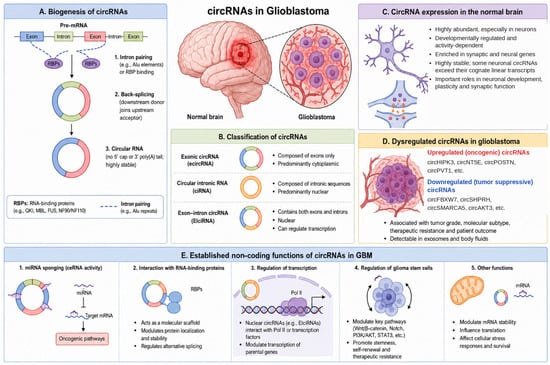
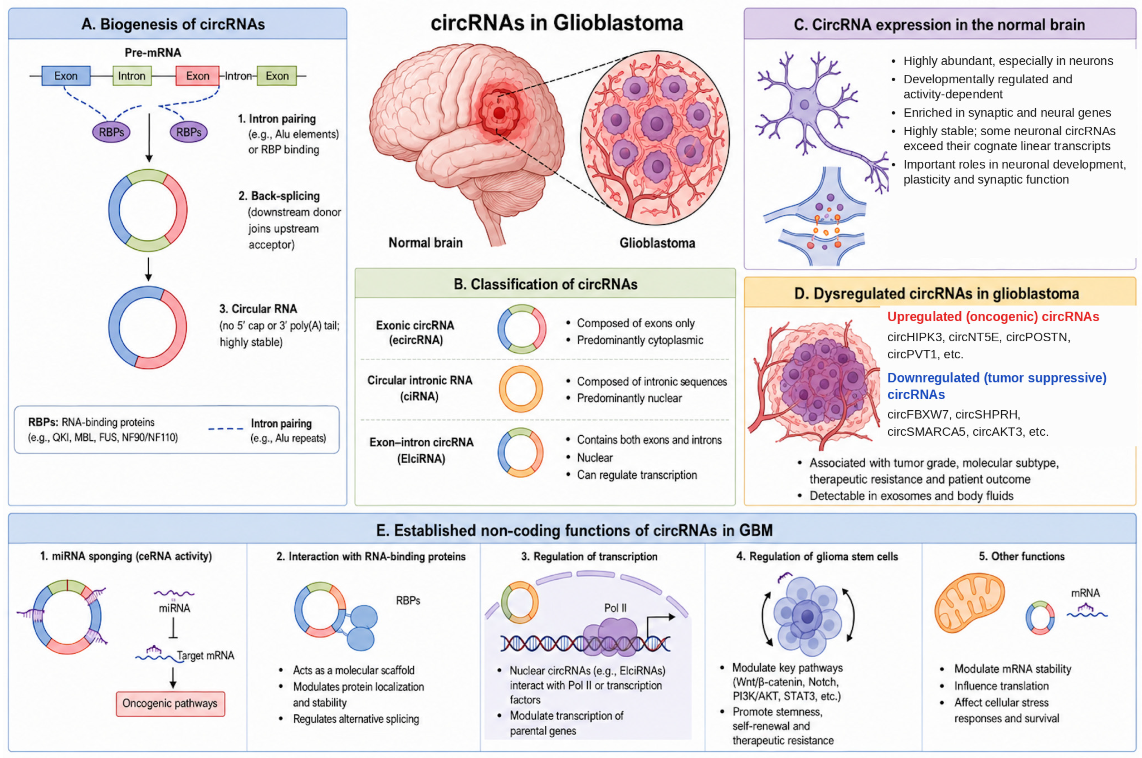
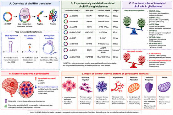
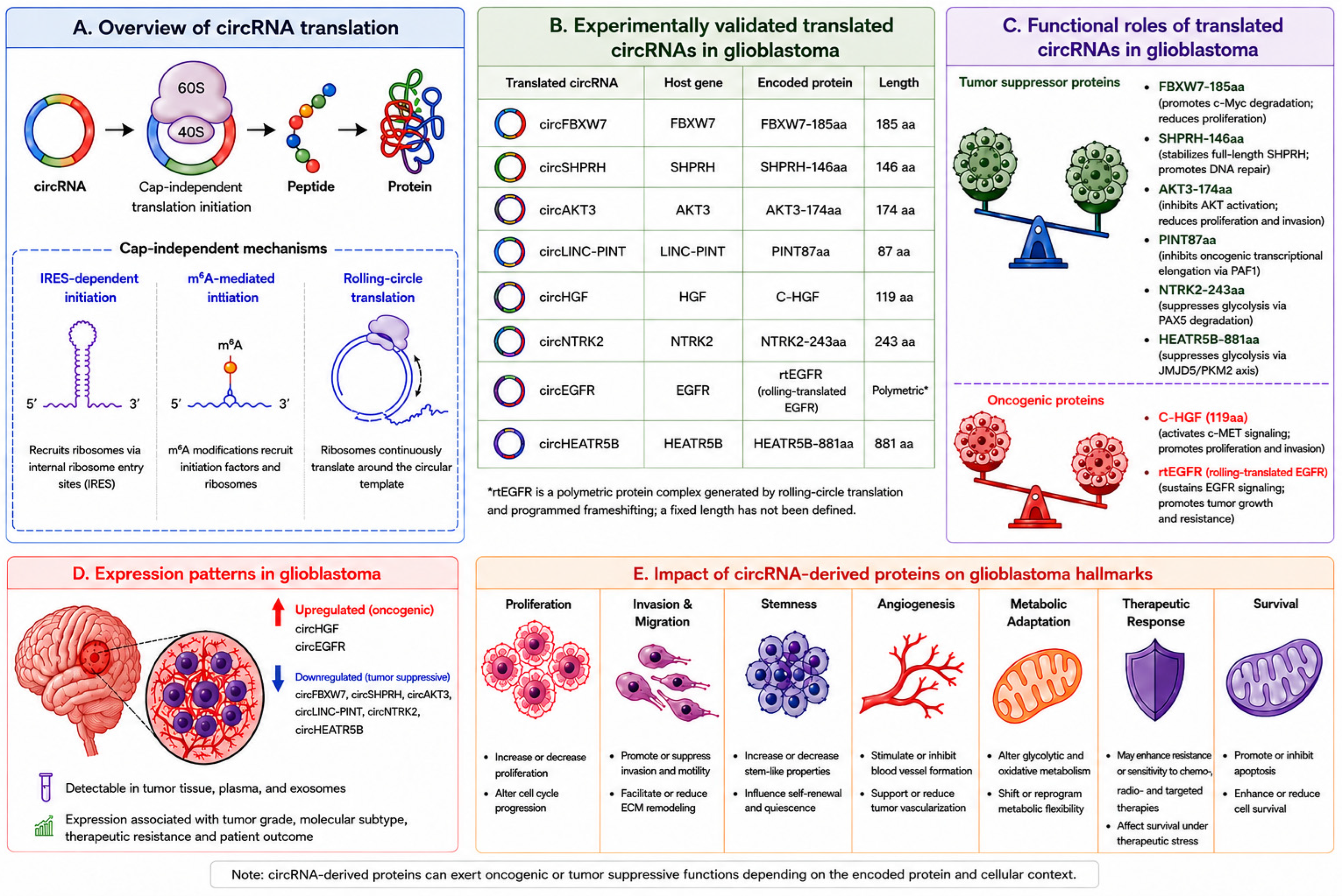
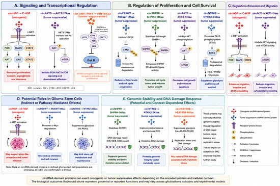
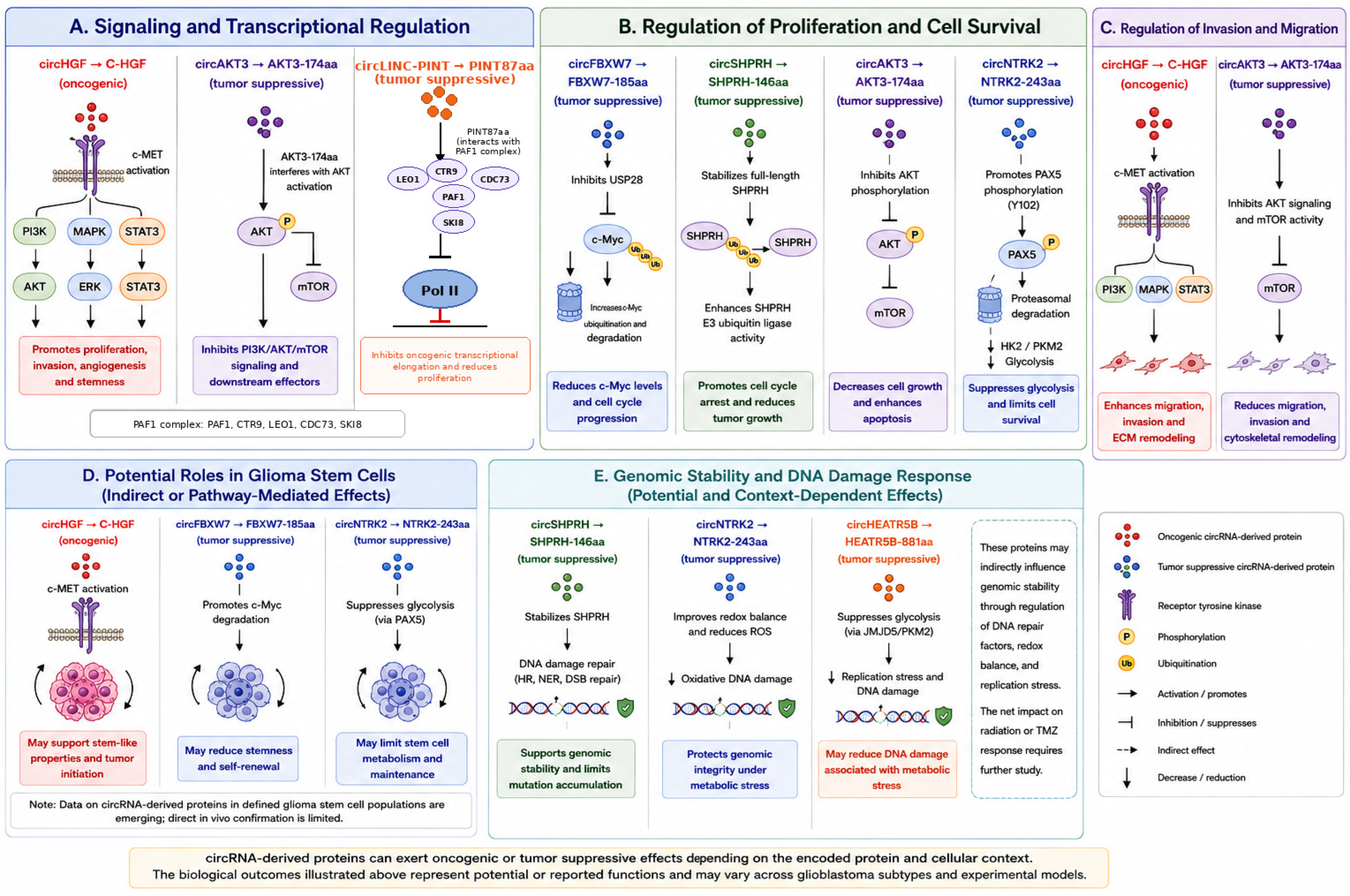
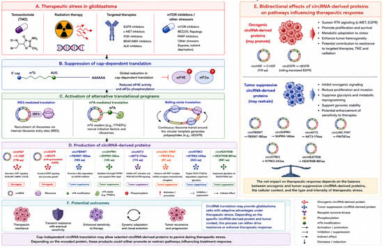
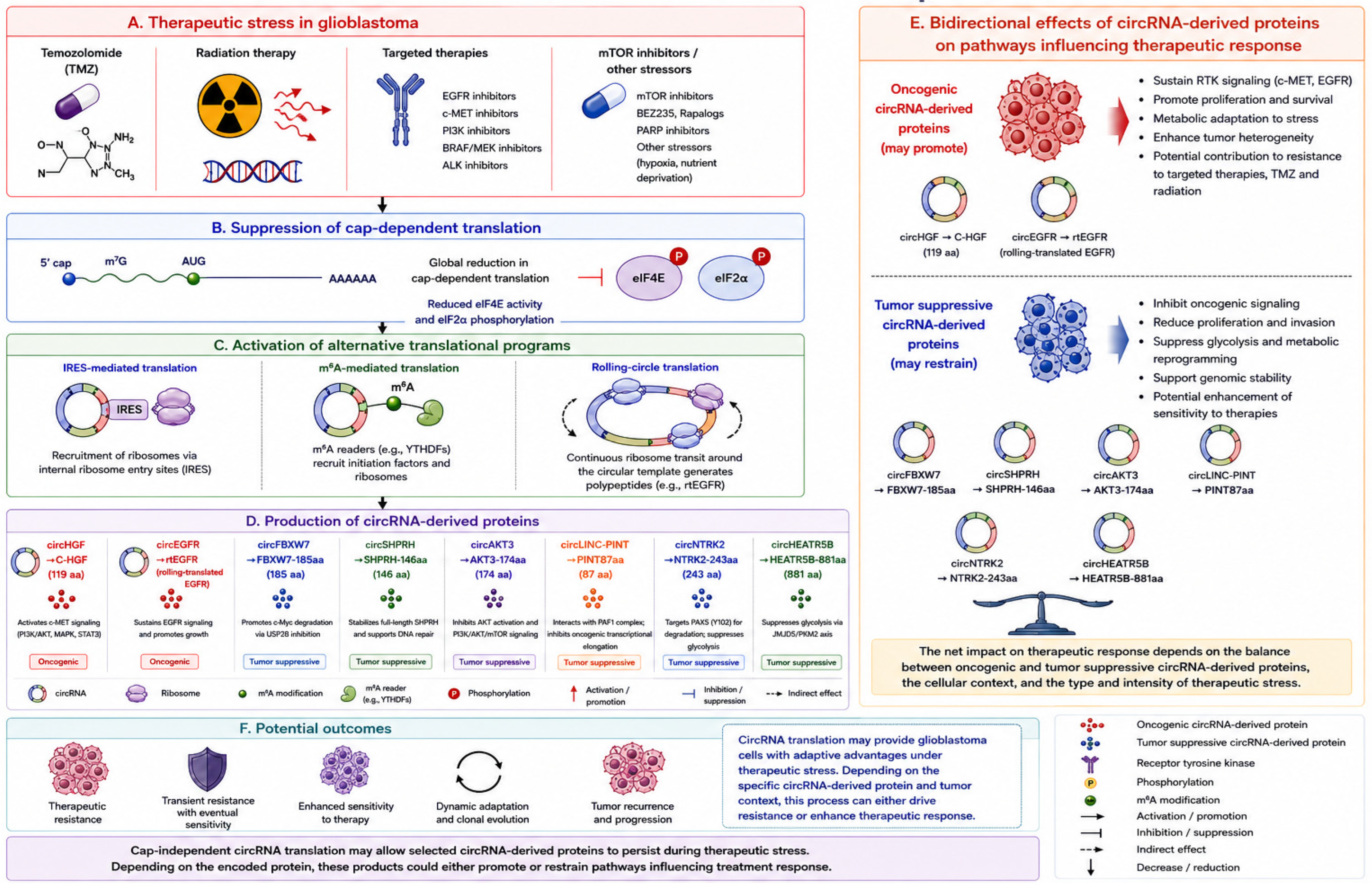

Disclaimer/Publisher’s Note: The statements, opinions and data contained in all publications are solely those of the individual author(s) and contributor(s) and not of MDPI and/or the editor(s). MDPI and/or the editor(s) disclaim responsibility for any injury to people or property resulting from any ideas, methods, instructions or products referred to in the content. |
© 2026 by the authors. Licensee MDPI, Basel, Switzerland. This article is an open access article distributed under the terms and conditions of the Creative Commons Attribution (CC BY) license.