


Abstract
Photobiomodulation (PBM) is a non-thermal therapeutic modality in which low-intensity red or near-infrared light is used to modulate biological activity. Experimental inner-ear studies report preservation of auditory neurons, attenuation of vestibular injury, improved survival of transplanted stem cells, and changes in otic organoid differentiation. These observations raise the possibility that PBM may influence differentiation-associated cellular states in addition to providing cytoprotection. The classical mechanistic model proposes that photons interact with mitochondrial chromophores, particularly cytochrome c oxidase, leading to changes in respiration, ATP, reactive oxygen species (ROS), nitric oxide, calcium, and downstream signaling; however, a universal primary photoreceptor has not been established. In a 2020 mouse embryonic-stem-cell-derived otic-organoid study, PBM was associated with more organoid-like structures, increased hair-cell-associated markers, increased ROS, and lower Hes5 expression. The RNA-sequencing comparison pooled five embryoid bodies into one sample per condition, limiting biological replication and statistical inference. Accordingly, the Hes5 result should be regarded as exploratory and does not demonstrate direct Notch inhibition. Studies in other regenerative tissues show that PBM can engage ROS-dependent PI3K/AKT/GSK3β/β-catenin signaling, providing a mechanistic precedent rather than proof for the inner ear. This review therefore presents a testable working model in which PBM may connect mitochondrial and redox responses to Wnt/β-catenin and Notch/HES5 pathways during otic differentiation, while emphasizing that mature hair-cell regeneration, new SGN generation, and hearing restoration have not yet been established.
Keywords:
photobiomodulation; inner ear; hair-cell differentiation; neural protection; Wnt; Notch; cell fate 1. Introduction
Sensorineural hearing loss is characterized by irreversible or poorly reversible damage to cochlear hair cells, supporting cells, synaptic structures, and spiral ganglion neurons (SGNs). Because mature mammalian cochlear hair cells have very limited spontaneous regenerative capacity, restoration of hearing after extensive sensory-cell or neuronal loss remains a major challenge [1]. Current clinical approaches, including hearing aids [2] and cochlear implantation, compensate for lost function but do not reconstruct damaged sensory or neural structures. Regenerative strategies therefore include stem-cell transplantation, endogenous progenitor activation, gene therapy, growth-factor delivery, and organoid-based approaches [3,4,5]. Foundational studies have established that ATOH1 is required for hair-cell development and that forced ATOH1 expression can induce hair-cell-like phenotypes [6,7,8,9,10]. Wnt/β-catenin signaling regulates cochlear progenitor proliferation and hair-cell differentiation, whereas Notch/HES signaling helps maintain supporting-cell states and restrict hair-cell fate [11,12,13,14,15,16]. These developmental pathways provide the biological framework for asking whether a physical stimulus such as PBM can modify progenitor state.
PBM uses low-intensity red or near-infrared light to modify cellular physiology and has been associated with mitochondrial, redox, inflammatory, proliferative, and differentiation responses [17,18,19]. Direct inner-ear evidence is nevertheless limited and unevenly distributed. Several of the studies most central to the present hypothesis—including the auditory-neuropathy, vestibular, otic-organoid, and transplantation studies—were produced by the same research group [20,21,22,23,24]. Independent studies provide complementary evidence for cytoprotective or stress-modulating effects in auditory-cell and noise-injury models [25,26], whereas a randomized placebo-controlled human study did not detect significant auditory benefit [27]. This concentration of evidence does not invalidate the findings, but it limits independent replication and should temper mechanistic and translational claims.
Throughout this review, “protection” denotes preservation of existing cells or function after injury; “repair” denotes partial restoration of damaged structures without proof of newly generated, integrated cells; and “regeneration” denotes the production or replacement of lost cells followed by maturation, structural integration, and functional contribution. Under these definitions, the current SGN evidence supports neural protection, not demonstrated neurogenesis or SGN regeneration. The central question is therefore whether PBM may become a component of a regenerative strategy, rather than whether it has already been shown to regenerate the inner ear.
2. PBM in the Inner Ear: From Cytoprotection Toward Regenerative Hypotheses
The early inner-ear PBM literature primarily supports tissue protection. In ouabain-induced auditory neuropathy, 808 nm PBM improved ABR thresholds and preserved SGNs, neurofilaments, and postsynaptic puncta [20]. The study did not demonstrate birth of new neurons, lineage conversion, or functional integration of new SGNs; it should therefore be interpreted as neural protection rather than regeneration. In gentamicin-induced vestibular dysfunction, near-infrared PBM improved vestibular function and reduced histological injury [21]. Independent studies likewise reported stress-modulating or protective effects in auditory cells and noise-exposed cochleae [25,26]. Together, these data support a plausible cytoprotective action across sensory and neural compartments, but they do not show replacement of lost inner-ear cells.
The distinction between survival and regeneration is especially important for cell transplantation. In the 2023 secondary-neurodegeneration study, PBM increased the persistence and distribution of transplanted GFP-positive embryonic stem cells, but neither ABR nor eABR recovered and nerve regeneration was not demonstrated [23]. One of three animals in the combined stem-cell-plus-PBM group had a deformed cochlea with widespread GFP-positive cells and absent neurofilament expression in the abnormal sample. A single animal cannot establish toxicity, but the finding is a safety signal that warrants cautious reporting, longer follow-up, lineage tracing, and larger studies. Enhanced survival is necessary for transplantation strategies, but it is insufficient without controlled lineage specification, maturation, synapse formation, and integration into the auditory circuit.
The current evidence therefore supports a transition from studying PBM solely as a cytoprotective intervention toward testing it as a possible adjunct to regeneration. That conceptual shift should not be confused with evidence that PBM itself regenerates hair cells or SGNs. A systematic review identified 17 eligible PBM studies across animal, human, and ex vivo models, with substantial heterogeneity in wavelength, dose, delivery, and endpoints [28]. Standardized dosimetry, independent replication, and functional endpoints are prerequisites for stronger claims (Table 1).
Table 1.
Representative PBM studies relevant to inner-ear regeneration. Dosimetry is reported as wavelength, irradiance, fluence, and exposure; NR = not reported; N/A = not applicable; calculated fluence values are identified explicitly.
Evidence-level criteria: “Moderate” indicates direct inner-ear observations with biological replication and/or support from independent studies, but incomplete causal or functional validation. “Preliminary” indicates limited direct evidence from a small number of studies or incompletely replicated experiments. “Very limited” indicates indirect, pooled, under-replicated, or association-only evidence. “Not established/Unknown” indicates no direct demonstration. Ratings also consider whether evidence is inner-ear-specific or extrapolated, independent replication, biological replicate number, association versus intervention, and whether endpoints are molecular, structural, or functional.
3. PBM and Inner-Ear Organoid Differentiation
The development of inner-ear organoids provides a useful platform for determining whether PBM may modify differentiation-associated cell states rather than simply preserve existing cells. In mouse embryonic-stem-cell-derived embryoid bodies, 630 nm LED PBM was associated with more organoid-like structures and increased expression of otic and hair-cell-associated markers, including Pax2, Pax8, Atoh1, and Myo7a. MYO7A-positive cells and FM1-43 uptake were also reported [22]. These endpoints support altered differentiation and the presence of hair-cell-like features, but they do not by themselves establish mature functional hair cells or regeneration in vivo.
The transcriptomic component of the 2020 study should be interpreted more cautiously than the original wording of this review implied. For each condition, five embryoid bodies were pooled to generate the RNA-seq sample, leaving no independent biological replicate at the sequencing level [22]. The observed decrease in Hes5 is therefore preliminary and hypothesis generating. Because HES5 is a downstream effector of Notch signaling, lower Hes5 expression is compatible with altered Notch-related transcription, but it does not prove direct inhibition of Notch. Direct evidence would require replicated measurements of NICD generation or localization, RBPJ-dependent transcription, and pathway perturbation or rescue.
The strengths and limitations of the organoid evidence should be considered together: PBM-associated differences were detected across organoid yield, marker expression, ROS, and cellular morphology, but the model remains an early developmental system, the transcriptomic comparison was under-replicated, and independent laboratories have not yet reproduced the cell-fate result. Future studies should predefine biological replicates, analyze individual organoids or independent differentiation batches, and connect molecular changes to electrophysiological maturation.
The effects of PBM on otic stem-cell behavior are not restricted to the 630 nm wavelength. A subsequent study examined PBM at wavelengths above 800 nm in stem cells within otic organoids and the scala media of the cochlea and demonstrated morphological changes in stem cells together with increased formation of otic vesicle-like structures, suggesting that near-infrared PBM can modify cellular behavior within an otic developmental environment [24]. The observation that both visible red and near-infrared wavelengths can influence otic stem-cell behavior raises an important question regarding the relationship between wavelength and molecular mechanism. The biological response to PBM is strongly dependent on wavelength, irradiance, fluence, exposure duration, treatment frequency, tissue depth, and cellular state, and a systematic review of PBM for hearing loss identified 17 eligible studies with considerable variation in treatment parameters [28]. Most animal studies used wavelengths between 808 and 830 nm, whereas inner-ear organoid studies included both 630 nm and wavelengths above 800 nm. Such heterogeneity complicates direct comparison between studies and makes it difficult to determine whether different wavelengths converge on a common molecular pathway or activate distinct biological mechanisms. A major future objective should therefore be to determine whether red and near-infrared PBM generate a common cellular state despite differences in photon absorption, tissue penetration, and intracellular targets.
ATOH1 is a central transcription factor in cochlear hair-cell specification [6,7,8,9,10], and Wnt/β-catenin and Notch signaling regulate the balance among progenitor proliferation, supporting-cell identity, and hair-cell differentiation [11,12,13,14,15,16]. Nevertheless, increased Atoh1 or Myo7a transcript abundance is a differentiation-associated molecular endpoint, not proof of functional regeneration. A mature hair cell must acquire organized stereocilia, mechanotransduction currents, appropriate electrophysiological properties, ribbon synapses, and functional connectivity. FM1-43 uptake in the PBM-treated organoids provides an additional functional clue [22], but ultrastructural, electrophysiological, and synaptic validation remains necessary.
Inner-ear organoids remain an attractive platform for mechanistic PBM research because they permit assessment of differentiation, spatial organization, developmental timing, and interactions among multiple cell populations [29]. PBM could be tested alongside defined Wnt activation and Notch inhibition schedules [11,16], but such combinations should be framed as experimental strategies rather than established protocols. The next step is to determine whether PBM reproducibly changes lineage allocation across independent differentiation batches and whether any increase in hair-cell-like cells is accompanied by maturation, synaptic connectivity, and functional integration.
4. Molecular Mechanisms of PBM: Mitochondria, ROS, Wnt, and Notch
A widely discussed PBM model proposes that photons interact with mitochondrial chromophores, particularly cytochrome c oxidase, thereby modifying electron transport, mitochondrial membrane potential, ATP production, ROS generation, and nitric oxide signaling [17,18]. Mitochondria are central to hair-cell bioenergetics and stress responses, and mitochondrial calcium handling can drive ROS production during hair-cell injury [30,31,32]. These observations make mitochondrial signaling biologically plausible in the inner ear, but they do not identify a universal PBM photoreceptor or show that the same mechanism controls otic differentiation. Reviews and isolated-enzyme experiments have questioned whether cytochrome c oxidase alone can account for red or near-infrared responses [33,34]. A contemporary working model should therefore allow multiple photoreceptive, mitochondrial, membrane, ion-channel, and intracellular mechanisms [35] (Table 2).
Table 2.
PBM-associated signaling pathways relevant to inner-ear regeneration.
ROS can act either as mediators of injury or as transient second messengers, depending on magnitude, timing, compartment, and cellular state. The otic-organoid study reported higher ROS after PBM together with altered differentiation markers [22], but did not establish that ROS was necessary for the phenotype. In a hair-follicle stem-cell model, by contrast, ROS scavenging and β-catenin manipulation provided causal evidence linking PBM-induced ROS to PI3K/AKT/GSK3β/β-catenin signaling and hair regeneration [36]. Neural-injury studies have also reported increased neuroprogenitor markers after transcranial PBM [37]. These cross-system findings support experimental hypotheses for the inner ear; they should not be presented as evidence that PBM has already activated the same regenerative pathways in cochlear progenitors.
Wnt/β-catenin signaling is a major regulator of cochlear development, sensory progenitor proliferation, and hair-cell differentiation [12,13,14]. Sequential Wnt activation and Notch inhibition can promote hair-cell production in postnatal cochlear models [16]. Outside the auditory system, PBM has increased Wnt-related readouts in outer-root-sheath cells [38] and produced causal β-catenin-dependent regeneration in hair follicles [36]. These studies provide mechanistic precedents, but direct PBM-induced Wnt/β-catenin activation has not been demonstrated in otic cells. Inner-ear studies should therefore use β-catenin reporters, pathway inhibitors, genetic perturbation, and rescue experiments before assigning Wnt a causal role.
Notch signaling regulates the balance between supporting-cell and hair-cell fates during cochlear development. NICD interacts with RBPJ to induce HES-family transcriptional repressors, whereas Notch inhibition can permit ATOH1-dependent hair-cell differentiation and, in selected injury models, hair-cell regeneration [11,15,16]. The exploratory decrease in Hes5 observed after PBM is therefore a reasonable basis for a hypothesis [22], but not evidence of direct Notch inhibition. The sequencing design used one pooled sample per condition, and proximal pathway readouts were not measured. Replicated NICD, RBPJ-reporter, and HES5 measurements, followed by gain- and loss-of-function rescue, are required to establish causality.
A testable possibility is that PBM-induced metabolic or redox changes alter the responsiveness of otic progenitors to multiple developmental pathways rather than directly targeting a single pathway. In this model, β-catenin activation and reduced Notch/HES5 activity could converge on ATOH1-associated differentiation. At present, the Wnt branch is extrapolated from other tissues and the Notch branch is supported only by an exploratory downstream transcript. The model should therefore guide experiments, not be interpreted as a demonstrated molecular sequence in the inner ear.
5. PBM as a Potential Modulator of Inner-Ear Cell State and the Regenerative Microenvironment
PBM may be most usefully conceptualized as a context-dependent physical modulator of the regenerative microenvironment rather than a single-target pharmacological treatment (Table 3). Stem-cell differentiation depends on growth factors, extracellular matrix, cell–cell interactions, metabolic state, and developmental signaling. PBM could modify cellular responsiveness through mitochondrial, redox, membrane, or ion-channel effects. In mature injured cochlear tissue, these effects may primarily support survival; during an organoid differentiation window, they may alter differentiation-associated cellular states. This context dependence may help explain why similar wavelengths or doses produce different outcomes across models.
Table 3.
Mechanistic evidence hierarchy and proposed experiments.
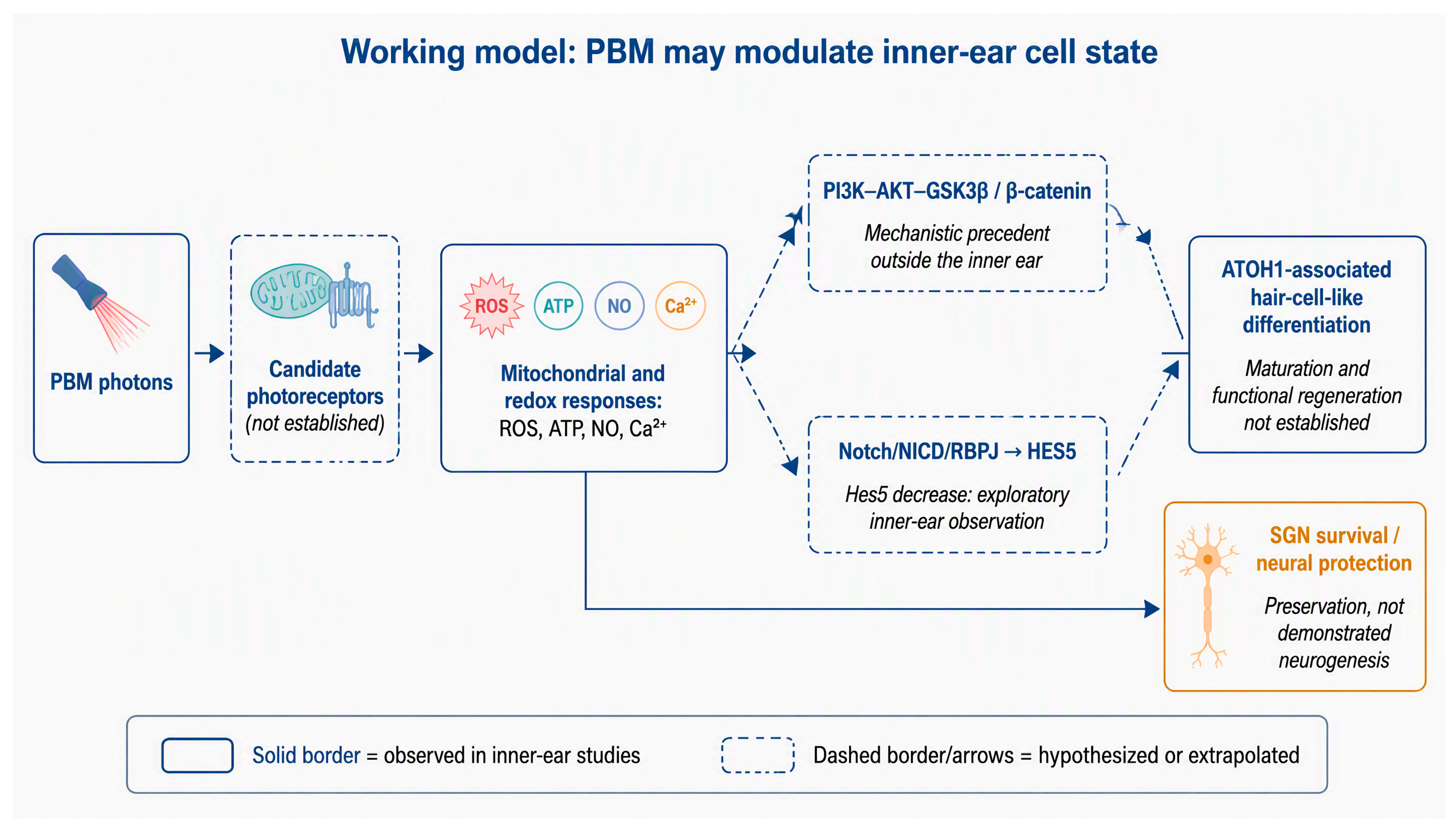
Figure 1 summarizes the proposed mechanism while distinguishing direct inner-ear observations from extrapolated or hypothetical steps. PBM may initiate responses through one or more unconfirmed photoreceptive mechanisms, followed by changes in mitochondrial activity and redox mediators. These events may engage Wnt/β-catenin, or Notch/HES5 signaling and thereby alter ATOH1-associated differentiation. The same upstream responses may also support SGN survival. The current data support mitochondrial/redox changes, exploratory otic differentiation associations, and neural protection; they do not yet establish the complete pathway, mature hair-cell regeneration, or new SGN generation.
Figure 1.
Working model for PBM-associated inner-ear responses. Solid borders and arrows denote observations reported in inner-ear models; dashed elements denote hypotheses or mechanistic extrapolations from other tissues. The decrease in Hes5 was observed in an exploratory pooled RNA-seq comparison and does not establish direct Notch inhibition. SGN data support neural protection rather than demonstrated neurogenesis. PBM, photobiomodulation; ROS, reactive oxygen species; NO, nitric oxide; SGN, spiral ganglion neuron.
6. From Association to Causality: Current Mechanistic Limitations and Experimental Strategies
The most important limitation of the current PBM literature is the weakness of direct causal evidence. Many studies report changes in markers, but few establish that the marker is required for the biological response. Reduced Hes5 after PBM does not prove direct Notch inhibition [22], and increased β-catenin in a non-otic model does not establish a cochlear Wnt mechanism [36,38]. The inner-ear evidence base is also concentrated in a small number of studies, several from the same research group [20,21,22,23,24], and the key RNA-seq comparison pooled embryoid bodies without independent sequencing-level biological replicates [22]. Additional limitations include uncertainty regarding the primary photoreceptor [33,34,35], heterogeneous wavelengths and dosimetry [28], short follow-up, small animal cohorts, and reliance on molecular or histological endpoints. Marker-positive cells should not be equated with regenerated functional cells.
The central challenge for the field is to establish a causal chain between photon exposure and regenerative outcome. Future studies should therefore move beyond single time-point measurements and investigate the temporal sequence of molecular events. A particularly informative experiment would simultaneously measure ROS, mitochondrial membrane potential, ATP, NICD, HES5, β-catenin, ATOH1, POU4F3, GFI1, and MYO7A at multiple time points after PBM. If ROS changes precede alterations in NICD or β-catenin and these changes subsequently precede ATOH1 induction and hair-cell differentiation, a temporal hierarchy would be established. However, temporal association alone would not prove causality. Causal evidence would require pathway perturbation. For example, if inhibition of ROS prevented PBM-induced HES5 suppression, ROS would be positioned upstream of Notch-related signaling. If forced NICD expression prevented PBM-induced ATOH1 and MYO7A expression, Notch signaling would be demonstrated as a necessary mediator. Similarly, if inhibition of β-catenin abolished PBM-induced differentiation, Wnt/β-catenin signaling would become a causal component of the response. Such experiments would transform the field from descriptive PBM biology into mechanistically defined regenerative biology.
7. Translational Perspectives and Future Applications
The most realistic near-term role of PBM is as an experimental adjunct that may improve the biological environment for regenerative therapies. Potential preclinical applications include supporting transplanted-cell survival, modulating defined differentiation windows, or preserving SGNs as a substrate for electrical stimulation. However, the 2023 transplantation study increased cell persistence without ABR/eABR recovery or demonstrated nerve regeneration, and one animal showed a deformed cochlea with widespread GFP-positive cells [23]. This isolated finding does not prove toxicity, but it argues for dose control, lineage restriction, tumorigenicity surveillance, and longer safety studies before translation.
Clinical plausibility must be separated from clinical evidence. PBM has theoretical advantages as a non-pharmacological, parameter-adjustable intervention, but light penetration, cochlear dose distribution, target specificity, and treatment timing remain unresolved. Human studies are sparse and heterogeneous; a randomized placebo-controlled study found no significant improvement in auditory outcomes [27], and no human study has demonstrated hair-cell or SGN regeneration. Stage-specific PBM protocols—survival, lineage specification, maturation, and integration—should therefore be tested first in adequately powered preclinical models with functional endpoints.
8. Conclusions
PBM research in the inner ear has expanded from cytoprotection to exploratory studies of stem-cell behavior and otic organoid differentiation. Current evidence supports preservation of SGNs and auditory neural structures, attenuation of vestibular or cochlear injury in selected models, and improved survival of transplanted cells [20,21,23,25,26]. The SGN findings constitute neural protection, not demonstrated regeneration. In otic organoids, PBM was associated with increased ROS, hair-cell-associated markers, and lower Hes5 expression [22]. Because the RNA-seq analysis used one pooled sample of five embryoid bodies per condition, the Hes5 result is preliminary and does not establish direct Notch inhibition. Evidence from hair follicles and other regenerative systems supports ROS-dependent signaling as a mechanistic precedent [36,37,38], but direct Wnt activation by PBM in the inner ear remains unproven. PBM should therefore be considered a potential regenerative signal or adjunct that may alter differentiation-associated cellular states, rather than an established inducer of inner-ear regeneration. The priority is to test causal pathway perturbations, independent biological replication, lineage tracing, mature hair-cell and synaptic endpoints, long-term ABR/eABR recovery, and safety. Until those criteria are met, claims of functional hair-cell regeneration, SGN regeneration, or clinical hearing restoration are not established.
Funding
This research was supported by the Basic Science Research Program through the National Research Foundation of Korea (NRF) (RS-2020-NR049585; RS-2021-NR065982; RS-2022-NR075691; RS-2025–25414419; RS-2026-25619632), and the Bio&Medical Technology Development Program of the National Research Foundation (NRF) funded by the Korean government (RS-2023–00220408).
Institutional Review Board Statement
Not applicable.
Data Availability Statement
No new data were created or analyzed in this study. Data sharing is not applicable to this article.
Acknowledgments
During the preparation of this manuscript/study, the author used OpenAI OpCo, LLC for the purposes of review and revision. The author has reviewed and edited the output and takes full responsibility for the content of this publication.
Conflicts of Interest
The author declares no conflicts of interest.
References
- Izumikawa, M.; Batts, S.A.; Miyazawa, T.; Swiderski, D.L.; Raphael, Y. Response of the flat cochlear epithelium to forced expression of Atoh1. Hear. Res. 2008, 240, 52–56. [Google Scholar] [CrossRef] [Scilit]
- Kang, M.; Seo, J.-H.; Song, J.-J.; Moon, I.J.; Park, Y.-H.; Park, M.K. Practical Guidelines for Smartphone-Based Hearing Aid Application and Personal Sound Amplification Device Usage to Correct Hearing Loss in Korea Based on a Randomized Controlled Trial by the Patient-Centered Clinical Research Coordinating Center. J. Audiol. Otol. 2025, 29, 237–241. [Google Scholar] [CrossRef] [Scilit]
- Devare, J.; Gubbels, S.; Raphael, Y. Outlook and future of inner ear therapy. Hear. Res. 2018, 368, 127–135. [Google Scholar] [CrossRef] [Scilit]
- Lee, M.Y.; Park, Y.H. Potential of Gene and Cell Therapy for Inner Ear Hair Cells. BioMed Res. Int. 2018, 2018, 8137614, Erratum in BioMed Res. Int. 2019, 2019, 9601260. https://doi.org/10.1155/2019/9601260. [Google Scholar] [CrossRef] [Scilit]
- Kim, D.K. Recent Advances in Functionalized Nanoparticles for Targeted and Controlled Inner Ear Therapy via Localized Cochlear Delivery. J. Audiol. Otol. 2025, 29, 159–165. [Google Scholar] [CrossRef] [Scilit]
- Izumikawa, M.; Minoda, R.; Kawamoto, K.; A Abrashkin, K.; Swiderski, D.L.; Dolan, D.F.; E Brough, D.; Raphael, Y. Auditory hair cell replacement and hearing improvement by Atoh1 gene therapy in deaf mammals. Nat. Med. 2005, 11, 271–276. [Google Scholar] [CrossRef] [Scilit]
- Lee, S.; Song, J.-J.; Beyer, L.A.; Swiderski, D.L.; Prieskorn, D.M.; Acar, M.; Jen, H.-I.; Groves, A.K.; Raphael, Y. Combinatorial Atoh1 and Gfi1 induction enhances hair cell regeneration in the adult cochlea. Sci. Rep. 2020, 10, 21397. [Google Scholar] [CrossRef] [Scilit]
- Liu, Y.; Yang, L.; Singh, S.; A Beyer, L.; Prieskorn, D.M.; Swiderski, D.L.; Groves, A.K.; Raphael, Y. Combinatorial Atoh1, Gfi1, Pou4f3, and Six1 gene transfer induces hair cell regeneration in the flat epithelium of mature guinea pigs. Hear. Res. 2024, 441, 108916. [Google Scholar] [CrossRef] [Scilit]
- Bermingham, N.A.; Hassan, B.A.; Price, S.D.; Vollrath, M.A.; Ben-Arie, N.; Eatock, R.A.; Bellen, H.J.; Lysakowski, A.; Zoghbi, H.Y. Math1: An essential gene for the generation of inner ear hair cells. Science 1999, 284, 1837–1841. [Google Scholar] [CrossRef] [Scilit]
- Zheng, J.L.; Gao, W.Q. Overexpression of Math1 induces robust production of extra hair cells in postnatal rat inner ears. Nat. Neurosci. 2000, 3, 580–586. [Google Scholar] [CrossRef] [Scilit]
- Jung, J.Y.; Avenarius, M.R.; Adamsky, S.; Alpert, E.; Feinstein, E.; Raphael, Y. siRNA targeting Hes5 augments hair cell regeneration in aminoglycoside-damaged mouse utricle. Mol. Ther. 2013, 21, 834–841. [Google Scholar] [CrossRef] [Scilit]
- Chai, R.; Kuo, B.; Wang, T.; Liaw, E.J.; Xia, A.; Jan, T.A.; Liu, Z.; Taketo, M.M.; Oghalai, J.S.; Nusse, R.; et al. Wnt signaling induces proliferation of sensory precursors in the postnatal mouse cochlea. Proc. Natl. Acad. Sci. USA 2012, 109, 8167–8172. [Google Scholar] [CrossRef] [Scilit]
- Jacques, B.E.; Puligilla, C.; Weichert, R.M.; Ferrer-Vaquer, A.; Hadjantonakis, A.-K.; Kelley, M.W.; Dabdoub, A. A dual function for canonical Wnt/β-catenin signaling in the developing mammalian cochlea. Development 2012, 139, 4395–4404. [Google Scholar] [CrossRef] [Scilit]
- Shi, F.; Hu, L.; Jacques, B.E.; Mulvaney, J.F.; Dabdoub, A.; Edge, A.S. β-Catenin is required for hair-cell differentiation in the cochlea. J. Neurosci. 2014, 34, 6470–6479. [Google Scholar] [CrossRef] [Scilit]
- Mizutari, K.; Fujioka, M.; Hosoya, M.; Bramhall, N.; Okano, H.J.; Okano, H.; Edge, A.S. Notch inhibition induces cochlear hair cell regeneration and recovery of hearing after acoustic trauma. Neuron 2013, 77, 58–69, Erratum in Neuron 2013, 78, 403. Erratum in Neuron 2015, 96, 341. [Google Scholar] [CrossRef] [Scilit]
- Ni, W.; Zeng, S.; Li, W.; Chen, Y.; Zhang, S.; Tang, M.; Sun, S.; Chai, R.; Li, H. Wnt activation followed by Notch inhibition promotes mitotic hair cell regeneration in the postnatal mouse cochlea. Oncotarget 2016, 7, 66754–66768. [Google Scholar] [CrossRef] [Scilit]
- de Freitas, L.F.; Hamblin, M.R. Proposed Mechanisms of Photobiomodulation or Low-Level Light Therapy. IEEE J. Sel. Top. Quantum Electron. 2016, 22, 7000417. [Google Scholar] [CrossRef] [Scilit]
- Chang, S.Y.; Lee, M.Y.; Chung, P.-S.; Kim, S.; Choi, B.; Suh, M.-W.; Rhee, C.-K.; Jung, J.Y. Enhanced mitochondrial membrane potential and ATP synthesis by photobiomodulation increases viability of the auditory cell line after gentamicin-induced intrinsic apoptosis. Sci. Rep. 2019, 9, 19248. [Google Scholar] [CrossRef] [Scilit]
- Choi, S.H.; Chang, S.; Biswas, R.; Chung, P.; Mo, S.; Lee, M.Y.; Ahn, J.C. Light-emitting diode irradiation using 660 nm promotes human fibroblast HSP90 expression and changes cellular activity and morphology. J. Biophotonics 2019, 12, e201900063. [Google Scholar] [CrossRef] [Scilit]
- Lee, M.Y.; Bae, S.-H.; Chang, S.-Y.; Lee, J.-H.; Kim, S.-H.; Ahn, J.-C.; Chung, P.-S.; Moy, W.; Jung, J.Y. Photobiomodulation by laser therapy rescued auditory neuropathy induced by ouabain. Neurosci. Lett. 2016, 633, 165–173. [Google Scholar] [CrossRef] [Scilit]
- Lee, M.Y.; Hyun, J.-H.; Suh, M.-W.; Ahn, J.-C.; Chung, P.-S.; Jung, J.Y. Treatment of peripheral vestibular dysfunction using photobiomodulation. J. Biomed. Opt. 2017, 22, 088001. [Google Scholar] [CrossRef] [Scilit]
- Chang, S.Y.; Carpena, N.T.; Mun, S.; Jung, J.Y.; Chung, P.-S.; Shim, H.; Han, K.; Ahn, J.-C.; Lee, M.Y. Enhanced Inner-Ear Organoid Formation from Mouse Embryonic Stem Cells by Photobiomodulation. Mol. Ther. Methods Clin. Dev. 2020, 17, 556–567. [Google Scholar] [CrossRef] [Scilit]
- Chang, S.Y.; Kim, E.; Carpena, N.T.; Lee, J.-H.; Kim, D.H.; Lee, M.Y. Photobiomodulation Can Enhance Stem Cell Viability in Cochlea with Auditory Neuropathy but Does Not Restore Hearing. Stem Cells Int. 2023, 2023, 6845571. [Google Scholar] [CrossRef] [Scilit]
- Chang, S.Y.; Lee, M.Y. Photobiomodulation with a wavelength >800 nm induces morphological changes in stem cells within otic organoids and scala media of the cochlea. Lasers Med. Sci. 2021, 36, 1917–1925. [Google Scholar]
- Bartos, A.; Grondin, Y.; Bortoni, M.E.; Ghelfi, E.; Sepulveda, R.; Carroll, J.; Rogers, R.A. Pre-conditioning with near infrared photobiomodulation reduces inflammatory cytokines and markers of oxidative stress in cochlear hair cells. J. Biophotonics 2016, 9, 1125–1135. [Google Scholar] [CrossRef] [Scilit]
- Tamura, A.; Matsunobu, T.; Tamura, R.; Kawauchi, S.; Sato, S.; Shiotani, A. Photobiomodulation rescues the cochlea from noise-induced hearing loss via upregulating nuclear factor κB expression in rats. Brain Res. 2016, 1646, 467–474. [Google Scholar] [CrossRef] [Scilit]
- Goodman, S.S.; Bentler, R.A.; Dittberner, A.; Mertes, I.B. The effect of low-level laser therapy on hearing. ISRN Otolaryngol. 2013, 2013, 916370. [Google Scholar] [CrossRef] [Scilit]
- Nikookam, Y.; Zia, N.; Lotfallah, A.; Muzaffar, J.; Davis-Manders, J.; Kullar, P.; Smith, M.; Bale, G.; Boyle, P.; Irving, R.; et al. The effect of photobiomodulation on hearing loss: A systematic review. Clin. Otolaryngol. 2024, 49, 41–61. [Google Scholar] [CrossRef] [Scilit]
- Moore, S.T.; Nakamura, T.; Nie, J.; Solivais, A.J.; Aristizábal-Ramírez, I.; Ueda, Y.; Manikandan, M.; Reddy, V.S.; Romano, D.R.; Hoffman, J.R.; et al. Generating high-fidelity cochlear organoids from human pluripotent stem cells. Cell Stem Cell 2023, 30, 950–961.e7. [Google Scholar] [CrossRef] [Scilit]
- Böttger, E.C.; Schacht, J. The mitochondrion: A perpetrator of acquired hearing loss. Hear. Res. 2013, 303, 12–19. [Google Scholar] [CrossRef] [Scilit]
- Esterberg, R.; Linbo, T.; Pickett, S.B.; Wu, P.; Ou, H.C.; Rubel, E.W.; Raible, D.W. Mitochondrial calcium uptake underlies ROS generation during aminoglycoside-induced hair cell death. J. Clin. Investig. 2016, 126, 3556–3566. [Google Scholar] [CrossRef] [Scilit]
- O’Sullivan, J.D.B.; Bullen, A.; Mann, Z.F. Mitochondrial form and function in hair cells. Hear. Res. 2023, 428, 108660. [Google Scholar] [CrossRef] [Scilit]
- Quirk, B.J.; Whelan, H.T. What Lies at the Heart of Photobiomodulation: Light, Cytochrome C Oxidase, and Nitric Oxide—Review of the Evidence. Photobiomodul. Photomed. Laser Surg. 2020, 38, 527–530. [Google Scholar] [CrossRef] [Scilit]
- Quirk, B.J.; Whelan, H.T. Effect of Red-to-Near Infrared Light on the Reaction of Isolated Cytochrome c Oxidase with Cytochrome c. Photomed. Laser Surg. 2016, 34, 631–637. [Google Scholar] [CrossRef] [Scilit]
- Hamblin, M.R.; Liebert, A. Photobiomodulation Therapy Mechanisms Beyond Cytochrome c Oxidase. Photobiomodul. Photomed. Laser Surg. 2022, 40, 75–77. [Google Scholar] [CrossRef] [Scilit]
- Jin, H.; Zou, Z.; Chang, H.; Shen, Q.; Liu, L.; Xing, D. Photobiomodulation therapy for hair regeneration: A synergetic activation of β-CATENIN in hair follicle stem cells by ROS and paracrine WNTs. Stem Cell Rep. 2021, 16, 1568–1583. [Google Scholar] [CrossRef] [Scilit]
- Xuan, W.; Vatansever, F.; Huang, L.; Hamblin, M.R. Transcranial low-level laser therapy enhances learning, memory, and neuroprogenitor cells after traumatic brain injury in mice. J. Biomed. Opt. 2014, 19, 108003. [Google Scholar] [CrossRef] [Scilit]
- Kim, J.E.; Woo, Y.J.; Sohn, K.M.; Jeong, K.H.; Kang, H. Wnt/β-catenin and ERK pathway activation: A possible mechanism of photobiomodulation therapy with light-emitting diodes that regulate the proliferation of human outer root sheath cells. Lasers Surg. Med. 2017, 49, 940–947. [Google Scholar] [CrossRef] [Scilit]

Disclaimer/Publisher’s Note: The statements, opinions and data contained in all publications are solely those of the individual author(s) and contributor(s) and not of MDPI and/or the editor(s). MDPI and/or the editor(s) disclaim responsibility for any injury to people or property resulting from any ideas, methods, instructions or products referred to in the content. |
© 2026 by the author. Licensee MDPI, Basel, Switzerland. This article is an open access article distributed under the terms and conditions of the Creative Commons Attribution (CC BY) license.