


Abstract
The kynurenine pathway (KP) constitutes the primary route of tryptophan catabolism, generating a spectrum of neuroactive metabolites that profoundly influence central nervous system function. Dysregulation of the KP is increasingly recognized as a critical pathogenic mechanism underlying diverse neuropathological conditions. This review critically evaluates the most widely cited mammalian cellular models currently utilized to delineate the causal role of KP alterations in neurological disease. Specifically, this article examines primary cell cultures, immortalized and tumor-derived cell lines, stem cell-derived systems, and ex vivo organotypic brain slices and tissues, highlighting their distinct methodological advantages, translational limitations, and specific enzymatic profiles. Across the described cellular systems, a recurring mechanistic theme emerges: quinolinic acid-driven mitochondrial dysfunction, oxidative stress, and NAD+ depletion converge in neurodegenerative conditions such as Alzheimer’s disease, Huntington’s disease, and amyotrophic lateral sclerosis. Conversely, kynurenic acid exhibits disorder-dependent—and at times opposing—roles, attenuating dopaminergic neurotoxicity in Parkinson’s disease models while contributing to synaptic pruning deficits in schizophrenia models. Furthermore, cellular models demonstrate that IDO1/TDO induction and downstream metabolite shifts are frequently cell type- and species-dependent, complicating direct extrapolation to human pathology. Because no single experimental system achieves complete physiological fidelity, elucidating the complex dynamics of the KP and identifying novel therapeutic targets requires the integration of data across complementary platforms.
1. Introduction
The kynurenine pathway (KP) of tryptophan metabolism has emerged as a mechanistic nexus linking immune activation, neuromodulation, and cellular energy metabolism, positioning it as a tractable therapeutic target in neurological and neuropsychiatric disorders [1,2]. The KP produces a variety of biologically active metabolites that interact bidirectionally with multiple physiological systems, including the endocrine, hematopoietic, immune, cardiovascular, and nervous systems, while the final product of the KP, NAD+, is necessary for vital cellular processes [3].
Neurodegenerative and neuropsychiatric disorders constitute one of the most significant challenges in contemporary medicine, affecting hundreds of millions of people worldwide and imposing a substantial socio-economic burden on society. Diseases such as Alzheimer’s disease (AD), Parkinson’s disease (PD), Huntington’s disease (HD), amyotrophic lateral sclerosis, multiple sclerosis, and neuropsychiatric conditions including schizophrenia, major depressive disorder (MDD), and bipolar disorder share a common yet incompletely understood feature: profound dysregulation of neuroimmune homeostasis and altered neurotransmitter metabolism [4,5,6]. Despite the various clinical manifestations that range from dysfunction to neuronal death, these conditions are united by evidence of neuroimmune dysregulation, though the nature, magnitude, and causal role of neuroinflammatory processes differ substantially across diagnostic categories [7,8]. In recent decades, accumulating evidence suggests that dysregulation and imbalances in the metabolites of the KP of tryptophan catabolism constitute a critical mechanistic link between neuroinflammation and neurodegeneration [9]. Factors such as aging, genetics, and environmental influences initiate the processes accompanying the development of neurological disorders and modulate KP enzyme expression and metabolite flux in a hypoxia-inducible factor 1-alpha (HIF-1α)-dependent manner [10], through proinflammatory cytokine signaling, particularly via IFN-γ [11], and through the upregulation of key enzymes by amyloid-β (Aβ) and hyperphosphorylated tau [12,13]. Additionally, gut microbiota dysbiosis represents a further upstream modulator of KP activity [14]. Altered KP metabolism contributes to the development of mitochondrial dysfunction and oxidative stress, which, together with hypoxia, inflammation, and abnormal protein aggregation, drive neuronal loss (Figure 1) [15].
Figure 1.
A schematic representation of the KP’s role in the cellular processes involved in neurodegeneration. Genetic, environmental, and microbiota-related factors converge on neuroinflammation and KP dysregulation, which promote neuronal damage through excitotoxicity, NAD+ depletion, oxidative stress, and mitochondrial dysfunction, culminating in apoptosis and neuronal loss. Gray indicates factors that contribute to the development of the pathological processes shown in orange. Minor and secondary interactions are simplified for clarity. NAD+, nicotinamide adenine dinucleotide.
Despite the compelling and consistent associations observed in clinical studies—including changes in QUIN and KYNA levels in cerebrospinal fluid of patients with AD, HD, depression [16,17,18,19], increased IDO1 expression in hippocampal sections in AD [20], and altered KYN/TRP ratios in schizophrenia [21]—correlational data from human cohorts are contradictory and have limited capacity to establish causality, dissect mechanisms, or evaluate therapeutic interventions. Human studies are confounded by disease heterogeneity, comorbidities, medication effects, and the inaccessibility of the living brain to direct biochemical interrogation. It is precisely for these reasons that well-established experimental models, including cell-based models—ranging from primary cultures and organotypic preparations to in vitro cell culture systems—are indispensable tools for advancing our mechanistic understanding of the KP’s involvement in neurological disease. Such models enable precise manipulation of individual enzymatic steps and targeted pharmacological and genetic modulation at defined nodes of the pathway, as well as directly evaluating the subsequent phenotypic, biochemical and functional consequences, thereby enabling causal inference that cannot be drawn from correlational human data alone.
The present review appraises the cellular experimental models of mammalian origin most frequently employed to elucidate the role of the KP in major neuroinflammatory, neurodegenerative, and neuropsychiatric disorders—primary cultures, immortalized and tumor-derived cell lines, stem cell-derived systems, organotypic brain slice cultures (OBSCs) and ex vivo preparations—with emphasis on the model-specific features, strengths, and limitations of KP expression and metabolism in each system, an essential prerequisite for correctly interpreting what these models reveal about the pathophysiological role of the KP in neurological disease and for guiding the development of KP-targeted therapeutic strategies (Table 1).
Table 1.
A comparative overview of the cellular models employed in the study of the kynurenine pathway.
2. Literature Search Strategy
For this review, a comprehensive literature search was conducted in the PubMed, Scopus and Google Scholar databases up to 25 June 2026 using various combinations of keywords. Representative search queries included the terms “kynurenine”, “kynurenine pathway”, as well as the names of individual KP metabolites and enzymes, combined with keywords related to specific neurological disorders (e.g., “Alzheimer’s disease”, “Parkinson’s disease”, “schizophrenia”, and “major depressive disorder”), cellular systems (e.g., “primary culture”, “microglia”, “astrocytes”, “neurons”, “glia”, “organoids”, “iPSC”, and “SH-SY5Y”), and mechanistic processes (e.g., “neuroinflammation”, “oxidative stress”, “AhR”, “mitochondrial dysfunction”, and “immune regulation”). No publication date restrictions were applied in order to ensure full coverage of the relevant literature. Studies not directly focused on both neurobiological disorders and various aspects of the kynurenine pathway were excluded. The reference lists of relevant articles were also screened to identify additional pertinent studies. In total, 251 sources were selected for inclusion in the review.
3. The General Characteristics of the Kynurenine Pathway
The KP represents the predominant catabolic route for tryptophan, accounting for more than 95% of its systemic degradation (Figure 2) [22,23].
Figure 2.
Tryptophan degradation via the kynurenine pathway in mammalian cells. The key enzymes of the KP, which are the most extensively studied and frequently cited in the literature, are highlighted in bold. Abbreviations: IDO 1/2, Indoleamine-2,3-dioxygenase 1/2; TDO, Tryptophan-2,3-dioxygenase; AFMID, Arylformamidase (also known as formamidase); KMO, Kynurenine 3-monooxygenase; KYNU, Kynureninase; KAT I-IV, Kynurenine aminotransferases I-IV; 3-HAO, 3-Hydroxyanthranilate-3,4-dioxygenase; ACMSD, Aminocarboxymuconate semialdehyde decarboxylase; AMSDH, Aminomuconate semialdehyde dehydrogenase; QPRT, Quinolinate phosphoribosyltransferase; NAD+, Nicotinamide adenine dinucleotide; CoA, coenzyme A.
The initiating and rate-limiting reaction of the KP is the oxidative cleavage of the indole ring of L-tryptophan (TRP), catalyzed by three enzymes: indoleamine-2,3-dioxygenase 1 and 2 (IDO1, IDO2) and tryptophan-2,3-dioxygenase (TDO, also known as TDO2) [24]. IDO1 is the most broadly expressed of these enzymes, with a wide distribution across both immune and non-immune cells, including macrophages, microglia, dendritic cells, astrocytes, fibroblasts, and epithelial cells. IDO1 is regarded as the principal driver of kynurenine production under inflammatory conditions. IDO1 exhibits relatively broad substrate promiscuity, accepting not only L-tryptophan but also D-tryptophan, tryptamine, 5-hydroxytryptophan, and serotonin as substrates [25]. Beyond its enzymatic role, IDO1 functions as a signaling molecule that shapes immune cell phenotypes toward immunoregulatory states [26,27]. A defining characteristic of IDO1 is its inducibility: expression is potently upregulated by IFN-γ and other proinflammatory mediators, including TNF-α, IL-6, and IL-10 [28,29]. IDO2, by contrast, is comparatively insensitive to cytokine stimulation, displays a restricted expression pattern predominantly confined to the liver, kidney, and antigen-presenting cells (dendritic cells and B cells), and exhibits markedly lower catalytic activity toward TRP than IDO1 [30,31]. TDO, in turn, is constitutively expressed primarily in the liver and brain, where it plays a central role in maintaining systemic TRP homeostasis [32,33]. All these enzymes convert TRP to N-formylkynurenine, which is rapidly hydrolyzed to L-kynurenine (KYN)—the central branch point metabolite of the pathway. From KYN, the pathway diverges into several distinct branches of critical neurobiological importance. Kynurenine aminotransferases (KATs) I–IV convert KYN to kynurenic acid (KYNA), an endogenous antagonist of ionotropic glutamate receptors (NMDA and AMPA) and the α7-nicotinic acetylcholine receptor (α7-nAChR), with broad neuroprotective and anti-inflammatory properties [34,35], although the affinity for α7-nAChR has been questioned [36]. KYNA also acts as a ligand for the G-protein receptor 35 (GPR35) and the aryl hydrocarbon receptor (AhR), which are two signaling receptors that function in both the brain and peripheral organs [37,38]. AhR regulates the function of various cells in both the innate and adaptive immune systems, including processes related to inflammation and carcinogenesis [39,40]. On the other hand, the enzyme kynurenine-3-monooxygenase (KMO) initiates a cascade of reactions leading to the formation of neurotoxic metabolites such as 3-hydroxykynurenine (3-HK) and its metabolite, xanthurenic acid (XANA), and 3-hydroxyanthranilic acid (3-HAA) [41,42]. Although the biological effects of the less characterized metabolite XANA remain insufficiently characterized, it has been demonstrated to function as an endogenous modulator of glutamatergic neurotransmission, leading to a net reduction in extracellular glutamate levels [43]. Subsequently, the KP again diverges into several metabolic branches. The unstable intermediate 2-amino-3-carboxymuconate-6-semialdehyde undergoes either enzymatic decarboxylation by ACMSD or spontaneous non-enzymatic cyclization to form quinolinic acid (QUIN), which is an endogenous agonist for NMDA receptors. This compound has a pronounced neurotoxic effect through overactivation of glutamatergic signaling and calcium-dependent toxicity [44,45]. In the human brain, ACMSD isoform I is expressed at low yet detectable levels relative to kidney and liver, whereas ACMSD isoform II is absent [46,47]. The terminal product of the pathway, nicotinamide adenine dinucleotide (NAD+), is an essential cofactor in cellular energy metabolism, DNA repair, and sirtuin-mediated signaling, thereby linking KP activity to mitochondrial function and cellular survival [48]. An alternative route, the glutarate pathway, leads to the formation of glutaryl-CoA, acetyl-CoA, and glutaric acid [49]. Picolinic acid (PIC) is formed via non-enzymatic cyclization in this pathway from an unstable intermediate metabolite and may have neuroprotective effects through inhibition of excitotoxic damage [50]. However, the exact mechanism of this process has not been fully elucidated [45,51]. Another less studied metabolite of the KP is cinnabaric acid (CA), formed as a result of oxidative non-enzymatic dimerization of 3-hydroxyanthranilic acid and found in trace amounts in the human brain [52]. Data indicate that CA possesses neuroprotective properties and is capable of modulating neurotransmission [53,54]. CA is an AhR ligand that stimulates the differentiation of human and mouse T cells producing IL-22 [55]. In addition to the aforementioned branches, there is an alternative pathway from KYN to 3-HAA, bypassing the enzyme KMO through anthranilic acid (AA). This alternative pathway competes with the KMO-dependent pathway and can alter the metabolic balance, affecting the production of a neurotoxic metabolite, 3-HAA [56,57]. Quinaldic acid (QUINA) and 8-hydroxyquinaldic acid (8-OH-QUINA) are poorly characterized metabolites of the KP, derived from KYNA and XANA, respectively [58,59]. Although administration of QUINA and 8-OH-QUINA does not affect the electrocorticogram in rats, these compounds may exhibit partial functional similarity to KYNA in physiological and pathological contexts [60,61,62].
The balance between all these branches and metabolites, and particularly the KYNA/QUIN ratio, has been proposed as a key determinant of neuronal vulnerability or resilience in the context of disease. Most of the metabolites produced from KYN have poor permeability through the blood–brain barrier, implying that the concentrations of these metabolites in the central nervous system (CNS) are predominantly regulated by local enzyme activity [63]. However, KYN is actively transported into the brain through the large neutral amino acid transporter (L-amino acid transporter 1/LAT1) [64]. Due to the relatively low basal activity of the enzymes TDO, IDO1, and IDO2 in the brain, approximately 60% of KYN found in the brain typically originates from the bloodstream. However, the local biosynthesis of this compound substantially increases when the activity of brain cells’ IDO is stimulated by an activated immune system [56].
A conceptually important feature of KP biology in the CNS is the pronounced cell-type specificity of enzyme expression, which has fundamental consequences for the spatial distribution of neuroprotective versus neurotoxic metabolites. Microglia predominantly express KMO and 3-HAO, rendering them the primary source of QUIN and 3-HK upon inflammatory activation [65,66]. Astrocytes, by contrast, highly express KATs—particularly KAT II (also known as AADAT), the dominant KAT isoform in the brain—and thus serve as the principal producers of neuroprotective KYNA [67]. Astrocytes also exhibit high expression of nicotinic acid phosphoribosyltransferase while maintaining negligible QPRT expression. Consequently, astrocytes preferentially engage the nicotinic acid rather than the quinolinate branch of the pathway to buffer against the functional consequences of oxidative stress under conditions of reduced NAD+ availability—an arrangement that further underscores their neuroprotective role within the CNS [68,69]. Neurons themselves have limited capacity for KP metabolism and are accordingly net consumers, rather than producers, of these metabolites [70]. This cellular compartmentalization implies that the local neuroimmune context—specifically the relative activation state of microglia versus astrocytes—determines the net neurochemical outcome of KP engagement [71]. Disruption of this cellular equilibrium, as occurs in chronic neuroinflammation or acute neuropsychiatric episodes, can shift the microenvironmental balance profoundly toward excess QUIN and deficient KYNA, creating conditions permissive for excitotoxic damage, synaptic dysfunction, and progressive neuronal loss [72]. Notably, KYN, at concentrations similar to those produced by astrocytes, induces a substantial production of QUIN in macrophages. These results suggest that astrocytes themselves have neuroprotective properties, reducing QUIN formation and increasing the synthesis of KYNA. Nevertheless, it is possible that during neuroinflammatory conditions, in the presence of macrophages and/or microglia, astrocytes may become indirectly neurotoxic due to the production of high KYN levels, which can be metabolized into the neurotoxic QUIN by nearby or infiltrating monocytic cells [73].
In addition to QUIN, other metabolites of KP have pro-neuroinflammatory and neurotoxic effects. Even relatively low levels of 3-HK and 3-HAA can induce neurotoxicity through the generation of oxidative stress [74,75,76,77]. At millimolar concentrations, PIC acts as a cofactor for macrophages, inducing the expression of macrophage inflammatory proteins 1-α and 1-β [78]. 3-HK significantly inhibits respiratory chain complexes I, II, and IV; 3-HAA inhibits complexes I and II; and AA inhibits complexes I–III—collectively inducing mitochondrial dysfunction and exerting cytotoxic effects. Simultaneously, the activity of the Na+, K+-ATPase remains unaffected in the presence of these metabolites [79].
However, the biological effects of KP metabolites are highly context- and concentration-dependent and resist simple classification as exclusively neuroprotective or neurotoxic. QUIN, widely regarded as an excitotoxic NMDA receptor agonist, nonetheless participates at physiological concentrations in cellular metabolism, NAD+ biosynthesis, and neurodevelopmental processes such as neuronal growth and synaptogenesis; pathological accumulation, by contrast, drives oxidative stress, mitochondrial dysfunction, and excitotoxic injury [80]. KYNA presents an analogous duality: despite its established role as a neuroprotective antagonist of glutamatergic signaling, excessive concentrations impair synaptic plasticity, neurotransmission, and cognitive function [81,82]. Likewise, metabolites such as 3-HK and 3-HAA exhibit dual redox properties, capable of exerting either antioxidant or pro-oxidant effects depending on the cellular microenvironment and inflammatory state [83]. The net functional impact of the kynurenine pathway on the nervous system is therefore governed not by the absolute level of any single metabolite, but by their relative balance, regional distribution, cell type-specific production, and the temporal dynamics of immune activation.
4. Primary Cell Cultures
Primary cell cultures provide the closest available approximation to physiological KP activity for cell type-specific investigations, enabling direct measurement of enzyme expression and metabolite profiles under defined experimental conditions—though significant limitations must be considered. The primary cell culture models described in the literature can be broadly categorized into neuronal, astrocytic, microglial, and human or animal-derived cultures. Primary cell cultures are used to measure metabolite levels, the expression of enzymes involved in the KP, and to conduct targeted modulation experiments, for example using IFN-γ or lipopolysaccharide (LPS). The use of primary mammalian neurons is constrained by the fact that, following terminal differentiation into mature neurons, cells lose their proliferative capacity. Intraspecies biological variability increases the sample sizes required to achieve adequate statistical power, substantially reducing the scalability of primary cultures for high-throughput screening applications; interspecies differences further complicate the interpretation of results by limiting their extrapolation across experimental systems and their translational relevance to human pathology.
An additional limitation is the pronounced cell type-specific organization of the KP in the brain: the use of single-cell-type primary cultures does not capture intercellular interactions that modulate KP activity. This limitation can be partially mitigated by co-culture systems. Using IFN-γ-stimulated purified cultures of human neurons, astrocytes, and microglia, it has been indicated that all three cell types express IDO; however, only microglia produce detectable amounts of QUIN [84]. Using primary cultures of fetal human astrocytes, divergent effects of QUIN across a concentration range spanning low physiological to high excitotoxic levels have been demonstrated. QUIN induced IL-1β production, astrogliosis, and inhibition of glutamine synthetase activity. At low concentrations (50 nM), QUIN treatment elicited a concomitant increase in glial fibrillary acidic protein (GFAP) levels and reduction in vimentin expression—a pattern consistent with reactive astrogliosis. At pathophysiological concentrations (>500 nM), QUIN induced a dose-dependent reciprocal shift in structural protein expression, characterized by upregulation of vimentin accompanied by a concurrent reduction in GFAP expression [85]. In another study. the decrease in cellular NAD+ levels correlated inversely with increasing extracellular lactate dehydrogenase activity in a dose-dependent manner at QUIN concentrations greater than 150 nM in human fetal astrocytes and neurons over 24 h. Concomitantly, upregulation of both mRNA and protein expression of the inducible (iNOS) and neuronal (nNOS) isoforms of nitric oxide synthase was observed [86].
In addition to the three principal types of primary neural cells, studies of the KP in neuroinflammatory processes have also examined the contribution of human peripheral blood mononuclear cells and oligodendrocytes. During the development of an immune response, activated macrophages can infiltrate the CNS through a compromised or regulated blood–brain barrier, influencing cellular processes in the brain and activating microglia [87,88]. It has been demonstrated that, following IFN-γ stimulation, macrophages—but not lymphocytes—produce de novo approximately 20-fold higher levels of QUIN than microglia [65,89]. IDO1-expressing monocytes activate the IDO1–KYN–AhR axis, exerting disease-specific immunomodulatory effects in PD [90]. In contrast, oligodendrocytes appear to exert a neuroprotective role by producing KYNA, likely due to the absence of IDO and TDO expression under basal conditions and after IFN-γ treatment [91]. The absence of these two regulatory KP enzymes in oligodendrocytes may be associated with heightened susceptibility to alloreactive T cells, given that IDO1 plays a pivotal role in immune regulation—most notably in the suppression of T-cell proliferation [92]. Nevertheless, the biological function and activity of the KP in this cell type remains poorly characterized. Notably, subdural infusion of KYNA in rats induces demyelination without oligodendrocyte necrosis, accompanied by pronounced local astrogliosis and in the absence of a cellular inflammatory response [93].
In contemporary studies of the KP in neuropsychiatric and neurodegenerative disorders, primary rat cultures are used considerably less frequently, despite the fact that a number of foundational studies in this field were performed in rat models. This decline likely reflects several factors. First, the mouse represents a more tractable model for genetic manipulation, enabling detailed investigation of the regulation of key KP enzymes such as IDO and KMO. Second, most modern experimental protocols, including primary culture techniques and high-throughput “omics” approaches, have been standardized for mouse models. Finally, the widespread use of mice in neuroimmunology further contributes to their predominance in KP research, given the close link between this metabolic cascade and immune regulation. At the same time, species-specific differences in metabolism and cellular responses in rats should be taken into account, as they may limit the extrapolation of findings to humans. For example, in rats, systemic inflammatory stimulation with LPS and immune activation do not result in a significant increase in QUIN levels in the brain, most likely due to the lack of upregulation of IDO and TDO expression [94,95,96]. Nevertheless, primary rat cultures continue to appear in KP-focused studies published after 2015. For instance, KYNA has been demonstrated to enhance dendritic arborization complexity and promote synaptogenesis in primary mature rat cortical neurons, as well as to protect against the reduction in neuronal structural complexity and the loss of co-localized synaptic marker expression induced by conditioned medium from IFN-γ-stimulated mixed glia and by direct QUIN treatment of neuronal cultures [97]. In another study investigating the relationship between disrupted copper metabolism and neurodegenerative conditions—including AD, PD, Menkes disease, and Wilson’s disease—co-incubation of CuSO4 with 3-HK or 3-HAA was shown to potentiate copper-induced mitochondrial membrane potential reduction, decrease in glutathione levels and viability of primary rat cortical astrocytes, while concurrently abrogating copper-induced ROS generation [98]. In contrast, the laboratory mouse has become the predominant model organism in contemporary KP research owing to its tractability for genetic manipulation, the availability of standardized experimental protocols, and its widespread use in neuroimmunological studies. At the same time, important interspecies differences in KP regulation and immune responses remain relevant when extrapolating findings to humans [99]. Primary mouse cultures more closely resemble human KP regulation than rat cultures and are cited more frequently. As in humans, immune activation in mice induces IDO in the brain, followed by increased QUIN production by microglia [100,101]. Examination of primary mouse neuronal, astrocytic, and microglial cultures revealed KAT II expression across all three cell types [102].
Within primary culture models of the KP, microglia occupy a particularly important position. In contrast to peripheral immune cells such as bone marrow-derived macrophages and monocytes, primary human microglia remain insufficiently characterized. This is due to several factors: first, they differ substantially from microglia of model organisms, particularly the house mouse, in both transcriptomic and functional profiles; second, their isolation from human tissue is limited by availability and ethical constraints; and third, existing isolation and culture methods likely alter their key properties [103]. In monoculture, microglia typically adopt an unramified, amoeboid morphology resembling that observed in damaged tissue and rapidly dedifferentiate ex vivo, losing their characteristic gene expression profile [104].
Mouse microglia demonstrate partial similarity to human microglia, and protocols exist for isolating pure microglial cultures from both neonatal and adult mouse brains [105,106]. However, overall, mouse microglia are less reactive, both in terms of secretory responses—to LPS, IL-1β, IFN-γ, or TNF-α—and phagocytic capacity. In particular, mouse microglia exhibit robust induction of nitric oxide (NO) secretion in response to IFN-γ or LPS—a response not observed in primary human microglia—whereas iNOS inhibition was found to exert no effect on IDO expression [107,108,109]. The effects of NO secretion on the remaining KP enzymes have not been evaluated.
5. Immortalized and Tumor-Derived Cell Lines
A critical confound in the use of immortalized and tumor-derived cell lines for KP research is the origin-dependent distortion of enzyme expression profiles: the provenance of these lines imposes constitutive patterns of KP enzyme activity that deviate substantially from those of primary adult brain cells, and these deviations are line-specific.
The origin of a cell line is also relevant in the context of age-related differences in TRP metabolism and KP activity in particular [110,111]. Cells of embryonic origin exhibit differences in phenotype and functional responses compared to those derived from the adult human brain [112,113,114]. Consistent with this, studies in rats have demonstrated that the aging brain undergoes a coordinated shift in the KP activity, characterized by declining TRP levels and TDO activity alongside reduced QPRT activity, and a concomitant elevation in IDO activity and the accumulation of KYN, KYNA, QUIN, and PIC [115].
The advantages and limitations of the major immortalized and tumor-derived cell lines most frequently cited in the context of kynurenine pathway research are summarized in Table 2.
Table 2.
Key features of the kynurenine pathway in various immortalized and tumor-derived cell lines.
Among the cell lines used to model neuronal processes and widely employed in the study of the KP, the neuroblastoma line SK-N-SH and its subclone SH-SY5Y deserve particular attention. SK-N-SH was established in 1970 from a metastatic bone marrow biopsy from a 4-year-old female cancer patient. SH-SY5Y is a thrice-cloned subline of the neuroblastoma cell line SK-N-SH (SK-N-SH -> SH-SY -> SH-SY5 -> SH-SY5Y), deposited at the ATCC® in 1970 by June L. Biedler [135]. SK-N-SH cells stimulated with IFN-γ have been shown to express IDO, TDO, KYNU, KAT-II and KMO, whereas IDO expression is absent in untreated SK-N-SH cells [70]. When SK-N-SH is employed as a model system, it is important to consider phenotypic heterogeneity and differential gene expression between cell subtypes within the line. Two principal cell subtypes can be distinguished within this neuroblastoma line: neuroblastic (N-type) cells and epithelial-like substrate-adherent (S-type) cells, with an intermediate (I-type) occasionally recognized as a third category [136]. N-type cells exhibit a morphology resembling neuroblasts and are positive for tyrosine hydroxylase (TH) and dopamine-β-hydroxylase, which are characteristic of catecholaminergic neurons, whereas epithelial-like S-type cells lack these properties [137].
The SH-SY5Y neuroblastoma cell line, which is enriched in N-type cells relative to the parental SK-N-SH culture, is the most widely cited in vitro tumor-derived model for studying neuronal processes, especially in the context of PD [118]. Using retinoic acid (RA)-based differentiation protocols, N-type SH-SY5Y cells can be directed toward a cholinergic neuronal phenotype, most likely of cortical identity, with a transcriptomic signature closely resembling that of neocortical brain tissue [138,139]. The use of additional differentiating agents produces distinct neuronal phenotypes and biochemical alterations [140]. For example, sequential treatment with retinoic acid followed by 12-O-tetradecanoylphorbol-13-acetate (TPA) promotes the acquisition of a dopaminergic neuronal phenotype and reduces cellular sensitivity to neurotoxic agents and neuroprotective substances [141,142]. Differentiated SH-SY5Y cells exhibit neurite outgrowth, generation of evoked action potentials, and functional currents mediated by NMDA receptors that are suppressed by MK-801 (an NMDA receptor antagonist), despite conflicting reports regarding the clear mRNA expression of NR1, NR2C, and NR2D subunits alongside weak expression of the NR2A and NR2B subunits [143,144]. NR2A and NR2B are the principal GluN2 subunits of the NMDA receptor and are associated with synaptic transmission as well as learning and memory functions [145].
SH-SY5Y cells are commonly employed as a neuronal model to investigate the effects of exogenous KP metabolite exposure on physiological and biochemical parameters, given that neurons are physiologically subject to paracrine influences from both glial cells and cells of the immune system. It has been demonstrated that low QUIN concentration (50–150 nm) induces neuritogenesis in SH-SY5Y neuroblastoma cells independently of NMDA receptor activation [146]. Conversely, supraphysiological QUIN concentrations (>4 mM) exert neurotoxic effects on SH-SY5Y cells, manifesting as elevated reactive oxygen species levels, reduced neurite density, and induction of apoptosis [147,148]. These opposing concentration-dependent effects may be attributable to excessive NR2B subunit activation by supraphysiological QUIN levels, which dysregulates downstream effectors—including CREB and BDNF—critically implicated in neuronal differentiation and neuritogenesis [149,150]. Furthermore, exposure of SH-SY5Y cells to near-physiological concentrations of 3-HK has been shown to produce no overt neurotoxic effect; however, mitochondrial fragmentation was observed alongside a significant increase in mitochondrial number. Supraphysiological levels of exogenous 3-HK application in vitro are associated with ROS-induced apoptosis [151].
A critical practical consideration is phenotypic drift: with increasing passage number, SH-SY5Y cultures tend to become enriched in S-type cells, which divide faster and outcompete N-type cells [116]. This represents a major source of reproducibility concerns in the literature: laboratories working with high-passage SH-SY5Y may be operating with a predominantly non-neuronal cell population. In addition, the activity of the glutarate branch of the TRP catabolism pathway in SK-N-SH and SH-SY5Y cell lines remains a subject of uncertainty. In the parental neuroblastoma cell line SK-N-SH, in contrast to primary neurons, the ACMSD enzyme is reportedly nearly absent, with no detectable production of picolinic acid, mechanistically shifting TRP metabolism toward the production of QUIN [70,152]. However, comparative metabolite profiling of the KP in SH-SY5Y cells reveals a tendency toward elevated picolinic acid levels upon differentiation with RA and BDNF [153]. Given that ACMSD is required for the formation of the picolinic acid precursor, 2-aminomuconate semialdehyde, these findings suggest the presence of basal ACMSD expression in SH-SY5Y cells. This discrepancy may stem from the fact that SH-SY5Y is an N-type-enriched subclone of SK-N-SH, or may reflect differences in the sensitivity of the analytical methods employed.
In the literature, various cell lines of glioblastoma and astrocytoma are used to model astrocytic processes. The A172 and U87-MG cell lines are of particular interest due to differences in the expression of key enzymes of the KP between them and primary astrocytes. The A172 cell line was established in 1972 from the brain tissue of a 53-year-old male patient diagnosed with glioblastoma [119]. A172 cells exhibit high expression and activity of KMO, as well as constitutive expression of IDO1, IDO2, and TDO, which makes this line the least representative of primary astrocytes, but nevertheless serves as a convenient model for studying the pharmacological modulation of enzymes involved in the KP [120,121,122]. The A172 cell line produces relatively high levels of KYN due to constitutively elevated TDO expression, which is dependent on the MEK/ERK pathway and remains unaltered following IFN-γ stimulation [154]. In accordance with observations in other glioma-derived cell lines, IFN-γ stimulation of A172 cells likely induces suppression of ACMSD expression, thereby diverting KP flux toward the neurotoxic QUIN branch—a pattern that deviates substantially from the metabolic profile of primary glial cells [155]. The extensive application of A172 cells in inhibitor screening studies has facilitated the development of complementary in silico modeling approaches [154,156].
The U87-MG cell line, also referred to as U87 cells, was obtained from a 44-year-old female patient at Uppsala University in 1966 and deposited at the ATCC in 1982. It was subsequently discovered, however, that the ATCC U87-MG line does not correspond to the original Uppsala University culture and represents an authentic human glioblastoma cell line of unknown origin [124,157]. Nevertheless, the ATCC U87-MG cell line remains the most widely used and versatile model, appearing in virtually all major studies in the field of neurological disorders. U87-MG cells display the lowest expression of KMO, constitutive TDO expression, and no detectable IDO1 or IDO2 basal expression [121]. Collectively, these features render U87-MG the closest approximation to primary astrocytes among the classical, widely employed glioblastoma cell lines. TDO-driven kynurenine accumulation promotes U87-MG cell proliferation and invasion via the AhR/AKT/CREB signaling axis and suppresses anti-tumor T-cell responses [158]. Hypoxia reversibly attenuates TDO production through an HIF-1α-dependent mechanism [156]. Treatment with IFN-γ induces IDO1 expression, promoting KYN release; both IDO1 induction and direct KYN treatment increase AhR expression and enhance its nuclear translocation [120,122,123]. Using the U87-MG model, activation of the KYN–AhR axis has been demonstrated to potentiate glioma invasion [122,158,159]. Furthermore, U87-MG cells have served as the experimental basis for demonstrating that KYNU is a functionally relevant enzyme whose inhibition suppresses complement activation, elicits a cellular stress response, and impairs glioblastoma cell viability [160].
In certain studies, A172 and U87-MG cultures are employed in parallel to interrogate shared features of glioblastoma KP biology. Using both lines, it was determined that activation of the AhR pathway leads to increased hpol κ mRNA and protein levels, thereby augmenting endogenous DNA damage in a manner likely contributing to the elevated replication stress and genomic instability characteristic of these tumors [161]. In a separate investigation, both A172 and U87-MG cells were found to express QPRT—in contrast to primary astrocytes—indicating that the quinolinate-dependent, rather than the nicotinate-dependent, NAD+ salvage pathway is preferentially operative in glioma cells. Additionally, the absence of 3-HAO expression in glioma cells, including A172 and U87-MG, implies that adequate intratumoral QUIN supply is contingent upon microglial paracrine provision in exchange for KYN secreted by tumor cells [69].
The immortalized cell line Human Microglia Clone 3 (HMC3) was established in 1995 via SV40-dependent immortalization of human embryonic microglia and was comprehensively characterized in a 2018 review [128]. Prior to authentication by ATCC, this culture had been used across different laboratories under alternative names, such as CHME3 and CHME-5; notably, there is evidence of cross-contamination of CHME-5 with rat glioblastoma cells [128,162]. This observation, together with the increasing use of HMC3 cells as a promising experimental model for elucidating the role of microglia in human disease, underscores the importance of employing authenticated cell lines and exercising caution when interpreting data obtained using this model. With respect to studies of the KP, HMC3 cells are frequently used to model pathological processes associated with AD [163,164,165,166]. In HMC3 cells, IFN-γ treatment markedly activates the KP, significantly reducing TRP levels while increasing concentrations of KYN, KYNA, AA, and 3-HAA; in contrast, LPS treatment does not induce KP activation [126]. It has also been shown that 3-HK promotes the induction of cellular senescence in HMC3 cells, stimulating the secretion of the neuroinflammatory cytokine IL-6 and increasing the expression of senescence markers p21 and p19, as well as senescence-associated β-galactosidase activity [166,167]. In another study, 3-HK significantly reduced the viability of HMC3 cells by inducing concentration-dependent pyroptosis via the mitochondrial pathway, leading to the release of the proinflammatory cytokine IL-1β [168]. However, transcriptomic analyses indicate that HMC3 cells more closely resemble astrocytic expression profiles than those of microglia [127]. Compared with primary microglial cells, HMC3 cells exhibit a phenotype resembling that of human pericytes and do not stain positively for any of the three commonly used microglial markers (Iba1, CD45, and PU.1) in vitro or in situ, instead expressing markers of vascular wall cells (PDGFRβ and NG2) [108]. These findings indicate that results obtained using the HMC3 model should be extrapolated to microglia with caution.
The murine microglial cell line BV2 represents another widely used in vitro model for studying neuroinflammation and the role of the KP in microglia. BV2 cells were generated in 1990 from primary microglia of neonatal C57BL/6 mice infected with a v-raf/v-myc retrovirus [169]. LPS stimulation of BV2 cells recapitulates, albeit to a lesser extent, responses observed in primary microglia, including increased expression of IDO1 and KMO and elevated levels of KYN [132]. 17β-estradiol attenuates LPS-induced upregulation of IDO1 expression in BV2 cells, an effect that is abrogated by previous treatment with small interfering RNA ERβ [170]. However, another study comparing gene expression profiles of primary microglia and BV2 cells demonstrated that the latter lack the inflammation-induced microglia-specific transcriptomic signature triggered by LPS, raising concerns about the translatability of such findings [133]. Experiments using BV2 microglial cells demonstrated that overexpression of miR-132 led to a reduction in QUIN levels without altering the activation status of microglia [130]. In BV2 cells, stimulation with Aβ has been demonstrated to increase QUIN levels relative to control cells, thereby activating microglia. Elevated QUIN concentrations impair mitophagy by inhibiting mitolysosome formation in microglial cells, leading to mitochondrial dysfunction and the accumulation of damaged mitochondria, which in turn contributes to cellular senescence and age-related neurodegenerative processes [134]. However, KYNA co-treatment reduced the proinflammatory cytokines TNF-α, IL-6 and Aβ phagocytosis [171]. IDO1 inhibition reduces BV2 cell size and filopodial extension, fluid uptake, and both macropinocytic and phagocytic activity, while concomitantly increasing IL-1β secretion and suppressing NLRP3 expression [172]. Exposure to O3 enhanced BV2 cell viability under QUIN treatment conditions without inducing oxidative or nitrosative stress or DNA damage, suggesting potential utility of ozone-based therapeutic approaches in neuroinflammatory disease [173]. IDO1 inhibitors (1-MT and berberine) markedly attenuated QUIN secretion in BV2 cultures and completely abrogated oligodendrocyte apoptosis in co-culture conditions [174]. Conditioned medium derived from IFN-γ-pretreated BV2 cells attenuated both neurite outgrowth and arborization complexity in primary cortical neurons isolated from rat pups [129].
Nevertheless, species-specific differences in KP expression between human and murine cell cultures may be greater than expected for an evolutionarily conserved pathway [94,95,175]. Illustrating this point, two murine conditionally immortalized oligodendrocyte cell lines differing in their degree of maturation not only exhibited distinct KP expression profiles from one another, but also diverged markedly from their human counterparts in key aspects of KP regulation. Notably, despite their mutual differences, both lines constitutively expressed TDO and upregulated its mRNA in response to IFN-γ—responses absent in human oligodendroglia [82,91,174]. It should be noted, however, that these interspecies discrepancies may be partly attributable to the conditionally immortalized status of the cell lines examined, rather than reflecting intrinsic species-specific differences in KP regulation per se.
6. Stem Cell-Derived Models
Induced pluripotent stem cell (iPSC)-derived and human embryonic stem cell (hESC)-derived cellular models, as well as organoids, exhibit substantially greater similarity to primary cultures and are therefore preferred due to their enhanced translational potential. iPSC-based models can be employed for the identification of candidate therapeutics using high-throughput screening approaches [176]. However, despite the increasing accessibility of stem cell–derived brain models, they remain relatively labor-intensive and costly compared with immortalized and tumor cell lines, and are still infrequently used in studies of the KP [114,177]. Among the key findings obtained using iPSC- and hESC-derived models in the context of KP research are those related to AD and schizophrenia.
It is important to consider KP activity in undifferentiated stem cells. Undifferentiated hESCs and iPSCs express key KP enzymes, including the rate-limiting enzymes IDO 1/2 and TDO, whose activity is associated with KYN production and release into the extracellular environment [114,178]. KYN acts as a paracrine signal that maintains stem cells in an undifferentiated state via activation of the AhR; the KYN–AhR complex further promotes the expression of IDO1 and AhR, establishing a positive feedback loop. Activation of KAT II and a decrease in KYN levels serve as biomarkers of differentiation [179]. Differentiation status, as well as patient-specific features, influence KP activity in iPSC-derived neurons (iNeurons), astrocytes (iAstrocytes), and microglia (iMicroglia). A comparative analysis of KP metabolite levels in human iAstrocytes derived from individuals with normal cognitive status and from a patient with late-stage AD revealed elevated KYN levels in patient-derived iAstrocytes. These cells also exhibited reduced glucose metabolism, which was restored to control levels following IDO1 inhibition [12]. Patient-specific iPSC-derived models of schizophrenia are currently being actively developed and utilized [180]. In iNeuron models derived from patients with schizophrenia carrying heterozygous deletions in NRXN1, increased expression of KAT III has been reported [181]. Although the use of growth factors to generate astrocytes, neurons, oligodendrocytes, and peripheral macrophages from stem cells began soon after the advent of iPSC technology, the generation of microglia proved more challenging [182]. iPSC-derived microglia (iMicroglia) have only been available since 2016 and their use has expanded rapidly, with numerous differentiation protocols now established [103,183,184,185]. Nevertheless, iMicroglia in monoculture tend to adopt an amoeboid morphology and exhibit spontaneous activation even in the absence of stimulation, requiring cues from neurons and astrocytes to maintain their identity and morphology [186].
In a human iPSC-derived dorsal forebrain organoid model with innately developing microglia, generated from patients with schizophrenia, induction of endogenous KYNA production and release led to a substantial decrease in microglial uptake of synaptic structures [187]. Using a murine cerebral organoid model, microplastic exposure has been demonstrated to elevate KYN and 3-HK levels, thereby promoting neurotoxicity through KP dysregulation [188].
The integration of organoid technology with CRISPR/Cas9 genome editing has further advanced the application of iPSCs in the study of neurodegenerative diseases [189]. When employing animal iPSC-derived models, potential interspecies differences must be taken into account. Illustrating this point, temporal transcriptomic responses to LPS have been shown to differ substantially between murine and human iPSC-based systems [190].
7. Organotypic Brain Slice Cultures and Ex Vivo Preparations
OBSCs and postmortem brain slices represent a physiologically relevant three-dimensional model of the brain. OBSCs preserve all major CNS cell populations and can be prepared from various brain regions, including those implicated in neurodegenerative processes [191,192]. This model constitutes a suitable experimental system for studying the KP, as it retains intercellular interactions necessary for maintaining the balance of its metabolites. These cultures preserve three-dimensional cellular architecture, intercellular connectivity, tissue-specific transport and diffusion processes, and neuronal functional activity for up to six weeks in vitro [193]. Limitations of this model include the absence of a functional vascular system and blood–brain barrier, axotomy, limited ability to assess the contributions of individual non-neuronal cell populations, and considerable methodological heterogeneity across studies [194,195,196].
Postmortem human OBSCs are the most representative, albeit limited, model in which the KP is primarily studied under conditions preserving native tissue architecture without prior experimental manipulation [197,198]. OBSCs constitute a particularly tractable model for the comparative investigation of cell type-specific and region-specific heterogeneity in KP enzyme activity. Additionally, OBSCs, in conjunction with data on the levels of metabolites in the kynurenine pathway across different brain regions under neuropathological conditions, allow for the design of experiments involving targeted pharmacological modulation [199,200]. Analyses of postmortem samples from patients with various neuropsychiatric and neurodegenerative disorders have revealed increased levels of 3-HK and QUIN in the neostriatum and cortex in Huntington’s disease, an elevated KYN/TRP ratio, and increased KYNA levels alongside altered KYNA/QUIN ratios associated with the severity of inflammatory responses in schizophrenia [21,201,202,203]. Inhibition of KMO attenuates post-ischemic neuronal death in organotypic hippocampal slice cultures [204]. Furthermore, in hippocampal sections from human AD brains, colocalization of TDO, QUIN, and Aβ-containing neurofibrillary tangles has been demonstrated [205]. However, the use of human organotypic slices is constrained by the need for ethical approval.
Animal brain tissue is more readily accessible; however, the region-specific activity of KP enzymes in such preparations remains incompletely characterized. In murine OBSCs, KAT II immunoreactivity was largely restricted to the soma, showing a perinuclear distribution in glial cells and partial extension into dendritic compartments in neurons [102,206]. Data from rat OBSCs indicate a predominant localization of KAT II in astrocytes, with little to no immunoreactivity detected in neurons or microglia [207]. This divergence in KAT II cellular distribution renders rat organotypic slices a less suitable model for investigating the KP in the context of human neurological disorders.
The greater availability of animal brain tissue enables experimental studies involving prior modulation of the KP. In organotypic slices of the rat dorsal raphe nucleus, administration of the proinflammatory factor IFN-γ reduced cell viability and increased IDO expression directly in neurons, rather than exclusively in microglia and astrocytes [208]. Moreover, animal slice models allow for the investigation of age-dependent dynamics of KP function, including differences between prenatal and postnatal periods, as well as the influence of maternal metabolites on fetal KP activity in utero. In mouse OBSCs, the prenatal period is characterized by predominance of the neuroprotective branch (KYNA), due to low KMO expression alongside a substantial contribution of KAT I, whereas a more balanced KP metabolism emerges postnatally [209]. In a separate study, ex vivo recordings of evoked local field potentials in coronal brain slices from mice born to dams fed a KYN-enriched diet during pregnancy revealed a prolonged contralateral response latency compared with the control group, indicating impaired white matter function [210]. In addition, studies frequently employ brain slices from genetically modified (knockout) animals, enabling validation of findings obtained in pharmacological experiments [205,209,211].
When employing OBSCs, consideration must also be given to potential alterations in KP activity arising during slice preparation itself—both as a consequence of postmortem biochemical changes and as a result of the capacity of CNS cells to mount adaptive structural and functional responses to mechanical injury and cellular stress, many of which are mediated through the KP [212,213].
8. Future Perspectives and Research Directions
Realizing the therapeutic potential of KP-targeted interventions will require reproducible, well-characterized experimental platforms whose complementary use can compensate for the limitations inherent to each model class.
The rapidly growing interest in iPSC- and hESC-derived models, coupled with their increasing accessibility and methodological maturation, is expected to substantially advance our understanding of the pivotal role of the KP in the pathogenesis of neurological disorders. A recently described iPSC-derived tri-culture system successfully recapitulated the transition from a homeostatic to a neuroinflammatory state, revealing dynamic interactions among microglia, astrocytes, and neurons during neuroinflammation [214,215]. This model represents a promising platform for dissecting the fine molecular mechanisms underlying KP regulation in neuropsychiatric and neurodegenerative conditions, surpassing the capabilities of earlier experimental approaches.
Among immortalized cellular models, the PC12 cell line serves as a potentially valuable yet underutilized system for KP research. PC12 cells were derived from a rat adrenal pheochromocytoma and established as a monoclonal line by Greene and Tischler in 1976 [216]. PC12 cultures exist in an undifferentiated state characterized by properties of chromaffin cells and the ability to synthesize catecholamines, primarily dopamine and norepinephrine. Upon treatment with nerve growth factor (NGF), these cells differentiate into neuron-like cells that are morphologically and functionally similar to sympathetic neurons and capable of synthesizing acetylcholine [217]. Differentiated PC12 cells extend neurites, express neuronal markers, exhibit electrical excitability, and are widely used to study neurodegeneration, apoptosis, neurotoxicity, and other neural processes [218,219]. The PC12 model has also been applied to AD research [220]. Evidence indicates that corticosterone-induced metabolic changes in PC12 cells resemble aspects of depression pathophysiology, with poorly differentiated PC12 cells representing a particularly suitable model for studying depressive states [221]. However, data on KP activity in PC12 cells remain scarce. High-performance liquid chromatography (HPLC) analysis of PC12 supernatants has confirmed active TRP catabolism and the presence of KP metabolites, including KYNA and QUIN [222]. IFN-γ stimulation increases IDO activity in PC12 cells [223]. Nevertheless, the rat origin of this cell line and interspecies differences in KP regulation between rodents and humans limit its translational applicability.
Despite the limited availability of human oligodendrocyte cultures, the paucity of data on KP activity in these cells may be partially addressed using rodent primary microglial models and, potentially, the oligodendrocyte cell line OLN-93 [224,225]. OLN-93 cells, derived from spontaneously transformed cells in primary rat brain glial cultures, retain expression of both KAT I and KAT II isoforms and are capable of synthesizing KYNA from exogenously supplied KYN [226,227].
Of particular note is a recent study in which a three-dimensional glioblastoma spheroid model was established incorporating all cell culture systems discussed in the present review: primary human astrocytes, and the U87-MG and HMC3 cell lines [228]. The resulting three-dimensional glioblastoma spheroids demonstrated structural stability and successfully recapitulated hallmark glioblastoma properties, including glial cell-dependent growth, invasion, and compactness, as well as the formation of a physical barrier restricting intercellular penetration. Moreover, the incorporation of astrocytes and microglia induced marked upregulation of IDO1, PTGES2, and PDL1 expression, indicative of glioblastoma stromal activation and the acquisition of immunosuppressive capacity—findings subsequently corroborated by the demonstrated suppression of NK-92 cell cytotoxicity against the spheroids. This model holds considerable promise for elucidating the reciprocal intercellular interactions mediated by KP activation in glioblastoma, and for high-throughput screening of KP-targeted inhibitors under conditions more faithfully recapitulating the complexity of the tumor microenvironment.
Beyond the model-specific methodological, ethical, biological, and translational limitations discussed in preceding sections, the interpretation of KP data across the cellular systems reviewed here is further constrained by general analytical sources of variability. With respect to gene expression analyses, whether or not total RNA is subjected to DNase treatment prior to reverse transcription can substantially affect results: omission of this step may yield spurious amplification products arising from genomic DNA when primers lack strict mRNA specificity, whereas its inclusion introduces additional sample processing steps that risk RNA degradation or material loss. The choice of qPCR primer pairs represents an additional source of discordance between studies, as primers differ in their sensitivity to alternative splicing and in the number of splice variants they amplify, in their dependence on RNA integrity as a function of amplicon length and proximity to the 3′ or 5′ end of the target transcript, and in amplification efficiency across commercial reverse transcription and qPCR kit combinations. HPLC-based quantification of KP metabolites is similarly affected by inter-laboratory variability, with published methods differing considerably in detection sensitivity and sample preparation procedures [229]. Furthermore, a substantial proportion of the available literature—including studies employing the cellular models reviewed here—has focused on a restricted set of KP nodes, most commonly the enzymes IDO, TDO, KMO, and KAT, and the metabolites KYN, KYNA, and QUIN, despite the considerably broader analytical capabilities afforded by contemporary analytical platforms. Collectively, variability in analytical methodology—spanning RNA sample preparation protocols and qPCR primer selection to the sensitivity of HPLC-based metabolite quantification and the breadth of metabolites measured—contributes substantially to inter-laboratory variability in KP data, compounding the model-specific limitations discussed above. Methodological standardization, encompassing harmonization of analytical protocols and a shift from targeted profiling of a restricted metabolite set toward comprehensive pathway assessment integrating metabolomic, transcriptomic, and functional readouts, is therefore a prerequisite for improving the reproducibility and translational potential of findings across the mammalian cellular model systems reviewed here.
Despite the growing interest in the KP and the availability of diverse experimental models, its role in the pathogenesis of neuropsychiatric and neurodegenerative disorders remains insufficiently understood, particularly in the context of rare diseases. Notably, there are currently no data on KP activity or metabolite levels in glutaric aciduria type I, a disorder caused by deficiency of glutaryl-CoA dehydrogenase (GCDH), despite the hypothesis that neurodegeneration in this condition may result from a metabolic shift toward QUIN production due to blockade of the glutarate pathway [230,231]. This gap is likely to be addressed in the near future with the development of relevant experimental models, including GCDH knockout cell lines and GCDH−/− mouse models [232,233,234]. Existing reviews of lysosomal storage disorders, such as Krabbe disease, metachromatic leukodystrophy, Niemann–Pick disease type C, and Menkes disease, highlight the presence of neuroinflammation and/or neurodegeneration associated with these conditions. However, the role of the KP in their pathogenesis has not yet been investigated, despite their well-established genetic basis [235,236,237,238].
Future studies should also address the interplay between the kynurenine pathway and other metabolically integrated immunoregulatory systems, particularly polyamine metabolism. Both kynurenines and polyamines modulate neuroinflammatory responses, redox homeostasis, mitochondrial activity, and glial cell function, while simultaneously participating in AhR-mediated immune regulation [239]. Furthermore, polyamine-dependent processes such as spermidine-mediated eIF5A hypusination may represent an additional layer of translational and metabolic control relevant to cellular responses induced by KP activation [240]. Defining these interactions more precisely may improve our understanding of how metabolic stress programs contribute to neuronal vulnerability and neurodegeneration.
9. Conclusions
Cellular models of mammalian origin have been instrumental in establishing the KP as a causal—rather than merely correlative—contributor to neurological and neuropsychiatric disease. Taken together, the evidence reviewed here points to both convergent and disorder-specific mechanisms. QUIN-mediated excitotoxicity, mitochondrial dysfunction, and impaired NAD+ salvage recur as a shared mechanistic axis across Alzheimer’s disease, Huntington’s disease, and amyotrophic lateral sclerosis models, from primary neurons and astrocytes to postmortem tissue. In contrast, KYNA exhibits context-dependent duality: neuroprotective against dopaminergic cell death in Parkinson’s disease models, yet implicated in excessive microglial synaptic pruning in schizophrenia-derived iPSC and organoid systems—underscoring that a single metabolite cannot be assigned a uniform pathogenic or protective role across disorders. Glial activation (BV2, primary microglia, postmortem microglia) emerges as a common upstream driver of IDO1/TDO2 induction and downstream QUIN/KYN accumulation in AD, MDD, multiple sclerosis, and amyotrophic lateral sclerosis, whereas tumor-derived models (U87-MG, A172) reveal a distinct, QPRT-dependent NAD+ salvage strategy that supports glioblastoma invasion via the KYN–AhR axis rather than classical neurodegenerative excitotoxicity. Table 3 summarizes the alterations in the KP across various neuropathological conditions, as characterized in the mammalian cellular models reviewed herein.
Table 3.
Features of the KP identified in mammalian cellular models of neurological disorders.
No currently available model system fully combines physiological fidelity, mechanistic tractability, and scalability. Primary cultures most closely approximate native KP enzyme expression but are constrained by donor/tissue availability, limited proliferative capacity, and pronounced inter- and intraspecies variability; immortalized and tumor-derived lines offer reproducibility and genetic tractability but frequently diverge from primary cells in basal enzyme expression (e.g., constitutive TDO activity and altered KMO/IDO profiles in glioma lines) and carry uncertain provenance in some cases (e.g., U87-MG); stem cell-derived models afford patient-specific and developmentally relevant readouts but remain comparatively costly and underutilized; and organotypic slice cultures preserve native tissue architecture at the expense of throughput and, for human tissue, availability. Notably, mouse-derived primary and immortalized systems more faithfully reproduce the human-like, IFN-γ-driven induction of IDO and QUIN production than rat-derived counterparts, which fail to upregulate IDO and TDO upon immune challenge—a species-specific divergence that should inform model selection in future studies. Several systems with demonstrated but underexploited potential—including the PC12 and OLN-93 cell lines, and the recently described iPSC-derived tri-culture and glioblastoma spheroid platforms—merit further validation and wider adoption.
Beyond model-specific limitations, interpretation of KP involvement in neuropathology across all systems reviewed here remains constrained by reliance on a narrow panel of pathway nodes (IDO, TDO, KMO, KAT; KYN, KYNA, QUIN) and by considerable inter-laboratory variability, including differences in RNA processing, qPCR primer design, and HPLC-based metabolite quantification. Furthermore, despite the breadth of models now available, the KP remains entirely unexplored in several genetically well-defined CNS disorders, including glutaric aciduria type I and the lysosomal storage diseases discussed above—gaps that are likely attributable to disease rarity rather than to any inherent unsuitability of existing modeling approaches. Addressing these limitations will require the combined use of complementary model systems, the development of disease-specific platforms (e.g., GCDH-deficient cell lines), and a shift from targeted profiling toward standardized, integrated multi-omics characterization of the KP across the cellular models discussed in this review.
Author Contributions
Conceptualization, E.S.P. and A.V.S.; investigation, E.S.P.; writing—original draft preparation, E.S.P.; writing—review and editing, A.V.S.; supervision, S.I.K. All authors have read and agreed to the published version of the manuscript.
Funding
This research received no external funding.
Institutional Review Board Statement
Not applicable.
Informed Consent Statement
Not applicable.
Data Availability Statement
No new data were created or analyzed in this study.
Acknowledgments
The figures were created using free design tool Inkscape v.1.4.30.0 (https://inkscape.org/, last accessed on 13 May 2026).
Conflicts of Interest
The authors declare no conflicts of interest.
Abbreviations
The following abbreviations are used in this manuscript:
| 1-MT | 1-Metyltryptophan |
| 3-HAA | 3-Hydroxyanthranilic acid |
| 3-HK | 3-Hydroxykynurenine |
| 8-OH-QUINA | 8-Hydroxyquinaldic acid |
| Aβ | Amyloid-β |
| AA | Anthranilic acid |
| AADAT | Alpha-aminoadipate aminotransferase |
| ACMSD | Aminocarboxymuconate semialdehyde decarboxylase |
| AD | Alzheimer’s disease |
| AhR | Aryl hydrocarbon receptor |
| AMPA | α-amino-3-hydroxy-5-methyl-4-isoxazolepropionic acid receptor |
| AMSDH | Aminomuconate semialdehyde dehydrogenase |
| ATCC | American Type Culture Collection |
| BDNF | Brain-derived neurotrophic factor |
| CA | Cinnabaric acid |
| CD45 | Protein tyrosine phosphatase receptor type C |
| CRISPR/Cas9 | Clustered regularly interspaced short palindromic repeats/CRISPR associated protein 9 |
| DNA | Deoxyribonucleic Acid |
| ERK | Extracellular signal-regulated kinase |
| GCDH | Glutaryl-CoA dehydrogenase |
| CNS | Central nervous system |
| GFAP | Glial fibrillary acid protein |
| GPR35 | G protein-coupled receptor 35 |
| HD | Huntington’s disease |
| hESC | Human embryonic stem cells |
| HIF | Hypoxia-inducible factor |
| Iba1 | Ionized calcium-binding adapter molecule 1 |
| IDO | Indoleamine 2,3-dioxygenase |
| IFN | Interferon |
| IL | Interleukin |
| iAstrocytes | iPSC-derived astrocytes |
| iMicroglia | iPSC-derived microglia |
| iNeurons | iPSC-derived neurons |
| iNOS | Inducible nitric oxide synthetase |
| iPSC | Induced pluripotent stem cells |
| KAT | Kynurenine aminotransferase |
| KMO | Kynurenine 3-monooxygenase |
| KP | Kynurenine pathway |
| KYN | L-Kynurenine |
| KYNA | Kynurenic acid |
| KYNU | Kynureninase |
| LPS | Lipopolysaccharide |
| MDD | Major depressive disorder |
| mRNA | Messenger ribonucleic acid |
| NG2 | Neural/glial antigen 2 |
| NGF | Nerve growth factor |
| NMDA | N-methyl-D-aspartate receptor |
| NO | Nitric oxide |
| nNOS | Neuronal nitric oxide synthetase |
| NAD+ | Nicotinamide adenine dinucleotide |
| OBSC | Organotypic brain slice culture |
| PD | Parkinson’s disease |
| PDGFRβ | Platelet-derived growth factor receptor beta |
| PIC | Picolinic acid |
| PU.1 | Transcription factor PU.1 |
| QPRT | Quinolinate phosphoribosyltransferase |
| QUIN | Quinolinic acid |
| QUINA | Quinaldic acid |
| ROS | Reactive oxygen species |
| TDO | Tryptophan 2,3-dioxygenase |
| TNF | Tumor necrosis factor |
| TRP | L-Tryptophan |
| XANA | Xanthurenic acid |
| α7-nAChR | Alpha-7 nicotinic acetylcholine receptor |
References
- Savitz, J. The Kynurenine Pathway: A Finger in Every Pie. Mol. Psychiatry 2020, 25, 131–147. [Google Scholar] [CrossRef] [Scilit] [PubMed]
- Davis, I.; Liu, A. What Is the Tryptophan Kynurenine Pathway and Why Is It Important to Neurotherapeutics? Expert Rev. Neurother. 2015, 15, 719–721. [Google Scholar] [CrossRef] [Scilit] [PubMed]
- Badawy, A.A.-B. Kynurenine Pathway and Human Systems. Exp. Gerontol. 2020, 129, 110770. [Google Scholar] [CrossRef] [Scilit] [PubMed]
- GBD 2016 Neurology Collaborators. Global, Regional, and National Burden of Neurological Disorders, 1990–2016: A Systematic Analysis for the Global Burden of Disease Study 2016. Lancet Neurol. 2019, 18, 459–480. [Google Scholar] [CrossRef] [Scilit] [PubMed]
- Pape, K.; Tamouza, R.; Leboyer, M.; Zipp, F. Immunoneuropsychiatry—Novel Perspectives on Brain Disorders. Nat. Rev. Neurol. 2019, 15, 317–328. [Google Scholar] [CrossRef] [Scilit] [PubMed]
- Banaszkiewicz, J.K.; Kukiełka, A.; Kudyk, E.; Walczak, Ł.J.; Wicha-Komsta, K.; Herbet, M.; Piątkowska-Chmiel, I.; Nowicki, G.; Mielnik, C.E.; Kocki, T. Modulation of the Kynurenine Pathway: A New Approach for Treating Neurodegeneration. Life 2026, 16, 266. [Google Scholar] [CrossRef] [Scilit] [PubMed]
- Zhang, W.; Xiao, D.; Mao, Q.; Xia, H. Role of Neuroinflammation in Neurodegeneration Development. Signal Transduct. Target Ther. 2023, 8, 267. [Google Scholar] [CrossRef] [Scilit] [PubMed]
- Kwon, H.S.; Koh, S.-H. Neuroinflammation in Neurodegenerative Disorders: The Roles of Microglia and Astrocytes. Transl. Neurodegener. 2020, 9, 42. [Google Scholar] [CrossRef] [Scilit] [PubMed]
- Pathak, S.; Nadar, R.; Kim, S.; Liu, K.; Govindarajulu, M.; Cook, P.; Watts Alexander, C.S.; Dhanasekaran, M.; Moore, T. The Influence of Kynurenine Metabolites on Neurodegenerative Pathologies. Int. J. Mol. Sci. 2024, 25, 853. [Google Scholar] [CrossRef] [Scilit] [PubMed]
- Adeyemi, O.S.; Awakan, O.J.; Afolabi, L.B.; Rotimi, D.E.; Oluwayemi, E.; Otuechere, C.A.; Ibraheem, O.; Elebiyo, T.C.; Alejolowo, O.; Arowolo, A.T. Hypoxia and the Kynurenine Pathway: Implications and Therapeutic Prospects in Alzheimer’s Disease. Oxidative Med. Cell. Longev. 2021, 2021, 5522981. [Google Scholar] [CrossRef] [Scilit] [PubMed]
- Stone, T.W.; Clanchy, F.I.L.; Huang, Y.-S.; Chiang, N.-Y.; Darlington, L.G.; Williams, R.O. An Integrated Cytokine and Kynurenine Network as the Basis of Neuroimmune Communication. Front. Neurosci. 2022, 16, 1002004. [Google Scholar] [CrossRef] [Scilit] [PubMed]
- Minhas, P.S.; Jones, J.R.; Latif-Hernandez, A.; Sugiura, Y.; Durairaj, A.S.; Wang, Q.; Mhatre, S.D.; Uenaka, T.; Crapser, J.; Conley, T.; et al. Restoring Hippocampal Glucose Metabolism Rescues Cognition across Alzheimer Disease Pathologies. Science 2024, 385, eabm6131. [Google Scholar] [CrossRef] [Scilit] [PubMed]
- Cortés Malagón, E.M.; López Ornelas, A.; Olvera Gómez, I.; Bonilla Delgado, J. The Kynurenine Pathway, Aryl Hydrocarbon Receptor, and Alzheimer’s Disease. Brain Sci. 2024, 14, 950. [Google Scholar] [CrossRef] [Scilit] [PubMed]
- Guo, Q.; He, Y.; Cao, T.; Lin, C.; Deng, M.; Wu, R.; Cai, H. Tryptophan-Kynurenine Metabolism: A Link between the Gut Microbiota Dysbiosis and Cognitive Impairment. Microbiol. Res. 2026, 307, 128481. [Google Scholar] [CrossRef] [Scilit] [PubMed]
- Sas, K.; Robotka, H.; Toldi, J.; Vécsei, L. Mitochondria, Metabolic Disturbances, Oxidative Stress and the Kynurenine System, with Focus on Neurodegenerative Disorders. J. Neurol. Sci. 2007, 257, 221–239. [Google Scholar] [CrossRef] [Scilit] [PubMed]
- Byrne, L.M.; Wild, E.J. Cerebrospinal Fluid Biomarkers for Huntington’s Disease. J. Huntingt. Dis. 2016, 5, 1–13. [Google Scholar] [CrossRef] [Scilit] [PubMed]
- Heilman, P.L.; Wang, E.W.; Lewis, M.M.; Krzyzanowski, S.; Capan, C.D.; Burmeister, A.R.; Du, G.; Escobar Galvis, M.L.; Brundin, P.; Huang, X.; et al. Tryptophan Metabolites Are Associated with Symptoms and Nigral Pathology in Parkinson’s Disease. Mov. Disord. 2020, 35, 2028–2037. [Google Scholar] [CrossRef] [Scilit] [PubMed]
- Raison, C.L.; Dantzer, R.; Kelley, K.W.; Lawson, M.A.; Woolwine, B.J.; Vogt, G.; Spivey, J.R.; Saito, K.; Miller, A.H. CSF Concentrations of Brain Tryptophan and Kynurenines during Immune Stimulation with IFN-α: Relationship to CNS Immune Responses and Depression. Mol. Psychiatry 2010, 15, 393–403. [Google Scholar] [CrossRef] [Scilit] [PubMed]
- Wennström, M.; Nielsen, H.M.; Orhan, F.; Londos, E.; Minthon, L.; Erhardt, S. Kynurenic Acid Levels in Cerebrospinal Fluid from Patients with Alzheimer’s Disease or Dementia with Lewy Bodies. Int. J. Tryptophan Res. 2014, 7, 1–7. [Google Scholar] [CrossRef] [Scilit] [PubMed]
- Bonda, D.J.; Mailankot, M.; Stone, J.G.; Garrett, M.R.; Staniszewska, M.; Castellani, R.J.; Siedlak, S.L.; Zhu, X.; Lee, H.; Perry, G.; et al. Indoleamine 2,3-Dioxygenase and 3-Hydroxykynurenine Modifications Are Found in the Neuropathology of Alzheimer’s Disease. Redox Rep. Commun. Free Radic. Res. 2013, 15, 161–168. [Google Scholar] [CrossRef] [Scilit] [PubMed]
- Antenucci, N.; D’Errico, G.; Fazio, F.; Nicoletti, F.; Bruno, V.; Battaglia, G. Changes in Kynurenine Metabolites in the Gray and White Matter of the Dorsolateral Prefrontal Cortex of Individuals Affected by Schizophrenia. Schizophrenia 2024, 10, 27. [Google Scholar] [CrossRef] [Scilit] [PubMed]
- Leklem, J.E. Quantitative Aspects of Tryptophan Metabolism in Humans and Other Species: A Review. Am. J. Clin. Nutr. 1971, 24, 659–672. [Google Scholar] [CrossRef] [Scilit] [PubMed]
- Haq, S.; Grondin, J.A.; Khan, W.I. Tryptophan-derived Serotonin-kynurenine Balance in Immune Activation and Intestinal Inflammation. FASEB J. 2021, 35, e21888. [Google Scholar] [CrossRef] [Scilit] [PubMed]
- Wang, X.; Chen, Z.; Chen, L.; Qiu, C. IDO Family: The Metabolic Crossroads Connecting Immunity, Nerves and Tumors. J. Transl. Med. 2026, 24, 289. [Google Scholar] [CrossRef] [Scilit] [PubMed]
- Pantouris, G.; Serys, M.; Yuasa, H.J.; Ball, H.J.; Mowat, C.G. Human Indoleamine 2,3-Dioxygenase-2 Has Substrate Specificity and Inhibition Characteristics Distinct from Those of Indoleamine 2,3-Dioxygenase-1. Amino Acids 2014, 46, 2155–2163. [Google Scholar] [CrossRef] [Scilit] [PubMed]
- Pallotta, M.T.; Orabona, C.; Bianchi, R.; Vacca, C.; Fallarino, F.; Belladonna, M.L.; Volpi, C.; Mondanelli, G.; Gargaro, M.; Allegrucci, M.; et al. Forced IDO1 Expression in Dendritic Cells Restores Immunoregulatory Signalling in Autoimmune Diabetes. J. Cell. Mol. Med. 2014, 18, 2082–2091. [Google Scholar] [CrossRef] [Scilit] [PubMed]
- Panfili, E.; Rezzi, S.J.; Adamo, A.; Mazzoletti, D.; Massarotti, A.; Miggiano, R.; Fallarini, S.; Ambrosino, S.; Coletti, A.; Molinaro, P.; et al. Identification of a Compound Inhibiting Both the Enzymatic and Nonenzymatic Functions of Indoleamine 2,3-Dioxygenase 1. ACS Pharmacol. Transl. Sci. 2024, 7, 3056–3070. [Google Scholar] [CrossRef] [Scilit] [PubMed]
- Al-Zoubi, R.M.; Elaarag, M.; Al-Qudimat, A.R.; Al-Hurani, E.A.; Fares, Z.E.; Farhan, A.; Al-Zoubi, S.R.; Khan, A.; Agouni, A.; Shkoor, M.; et al. IDO and TDO Inhibitors in Cancer Immunotherapy: Mechanisms, Clinical Development, and Future Directions. Front. Pharmacol. 2025, 16, 1632446. [Google Scholar] [CrossRef] [Scilit] [PubMed]
- Munn, D.H.; Mellor, A.L. Indoleamine 2,3 Dioxygenase and Metabolic Control of Immune Responses. Trends Immunol. 2013, 34, 137–143. [Google Scholar] [CrossRef] [Scilit] [PubMed]
- Suvieri, C.; Belladonna, M.L.; Volpi, C. The Two Sides of Indoleamine 2,3-Dioxygenase 2 (IDO2). Cells 2024, 13, 1894. [Google Scholar] [CrossRef] [Scilit] [PubMed]
- Merlo, L.M.F.; DuHadaway, J.B.; Montgomery, J.D.; Peng, W.-D.; Murray, P.J.; Prendergast, G.C.; Caton, A.J.; Muller, A.J.; Mandik-Nayak, L. Differential Roles of IDO1 and IDO2 in T and B Cell Inflammatory Immune Responses. Front. Immunol. 2020, 11, 1861. [Google Scholar] [CrossRef] [Scilit] [PubMed]
- Badawy, A.A.-B. Kynurenine Pathway of Tryptophan Metabolism: Regulatory and Functional Aspects. Int. J. Tryptophan Res. IJTR 2017, 10, 1178646917691938. [Google Scholar] [CrossRef] [Scilit] [PubMed]
- Zhai, L.; Spranger, S.; Binder, D.C.; Gritsina, G.; Lauing, K.L.; Giles, F.J.; Wainwright, D.A. Molecular Pathways: Targeting IDO1 and Other Tryptophan Dioxygenases for Cancer Immunotherapy. Clin. Cancer Res. Off. J. Am. Assoc. Cancer Res. 2015, 21, 5427–5433. [Google Scholar] [CrossRef] [Scilit] [PubMed]
- Hilmas, C.; Pereira, E.F.R.; Alkondon, M.; Rassoulpour, A.; Schwarcz, R.; Albuquerque, E.X. The Brain Metabolite Kynurenic Acid Inhibits A7 Nicotinic Receptor Activity and Increases Non-A7 Nicotinic Receptor Expression: Physiopathological Implications. J. Neurosci. 2001, 21, 7463–7473. [Google Scholar] [CrossRef] [Scilit] [PubMed]
- Han, Q.; Cai, T.; Tagle, D.A.; Li, J. Structure, Expression, and Function of Kynurenine Aminotransferases in Human and Rodent Brains. Cell. Mol. Life Sci. CMLS 2009, 67, 353–368. [Google Scholar] [CrossRef] [Scilit] [PubMed]
- Stone, T.W. Does Kynurenic Acid Act on Nicotinic Receptors? An Assessment of the Evidence. J. Neurochem. 2020, 152, 627–649. [Google Scholar] [CrossRef] [Scilit] [PubMed]
- Wang, J.; Simonavicius, N.; Wu, X.; Swaminath, G.; Reagan, J.; Tian, H.; Ling, L. Kynurenic Acid as a Ligand for Orphan G Protein-Coupled Receptor GPR35 *. J. Biol. Chem. 2006, 281, 22021–22028. [Google Scholar] [CrossRef] [Scilit] [PubMed]
- Sawa-Wejksza, K.; Parada-Turska, J.; Turski, W. The Pharmacological Evidences for the Involvement of AhR and GPR35 Receptors in Kynurenic Acid-Mediated Cytokine and Chemokine Secretion by THP-1-Derived Macrophages. Molecules 2025, 30, 3133. [Google Scholar] [CrossRef] [Scilit] [PubMed]
- Nguyen, N.T.; Hanieh, H.; Nakahama, T.; Kishimoto, T. The Roles of Aryl Hydrocarbon Receptor in Immune Responses. Int. Immunol. 2013, 25, 335–343. [Google Scholar] [CrossRef] [Scilit] [PubMed]
- Avilla, M.N.; Malecki, K.M.C.; Hahn, M.E.; Wilson, R.H.; Bradfield, C.A. The Ah Receptor: Adaptive Metabolism, Ligand Diversity, and the Xenokine Model. Chem. Res. Toxicol. 2020, 33, 860–879. [Google Scholar] [CrossRef] [Scilit] [PubMed]
- Muzio, L.; Viotti, A.; Martino, G. Microglia in Neuroinflammation and Neurodegeneration: From Understanding to Therapy. Front. Neurosci. 2021, 15, 742065. [Google Scholar] [CrossRef] [Scilit] [PubMed]
- Majláth, Z.; Toldi, J.; Vécsei, L. The Role of the Kynurenine Pathway in Neurodegenerative Diseases. In Targeting the Broadly Pathogenic Kynurenine Pathway; Mittal, S., Ed.; Springer International Publishing: Cham, Switzerland, 2015; pp. 193–204. ISBN 978-3-319-11870-3. [Google Scholar]
- Sathyasaikumar, K.V.; Tararina, M.; Wu, H.-Q.; Neale, S.A.; Weisz, F.; Salt, T.E.; Schwarcz, R. Xanthurenic Acid Formation from 3-Hydroxykynurenine in the Mammalian Brain: Neurochemical Characterization and Physiological Effects. Neuroscience 2017, 367, 85–97. [Google Scholar] [CrossRef] [Scilit] [PubMed]
- Lugo-Huitrón, R.; Ugalde Muñiz, P.; Pineda, B.; Pedraza-Chaverrí, J.; Ríos, C.; Pérez-de la Cruz, V. Quinolinic Acid: An Endogenous Neurotoxin with Multiple Targets. Oxidative Med. Cell. Longev. 2013, 2013, 104024. [Google Scholar] [CrossRef] [Scilit] [PubMed]
- Newton, A.; McCann, L.; Huo, L.; Liu, A. Kynurenine Pathway Regulation at Its Critical Junctions with Fluctuation of Tryptophan. Metabolites 2023, 13, 500. [Google Scholar] [CrossRef] [Scilit] [PubMed]
- Pucci, L.; Perozzi, S.; Cimadamore, F.; Orsomando, G.; Raffaelli, N. Tissue Expression and Biochemical Characterization of Human 2-Amino 3-Carboxymuconate 6-Semialdehyde Decarboxylase, a Key Enzyme in Tryptophan Catabolism. FEBS J. 2007, 274, 827–840. [Google Scholar] [CrossRef] [Scilit] [PubMed]
- Fukuoka, S.-I.; Ishiguro, K.; Tanabe, A.; Egashira, Y.; Sanada, H.; Fukuwatari, T.; Shibata, K. Identification and Expression of α Cdna Encoding Human 2-Amino-3- Carboxymuconate-6-Semialdehyde Decarboxylase (Acmsd): A Key Enzyme for the Tryptophan-Niacine Pathway and Quinolinate Hypothesis. In Developments in Tryptophan and Serotonin Metabolism; Allegri, G., Costa, C.V.L., Ragazzi, E., Steinhart, H., Varesio, L., Eds.; Springer: Boston, MA, USA, 2003; pp. 443–453. ISBN 978-1-4615-0135-0. [Google Scholar]
- Li, F.; Wu, C.; Wang, G. Targeting NAD Metabolism for the Therapy of Age-Related Neurodegenerative Diseases. Neurosci. Bull. 2024, 40, 218–240. [Google Scholar] [CrossRef] [Scilit] [PubMed]
- Colabroy, K.L.; Begley, T.P. Tryptophan Catabolism: Identification and Characterization of a New Degradative Pathway. J. Bacteriol. 2005, 187, 7866–7869. [Google Scholar] [CrossRef] [Scilit] [PubMed]
- Grant, R.S.; Coggan, S.E.; Smythe, G.A. The Physiological Action of Picolinic Acid in the Human Brain. Int. J. Tryptophan Res. IJTR 2009, 2, 71–79. [Google Scholar] [CrossRef] [Scilit] [PubMed]
- Yang, Y.; Davis, I.; Matsui, T.; Rubalcava, I.; Liu, A. Quaternary structure of α-amino-β-carboxymuconate-ϵ-semialdehyde decarboxylase (ACMSD) controls its activity. J. Biol. Chem. 2019, 294, 11609. [Google Scholar] [CrossRef] [Scilit] [PubMed]
- Ulivieri, M.; Wierońska, J.M.; Lionetto, L.; Martinello, K.; Cieslik, P.; Chocyk, A.; Curto, M.; Di Menna, L.; Iacovelli, L.; Traficante, A.; et al. The Trace Kynurenine, Cinnabarinic Acid, Displays Potent Antipsychotic-Like Activity in Mice and Its Levels Are Reduced in the Prefrontal Cortex of Individuals Affected by Schizophrenia. Schizophr. Bull. 2020, 46, 1471–1481. [Google Scholar] [CrossRef] [Scilit] [PubMed]
- Fazio, F.; Lionetto, L.; Molinaro, G.; Bertrand, H.O.; Acher, F.; Ngomba, R.T.; Notartomaso, S.; Curini, M.; Rosati, O.; Scarselli, P.; et al. Cinnabarinic Acid, an Endogenous Metabolite of the Kynurenine Pathway, Activates Type 4 Metabotropic Glutamate Receptors. Mol. Pharmacol. 2012, 81, 643–656. [Google Scholar] [CrossRef] [Scilit] [PubMed]
- Fazio, F.; Lionetto, L.; Curto, M.; Iacovelli, L.; Copeland, C.S.; Neale, S.A.; Bruno, V.; Battaglia, G.; Salt, T.E.; Nicoletti, F. Cinnabarinic Acid and Xanthurenic Acid: Two Kynurenine Metabolites That Interact with Metabotropic Glutamate Receptors. Neuropharmacology 2017, 112, 365–372. [Google Scholar] [CrossRef] [Scilit] [PubMed]
- Lowe, M.M.; Mold, J.E.; Kanwar, B.; Huang, Y.; Louie, A.; Pollastri, M.P.; Wang, C.; Patel, G.; Franks, D.G.; Schlezinger, J.; et al. Identification of Cinnabarinic Acid as a Novel Endogenous Aryl Hydrocarbon Receptor Ligand That Drives IL-22 Production. PLoS ONE 2014, 9, e87877. [Google Scholar] [CrossRef] [Scilit] [PubMed]
- Pocivavsek, A.; Schwarcz, R.; Erhardt, S. Neuroactive Kynurenines as Pharmacological Targets: New Experimental Tools and Exciting Therapeutic Opportunities. Pharmacol. Rev. 2024, 76, 978–1008. [Google Scholar] [CrossRef] [Scilit] [PubMed]
- Murata, S.; Oxenkrug, G.; Halaris, A. Contextual Regulation of the Kynurenine Pathway and Its Relevance for Personalized Psychiatry. J. Pers. Med. 2026, 16, 118. [Google Scholar] [CrossRef] [Scilit] [PubMed]
- Kaihara, M.; Price, J.M.; Takahashi, H. The Conversion of Kynurenic Acid to Quinaldic Acid by Humans and Rats. J. Biol. Chem. 1956, 223, 705–708. [Google Scholar] [CrossRef] [Scilit] [PubMed]
- Takahashi, H.; Price, J.M. Dehydroxylation of Xanthurenic Acid to 8-Hydroxy-Quinaldic Acid. J. Biol. Chem. 1958, 233, 150–153. [Google Scholar] [CrossRef] [Scilit] [PubMed]
- Yokoi, I.; Nishijima, Y.; Uchida, A.; Kabuto, H.; Yamamoto, N.; Ogawa, N. Effects of Kynurenine Metabolites on the Electrocorticographic Activity in the Rat. J. Neural Transm. 1998, 105, 147–160. [Google Scholar] [CrossRef] [Scilit] [PubMed]
- Dündar, P.A.; İmamoğlu, N.N. Cytotoxic Effects of Kynurenic Acid and Quinaldic Acid in Hepatocellular Carcinoma (HepG2) Cell Line. Proceedings 2017, 1, 1045. [Google Scholar] [CrossRef] [Scilit]
- Walczak, K.; Langner, E.; Szalast, K.; Makuch-Kocka, A.; Pożarowski, P.; Plech, T. A Tryptophan Metabolite, 8-Hydroxyquinaldic Acid, Exerts Antiproliferative and Anti-Migratory Effects on Colorectal Cancer Cells. Molecules 2020, 25, 1655. [Google Scholar] [CrossRef] [Scilit] [PubMed]
- Gál, E.M.; Sherman, A.D. Synthesis and Metabolism of L-Kynurenine in Rat Brain. J. Neurochem. 1978, 30, 607–613. [Google Scholar] [CrossRef] [Scilit] [PubMed]
- Fukui, S.; Schwarcz, R.; Rapoport, S.I.; Takada, Y.; Smith, Q.R. Blood-Brain Barrier Transport of Kynurenines: Implications for Brain Synthesis and Metabolism. J. Neurochem. 1991, 56, 2007–2017. [Google Scholar] [CrossRef] [Scilit] [PubMed]
- Guillemin, G.J.; Smith, D.G.; Smythe, G.A.; Armati, P.J.; Brew, G.J. Expression of The Kynurenine Pathway Enzymes in Human Microglia and Macrophages. In Developments in Tryptophan and Serotonin Metabolism; Allegri, G., Costa, C.V.L., Ragazzi, E., Steinhart, H., Varesio, L., Eds.; Springer: Boston, MA, USA, 2003; pp. 105–112. ISBN 978-1-4615-0135-0. [Google Scholar]
- Espey, M.G.; Chernyshev, O.N.; Reinhard, J.F.; Namboodiri, M.A.; Colton, C.A. Activated Human Microglia Produce the Excitotoxin Quinolinic Acid. Neuroreport 1997, 8, 431–434. [Google Scholar] [CrossRef] [Scilit] [PubMed]
- Schwarcz, R.; Pellicciari, R. Manipulation of Brain Kynurenines: Glial Targets, Neuronal Effects, and Clinical Opportunities. J. Pharmacol. Exp. Ther. 2002, 303, 1–10. [Google Scholar] [CrossRef] [Scilit] [PubMed]
- Grant, R.S.; Kapoor, V. Murine Glial Cells Regenerate NAD, after Peroxide-Induced Depletion, Using Either Nicotinic Acid, Nicotinamide, or Quinolinic Acid as Substrates. J. Neurochem. 1998, 70, 1759–1763. [Google Scholar] [CrossRef] [Scilit] [PubMed]
- Sahm, F.; Oezen, I.; Opitz, C.A.; Radlwimmer, B.; von Deimling, A.; Ahrendt, T.; Adams, S.; Bode, H.B.; Guillemin, G.J.; Wick, W.; et al. The Endogenous Tryptophan Metabolite and NAD+ Precursor Quinolinic Acid Confers Resistance of Gliomas to Oxidative Stress. Cancer Res. 2013, 73, 3225–3234. [Google Scholar] [CrossRef] [Scilit] [PubMed]
- Guillemin, G.J.; Cullen, K.M.; Lim, C.K.; Smythe, G.A.; Garner, B.; Kapoor, V.; Takikawa, O.; Brew, B.J. Characterization of the Kynurenine Pathway in Human Neurons. J. Neurosci. 2007, 27, 12884–12892. [Google Scholar] [CrossRef] [Scilit] [PubMed]
- Argolo, D.S.; de Oliveira, L.M.G.; Guillemin, G.J.; Barreto, G.E.; Butt, A.M.; Costa, S.L.; Costa, M.d.F.D. Tryptophan Metabolism Through the Kynurenine Pathway in Glial Cells. Neuroglia 2025, 6, 14. [Google Scholar] [CrossRef] [Scilit]
- Vécsei, L.; Szalárdy, L.; Fülöp, F.; Toldi, J. Kynurenines in the CNS: Recent Advances and New Questions. Nat. Rev. Drug Discov. 2013, 12, 64–82. [Google Scholar] [CrossRef] [Scilit] [PubMed]
- Guillemin, G.J.; Kerr, S.J.; Smythe, G.A.; Smith, D.G.; Kapoor, V.; Armati, P.J.; Croitoru, J.; Brew, B.J. Kynurenine Pathway Metabolism in Human Astrocytes: A Paradox for Neuronal Protection. J. Neurochem. 2001, 78, 842–853. [Google Scholar] [CrossRef] [Scilit] [PubMed]
- Mor, A.; Tankiewicz-Kwedlo, A.; Krupa, A.; Pawlak, D. Role of Kynurenine Pathway in Oxidative Stress during Neurodegenerative Disorders. Cells 2021, 10, 1603. [Google Scholar] [CrossRef] [Scilit] [PubMed]
- Nakagami, Y.; Saito, H.; Katsuki, H. 3-Hydroxykynurenine Toxicity on the Rat Striatum in Vivo. Jpn. J. Pharmacol. 1996, 71, 183–186. [Google Scholar] [CrossRef] [Scilit] [PubMed]
- Reyes-Ocampo, J.; Ramírez-Ortega, D.; Cervantes, G.I.V.; Pineda, B.; Balderas, P.M.d.O.; González-Esquivel, D.; Sánchez-Chapul, L.; Lugo-Huitrón, R.; Silva-Adaya, D.; Ríos, C.; et al. Mitochondrial Dysfunction Related to Cell Damage Induced by 3-Hydroxykynurenine and 3-Hydroxyanthranilic Acid: Non-Dependent-Effect of Early Reactive Oxygen Species Production. Neurotoxicology 2015, 50, 81–91. [Google Scholar] [CrossRef] [Scilit] [PubMed]
- Okuda, S.; Nishiyama, N.; Saito, H.; Katsuki, H. 3-Hydroxykynurenine, an Endogenous Oxidative Stress Generator, Causes Neuronal Cell Death with Apoptotic Features and Region Selectivity. J. Neurochem. 1998, 70, 299–307. [Google Scholar] [CrossRef] [Scilit] [PubMed]
- Bosco, M.C.; Rapisarda, A.; Massazza, S.; Melillo, G.; Young, H.; Varesio, L. The Tryptophan Catabolite Picolinic Acid Selectively Induces the Chemokines Macrophage Inflammatory Protein-1 Alpha and -1 Beta in Macrophages. J. Immunol. 2000, 164, 3283–3291. [Google Scholar] [CrossRef] [Scilit] [PubMed]
- Schuck, P.F.; Tonin, A.; da Costa Ferreira, G.; Viegas, C.M.; Latini, A.; Duval Wannmacher, C.M.; de Souza Wyse, A.T.; Dutra-Filho, C.S.; Wajner, M. Kynurenines Impair Energy Metabolism in Rat Cerebral Cortex. Cell. Mol. Neurobiol. 2006, 27, 147–160. [Google Scholar] [CrossRef] [Scilit] [PubMed]
- Schwarcz, R.; Bruno, J.P.; Muchowski, P.J.; Wu, H.-Q. Kynurenines in the Mammalian Brain: When Physiology Meets Pathology. Nat. Rev. Neurosci. 2012, 13, 465–477. [Google Scholar] [CrossRef] [Scilit] [PubMed]
- Stone, T.W.; Darlington, L.G. Endogenous Kynurenines as Targets for Drug Discovery and Development. Nat. Rev. Drug Discov. 2002, 1, 609–620. [Google Scholar] [CrossRef] [Scilit] [PubMed]
- Sellgren, C.M.; Kegel, M.E.; Bergen, S.E.; Ekman, C.J.; Olsson, S.; Larsson, M.; Vawter, M.P.; Backlund, L.; Sullivan, P.F.; Sklar, P.; et al. A Genome-Wide Association Study of Kynurenic Acid in Cerebrospinal Fluid: Implications for Psychosis and Cognitive Impairment in Bipolar Disorder. Mol. Psychiatry 2016, 21, 1342–1350. [Google Scholar] [CrossRef] [Scilit] [PubMed]
- Stone, T.W.; Stoy, N.; Darlington, L.G. An Expanding Range of Targets for Kynurenine Metabolites of Tryptophan. Trends Pharmacol. Sci. 2013, 34, 136–143. [Google Scholar] [CrossRef] [Scilit] [PubMed]
- Guillemin, G.J.; Smythe, G.; Takikawa, O.; Brew, B.J. Expression of Indoleamine 2,3-Dioxygenase and Production of Quinolinic Acid by Human Microglia, Astrocytes, and Neurons. Glia 2005, 49, 15–23. [Google Scholar] [CrossRef] [Scilit] [PubMed]
- Ting, K.K.; Brew, B.J.; Guillemin, G.J. Effect of Quinolinic Acid on Human Astrocytes Morphology and Functions: Implications in Alzheimer’s Disease. J. NeuroInflamm. 2009, 6, 36. [Google Scholar] [CrossRef] [Scilit] [PubMed]
- Braidy, N.; Grant, R.; Adams, S.; Brew, B.J.; Guillemin, G.J. Mechanism for Quinolinic Acid Cytotoxicity in Human Astrocytes and Neurons. Neurotox. Res. 2009, 16, 77–86. [Google Scholar] [CrossRef] [Scilit] [PubMed]
- Denieffe, S.; Kelly, R.J.; McDonald, C.; Lyons, A.; Lynch, M.A. Classical Activation of Microglia in CD200-Deficient Mice Is a Consequence of Blood Brain Barrier Permeability and Infiltration of Peripheral Cells. Brain Behav. Immun. 2013, 34, 86–97. [Google Scholar] [CrossRef] [Scilit] [PubMed]
- Takata, F.; Nakagawa, S.; Matsumoto, J.; Dohgu, S. Blood-Brain Barrier Dysfunction Amplifies the Development of Neuroinflammation: Understanding of Cellular Events in Brain Microvascular Endothelial Cells for Prevention and Treatment of BBB Dysfunction. Front. Cell. Neurosci. 2021, 15, 661838. [Google Scholar] [CrossRef] [Scilit] [PubMed]
- Jones, S.P.; Franco, N.F.; Varney, B.; Sundaram, G.; Brown, D.A.; de Bie, J.; Lim, C.K.; Guillemin, G.J.; Brew, B.J. Expression of the Kynurenine Pathway in Human Peripheral Blood Mononuclear Cells: Implications for Inflammatory and Neurodegenerative Disease. PLoS ONE 2015, 10, e0131389. [Google Scholar] [CrossRef] [Scilit] [PubMed]
- Gonçalves, M.; Rodrigues-Santos, P.; Januário, C.; Cosentino, M.; Pereira, F.C. Indoleamine 2,3-Dioxygenase (IDO1)—Can Dendritic Cells and Monocytes Expressing This Moonlight Enzyme Change the Phase of Parkinson’s Disease? Int. Immunopharmacol. 2024, 133, 112062. [Google Scholar] [CrossRef] [Scilit] [PubMed]
- Lim, C.K.; Smythe, G.A.; Stocker, R.; Brew, B.J.; Guillemin, G.J. Characterization of the Kynurenine Pathway in Human Oligodendrocytes. Int. Congr. Ser. 2007, 1304, 213–217. [Google Scholar] [CrossRef] [Scilit]
- Mellor, A.L.; Munn, D.H. Tryptophan Catabolism and Regulation of Adaptive Immunity. J. Immunol. 2003, 170, 5809–5813. [Google Scholar] [CrossRef] [Scilit] [PubMed]
- Dabrowski, W.; Kwiecien, J.M.; Rola, R.; Klapec, M.; Stanisz, G.J.; Kotlinska-Hasiec, E.; Oakden, W.; Janik, R.; Coote, M.; Frey, B.N.; et al. Prolonged Subdural Infusion of Kynurenic Acid Is Associated with Dose-Dependent Myelin Damage in the Rat Spinal Cord. PLoS ONE 2015, 10, e0142598. [Google Scholar] [CrossRef] [Scilit] [PubMed]
- Murakami, Y.; Saito, K. Species and Cell Types Difference in Tryptophan Metabolism. Int. J. Tryptophan Res. 2013, 6, 47–54. [Google Scholar] [CrossRef] [Scilit] [PubMed]
- Badawy, A.A.-B.; Guillemin, G.J. Species Differences in Tryptophan Metabolism and Disposition. Int. J. Tryptophan Res. 2022, 15, 11786469221122511. [Google Scholar] [CrossRef] [Scilit] [PubMed]
- Heyes, M.P.; Saito, K.; Chen, C.Y.; Proescholdt, M.G.; Nowak, T.S.; Li, J.; Beagles, K.E.; Proescholdt, M.A.; Zito, M.A.; Kawai, K.; et al. Species Heterogeneity between Gerbils and Rats: Quinolinate Production by Microglia and Astrocytes and Accumulations in Response to Ischemic Brain Injury and Systemic Immune Activation. J. Neurochem. 1997, 69, 1519–1529. [Google Scholar] [CrossRef] [Scilit] [PubMed]
- O’Reilly, K.; O’Farrell, K.; Midttun, O.; Rakovets, Y.; David -Bercholz, J.; Harkin, A. Kynurenic Acid Protects Against Reactive Glial-Associated Reductions in the Complexity of Primary Cortical Neurons. J. Neuroimmune Pharmacol. 2021, 16, 679–692. [Google Scholar] [CrossRef] [Scilit] [PubMed]
- Ramírez-Ortega, D.; Ramiro-Salazar, A.; González-Esquivel, D.; Ríos, C.; Pineda, B.; Pérez de la Cruz, V. 3-Hydroxykynurenine and 3-Hydroxyanthranilic Acid Enhance the Toxicity Induced by Copper in Rat Astrocyte Culture. Oxidative Med. Cell. Longev. 2017, 2017, 2371895. [Google Scholar] [CrossRef] [Scilit] [PubMed]
- Rosenthal, N.; Brown, S. The Mouse Ascending: Perspectives for Human-Disease Models. Nat. Cell Biol. 2007, 9, 993–999. [Google Scholar] [CrossRef] [Scilit] [PubMed]
- Dostal, C.R.; Carson Sulzer, M.; Kelley, K.W.; Freund, G.G.; McCusker, R.H. Glial and Tissue-Specific Regulation of Kynurenine Pathway Dioxygenases by Acute Stress of Mice. Neurobiol. Stress 2017, 7, 1–15. [Google Scholar] [CrossRef] [Scilit] [PubMed]
- Roy, E.J.; Takikawa, O.; Kranz, D.M.; Brown, A.R.; Thomas, D.L. Neuronal Localization of Indoleamine 2,3-Dioxygenase in Mice. Neurosci. Lett. 2005, 387, 95–99. [Google Scholar] [CrossRef] [Scilit] [PubMed]
- Jenei, G.; Balog, E.; Pesti, I.; Toldi, J.; Vécsei, L.; Kis, Z. Expression of Kynurenine Aminotransferase-2 in Different Mouse Brain-Derived Cells: A Comprehensive Study in Cell Cultures. Tissue Cell 2026, 98, 103230. [Google Scholar] [CrossRef] [Scilit] [PubMed]
- Muffat, J.; Li, Y.; Yuan, B.; Mitalipova, M.; Omer, A.; Corcoran, S.; Bakiasi, G.; Tsai, L.-H.; Aubourg, P.; Ransohoff, R.M.; et al. Efficient Derivation of Microglia-like Cells from Human Pluripotent Stem Cells. Nat. Med. 2016, 22, 1358–1367. [Google Scholar] [CrossRef] [Scilit] [PubMed]
- Stansley, B.; Post, J.; Hensley, K. A Comparative Review of Cell Culture Systems for the Study of Microglial Biology in Alzheimer’s Disease. J. NeuroInflamm. 2012, 9, 115. [Google Scholar] [CrossRef] [Scilit] [PubMed]
- Grabert, K.; McColl, B.W. Isolation and Phenotyping of Adult Mouse Microglial Cells. In Macrophages: Methods and Protocols; Rousselet, G., Ed.; Springer: New York, NY, USA, 2018; pp. 77–86. ISBN 978-1-4939-7837-3. [Google Scholar]
- Daniele, S.G.; Edwards, A.A.; Maguire-Zeiss, K.A. Isolation of Cortical Microglia with Preserved Immunophenotype and Functionality From Murine Neonates. J. Vis. Exp. JoVE 2014, 83, e51005. [Google Scholar] [CrossRef] [Scilit] [PubMed]
- Peterson, P.K.; Hu, S.; Anderson, W.R.; Chao, C.C. Nitric Oxide Production and Neurotoxicity Mediated by Activated Microglia from Human versus Mouse Brain. J. Infect. Dis. 1994, 170, 457–460. [Google Scholar] [CrossRef] [Scilit] [PubMed]
- Woolf, Z.; Stevenson, T.J.; Lee, K.; Highet, B.; Macapagal Foliaki, J.; Ratiu, R.; Rustenhoven, J.; Correia, J.; Schweder, P.; Heppner, P.; et al. In Vitro Models of Microglia: A Comparative Study. Sci. Rep. 2025, 15, 15621. [Google Scholar] [CrossRef] [Scilit] [PubMed]
- Wang, Y.; Lawson, M.; Kelley, K.; Dantzer, R. Primary Murine Microglia Are Resistant to Nitric Oxide Inhibition of Indoleamine 2, 3 Dioxygenase. Brain Behav. Immun. 2010, 24, 1249–1253. [Google Scholar] [CrossRef] [Scilit] [PubMed]
- Giuliano, M.G.; Tognini, P. Tryptophan Metabolism in Neurodevelopment and Its Implications For Neurodevelopmental Disorders. Mol. Neurobiol. 2025, 63, 97. [Google Scholar] [CrossRef] [Scilit] [PubMed]
- Schwarcz, R.; Stone, T.W. The Kynurenine Pathway and the Brain: Challenges, Controversies and Promises. Neuropharmacology 2017, 112, 237–247. [Google Scholar] [CrossRef] [Scilit] [PubMed]
- Freitag, K.; Eede, P.; Ivanov, A.; Sterczyk, N.; Schneeberger, S.; Borodina, T.; Sauer, S.; Beule, D.; Heppner, F.L. Diverse but Unique Astrocytic Phenotypes during Embryonic Stem Cell Differentiation, Culturing and Development. Commun. Biol. 2023, 6, 40. [Google Scholar] [CrossRef] [Scilit] [PubMed]
- Patani, R.; Lewis, P.A.; Trabzuni, D.; Puddifoot, C.A.; Wyllie, D.J.A.; Walker, R.; Smith, C.; Hardingham, G.E.; Weale, M.; Hardy, J.; et al. Investigating the Utility of Human Embryonic Stem Cell-Derived Neurons to Model Ageing and Neurodegenerative Disease Using Whole-Genome Gene Expression and Splicing Analysis. J. Neurochem. 2012, 122, 738. [Google Scholar] [CrossRef] [Scilit] [PubMed]
- Summers, B.S.; Thomas Broome, S.; Pang, T.W.R.; Mundell, H.D.; Koh Belic, N.; Tom, N.C.; Ng, M.L.; Yap, M.; Sen, M.K.; Sedaghat, S.; et al. A Review of the Evidence for Tryptophan and the Kynurenine Pathway as a Regulator of Stem Cell Niches in Health and Disease. Int. J. Tryptophan Res. 2024, 17, 11786469241248287. [Google Scholar] [CrossRef] [Scilit] [PubMed]
- Braidy, N.; Guillemin, G.J.; Mansour, H.; Chan-Ling, T.; Grant, R. Changes in Kynurenine Pathway Metabolism in the Brain, Liver and Kidney of Aged Female Wistar Rats. FEBS J. 2011, 278, 4425–4434. [Google Scholar] [CrossRef] [Scilit] [PubMed]
- Shipley, M.M.; Mangold, C.A.; Szpara, M.L. Differentiation of the SH-SY5Y Human Neuroblastoma Cell Line. J. Vis. Exp. 2016, 108, 53193. [Google Scholar] [CrossRef] [Scilit] [PubMed]
- Lopes, F.M.; Bristot, I.J.; da Motta, L.L.; Parsons, R.B.; Klamt, F. Mimicking Parkinson’s Disease in a Dish: Merits and Pitfalls of the Most Commonly Used Dopaminergic In Vitro Models. NeuroMol. Med. 2017, 19, 241–255. [Google Scholar] [CrossRef] [Scilit] [PubMed]
- Lopez-Suarez, L.; Awabdh, S.A.; Coumoul, X.; Chauvet, C. The SH-SY5Y Human Neuroblastoma Cell Line, a Relevant in Vitro Cell Model for Investigating Neurotoxicology in Human: Focus on Organic Pollutants. Neurotoxicology 2022, 92, 131–155. [Google Scholar] [CrossRef] [Scilit] [PubMed]
- Kiseleva, L.N.; Kartashev, A.V.; Vartanyan, N.L.; Pinevich, A.A.; Samoilovich, M.P. A172 and T98G Cell Lines Characteristics. Cell Tissue Biol. 2016, 10, 341–348. [Google Scholar] [CrossRef] [Scilit]
- Cervantes, G.I.V.; Pineda, B.; Ortega, D.R.; Salazar, A.; Esquivel, D.F.G.; Rembao, D.; Vega, S.Z.; Gómez-Manzo, S.; la Cruz, G.P.d.; Cruz, V.P. De la Kynurenine Monooxygenase Expression and Activity in Human Astrocytomas. Cells 2021, 10, 2028. [Google Scholar] [CrossRef] [Scilit] [PubMed]
- Pilotte, L.; Larrieu, P.; Stroobant, V.; Colau, D.; Dolušić, E.; Frédérick, R.; De Plaen, E.; Uyttenhove, C.; Wouters, J.; Masereel, B.; et al. Reversal of Tumoral Immune Resistance by Inhibition of Tryptophan 2,3-Dioxygenase. Proc. Natl. Acad. Sci. USA 2012, 109, 2497–2502. [Google Scholar] [CrossRef] [Scilit] [PubMed]
- Du, L.; Xing, Z.; Tao, B.; Li, T.; Yang, D.; Li, W.; Zheng, Y.; Kuang, C.; Yang, Q. Both IDO1 and TDO Contribute to the Malignancy of Gliomas via the Kyn–AhR–AQP4 Signaling Pathway. Signal Transduct. Target Ther. 2020, 5, 10, Erratum in Signal Transduct. Target Ther. 2021, 6, 385. [Google Scholar] [CrossRef] [Scilit] [PubMed]
- Kesarwani, P.; Prabhu, A.; Kant, S.; Kumar, P.; Graham, S.F.; Buelow, K.; Wilson, G.; Miller, C.R.; Chinnaiyan, P. Tryptophan Metabolism Contributes to Radiation-Induced Immune Checkpoint Reactivation in Glioblastoma. Clin. Cancer Res. Off. J. Am. Assoc. Cancer Res. 2018, 24, 3632–3643. [Google Scholar] [CrossRef] [Scilit] [PubMed]
- Dolgin, E. Venerable Brain-Cancer Cell Line Faces Identity Crisis. Nature 2016, 537, 149–150. [Google Scholar] [CrossRef] [Scilit] [PubMed]
- Villani, S.; Fallarini, S.; Rezzi, S.J.; Di Martino, R.M.C.; Aprile, S.; Del Grosso, E. Selective Inhibition of Indoleamine and Tryptophan 2,3-Dioxygenases: Comparative Study on Kynurenine Pathway in Cell Lines via LC-MS/MS-Based Targeted Metabolomics. J. Pharm. Biomed. Anal. 2024, 237, 115750. [Google Scholar] [CrossRef] [Scilit] [PubMed]
- Gunasegaran, B.; Krishnamurthy, S.; Chow, S.S.; Villanueva, M.D.; Guller, A.; Ahn, S.B.; Heng, B. Comparative Analysis of HMC3 and C20 Microglial Cell Lines Reveals Differential Myeloid Characteristics and Responses to Immune Stimuli. Immunology 2025, 175, 84–102. [Google Scholar] [CrossRef] [Scilit] [PubMed]
- Rahm, K.K.; Kinghorn, B.S.; Moody, M.J.; Stone, B.C.; Strong, K.C.; Kim, B.S.; Chang, Y.J.; Sleight, S.N.; Nitz, A.A.; Hansen, D.V.; et al. Cellf-Deception: Human Microglia Clone 3 (HMC3) Cells Exhibit More Astrocyte-like than Microglia-like Gene Expression. Front. BioInform. 2025, 5, 1681811. [Google Scholar] [CrossRef] [Scilit] [PubMed]
- Dello Russo, C.; Cappoli, N.; Coletta, I.; Mezzogori, D.; Paciello, F.; Pozzoli, G.; Navarra, P.; Battaglia, A. The Human Microglial HMC3 Cell Line: Where Do We Stand? A Systematic Literature Review. J. NeuroInflamm. 2018, 15, 259. [Google Scholar] [CrossRef] [Scilit] [PubMed]
- O’Farrell, K.; Fagan, E.; Connor, T.J.; Harkin, A. Inhibition of the Kynurenine Pathway Protects against Reactive Microglial-Associated Reductions in the Complexity of Primary Cortical Neurons. Eur. J. Pharmacol. 2017, 810, 163–173. [Google Scholar] [CrossRef] [Scilit] [PubMed]
- Kezai, A.M.; Badiane, P.Y.; Hennart, B.; Allorge, D.; Marion, S.; Hébert, S.S. MicroRNA-132 Regulates Quinolinic Acid Production in the Brain during LPS-Induced Neuroinflammation. Front. Immunol. 2025, 16, 1644783. [Google Scholar] [CrossRef] [Scilit] [PubMed]
- Visentin, A.P.V.; Santos, J.M.; Pereira, A.; Scariot, F.J.; Ramos, A.P.; Touguinha, L.B.; Machado, A.K.; Salvador, M.; Branco, C.S. The Protective Effects of Araucaria Angustifolia (Bertol.) on Neurodegeneration Induced by LPS and Kynurenine Exposure in BV-2 Microglial Cells. Mol. Biol. Rep. 2025, 52, 517. [Google Scholar] [CrossRef] [Scilit] [PubMed]
- Garrison, A.M.; Parrott, J.M.; Tuñon, A.; Delgado, J.; Redus, L.; O’Connor, J.C. Kynurenine Pathway Metabolic Balance Influences Microglia Activity: Targeting Kynurenine Monooxygenase to Dampen Neuroinflammation. Psychoneuroendocrinology 2018, 94, 1–10. [Google Scholar] [CrossRef] [Scilit] [PubMed]
- Das, A.; Kim, S.H.; Arifuzzaman, S.; Yoon, T.; Chai, J.C.; Lee, Y.S.; Park, K.S.; Jung, K.H.; Chai, Y.G. Transcriptome Sequencing Reveals That LPS-Triggered Transcriptional Responses in Established Microglia BV2 Cell Lines Are Poorly Representative of Primary Microglia. J. NeuroInflamm. 2016, 13, 182. [Google Scholar] [CrossRef] [Scilit] [PubMed]
- Dongol, A.; Chen, X.; Zheng, P.; Seyhan, Z.B.; Huang, X.-F. Quinolinic Acid Impairs Mitophagy Promoting Microglia Senescence and Poor Healthspan in C. Elegans: A Mechanism of Impaired Aging Process. Biol. Direct 2023, 18, 86. [Google Scholar] [CrossRef] [Scilit] [PubMed]
- Kovalevich, J.; Santerre, M.; Langford, D. Considerations for the Use of SH-SY5Y Neuroblastoma Cells in Neurobiology. Methods Mol. Biol. 2021, 2311, 9–23. [Google Scholar] [CrossRef] [Scilit] [PubMed]
- Horvat, S.; Zidar, N.; Kos, J.; Pišlar, A. α-Enolase and γ-Enolase Expression in Enriched S- and N-Type SH-SY5Y Cells: Regulatory Role of Cathepsin X. Mol. Neurobiol. 2025, 62, 10006–10019. [Google Scholar] [CrossRef] [Scilit] [PubMed]
- Ross, R.A.; Spengler, B.A.; Biedler, J.L. Coordinate Morphological and Biochemical Interconversion of Human Neuroblastoma Cells. JNCI J. Natl. Cancer Inst. 1983, 71, 741–747. [Google Scholar] [CrossRef] [Scilit]
- Chiocchetti, A.G.; Haslinger, D.; Stein, J.L.; de la Torre-Ubieta, L.; Cocchi, E.; Rothämel, T.; Lindlar, S.; Waltes, R.; Fulda, S.; Geschwind, D.H.; et al. Transcriptomic Signatures of Neuronal Differentiation and Their Association with Risk Genes for Autism Spectrum and Related Neuropsychiatric Disorders. Transl. Psychiatry 2016, 6, e864. [Google Scholar] [CrossRef] [Scilit] [PubMed]
- Xicoy, H.; Wieringa, B.; Martens, G.J.M. The SH-SY5Y Cell Line in Parkinson’s Disease Research: A Systematic Review. Mol. Neurodegener. 2017, 12, 10. [Google Scholar] [CrossRef] [Scilit] [PubMed]
- Correia, A.S.; Silva, I.; Reguengo, H.; Oliveira, J.C.; Vasques-Nóvoa, F.; Cardoso, A.; Vale, N. The Effect of the Stress Induced by Hydrogen Peroxide and Corticosterone on Tryptophan Metabolism, Using Human Neuroblastoma Cell Line (SH-SY5Y). Int. J. Mol. Sci. 2023, 24, 4389. [Google Scholar] [CrossRef] [Scilit] [PubMed]
- Xie, H.; Hu, L.; Li, G. SH-SY5Y Human Neuroblastoma Cell Line: In Vitro Cell Model of Dopaminergic Neurons in Parkinson’s Disease. Chin. Med. J. 2010, 123, 1086–1092. [Google Scholar] [CrossRef]
- Oc, I.; Lc, C.; Bo, P. SH-SY5Y Cell Line In Vitro Models for Parkinson Disease Research-Old Practice for New Trends. J. Integr. Neurosci. 2023, 22, 20. [Google Scholar] [CrossRef] [Scilit] [PubMed]
- Wang, Y.-J.; Chen, Y.-H.; Hwang, E.; Yeh, C.-J.; Liu, Y.-X.; Chen, H.-H.; Wu, S.-N. Functional Expression of NMDA Receptors in SH-SY5Y Neuroblastoma Cells Following Long-Term RA/BDNF-Induced Differentiation. NeuroSci 2025, 6, 47. [Google Scholar] [CrossRef] [Scilit] [PubMed]
- Lee, S.I.; Yun, J.; Baek, J.-Y.; Jeong, Y.-J.; Kim, J.-A.; Kang, J.S.; Park, S.H.; Kim, S.K.; Park, S.-K. NgR1 Expressed in P19 Embryonal Carcinoma Cells Differentiated by Retinoic Acid Can Activate STAT3. Korean J. Physiol. Pharmacol. Off. J. Korean Physiol. Soc. Korean Soc. Pharmacol. 2015, 19, 105–109. [Google Scholar] [CrossRef] [Scilit] [PubMed]
- Peng, J.; Wang, G.; Han, Y.; Qu, L.; Wang, M.; Arnsten, A.F.T.; Cai, J.; Li, B. Prefrontal Cortical NR2B-Containing NMDA Receptors Are Essential for Spatial Working Memory Performance. Transl. Psychiatry 2025, 15, 357. [Google Scholar] [CrossRef] [Scilit] [PubMed]
- Hernandez-Martinez, J.-M.; Forrest, C.M.; Darlington, L.G.; Smith, R.A.; Stone, T.W. Quinolinic Acid Induces Neuritogenesis in SH-SY5Y Neuroblastoma Cells Independently of NMDA Receptor Activation. Eur. J. Neurosci. 2017, 45, 700–711. [Google Scholar] [CrossRef] [Scilit] [PubMed]
- Rostami, M.; Vaezzade, S.; Gheybi, E.; Azadi, H.; Rajabian, A.; Forouzanfar, F.; Soukhtanloo, M. Alpha Lipoic Acid Protects Human SH-SY5Y Cells Against Quinolinic Acid-Induced Toxicity: Focusing on ROS Levels and Cell Cycle. Acta Biochim. Iran. 2023, 1, 201–208. [Google Scholar] [CrossRef] [Scilit]
- Abbasinezhad-Moud, F.; Mirzavi, F.; Rakhshandeh, H.; Mohebbati, R.; Forouzanfar, F.; Jalili-Nik, M.; Azadi, N.; Sanati, M.; Afshari, A.R.; Soukhtanloo, M. The Effects of Urolithin B and Auraptene on Quinolinic Acid-Induced Toxicity in the SH-SY5Y Neuroblastoma Cell Line. Altern. Lab. Anim. 2023, 51, 30–38. [Google Scholar] [CrossRef] [Scilit] [PubMed]
- Xie, Y. The Mechanism of Quinolinic Acid-Induced Neurite Lesions: The Role of NR2B, ROS and Autophagy. Doctoral Dissertation, University of Wollongong, Wollongong, Australia, 2022. [Google Scholar]
- Attoff, K.; Johansson, Y.; Cediel-Ulloa, A.; Lundqvist, J.; Gupta, R.; Caiment, F.; Gliga, A.; Forsby, A. Acrylamide Alters CREB and Retinoic Acid Signalling Pathways during Differentiation of the Human Neuroblastoma SH-SY5Y Cell Line. Sci. Rep. 2020, 10, 16714. [Google Scholar] [CrossRef] [Scilit] [PubMed]
- Cassidy, J. Elucidating the Role of 3-Hydroxykynurenine on Neuronal Physiology. Doctoral Dissertation, University of Leicester, Leicester, UK, 2026. [Google Scholar]
- Thirtamara-Rajamani, K.; Li, P.; Escobar Galvis, M.L.; Labrie, V.; Brundin, P.; Brundin, L. Is the Enzyme ACMSD a Novel Therapeutic Target in Parkinson’s Disease? J. Park. Dis. 2017, 7, 577–587. [Google Scholar] [CrossRef] [Scilit] [PubMed]
- Haslinger, D.; Waltes, R.; Yousaf, A.; Lindlar, S.; Schneider, I.; Lim, C.K.; Tsai, M.-M.; Garvalov, B.K.; Acker-Palmer, A.; Krezdorn, N.; et al. Loss of the Chr16p11.2 ASD Candidate Gene QPRT Leads to Aberrant Neuronal Differentiation in the SH-SY5Y Neuronal Cell Model. Mol. Autism 2018, 9, 56. [Google Scholar] [CrossRef] [Scilit] [PubMed]
- Yoshioka, S.; Ikeda, T.; Fukuchi, S.; Kawai, Y.; Ohta, K.; Murakami, H.; Ogo, N.; Muraoka, D.; Takikawa, O.; Asai, A. Identification and Characterization of a Novel Dual Inhibitor of Indoleamine 2,3-Dioxygenase 1 and Tryptophan 2,3-Dioxygenase. Int. J. Tryptophan Res. 2022, 15, 11786469221138456. [Google Scholar] [CrossRef] [Scilit] [PubMed]
- Adams, S.; Teo, C.; McDonald, K.L.; Zinger, A.; Bustamante, S.; Lim, C.K.; Sundaram, G.; Braidy, N.; Brew, B.J.; Guillemin, G.J. Involvement of the Kynurenine Pathway in Human Glioma Pathophysiology. PLoS ONE 2014, 9, e112945. [Google Scholar] [CrossRef] [Scilit] [PubMed]
- Mohapatra, S.R.; Sadik, A.; Tykocinski, L.-O.; Dietze, J.; Poschet, G.; Heiland, I.; Opitz, C.A. Hypoxia Inducible Factor 1α Inhibits the Expression of Immunosuppressive Tryptophan-2,3-Dioxygenase in Glioblastoma. Front. Immunol. 2019, 10, 2762. [Google Scholar] [CrossRef] [Scilit] [PubMed]
- Allen, M.; Bjerke, M.; Edlund, H.; Nelander, S.; Westermark, B. Origin of the U87MG Glioma Cell Line: Good News and Bad News. Sci. Transl. Med. 2016, 8, 354re3. [Google Scholar] [CrossRef] [Scilit] [PubMed]
- Zhong, C.; Peng, L.; Tao, B.; Yin, S.; Lyu, L.; Ding, H.; Yang, X.; Peng, T.; He, H.; Zhou, P. TDO2 and Tryptophan Metabolites Promote Kynurenine/AhR Signals to Facilitate Glioma Progression and Immunosuppression. Am. J. Cancer Res. 2022, 12, 2558–2575. [Google Scholar] [CrossRef] [Scilit] [PubMed]
- Ma, W.; Ye, L.; Zhong, C.; Li, J.; Ye, F.; Lv, L.; Yu, Y.; Jiang, S.; Zhou, P. Kynurenine Produced by Tryptophan 2,3-Dioxygenase Metabolism Promotes Glioma Progression through an Aryl Hydrocarbon Receptor-Dependent Signaling Pathway. Cell Biol. Int. 2022, 46, 1577–1587. [Google Scholar] [CrossRef] [Scilit] [PubMed]
- Hasan, A.U.; Sato, S.; Obara, M.; Kondo, Y.; Taira, E. Targeting the Kynureninase–HDAC6–Complement Axis as a Novel Therapeutic Strategy in Glioblastoma. Epigenomes 2025, 9, 27. [Google Scholar] [CrossRef] [Scilit] [PubMed]
- Bostian, A.C.L.; Maddukuri, L.; Reed, M.R.; Savenka, T.; Hartman, J.H.; Davis, L.; Pouncey, D.L.; Miller, G.P.; Eoff, R.L. Kynurenine Signaling Increases DNA Polymerase Kappa Expression and Promotes Genomic Instability in Glioblastoma Cells. Chem. Res. Toxicol. 2016, 29, 101–108. [Google Scholar] [CrossRef] [Scilit] [PubMed]
- Garcia-Mesa, Y.; Jay, T.R.; Checkley, M.A.; Luttge, B.; Dobrowolski, C.; Valadkhan, S.; Landreth, G.E.; Karn, J.; Alvarez-Carbonell, D. Immortalization of Primary Microglia: A New Platform to Study HIV Regulation in the Central Nervous System. J. Neurovirol. 2017, 23, 47–66. [Google Scholar] [CrossRef] [Scilit] [PubMed]
- Privitera, A.; Cardaci, V.; Zupan, M.C.; Pietro, L.D.; Carota, G.; Sibbitts, J.; Mangione, R.; Graziani, A.; Buccarello, L.; Bellia, F.; et al. Carnosine Protects Human Microglia against Aβ Oligomers through a Multimodal Mechanism of Action: Inhibition of Oxidative Stress, Rescue of Cellular Energy Status, and Enhancement of Phagocytosis. Front. Immunol. 2026, 17, 1768094. [Google Scholar] [CrossRef] [Scilit] [PubMed]
- Huang, R.; Lin, B.-B.; Lu, Z.; Hao, Y.; Li, C.; Lin, Z.; Zhang, Y.; Wei, N.; Chen, J. Identification of Plasma Inflammatory Biomarkers for Alzheimer’s Disease Reveals IFN-γ as a Regulator of ACSL1-Mediated Microglia Phenotype. Front. Immunol. 2026, 17, 1770509. [Google Scholar] [CrossRef] [Scilit] [PubMed]
- Caruso, G.I.; Spampinato, S.F.; Costantino, G.; Merlo, S.; Sortino, M.A. SIRT1-Dependent Upregulation of BDNF in Human Microglia Challenged with Aβ: An Early but Transient Response Rescued by Melatonin. Biomedicines 2021, 9, 466. [Google Scholar] [CrossRef] [Scilit] [PubMed]
- Hill, W.; Kondrikov, D.; Dixon, S.; Wang, G. Inhibiting KMO Blocks Kynurenine Metabolites’ Induction of Senescence in Human Neurons, Microglia, and 5xFAD Mice. Innov. Aging 2025, 9, igaf122.3967. [Google Scholar] [CrossRef] [Scilit]
- Bonaparte, J.; Cook, M.; Dixon, S.; Wright, C.; Hill, W.; Wang, G. Kynurenine Metabolites Induce Microglial Cell Senescence and Stimulate Neuroinflammation. Innov. Aging 2023, 7, 1073. [Google Scholar] [CrossRef] [Scilit]
- Shen, X.; Ji, W.; Zheng, H.; Yu, B.; Wan, Y.; Cai, H.; Dai, H.; Zhong, H. Peripheral Cytokine Dysregulation, Microglial Dysfunction in Adolescent Major Depressive Disorder: Neuroimmune Crosstalk Implications. Psychoneuroendocrinology 2026, 183, 107656. [Google Scholar] [CrossRef] [Scilit] [PubMed]
- Blasi, E.; Barluzzi, R.; Bocchini, V.; Mazzolla, R.; Bistoni, F. Immortalization of Murine Microglial Cells by a V-Raf/v-Myc Carrying Retrovirus. J. Neuroimmunol. 1990, 27, 229–237. [Google Scholar] [CrossRef] [Scilit] [PubMed]
- Jiang, X.; Yu, X.; Hu, S.; Dai, H.; Zhang, H.; Hang, Y.; Xie, X.; Yang, Y.; Wu, F. Effects of E2 on the IDO1-Mediated Metabolic KYN Pathway in OVX Female Mice. J. Cell. Mol. Med. 2024, 28, e70179. [Google Scholar] [CrossRef] [Scilit] [PubMed]
- Steiner, L.; Gold, M.; Mengel, D.; Dodel, R.; Bach, J.-P. The Endogenous A7 Nicotinic Acetylcholine Receptor Antagonist Kynurenic Acid Modulates Amyloid-β-Induced Inflammation in BV-2 Microglial Cells. J. Neurol. Sci. 2014, 344, 94–99. [Google Scholar] [CrossRef] [Scilit] [PubMed]
- Ji, R.; Ma, L.; Chen, X.; Sun, R.; Zhang, L.; Saiyin, H.; Wei, W. Characterizing the Distributions of IDO-1 Expressing Macrophages/Microglia in Human and Murine Brains and Evaluating the Immunological and Physiological Roles of IDO-1 in RAW264.7/BV-2 Cells. PLoS ONE 2021, 16, e0258204. [Google Scholar] [CrossRef] [Scilit] [PubMed]
- Zatti, P.H.; da Silva, N.P.; Rigotti, M.; Scariot, F.J.; Davidson, C.B.; Machado, A.K.; Branco, C.S. Protective Effects of Ozone against Quinolinic Acid-Induced Redox Imbalance in Murine BV-2 Microglial Cells. Neurotox. Res. 2026, 44, 21. [Google Scholar] [CrossRef] [Scilit] [PubMed]
- Sundaram, G.; Brew, B.J.; Jones, S.P.; Adams, S.; Lim, C.K.; Guillemin, G.J. Quinolinic Acid Toxicity on Oligodendroglial Cells: Relevance for Multiple Sclerosis and Therapeutic Strategies. J. NeuroInflamm. 2014, 11, 204. [Google Scholar] [CrossRef] [Scilit] [PubMed]
- Allegri, G.; Bertazzo, A.; Biasiolo, M.; Costa, C.V.L.; Ragazzi, E. Kynurenine Pathway Enzymes in Different Species of Animals. Adv. Exp. Med. Biol. 2003, 527, 455–463. [Google Scholar] [CrossRef] [Scilit] [PubMed]
- Li, J.; Fraenkel, E. Phenotyping Neurodegeneration in Human iPSCs. Annu. Rev. Biomed. Data Sci. 2021, 4, 83–100. [Google Scholar] [CrossRef] [Scilit] [PubMed]
- Heinzelmann, E.; Piraino, F.; Costa, M.; Roch, A.; Norkin, M.; Garnier, V.; Homicsko, K.; Brandenberg, N. IPSC-Derived and Patient-Derived Organoids: Applications and Challenges in Scalability and Reproducibility as Pre-Clinical Models. Curr. Res. Toxicol. 2024, 7, 100197. [Google Scholar] [CrossRef] [Scilit] [PubMed]
- Someya, S.; Tohyama, S.; Kameda, K.; Tanosaki, S.; Morita, Y.; Sasaki, K.; Kang, M.-I.; Kishino, Y.; Okada, M.; Tani, H.; et al. Tryptophan Metabolism Regulates Proliferative Capacity of Human Pluripotent Stem Cells. iScience 2021, 24, 102090. [Google Scholar] [CrossRef] [Scilit] [PubMed]
- Yamamoto, T.; Hatabayashi, K.; Arita, M.; Yajima, N.; Takenaka, C.; Suzuki, T.; Takahashi, M.; Oshima, Y.; Hara, K.; Kagawa, K.; et al. Kynurenine Signaling through the Aryl Hydrocarbon Receptor Maintains the Undifferentiated State of Human Embryonic Stem Cells. Sci. Signal. 2019, 12, eaaw3306. [Google Scholar] [CrossRef] [Scilit] [PubMed]
- Dobrindt, K.; Zhang, H.; Das, D.; Abdollahi, S.; Prorok, T.; Ghosh, S.; Weintraub, S.; Genovese, G.; Powell, S.K.; Lund, A.; et al. Publicly Available hiPSC Lines with Extreme Polygenic Risk Scores for Modeling Schizophrenia. Complex Psychiatry 2021, 6, 68–82. [Google Scholar] [CrossRef] [Scilit] [PubMed]
- Sebastian, R.; Song, Y.; Pak, C. Probing the Molecular and Cellular Pathological Mechanisms of Schizophrenia Using Human Induced Pluripotent Stem Cell Models. Schizophr. Res. 2024, 273, 4–23. [Google Scholar] [CrossRef] [Scilit] [PubMed]
- Jäntti, H.; Kistemaker, L.; Buonfiglioli, A.; De Witte, L.D.; Malm, T.; Hol, E.M. Emerging Models to Study Human Microglia In Vitro. In Microglia: Physiology, Pathophysiology and Therapeutic Potential; Tremblay, M.-È., Verkhratsky, A., Eds.; Springer International Publishing: Cham, Switzerland, 2024; pp. 545–568. ISBN 978-3-031-55529-9. [Google Scholar]
- Pandya, H.; Shen, M.J.; Ichikawa, D.M.; Sedlock, A.B.; Choi, Y.; Johnson, K.R.; Kim, G.; Brown, M.A.; Elkhaloun, A.G.; Maric, D.; et al. Differentiation of human and murine induced pluripotent stem cells to microglia-like cells. Nat. Neurosci. 2017, 20, 753. [Google Scholar] [CrossRef] [Scilit] [PubMed]
- Douvaras, P.; Sun, B.; Wang, M.; Kruglikov, I.; Lallos, G.; Zimmer, M.; Terrenoire, C.; Zhang, B.; Gandy, S.; Schadt, E.; et al. Directed Differentiation of Human Pluripotent Stem Cells to Microglia. Stem Cell Rep. 2017, 8, 1516–1524. [Google Scholar] [CrossRef] [Scilit] [PubMed]
- McQuade, A.; Coburn, M.; Tu, C.H.; Hasselmann, J.; Davtyan, H.; Blurton-Jones, M. Development and Validation of a Simplified Method to Generate Human Microglia from Pluripotent Stem Cells. Mol. Neurodegener. 2018, 13, 67. [Google Scholar] [CrossRef] [Scilit] [PubMed]
- Baxter, P.S.; Dando, O.; Emelianova, K.; He, X.; McKay, S.; Hardingham, G.E.; Qiu, J. Microglial Identity and Inflammatory Responses Are Controlled by the Combined Effects of Neurons and Astrocytes. Cell Rep. 2021, 34, 108882. [Google Scholar] [CrossRef] [Scilit] [PubMed]
- Orhan, F.; Malwade, S.; Khanlarkhani, N.; Gkogka, A.; Langeder, A.; Jungholm, O.; Koskuvi, M.; Lehtonen, Š.; Schwieler, L.; Jardemark, K.; et al. Kynurenic Acid and Promotion of Activity-Dependent Synapse Elimination in Schizophrenia. Am. J. Psychiatry 2025, 182, 389–400. [Google Scholar] [CrossRef] [Scilit] [PubMed]
- Park, S.B.; Jo, J.H.; Kim, S.S.; Jung, W.H.; Bae, M.-A.; Koh, B.; Kim, K.Y. Microplastics Accumulation Induces Kynurenine-Derived Neurotoxicity in Cerebral Organoids and Mouse Brain. Biomol. Ther. 2025, 33, 447–457. [Google Scholar] [CrossRef] [Scilit] [PubMed]
- Guo, X.; Wang, X.; Wang, J.; Ma, M.; Ren, Q. Current Development of iPSC-Based Modeling in Neurodegenerative Diseases. Int. J. Mol. Sci. 2025, 26, 3774. [Google Scholar] [CrossRef] [Scilit] [PubMed]
- Sabogal-Guáqueta, A.M.; Marmolejo-Garza, A.; Trombetta-Lima, M.; Oun, A.; Hunneman, J.; Chen, T.; Koistinaho, J.; Lehtonen, S.; Kortholt, A.; Wolters, J.C.; et al. Species-Specific Metabolic Reprogramming in Human and Mouse Microglia during Inflammatory Pathway Induction. Nat. Commun. 2023, 14, 6454. [Google Scholar] [CrossRef] [Scilit] [PubMed]
- Croft, C.L.; Futch, H.S.; Moore, B.D.; Golde, T.E. Organotypic Brain Slice Cultures to Model Neurodegenerative Proteinopathies. Mol. Neurodegener. 2019, 14, 45. [Google Scholar] [CrossRef] [Scilit] [PubMed]
- Humpel, C. Organotypic Brain Slice Cultures: A Review. Neuroscience 2015, 305, 86–98. [Google Scholar] [CrossRef] [Scilit] [PubMed]
- Plug, B.C.; Revers, I.M.; Breur, M.; González, G.M.; Timmerman, J.A.; Meijns, N.R.C.; Hamberg, D.; Wagendorp, J.; Nutma, E.; Wolf, N.I.; et al. Human Post-Mortem Organotypic Brain Slice Cultures: A Tool to Study Pathomechanisms and Test Therapies. Acta Neuropathol. Commun. 2024, 12, 83. [Google Scholar] [CrossRef] [Scilit] [PubMed]
- Roux, A.; Wang, X.; Becker, K.; Ma, J. Modeling α-Synucleinopathy in Organotypic Brain Slice Culture with Preformed α-Synuclein Amyloid Fibrils. J. Park. Dis. 2020, 10, 1397–1410. [Google Scholar] [CrossRef] [Scilit] [PubMed]
- Petralia, C.C.T.; D’amico, A.G.; D’Agata, V.; Broggi, G.; Barbagallo, G.M.V. Ex Vivo Organotypic Brain Slice Models for Glioblastoma: A Systematic Review. Cancers 2026, 18, 372. [Google Scholar] [CrossRef] [Scilit] [PubMed]
- Humpel, C. Three-dimensional organotypic mouse brain slices to study Alzheimer’s disease pathologies: A review. Front. Dement. 2025, 4, 1585124. [Google Scholar] [CrossRef] [Scilit] [PubMed]
- Du, F.; Okuno, E.; Whetsell, W.O.; Köhler, C.; Schwarcz, R. Immunohistochemical Localization of Quinolinic Acid Phosphoribosyltransferase in the Human Neostriatum. Neuroscience 1991, 42, 397–406. [Google Scholar] [CrossRef] [Scilit] [PubMed]
- Du, F.; Okuno, E.; Whetsell, W.O.; Köhler, C.; Schwarcz, R. Distribution of Quinolinic Acid Phosphoribosyltransferase in the Human Hippocampal Formation and Parahippocampal Gyrus. J. Comp. Neurol. 1990, 295, 71–82. [Google Scholar] [CrossRef] [Scilit] [PubMed]
- Herédi, J.; Cseh, E.K.; Berkó, A.M.; Veres, G.; Zádori, D.; Toldi, J.; Kis, Z.; Vécsei, L.; Ono, E.; Gellért, L. Investigating KYNA Production and Kynurenergic Manipulation on Acute Mouse Brain Slice Preparations. Brain Res. Bull. 2019, 146, 185–191. [Google Scholar] [CrossRef] [Scilit] [PubMed]
- Balog, E.; Jenei, G.; Berkó, A.M.; Lőrinczi, B.; Szatmári, I.; Vécsei, L.; Toldi, J.; Kis, Z. Age-Dependent Changes in NMDA-Induced Excitotoxicity and Neuromodulatory Effects of Kynurenic Acid and Its Analogue in Mouse Brain Slices. Neurosci. Lett. 2025, 854, 138220. [Google Scholar] [CrossRef] [Scilit] [PubMed]
- Guidetti, P.; Luthi-Carter, R.E.; Augood, S.J.; Schwarcz, R. Neostriatal and Cortical Quinolinate Levels Are Increased in Early Grade Huntington’s Disease. Neurobiol. Dis. 2004, 17, 455–461. [Google Scholar] [CrossRef] [Scilit] [PubMed]
- Kindler, J.; Lim, C.K.; Weickert, C.S.; Boerrigter, D.; Galletly, C.; Liu, D.; Jacobs, K.R.; Balzan, R.; Bruggemann, J.; O’Donnell, M.; et al. Dysregulation of Kynurenine Metabolism Is Related to Proinflammatory Cytokines, Attention, and Prefrontal Cortex Volume in Schizophrenia. Mol. Psychiatry 2020, 25, 2860–2872. [Google Scholar] [CrossRef] [Scilit] [PubMed]
- Plitman, E.; Iwata, Y.; Caravaggio, F.; Nakajima, S.; Chung, J.K.; Gerretsen, P.; Kim, J.; Takeuchi, H.; Chakravarty, M.M.; Remington, G.; et al. Kynurenic Acid in Schizophrenia: A Systematic Review and Meta-Analysis. Schizophr. Bull. 2017, 43, 764–777. [Google Scholar] [CrossRef] [Scilit] [PubMed]
- Carpenedo, R.; Meli, E.; Peruginelli, F.; Pellegrini-Giampietro, D.E.; Moroni, F. Kynurenine 3-Mono-Oxygenase Inhibitors Attenuate Post-Ischemic Neuronal Death in Organotypic Hippocampal Slice Cultures. J. Neurochem. 2002, 82, 1465–1471. [Google Scholar] [CrossRef] [Scilit] [PubMed]
- Wu, W.; Nicolazzo, J.A.; Wen, L.; Chung, R.; Stankovic, R.; Bao, S.S.; Lim, C.K.; Brew, B.J.; Cullen, K.M.; Guillemin, G.J. Expression of Tryptophan 2,3-Dioxygenase and Production of Kynurenine Pathway Metabolites in Triple Transgenic Mice and Human Alzheimer’s Disease Brain. PLoS ONE 2013, 8, e59749. [Google Scholar] [CrossRef] [Scilit] [PubMed]
- Herédi, J.; Berkó, A.M.; Jankovics, F.; Iwamori, T.; Iwamori, N.; Ono, E.; Horváth, S.; Kis, Z.; Toldi, J.; Vécsei, L.; et al. Astrocytic and Neuronal Localization of Kynurenine Aminotransferase-2 in the Adult Mouse Brain. Brain Struct. Funct. 2017, 222, 1663–1672. [Google Scholar] [CrossRef] [Scilit] [PubMed]
- Guidetti, P.; Hoffman, G.E.; Melendez-Ferro, M.; Albuquerque, E.X.; Schwarcz, R. Astrocytic Localization of Kynurenine Aminotransferase II in the Rat Brain Visualized by Immunocytochemistry. Glia 2007, 55, 78–92. [Google Scholar] [CrossRef] [Scilit] [PubMed]
- Hochstrasser, T.; Ullrich, C.; Sperner-Unterweger, B.; Humpel, C. Inflammatory Stimuli Reduce Survival of Serotonergic Neurons and Induce Neuronal Expression of Indoleamine 2,3-Dioxygenase in Rat Dorsal Raphe Nucleus Organotypic Brain Slices. Neuroscience 2011, 184, 128–138. [Google Scholar] [CrossRef] [Scilit] [PubMed]
- Notarangelo, F.M.; Beggiato, S.; Schwarcz, R. Assessment of Prenatal Kynurenine Metabolism Using Tissue Slices: Focus on the Neosynthesis of Kynurenic Acid in Mice. Dev. Neurosci. 2019, 41, 102–111. [Google Scholar] [CrossRef] [Scilit] [PubMed]
- Beggiato, S.; Brown, P.L.; Milosavljevic, S.; Thomas, M.A.R.; Piroli, M.V.; Sathyasaikumar, K.V.; Notarangelo, F.M.; Schwarcz, R.; Pocivavsek, A. Functional Impairments in Learning and Signal Propagation Following Prenatal Kynurenine Treatment in Mice. Eur. J. Neurosci. 2025, 62, e70185. [Google Scholar] [CrossRef] [Scilit] [PubMed]
- Imbeault, S.; Gubert Olivé, M.; Jungholm, O.; Erhardt, S.; Wigström, H.; Engberg, G.; Jardemark, K. Blockade of KAT II Facilitates LTP in Kynurenine 3-Monooxygenase Depleted Mice. Int. J. Tryptophan Res. IJTR 2021, 14, 11786469211041368. [Google Scholar] [CrossRef] [Scilit] [PubMed]
- Yan, E.B.; Frugier, T.; Lim, C.K.; Heng, B.; Sundaram, G.; Tan, M.; Rosenfeld, J.V.; Walker, D.W.; Guillemin, G.J.; Morganti-Kossmann, M.C. Activation of the Kynurenine Pathway and Increased Production of the Excitotoxin Quinolinic Acid Following Traumatic Brain Injury in Humans. J. NeuroInflamm. 2015, 12, 110. [Google Scholar] [CrossRef] [Scilit] [PubMed]
- Dehhaghi, M.; Heng, B.; Guillemin, G.J. The Kynurenine Pathway in Traumatic Brain Injuries and Concussion. Front. Neurol. 2023, 14, 1210453. [Google Scholar] [CrossRef] [Scilit] [PubMed]
- Kobayashi, H.; Kato, H.; Taniguchi, M.; Endoh-Yamagami, S. Tri-Culture System Reveals an Activation Cascade from Microglia Through Astrocytes to Neurons During Neuroinflammation. J. Neurochem. 2026, 170, e70412. [Google Scholar] [CrossRef] [Scilit] [PubMed]
- Li, H.L.; Ohmiya, H.; Sakamoto, S.; Yugami, M.; Oki, A.; Furusawa, M.; Ling, Y. Microglial Dynamics and Ferroptosis Induction in Human iPSC-Derived Neuron-Astrocyte-Microglia Tri-Cultures. FEBS Open Bio 2026, 16, 1060–1073. [Google Scholar] [CrossRef] [Scilit] [PubMed]
- Greene, L.A.; Tischler, A.S. Establishment of a Noradrenergic Clonal Line of Rat Adrenal Pheochromocytoma Cells Which Respond to Nerve Growth Factor. Proc. Natl. Acad. Sci. USA 1976, 73, 2424–2428. [Google Scholar] [CrossRef] [Scilit] [PubMed]
- Schubert, D.; Heinemann, S.; Kidokoro, Y. Cholinergic Metabolism and Synapse Formation by a Rat Nerve Cell Line. Proc. Natl. Acad. Sci. USA 1977, 74, 2579–2583. [Google Scholar] [CrossRef] [Scilit] [PubMed]
- Shafer, T.J.; Atchison, W.D. Transmitter, Ion Channel and Receptor Properties of Pheochromocytoma (PC12) Cells: A Model for Neurotoxicological Studies. Neurotoxicology 1991, 12, 473–492. [Google Scholar] [PubMed]
- Saminathan, R.; Pachiappan, A.; Feng, L.; Rowan, E.G.; Gopalakrishnakone, P. Transcriptome Profiling of Neuronal Model Cell PC12 from Rat Pheochromocytoma. Cell. Mol. Neurobiol. 2009, 29, 533–548. [Google Scholar] [CrossRef] [Scilit] [PubMed]
- Xie, D.; Deng, T.; Zhai, Z.; Sun, T.; Xu, Y. The Cellular Model for Alzheimer’s Disease Research: PC12 Cells. Front. Mol. Neurosci. 2023, 15, 1016559. [Google Scholar] [CrossRef] [Scilit] [PubMed]
- Tian, J.; Liu, S.; He, X.; Xiang, H.; Chen, J.; Gao, Y.; Zhou, Y.; Qin, X. Metabolomics Studies on Corticosterone-Induced PC12 Cells: A Strategy for Evaluating an in Vitro Depression Model and Revealing the Metabolic Regulation Mechanism. Neurotoxicol. Teratol. 2018, 69, 27–38. [Google Scholar] [CrossRef] [Scilit] [PubMed]
- Argolo, D.S.; Oliveira, L.M.G.; dos Santos, C.C.; da Penha Gonçalves, L.V.; Loiola, E.C.; de Freitas Souza, B.S.; Barreto, G.E.; Butt, A.M.; David, J.M.; Branco, A.; et al. Agathisflavone Modulates the Kynurenine Pathway and Glial Inflammatory Responses with Implications for Neuroprotection. Int. J. Mol. Sci. 2025, 26, 11951. [Google Scholar] [CrossRef] [Scilit] [PubMed]
- Yu, D.; Tao, B.-B.; Yang, Y.-Y.; Du, L.-S.; Yang, S.-S.; He, X.-J.; Zhu, Y.-W.; Yan, J.-K.; Yang, Q. The IDO Inhibitor Coptisine Ameliorates Cognitive Impairment in a Mouse Model of Alzheimer’s Disease. J. Alzheimer’s Dis. JAD 2015, 43, 291–302. [Google Scholar] [CrossRef] [Scilit] [PubMed]
- Kim, H.; Kim, B.J.; Koh, S.; Cho, H.J.; Jin, X.; Kim, B.G.; Choi, J.Y. A Primary Culture Method for the Easy, Efficient, and Effective Acquisition of Oligodendrocyte Lineage Cells from Neonatal Rodent Brains. Heliyon 2024, 10, e29359. [Google Scholar] [CrossRef] [Scilit] [PubMed]
- Richter-Landsberg, C.; Heinrich, M. OLN-93: A New Permanent Oligodendroglia Cell Line Derived from Primary Rat Brain Glial Cultures. J. Neurosci. Res. 1996, 45, 161–173. [Google Scholar] [CrossRef]
- Langner, E.; Lemieszek, M.K.; Kwiecień, J.M.; Rajtar, G.; Rzeski, W.; Turski, W.A. Kynurenic Acid Induces Impairment of Oligodendrocyte Viability: On the Role of Glutamatergic Mechanisms. Neurochem. Res. 2017, 42, 838–845. [Google Scholar] [CrossRef] [Scilit] [PubMed]
- Wejksza, K.; Rzeski, W.; Okuno, E.; Kandefer-Szerszen, M.; Albrecht, J.; Turski, W.A. Demonstration of Kynurenine Aminotransferases I and II and Characterization of Kynurenic Acid Synthesis in Oligodendrocyte Cell Line (OLN-93). Neurochem. Res. 2005, 30, 963–968. [Google Scholar] [CrossRef] [Scilit] [PubMed]
- Heinrich, M.A.; Huynh, N.-T.; Heinrich, L.; Prakash, J. Understanding Glioblastoma Stromal Barriers against NK Cell Attack Using Tri-Culture 3D Spheroid Model. Heliyon 2024, 10, e24808. [Google Scholar] [CrossRef] [Scilit] [PubMed]
- Hossen, M.M.; Fleiss, B.; Zakaria, R. The Current State in Liquid Chromatography-Mass Spectrometry Methods for Quantifying Kynurenine Pathway Metabolites in Biological Samples: A Systematic Review. Crit. Rev. Clin. Lab. Sci. 2025, 62, 437–453. [Google Scholar] [CrossRef] [Scilit] [PubMed]
- Heyes, M.P. Hypothesis: A Role for Quinolinic Acid in the Neuropathology of Glutaric Aciduria Type I. Can. J. Neurol. Sci. 1987, 14, 441–443. [Google Scholar] [CrossRef] [Scilit] [PubMed]
- Varadkar, S.; Surtees, R. Glutaric Aciduria Type I and Kynurenine Pathway Metabolites: A Modified Hypothesis. J. Inherit. Metab. Dis. 2004, 27, 835–842. [Google Scholar] [CrossRef] [Scilit] [PubMed]
- Tian, F.; Fu, X.; Gao, J.; Ying, Y.; Hou, L.; Liang, Y.; Ning, Q.; Luo, X. Glutaric Acid-Mediated Apoptosis in Primary Striatal Neurons. BioMed Res. Int. 2014, 2014, 484731. [Google Scholar] [CrossRef] [Scilit] [PubMed]
- Schuurmans, I.M.E.; Wu, K.M.; van Karnebeek, C.D.M.; Nadif Kasri, N.; Garanto, A. Generation of an Induced Pluripotent Stem Cell Line Carrying a Biallelic Deletion (SCTCi019-A) in GCDH Using CRISPR/Cas9. Stem Cell Res. 2023, 69, 103069. [Google Scholar] [CrossRef] [Scilit] [PubMed]
- Schuurmans, I.M.E.; van Karnebeek, C.D.M.; Hoogendoorn, A.D.M.; Ribes, A.; Nadif Kasri, N.; Garanto, A. Generation of hiPSC Lines from Four Glutaric Aciduria Type I (GA1) Patients Carrying Pathogenic Biallelic Variants in GCDH. Stem Cell Res. 2024, 79, 103481. [Google Scholar] [CrossRef] [Scilit] [PubMed]
- Maghazachi, A.A. Globoid Cell Leukodystrophy (Krabbe Disease): An Update. ImmunoTargets Ther. 2023, 12, 105–111. [Google Scholar] [CrossRef] [Scilit] [PubMed]
- Shaimardanova, A.A.; Chulpanova, D.S.; Solovyeva, V.V.; Mullagulova, A.I.; Kitaeva, K.V.; Allegrucci, C.; Rizvanov, A.A. Metachromatic Leukodystrophy: Diagnosis, Modeling, and Treatment Approaches. Front. Med. 2020, 7, 576221. [Google Scholar] [CrossRef] [Scilit] [PubMed]
- Tirelli, C.; Rondinone, O.; Italia, M.; Mira, S.; Belmonte, L.A.; De Grassi, M.; Guido, G.; Maggioni, S.; Mondoni, M.; Miozzo, M.R.; et al. The Genetic Basis, Lung Involvement, and Therapeutic Options in Niemann–Pick Disease: A Comprehensive Review. Biomolecules 2024, 14, 211. [Google Scholar] [CrossRef] [Scilit] [PubMed]
- Datta, D.; Bose, P. Comprehensive insights into Menkes disease: Genetics, pathophysiology, animal models, diagnosis, and therapeutic strategies. World J. Biol. Pharm. Health Sci. 2024, 18, 87–92. [Google Scholar] [CrossRef] [Scilit]
- Proietti, E.; Rossini, S.; Grohmann, U.; Mondanelli, G. Polyamines and Kynurenines at the Intersection of Immune Modulation. Trends Immunol. 2020, 41, 1037–1050. [Google Scholar] [CrossRef] [Scilit] [PubMed]
- Kar, R.K.; Hanner, A.S.; Starost, M.F.; Springer, D.; Mastracci, T.L.; Mirmira, R.G.; Park, M.H. Neuron-Specific Ablation of eIF5A or Deoxyhypusine Synthase Leads to Impairments in Growth, Viability, Neurodevelopment, and Cognitive Functions in Mice. J. Biol. Chem. 2021, 297, 101333. [Google Scholar] [CrossRef] [Scilit] [PubMed]
- Rahman, A.; Ting, K.; Cullen, K.M.; Braidy, N.; Brew, B.J.; Guillemin, G.J. The Excitotoxin Quinolinic Acid Induces Tau Phosphorylation in Human Neurons. PLoS ONE 2009, 4, e6344. [Google Scholar] [CrossRef] [Scilit] [PubMed]
- Guillemin, G.J.; Smythe, G.A.; Veas, L.A.; Takikawa, O.; Brew, B.J. Aβ1-42 Induces Production of Quinolinic Acid by Human Macrophages and Microglia. NeuroReport 2003, 14, 2311–2315. [Google Scholar] [CrossRef] [Scilit] [PubMed]
- Lee, D.Y.; Lee, K.-S.; Lee, H.J.; Noh, Y.H.; Kim, D.H.; Lee, J.Y.; Cho, S.H.; Yoon, O.J.; Lee, W.B.; Kim, K.Y.; et al. Kynurenic Acid Attenuates MPP+-Induced Dopaminergic Neuronal Cell Death via a Bax-Mediated Mitochondrial Pathway. Eur. J. Cell Biol. 2008, 87, 389–397. [Google Scholar] [CrossRef] [Scilit] [PubMed]
- Guidetti, P.; Bates, G.P.; Graham, R.K.; Hayden, M.R.; Leavitt, B.R.; MacDonald, M.E.; Slow, E.J.; Wheeler, V.C.; Woodman, B.; Schwarcz, R. Elevated Brain 3-Hydroxykynurenine and Quinolinate Levels in Huntington Disease Mice. Neurobiol. Dis. 2006, 23, 190–197. [Google Scholar] [CrossRef] [Scilit] [PubMed]
- Stepanova, P.; Srinivasan, V.; Lindholm, D.; Voutilainen, M.H. Cerebral Dopamine Neurotrophic Factor (CDNF) Protects against Quinolinic Acid-Induced Toxicity in in Vitro and in Vivo Models of Huntington’s Disease. Sci. Rep. 2020, 10, 19045. [Google Scholar] [CrossRef] [Scilit] [PubMed]
- Flint Beal, M.; Matson, W.R.; Storey, E.; Milbury, P.; Ryan, E.A.; Ogawa, T.; Bird, E.D. Kynurenic Acid Concentrations Are Reduced in Huntington’s Disease Cerebral Cortex. J. Neurol. Sci. 1992, 108, 80–87. [Google Scholar] [CrossRef] [Scilit] [PubMed]
- Li, C.-H.; Wang, B.; Gao, R.-B.; Peng, Y.; Chen, K.; Li, X.-H.; Li, P. TLR4 Mediates Post-Traumatic Depression via Kynurenine Pathway Activation in a Murine Traumatic Brain Injury Model. Front. Pharmacol. 2026, 17, 1744031. [Google Scholar] [CrossRef] [Scilit] [PubMed]
- Steiner, J.; Walter, M.; Gos, T.; Guillemin, G.J.; Bernstein, H.-G.; Sarnyai, Z.; Mawrin, C.; Brisch, R.; Bielau, H.; Zu Schwabedissen, L.; et al. Severe Depression Is Associated with Increased Microglial Quinolinic Acid in Subregions of the Anterior Cingulate Gyrus: Evidence for an Immune-Modulated Glutamatergic Neurotransmission? J. Neuroinflamm. 2011, 8, 94. [Google Scholar] [CrossRef] [Scilit] [PubMed]
- Miller, C.L.; Llenos, I.C.; Dulay, J.R.; Weis, S. Upregulation of the Initiating Step of the Kynurenine Pathway in Postmortem Anterior Cingulate Cortex from Individuals with Schizophrenia and Bipolar Disorder. Brain Res. 2006, 1073–1074, 25–37. [Google Scholar] [CrossRef] [Scilit] [PubMed]
- Chen, Y.; Stankovic, R.; Cullen, K.M.; Meininger, V.; Garner, B.; Coggan, S.; Grant, R.; Brew, B.J.; Guillemin, G.J. The Kynurenine Pathway and Inflammation in Amyotrophic Lateral Sclerosis. Neurotox. Res. 2010, 18, 132–142. [Google Scholar] [CrossRef] [Scilit] [PubMed]
- Cozzi, A.; Carpenedo, R.; Moroni, F. Kynurenine Hydroxylase Inhibitors Reduce Ischemic Brain Damage: Studies with (m -Nitrobenzoyl)-Alanine (mNBA) and 3,4-Dimethoxy-[-N-4-(Nitrophenyl)Thiazol-2YL]-Benzenesulfonamide (Ro 61-8048) in Models of Focal or Global Brain Ischemia. J. Cereb. Blood Flow Metab. 1999, 19, 771–777. [Google Scholar] [CrossRef] [Scilit] [PubMed]
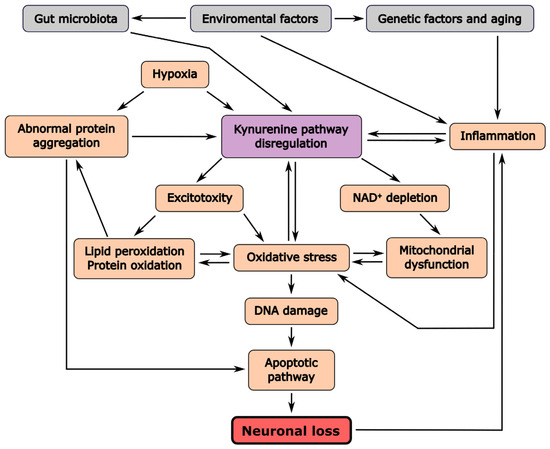
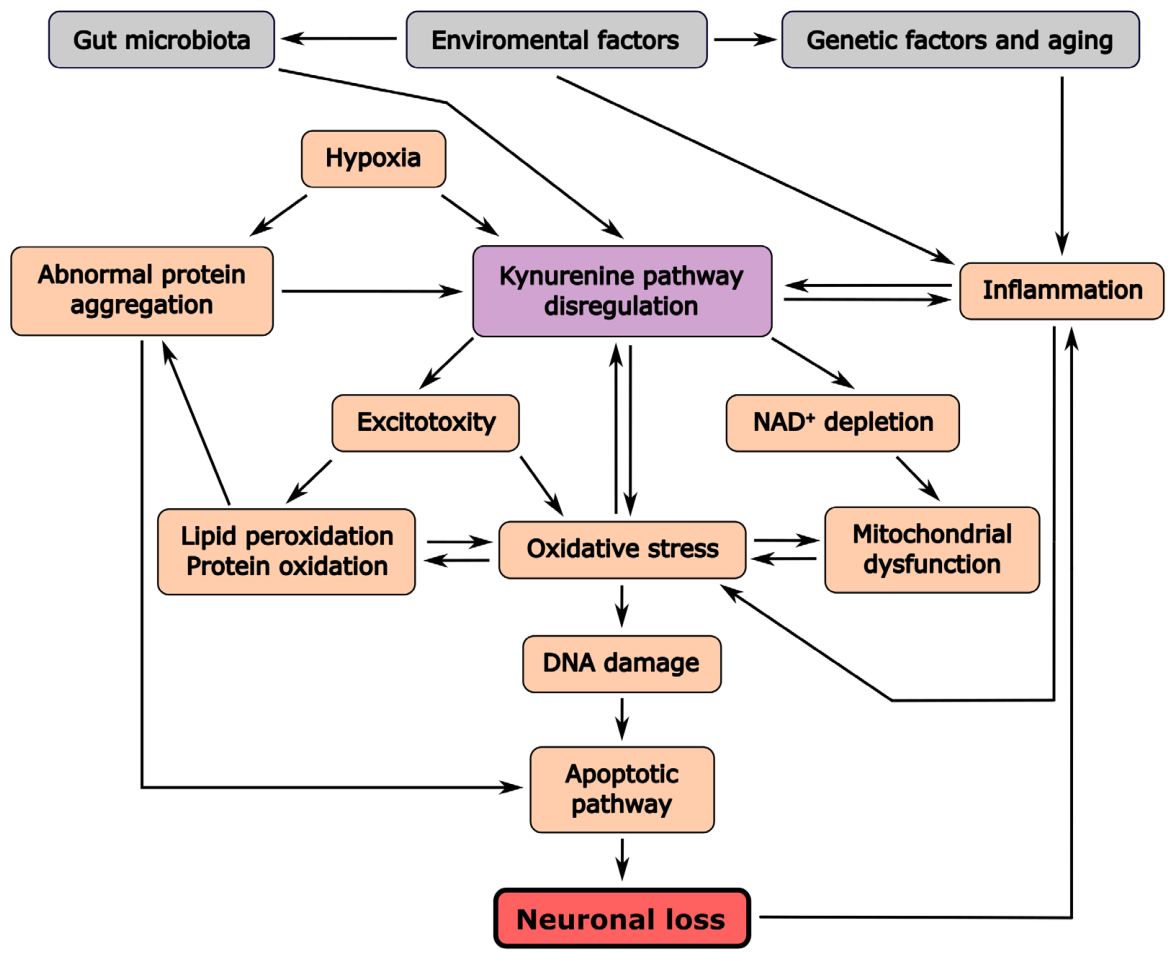
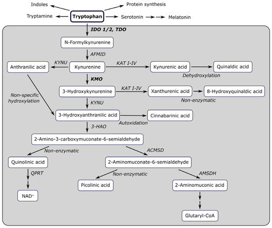
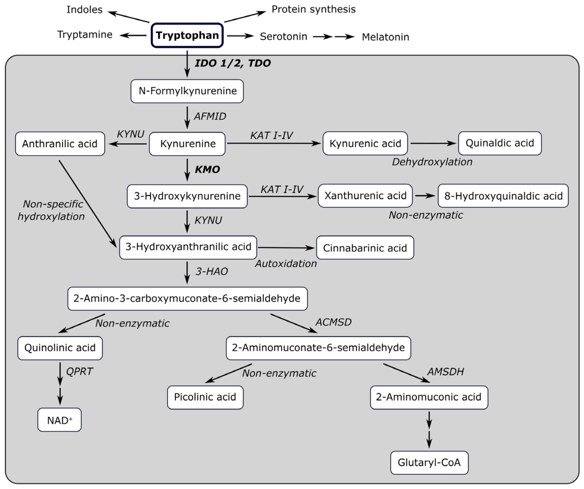

Disclaimer/Publisher’s Note: The statements, opinions and data contained in all publications are solely those of the individual author(s) and contributor(s) and not of MDPI and/or the editor(s). MDPI and/or the editor(s) disclaim responsibility for any injury to people or property resulting from any ideas, methods, instructions or products referred to in the content. |
© 2026 by the authors. Licensee MDPI, Basel, Switzerland. This article is an open access article distributed under the terms and conditions of the Creative Commons Attribution (CC BY) license.