


Abstract
Detecting a KIT D816V mutation in patients with systemic mastocytosis (SM) needs a high-sensitivity assay. Additionally, high mutant KIT transcriptional activity and multilineage involvement are strong unfavorable prognostic parameters in SM. The current frequently used quantitative PCR-based KIT D816V assays have important analytical limitations, including considerable analytical variation in the low-concentration range, a need for a calibration curve, less multiplexing capabilities, and higher susceptibility to interference by PCR inhibitors. We developed, validated, and explored the diagnostic relevance of a novel multiplex KIT D816V cDNA droplet digital PCR (ddPCR), using ABL1 and KIT as reference genes. Furthermore, we correlated the expressed allele burden (EAB) with variant allele frequency (VAF) and percentage of neoplastic mast cells (nMC%) in bone marrow samples of 79 SM patients. Our assay shows very low background signals and high assay precision, leading to a linear dynamic range spanning five orders of magnitude and very low limits of detection and quantification of 0.062% (EAB/ABL1) and 0.027% (EAB/KIT). The assay detects median 57 times more mutant cDNA copies than mutant gDNA copies in SM patients and positivity in 83% of SM-suspected patients with negative gDNA ddPCR results. Furthermore, correlations between EAB, VAF, and nMC% suggest the presence of KIT D816V in non-MCs, either with or without transcriptional activity. Using our novel high-sensitivity mRNA-based multiplex ddPCR KIT D816V assay returns high diagnostic yield in patients with suspicion for SM. We recommend incorporating our assay in routine SM diagnostics to determine low-burden KIT D816V.
1. Introduction
Mast cells (MCs) are hematopoietic cells of the innate immune system, primarily responsible for the IgE-mediated immune response, and they reside in several organs and tissues, including bone marrow (BM), the spleen, the liver, lymph nodes, and the gastrointestinal tract. A somatic point mutation in the KIT gene resulting in an aspartic acid-to-valine substitution at codon 816, i.e., KIT D816V, causes constitutive activity of the c-KIT receptor (CD117). In MCs, this leads to their increased survival and excessive proliferation, resulting in systemic mastocytosis (SM) [1]. The WHO classification of systemic MC disorders depends on the presence of so-called B (burden of disease) and C (cytoreduction required) findings and ranges from relatively mild variants, such as monoclonal MC activation syndrome (MMCAS), bone marrow mastocytosis (BMM, SM without skin lesions), indolent SM (ISM, SM with skin lesions) and smoldering SM (SSM), to advanced SM (AdvSM) including aggressive SM (ASM), mast cell leukemia (MCL), and SM with an associated hematological neoplasm (SM-AHN) [2].
The diagnosis of SM requires the assessment of pathologic MCs in BM and includes the detection of one of the most important diagnostic markers, i.e., the KIT D816V mutation [2]. However, very low numbers of KIT D816V-mutated BM MC often hamper the detection of this mutation [3,4], especially when performing the analysis of gDNA [5,6]. Given the strong cKIT (CD117) expression in MCs [7], several real-time qPCR assays have been developed for sensitive mRNA-based detection of KIT D816V [8,9,10,11]. However, qPCRs exhibit several important analytical disadvantages when compared to the droplet digital PCR (ddPCR) technique, like a larger analytical variation in the low-concentration range, the need for a calibration curve, less multiplexing capabilities with a higher chance of pipetting bias, and a higher susceptibility to interference by PCR inhibitors [12,13,14,15]. Moreover, the sensitivity of ddPCR assays can be readily improved by running additional wells and combining the results. The European Leukemia Net (ELN) has published recommendations for monitoring measurable residual disease (MRD) in patients with acute myeloid leukemia (AML) [16]. To achieve high-sensitivity MRD analysis, the ELN recommends molecular MRD assessment with a quantitative polymerase chain reaction (PCR) and the use of cDNA over gDNA for genes with high expression levels in AML cells, such as mutant Nucleophosmin 1 (NPM1). Furthermore, the ELN advises that transcript levels are normalized to the expression of the ABL1 housekeeping gene. Because high analytical sensitivity is essential in SM diagnostics, we developed and validated an mRNA-based KIT D816V ddPCR assay with very high precision, sensitivity, and specificity. Furthermore, previously published qPCR assays used either the expression of the housekeeping ABL1 gene or the expression of the wild-type KIT gene for normalization or as a reference gene [8,9,10,11]. To achieve the benefits of using both reference genes, we designed a multiplex ddPCR assay with both ABL1 and KIT as reference genes.
Recently published data have revealed that high mutant KIT transcriptional activity and the presence of a KIT mutation in other non-MC myeloid and/or lymphoid cells, named multilineage involvement, seem to be strong unfavorable prognostic parameters in SM [17]. To enhance our understanding of KIT D816V transcriptional activity and patterns of clonal involvement, we correlated the transcriptional activity with the variant allele frequency (VAF) and actual percentage of neoplastic mast cells (nMC%) in bone marrow samples of SM patients.
2. Results
2.1. Assay Design
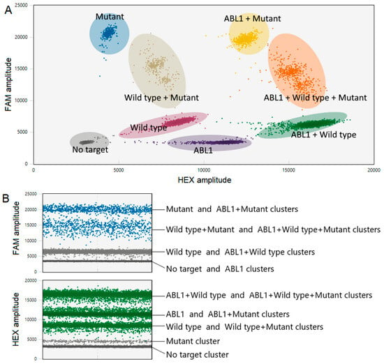
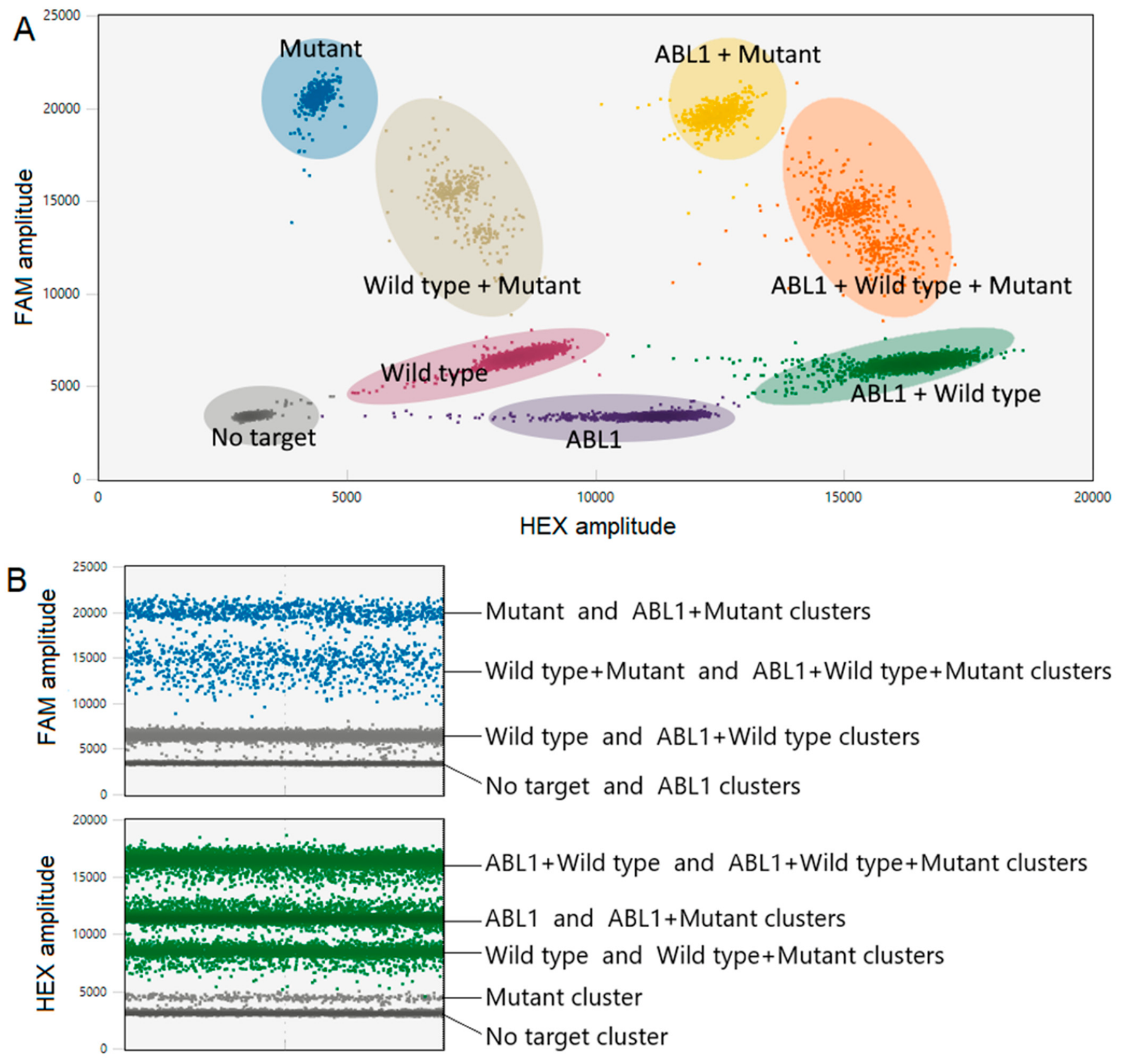
A multiplex KIT D816V ddPCR assay was developed to quantify KIT D816V expression levels normalized to both ABL1 and KIT transcripts, enabling a highly accurate measurement of mutant KIT transcriptional activity and optimal comparison with mutant KIT VAF. A representative example of a 2D plot of a multiplex ddPCR reaction with clearly separated droplet clusters is shown in Figure 1A. The KIT wild-type and ABL1 clusters are diagonally segregated and outlined with a pencil tool. High PCR efficiency was assessed by observing high consistency and segregation of clusters. The thresholds for the KIT D816V mutation detection (FAM signal), KIT wild-type detection (HEX-LOW signal), and ABL1 detection (HEX-HIGH signal) are manually set to maximize the sensitivity and specificity. Slight fluorescence overlap between HEX and FAM channels improved the separation of the violet wild-type cluster and the green ABL1+ wild-type cluster from the purple ABL1 cluster, as also illustrated in 1D plots (Figure 1B).
Figure 1.
Representative fluorescence amplitude plots. A representative 2D fluorescence amplitude plot of a ddPCR analysis of a KIT D816V-positive sample (A). The black cluster on the plot represents the empty droplets, the blue cluster represents the droplets that are only positive for the KIT D816V mutant, the violet cluster represents the droplets that are only positive for the wild-type KIT, the purple cluster represents the droplets that are only positive for the ABL1 reference target, the green cluster represents the droplets that are positive for both ABL1 and wild-type KIT, the gray cluster represents the droplets that are positive for both the wild-type KIT and KIT D816V, the yellow cluster represents the droplets that are positive for both ABL1 and KIT D816V, and the orange cluster represents the droplets that are positive for ABL1, KIT D816V and KIT wild-type. Representative 1D plots for all clusters (B). Results of KIT D816V mutant (blue FAM channel), KIT wild type (green HEX-LOW channel), and ABL1 (green HEX-HIGH channel) are shown.
2.2. Linearity and Dynamic Range
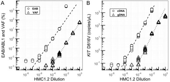
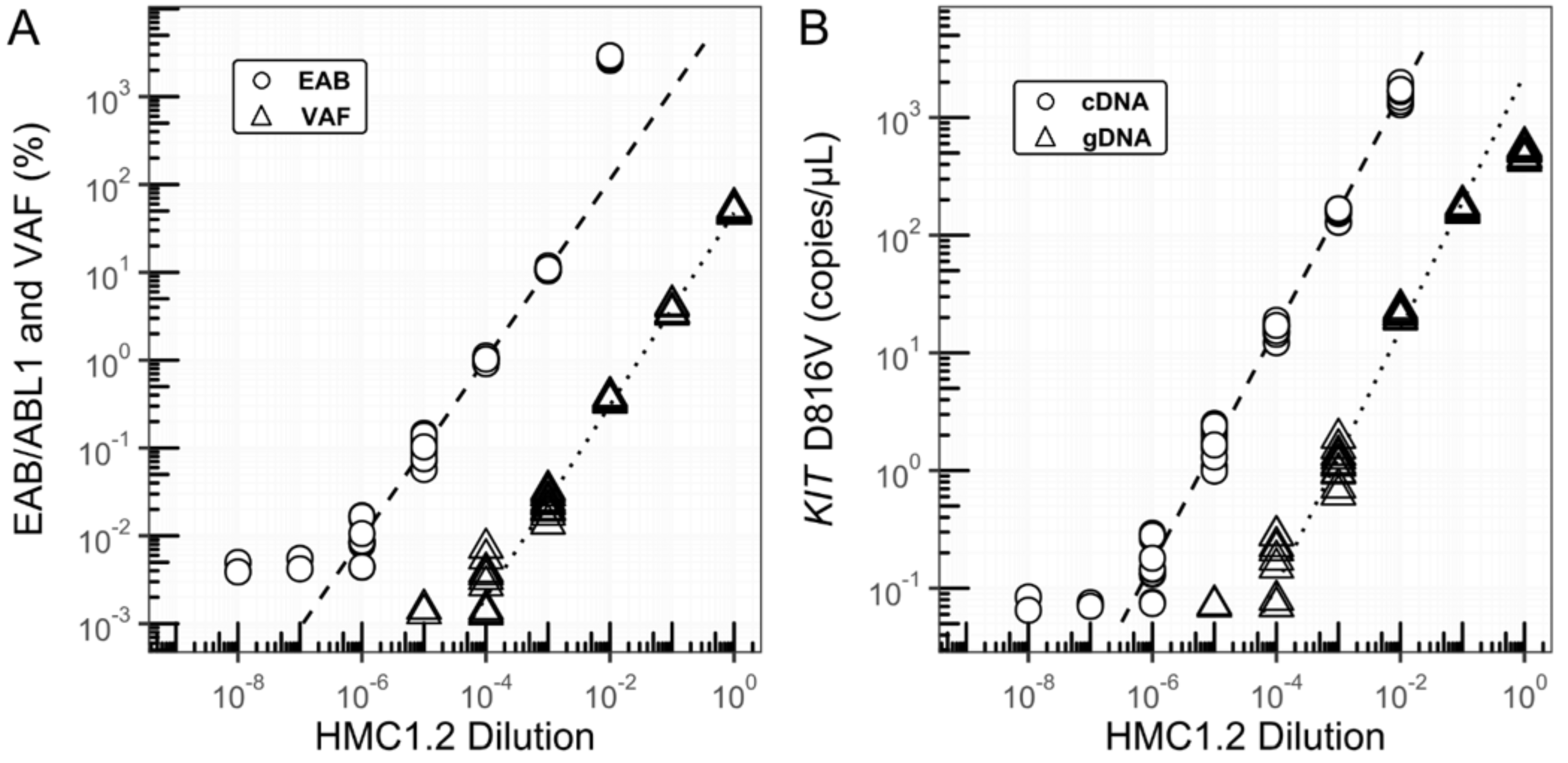
To determine the linearity and dynamic range, tenfold cDNA dilution series up to a 10−8 dilution from the human mast cell line 1.2 (HMC-1.2) [18], harboring a heterozygous KIT D816V mutation, were tested in duplicate at six different time points. The undiluted and 10−1 dilution samples were not included because these dilutions oversaturated the ddPCR reaction. The 10−1 and 10−2 dilution samples were diluted in DEPC H2O. All subsequent dilution steps were performed in cDNA from pooled KIT D816V-negative normal bone marrow (NBM) samples from healthy BM transplantation donors. The dilution series extended to positive ABL1-normalized KIT D816V values up to 10−6 dilution and was linear with an optimal R2 of 0.99 (Figure 2). The 10−2 dilution exhibited a slightly higher expressed allele burden (EAB) than the trend line, possibly due to a lower ABL1 expression level in the HMC-1.2 cell line.
Figure 2.
Linearity and dynamic range. Linearity and dynamic range of KIT D816V ABL1-normalized expressed allele burden (EAB/ABL1) and variant allele frequency (VAF) (A), and actual copy numbers (B) using HMC-1.2 serial dilutions. The undiluted and the 10−1 dilution samples were not included for the cDNA measurements because these dilutions oversaturated the ddPCR reaction. The dashed trendlines represent measurements of dilution series of HMC-1.2 cDNA, and the dotted trendlines represent measurements of dilution series of HMC-1.2 gDNA.
In addition, tenfold serial dilutions of HMC-1.2 gDNA in normal bone marrow (NBM) gDNA, ranging from undiluted (100) to a 10−6 dilution, were tested in duplicate at six different time points (Figure 2). The dynamic range of the VAF analysis spanned five orders of magnitude, ranging from undiluted to a 10−4 dilution, with an optimal R2 linearity of 0.99.
2.3. Analytical Sensitivity and Specificity
The limit of blank (LoB) was estimated by measuring six cDNA NBM samples at ten different time points and was calculated at a 95% confidence level as the mean value + 1.645 × standard deviation (SD). The LoB was 0.007% EAB when using ABL1 as a reference gene and 0.013% EAB when using KIT as a reference gene (Table 1).
Table 1.
Analytical sensitivity and specificity parameters of the cDNA and gDNA assay and the HMC-1.2 results.
For calculating the limit of detection (LoD), aliquots from the six split NBM samples were spiked with the smallest possible amount of HMC-1.2 that returned a positive KIT D816V signal higher than the LoB in more than 95% of measurements, which was determined as the 2 × 10⁻⁶ HMC-1.2 dilution. These low-concentration samples were measured in parallel with the KIT D816V-negative samples. The LoD was calculated with a 95% confidence level as the LoB + 1.645 × SD of the low concentration sample and turned out to be 0.062% EAB when using ABL1 as the reference gene and 0.027% EAB when using KIT as the reference gene. The limit of quantification (LoQ) was defined as the concentration where the inter-assay coefficient of variation (CV) equals 35%.
The inter-assay CV of the low-concentration sample used for establishing the LoD, i.e., the 2 × 10⁻⁶ HMC-1.2 dilution, was 24.1% when using ABL1 as the reference gene and 34.1% when using KIT as the reference gene (Table 2). Because the LoDs exhibited CVs lower than 35%, the LoQs were defined as equal to the LoDs. The LoB, LoD, LoQ, and intra- and inter-assay CV values of the ddPCR gDNA assay were also determined by similar procedures. The inter-assay CV of the low-concentration sample used for establishing the LoD, i.e., the 1 × 10⁻³ HMC-1.2 dilution, yielded VAF results that were comparable to the results found previously [19], and comparable to our cDNA assay, with an LoB, LoD, and LoQ of 0.002%, 0.024%, and 0.041%, respectively (Table 1).
Table 2.
Intra-assay and inter-assay variation (CV%) of the cDNA and gDNA assay.
2.4. Normalization to ABL1 Versus KIT
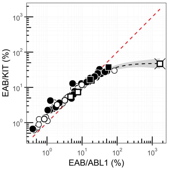
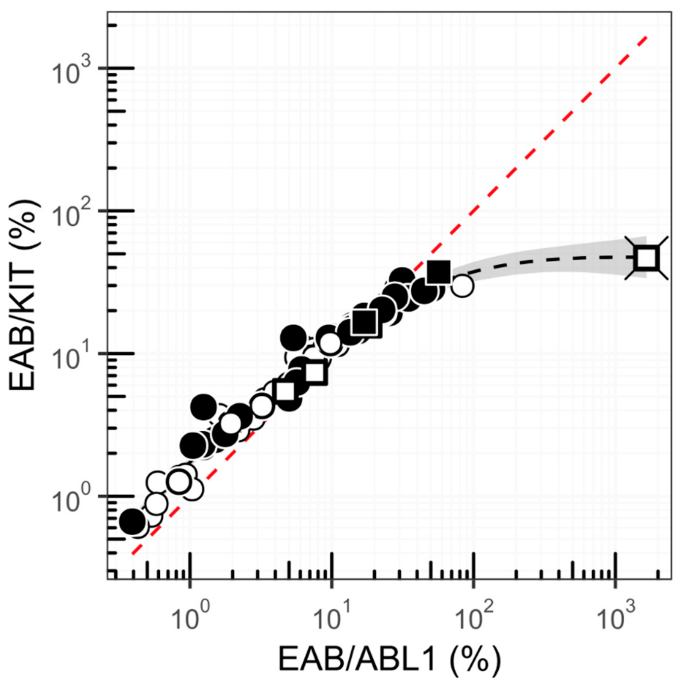
The choice of reference gene impacts the accuracy and precision of the KIT D816V quantification. Therefore, we compared the use of ABL1 versus total KIT as reference genes across SM patient samples (Figure 3). Both methods revealed a nonlinear correlation, in which a low tumor load (<0.1%) resulted in a trend toward slightly higher total KIT-normalized values, whereas a higher load showed higher ABL1-normalized results.
Figure 3.
Normalization with ABL1 versus KIT reference gene. Correlation (black dashed line) between ABL1-normalized KIT D816V expression (KIT D816V/ABL1) versus total KIT-normalized KIT D816V expression (KIT D816V/KIT total) in 73 patients with an ISM (circles) and six patients with an AdvSM (squares). The red dashed line follows y = x. Abbreviation: EAB (expressed allele burden).
2.5. Sensitivity of cDNA Versus gDNA Analyses
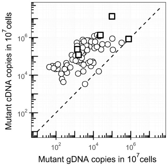
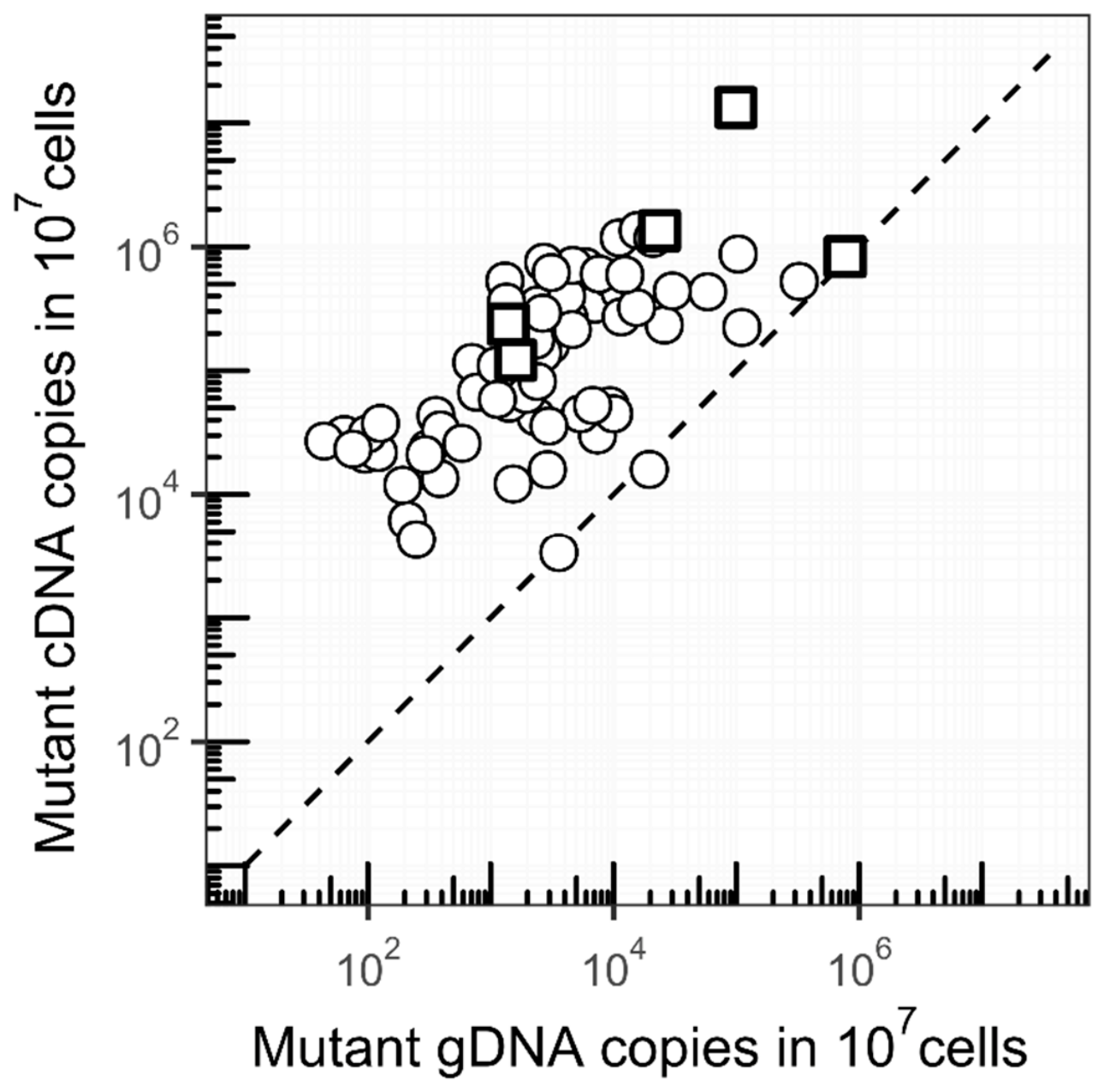
The abundance of KIT D816V mutant cDNA and gDNA copies in split BM samples (107 cells) from a group of 73 patients with ISM and six patients with advanced SM (AdvSM) was compared (Figure 4). The median number of mutant cDNA copies was 57 (range 1–614) times higher than mutant gDNA copies. Higher mutant abundance when using RNA instead of DNA samples increases detection probability and sensitivity, especially in cases with a low mutant burden where detection is affected by stochastic effects.
Figure 4.
Sensitivity of cDNA versus gDNA analyses for KIT D816V detection in bone marrow (BM). The abundance of KIT D816V mutant cDNA and gDNA copies in split BM samples (107 leukocytes) from 73 patients with an ISM (circles) and six patients with an AdvSM (squares). The dashed line follows y = x.
To illustrate the impact of using our high-sensitivity KIT D816V cDNA ddPCR assay, samples from a group of 18 patients with a high suspicion for SM, but insufficient diagnostic criteria to establish the diagnosis of SM, and negative KIT D816V gDNA ddPCR results were evaluated. Fifteen out of 18 patients with negative KIT D816V gDNA ddPCR results demonstrated positive KIT D816V expression levels, with KIT EAB/ABL1 ranging from 0.021% to 0.865%, and KIT EAB/KIT ranging from 0.028% to 0.632%. Seven out of 15 patients also showed MCs with aberrant CD25 expression, supporting the presence of abnormal MCs. Constantly negative no-template control (NTC) values and no-amplification control (NAC) values below the LoB made the possibility of false-positive background signals unlikely. So, the cDNA ddPCR analysis detected KIT D816V expression in 83% (15/18) of SM-suspected patients with negative gDNA ddPCR results.
2.6. Correlations Between KIT D816V VAF, KIT D816V EAB and nMC%
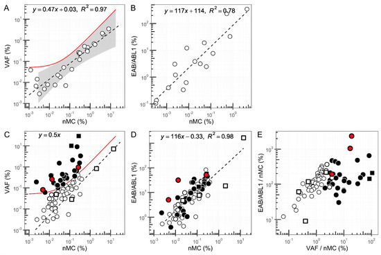
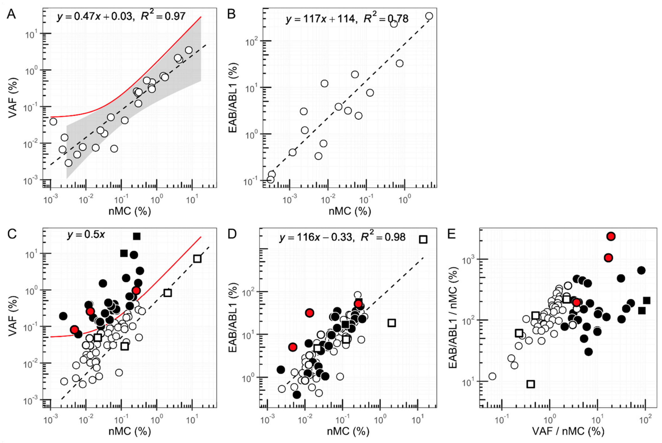
We also correlated the KIT D816V EAB and VAF with the actual percentage of neoplastic BM MCs (nMC%). Firstly, we determined the analytical variance in the correlation results of the flow cytometric and PCR analyses by testing tenfold HMC-1.2 gDNA and cDNA dilution series up to 10−3 dilutions in duplicate (Figure 5A,B). The KIT D816V VAF results showed good agreement with the expected distribution around 0.5 times the nMC%, as aberrant MCs exhibit one heterozygous KIT D816V allele and one KIT wild-type allele, with an R2 of 0.97 (Figure 5A). The upper limit values of this correlation were defined as the cut-off value for detecting non-MC-restricted KIT D816V. Of note, at lower nMC%, the upper bound was set at a relatively higher VAF value due to higher analytical variance. In addition, the KIT D816V EAB showed more than 100-fold higher values than the nMC%, with a good correlation with an R2 of 0.78 (Figure 5B).
Figure 5.
Correlations between KIT D816V VAF, KIT D816V EAB, and nMC%. Correlation between the KIT D816V VAF and nMC% in HMC-1.2 gDNA tenfold dilution series in gDNA from pooled NBM samples (A). The undiluted sample was not included because this sample oversaturated the ddPCR reaction. The dashed black line follows the trendline y = 0.47x + 0.03. The correlation is shown with a confidence interval covering the measurement range of 99.999%. The solid red line corresponds to the upper boundary of this range and is used as a cut-off value for detecting non-MC-restricted involvement of KIT D816V. Correlation between the KIT D816V EAB/ABL1 and nMC% in HMC-1.2 cDNA tenfold dilution series in cDNA from pooled NBM samples. (B). The undiluted and 10% dilution samples were not included in the cDNA assay because these samples oversaturated the ddPCR reaction. The dashed black line follows the trendline y = 117x + 114. Correlation between the VAF and the nMC% in BM samples of 73 patients with an ISM (circles) and six patients with an AdvSM (squares) (C). The cut-off value for detecting non-MC-restricted involvement of KIT D816V is indicated as a solid red line. Patients who showed higher VAFs than expected relative to the nMC% are indicated with filled circles (ISM patients) or filled squares (AdvSM patients). The dashed black line follows y = 0.5x. Correlations between the KIT D816V EAB/ABL1 and the nMC% in BM samples of 73 patients with an ISM (circles) and six patients with an AdvSM (squares) (D). The dashed black line follows the trendline y = 116x − 0.33. Correlation between the KIT D816V EAB/ABL1 (KIT D816V EAB/nMC%) and the KIT D816V VAF (KIT D816V VAF/nMC%) after normalizing both for the nMC% (E). Results from three ISM patients with a B-finding are indicated in red. VAF: variant allele fraction, EAB: expressed allele. burden, nMC%: percentage of neoplastic mast cells in bone marrow.
Next, we tested bone marrow samples of 79 SM patients and also found a positive correlation between the VAF and nMC% (Spearman ρ = 0.64, p < 0.01) (Figure 5C). However, 28 out of 73 patients with ISM and two out of six patients with AdvSM showed VAFs higher than 0.5 times the nMC%, suggesting that other non-MC BM cells also harbored the KIT D816V mutation, i.e., possible presence of multilineage involvement. Further, the KIT D816V EAB showed a distribution around about 100 times the nMC% and a strong correlation with an R2 of 0.98 (Zou’s 95% confidence interval: 0.06–0.29; p < 0.01) (Figure 5D). Notably, even most patients with a presumed non-MC-restricted involvement had correlations between the KIT D816V EAB and nMC% that did not deviate from the main trendline.
Finally, we correlated the EAB and the VAF after correction for the nMC% (Figure 5E). As expected, patients with VAFs around 0.5 times the nMC% showed EAB/nMC% ratios of about 100, comparable to the transcriptional activity of the HMC-1.2 cells. Additionally, most patients with VAFs higher than 0.5 times the nMC% demonstrated EAB/nMC% ratios also around 100. However, a few patients with VAFs higher than 0.5 times showed much higher EAB/nMC% ratios, and the two patients with the highest EAB/nMC% ratios (2.358% and 1.052%) exhibited an ISM with a B-finding, while only one out of the remaining 71 ISM patients had a B-finding (Figure 5E).
3. Discussion
Here we present, for the first time, a multiplex mRNA-based KIT D816V ddPCR assay that covers both ABL1 and KIT wild-type reference genes, enabling highly accurate measurement of mutant KIT transcriptional activity leading to a very high diagnostic yield. Our assay shows extremely low background signals with LoBs of 0.007% (EAB/ABL1) and 0.013% (EAB/KIT) and a high assay precision, leading to very low LoD/LoQ values of 0.062% (EAB/ABL1) and 0.027% (EAB/KIT). The linear dynamic range spans five orders of magnitude with a slope of 0.999 and an R2 of 0.99, indicating excellent PCR efficiency. We employed a ddPCR method instead of the conventionally used real-time qPCR because of the analytical advantages of direct and accurate quantification without the need for a calibration curve, the multiplexing capacity with reduced pipetting bias, and a lower impact of PCR inhibitors [12,13,14,15]. Moreover, using the same analytical ddPCR technique, i.e., our mRNA-based ddPCR assay and a widely used and extensively validated DNA-based ddPCR assay [19], enabled an optimal comparison between KIT D816V transcriptional activity and VAF. However, although ddPCR provides several analytical advantages over qPCR, its broader implementation in routine clinical diagnostics may be limited by higher reagent and operational costs, requirement for specialized instrumentation and technical expertise, and its currently lower availability in routine diagnostic laboratories compared with qPCR-based platforms.
We designed a multiplex ddPCR assay with both ABL1 and KIT as reference genes to achieve the benefits of using both reference genes. For exact quantification, a reference gene expressed by all cells, such as the ABL1 housekeeping gene, is required. Furthermore, including the KIT wild-type probe in the assay reduces possible non-specific binding of the KIT D816V probe to the wild-type allele. Moreover, our assay offers the opportunity to compare results with data from other studies that either used ABL1 or KIT as a reference gene [8,9,10,11]. Finally, the use of a KIT reference facilitates the optimal comparison between mutant KIT expression and VAF signals [20]. To achieve high sensitivity, maximum input of KIT D816V transcripts without oversaturation of the ddPCR reaction is needed [21]. Clonal MCs express high amounts of heterozygous KIT D816V and KIT wild-type transcripts and relatively low numbers of ABL1 transcripts. When using only one reference gene in the assay, ABL1 offers the advantage of having less impact on the risk of oversaturation and therefore enables higher cDNA input in the assay, with the possibility to detect very low numbers of mutant KIT transcripts. In addition, using KIT as the only reference gene also enables high sample input, because only signals from KIT-expressing CD117-positive cells, including MCs, myeloid blasts, and promyelocytes, are measured.
However, despite all these advantages, there are some drawbacks. Firstly, the use of a second reference gene impairs the maximum sample input, although this effect seems to be limited. The only extra positive droplets in the assay are the droplets that are only positive for the second reference gene, and a high sample input gives relatively fewer droplets with copies of only one gene. Additionally, ABL1-normalized KIT expression results might be influenced by variable levels of ABL1 expression [21], potentially causing a risk of falsely increased or decreased KIT D816V expression values.
Furthermore, we compared the results of our mRNA-based ddPCR assay with the results of the commercially available and widely used gDNA-based ddPCR [18]. Although both assays demonstrated similar high analytical performances, the cDNA measurements proved superior to the gDNA results, as observed by finding a median 57-fold higher abundance of mutant KIT cDNA transcripts compared to mutant KIT gDNA alleles in BM samples of SM patients. The diagnostic impact of the much higher sensitivity is evident, as our data showed that the cDNA assay was able to detect KIT D816V transcripts in 83% of the patients with a clinical suspicion for SM but negative KIT gDNA ddPCR results. Taken together, these findings demonstrate that our novel cDNA ddPCR assay detects KIT D816V mutants with significantly higher sensitivity in BM samples than the gDNA ddPCR assay, while retaining high specificity.
To evaluate the relevance of measuring KIT transcriptional activity to better understand patterns of clonal mutant KIT involvement, we correlated the transcriptional activity with the VAF and the nMC% and found three different types of patients. First, 49 out of 79 (62%) patients, including four patients with an AdvSM, showed VAFs around 0.5 times the nMC% and EAB/nMC% ratios comparable to the transcriptional activity of HMC-1.2 cells, indicating MC-restricted KIT D816V expression. Second, most remaining patients, including two with an AdvSM, demonstrated VAFs higher than 0.5 times the nMC%, but the EAB/nMC% ratios were still comparable with the transcriptional activity of HMC-1.2 cells, suggesting that in these patients, non-MCs also carry the KIT D816V mutation in their DNA (i.e., possible presence of multilineage involvement), but have no transcriptional activity. These findings are in line with data from Escribano et al. [22] and Teodosio et al. [23], demonstrating multilineage involvement in 9.5% and 30% SM patients, respectively, and with results from several other studies showing multilineage involvement in CD117-negative cells, such as granulocytes, monocytes, and lymphocytes [23,24,25]. However, the interpretation of multilineage KIT D816V involvement should be done carefully because it remains indirect, i.e., no sorted-cell or single-cell confirmation was performed. Moreover, alternative explanations, such as the KIT D816V copy number gain or loss of heterozygosity or the KIT wild-type copy number loss, were not excluded. Finally, some patients with suggested multilineage involvement showed clearly higher EAB/nMC% ratios, which might suggest that the KIT D816V mutation is not only present in the DNA but also expressed in non-MCs, such as CD117-positive early pluripotent progenitor cells, myeloblasts, and promyelocytes [26]. Remarkably, the two patients with the highest EAB/nMC% ratios both exhibited an ISM with a B-finding, i.e., one had hepatomegaly, and the other one had splenomegaly, while only one out of the remaining 71 ISM patients had a B-finding. These findings seem to be in line with data, showing that ISM patients who evolve to aggressive disease subtypes harbor the KIT D816V mutation in early CD117-positive hematopoietic precursors [22,23,24,25]. However, other possible explanations, like increased KIT D816V transcription in MCs or variable RNA integrity, can give the same results and were not excluded. Therefore, there is far too little data available to claim this association. This observation must be interpreted as preliminary and hypothesis-generating. So, our findings suggest that increased VAF/nMC% and EAB/nMC% ratios might possibly be used as surrogate markers for multilineage involvement of KIT mutations in SM patients. This is of special relevance because complex and not widely available cell sorting techniques are required to determine mutant KIT multilineage involvement.
Our study has some limitations. Samples were primarily collected for clinical testing, and the inclusion of patients and data was performed retrospectively. This resulted in limited sample volumes being available for analyses, restricting some aspects of the analytical and clinical validations, including meaningful statistical testing in some small subgroups and evaluation of our assay in a large number of patients. In addition, no cell sorting analyses to confirm the presence of multilineage involvement or testing possible other explanations were performed due to the retrospective nature of our study. Consequently, further clinical multicenter validation studies in larger patient cohorts, including cell sorting experiments, are needed to confirm our findings concerning the relevance of correlating KIT D816V transcriptional activity with VAF and tumor load in the routine clinical practice of SM patients.
4. Materials and Methods
4.1. KIT D816V cDNA Droplet Digital PCR Assay Design
A multiplex ddPCR assay was developed to quantify KIT D816V (NM_000222.2, GRCh37 chromosome 4: 55,524,085-55,606,881) expression, together with KIT wild-type and ABL1 (NM_007313.2, GRCh37 chromosome 9: 133,589,333-133,763,062) reference transcripts, as was done before for different Nucleophosmin 1 (NPM1) mutations [27]. For the measurement of KIT expression, primers and a FAM (Fluorescein)-labeled KIT D816V probe and a HEX (Hexachloro-Fluorescein)-labeled KIT wild-type probe were designed (Supplementary Materials, Selection of primers and probes and Table S1). For measurement of reference ABL1 transcripts, a commercially available mix of primers and a HEX-labeled probe (Bio-Rad Laboratories, Hercules, CA, USA) was included in the assay. The optimal annealing temperature was determined at 57 °C by testing a temperature gradient (Supplementary Materials, Selection of optimal annealing temperature and Figure S1).
4.2. Droplet Digital PCR Protocols
KIT D816V cDNA levels were analyzed using an optimized ddPCR protocol. Briefly, the PCR mix contained ddPCR Multiplex Supermix, KIT primers, a KIT D816V probe and a KIT wild-type probe, and ABL1 primers and probe. Data were analyzed on a QX200 droplet reader (Bio-Rad Laboratories, Hercules, CA, USA) with QX manager or Quantasoft Analysis Pro software (version 1.0.596) to distinguish KIT D816V (FAM), KIT wild-type (HEX-LOW), and ABL1 (HEX-HIGH) signals. A detailed protocol is described in the Supplementary Materials (Supplementary Materials, KIT D816V Droplet Digital PCR Protocol and Table S2). Results were expressed as a fraction (%) normalized to ABL1 reference gene expression (KIT D816V copies divided by ABL1 copies × 100%), or as a fraction (%) normalized to the total KIT expression (KIT D816V copies divided by KIT D816V copies + KIT wild-type copies × 100%).
For KIT D816V gDNA analysis, a commercially available and extensively validated ddPCR assay [19] was purchased from Bio-Rad Laboratories (Hercules, CA, USA) and used according to the manufacturer’s recommendations. Results were expressed as variant allele frequency (VAF), i.e., the percentage mutated KIT D816V copies divided by the total number of KIT copies × 100%.
4.3. Study Samples
For analytical validation experiments, RNA and gDNA from the human mast cell line 1.2 (HMC-1.2) [18], harboring a heterozygous KIT D816V mutation, and normal bone marrow (NBM) samples from healthy BM transplantation donors, were used. For testing clinical utility, BM RNA and gDNA samples from patients referred to the University Medical Center of Groningen between 2014 and 2024 were selected, based on symptoms of MC activation or established SM diagnosis. SM diagnosis and classification were established according to the World Health Organization (WHO) criteria for SM, as previously described [28,29]. Samples for this study were retrospectively included if flow cytometric results and split RNA and gDNA samples were available to perform the analyses needed for this study. RNA and gDNA BM samples from 18 individuals with a high suspicion of SM but negative gDNA KIT D816V ddPCR results were selected to assess the impact of using the highly sensitive assay on diagnostic accuracy. Also, RNA and gDNA BM samples from 73 patients with indolent SM (ISM) and from 6 patients with advanced SM (AdvSM) were processed for PCR analyses to determine the EAB and VAF levels. Further details about sample collection, RNA and gDNA isolation, and cDNA synthesis are described in the Supplementary Materials section (Supplementary Materials, Sample collection, RNA and gDNA isolation, and cDNA synthesis and Tables S3 and S4).
4.4. Flow Cytometric Analysis
Flow cytometric analysis was performed in all patient samples as part of the routine diagnostic procedure. The actual percentage of neoplastic MCs in BM (nMC%) was defined as the population of CD25-positive MCs using all CD45-positve BM cells as the denominator. Further details on the flow cytometric analysis have been described previously [30].
4.5. Analytical Assay Validation
The KIT D816V mRNA ddPCR assay was validated in accordance with the EP05 and EP17 guidelines of the Clinical and Laboratory Standards Institute (CLSI) and the Digital MIQE guidelines [31,32,33]. The analytical sensitivity and specificity were determined using the limit of blank (LoB), limit of detection (LoD), and limit of quantification (LoQ). Intra- and inter-assay variability was quantified with the coefficients of variation (CV). Finally, the performance of our new assay was compared with that of a widely used and commercially available KIT D816V gDNA ddPCR assay [19] and a qPCR previously used for routine clinical diagnostics (Supplementary Materials, Method comparison between ddPCR and qPCR and Figure S2).
4.6. Statistical Analysis
The significance of correlations was determined using Spearman’s rank correlation coefficient (ρ) due to non-normal and/or skewed distributions. Differences between correlations were evaluated using Zou’s 95% confidence interval method [34]. All distributions were tested for normality using Q-Q plots. All statistical analyses and graphical representations were performed using RStudio version 1.4.1106 (PBC).
5. Conclusions
We present the development, validation, and exploratory clinical evaluation of a high-sensitivity multiplex mRNA-based KIT D816V ddPCR assay. Implementation of this highly sensitive assay in the daily clinical practice of SM will increase the diagnostic yield in bone marrow cells, especially when no KIT mutation is found with DNA assays. Furthermore, correlating KIT D816V transcriptional activity with VAF and mast cell tumor load might show promise as a surrogate approach for identifying multilineage KIT mutation involvement, pending confirmation by cell-sorting or single-cell investigations. Additionally, further prospective multicenter clinical validation studies in larger patient cohorts are needed to prove its possible value in risk assessment.
Supplementary Materials
The following supporting information can be downloaded at https://www.mdpi.com/article/10.3390/ijms27146314/s1.
Author Contributions
Conceptualization, A.A., I.P.K., and A.B.M.; Methodology, A.A., A.S., L.B.H., A.T.J.W., and A.B.M.; Software, A.A.; Validation, A.A., A.S., L.B.H., A.T.J.W., and A.B.M.; Formal analysis, A.A., A.S., L.B.H., M.H., and A.B.M.; Investigation, A.A. and A.B.M.; Resources, I.P.K.; Data curation, A.A., I.P.K., and A.B.M.; Writing—original draft, A.A., I.M., and A.B.M.; Writing—review and editing, A.A., A.S., L.B.H., A.T.J.W., M.H., I.M., S.K.K., H.N.G.O.E., I.P.K., and A.B.M.; Visualization, A.A. and I.M.; Supervision, I.P.K. and A.B.M.; Project administration, A.A. All authors have read and agreed to the published version of the manuscript.
Funding
This work received no external funding.
Institutional Review Board Statement
The study was conducted in accordance with the Declaration of Helsinki. According to the Medical Ethical Review Board of the University Medical Center Groningen, this observational, non-interventional, retrospective study is classified as non-WMO and has been approved for the publication of subject data (METc number METc 2024/199, UMCG RR number 19272, date: 26 March 2024).
Informed Consent Statement
The study was approved by the Institutional Review Board, and the requirement for informed consent was waived due to the retrospective nature of the study.
Data Availability Statement
The data that support the findings of this study are available from the corresponding author upon reasonable request. Not all supporting data are publicly available due to privacy restrictions.
Conflicts of Interest
The authors declare no conflicts of interest.
References
- Akin, C.; Gülen, T.; Castells, M.C.; Oude Elberink, J.N.G.; Valent, P. Diagnosis and Management of Patients with Mast Cell Activation Syndromes: Status 2026. J. Allergy Clin. Immunol. Pract. 2026, 14, 19–28. [Google Scholar] [CrossRef] [Scilit] [PubMed]
- Verstovsek, S.; Álvarez-Twose, I.; Colmenero, I.; Cozzolino, I.; Sotlar, K. Systemic mastocytosis. In WHO Classification of Tumours, Haematolymphoid Tumours Part A, 5th ed.; Ahmed, F., Cree, I.A., De Jong, D., Fonseca, A.F.A., Goldman-Lévy, G., Lokuhetty, D., Eds.; IARC: Lyon, France, 2024; Volume 11, pp. 64–70. [Google Scholar]
- Boggs, N.A.; Sun, X.; Lyons, J.J.; McMurray, J.C.; Rose, D.M.; Pryor, E.M.; Metcalfe, D.D.; Maric, I. Challenges in applying diagnostic criteria for systemic mastocytosis. Blood Adv. 2023, 7, 3150–3154. [Google Scholar] [CrossRef] [Scilit] [PubMed]
- Pardanani, A.; Chen, D.; Abdelrahman, R.A.; Reichard, K.K.; Zblewski, D.; Wood, A.J.; McClure, R.F.; Butterfield, J.H.; Hanson, C.A.; Tefferi, A. Clonal mast cell disease not meeting WHO criteria for diagnosis of mastocytosis: Clinicopathologic features and comparison with indolent mastocytosis. Leukemia 2013, 27, 2091–2094. [Google Scholar] [CrossRef] [Scilit] [PubMed]
- Jawhar, M.; Schwaab, J.; Horny, H.; Sotlar, K.; Naumann, N.; Fabarius, A.; Valent, P.; Cross, N.C.P.; Hofman, W.-K.; Metzgeroth, G.; et al. Impact of centralized evaluation of bone marrow histology in systemic mastocytosis. Eur. J. Clin. Investig. 2016, 46, 392–397. [Google Scholar] [CrossRef] [Scilit]
- Muñoz-González, J.I.; García-Montero, A.C.; Orfao, A.; Álvarez-Twose, I. Pathogenic and diagnostic relevance of KIT in primary mast cell activation disorders. Ann. Allergy Asthma Immunol. 2021, 127, 427–434. [Google Scholar] [CrossRef] [Scilit] [PubMed]
- Teodoio, C.; Mayado, A.; Sánchez-Muñoz, L.; Morgado, J.M.; Jara-Acevedo, M.; Álvarez-Twose, I.; García-Montero, A.C.; Matito, A.; Caldas, C.; Escribano, L.; et al. The immunophenotype of mast cells and its utility in the diagnostic work-up of systemic mastocytosis. J. Leukoc. Biol. 2015, 97, 49–59. [Google Scholar]
- Kristensen, T.; Broesby-Olsen, S.; Vestergaard, H.; Bindslev-Jensen, C.; Møller, M.B. Comparison of gDNA-based versus mRNA-based KIT D816V mutation analysis reveals large differences between blood and bone marrow in systemic mastocytosis. Br. J. Haematol. 2017, 178, 330–332. [Google Scholar] [PubMed]
- De Matteis, G.; Zanotti, R.; Colarossi, S.; De Benedittis, C.; García-Montero, A.C.; Bonifacio, M.; Sartori, M.; Aprili, F.; Caruso, B.; Paviati, E.; et al. The impact of sensitive KIT D816V detection on recognition of Indolent Systemic Mastocytosis. Leuk. Res. 2015, 39, 273–278. [Google Scholar] [CrossRef] [Scilit] [PubMed]
- Erben, P.; Schwaab, J.; Metzgeroth, G.; Horny, H.P.; Jawhar, M.; Sotlar, K.; Fabarius, A.; Teichmann, M.; Schneider, S.; Ernst, T.; et al. The KIT D816V expressed allele burden for diagnosis and disease monitoring of systemic mastocytosis. Ann. Hematol. 2014, 93, 81–88. [Google Scholar] [PubMed]
- Lawley, W.; Hird, H.; Mallinder, P.; McKenna, S.; Hargadon, B.; Murray, A.; Bradding, P. Detection of an activating c-kit mutation by real-time PCR in patients with anaphylaxis. Mutat. Res. 2005, 572, 1–13. [Google Scholar] [CrossRef] [Scilit] [PubMed]
- Bill, M.; Grimm, J.; Jentzsch, M.; Kloss, L.; Goldmann, K.; Schulz, J.; Beinicke, S.; Häntschel, J.; Cross, M.; Vucinic, V.; et al. Digital droplet PCR-based absolute quantification of pre-transplant NPM1 mutation burden predicts relapse in acute myeloid leukemia patients. Ann. Hematol. 2018, 97, 1757–1765. [Google Scholar] [CrossRef] [Scilit] [PubMed]
- Olmedillas-López, S.; García-Arranz, M.; García-Olmo, D. Current and Emerging Applications of Droplet Digital PCR in Oncology. Mol. Diagn. Ther. 2017, 21, 493–510. [Google Scholar] [CrossRef] [Scilit] [PubMed]
- Galimberti, S.; Balducci, S.; Guerrini, F.; Del Re, M.; Cacciola, R. Digital Droplet PCR in Hematologic Malignancies: A New Useful Molecular Tool. Diagnostics 2022, 12, 1305. [Google Scholar] [CrossRef] [Scilit] [PubMed]
- Bacher, U.; Dicker, F.; Haferlach, C.; Alpermann, T.; Rose, D.; Kern, W.; Haferlach, T.; Schnittger, S. Quantification of rare NPM1 mutation subtypes by digital PCR. Br. J. Haematol. 2014, 167, 710–714. [Google Scholar] [CrossRef] [Scilit] [PubMed]
- Cloos, J.; Valk, P.J.M.; Thiede, C.; Döhner, K.; Roboz, G.J.; Wood, B.L.; Walter, R.B.; Wang, S.; Wierzbowska, A.; Wei, A.H.; et al. 2025 update on MRD in acute myeloid leukemia: A consensus document from the ELN-DAVID MRD Working Party. Blood 2026, 147, 1147–1167. [Google Scholar] [CrossRef] [Scilit] [PubMed]
- Naumann, N.; Lübke, J.; Baumann, S.; Schwaab, J.; Hoffmann, O.; Kreil, S.; Dangelo, V.; Reiter, L.; Bugert, P.; Kristensen, T.; et al. Adverse Prognostic Impact of the KIT D816V Transcriptional Activity in Advanced Systemic Mastocytosis. Int. J. Mol. Sci. 2021, 22, 2562. [Google Scholar] [CrossRef] [Scilit] [PubMed]
- Sundström, M.; Vliagoftis, H.; Karlberg, P.; Butterfield, J.H.; Nilsson, K.; Metcalfe, D.D.; Nilsson, G. Functional and phenotypic studies of two variants of a human mast cell line with a distinct set of mutations in the c-kit proto-oncogene. Immunology 2003, 108, 89–97. [Google Scholar] [CrossRef] [Scilit] [PubMed]
- Greiner, G.; Gurbisz, M.; Ratzinger, F.; Witzeneder, N.; Simonitsch-Klupp, I.; Mitterbauer-Hohendanner, G.; Mayerhofer, M.; Müllauer, L.; Sperr, W.R.; Valent, P.; et al. Digital PCR: A Sensitive and Precise Method for KIT D816V Quantification in Mastocytosis. Clin. Chem. 2018, 64, 547–555. [Google Scholar] [CrossRef] [Scilit] [PubMed]
- Jennings, L.J. Normalization of NPM1 mutant transcript to the wild-type transcript. eJHaem 2022, 3, 1343–1345. [Google Scholar] [CrossRef] [Scilit] [PubMed]
- Falchi, L.; Verstovsek, S. Kit Mutations. Immunol. Allergy Clin. N. Am. 2018, 38, 411–428. [Google Scholar] [CrossRef] [Scilit]
- Escribano, L.; Álvarez-Twose, I.; Sánchez-Muñoz, L.; Garcia-Montero, A.C.; Núñez, R.; Almeida, J.; Jara-Acevedo, M.; Teodósio, C.; García-Cosío, M.; Bellas, C.; et al. Prognosis in adult indolent systemic mastocytosis: A long-term study of the Spanish Network on Mastocytosis in a series of 145 patients. J. Allergy Clin. Immunol. 2009, 124, 514–521. [Google Scholar] [CrossRef] [Scilit] [PubMed]
- Teodósio, C.; García-Montero, A.C.; Jara-Acevedo, M.; Álvarez-Twose, I.; Sánchez-Muñoz, L.; Almeida, J.; Morgado, J.M.; Matito, A.; Escribano, L.; Orfao, A. An immature immunophenotype of bone marrow mast cells predicts for multilineage D816V KIT mutation in systemic mastocytosis. Leukemia 2012, 26, 951–958. [Google Scholar] [PubMed]
- Garcia-Montero, A.C.; Jara-Acevedo, M.; Teodosio, C.; Luz Sanchez, M.; Nunez, R.; Prados, A.; Aldanondo, I.; Sanchez, L.; Dominguez, M.; Botana, L.M.; et al. KIT mutation in mast cells and other bone marrow hematopoietic cell lineages in systemic mast cell disorders: A prospective study of the Spanish Network on Mastocytosis (REMA) in a series of 113 patients. Blood 2006, 108, 2366–2372. [Google Scholar] [CrossRef] [Scilit] [PubMed]
- Sperr, W.R.; Horny, H.P.; Lechner, K.; Valent, P. Clinical and Biologic Diversity of Leukemias Occurring in Patients with Mastocytosis. Leuk. Lymphoma 2000, 37, 473–486. [Google Scholar] [CrossRef] [Scilit] [PubMed]
- Garcia-Montero, A.C.; Jara-Acevedo, M.; Alvarez-Twose, I.; Teodosio, C.; Sanchez-Muñoz, L.; Muñiz, C.; Muñoz-Gonzalez, J.I.; Mayado, A.; Matito, A.; Caldas, C.; et al. KIT D816V–mutated bone marrow mesenchymal stem cells in indolent systemic mastocytosis are associated with disease progression. Blood 2016, 127, 761–768. [Google Scholar] [CrossRef] [Scilit] [PubMed]
- Wierenga, A.T.J.; Hesp, L.B.; Simpelaar, A.; Morsink, L.M.; Woolthuis, C.M.; Schuringa, J.J.; Minovic, I.; Huls, G.; Mulder, A.B. Validation of a Multiplex mRNA- and gDNA-Based Droplet Digital PCR Assay in Acute Myeloid Leukemia Patients with an NPM1 Mutation. Clin. Chem. 2026, 72, 281–290. [Google Scholar] [PubMed]
- van Doormaal, J.J.; van der Veer, E.; van Voorst Vader, P.C.; Kluin, P.M.; Mulder, A.B.; van der Heide, S.; Arends, S.; Kluin-Nelemans, J.C.; Oude Elberink, J.N.G.; de Monchy, J.G. Tryptase and histamine metabolites as diagnostic indicators of indolent systemic mastocytosis without skin lesions. Allergy 2012, 67, 683–690. [Google Scholar] [CrossRef] [Scilit] [PubMed]
- Onnes, M.C.; Alheraky, A.; Nawijn, M.C.; Sluijter, T.E.; Mulder, A.B.; Arends, S.; Oude Elberink, H.N.G. Detection of clonal mast cell disease in wasp venom allergic patients with normal tryptase. Clin. Transl. Allergy 2022, 12, e12174. [Google Scholar] [CrossRef] [Scilit] [PubMed]
- Alheraky, A.; Meijer, K.; Nijk, M.T.; Klein, S.K.; Oude Elberink, J.N.G.; Kema, I.P.; Mulder, A.B. Critical Pitfalls in the Flow Cytometric Analysis of Mast Cells in Patients with Systemic Mastocytosis. Cytom. Part A 2025, 107, 538–550. [Google Scholar] [CrossRef] [Scilit]
- Armbruster, D.A.; Pry, T. Limit of blank, limit of detection and limit of quantitation. Clin. Biochem. Rev. 2008, 29, S49–S52. [Google Scholar] [PubMed]
- Whale, A.S.; De Spiegelaere, W.; Trypsteen, W.; Nour, A.A.; Bae, Y.-K.; Benes, V.; Burke, D.; Cleveland, M.; Corbisier, P.; Devonshire, A.S.; et al. The Digital MIQE Guidelines Update: Minimum Information for Publication of Quantitative Digital PCR Experiments for 2020. Clin. Chem. 2020, 66, 1012–1029, Erratum in Clin. Chem. 2020, 66, 1464. [Google Scholar] [CrossRef] [Scilit]
- Clinical and Laboratory Standards Institute (CLSI). EP17-A: Protocols for Determination of Limits of Detection and Limits of Quantitation, Approved Guideline; CLSI: Wayne, PA, USA, 2004. [Google Scholar]
- Diedenhofen, B.; Musch, J. cocor: A Comprehensive Solution for the Statistical Comparison of Correlations. PLoS ONE 2015, 10, e0121945, Erratum in PLoS ONE 2015, 10, e0131499. [Google Scholar] [CrossRef] [Scilit] [PubMed]

Disclaimer/Publisher’s Note: The statements, opinions and data contained in all publications are solely those of the individual author(s) and contributor(s) and not of MDPI and/or the editor(s). MDPI and/or the editor(s) disclaim responsibility for any injury to people or property resulting from any ideas, methods, instructions or products referred to in the content. |
© 2026 by the authors. Licensee MDPI, Basel, Switzerland. This article is an open access article distributed under the terms and conditions of the Creative Commons Attribution (CC BY) license.