


Abstract
During Agrobacterium rhizogenes–plant interaction, the rolB gene is transferred into the plant genome and is stably inherited in the plant’s offspring. Among the numerous effects of rolB on plant metabolism, including the activation of secondary metabolism, its effect on plant defense systems has not been sufficiently studied. In this work, we performed a proteomic analysis of rolB-expressing Arabidopsis thaliana plants with particular focus on defense proteins. We found a total of 77 overexpressed proteins and 64 underexpressed proteins in rolB-transformed plants using two-dimensional gel electrophoresis and MALDI mass spectrometry. In the rolB-transformed plants, we found a reduced amount of scaffold proteins RACK1A, RACK1B, and RACK1C, which are known as receptors for activated C-kinase 1. The proteomic analysis showed that rolB could suppress the plant immune system by suppressing the RNA-binding proteins GRP7, CP29B, and CP31B, which action are similar to the action of type-III bacterial effectors. At the same time, rolB plants induce the massive biosynthesis of protective proteins VSP1 and VSP2, as well as pathogenesis-related protein PR-4, which are markers of the activated jasmonate pathway. The increased contents of glutathione-S-transferases F6, F2, F10, U19, and DHAR1 and the osmotin-like defense protein OSM34 were found. The defense-associated protein PCaP1, which is required for oligogalacturonide-induced priming and immunity, was upregulated. Moreover, rolB-transformed plants showed the activation of all components of the PYK10 defense complex that is involved in the metabolism of glucosinolates. We hypothesized that various defense systems activated by rolB protect the host plant from competing phytopathogens and created an effective ecological niche for A. rhizogenes. A RolB → RACK1A signaling module was proposed that might exert most of the rolB-mediated effects on plant physiology. Our proteomics data are available via ProteomeXchange with identifier PXD037959.
1. Introduction
Of the plast family of Agrobacterium T-DNA oncogenes, rolB from A. rhizogenes (Rhizobium rhizogenes) and 6b from A. tumefaciens have been studied extensively for many years [1,2]. The rolB oncogene of A. rhizogenes causes a tumor phenotype of transformed plants and perturbs the hormonal signaling pathways [3,4]. The rolB gene promotes de novo meristem formation in plant tissues [5,6], decreases ROS levels, and represses apoptosis [7,8]. These traits are associated with the high resistance of rolB-transformed cells to the ROS-inducing herbicides [7]. It was recently shown that rolB confers tolerance of Arabidopsis plants to drought stress [9]. Bettini et al. [10,11] showed the role of rolB in photosynthesis, demonstrating significantly increased non-photochemical quenching in rolB-transformed tomato plants. Wang et al. [12] suggested a role of rolB in lipid transport and metabolism.
New data about the influence of rolB on the development and morphology of plants is constantly being published. Kalanchoë blossfeldiana plants showed altered root system architecture depending on the strength of rolB expression [13] and showed a smaller plant size with fewer leaves [14]. The Ib-rolB/C gene isolated from naturally transgenic Ipomoea batatas plants induces early flowering phenotype and altered leaf morphology in transgenic Arabidopsis thaliana plants [15]. The involvement of the Ib-rolB/C gene in the control of flowering is due to its influence on the expression of genes encoding FLOWERING LOCUS T (FT), TWIN SISTER of FT (TSF), and the MADS-box protein FLOWERING LOCUS C (FLC). A mediator of the red- and far-red light signaling pathway, the PIF4 gene (encoding phytochrome-interacting factor PIF4) was significantly overexpressed in Ib-rolB/C-transgenic lines [15], this supporting the important observation of Bettini et al. [11], which points to a role for rolB in far-red enriched light dissipation pathways.
Tomato plants transformed with the rolB gene showed a high level of resistance to fungal pathogens [16]. Some transformed lines were completely resistant to Alternaria solani and Fusarium oxysporum that are air and soilborne pathogens, respectively. The production of secondary metabolites has been also attributed to defense functions activated by 6b and RolB. It has been hypothesized that the enhancement of phytoalexin biosynthesis is associated with the suppression of competing microorganisms by agrobacteria [17,18]. Nevertheless, the defense functions of the host plant modulated by the oncogene remain largely unknown.
We previously conducted a proteomics experiment with transgenic Arabidopsis calli in which the rolB plant oncogene was constitutively expressed [19]. In particular, we noted that the abundance of the receptor for activated C kinase 1A (RACK1A) was decreased in rolB-transformed cells. RACK1 is a WD-40-type scaffold protein that is conserved in eukaryotes, and it plays regulatory roles in diverse signal transduction and stress response pathways. RACK1A, being a versatile protein capable of interacting directly or indirectly with many ligands, is positioned as a central hub for the integration of multiple pathways affecting key cellular functions [20,21,22]. If the RolB oncoprotein affects RACK1A abundance, this could explain the pleiotropic effects observed in transformed plants. Indeed, it is known that RACK1A is involved in immune responses [23,24] and exerts important functions in disease-resistance against fungal and bacterial phytopathogens [21,23,25,26].
The role of RACK1 in plant–virus interactions has been studied in the model of Red clover necrotic mosaic virus in tobacco [27]. This virus captures RACK1 and uses it for its own replication. If RACK1 is depleted, viral translation is impaired, which result in unsuccessful viral infection. The downregulation of RACK1 inhibited viral translation and ROS production mediated by the p27 virus replication protein. Since ROS is essential for the infection process, downregulation of RACK1 significantly limits viral infection [27]. A similar effect of RACK1 silencing was reported for distantly related human and fly viruses [28,29]. Importantly, the inhibition of RACK1 did not affect Drosophila or human cell viability and proliferation [28]. Arabidopsis rack1a mutants were viable and less sensitive to auxin and displayed pleiotropic phenotypes resembling the phenotypes of some auxin mutants [30].
Since RACK1A is a pleiotropic regulator and is involved in modulating protection against abiotic and biotic stress, ROS metabolism, protein biosynthesis, photosynthesis, hormonal responses, developmental processes, and miRNA production, it functions at different layers of signaling pathways [21,31]. Guo et al. [31] found 215 RACK1A-interacting proteins. In total, 295 RACK1A-interacting proteins have been reported so far (https://thebiogrid.org/23627/summary/arabidopsis-thaliana/atarca.html, accessed on 15 December 2022).
Because the function of the RolB protein is not clear, we tried to determine which protective proteins are activated in rolB-transformed plants and to understand whether these proteins are included in the signaling network of the PACK1A regulator. It has been assumed that some defense proteins would be activated based on data on the resistance of transformed plants to phytopathogens [16]. However, it was not clear what protective signaling systems were involved. Proteomic analysis could help identify these signaling systems, or at least provide a working hypothesis for a defense mechanism.
Therefore, the aim of the present investigation was to study changes in protein levels in Arabidopsis transgenic plants expressing rolB. We performed a proteomic analysis with particular emphasis on defense proteins. Our analysis showed that rolB affects expression of numerous defense proteins regulated by different regulatory pathways.
2. Results
2.1. AtB-1 Line
In the present study, we used a plant clone called AtB-1, which is a cloned variant of the A4-rolB-transformed B5 line [9]. The phenotype of the B5 line was described in detail in a recent article by Veremeichik et al. [9] and is similar to the previously described phenotype of Arabidopsis plants transformed with the rolB gene [32]. The AtB-1 line does not show signs of necrosis or dwarfism and thus is suitable for investigating the effects of rolB. To quantify the copy number of rolB cDNA (equivalent to mRNA copy numbers) in the AtB-1 line, we used the absolute quantification method with the qPCR technique. The reaction efficiency and linearity for the serially diluted standard (PCR amplicon of rolB) were of good quality (Supplementary Figure S1). According to our data, 1198 ± 70 copies of rolB cDNA were transcribed from 1 µg of total plant RNA.
2.2. General Description of Proteomic Analysis
Total protein fractions from control and rolB-transformed A. thaliana plants were separated by two-dimensional gel electrophoresis. Overall, 1500 proteins were resolved on 2-D gels, and 300 of these were identified using MALDI mass spectrometry. A total of 77 overexpressed proteins and 64 underexpressed proteins were identified in the rolB-transformed plants. These proteins were grouped according to their function using the UniProtKB and TAIR databases. Functional groups are represented by the following proteins (the number of upregulated/downregulated proteins is indicated in brackets): chaperones (5/7), proteins involved in plant defense (21/4), response to abiotic stress (9/4), photosynthesis (2/14), response to oxidative stress and maintenance of redox homeostasis (13/0), plant development (5/0), primary metabolism (18/20), and protein biosynthesis (0/12). These proteins are presented in Table 1 and Table 2.
Table 1.
Proteins upregulated in rolB-expressing Arabidopsis plants.
Table 2.
Proteins downregulated in rolB-expressing Arabidopsis plants.
2.3. Defense Reactions and Plant Immunity
We found that 21 defense-related proteins were upregulated in rolB plants (Table 1). A compact group of proteins constitutes the endoplasmic reticulum (ER) PYK10 defense complex. In Arabidopsis, the PYK10 complex forms protein aggregates inside cells consisting of β-glucosidases, GDSL lipase-like proteins, and cytosolic jacalin-related lectins [33,34]. Components of the ER PYK10 complex (such as PYK10-binding protein 1; beta-glucosidases 23 (PYK10), 37, and 18; GDSL esterase/lipase ESM1; jacalin-related lectin 23; jacalin-related lectin 35; and two defense lectin-like proteins) were upregulated (Table 1), suggesting that rolB is involved in plant immunity and possibly in the metabolism of glucosinolates that protect plants from pests and fungi [33,35]. Other upregulated defense proteins were represented by pathogenesis-related protein 4 (PR-4/HEL), osmotin-like protein OSM34, vegetative storage proteins 1 and 2 (VSP1 and VSP2), polygalacturonase inhibitor 1 (PGIP1), class V chitinase, avirulence-induced gene 2 protein B, and plasma membrane-associated cation-binding protein 1 (PCaP1). A representative view of relative content of some defense proteins is shown in Figure 1 and Supplementary Figure S2.
Figure 1.
Induction of defense proteins by the rolB gene. Fragments of two-dimensional gels of protein fractions from the control (At) and rolB-transformed AtB-1 (AtB) plants obtained in three separate experiments are presented. Gels were scanned using a VersaDoc MP 4000 system with PDQuest 8.0.1 Advanced software as described in Section 4.3. Protein expression is presented as the average total spot intensity in a group of three repeated gels. (A) hevein-like preproprotein PR-4/HEL, marked with boxes (this protein was divided on gels into two isoforms by isoelectric mobility and by mass); (B) vegetative storage protein 1 (VSP1, boxes) and vegetative storage protein 2 (VSP2, circles).
PR-4/HEL was reported to have a strong antifungal activity by crossing the pathogen hyphal membrane and interacting with a fungal fruiting body lectin [36]. OSM34 also has antifungal activity and belonged to the PR-5 family of defense proteins [37,38]. It is also involved in abiotic stress reactions via the ABA signaling pathway [39]. VSP1 and VSP2 are part of the local response of plants to herbivore attack [40]. PGIP1 limits the growth of invasive fungal pathogens and provides protection against cyst nematodes [41,42]. Some upregulated proteins involved in the response to oxidative stress and redox homeostasis also play a protective role against pathogens. These are peroxidase 34 and glutathione S-transferase F6 (Table 1).
Several defense proteins were downregulated in rolB plants (Table 2). These are members of RNA-binding proteins: glycine-rich RNA-binding protein 7 (GRP7) and two related RNA-binding proteins CP29B and CP31B, which are collectively involved in innate immune response [43]. Probable glucan 1,3-alpha-glucosidase, which is required for sustained activation of EFR-mediated signaling, and polyadenylate-binding protein 8 (PABP-8), involved in plant–virus interaction, were also downregulated.
2.4. Primary Metabolism
Changes in the primary metabolism caused by the expression of the rolB gene in plants have been noted previously [44]. Our data also indicate a large number of proteins of this group whose expression increased or decreased in rolB-transformed plants (Table 1 and Table 2). Upregulated proteins involved in carbohydrate metabolism are represented by aldose 1-epimerase, galactose mutarotase-like superfamily protein, and UDP-glucuronic acid decarboxylase 3 (Table 1). Of these proteins, aldose 1-epimerase showed a tenfold increase in abundance in the rolB-transformed plants. This is an interesting enzyme for our analysis, as Phytophthora pathogens use their own aldose 1-epimerase as a pathogenicity effector [45]. Given that fungi and bacteria do not contain genes encoding aldose 1-epimerases [45], rolB may create a more favorable niche for A. rhizogenes by activating plant aldose 1-epimerase for efficient sugar uptake. A similar function of other plast genes, rolC and 6b, was previously proposed by Otten’s group as an ancestral property of the plast genes [46].
Upregulated proteins involved in lipid metabolism are represented by 3-ketoacyl-CoA thiolase 2 (KAT2), UDP-sulfoquinovose synthase, and short-chain dehydrogenase/reductase SDRA (Table 2). This data is consistent with Wang et al. [12], who proposed a role for rolB in lipid metabolism. Interestingly, KAT2 is involved in inflorescence development by regulating peroxisomal β-oxidation [47] and benzoylated glucosinolate biosynthesis [48], i.e., KAT2 may be involved in both processes affected by rolB. At the same time, several proteins involved in lipid metabolism were suppressed, such as the GDSL esterase/lipase At1g29660, acetyl-coenzyme A carboxylase carboxyl transferase, and 3-oxoacyl-[acyl-carrier-protein] synthase I (Table 2).
Other downregulated proteins included proteins involved in amino acid biosynthesis and protein biosynthesis (Table 2), supporting the well-known inhibitory effect of rolB on growth. Interestingly, three proteins involved in protein degradation (ubiquitin carboxyterminal hydrolase 6, puromycin-sensitive aminopeptidase, and DNA-DAMAGE INDUCIBLE 1 protein) were inhibited, which might indicate that rolB simultaneously affects two oppositely directed processes in plants, such as protein biosynthesis and protein degradation.
2.5. Chaperones
Previously, we showed that rolB affects the expression of chaperone-type proteins such as heat-shock proteins (HSPs) and cyclophilins [19]. In the present investigation, we detected increased expression of heat shock 70 kDa proteins BIP1 and BIP2 (AtHsp70-11 and AtHsp70-12) in rolB-transformed Arabidopsis plants (Table 1, Supplementary Table S2). Expression of peptidyl-prolyl cis-trans isomerases CYP18-3 (ROC1), CYP19-3 (ROC2), CYP19-1 (ROC3), and CYP18-4 (ROC5) was also increased. ROC1 induces plant immunity via ROC1–RIN4 interaction and regulation of RPM1 level [49] and confers Arabidopsis cold tolerance by modulating jasmonic acid signaling [50]. ROC2 regulates seedling growth by affecting the expression levels of abscisic acid signaling genes [51]. ROC3 is also involved in the control of plant immunity [52].
Several proteins having chaperone function were downregulated in rolB plants. These are Hsp70-Hsp90 organizing protein 1 (HOP1); chaperone proteins ClpB1, ClpB3, and ClpC1; and ankyrin repeat domain-containing protein 2A (AKR2A) (Table 2). HOP proteins form large protein complexes as a part of defensome in ER, along with chitin elicitor receptor kinase 1 (CERK1), RAC1, and HSP90, thereby participating in plant antifungal defense and antiviral defense [53]. HOP proteins are also involved in plant response to cellular and environmental stresses, especially in long-term acquired thermotolerance [53]. Among downregulated proteins, we detected chaperone proteins ClpB1 and ClpB3, which are involved in establishing heat stress tolerance, and chaperone protein ClpC1, which is necessary for proper chloroplast organization. Another downregulated protein was ankyrin repeat domain-containing protein 2A (AKR2A) exhibiting chaperone activity toward chloroplast outer envelope membrane, mitochondrion outer membrane, and ER membrane. The next downregulated proteins were dehydrins ERD10 and ERD14 (Table 2). ERD10 and ERD14 are members of the dehydrin family that accumulate in response to abiotic stresses to prevent the heat-induced aggregation of various proteins [54].
2.6. Photosynthesis
There are two opposite points of view on the effect of rolB on the photosynthetic apparatus. Chlorophyll content was reduced in Kalanchoë blossfeldiana plants expressing rolB compared to WT plants [14]. Kodahl et al. [32] suggested that the light green color of A. thaliana transformed with rolB was caused by a decrease in chlorophyll content. On the contrary, in rolB-transformed tomato plants, the efficiency of photosynthesis was higher than in control plants, and the content of chlorophyll also increased and the process of non-photochemical quenching was activated [10]. This information was complemented by an observation indicating that the light energy, directed through regulated or unregulated dissipation pathways, differed between rolB transgenic and control plants, particularly after exposure to far-red-enriched light [11].
We have shown that ferredoxin-NADP-reductase and root isoenzyme 1/RFNR 1 are activated 2-5-fold in rolB plants (Table 1). This is important because ferredoxin--NADP+ reductases catalyze the final step in linear electron transfer (LET), and their inhibition decreases photosynthetic capacity [55]. High-light grown plants have a higher overall capacity for LET, coupled with increased resistance to photoinhibition.
However, 14 chloroplastic photosynthetic proteins were downregulated in rolB-transformed plants (Table 2). Five of these are involved in chlorophyll biosynthesis: magnesium protoporphyrin IX methyltransferase, coproporphyrinogen-III oxidase 1, uroporphyrinogen decarboxylase 2, protochlorophyllide reductase B, and protochlorophyllide reductase C. Leaf isozymes, such as ferredoxin–NADP reductase 1/FNR 1 and ferredoxin–NADP reductase/FNR 2, were almost two-fold downregulated. It turns out that the leaf forms of FNR are inhibited, and the root form is activated, but the physiological role of such differences is not yet clear.
The expression of some proteins responsible for photosynthetic electron transport (ATP synthase gamma chain 1/ATPC1, plastocyanin major isoform, and cytochrome b6-f complex iron-sulfur subunit) and for chloroplast organization (protein TIC 40, signal recognition particle 54 kDa protein, and membrane-associated protein VIPP1) are also inhibited. Since the increased level of FNR 1, FNR 2, ATPC1, and VIPP1 correlates with high light intensity [55], it can be assumed that the decrease in the expression of these proteins in rolB plants reflects plants acclimatizing to low light intensity.
2.7. RACK1-Associated Proteins
We tested the hypothesis that the rolB gene-mediated physiological effects might be manifested through RACK1 proteins. If RACK1A, RACK1B, and RACK1C are suppressed in Arabidopsis plants, it would result in various changes in physiological functions. Indeed, the abundance of RACK1A, RACK1B, and RACK1C was decreased in the rolB-transformed plants, as shown in Figure 2 and Table 1. RACK1A expression was suppressed 1.5-fold both in rolB-transformed plants (Table 2) and in calluses [19] and was inhibited almost 4-fold for RACK1B and RACK1C (Table 1).
Figure 2.
Downregulation of RACK1 proteins in rolB-expressing Arabidopsis plants. Gels were scanned using a VersaDoc MP 4000 system with PDQuest 8.0.1 Advanced software as described in Section 4.3. Protein expression is presented as the average total spot intensity in a group of three repeated gels. Fragments of 2-D gels of protein fractions from the control (At) and rolB-transformed AtB-1 line (AtB) are presented in triplicate: RACK1A, marked with polygons; RACK1B (boxes) and RACK1C (circles).
The BioGRID annotation (https://thebiogrid.org/23627/summary/arabidopsis-thaliana/atarca.html, accessed on 15 December 2022) shows that the most abundant groups of RACK1A-interacting proteins are represented by ROS-detoxifying enzymes, ribosomal proteins, and photosynthetic proteins.
It is known that RACK1A physically interacts with numerous ROS-detoxifying proteins such as APX1, CAT3, CSD1, FCD1, DHAR3, GPX1, and others (BioGRID annotation). In our analysis, we detected proteins of this group, including peroxidases, peroxiredoxins, glutathione transferases, superoxide dismutases, ascorbate peroxidase 1, and others that are listed in Table 1. All of these were upregulated in the rolB-transformed plants.
A large group of proteins related to protein biosynthesis also interacts with RACK1A. They include 20S, 40S, 50S, and 60S ribosomal proteins (BioGRID). We found that 30S ribosomal protein S1, 40S ribosomal protein S3-1, 40S ribosomal protein S3-3, 50S ribosomal protein L4, 50S ribosomal protein L12-1, 50S ribosomal protein L1, 60S ribosomal protein L5-1, and 60S ribosomal protein L5-2 were downregulated in the rolB-transformed plants (Table 2).
The third large group of RACK1A-interacting proteins is represented by proteins of the photosynthetic apparatus such as CAB1, CAB3, LHCA1, LHCA2, LHCA3, APE1, and others (BioGRID). Among the upregulated proteins in the rolB-transformed plants were FNR 1 (ferredoxin-NADP reductase, root isozyme 1) and bifunctional protein FolD2 (tetrahydrofolate dehydrogenase/cyclohydrolase 2). Several photosynthetic proteins were downregulated (Table 2).
Table 3 presents RACK1A-interacting proteins whose abundances were changed in AtB-1 plants. The expression of 11 RACK1A-interacting proteins was increased in rolB plants while expression of four proteins was decreased. Most (6/11) of the upregulated proteins are related to plant defense (Table 3). These results suggest that some of the processes affected by the rolB gene, such as antioxidant defense [7], plant growth suppression [14,32], and changes in the photosynthetic process [10,11], may be associated with changes in the content of RACK1 proteins.
Table 3.
RACK1A-interacting proteins which abundance was changed in rolB-plants, line AtB-1.
2.8. CERK1-Associated Proteins
Being involved in defense reactions against plant pathogens, RACK1A acts as an adaptor in the formation of complexes with heterotrimeric G proteins, linking it to the MAPK cascade in the PrpL/ArgC-triggered immune signaling pathway [23,24]. Arabidopsis G proteins have also been implicated in plant immunity mediated by multiple receptor-like kinases (RLKs) including flagellin sensing 2 (FLS2), receptor-like kinase EFR, and chitin elicitor receptor kinase 1 CERK1, which recognize bacterial flagellin, elongation factor-Tu (EF-Tu), and fungal chitin, respectively [66]. Using the BioGRID, IntAct, and STRING databases, we searched for FLS2-, EFR-, and CERK1-interacting partners that were detected in our analysis. We found that the Arabidopsis rolB interactome is enriched with proteins physically interacting with CERK1 but not with FLS2 or EFR. The CERK1-interacting proteins were represented by annexin D1, superoxide dismutase [Cu-Zn] 2, vegetative storage protein 2, beta-glucosidase 1 (AtBG1), L-ascorbate peroxidase 1 (Table 1), and RGG repeats nuclear RNA binding protein C and ferredoxin-NADP reductase FNR 2 (Table 2).
2.9. Gene Expression
To confirm the results of the proteomic analysis, we performed qPCR analysis with individual genes corresponding to the most interesting proteins whose abundances changed. As expected, the expression of the RACK1A, RACK1B, and RACK1C genes was reduced in rolB-transformed plants (Figure 3A, Supplementary Table S2). In accordance with the proteomic data, the expression of genes involved in defense reactions and plant immunity, such as PR-4/HEL, VSP1, and VSP2, as well as encoding components of the ER-PYK10 defense complex (PBP1 and BGLU23/PYK10), increased (Figure 3B). The downregulation of RBG7 encoding GRP7 was also consistent with the proteomic data (Figure 3B, Table 2). In line with the proteomic data, the expression of genes corresponding to peptidyl-prolyl cis-trans isomerases ROC1 (CYP18-3), ROC2 (CYP19-3), ROC3 (CYP19-1) and ROC5 (CYP18-4) was increased in transgenic Arabidopsis plants (Figure 3E, Supplementary Table S2).
Figure 3.
qPCR analysis of selected genes in Arabidopsis normal (At) and rolB-transformed AtB-1 line (AtB). (A) expression of RACK1 genes; (B) expression of genes involved in defense reactions and plant immunity; (C) CERK1-associated genes; (D,E) chaperone-type genes. Data are presented as mean relative expression fold ± standard error across three different experiments with three technical replicates. Asterisks above the bars indicate significantly different mean values (* p < 0.05; ** p < 0.01), Fisher’s LSD.
The hypothesis that CERK1-associated genes are activated in rolB plants was tested. The qPCR analysis showed that the expression of the CERK1 gene is not activated in the rolB-transformed plants (Figure 3C). Because the connection between the CERK1-based receptor complexes with the canonical MAPK cascade is well established [67], and because the activation of the MAPK cascade is essential for plant immunity, the expression of several MAP kinase genes was studied. There were no significant differences in expression levels of the MPK3, MPK4, and MPK6 genes in the WT and rolB-transformed plants (Figure 3C). Therefore, the hypothesis of activation of the CERK1-mediated pathway by the rolB gene and concomitant activation of MAP kinase cascades was not confirmed at the level of gene expression.
It was previously suggested that rolB-transformed cells activate chaperone proteins as a cell response to a biochemical imbalance caused by oncogene invasion [19]. Heat-shock 70-kDa proteins 6 and 7 (Hsp70-6 and Hsp70-7), Hsp90-5, 20-kDa chaperonin (Cpn10), and chaperonin 60 subunit α1 were activated in rolB-expressing Arabidopsis cells [19]. Such Hsp70 proteins as AtHsp70-10, AtHsp70-14, and AtHsp70-15 were in equal abundance in control and rolB-expressing calli [19]. Here, we showed the enhanced expression of AtHsp70-11 and AtHsp70-12 (also known as BIP1 and BIP2, Table 1), and the qPCR analysis confirmed the increased expression of the corresponding genes in the rolB-transformed plants. The present analysis showed that expression of selected chaperone genes did not completely coincide with that in rolB-calli. Although Cpn10 was activated in the rolB-transformed plants as in rolB-calli, no significant differences were observed in the expression levels of the Hsp70-7 and Hsp90-5 genes between control and rolB plants while these genes were upregulated in rolB-expressing calli. This information confirmed the involvement of heat-shock proteins in the functioning of the rolB gene. At the same time, various forms are induced differently in plants and calluses.
A significant role of CYP20-3 (ROC4) in rolB effects has been proposed [19]. CYP20-3 connects redox and light signals to cysteine biosynthesis and stress responses in chloroplasts [68] and is known to be a key effector protein that links hormone signaling to redox homeostasis during stress responses. CYP20-3 interacts with the 26S proteasome subunit RPT2A and RACK1A. CYP20-3 was not found in the present analysis, but we were interested in analysis of its gene expression to compare results on calli and plants. As in rolB-calli, mRNA levels of CYP20-3 were five times lower in rolB-transformed plants (Figure 3E, Supplementary Table S2).
The VH1-interacting kinase (VIK) was also not found in our 2D gels. The expression of the gene encoding this interesting protein (VIK is involved in the defense response to fungi, the negative regulation of programmed cell death, regulation of the plant-type hypersensitive response, and responses water deprivation; TAIR annotation) was studied. In contrast to rolB-calli, VIK expression was not changed rolB-plants, which weakens interest in this line of further research.
In general, all data on gene expression were consistent with the proteomic analysis. Some selected genes of interest were expressed differently in rolB-transformed calluses and plants, probably indicating their dependence on the level of cell differentiation.
3. Discussion
3.1. Defense Response
We found an unexpectedly large number of defense-related proteins whose expression was increased in rolB-transformed Arabidopsis plants (Table 1). Previously, it was noted that some effects of rolB on plant physiology can be realized through the primary effects of the oncogene, as well as by triggering several layers of the host’s immune compensatory reactions [19]. From the side of the pathogen, suppression of the plant immune responses is usually observed for successful colonization. Plants adapt to invasion by activating defense mechanisms. On the other hand, a pathogen can use the plant’s defenses to its advantage to limit the spread of competing microorganisms and maintain its own ecological niche. The strategy of A. rhizogenes to suppress the plant immune system appears to be essential for the pathogen in the early stages of plant colonization. However, then, after the integration of T-DNA into the host genome, including the integration of the rolB oncogene, it becomes possible to manipulate plant signaling systems for the benefit of the Agrobacterium.
The type III effector HopU1 of Pseudomonas syringae can suppress plant innate immunity by interacting with RNA-binding proteins GRP7, CP29B, and CP31B [69]. HopU1-induced ADP ribosylation of these RNA-binding proteins suppresses host immunity by affecting RNA metabolism and the plant defense transcriptome. There is an analogy between the effects mediated by RolB and some type III proteins of P. syringae. The effector HopAO1 (HopPtoD2) protein of P. syringae is injected from bacterial cells into plant cells to promote bacterial growth by the suppression of the innate immunity of the host cells. Interestingly, type-III effectors simultaneously activate pathways associated with stress resistance [70]. It was shown that both RolB and HopAO1 possess protein tyrosine phosphatase activity [71,72,73], suppress induced ROS in plants [7,73], and suppress induced programmed cell death [8,72]. Because GRP7, CP29B, and CP31B proteins were downregulated (Table 2), we suggest that immunity of rolB-expressing plants might be inhibited in this way. A member of the RolB family, the 6b oncoprotein encoded by the 6b gene from A. tumefaciens, also displays ADP ribosylation activity in the presence of plant co-factors [74]. ADP ribosylation of GRP7 by the pathogen effector HopU1 is important for pathogen invasion because pathogens must block or avoid pattern-triggered immunity (PTI) to cause disease. GRP7 plays a critical role in plant immunity since the abundance and activity of GRP7 are required for establishing optimal PTI response [43]. Therefore, decreased level of GRP7 and related CP29B and CP31B proteins suggests that normal PTI response in Arabidopsis rolB-expressing plants might be disturbed.
A decrease in the expression of RACK1 proteins (Table 2) in rolB plants might indicate a suppression of the immune status of plants since the formation of immune complexes consisting of the RACK1A, accessory proteins, and the MAPK cascade might be disrupted [23,24,25,75]. Among proteins identified in the proteomic analysis, we selected proteins that physically interact with RACK1A (using BioGRID database and data of Islas-Flores et al. [21]). We found that expression of 11 RACK1A-interacting proteins was increased in rolB plants while the expression of four proteins was decreased (Table 3). Surprisingly, most (6/11) of the upregulated proteins were related to plant defense (Table 3). This means that a decrease in the level of RACK1A in rolB plants could not lead to a weakening of plant defense responses. Consistent with this suggestion, it has been reported that during the interaction of tobacco with Red clover necrotic mosaic virus, RACK1 suppression inhibits translation of the virus [27].
Some defense systems are activated in rolB plants. Our analysis presents major components of the ER-PYK10 defense complex, such as PYK10 binding protein 1/PBP1, beta-glucosidases 18 (AtBG1), 23 (PYK10) and 37, GDSL esterase/lipase ESM1, jacalin-related lectin 23, jacalin-related lectin 35, and other protective lectin-like proteins. All of these proteins were upregulated (Table 1). Since ER bodies in Brassicaceae plants are important for defense against pests, nematodes, and various microbes, including fungal pathogens, at least in part through degradation products of glucosinolates [33,34,35], these results imply that rolB can activate the ER-body defense responses. In addition, the expression of nitrile-specifier protein 5/NSP5, which converts glucosinolates to their corresponding simple nitriles via the myrosinase pathway, was also upregulated. An independent level of defense is represented by indole glucosinolates, which play an important role in plant immunity and are biosynthesized through regulation by transcription factors MYB and MYC. In fact, increased levels of indole glucosinolates have been found in Arabidopsis cells expressing rolB [76].
On 2-D gels of rolB-plant protein fractions, we noted the appearance of massive protein spots (Figure 1) corresponding to three proteins: vegetative storage proteins 1 and 2 (VSP1 and VSP2), which are acid phosphatases and hevein-like preproprotein HEL (synonym: pathogenesis-related 4, PR-4). VSP1 is a MeJA-inducible wound-responsive protein that is involved in the local response of plants to herbivore attacks [40]. VSP2 possesses strong anti-insect activity [77] and acts via the jasmonate pathway to inhibit Pseudomonas syringae pv. tomato DC3000 during plant-pathogen interaction [78,79]. PR-4 binds chitin and is involved in defense response to fungus and herbivores [80,81]. The genes encoding these three proteins are markers of the jasmonate pathway. Therefore, the jasmonate signaling pathway might be activated in rolB-expressing plants.
The plasma membrane-associated cation-binding protein 1/PCaP1 is required for oligogalacturonide and flagellin-induced priming and immunity [82] and negatively regulates intercellular movement of Bamboo mosaic virus [83]. Its increased expression in rolB plants (Table 1) indicates a preformed immune response. Another upregulated protein is nitrilase NIT2, which is essential for plant defense and R-gene mediated resistance response against P. syringae pv. tomato [84].
Among classical phytopathogenic effectors, such as receptor-like kinases FLS2, EFR and CERK1, we found changes in expression of CERK1-interacting proteins. Recent data indicate that Arabidopsis CERK1 plays multifaceted roles beyond chitin signaling and can mediate the crosstalk between chitin signaling and other biotic or abiotic stress signaling [85,86]. In addition to the VSP2-CERK1 interaction, we found several other proteins that physically interact with CERK1 (BioGRID annotation; see Section 2.8). The expression of five of these was increased, and the expression of two was decreased. Indeed, a bioinformatics analysis showed that CERK1-interacting proteins detected in our proteomic experiment (annexin D1, superoxide dismutase [Cu-Zn] 2, vegetative storage protein 2, beta-glucosidase 1 (AtBG1), L-ascorbate peroxidase 1, RGG repeats nuclear RNA binding protein C, and ferredoxin-NADP reductase) are involved in chitin signaling, as well as biotic or abiotic stress signaling, indicating that a part of rolB-mediated effects might be realized via the CERK1 signaling pathway. However, neither the CERK1 gene itself nor the genes encoding related MAP kinases (MPK3, MPK4, and MPK6; Figure 3) are activated in rolB plants. It seems likely that the CERK1-based receptor complex with the MAPK cascade [67] does not function in rolB-transformed plants.
A proteomic analysis of A. thaliana performed by Mukherjee et al. [80] showed a high induction of glutathione S-transferases, PR-4, and osmotin in plants infected with fungal pathogen Alternaria brassicicola. In our analysis, we also showed increase in abundance of glutathione S-transferases F6, F2, F10, U19, and DHAR1, PR-4 and the osmotin-like protein OSM34 (Table 1). With this data in mind, it is now possible to explain why rolB tomato plants were resistant to A. solani [16]. At the same time, we confirmed the data that the pool of antioxidant enzymes is activated in rolB plants, which gives a high resistance to ROS-inducing herbicides [7]. Having constantly activated defense and antioxidant systems, rolB plants rearrange their metabolism with concomitant growth inhibition. These processes are accompanied by an apparently repressed protein biosynthesis system because many ribosomal proteins and elongation factors are downregulated in rolB plants (Table 2).
The influence of rolB on chaperone proteins was discussed earlier [19]. Chaperone-type proteins were also identified in this work, of which the heat shock 70 kDa proteins BIP1 and BIP2 (AtHsp70-11 and AtHsp70-12), peptidyl-prolyl cis-trans isomerases CYP18-3 (ROC1), CYP19-3 (ROC2), CYP19-1 (ROC3), and CYP18-4 (ROC5) were upregulated. ROC1 plays an important role in plant defense, acting both in PTI and effector-triggered immunity (ETI) as a modifier of RIN4 (RPM1-interacting protein 4) configuration and regulator of RPM1 (resistance to P. syringae pv. maculicola 1) and RPS2 (resistance to P. syringae 2) proteins [49]. One significant difference was found between proteomic analyses of the rolB calli [19] and rolB plants (this study): cyclophyllin amounts were decreased in calli and increased in plants. It is not yet clear where this difference comes from, but it has been confirmed at the level of protein and gene expression. It is known that cyclophyllins inhibit Agrobacterium-mediated transformation in both tobacco and Arabidopsis [87]. The significance of this relationship in vivo is presently unknown, although it can be assumed that cyclophyllins play a role not only in the transfer of T-DNA [87], but also in a stably transformed plant. The mode of action of increased (or decreased) levels of ROC1 on the immune system affected by the RIN4-RPM1-RPS2 module depends on the type of pathogen-employed effectors [49]. Therefore, it remains to be seen how an increase in the expression of ROC1 affects immunity of rolB-transformed plants. Interestingly, ROC1 and PYK10-binding protein 1 are both RIN4-associated proteins [49]. The expression of both proteins is increased in rolB plants. The relationship between the ER defense complex PYK10 and RIN4-based module is currently unknown [49]. Similarly, the current state of relatively low coverage of protein–protein interactions in the Arabidopsis interactome does not allow establishing a connection between signaling systems based on RACK1 and CEPK1. So far, only one protein is known, the serine/threonine-protein kinase WNK8, which can regulate both systems (https://thebiogrid.org/19454/summary/arabidopsis-thaliana/wnk8.html, accessed on 15 December 2022).
Therefore, it is still not possible to combine the described multiple interactions into a single signaling network. Most likely, the RolB protein has multiple targets, similar to the 6b protein encoded by 6b gene from A. tumefaciens [74]. In turn, these multiple targets may represent pleiotropic regulators that trigger cascades of protein interactions. RACK1A, in addition to the defense function, has other functions, such as interaction with the microRNA processing machinery, through the SERRATE (SE) and Argonaute (AGO1) proteins [88]. The involvement of rolB in the modulation of SE and AGO1 expression was previously shown [89], and specific interactions of 6b protein with SE and AGO1 were also reported [74].
3.2. ROS Metabolism and Abiotic Stress Response
It has been shown that important ROS generators, NADPH oxidase genes AtRbohD and AtRbohF and corresponding proteins, are upregulated in rolB-expressing cells [90], and the deletion of rolB from wild-type hairy roots decreases ROS level in rolB-deficient hairy roots [12]. This data could indicate that ROS levels in rolB-transformed plants should be increased. However, according to previous data indicating decreased ROS levels and increased expression of genes encoding ROS-detoxifying enzymes in rolB-transformed cells [7], we observed the activation of ROS-detoxifying enzymes (peroxiredoxin-2B, peroxiredoxin-2F, glutathione S-transferase DHAR1, L-ascorbate peroxidase 1, superoxide dismutase [Cu-Zn] 2, superoxide dismutase [Fe] 1, and peroxidase 34/PRX34) in this work (Table 1). We found a strong, more than seven-fold, activation of expression of the apoplastic class III peroxidase 34 (PRX34) in rolB plants (Table 1). This result is consistent with previously published data that rolB induces the expression of class-III peroxidases [91]. PRX34 is an important player in ROS generation via apoplastic oxidative burst during plant–pathogen interactions, along with AtRbohD and AtRbohF, and PRX34 is positioned as major component of PTI [92,93].
Recent studies indicate that rolB confers drought tolerance in transgenic Arabidopsis plants and increases flavonoid biosynthesis [9]. These traits are associated with a change in ROS metabolism, which includes two opposite processes, such as rolB-dependent activation of ROS production [12,90] and ROS detoxification [7,19] and present study, which together result in ROS homeostasis under stress conditions. Drought tolerance of rolB-transformed plants is in accordance with the proteomics data and can be explained by the upregulation of many proteins involved in the response to water deprivation. These are plasma membrane-associated cation-binding protein 1/PCaP1, annexin D1, NADPH-dependent aldo-keto reductase, aldo-keto reductase family 4 member C8, glutathione S-transferase F10, glutathione S-transferase F6, and glutathione S-transferase U19 (Table 1). Thus, we observe two oppositely directed processes in rolB-transformed cells; the generation of ROS by activating NADPH oxidases and peroxidase PRX34 and ROS scavenging by activating antioxidant enzymes and flavonoid biosynthesis. The resulting effect is the establishment of the intracellular ROS level of about 60% of normal level and the stabilization of the redox balance, as revealed by stabilization of the reduced glutathione/oxidized glutathione (GSH/GSSG) ratio in rolB-expressing cells [7]. This situation resembles the process known as ROS-mediated acclimation of plants to stress combinations [94]. Therefore, cells expressing rolB are better prepared for both biotic and abiotic stresses, albeit at the expense of a trade-off between defense processes and growth.
4. Materials and Methods
4.1. Plant Material
Wild-type (WT) Arabidopsis thaliana (Columbia-0) plants were obtained from seeds purchased from RIKEN BioResource Research Center (Ibaraki, Japan). RolB-transgenic plants were established with the floral dip method as previously described [9,95]. Plants were transformed with A. tumefaciens strain GV3101 containing the pPCV002-CaMVBT construct (rolB under the control of 35S CaMV promoter) [96]. WT and rolB plants were cultured on half strength hormone-free W0 solid medium with 16 h/8 h light/dark cycles (24 °C/120 µmol m−2 s−1; light source: warm white LED lamps with emission maxima at 581 nm and 448 nm) for three weeks. The W0 medium contained standard Murashige and Skoog macrosalts, microsalts, and Fe-EDTA, with the exception of NH4NO3, whose concentration was decreased to 400 mg/L. The following components were added to W0 medium (mg/L): thiamine-HCl (0.2), nicotinic acid (0.5), pyridoxine-HCl (0.5), meso-inositol (100), peptone (100), sucrose (10,000), and agar (6000). All reagents were obtained from Sigma-Aldrich (St. Louis, MO, USA, “Tissue Culture Grade”).
In the present study, we used a plant clone called AtB-1 derived from A4-rolB-transformed line B5 [9]. Briefly, the seeds of antibiotic resistant F1 plants were germinated in the presence of kanamycin. Well growing kanamycin resistant F2 plants were analyzed to determine the transfer and expression of A4-rolB. Based on molecular analysis and morphological features characteristic of transformed plants (dwarfing and abnormal flowering), clone B5 was selected. From the second generation F2, the F3 generation of B5 line was selected to obtain plants for proteomic analysis. The F3 generation of B5 line exhibited a faster transition into the reproductive phase and earlier flowering compared to WT plants, but it did not show signs of dwarfism [9]. Real-time PCR analysis showed that the haploid B5 genome carries a single copy of the integrated T-DNA [9]. Next, the B5 line growing in soil was transferred to an in vitro culture (to avoid microbial contamination, which could distort the results of proteomic analysis) under the name AtB-1.
AtB-1 represented a moderately expressing rolB line, in which the oncogene was expressed at the level of 1200 copies of rolB cDNA from 1 µg of the total plant RNA. No signs of cell death or necrosis were observed in AtB-1 plants; rolB-transformed and WT plants grew well and demonstrated normal green color.
4.2. 2D-Gel Electrophoresis
Reagents were purchased from Sigma-Aldrich (St. Louis, MO, USA), unless otherwise noted. Proteins were isolated from the aerial parts of WT and AtB-1 A. thaliana plants (1 g fresh weight) using a phenol extraction methanol/ammonium acetate precipitation method as described [19]. A protein from each extraction type was quantified using Bradford assay. For isoelectric focusing, dried protein pellets were dissolved in IPG buffer, containing 9.5 M urea with thiourea, 4% w/v CHAPS, 65 mM DTT, 2% Pharmalyte pH 3-10 (GE Healthcare, Uppsala, Sweden), and 0.01% w/ bromophenol blue. Protein probe diluted in IPG buffer was loaded to 18-cm Immobiline DryStrip pH 3–10 NL (GE Healthcare, Uppsala, Sweden) according to the manufacturer’s recommendations by passive rehydration for 12 h at 20 °C. IEF was performed in a Protean IEF Cell (Bio-Rad Laboratories Inc., Hercules, CA, USA) for 60,000 V-h as described [19]. For SDS-PAGE, 12% polyacrylamide gels with 4% stacking gels were run in a Protean II xi cell (Bio-Rad Laboratories Inc., Hercules, CA, USA). The gels were stained with Coomassie Brilliant Blue G-250. A set of three control and three experimental gels was used in the analysis.
4.3. Quantification of Protein Expression
Gels were scanned using the VersaDoc MP 4000 System (Bio-Rad Laboratories Inc., Hercules, CA, USA). PDQuest 8.0.1 Advanced software (Bio-Rad Laboratories Inc., Hercules, CA, USA) was used for the analysis of the protein maps. The Spot Detection Wizard was used to select the parameters for spot detection, such as a faint spot and a large spot cluster. The results of automated spot detection were checked and manually corrected. A local regression model (Loess) was used for normalization of spot intensity. The protein expression was accessed using PDQuest 8.0.1 Advanced software and was presented as mean total intensity of a defined spot in a replicate gel group. Spot quantity is the sum of the intensities of pixels inside the boundary. The fold of protein expression change was accessed based on mean protein intensity. For quantitative differentiation, a 1.5-fold change or higher in the average spot intensity was regarded as significant. Statistical significance of differences was assessed using Student’s t-test at a significance level of 0.05 in three replicates.
4.4. Experimental Design and Statistical Rationale
Three biological experiments were carried out with three technical replicates. The total number of samples analyzed by MALDI was 300. The number of technical replicates for protein identification by MALDI mass spectrometry was 2-3 (up to 5 for important and low-abundance proteins). Individual protein spots, selected on the basis of image-analysis output, were excised and digested in-gel with trypsin (Trypsin V511, Promega, Madison, WI, USA) as previously described [19]. For MALDI-TOF identification, 0.5–1 μL of the sample (50% solution of acetonitrile in water, 0.1% TFA) was placed on a ground steel MALDI target plate or AnchorChip or SmallAnchor (depending on the protein quantity), and 0.5–1 μL of the matrix (α-cyano-4-hydroxycinnamic acid) (Bruker Daltonics, Bremen, Germany) was added.
4.5. MALDI-TOF Mass Spectrometry and Protein Identification
All mass spectra were acquired with an Autoflex (Bruker Daltonics, Bremen, Germany) MALDI-TOF mass spectrometer with a nitrogen laser operated in the positive reflector mode (standard method RP 700-3500 Da.par) under the control of FlexControl software (version 3.4; Bruker Daltonics, Bremen, Germany). The analysis was performed in the automatic mode (AutoXecute—automatic Run). The spectra were externally calibrated using the CalibratePeptideStandards. FAMSMethod, and a standard calibration mixture (Protein Calibration Standard I, Bruker Daltonics, Bremen, Germany). The data files were transferred to Flexanalysis software version 3.4 (Bruker Daltonics, Bremen, Germany) for automated peak extraction. Assignment of the first monoisotopic signals in the spectra was performed automatically using the signal detection algorithm SNAP (Bruker Daltonics, Bremen, Germany). For MS and MS/MS analyses, we used the PMF.FAMSMethod and SNAP_full_process. FALIFTMethod, respectively. Each spectrum was obtained by averaging 1500–5000 laser shots (300 shots in a step) acquired at the minimum laser power. The data was analyzed using BioTools (version 3.2; Bruker Daltonics, Bremen, Germany). A peptide mass tolerance of 0.5 Da and a fragment mass tolerance of 0.5 Da were adopted for database searches. The m/z spectra were searched against the Arabidopsis thaliana NCBInr and SwissProt databases using the Mascot search engine. Threshold score was 40. Further data were analyzed using UniProtKB (http://www.uniprot.org/uniprot/, accessed on 12 June 2022) and other specialized databases and programs. The mass spectrometry proteomics data has been deposited to the ProteomeXchange Consortium via the PRIDE [97] partner repository with the dataset identifier PXD037959 and 10.6019/PXD037959.
4.6. RNA Isolation, cDNA Synthesis, and Real-Time PCR
4.6.1. RNA Isolation and cDNA Synthesis
RNA samples were isolated from the aerial parts of WT and AtB-1 plants cultivated in vitro for 21–24 days. The experiments were carried out in triplicate. RNA concentration, purity, and integrity were estimated with OD measurement via BioSpec-nano (Shimadzu Europa GmbH, Duisburg, Germany) and non-denaturing agarose gel electrophoresis. The total RNA (1 μg) was reverse transcribed using M-MuLV–RH Kit (Biolabmix, Novosibirsk, Russia) with the oligo(dT)20 primer. The cDNA produced was then diluted 5-fold with nuclease-free water and stored at −20 °C for subsequent analysis. The reaction with all ingredients, including the same amount of mRNA (1 μg), except reverse transcriptase, was used as a control without reverse transcriptase
4.6.2. Quantitative Real-Time PCR
Quantitative real-time PCR (qPCR) analysis was performed using a CFX96 (Bio-Rad Laboratories, Hercules, CA, USA) with 5× qPCRmix-HS SYBR master mix (Evrogen, Moscow, Russia) following the manufacturer’s recommendations. Three biological replicates, resulting from three different RNA extractions, were used for analysis. All samples, including the external standards, no reverse transcriptase control, and no template control, were run in triplicate. A. thaliana RHIP1 was used as a reference gene [98,99]. The primer sets used in the analysis are listed in Supplementary Table S1. Data was analyzed using CFX Manager Software (Bio-Rad Laboratories Inc., Hercules, CA, USA).
4.6.3. Absolute Quantification
Absolute RT-PCR links the PCR signal to the entered copy numbers using a calibration curve. PCR amplicon of rolB was used as a standard. PCR amplicon of 780 bp was amplified from A. tumefaciens GV3101 strain carrying pPCV002-CaMVBT construct by PCR with gene-specific primers rolB_780-D and rolB_780-R [10] (Supplementary Table S1). The product was then separated by agarose gel electrophoresis followed by purification using the Cleanup Standard DNA Purification Kit (Evrogen, Moscow, Russia). The concentration of the purified DNA standard was determined with a BioSpec-nano (Shimadzu, Europa GmbH, Duisburg, Germany) and converted to number of copies per microliter using its molecular weight and Avogadro constant according to [100]. The standard was finally prepared for qPCR in 10-fold dilution series, ranging from 1.25 × 101 to 1.25 × 10⁵ copies per 1 µL. A calibration curve was generated using CFX Manager software (Bio-Rad Laboratories, Hercules, CA, USA) by plotting the quantitative cycle (Cq) corresponding to each standard dilution as a function of the value of its corresponding logarithmic concentration number (expressed in copies per 1 µL).
4.6.4. Statistical Analyses
The statistical analysis was performed using Statistica 10.0 (StatSoft Inc., Tulsa, OK, USA), with the level of statistical significance being regarded as p < 0.05. Two independent categories were compared using the Student’s t test while comparisons among multiple groups were performed using analysis of variance (ANOVA), followed by a multiple comparison protocol. The inter-group comparison was made using Fisher’s protected least significant difference (PLSD) post-hoc test.
5. Conclusions
Tyrosine (de)phosphorylating activity is a prerequisite for the proper activity of RACK1 in animal models [101], where RACK1 deregulation is often associated with tumor progression. Likewise, tyrosine phosphorylation regulates Arabidopsis RACK1A activity [102]. Since RolB has been shown to have tyrosine phosphatase activity (https://www.uniprot.org/uniprot/P20402, accessed on 3 April 2022 [71]), we suggest that RACK1A is one of the potential targets for the RolB protein. The RolB → RACK1A signaling chain may be sufficient for physiological effects mediated by RACK1A, such as modulation of abiotic and biotic stress defense, ROS metabolism, protein biosynthesis, photosynthesis, hormonal responses, developmental processes, and miRNA production. This scenario seems realistic, since all these features are inherent in rolB-transformed plants and are described in the literature and in this study.
Summarizing the results of the proteomic analysis, we conclude that rolB causes changes in the functioning of various defense systems in transformed Arabidopsis plants. The primary effect of the oncogene aimed at suppressing the plant’s immune system appears to include suppression of PTI, by analogy with the action of bacterial type-III effectors, such as HopU1 of P. syringae. However, after the successful invasion and formation of tumor tissue, the rolB oncogene induces a defense effect through various pathways. The first of these is the suppression of RACK1A, RACK1B, and RACK1C proteins, similar to the effect of viral translation inhibition caused by the suppression of RACK1 when tobacco interacts with Red clover necrotic mosaic virus [27]. Simultaneously, the rolB gene induces the biosynthesis of glucosinolates [76], which are defense compounds against pests and fungi. We then showed that plants transformed with the rolB gene induce massive biosynthesis of jasmonate-induced defense proteins VSP1, VSP2, and PR-4 to protect plants from fungi and herbivores. The induction of PGIP-1 and other related proteins is an important factor in resistance to phytopathogenic fungi. In addition, rolB plants activate components of the PYK10 ER defense complex involved in the metabolism of glucosinolates. Therefore, we believe that most rolB-activated defense systems are aimed at protecting the host from competing phytopathogens and creating an effective ecological niche for A. rhizogenes.
Supplementary Materials
The following supporting information can be downloaded at: https://www.mdpi.com/article/10.3390/ijms24031880/s1. References are cited in [103,104,105,106].
Author Contributions
Conceptualization, V.P.B. and Y.V.V.; proteomics analysis, Y.V.V. and A.A.M.; mass-spectroscopy, D.V.B.; qPCR analysis, A.A.M.; plant lines, Y.V.V. and A.A.M.; writing—original draft preparation, V.P.B., Y.V.V. and A.A.M.; writing—review and editing, V.P.B., Y.V.V., A.A.M. and D.V.B.; supervision, V.P.B. All authors have read and agreed to the published version of the manuscript.
Funding
This research was funded by the Russian Science Foundation, Grant no. 20-16-00016.
Institutional Review Board Statement
Not applicable.
Informed Consent Statement
Not applicable.
Data Availability Statement
The mass spectrometry data have been deposited to ProteomeXchange via the PRIDE partner repository with the dataset identifier PXD037959 (Project DOI: 10.6019/PXD037959).
Acknowledgments
The analyses described in this work were performed using equipment from the Instrumental Centre for Biotechnology and Gene Engineering at the Federal Scientific Centre of East Asia Terrestrial Biodiversity of the Far East Branch of the Russian Academy of Sciences. Financial support was provided by the Russian Science Foundation, Grant no. 20-16-00016.
Conflicts of Interest
The authors declare no conflict of interest.
References
- Otten, L. The Agrobacterium Phenotypic Plasticity (Plast) Genes. Curr. Top Microbiol. Immunol. 2018, 418, 375–419. [Google Scholar] [CrossRef] [Scilit]
- Mauro, M.L.; Bettini, P.P. Agrobacterium rhizogenes rolB oncogene: An intriguing player for many roles. Plant Physiol. Biochem. 2021, 165, 10–18. [Google Scholar] [CrossRef] [Scilit]
- Delbarre, A.; Muller, P.; Imhoff, V.; Barbier-Brygoo, H.; Maurel, C.; Leblanc, N.; Perrot-Rechenmann, C.; Guern, J. The rolB gene of Agrobacterium rhizogenes does not increase the auxin sensitivity of tobacco protoplasts by modifying the intracellular auxin concentration. Plant Physiol. 1994, 105, 563–569. [Google Scholar] [CrossRef] [Scilit]
- Cecchetti, V.; Pomponi, M.; Altamura, M.M.; Pezzotti, M.; Marsilio, S.; D’Angeli, S.; Tornielli, G.B.; Costantino, P.; Cardarelli, M. Expression of rolB in tobacco flowers affects the coordinated processes of anther dehiscence and style elongation. Plant J. 2004, 38, 512–525. [Google Scholar] [CrossRef] [Scilit] [PubMed]
- Altamura, M.M.; Capitani, F.; Gazza, L.; Capone, I.; Costantino, P. The plant oncogene rolB stimulates the formation of flowers and root meristemoids in tobacco thin cell layers. New Phytol. 1994, 126, 283–293. [Google Scholar] [CrossRef] [Scilit]
- Koltunow, A.M.; Johnson, S.D.; Lynch, M.; Yoshihara, T.; Costantino, P. Expression of rolB in apomictic Hieracium piloselloides Vill. causes ectopic meristems in planta and changes in ovule formation, where apomixis initiates at higher frequency. Planta 2001, 214, 196–205. [Google Scholar] [CrossRef] [Scilit]
- Bulgakov, V.P.; Gorpenchenko, T.Y.; Veremeichik, G.N.; Shkryl, Y.N.; Tchernoded, G.K.; Bulgakov, D.V.; Aminin, D.L.; Zhuravlev, Y.N. The rolB gene suppresses reactive oxygen species in transformed plant cells through the sustained activation of antioxidant defense. Plant Physiol. 2012, 158, 1371–1381. [Google Scholar] [CrossRef] [Scilit] [PubMed]
- Gorpenchenko, T.Y.; Aminin, D.L.; Vereshchagina, Y.V.; Shkryl, Y.N.; Veremeichik, G.N.; Tchernoded, G.K.; Bulgakov, V.P. Can plant oncogenes inhibit programmed cell death? The rolB oncogene reduces apoptosis-like symptoms in transformed plant cells. Plant Signal. Behav. 2012, 7, 1058–1061. [Google Scholar] [CrossRef] [Scilit]
- Veremeichik, G.N.; Shkryl, Y.N.; Rusapetova, T.V.; Silantieva, S.A.; Grigorchuk, V.P.; Velansky, P.V.; Brodovskaya, E.V.; Konnova, Y.A.; Khopta, A.A.; Bulgakov, D.V.; et al. Overexpression of the A4-rolB gene from the pRiA4 of Rhizobium rhizogenes modulates hormones homeostasis and leads to an increase of flavonoid accumulation and drought tolerance in Arabidopsis thaliana transgenic plants. Planta 2022, 256, 8. [Google Scholar] [CrossRef] [Scilit]
- Bettini, P.P.; Marvasi, M.; Fani, F.; Lazzara, L.; Cosi, E.; Melani, L.; Mauro, M.L. Agrobacterium rhizogenes rolB gene affects photosynthesis and chlorophyll content in transgenic tomato (Solanum lycopersicum L.) plants. J. Plant Physiol. 2016, 204, 27–35. [Google Scholar] [CrossRef] [Scilit] [PubMed]
- Bettini, P.P.; Lazzara, L.; Massi, L.; Fani, F.; Mauro, M.L. Effect of far-red light exposure on photosynthesis and photoprotection in tomato plants transgenic for the Agrobacterium rhizogenes rolB gene. J. Plant Physiol. 2020, 245, 153095. [Google Scholar] [CrossRef] [Scilit] [PubMed]
- Wang, J.H.; Lin, H.H.; Liu, C.T.; Lin, T.C.; Liu, L.Y.; Lee, K.T. Transcriptomic analysis reveals that reactive oxygen species and genes encoding lipid transfer protein are associated with tobacco hairy root growth and branch development. Mol. Plant Microbe Interact. 2014, 27, 678–687. [Google Scholar] [CrossRef] [Scilit] [PubMed]
- Favero, B.T.; Tan, Y.; Chen, X.; Müller, R.; Lütken, H. Kalanchoë blossfeldiana naturally transformed with Rhizobium rhizogenes exhibits superior root phenotype. Plant Sci. 2022, 321, 111323. [Google Scholar] [CrossRef] [Scilit] [PubMed]
- Favero, B.T.; Tan, Y.; Lin, Y.; Hansen, H.B.; Shadmani, N.; Xu, J.; He, J.; Müller, R.; Almeida, A.; Lütken, H. Transgenic Kalanchoë blossfeldiana, containing individual rol genes and open reading frames under 35S promoter, exhibit compact habit, reduced plant growth, and altered ethylene tolerance in flowers. Front. Plant Sci. 2021, 12, 672023. [Google Scholar] [CrossRef] [Scilit]
- Shkryl, Y.; Yugay, Y.; Vasyutkina, E.; Chukhlomina, E.; Rusapetova, T.; Bulgakov, V. The RolB/RolC homolog from sweet potato promotes early flowering and triggers premature leaf senescence in transgenic Arabidopsis thaliana plants. Plant Physiol. Biochem. 2022, 193, 50–60. [Google Scholar] [CrossRef] [Scilit]
- Arshad, W.; Haq, I.U.; Waheed, M.T.; Mysore, K.S.; Mirza, B. Agrobacterium-mediated transformation of tomato with rolB gene results in enhancement of fruit quality and foliar resistance against fungal pathogens. PLoS ONE 2014, 9, e96979. [Google Scholar] [CrossRef] [Scilit]
- Clément, B.; Perot, J.; Geoffroy, P.; Legrand, M.; Zon, J.; Otten, L. Abnormal accumulation of sugars and phenolics in tobacco roots expressing the Agrobacterium T-6b oncogene and the role of these compounds in 6b-induced growth. Mol. Plant Microbe Interact. 2007, 20, 53–62. [Google Scholar] [CrossRef] [Scilit]
- Bulgakov, V.P.; Shkryl, Y.N.; Veremeichik, G.N.; Gorpenchenko, T.Y.; Vereshchagina, Y.V. Recent advances in the understanding of Agrobacterium rhizogenes-derived genes and their effects on stress resistance and plant metabolism. In Biotechnology of Hairy Root Systems; Advances in Biochemical Engineering/Biotechnology Series; Springer: Berlin/Heidelberg, Germany, 2013; Volume 134, pp. 1–22. [Google Scholar] [CrossRef] [Scilit]
- Bulgakov, V.P.; Vereshchagina, Y.V.; Bulgakov, D.V.; Veremeichik, G.N.; Shkryl, Y.N. The rolB plant oncogene affects multiple signaling protein modules related to hormone signaling and plant defense. Sci. Rep. 2018, 8, 2285. [Google Scholar] [CrossRef] [Scilit]
- Adams, D.R.; Ron, D.; Kiely, P.A. RACK1, A multifaceted scaffolding protein: Structure and function. Cell Comm. Signal. 2011, 9, 22. [Google Scholar] [CrossRef] [Scilit]
- Islas-Flores, T.; Rahman, A.; Ullah, H.; Villanueva, M.A. The Receptor for Activated C Kinase in plant signaling: Tale of a promiscuous little molecule. Front. Plant Sci. 2015, 6, 1090. [Google Scholar] [CrossRef] [Scilit]
- Alshammari, S.O.; Dakshanamurthy, S.; Ullah, H. Small compounds targeting tyrosine phosphorylation of scaffold protein Receptor for Activated C Kinase1A (RACK1A) regulate auxin mediated lateral root development in Arabidopsis. Plant Signal. Behav. 2021, 16, 1899488. [Google Scholar] [CrossRef] [Scilit] [PubMed]
- Cheng, Z.; Li, J.F.; Niu, Y.; Zhang, X.C.; Woody, O.Z.; Xiong, Y.; Djonović, S.; Millet, Y.; Bush, J.; McConkey, B.J.; et al. Pathogen-secreted proteases activate a novel plant immune pathway. Nature 2015, 521, 213–216. [Google Scholar] [CrossRef] [Scilit] [PubMed]
- Meng, X.; Shan, L.; He, P. Stack Heterotrimeric G Proteins and MAPK Cascades on a RACK. Mol. Plant 2015, 8, 1691–1693. [Google Scholar] [CrossRef] [Scilit]
- Nakashima, A.; Chen, L.; Thao, N.P.; Fujiwara, M.; Wong, H.L.; Kuwano, M.; Umemura, K.; Shirasu, K.; Kawasaki, T.; Shimamoto, K. RACK1 functions in rice innate immunity by interacting with the Rac1 Immune Complex. Plant Cell 2008, 20, 2265–2279. [Google Scholar] [CrossRef] [Scilit] [PubMed]
- Wang, B.; Yu, J.; Zhu, D.; Chang, Y.; Zhao, Q. Maize ZmRACK1 is involved in the plant response to fungal phytopathogens. Int. J. Mol. Sci. 2014, 15, 9343–9359. [Google Scholar] [CrossRef] [Scilit]
- Hyodo, K.; Suzuki, N.; Okuno, T. Hijacking a host scaffold protein, RACK1, for replication of a plant RNA virus. New Phytol. 2019, 221, 935–945. [Google Scholar] [CrossRef] [Scilit]
- Majzoub, K.; Hafirassou, M.L.; Meignin, C.; Goto, A.; Marzi, S.; Fedorova, A.; Verdier, Y.; Vinh, J.; Hoffmann, J.A.; Martin, F.; et al. RACK1 controls IRES-mediated translation of viruses. Cell 2014, 159, 1086–1095. [Google Scholar] [CrossRef] [Scilit]
- Ullah, H.; Hou, W.; Dakshanamurthy, S.; Tang, Q. Host targeted antiviral (HTA): Functional inhibitor compounds of scaffold protein RACK1 inhibit herpes simplex virus proliferation. Oncotarget 2019, 10, 3209–3226. [Google Scholar] [CrossRef] [Scilit]
- Chen, J.G.; Ullah, H.; Temple, B.; Liang, J.; Guo, J.; Alonso, J.M.; Ecker, J.R.; Jones, A.M. RACK1 mediates multiple hormone responsiveness and developmental processes in Arabidopsis. J. Exp. Bot. 2006, 57, 2697–2708. [Google Scholar] [CrossRef] [Scilit]
- Guo, J.; Hu, Y.; Zhou, Y.; Zhu, Z.; Sun, Y.; Li, J.; Wu, R.; Miao, Y.; Sun, X. Profiling of the Receptor for Activated C Kinase 1a (RACK1a) interaction network in Arabidopsis thaliana. Biochem. Biophys. Res. Commun. 2019, 520, 366–372. [Google Scholar] [CrossRef] [Scilit]
- Kodahl, N.; Müller, R.; Lütken, H. The Agrobacterium rhizogenes oncogenes rolB and ORF13 increase formation of generative shoots and induce dwarfism in Arabidopsis thaliana (L.) Heynh. Plant Sci. 2016, 252, 22–29. [Google Scholar] [CrossRef] [Scilit] [PubMed]
- Yamada, K.; Hara-Nishimura, I.; Nishimura, M. Unique defense strategy by the endoplasmic reticulum body in plants. Plant Cell Physiol. 2011, 52, 2039–2049. [Google Scholar] [CrossRef] [Scilit] [PubMed]
- Nakano, R.T.; Yamada, K.; Bednarek, P.; Nishimura, M.; Hara-Nishimura, I. ER bodies in plants of the Brassicales order: Biogenesis and association with innate immunity. Front. Plant Sci. 2014, 5, 73. [Google Scholar] [CrossRef] [Scilit] [PubMed]
- Nakano, R.T.; Piślewska-Bednarek, M.; Yamada, K.; Edger, P.P.; Miyahara, M.; Kondo, M.; Böttcher, C.; Mori, M.; Nishimura, M.; Schulze-Lefert, P.; et al. PYK10 myrosinase reveals a functional coordination between endoplasmic reticulum bodies and glucosinolates in Arabidopsis thaliana. Plant J. 2017, 89, 204–220. [Google Scholar] [CrossRef] [Scilit]
- Bertini, L.; Proietti, S.; Aleandri, M.P.; Mondello, F.; Sandini, S.; Caporale, C.; Caruso, C. Modular structure of HEL protein from Arabidopsis reveals new potential functions for PR-4 proteins. Biol. Chem. 2012, 393, 1533–1546. [Google Scholar] [CrossRef] [Scilit] [PubMed]
- Ibeas, J.I.; Yun, D.J.; Damsz, B.; Narasimhan, M.L.; Uesono, Y.; Ribas, J.C.; Lee, H.; Hasegawa, P.M.; Bressan, R.A.; Pardo, J.M. Resistance to the plant PR-5 protein osmotin in the model fungus Saccharomyces cerevisiae is mediated by the regulatory effects of SSD1 on cell wall composition. Plant J. 2001, 25, 271–280. [Google Scholar] [CrossRef] [Scilit]
- Hao, J.; Wu, W.; Wang, Y.; Yang, Z.; Liu, Y.; Lv, Y.; Zhai, Y.; Yang, J.; Liang, Z.; Huang, K.; et al. Arabidopsis thaliana defense response to the ochratoxin A-producing strain (Aspergillus ochraceus 3.4412). Plant Cell Rep. 2015, 34, 705–719. [Google Scholar] [CrossRef] [Scilit]
- Park, E.J.; Kim, T.H. Arabidopsis OSMOTIN 34 functions in the ABA signaling pathway and is regulated by proteolysis. Int. J. Mol. Sci. 2021, 22, 7915. [Google Scholar] [CrossRef] [Scilit]
- Berger, S.; Mitchell-Olds, T.; Stotz, H.U. Local and differential control of vegetative storage protein expression in response to herbivore damage in Arabidopsis thaliana. Physiol. Plant. 2002, 114, 85–91. [Google Scholar] [CrossRef] [Scilit]
- Shah, S.J.; Anjam, M.S.; Mendy, B.; Anwer, M.A.; Habash, S.S.; Lozano-Torres, J.L.; Grundler, F.M.W.; Siddique, S. Damage-associated responses of the host contribute to defence against cyst nematodes but not root-knot nematodes. J. Exp. Bot. 2017, 68, 5949–5960. [Google Scholar] [CrossRef] [Scilit]
- Liu, N.; Sun, Y.; Wang, P.; Duan, H.; Ge, X.; Li, X.; Pei, Y.; Li, F.; Hou, Y. Mutation of key amino acids in the polygalacturonase-inhibiting proteins CkPGIP1 and GhPGIP1 improves resistance to Verticillium wilt in cotton. Plant J. 2018, 96, 546–561. [Google Scholar] [CrossRef] [Scilit] [PubMed]
- Nicaise, V.; Joe, A.; Jeong, B.R.; Korneli, C.; Boutrot, F.; Westedt, I.; Staiger, D.; Alfano, J.R.; Zipfel, C. Pseudomonas HopU1 modulates plant immune receptor levels by blocking the interaction of their mRNAs with GRP7. EMBO J. 2013, 32, 701–712. [Google Scholar] [CrossRef] [Scilit] [PubMed]
- Makowski, W.; Królicka, A.; Tokarz, B.; Miernicka, K.; Kołton, A.; Pięta, Ł.; Malek, K.; Ekiert, H.; Szopa, A.; Tokarz, K.M. Response of physiological parameters in Dionaea muscipula J. Ellis teratomas transformed with rolB oncogene. BMC Plant Biol. 2021, 21, 564. [Google Scholar] [CrossRef] [Scilit] [PubMed]
- Xu, Y.; Zhang, Y.; Zhu, J.; Sun, Y.; Guo, B.; Liu, F.; Huang, J.; Wang, H.; Dong, S.; Wang, Y.; et al. Phytophthora sojae apoplastic effector AEP1 mediates sugar uptake by mutarotation of extracellular aldose and is recognized as a MAMP. Plant Physiol. 2021, 187, 321–335. [Google Scholar] [CrossRef] [Scilit]
- Mohajjel-Shoja, H.; Clément, B.; Perot, J.; Alioua, M.; Otten, L. Biological activity of the Agrobacterium rhizogenes-derived trolC gene of Nicotiana tabacum and its functional relation to other plast genes. Mol. Plant Microbe Interact. 2011, 24, 44–53. [Google Scholar] [CrossRef] [Scilit]
- Wiszniewski, A.A.; Bussell, J.D.; Long, R.L.; Smith, S.M. Knockout of the two evolutionarily conserved peroxisomal 3-ketoacyl-CoA thiolases in Arabidopsis recapitulates the abnormal inflorescence meristem 1 phenotype. J. Exp. Bot. 2014, 65, 6723–6733. [Google Scholar] [CrossRef] [Scilit]
- Bussell, J.D.; Reichelt, M.; Wiszniewski, A.A.; Gershenzon, J.; Smith, S.M. Peroxisomal ATP-binding cassette transporter COMATOSE and the multifunctional protein abnormal INFLORESCENCE MERISTEM are required for the production of benzoylated metabolites in Arabidopsis seeds. Plant Physiol. 2014, 164, 48–54. [Google Scholar] [CrossRef] [Scilit]
- Ray, S.K.; Macoy, D.M.; Kim, W.Y.; Lee, S.Y.; Kim, M.G. Role of RIN4 in Regulating PAMP-Triggered Immunity and Effector-Triggered Immunity: Current Status and Future Perspectives. Mol. Cells 2019, 42, 503–511. [Google Scholar] [CrossRef] [Scilit]
- Weng, Y.; Ge, L.; Jia, S.; Mao, P.; Ma, X. Cyclophilin AtROC1S58F confers Arabidopsis cold tolerance by modulating jasmonic acid signaling and antioxidant metabolism. Plant Physiol. Biochem. 2020, 152, 81–89. [Google Scholar] [CrossRef] [Scilit]
- Jung, H.; Jo, S.H.; Park, H.J.; Lee, A.; Kim, H.S.; Lee, H.J.; Cho, H.S. Golgi-localized cyclophilin 21 proteins negatively regulate ABA signalling via the peptidyl prolyl isomerase activity during early seedling development. Plant Mol. Biol. 2020, 102, 19–38. [Google Scholar] [CrossRef] [Scilit]
- Abdeeva, I.A.; Pogorelko, G.V.; Maloshenok, L.G.; Mokrykova, M.V.; Fursova, O.V.; Bruskin, S.A. Search for partner proteins of A. thaliana immunophilins involved in the control of plant immunity. Molecules 2018, 23, 953. [Google Scholar] [CrossRef] [Scilit] [PubMed]
- Toribio, R.; Mangano, S.; Fernández-Bautista, N.; Muñoz, A.; Castellano, M.M. HOP, a co-chaperone involved in response to stress in plants. Front. Plant Sci. 2020, 11, 591940. [Google Scholar] [CrossRef] [Scilit] [PubMed]
- Kovacs, D.; Kalmar, E.; Torok, Z.; Tompa, P. Chaperone activity of ERD10 and ERD14, two disordered stress-related plant proteins. Plant Physiol. 2008, 147, 381–390. [Google Scholar] [CrossRef] [Scilit]
- Flannery, S.E.; Hepworth, C.; Wood, W.H.J.; Pastorelli, F.; Hunter, C.N.; Dickman, M.J.; Jackson, P.J.; Johnson, M.P. Developmental acclimation of the thylakoid proteome to light intensity in Arabidopsis. Plant J. 2021, 105, 223–244. [Google Scholar] [CrossRef] [Scilit] [PubMed]
- Singh, H.; Kaur, K.; Singh, M.; Kaur, G.; Singh, P. Plant cyclophilins: Multifaceted proteins with versatile roles. Front. Plant Sci. 2020, 11, 585212. [Google Scholar] [CrossRef] [Scilit] [PubMed]
- Dixon, D.P.; Edwards, R. Selective binding of glutathione conjugates of fatty acid derivatives by plant glutathione transferases. J. Biol. Chem. 2009, 284, 21249–21256. [Google Scholar] [CrossRef] [Scilit]
- Zhao, L.; Wang, C.; Zhu, F.; Li, Y. Mild osmotic stress promotes 4-methoxy indolyl-3-methyl glucosinolate biosynthesis mediated by the MKK9-MPK3/MPK6 cascade in Arabidopsis. Plant Cell Rep. 2017, 36, 543–555. [Google Scholar] [CrossRef] [Scilit]
- Lyou, S.H.; Park, H.J.; Jung, C.; Sohn, H.B.; Lee, G.; Kim, C.H.; Kim, M.; Choi, Y.D.; Cheong, J.J. The Arabidopsis AtLEC gene encoding a lectin-like protein is up-regulated by multiple stimuli including developmental signal, wounding, jasmonate, ethylene, and chitin elicitor. Mol. Cells 2009, 27, 75–81. [Google Scholar] [CrossRef] [Scilit]
- Wang, R.; Zeng, Z.; Guo, H.; Tan, H.; Liu, A.; Zhao, Y.; Chen, L. Over-expression of the Arabidopsis formate dehydrogenase in chloroplasts enhances formaldehyde uptake and metabolism in transgenic tobacco leaves. Planta 2018, 247, 339–354. [Google Scholar] [CrossRef] [Scilit]
- Myouga, F.; Hosoda, C.; Umezawa, T.; Iizumi, H.; Kuromori, T.; Motohashi, R.; Shono, Y.; Nagata, N.; Ikeuchi, M.; Shinozaki, K. A heterocomplex of iron superoxide dismutases defends chloroplast nucleoids against oxidative stress and is essential for chloroplast development in Arabidopsis. Plant Cell 2008, 20, 3148–3162. [Google Scholar] [CrossRef] [Scilit]
- Barros, J.; Escamilla-Trevino, L.; Song, L.; Rao, X.; Serrani-Yarce, J.C.; Palacios, M.D.; Engle, N.; Choudhury, F.K.; Tschaplinski, T.J.; Venables, B.J.; et al. 4-Coumarate 3-hydroxylase in the lignin biosynthesis pathway is a cytosolic ascorbate peroxidase. Nat. Commun. 2019, 10, 1994. [Google Scholar] [CrossRef] [Scilit] [PubMed]
- Jamet, E.; Roujol, D.; San-Clemente, H.; Irshad, M.; Soubigou-Taconnat, L.; Renou, J.P.; Pont-Lezica, R. Cell wall biogenesis of Arabidopsis thaliana elongating cells: Transcriptomics complements proteomics. BMC Genom. 2009, 10, 505. [Google Scholar] [CrossRef] [Scilit]
- DiMario, R.J.; Clayton, H.; Mukherjee, A.; Ludwig, M.; Moroney, J.V. Plant carbonic anhydrases: Structures, locations, evolution, and physiological roles. Mol. Plant 2017, 10, 30–46. [Google Scholar] [CrossRef] [Scilit] [PubMed]
- Zhang, L.; Sakamoto, W. Possible function of VIPP1 in maintaining chloroplast membranes. Biochim. Biophys. Acta 2015, 1847, 831–837. [Google Scholar] [CrossRef] [Scilit] [PubMed]
- Liu, J.; Ding, P.; Sun, T.; Nitta, Y.; Dong, O.; Huang, X.; Yang, W.; Li, X.; Botella, J.R.; Zhang, Y. Heterotrimeric G proteins serve as a converging point in plant defense signaling activated by multiple receptor-like kinases. Plant Physiol. 2013, 161, 2146–2158. [Google Scholar] [CrossRef] [Scilit] [PubMed]
- Yamada, K.; Yamaguchi, K.; Shirakawa, T.; Nakagami, H.; Mine, A.; Ishikawa, K.; Fujiwara, M.; Narusaka, M.; Narusaka, Y.; Ichimura, K.; et al. The Arabidopsis CERK1-associated kinase PBL27 connects chitin perception to MAPK activation. EMBO J. 2016, 35, 2468–2483. [Google Scholar] [CrossRef] [Scilit] [PubMed]
- Zhang, Y.; Li, B.; Xu, Y.; Li, H.; Li, S.; Zhang, D.; Mao, Z.; Guo, S.; Yang, C.; Weng, Y.; et al. The cyclophilin CYP20-2 modulates the conformation of BRASSINAZOLE-RESISTANT1, which binds the promoter of FLOWERING LOCUS D to regulate flowering in Arabidopsis. Plant Cell 2013, 25, 2504–2521. [Google Scholar] [CrossRef] [Scilit]
- Fu, Z.Q.; Guo, M.; Jeong, B.R.; Tian, F.; Elthon, T.E.; Cerny, R.L.; Staiger, D.; Alfano, J.R. A type III effector ADP-ribosylates RNA-binding proteins and quells plant immunity. Nature 2007, 447, 284–288. [Google Scholar] [CrossRef] [Scilit]
- Truman, W.; de Zabala, M.T.; Grant, M. Type III effectors orchestrate a complex interplay between transcriptional networks to modify basal defence responses during pathogenesis and resistance. Plant J. 2006, 46, 14–33. [Google Scholar] [CrossRef] [Scilit]
- Filippini, F.; Rossi, V.; Marin, O.; Trovato, M.; Costantino, P.; Downey, P.M.; Schiavo, F.L.; Terzi, M. A plant oncogene as a phosphatase. Nature 1996, 379, 499–500. [Google Scholar] [CrossRef] [Scilit]
- Espinosa, A.; Guo, M.; Tam, V.C.; Fu, Z.Q.; Alfano, J.R. The Pseudomonas syringae type III-secreted protein HopPtoD2 possesses protein tyrosine phosphatase activity and suppresses programmed cell death in plants. Mol. Microbiol. 2003, 49, 377–387. [Google Scholar] [CrossRef] [Scilit] [PubMed]
- Bretz, J.R.; Mock, N.M.; Charity, J.C.; Zeyad, S.; Baker, C.J.; Hutcheson, S.W. A translocated protein tyrosine phosphatase of Pseudomonas syringae pv. tomato DC3000 modulates plant defence response to infection. Mol. Microbiol. 2003, 49, 389–400. [Google Scholar] [CrossRef] [Scilit]
- Wang, M.; Soyano, T.; Machida, S.; Yang, J.; Jung, C.; Chua, N.; Yuan, Y.A. Molecular insights into plant cell proliferation disturbance by Agrobacterium protein 6b. Genes Dev. 2011, 25, 64–76. [Google Scholar] [CrossRef] [Scilit]
- Su, J.; Xu, J.; Zhang, S. RACK1, scaffolding a heterotrimeric G protein and a MAPK cascade. Trends Plant Sci. 2015, 20, 405–407. [Google Scholar] [CrossRef] [Scilit] [PubMed]
- Bulgakov, V.P.; Veremeichik, G.N.; Grigorchuk, V.P.; Rybin, V.G.; Shkryl, Y.N. The rolB gene activates secondary metabolism in Arabidopsis calli via selective activation of genes encoding MYB and bHLH transcription factors. Plant Physiol. Biochem. 2016, 102, 70–79. [Google Scholar] [CrossRef] [Scilit] [PubMed]
- Liu, Y.; Ahn, J.E.; Datta, S.; Salzman, R.A.; Moon, J.; Huyghues-Despointes, B.; Pittendrigh, B.; Murdock, L.L.; Koiwa, H.; Zhu-Salzman, K. Arabidopsis vegetative storage protein is an anti-insect acid phosphatase. Plant Physiol. 2005, 139, 1545–1556. [Google Scholar] [CrossRef] [Scilit]
- Jia, X.; Zeng, H.; Wang, W.; Zhang, F.; Yin, H. Chitosan oligosaccharide induces resistance to Pseudomonas syringae pv. tomato DC3000 in Arabidopsis thaliana by activating both salicylic acid- and jasmonic acid-mediated pathways. Mol. Plant Microbe Interact. 2018, 31, 1271–1279. [Google Scholar] [CrossRef] [Scilit]
- Howlader, P.; Bose, S.K.; Jia, X.; Zhang, C.; Wang, W.; Yin, H. Oligogalacturonides induce resistance in Arabidopsis thaliana by triggering salicylic acid and jasmonic acid pathways against Pst DC3000. Int. J. Biol. Macromol. 2020, 164, 4054–4064. [Google Scholar] [CrossRef] [Scilit]
- Mukherjee, A.K.; Carp, M.J.; Zuchman, R.; Ziv, T.; Horwitz, B.A.; Gepstein, S. Proteomics of the response of Arabidopsis thaliana to infection with Alternaria brassicicola. J. Proteom. 2010, 73, 709–720. [Google Scholar] [CrossRef] [Scilit]
- Moreno, J.E.; Tao, Y.; Chory, J.; Ballaré, C.L. Ecological modulation of plant defense via phytochrome control of jasmonate sensitivity. Proc. Natl. Acad. Sci. USA 2009, 106, 4935–4940. [Google Scholar] [CrossRef] [Scilit]
- Giovannoni, M.; Marti, L.; Ferrari, S.; Tanaka-Takada, N.; Maeshima, M.; Ott, T.; De Lorenzo, G.; Mattei, B. The plasma membrane-associated Ca2+-binding protein, PCaP1, is required for oligogalacturonide and flagellin-induced priming and immunity. Plant Cell Environ. 2021, 44, 3078–3093. [Google Scholar] [CrossRef] [Scilit]
- Huang, Y.P.; Huang, Y.W.; Chen, I.H.; Shenkwen, L.L.; Hsu, Y.H.; Tsai, C.H. Plasma membrane-associated cation-binding protein 1-like protein negatively regulates intercellular movement of BaMV. J. Exp. Bot. 2017, 68, 4765–4774. [Google Scholar] [CrossRef] [Scilit] [PubMed]
- Choi du, S.; Lim, C.W.; Hwang, B.K. Proteomics and functional analyses of Arabidopsis nitrilases involved in the defense response to microbial pathogens. Planta 2016, 244, 449–465. [Google Scholar] [CrossRef] [Scilit] [PubMed]
- Zhou, Q.; Liu, J.; Wang, J.; Chen, S.; Chen, L.; Wang, J.; Wang, H.B.; Liu, B. The juxtamembrane domains of Arabidopsis CERK1, BAK1, and FLS2 play a conserved role in chitin-induced signaling. J. Integr. Plant Biol. 2020, 62, 556–562. [Google Scholar] [CrossRef] [Scilit] [PubMed]
- Gong, B.Q.; Wang, F.Z.; Li, J.F. Hide-and-seek: Chitin-triggered plant immunity and fungal counterstrategies. Trends Plant Sci. 2020, 25, 805–816. [Google Scholar] [CrossRef] [Scilit] [PubMed]
- Romano, P.G.; Horton, P.; Gray, J.E. The Arabidopsis cyclophilin gene family. Plant Physiol. 2004, 134, 1268–1282. [Google Scholar] [CrossRef] [Scilit]
- Speth, C.; Willing, E.M.; Rausch, S.; Schneeberger, K.; Laubinger, S. RACK1 scaffold proteins influence miRNA abundance in Arabidopsis. Plant J. 2013, 76, 433–445. [Google Scholar] [CrossRef] [Scilit]
- Bulgakov, V.P.; Veremeichik, G.N.; Shkryl, Y.N. The rolB gene activates the expression of genes encoding microRNA processing machinery. Biotechnol. Lett. 2015, 37, 921–925. [Google Scholar] [CrossRef] [Scilit]
- Veremeichik, G.; Bulgakov, V.; Shkryl, Y. Modulation of NADPH-oxidase gene expression in rolB-transformed calli of Arabidopsis thaliana and Rubia cordifolia. Plant Physiol. Biochem. 2016, 105, 282–289. [Google Scholar] [CrossRef] [Scilit]
- Shkryl, Y.N.; Veremeichik, G.N.; Bulgakov, V.P.; Avramenko, T.V.; Günter, E.A.; Ovodov, Y.S.; Muzarok, T.I.; Zhuravlev, Y.N. The production of class III plant peroxidases in transgenic callus cultures transformed with the rolB gene of Agrobacterium rhizogenes. J. Biotechnol. 2013, 168, 64–70. [Google Scholar] [CrossRef] [Scilit]
- Daudi, A.; Cheng, Z.; O’Brien, J.A.; Mammarella, N.; Khan, S.; Ausubel, F.M.; Bolwell, G.P. The apoplastic oxidative burst peroxidase in Arabidopsis is a major component of pattern-triggered immunity. Plant Cell 2012, 24, 275–287. [Google Scholar] [CrossRef] [Scilit] [PubMed]
- Kámán-Tót, E.; Dankó, T.; Gullner, G.; Bozsó, Z.; Palkovics, L.; Pogány, M. Contribution of cell wall peroxidase- and NADPH oxidase-derived reactive oxygen species to Alternaria brassicicola-induced oxidative burst in Arabidopsis. Mol. Plant Pathol. 2019, 20, 485–499. [Google Scholar] [CrossRef] [Scilit] [PubMed]
- Choudhury, F.K.; Rivero, R.M.; Blumwald, E.; Mittler, R. Reactive oxygen species, abiotic stress and stress combination. Plant J. 2017, 90, 856–867. [Google Scholar] [CrossRef] [Scilit] [PubMed]
- Shkryl, Y.; Yugay, Y.; Avramenko, T.; Grigorchuk, V.; Gorpenchenko, T.; Grischenko, O.; Bulgakov, V. CRISPR/Cas9-mediated knockout of HOS1 reveals its role in the regulation of secondary metabolism in Arabidopsis thaliana. Plants 2021, 10, 104. [Google Scholar] [CrossRef] [Scilit] [PubMed]
- Spena, A.; Schmülling, T.; Koncz, C.; Schell, J.S. Independent and synergistic activity of rolA, B and C loci in stimulating abnormal growth in plants. EMBO J. 1987, 6, 3891–3899. [Google Scholar] [CrossRef] [Scilit] [PubMed]
- Perez-Riverol, Y.; Bai, J.; Bandla, C.; García-Seisdedos, D.; Hewapathirana, S.; Kamatchinathan, S.; Kundu, D.J.; Prakash, A.; Frericks-Zipper, A.; Eisenacher, M.; et al. The PRIDE database resources in 2022: A hub for mass spectrometry-based proteomics evidences. Nucleic Acids Res. 2022, 50, D543–D552. [Google Scholar] [CrossRef] [Scilit]
- Czechowski, T.; Stitt, M.; Altmann, T.; Udvardi, M.K.; Scheible, W.R. Genome-wide identification and testing of superior reference genes for transcript normalization in Arabidopsis. Plant Physiol. 2005, 139, 5–17. [Google Scholar] [CrossRef] [Scilit]
- Brzezinka, K.; Altmann, S.; Czesnick, H.; Nicolas, P.; Gorka, M.; Benke, E.; Kabelitz, T.; Jähne, F.; Graf, A.; Kappel, C.; et al. Arabidopsis FORGETTER1 mediates stress-induced chromatin memory through nucleosome remodeling. Elife 2016, 5, e17061. [Google Scholar] [CrossRef] [Scilit]
- Dhanasekaran, S.; Doherty, T.M.; Kenneth, J.; TB Trials Study Group. Comparison of different standards for real-time PCR-based absolute quantification. J. Immunol. Methods 2010, 354, 34–39. [Google Scholar] [CrossRef] [Scilit]
- Meyer, S.A.; Jirtle, R.L. Old dance with a new partner: EGF receptor as the phenobarbital receptor mediating Cyp2B expression. Sci. Signal. 2013, 6, pe16. [Google Scholar] [CrossRef] [Scilit]
- Sabila, M.; Kundu, N.; Smalls, D.; Ullah, H. Tyrosine phosphorylation based homo-dimerization of Arabidopsis RACK1A proteins regulates oxidative stress signaling pathways in yeast. Front. Plant Sci. 2016, 7, 176. [Google Scholar] [CrossRef] [Scilit] [PubMed]
- Grishchenko, O.V.; Kiselev, K.V.; Tchernoded, G.K.; Fedoreyev, S.A.; Veselova, M.V.; Bulgakov, V.P.; Zhuravlev, Y.N. RolB gene-induced production of isoflavonoids in transformed Maackia amurensis cells. Appl. Microbiol. Biotechnol. 2016, 100, 7479–7489. [Google Scholar] [CrossRef] [Scilit] [PubMed]
- Ye, L.; Li, L.; Wang, L.; Wang, S.; Li, S.; Du, J.; Zhang, S.; Shou, H. MPK3/MPK6 are involved in iron deficiency-induced ethylene production in Arabidopsis. Front. Plant Sci. 2015, 6, 953. [Google Scholar] [CrossRef] [Scilit] [PubMed]
- Wingenter, K.; Trentmann, O.; Winschuh, I.; Hörmiller, I.I.; Heyer, A.G.; Reinders, J.; Schulz, A.; Geiger, D.; Hedrich, R.; Neuhaus, H.E. A member of the mitogen-activated protein 3-kinase family is involved in the regulation of plant vacuolar glucose uptake. Plant J. 2011, 68, 890–900. [Google Scholar] [CrossRef] [Scilit] [PubMed]
- Arraño-Salinas, P.; Domínguez-Figueroa, J.; Herrera-Vásquez, A.; Zavala, D.; Medina, J.; Vicente-Carbajosa, J.; Meneses, C.; Canessa, P.; Moreno, A.A.; Blanco-Herrera, F. WRKY7, -11 and -17 transcription factors are modulators of the bZIP28 branch of the unfolded protein response during PAMP-triggered immunity in Arabidopsis thaliana. Plant Sci. 2018, 277, 242–250. [Google Scholar] [CrossRef] [Scilit] [PubMed]
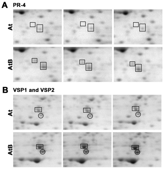
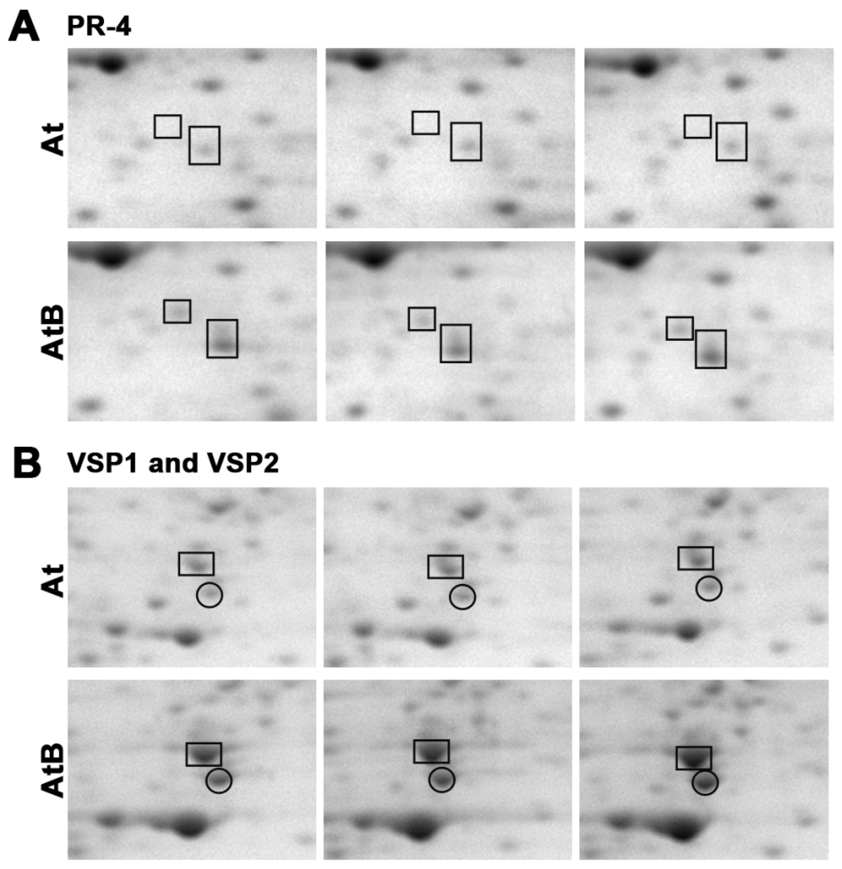
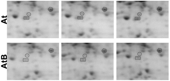
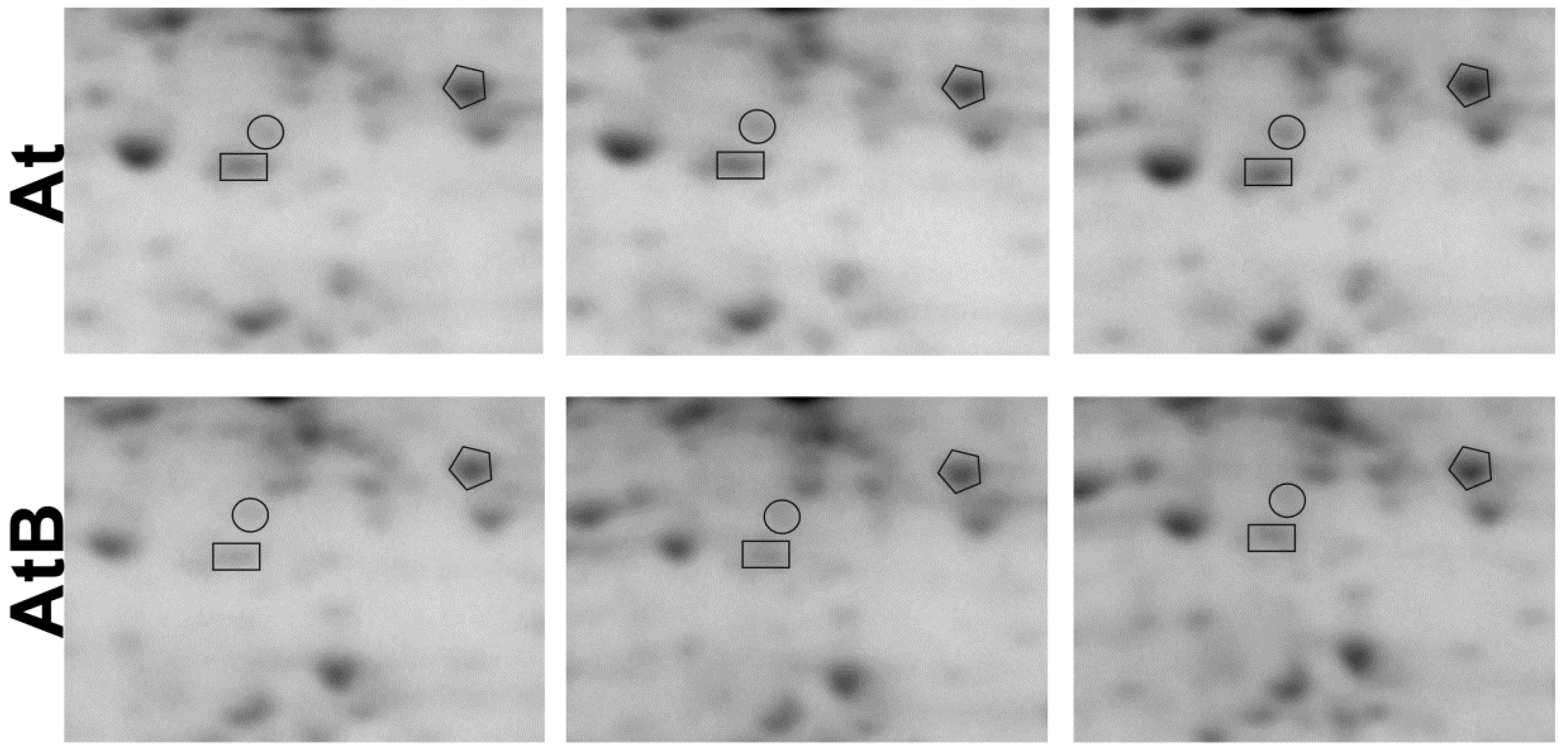
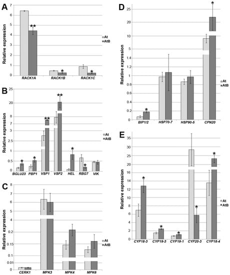
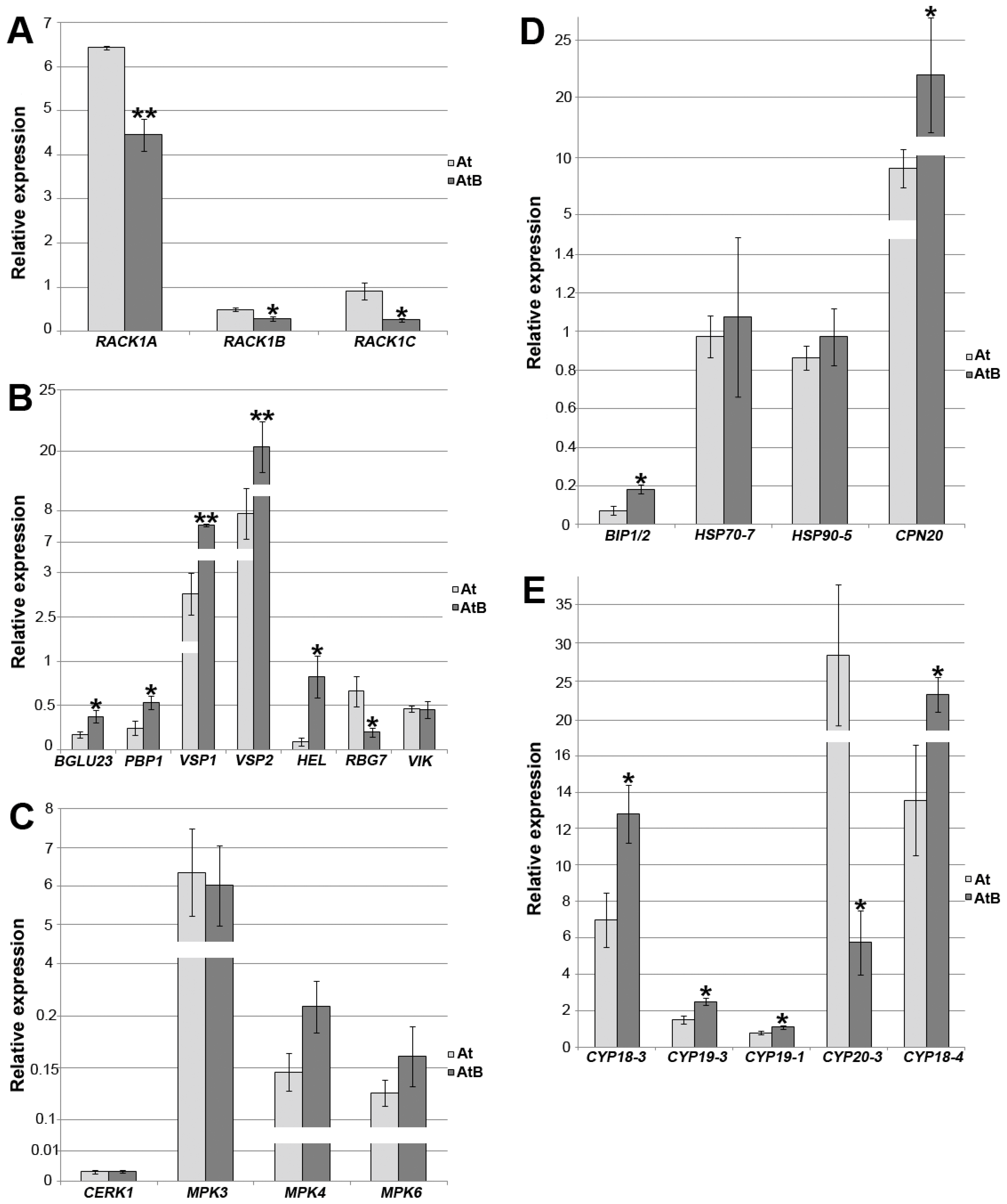

Disclaimer/Publisher’s Note: The statements, opinions and data contained in all publications are solely those of the individual author(s) and contributor(s) and not of MDPI and/or the editor(s). MDPI and/or the editor(s) disclaim responsibility for any injury to people or property resulting from any ideas, methods, instructions or products referred to in the content. |
© 2023 by the authors. Licensee MDPI, Basel, Switzerland. This article is an open access article distributed under the terms and conditions of the Creative Commons Attribution (CC BY) license (https://creativecommons.org/licenses/by/4.0/).