


Abstract
This review provides a comprehensive overview of the modulatory actions of plant-derived constituents on membrane ion channels in various cell types. Among their diverse bioactivities, ion channel regulation—governing membrane excitability, signal transduction, and cellular homeostasis—has emerged as a critical mechanistic basis for their pharmacological effects. Twenty-four representative phytoconstituents are discussed and classified into five major categories based on their structural features: alkaloids, terpenoids, lignans and acetogenins, polyphenols, and other aromatic and conjugated compounds. Across these categories, the reviewed compounds exhibit distinct and often highly specific effects on the amplitude and gating kinetics of multiple ionic currents, including voltage-gated Na+ currents (INa), delayed-rectifier K+ currents (IK(DR)), M-type K+ currents (IK(M)), hyperpolarization-activated cation currents (Ih), erg-mediated K+ currents (IK(erg)), inwardly rectifying K+ currents, and Ca2+-activated K+ currents (IK(Ca)). Alkaloids predominantly suppress voltage-gated K+ currents, with notable exceptions such as aconitine, which alters the properties of both INa and IK(DR), thereby contributing to its proarrhythmic toxicity. Terpenoids, including cannabidiol, croton diterpenoids, lutein, thymol, and triptolide, exert multifaceted effects on IK(M), Ih, inwardly rectifying K+ currents, and Ca2+-activated K+ channels. Lignans and acetogenins, such as gomisin A, honokiol, sesamin, and squamocin, primarily modulate INa, Ih, and IK(Ca), with several compounds demonstrating strong links between ion-channel modulation and anti-neoplastic or neuroprotective actions. Polyphenolic compounds, including curcumin, eugenol, resveratrol, gastrodigenin, gastrodin, and pterostilbene, display diverse ion-channel targeting profiles, influencing multiple Na+ and K+ channel subtypes. Other aromatic or conjugated compounds, such as isoplumbagin, plumbagin, and verteporfin, regulate IK(erg) and IK(Ca), potentially contributing to both therapeutic efficacy and adverse effects. Collectively, the compound-specific modulation of current amplitude and gating kinetics offers valuable mechanistic insight into the pharmacological and toxicological significance of plant-derived natural products, highlighting the functional role of ion channel evaluation in guiding their therapeutic development and ensuring safety assessment.
1. Introduction
Plant-derived natural products, often referred to as phytochemicals, are secondary metabolites that plants produce to protect themselves from environmental stress, pests, and diseases. These compounds provide humans with a vast repertoire of bioactive properties that may contribute to the prevention or treatment of a wide range of diseases and disorders [1,2,3,4]. Among these bioactive properties, their regulatory effects on ion channels in the cell membranes of different cell types have long been an important subject of academic research. This review paper examines 24 phytoconstituents derived from plant natural products. Based on their major structural features, these compounds are broadly classified into five main categories: alkaloids, terpenoids, lignans and acetogenins, polyphenols, and other aromatic and conjugated systems (Figure 1). We present a comprehensive overview of the currently recognized modulatory effects of these compounds on membrane ion channels across a broad range of mammalian cell types.
Figure 1.
Graphic representation showing the five major categories of phytoconstituents and their potential modulation of ion channels. The five categories are alkaloids (pink), terpenoids (light blue), lignans and acetogenins (brown), polyphenols (orange), and other aromatic and conjugated systems (light yellow). Within each box, the phytochemicals discussed in this article are listed individually.
The GH3 cells employed in this study are immortalized rat pituitary somatolactotrophs derived from a pituitary adenoma and are considered excitatory endocrine cells [5,6,7]. In pituitary cells, somatolactotrophs are classified as excitatory endocrine cells, whereas other types of pituitary cells are considered inhibitory endocrine cells. Excitatory endocrine cells are characterized by a high intrinsic level of excitability under basal conditions and are typically subject to suppression by inhibitory neurotransmitters, such as dopamine. In contrast, inhibitory endocrine cells exhibit low basal excitability and require stimulation by excitatory hormones to become activated [8]. It is important to note that the pituitary gland contains a wide variety of cell types, including corticotrophs, thyrotrophs, gonadotrophs, melanotrophs, somatotrophs, and lactotrophs [6]. Therefore, isolating and obtaining healthy pituitary cells remains technically challenging, and performing electrophysiological studies on these primary cells is even more difficult. Successfully isolating and efficiently obtaining immortalized cell lines derived from the human pituitary gland, and performing electrophysiological characterization of these cells, will constitute an important task for future research [6,8].
Alternatively, regarding treatment, pituitary tumors are primarily treated through surgical resection, most commonly performed using a transsphenoidal approach, as well as stereotactic radiosurgery techniques such as Gamma Knife. In cases where residual tumor tissue remains after surgery, various pharmacological therapies are employed to reduce the risk of recurrence or further growth [9,10]. Notably, voltage-dependent ion channels have emerged as promising potential therapeutic targets for future treatment strategies [6,11,12]. In addition to GH3 cells, this review also highlights the utility of other cell types, including insulinoma cells, glioma cells, Leydig tumor cells, and Rolf B1.T olfactory neurons.
2. Alkaloids
2.1. Aconitine
Aconitine is an intensely poisonous alkaloid derived from plants of the genus Aconitum (Ranunculaceae) [13,14]. This compound is a proarrhythmic agent known to open tetrodotoxin-sensitive Na+ channels in electrically excitable cells [15]. Aconitine-containing herbal extracts have also been reported to exert inhibitory effects on the proliferation of various neoplastic cells [6,8]. Studies have reported that this compound suppresses the delayed-rectifier K+ current (IK(DR)) in neurons, cardiomyocytes, and immune cells, leading to current inactivation upon membrane depolarization [16,17,18,19]. Recent findings indicate that aconitine can induce both early and delayed afterdepolarizations in neonatal cardiac cells and can trigger ventricular polymorphic tachycardia in rats [17,19]. These findings underscore the importance of understanding the electrophysiological impact of aconitine and similar compounds, considering their potential therapeutic applications and associated toxicities. Prior to evaluating the therapeutic potential of Aconitum extracts, such as their antitumor effects, it is essential to thoroughly assess the possible toxicities associated with these reagents.
2.2. Arecoline
Areca nut, derived from Areca catechu (family Arecaceae), has been widely used in China for its medical properties and psychoactive stimulant effects [20]. However, it is also recognized as a major risk for the development of oral cancer. Arecoline, a principal alkaloid isolated from the areca nut (chemically defined as 1,2,5,6-tetrahydro-1-methyl-3-pyridinecarboxylic acid methyl ester), acts as a non-selective agonist of muscarinic receptors. Extensive studies have demonstrated that arecoline exhibits carcinogenic, cytotoxic, and immunotoxic effects [21,22].
Arecoline has been reported to exert neuroprotective effects in the context of Alzheimer’s disease [23]. It also promotes the production of connective tissue growth factor in human buccal mucosal fibroblasts and induces apoptosis in HaCaT keratinocytes [21,22]. Previous studies have shown that, in U373 and U87MG glioma cells, arecoline inhibits intermediate-conductance Ca2+-activated K+ channels (KCa3.1; encoded by KCNN4) in a concentration-, voltage- and state-dependent fashion [24]. The dissociation constant for this inhibitory action was estimated to be 11.2 μM [24]. The resulting membrane depolarization induced by arecoline is therefore primarily attributable to its suppression of KCa3.1 channel activity in these cells. Notably, this effect appears to occur independently of muscarinic receptor activation [24]. Compounds such as arecoline that target KCa3.1 channels may thus be further developed as promising adjunctive agents to enhance the efficacy of cytotoxic therapies for malignant gliomas and other neoplastic diseases [25].
2.3. Berberine
Berberine is an isoquinoline alkaloid present in a variety of medicinal plants, particularly those belonging to the genera Berberis (Berberudaceae) and Coptis (Ranunculaceae). Extracts from these plants have long been used in traditional medicine for the treatment of conditions such as jaundice, dysentery, hypertension, and other ailments [26]. In addition, berberine itself has been shown to exert anti-proliferative effects on human teratocarcinoma and hepatoma cells [27].
An earlier study demonstrated that, in human myeloma cells, berberine directly exerts differential inhibitory effects on delayed-rectifier K+ current (IK(DR)) and Ca2+-activated K+ current (IK(Ca)) in a concentration-dependent manner [28]. Consistent with these electrophysiological findings, berberine markedly suppresses cellular proliferation. The close correspondence between the potency (IC50 values) for K+ current inhibition and for the reduction in cell proliferation suggests that block of IK(DR) and IK(Ca) may represent a key mechanism underlying its antineoplastic activity [28]. Beyond its effects on ion channels, berberine has also been reported to enhance mitochondrial function and to upregulate pro-apoptotic proteins, including cytochrome c, Bax, and caspase. Collectively, these findings indicate that berberine is a biologically active compound with considerable therapeutic potential for the treatment of a range of diseases, including glioma and Alzheimer’s disease [29,30,31].
3. Terpenoids
3.1. Cannabidiol
Cannabidiol, which is considered a terpenophenol, is a non-psychoactive cannabinoid derived from the Cannabis plant (Cannabaceae), known for its potential therapeutic effects. It is among over 100 cannabinoids present in the plant and has been shown to be effective in treating various medical conditions, such as epilepsy, bipolar disorder, inflammation, and cancer [32]. Recent studies have demonstrated that cannabidiol influences the activity in the hypothalamic–pituitary–adrenal axis and modulates several ionic currents in excitable cells, including voltage-gated Na+ currents (INa), M-type K+ currents (IK(M)), and hyperpolarization-activated cation currents (Ih) [33,34,35].
Cannabidiol exposure was noted to result in a concentration-dependent suppression of IK(M) in pituitary GH3 lactotrophs, with an IC50 of 3.6 μM [35]. This compound also results in the suppression of Ih in the same cell type with an IC50 of 3.3 μM [35]. These findings suggest that the responsiveness of these ionic currents is complex and influenced by various factors, including the resting membrane potential, cannabidiol concentration, patterns of action potential firing, or combinations of these variables [35].
3.2. Ent-Kaurane-Type Diterpenoids
The genus Croton (Euphorbiaceae) includes about 1300 species that are widely distributed throughout tropical regions. C. tonkinensis Gagnep., known as Kho sam cho la in Vietnamese, is a tropical shrub native to northern Vietnam and has been used commonly in Vietnam to treat different disorders [36]. C. tonkinensis is a rich source of diterpenoids [36], and its extracts were reported to exert anti-inflammatory and cancer chemopreventive activities [36,37].
Studies have shown that the fractions isolated from C. tonkinensis—specifically croton-01 (ent-18-acetoxy-7α-hydroxykaur-16-en-15-one), croton-02 (ent-7α,14β-dihydroxykaur-16-en-15-one), and croton-03 (ent-1β-acetoxy-7α,14β-dihydroxykaur-16-en-15-one)—are capable of modulating ionic currents in microglial SM826 cells, including the IK(DR) (encoded by KV1.3) and the inwardly rectifying K+ current (encoded by Kir2.1) [38]. The notable findings showed that in these microglial cells, croton-03 differentially suppresses KV1.3- and Kir2.1-encoded currents in a concentration-, time-, and state-dependent manner. Voltage-dependent blocking by croton-03 of KV1.3-encoded current with Kd (apparent affinity constant) and δ (fractional electrical distance) values of 5.17 μM and 0.091, respectively, was also estimated [39]. Therefore, the presence of these ent-kaurane diterpenoids is likely to disrupt cellular function by modulating the activity of these ionic currents [37,39].
Another report further demonstrated that the presence of croton-01, croton-02, and croton-03 produces concentration-dependent inhibition of Ih in pituitary tumor (GH3) cells with effective IC50 values of 2.89, 6.25, and 2.84 μM, respectively [40]. Cell exposure to croton-03 was also noted to decrease the voltage-dependent hysteresis of this current in response to long-lasting isosceles triangular ramp pulse [40]. Therefore, croton derivatives and other structurally related ent-kaurane-type diterpenoids could represent intriguing compounds that use the open/activated state of the HCN channels as a substrate.
3.3. Lutein
Lutein (a xanthophyll carotenoid; β,ε-carotene-3,3′-diol), derived from a (6R)-β,ε-carotene backbone, is one of the few xanthophylls that is found not only in fruits and vegetables but also at high concentrations in the macula of the human retina, where it is believed to function as a yellow light filter [41]. Previous work has shown that, in pituitary GH3 cells, exposure to lutein suppresses the hyperpolarization-activated cation current (Ih) in a concentration-, state-, voltage-, and hysteresis-dependent manner [42]. In addition, lutein induces a hyperpolarizing shift in the steady-state activation curve of this current.
Furthermore, in the continued presence of 3 μM lutein, the subsequent application of oxaliplatin or ivabradine was able to modulate the lutein-induced inhibition of Ih in GH3 cells, as illustrated in Figure 2. Oxaliplatin and ivabradine have been reported to enhance and suppress Ih, respectively [43,44]. Cyclic nucleotide-gated (CNG) channels (e.g., CNGA2) have previously been shown to exhibit pronounced hysteresis in their ion currents [45]. As demonstrated in Figure 3, the results further reveal that the magnitude of Ih elicited by a double-ramp pulse protocol can influence the patterns of burst firing in various excitable cells, including central neurons [42,46].
Figure 2.
Comparative effects of lutein, lutein plus oxaliplatin (OXAL), and lutein plus ivabradine on the amplitude of hyperpolarization-activated cation current (Ih) recorded from pituitary GH3 cells. Whole-cell current recordings were obtained from cells superfused with Ca2+-free Tyrode’s solution containing 1 μM tetrodotoxin, while the recording pipette was filled with a K+-rich internal solution. (A) Representative superimposed Ih traces recorded under control conditions (a, black), in the presence of 3 μM lutein alone (b, red), and during co-application of 3 μM lutein with 10 μM oxaliplatin (OXAL) (c, brown). The upper part shows the voltage protocol (blue) applied to the cell. Notably, Ih displays slow activation during sustained hyperpolarization. (B) Summary scatter plot illustrating the effects of lutein, lutein plus OXAL, and lutein plus ivabradine on Ih amplitude (mean ± SEM; n = 7 for each point). Current amplitudes were measured at the end of a 2-s hyperpolarizing step from −40 to −120 mV. For combination treatments, lutein was applied prior to the subsequent addition of OXAL or ivabradine. * p < 0.05 versus control; ** p < 0.05 compared with the lutein-alone (3 μM) group. This figure is adapted from Ref. [42] and is distributed under the Creative Commons Attribution (CC BY) license.
Figure 3.
Modulation by lutein of voltage-dependent hysteresis strength in Ih recorded from GH3 cells. (A) Representative current traces illustrating the relationship between forward (descending) or reverse (ascending) Ih and membrane potential, evoked by an inverted double (isosceles-triangular) ramp pulse (protocol shown at the top, blue) in the absence (a, black) and presence (b, red) of 3 μM lutein. The dashed black arrows along the current trajectory under control conditions (i.e., in the absence of lutein) indicate an anti-clockwise direction of Ih, reflecting time-dependent changes during activation measured in the absence or presence of lutein. Notably, Ih exhibits inward rectification along with pronounced voltage-dependent hysteresis. (B) Summary data showing the effects of lutein (1 or 3 μM) and lutein (3 μM) in combination with ivabradine (3 μM) on the Δarea of voltage-dependent hysteresis. The Δarea corresponds to the shaded region enclosed by the Ih trajectories during the downsloping and upsloping limbs of the triangular ramp pulse (mean ± SEM; n = 7 for each point). The results demonstrate the presence of ramp pulse induced hysteresis in Ih, and lutein reduced the Δarea in concentration-dependent manner. * p < 0.05 compared with control; ** p < 0.05 compared with the lutein-alone (3 μM) group. This figure is adapted from Ref. [42] and is distributed under the Creative Commons Attribution (CC BY) license.
During these current measurements, the tested cell was held at −40 mV and a long-lasting inverted double ramp pulse was applied [43]. This ramp protocol consists of a downsloping (forward) phase from −40 to −150 mV, followed by an upsloping (backward) return to −40 mV, with a total duration of 3.2 s (corresponding to a ramp speed of ±69 mV/s), as illustrated in the upper part of Figure 3A. As shown in Figure 3, voltage-dependent hysteresis of Ih—defined as the relationship between Ih and membrane potential during the forward and backward limbs—was evident upon activation by this triangular double ramp pulse [47].
Notably, the Ih amplitude evoked during the downsloping phase of the inverted triangular ramp was smaller (in absolute value) than that elicited during the upsloping phase, as indicated by the dashed arrows along the current trajectory (Figure 3A). For example, under control conditions (i.e., in the absence of lutein), the Ih amplitude measured at −120 mV differed significantly (p < 0.05) between the two phases, with values of 48 ± 8 pA during the downsloping limb and 249 ± 11 pA during the upsloping limb.
Upon exposure to lutein (3 μM), Ih recorded during the downsloping limb showed a small reduction compared to that observed during the upsloping limb of the triangular ramp pulse. Consequently, the extent of lutein-induced current inhibition of Ih differed significantly between the forward and reverse phases of the ramp protocol. As further quantified in Figure 3B, the degree of Ih hysteresis in GH3 cells, in the absence or presence of lutein, was assessed by measuring the ∆area under the curve (i.e., the shaded region in A) [42].
Specifically, in addition to reducing Ih magnitude, application of 3 μM lutein significantly decreased the Δarea associated with the inverted triangular ramp pulse, from 17.7 ± 2.4 to 8.8 ± 1.6 mV·nA (n = 7, p < 0.05). Following washout of lutein, the hysteretic area recovered to 17.2 ± 2.3 mV·nA. Moreover, during continued exposure to 3 μM lutein, subsequent addition of 3 μM ivabradine produced a further significant reduction in the ∆area of voltage-dependent hysteresis.
To further investigate the effects of lutein on sag potential in GH3 cells, the researchers employed current-clamp recordings [42]. Sag potential, elicited by hyperpolarizing current stimuli, is known to be closely associated with the presence of Ih in various types of excitable cells [47,48,49,50]. As illustrated in Figure 4, applying a 2-s hyperpolarizing current step of approximately pA induced a characteristic sag potential, defined as an initial rapid hyperpolarization followed by a gradual depolarizing “sag” in membrane potential during sustained current injection [49].
Figure 4.
Effect of lutein on sag potential in GH3 cells. Current-clamp recordings were performed in cells bathed in normal Tyrode’s solution containing 1.8 mM CaCl2 and 1 μM tetrodotoxin. A prolonged hyperpolarizing current step (2 s duration) was applied to evoke sag potential. (A) Representative membrane potential traces obtained under control conditions (a, black) and during exposure to 1 μM lutein (b, blue) or 3 μM lutein (c, red). The upper part shows the corresponding hyperpolarizing current stimulus applied to the recorded cell. (B) Summary scatter plot illustrating the effect of lutein on sag potential amplitude (mean ± SEM; n = 7 for each point). Under current-clamp conditions, sag amplitude was defined as the difference between the initial peak hyperpolarization and the steady-state level at the end of the current pulse, measured in the absence or presence of lutein. * p < 0.05 versus control; ** p < 0.05 compared with the lutein-alone (1 μM) group. This figure is adapted from Ref. [42] and is distributed under the Creative Commons Attribution (CC BY) license.
Exposure to ivabradine (3 μM), a known Ih inhibitor, significantly reduced the amplitude of sag potential. Similarly, treatment with lutein at concentrations of 1 or 3 μM markedly attenuated the sag potential evoked by the hyperpolarizing stimulus [42].
These findings suggest that, in addition to its established antioxidative and anti-inflammatory properties [51], lutein can modulate Ih by reducing its amplitude and altering its gating and hysteresis behavior. Consequently, lutein’s effects on ionic currents may contribute to changes in spontaneous action potential activity in electrically excitable cells, assuming similar effects occur in vivo [42].
3.4. Thymol
Thymol, a monoterpenoid phenol derived from thyme (Thymus vulgaris, Lamiaceae) essential oil, has been widely used as an antiseptic and antimicrobial agent [52,53,54,55]. Its aromatic properties make it a common ingredient in mouthwashes and dental preparations for treating oral infections [52]. Beyond oral care applications, thymol serves as a stabilizer in several therapeutic agents, including the anesthetic halothane, where accumulates in vaporizers during prolonged use. Notably, adverse effects following massive mouthwash ingestion have been attributed to thymol [56,57].
Previous studies have revealed that its biological activities may be partly mediated through modulation of ion channels. In particular, previous studies indicate that thymol influences Ca2+ signaling in pituitary GH3 cells by enhancing capacitative Ca2+ entry and depleting intracellular Ca2+ stores, involving both thapsigargin-sensitive and thapsigargin-insensitive pools [58]. Moreover, thymol was reported to stimulate IK(Ca) in GH3 cells. These stimulatory effects on Ca2+ dynamics may contribute to the cellular mechanisms underlying the impact of thymol on neuroendocrine or endocrine functions.
3.5. Triptolide
Triptolide is a diterpene triepoxide derived from the traditional Chinese medicinal plant Trypterygium wilfordii Hook. F. (Celastraceae). It exhibits a wide range of biological activities, including antitumor, immunosuppressive, and antifertility effects [59]. In the central nervous system (CNS), triptolide has been shown to exert neuroprotective properties and to promote axon growth in dopaminergic neurons [60]. Additionally, it facilitates spinal cord repair in animal models of spinal cord injury by reducing astrogliosis and inflammation [61]. Triptolide has also been reported to inhibit the proliferation and invasion of malignant glioma cells [62] and to induce apoptosis in these cells [63]. Owing to its low molecular weight and high lipid solubility, triptolide can readily cross the blood–brain barrier, enabling it to exert significant effects on glial and glioma cells [60,64].
Previous studies showed that in human glioma cells (U373 cells), triptolide inhibits the inwardly rectifying K+ current in a concentration-dependent manner with an IC50 of 0.72 μM [64]. Triptolide suppresses the activity of the inwardly rectifying K+ channels in these cells with no change in single-channel conductance. The biophysical properties of the inwardly rectifying K+ currents in U373 cells resemble the Kir4.1-encoded current because of positive mRNA detection of KCNJ10 (Kir4.1) and high sensitivity to inhibition by BaCl2. KCNJ10, the gene that codes for Kir4.1, is recognized as a putative seizure susceptibility gene in mice and humans [64,65]. Therefore, the pharmacological actions of triptolide as described recently [59,60,61,62,63] could be partly, if not entirely, connected with its inhibition of the inwardly rectifying K+ channels in glial or glioma cells. However, it should be noted that mRNA expression for other Kir channels may also occur in U373 cells.
We conducted a detailed analysis of the atomic interaction between the KCNJ10 protein [66] and triptolide using PyRx 0.8 software, employing its embedded AutoDock (PyRx 0.8; AutoDock Vina 11.2) function. Figure 5 illustrates the predicted docking sites of the triptolide molecule. Notably, during the docking analysis with the KCNJ10 channel, triptolide was observed to form hydrogen bonds with residues Arg36(A) and Ile179(A), with bond lengths of 3.08 and 3.06 Å, respectively. Moreover, triptolide exhibited hydrophobic interactions with several residues, including Lys33(A), Asp34(A), Glu177(A), Arg180(A), Phe181(A), Gln183(A), and Thr198(A). These findings suggest a strong binding affinity between triptolide and the amino acid residues of the KCNJ10 channel, estimated at −7.8 kcal/mol. This interaction predominantly occurs within the intracellular domain of chain A in the KCNJ10 channel. This predicted interaction therefore raises the possibility that triptolide-mediated alterations in the magnitude and gating kinetics of the inwardly rectifying K+ currents may occur independently of its binding to muscarinic receptors [67].
Figure 5.
Docking interaction between the triptolide molecule and the KCNJ10 (or Kir4.1-encoded) channel. The left graph refers to the docking prediction between triptolide and the KCNJ10 channel. The protein structure of KCNJ10 was obtained from the Protein Data Bank (PDB ID: 8I5M), and the three-dimensional chemical structure of triptolide was retrieved from PubChem (Compound CID: 107985). On the left, the red dashed box highlights a snapshot of triptolide engaging hydrophobic interactions and forming hydrogen bonds with the channel, which is enlarged and illustrated on the right. The green dashed line indicates hydrogen bond formation, with bond lengths estimated at 3.06 and 3.08 Å. Of note, in this and the following figures showing predicted docking, the red arcs, with spokes pointing toward the triptolide molecule, denote hydrophobic contacts, and the corresponding graph in the right panel indicates an expanded display of red dashed box with a curve arrow in the left panel.
4. Lignans and Acetogenins
4.1. Gomisin A
Gomisin A (also known as wuweizichu B, wǔwèizi chún yĭ) is a dibenzocyclooctadiene lignan isolated from the hexane fraction of the fruits of Schisandra chinensis (Schisandraceae) [68]. This dietary compound exhibits a broad spectrum of pharmacological activities, including anti-inflammatory, anti-oxidant, antihypertensive, neuroprotective, and antiproliferative effects [68]. Previous studies have demonstrated that gomisin A confers protective effects against hepatic and renal injury induced by carbon tetrachloride (CCl4), as well as against striatal toxicity caused by nitropropionic acid, primarily through modulation of the MAPK signal transduction pathway [69,70].
In addition, gomisin A suppresses the expression of key inflammatory mediators—including COX-2, iNOS, IL-6, TNF-α and NOּ—by inhibiting RIP2-mediated activation of the NF-κB activation in mouse macrophages [71]. It has also been reported to exert antioxidative effects during osteoblast differentiation and in vascular endothelial cells [68,72]. Furthermore, gomisin A can induce apoptotic responses in human colon carcinoma HCT-116 cells [73], highlighting its potential anticancer properties.
Pharmacokinetic studies have indicated that active lignan components of S. chinensis are detectable in vivo following intragastric administration in rats [67], supporting their bioavailability. Moreover, crude extracts of S. chinensis have been shown to reduce prolactin production in pituitary GH3 cells, suggesting therapeutical applications in the treatment of hyperprolactinemia and prolactin-secreting tumors [74].
A notable study reported that gomisin A exerts a suppressive effect on voltage-gated Na+ currents (INa) in GH3 cells in a concentration-, time- and state-dependent manner. Exposure to gomisin A was found to accelerate the inactivation kinetics of INa, with a particularly pronounced effect on the slow component of current inactivation [75]. The inhibitory action developed rapidly and was readily reversible upon washout. This effect was temporally associated with a marked increase in the inactivation rate of currents evoked by brief depolarizing pulses [75,76], while the activation kinetics of INa remained largely unaffected.
Further analysis revealed that, in GH3 cells, gomisin A differentially inhibits the transient or late components of INa, with IC50 value of 6.2 or 0.73 μM, respectively [75], indicating a greater potency toward the late current component. The modulatory effects of gomisin A on both the amplitude and gating properties of INa are therefore thought to arise from its direct interaction with voltage-gated Na+ (NaV) channel or their accessory subunits [76].
Subsequently, molecular docking analysis was performed to evaluate the interaction between the SCN9A (NaV1.7) channel protein and gomisin A using PyRx 0.8 software. The predicted binding sites of gomisin A on the channel are depicted in Figure 6. The docking results indicate that gomisin A forms hydrophobic interactions with multiple amino acid residues, including Ser32(I), Ser820(B), Val821(B), Leu875(A), Phe876(A), Val914(A), Tyr915(A), Pro916(A), and Tyr917(A), which are primarily located within the membrane-embedded regions of the channel protein.
Figure 6.
Docking analysis of NaV1.7 (SCN9A) and gomisin A. The protein structure of SCN9A was retrieved from the Protein Data Bank (PDB ID: 6N4Q), while the three-dimensional structure of gomisin A was obtained from PubChem (CID: 15608605). Molecular docking was performed using PyRx, and the optimal binding pose of gomisin A within SCN9A is highlighted by the red dashed box on the left panel.
Given the experimentally observed inhibitory effect of gomisin A on INa, together with the predicted binding interactions with the SCN9A protein, these findings suggest that gomisin A may directly modulate NaV1.7 channel activity. Consequently, this compound holds potential as a modulator of NaV1.7 function across different cell types, thereby influencing their electrophysiological behavior.
4.2. Honokiol
Honokiol, a hydroxylated biphenyl compound isolated from Magnolia officinalis and other species within the Magnoliaceae family, has long been utilized in traditional Asian herbal medicines, including Houpo, Hou p’u, or Saiboku-to [77]. It is widely recognized as a promising natural compound with diverse biological activities, particularly due to its ability to influence multiple cellular processes across various cancer models [78]. In addition to its anticancer properties, previous studies have demonstrated that honokiol can regulate the functional activity of both neuroendocrine or endocrine systems. For instance, it has been shown to induce cell cycle arrest and trigger programmed cell death in human thyroid tumor cells, both in vitro and in vivo [79]. Furthermore, several studies have reported that M. officinalis bark and honokiol can modulate catecholamine secretion in adrenal chromaffin cells [80]. Beyond these effects, honokiol has also been found to exhibit antidepressant-like activity, potentially through normalization of the hypothalamic-pituitary-adrenal axis [81].
Previous studies have shown that honokiol produces a concentration-dependent inhibition of Ih in GH3 cells, with an IC50 value of 2.1 μM (Figure 7) [82]. In addition to reducing current amplitude, honokiol shifts the steady-state activation curve of Ih toward a more negative potential without altering the apparent gating charge. This compound also attenuates the voltage-dependent hysteresis of Ih elicited by prolonged triangular ramp pulse. Moreover, honokiol has been reported to suppress Ih amplitude in Rolf B1.T olfactory neurons [82].
Figure 7.
Concentration-dependent inhibition of the hyperpolarization-activated cation current (Ih) by honokiol (HNK) in GH3 cells. (A) Representative Ih traces recorded under control conditions and following exposure to honokiol. Cells were perfused with Ca2+-free Tyrode’s solution containing 1 μM tetrodotoxin, and recording pipettes were filled with a K+-based internal solution. Hyperpolarizing voltage steps from −40 to −120 mV (2 s duration) were applied. Trace a corresponds to control conditions, while traces b and c were obtained after application of 3 μM and 10 μM honokiol, respectively. The upper part illustrates the voltage-clamp protocol used. (B) Concentration-response relationship for honokiol-mediated inhibition of Ih (mean ± SEM; n = 8 for each data point). The data were fitted with a red sigmoidal curve using the Hill equation, yielding an IC50 value of 2.1 μM. This figure is adapted from Ref. [82] and is distributed under the Creative Commons Attribution (CC BY) license.
These findings collectively demonstrate that honokiol effectively inhibits Ih in both pituitary GH3 cells and Rolf B1.T olfactory neurons. The onset of this inhibition is rapid, suggesting that it may contribute to the compound’s modulatory effects on the functional activity of sensory neurons. Such suppression of Ih is likely to influence the firing behavior of electrically excitable cells, thereby altering neuronal excitability and over cellular function [82].
To examine how honokiol interacts with the HCN channel protein, we used the structure of HCN3 protein from PDB. The HCN3 channel protein structure was acquired from PDB (PDB ID: 8IO3), and PyRx 0.8 software was used to see how the honokiol molecule docks into the channel protein. Figure 8 presents the predicted docking sites of honokiol for interaction with amino-acid residues of the HCN3 channel. Specifically, the honokiol molecule was predicted to form hydrophobic contacts with residuals Phe60(D), Tyr89(D), Phe94(D), Ile237(D), and Phe238(D), as well as hydrogen bonds with residuals Ser90(D) and Asp91(D), with estimated distances of 2.95 and 3.10 Å, respectively. It is therefore anticipated that honokiol can interact with HCN channels to modulate the amplitude and gating kinetics of Ih [82].
Figure 8.
Predicted docking between the HCN3 channel and the honokiol molecule. The left panel depicts the HCN3-channel protein structure acquired from PDB (PDB ID: 8IO3), and the three-dimensional structure of honokiol from PubChem (Compound CID: 72303). PyRx software was used to show that the HCN3 channel structure can be optimally docked by the honokiol molecule. The right panel depicts an expansion of the red dashed box with a curved arrow in the left panel. This diagram of the interaction between the HCN3 channel and the honokiol was generated using LigPlot+ Version 2.3.
Recent reports have demonstrated the ability of honokiol to exert antioxidant, anti-inflammatory, neurotrophic, and anti-apoptotic effects [83]. We also conducted a predicted docking analysis between honokiol and superoxide dismutase (SOD). SOD is a key antioxidant enzyme that plays a central role in controlling oxidative stress and modulating inflammation. We explored how SOD protein could be optimally docked with honokiol molecule by using PyRx software. The protein structure of SOD was obtained PDB (PDB ID: 1DO5). The predicted docking sites of the honokiol molecule with which the amino-acid residues can interact are presented in Figure 9. Notably, the honokiol molecule was predicted to form hydrophobic contacts with certain residues, including Asn142(B), Asn142(C), Ser143(B), Ser143(C), Gly145(C), Asn146(C), Ser233(A), Ala234(A), Gly235(A), and Leu236(A). The honokiol molecule was also predicted to form a hydrogen bond with residue Asp136(B) with a distance of 2.79 Å. These docking results suggest that honokiol can bind to SOD protein with a binding affinity of −7.4 kcal/mol. In high-resolution X-ray diffraction structures, the remaining water molecules are those associated with the protein, where other PDB structures, such as 8I5M (KCN10 channel), 6N4Q (SCN9A channel), and 8IO3 (HCN3 channel), determined by electron microscopy, do not display any water atoms.
Figure 9.
Molecular docking analysis of honokiol with superoxide dismutase. The protein structure of superoxide dismutase was acquired from PDB (PDB: 1DO5). The structure of superoxidase dismutase was docked with the honokiol molecule by using PyRx software, as indicated in the left side. The diagram of the interaction between superoxide peroxide protein and the honokiol molecule was generated by LigPlot+ (right side). The small green dots represent water molecules within the structure, with a total of 26 atoms included in this group.
4.3. Ganoderma Triterpenoids
Ganoderma mushrooms (known as Língzhī in Chinese and Reishi in Japanese) have long been used in traditional Chinese medicine, and are now recognized worldwide as a dietary supplement. Among the various species, Ganoderma lucidum (family Ganodermataceae) is the most extensively utilized and is commonly cultivated under controlled conditions to ensure a consistent chemical profile [84].
G. lucidum has been reported to exhibit a broad range of pharmacological activities, including antihypertensive, hypoglycemic, and hypocholesterolemic effects, along with other medicinal benefits [84,85]. Its major bioactive constituents are primarily triterpenoids [85,86]. In addition, G. lucitum has been shown to confer cardioprotective effects in animal models, likely by reducing oxidative stress associated with myocardial injury [87].
Triterpenes are a class of terpenes characterized by a C30 carbon backbone. Triterpenoids, their oxidized derivatives, typically have molecular weights ranging from 400 to 600 Da and exhibit structurally complex and highly oxidized chemical frameworks [88]. The triterpenoid fraction of Ganoderma, which includes more than 300 lanostane-type compounds, has been increasingly recognized for its biological activities. These include potent antioxidant effects the contribute to the prevention of myocardial injury, as well as notable neuroprotective properties [84,86,87,88].
In addition, Ganoderma triterpenoids have been shown to suppress inflammatory response by directly scavenging free radicals and by enhancing endogenous antioxidant enzymes, thereby reducing lipid peroxidation in animal models such as chicken liver and mice [87,89]. Furthermore, aqueous extract of Ganoderma has been reported to exhibit anticonvulsant, antidepressive, anxiolytic, and antinociceptive effects [90].
Previous studies have shown that the inhibitory effects of Ganoderma triterpenoids of Ih in GH3 cells extend beyond a simple reduction in current magnitude. These compounds also modify the kinetic properties of Ih, indicating a dose-, time- and state-dependent mode of inhibition [91]. In addition, the presence of Ganoderma triterpenoids shifts the steady-state activation curve of Ih along the voltage axis toward more hyperpolarized potentials.
The IC50 value for Ganoderma triterpenoids-mediated inhibition of Ih observed in GH3 cells (11.7 μg/mL) is comparable to the concentrations previously reported to produce antioxidative and neuroprotective effects [85,91]. These findings suggest that the modulation of Ih may represent an early, upstream mechanism contributing to the compounds’ overall pharmacological actions, including their ability to regulate intracellular oxidative stress [84,85].
4.4. Sesamin and Sesamolin
Sesame seeds and sesame oil, derived from Sesamum indicum (family Pedaliaceae), have long been valued as health-promoting foods in many Asian countries [83]. Compared with edible oils obtained from other plant seeds, sesame oil exhibits remarkably oxidative stability. This stability is largely attributed to its high content of lipid-soluble furofuran lignans, particularly sesamin and sesamolin, which possess potent antioxidant properties [92].
Recent studies have highlighted that sesamin and sesamolin, the principal furofuran lignans found in sesame oil, possess a wide range of biological activities. These compounds have been shown to inhibit lipid peroxidation in erythrocytes [93], reduce intestinal cholesterol absorption, and suppress hepatic 3-hydroxy-3-methylglutaryl coenzyme-A (HMG-CoA) reductase activity [94]. In addition, they have demonstrated chemopreventive effects against chemically induced mammary carcinogenesis, as well as the ability to inhibit Δ5-desaturase activity and the elongation of C18 fatty acids. Both lignans also provide protective effects in hypoxic neuronal and PC12 cells, primarily through the suppression of reactive oxygen species (ROS) generation and mitogen-activated protein kinase (MAPK) signaling pathways [95]. Furthermore, sesamin and sesamolin have been reported to exert antihypertensive and cardioprotective effects [96].
Previous investigations have demonstrated that sesamin and sesamolin exert differential and potent inhibitory effects on both the transient and late components of INa in pituitary GH3 cells, in a concentration-dependent manner [97]. Specifically, sesamin was found to reduce the peak and sustained INa with IC50 values of 7.2 and 0.6 μM, respectively [97]. In addition to decreasing current amplitude, sesamin alters the inactivation kinetics of INa during brief depolarizing pulses.
Based on a Markovian model derived from the SCN8A channel [97,98], the sesamin-induced modulation of NaV channel activity is thought to result from a decreased probability of channel occupying the open (O) and open-blocked (OB) states. These findings indicate that inhibition of NaV channels by sesamin may represent a key ionic mechanism underlying its ability to modulate the functional behavior of various electrically excitable cells, provided that similar effects occur in vivo [98]. Furthermore, sesamin and structurally related lignans may act as modulators of NaV channels in neoplastic or neuroendocrine tumor cells, suggesting potential applications in cancer neuroscience through the suppression of tumor progression, invasiveness, and metastasis [76,99].
4.5. Squamocin
Squamocin is a bis-tetrahydrofuran acetogenin that has been isolated from various members of Annonaceae family, including Annona squamosa (custard apple), Annona muricata (soursop), and Asimina triloba (pawpaw). Structurally, it features an extended aliphatic chain terminated by an α,β-unsaturated γ-lactone moiety, along with two tetrahydrofuran rings and several oxygen-containing functional groups distributed along the backbone.
This compound has attracted attention due to its diverse biological activities. It exhibits notable insecticidal properties [100] and has demonstrated potential anti-cancer effects [101]. Mechanistically, squamocin has been reported to inhibit mitochondrial NADH:ubiquinone oxidoreductase (complex I) [102], thereby affecting cellular energy metabolism. Furthermore, studies on asimicin, a structurally related analogue, suggest that its cytotoxic effects may involve alterations in membrane structure and dynamics [103]. More recent findings indicate that squamocin can trigger apoptosis in HL-60 leukemia cells, an effect that appears to be mediated through the activation of stress-activated protein kinase [104].
Earlier investigations have reported that squamocin exerts a stimulatory effect on Ca2+-activated K+ current (IK(Ca)) in human coronary smooth muscle cells [105]. This enhancement appears to be closely associated with elevations in intracellular Ca2+ levels. Supporting this interpretation, the observed increase in IK([Ca2+]i) amplitude induced by squamocin is markedly reduced when extracellular Ca2+ is removed, indicating that both Ca2+ influx from the extracellular space and Ca2+ release from intracellular stores contribute to the overall response. These findings suggest that squamocin may facilitate the opening of membrane ion channels with high Ca2+ permeability [105], thereby elevating intracellular Ca2+ and secondarily augmenting IK(Ca). Nevertheless, the precise cellular and molecular mechanisms underlying the actions of squamocin and related acetogenins across different cell types remain incompletely understood and warrant further investigation.
5. Polyphenols
5.1. Curcumin
Curcumin (diferuloylmethane, 1E,6E-1,7-bis(4-hydroxy-3-methoxyphenyl)-1,6-heptadiene-3,5-dione), a major constituent of the spice turmeric obtained from Curcuma longa (Zingiberaceae), is a bright yellow diarylheptanoid produced by some plants. It is the principal curcuminoid of turmeric (Curcuma longa), a member of the ginger family (Zingiberaceae). This compound is largely used as a herbal supplement, cosmetics ingredient, food flavoring and food coloring [106]. Particularly, this nutraceutical compound has been demonstrated to possess beneficial properties in a variety of diseases ranging from cancer to diabetes mellitus [107]. For example, curcumin has been recently reported to influence insulin release from isolated pancreatic islets [108].
Curcumin has been reported to exert a concentration- and state-dependent depressant action on IK(DR) in pancreatic β-cells, particularly in the rat insulinoma cell line INS-1 [109]. The inhibitory action on this K+ current tends to correlate in time with a significant increase in the inactivation rate of the currents in response to membrane depolarization, while the activation kinetics of the current remained unaltered in the presence of curcumin [109].
The estimated peak of plasma curcumin concentration was previously reported to reach 3.14 μg/mL (around 8.57 μM) [110], a value which is apparently greater than the KD (1.26 μM) and IC50 (3.32 μM) required for curcumin-mediated inhibition of IK(DR) seen in INS-1 cells [109]. Therefore, the inhibitory effects of curcumin and curcuminoids on these K+ currents in pancreatic β-cells are expected to occur at concentrations achievable in the human organisms, although whether the effects of curcumin and other curcuminoid derivatives on ionic currents in humans still remains to be further delineated.
5.2. Columbianadin
Columbianadin is a prominent bioactive compound isolated from the root of Angelica pubescens Maxim. f. biserrata (Apiaceae), a medicinal plant commonly known as Angelicae Pubescentis Radix, or “Duho (Dú huó)” in traditional Chinese medicine. Chemically, columbianandin belongs to the coumarin family and is classified as an angular dihydrofuranocoumarin.
This compound has been widely recognized for its diverse pharmacological properties, including analgesic, anti-inflammatory, and anti-neoplastic activities [103,104]. It has been shown to influence cellular processes by modulating proliferation and promoting apoptosis [111]. In addition, experimental studies have demonstrated that columbianadin can attenuate inflammatory responses triggered by agents such as carrageenan and lipopolysaccharide, further supporting its potential therapeutic value in inflammatory conditions [111,112].
It is noteworthy that columbianadin suppresses INa in GH3 cells in a manner that depends on concentration and channel state [113]. Detailed analyses indicate that it exerts a differential inhibitory effect on the transient (peak) and sustained (late) components of INa elicited by rapid membrane depolarization with respective IC50 values of 14.7 and 2.8 μM [113]. In addition, columbianadin shifts the midpoint of the steady0-state inactivation curve toward more negative potentials, while leaving the activation time course of the current largely unaffected.
Further observations show that columbianadin can counteract the tefluthrin-induced enhancement of persistent INa during both the ascending and descending phases of an upright isosceles-triangular ramp. Tefluthrin, a pyrethroid insecticide known to augment INa [114,115], thereby serves as a useful pharmacological tool in this context.
In HL-1 cardiomyocytes, columbianadin similarly reduces INa amplitude and accelerates the slow component of current inactivation [113]. Collectively, these findings suggest that the compound modulates both the magnitude and gating kinetics of INa. Such electrophysiological effects are likely to occur upstream of its reported influence on intracellular signaling pathways, including cytosolic NOD1/NF-κB activation [116] and on the regulation of antioxidant enzyme activity [117]. Consequently, these ion channel-modulating properties may contribute to the broader physiological actions of columbianadin in electrically excitable cells, such as GH3 cells or HL-1 cardiomyocytes, under in vivo conditions.
5.3. Eugenol
Eugenol (4-allyl-2-methoxyphenol) is an aromatic molecule found in several plants including clove (Syzygium aromaticum, Myrtaceae), bay leaves (Laurus nobilis, Lauraceae), and allspice (Pimenta dioica, Myrtaceae), and has been used in dental practice to relieve pain arising from a variety of sources, such as pulpal inflammations and dentin hypersensitivity [118]. Furthermore, eugenol is neuroprotective against excitotoxicity, cerebral ischemia and the toxic effects of amyloid-β peptides [119,120]. This compound has been demonstrated to suppress epileptiform field potentials and spreading depression in hippocampus and neocortex. These results suggest that the anti-convulsive properties of eugenol are mediated through the effects on neuronal ion fluxes by blocking NaV channels [121,122,123].
The evidence has emerged that the effects on ion channels may be an important mechanism underlying eugenol-induced actions in neurons. Previous studies demonstrated the ability of eugenol to bind to vanilloid receptors and, in turn, to activate non-selective cation channels [124]. A recent work has also reported that this compound produced an inhibition of voltage-gated Ca2+ current through CaV2.3 channels in the E52 cell line [124]. Such inhibition is thought to be direct and independent of the binding to vanilloid receptors or to β-adrenergic receptors [124,125].
Previous reports have demonstrated that the exposure to eugenol differentially inhibited the transient and late components of INa in differentiated NG108-15 neuronal cells in a concentration-dependent manner [126]. The IC50 values of eugenol required for the inhibition of the transient and late INa were 8.9 and 1.6 μM, respectively [126]. Eugenol can also diminish the amplitude of persistent INa evoked by long-lasting ramp pulse, and tefluthrin reversed the eugenol-induced inhibition of this current. This compound could decrease the frequency of spontaneous action potentials. It is therefore anticipated that the observed effects of this agent on ionic currents could be an important mechanism underlying its actions in neurons occurring in vivo [120,123,126].
5.4. Resveratrol
Resveratrol (trans-3,4′,5-trihydroxystilbene) is a polyphenolic phytoalexin naturally occurring in a variety of plant sources, such as grape (Vitis vinifera, family Vitaceae), peanuts (Arachis hypogaea, family Fabaceae), and several types of berries. A growing body of evidence has demonstrated that resveratrol exerts a wide range of pharmacological actions, including anti-platelet, anti-carcinogenic, anti-viral, and cardioprotective effects [127]. In addition, increasing studies suggest that resveratrol possesses notable neuroprotective properties [3,127,128,129]. Specifically, it has been shown to mitigate convulsions and neurotoxicity induced by kainic acid, as well as to confer protection against seizures triggered by pentylenetetrazol [130,131].
A notable observation is that resveratrol suppresses action potential firing in rat cortical neurons, and effect that appears to involve both enhancement of IK(Ca) and inhibition of INa [132]. The increase in IK(Ca) amplitude is thought to result from activation of large-conductance Ca2+-activated K+ channels [126,132,133]. In parallel, the inhibitory action of resveratrol on INa has been proposed to contribute to its analgesic properties. Through these concurrent modulatory effects on IK(Ca) and INa, resveratrol may hold promise as a broad-spectrum anti-seizure agent.
5.5. Gastrodigenin and Gastrodin
Gastrodigenin (p-hydroxybenzyl alcohol, also known as 4-hydroxybenzyl alcohol) is a phenolic constituent derived from the traditional Chinese medicinal herb Gastrodia elata Blume (family Orchidaceae), commonly referred to as Tian Ma in Chinese and Chunma in Korean [134]. Its glucoside form, gastrodin, represents the principal bioactive compound isolated from this plant. For centuries, the dried rhizomes of G. elata have been extensively utilized for the management of various neurological and psychiatric conditions, including seizures, vertigo, cognitive decline, depressive disorders, and migraines [135,136,137,138]. In recent years, growing attention has been directed toward elucidating the molecular and cellular mechanisms by which the active components of G. elata produce their diverse pharmacological effects.
Previous investigations have examined the effects of two structurally related compounds, gastrodigenin and its glucoside gastrodin, on various ionic currents in pituitary cells and hippocampal neurons. In pituitary GH3 lactotrophs, both agents were found to suppress the amplitude of IK(M) in a time- and concentration-dependent manner [131]. The IC50 value for gastrodigenin and gastrodin in these cells were estimated to be 12.1 and 19.4 μM, respectively [138].
Notably, the inhibition of IK(M) by gastrodigenin was accompanied by a slowing of the current’s activation kinetics, whereas gastrodin produced an apparent acceleration of the activation time course. In addition, gastrodigenin shifted the steady-state activation curve of IK(M) toward more hyperpolarized potentials without altering the apparent gating charge [138]. Both components were also effective in reducing IK(M) amplitude in hippocampal mHippoE-14 neurons.
Given that gastrodin is capable of crossing the blood–brain barrier, these findings suggest that modulation of ionic currents—particularly low threshold IK(M), which is characterized by slow activation and deactivation kinetics—may represent an important mechanism underlying the actions of these compounds in endocrine or neuroendocrine cells, as well as in central neurons, provided that similar effects occur in vivo [136,137,138].
5.6. Pterostilbene
Pterostilbene (trans-3,5-dimethoxy-4′-hydroxystilbene) is a natural dimethylated analog of resveratrol isolated from Pterocarpus marsupium Roxb. (Fabaceae), a tree native to India, Nepal, and Sri Lanka. The extracts of P. marsupium containing pterostilbene have been traditionally used in Ayurvedic medicine for treating various disorders [139]. This compound has been reported to have benefits for the prevention or treatment of different kinds of cancers, as mounting evidence has demonstrated its inhibitory effects on almost every cellular event that promotes tumor progression toward metastasis in both apoptosis-dependent and apoptosis-independent manners [140,141,142].
In earlier studies, the addition of pterostilbene inhibited Ih effectively in a concentration- and time-dependent manner in pituitary GH3 cells. The I–V relationships of Ih established at various levels of hyperpolarizing steps were also established and depicted in Figure 10. It was noted that the presence of pterostilbene (1 μM) significantly reduced the slope of the linear fit of Ih amplitudes to the voltages between −130 and −100 mV from 27.2 ± 1.3 to 13.1 ± 1.1 nS (n = 8, p < 0.05). The steady state activation curve of Ih was distinctly shifted to more hyperpolarizing potentials by 11 mV, producing channel opening at more negative voltages. The results thus demonstrate that pterostilbene has a conceivable depressant action on Ih functionally expressed in GH3 cells [143].
Figure 10.
Effect of pterostilbene (PTER) on the current-voltage (I–V) relationships of Ih in GH3 cells. Current traces were recorded during 2-s voltage steps to a family of membrane potentials ranging between −130 and −40 mV in 10-mV steps from a holding potential of −40 mV, as indicated in the uppermost part of (A). (A) Representative Ih traces achieved under the control condition (i.e., pterostilbene was not present) (upper) and during the exposure to 1 μM pterostilbene (lower). Arrowhead in each panel depicts the zero-current level and the calibration mark shown in the right lower side applies to all current traces. Of note, there are varying durations in voltage-clamp profile for better illustration. (B) Averaged I–V relations of Ih achieved in the absence (■) and presence (□) of 1 μM pterostilbene (mean ± SEM; n = 9 for each data point). Current amplitude was measured at the end of each voltage step. This figure is adapted from Ref. [143] and is published under the Creative Commons Attribution (CC BY) license.
Furthermore, as shown in Figure 11, the presence of pterostilbene raised the amplitude of macroscopic IK(Ca) in GH3 cells. In this set of whole-cell current recordings, GH3 cells were bathed in normal Tyrode’s solution containing 1.8 mM CaCl2, and the recording pipette was filled with a K+-enriched solution. In an attempt to inactivate most of the voltage-gated K+ currents, we then maintained the examined cells at the level of 0 mV, and then applied a series of voltage pulses between 0 and +50 mV with 10-mV steps. Within 1 min of exposing GH3 cells to pterostilbene (3 μM), the amplitude of IK(Ca) elicited by this voltage profile evidently rose (Figure 11). For example, the addition of 3 μM pterostilbene increased IK(Ca) amplitude elicited by depolarizing pulse from 0 to +50 mV from 568 ± 35 to 1005 ± 89 pA (n = 8, p < 0.05). As the compound was washed out, current amplitude returned to 621 ± 38 pA (n = 8). The averaged I–V relationships of IK(Ca) amplitude in the control, during the exposure to 3 μM pterostilbene and after washout of the compound were depicted in Figure 11B. The addition of 3 μM pterostilbene substantially increased the whole-cell conductance of IK(Ca) measured at the voltages between +30 and +50 mV to 19.6 ± 0.8 nS (n = 8, p < 0.05) from control value of 12.6 ± 0.5 nS (n = 8). Therefore, pterostilbene can increase the amplitude of IK(Ca) observed in these cells and its stimulation of IK(Ca) was largely attributable to the activation of large-conductance Ca2+-activated K+ channels [143].
Figure 11.
Stimulatory effect of pterostilbene (PTER) on Ca2+-activated K+ current (IK(Ca)) identified in GH3 cells. This set of whole-cell current recordings was conducted in cells suspended in normal Tyrode’s solution which contained 1.8 mM CaCl2. (A) Representative IK(Ca) traces obtained during the voltage steps to various membrane potentials between 0 and +50 mV in 10-mV steps from a holding potential of 0 mV (as indicated in the uppermost part of (A)). Records shown in the upper and lower panels were obtained under control condition and during the exposure to 3 μM pterostilbene, respectively. The uppermost part depicts the voltage pulses applied. The red dashed curved arrow indicates that the amplitude of IK(Ca) increases as the cell membrane depolarizes, demonstrating its outwardly rectifying properties. (B) Averaged I–V relations of IK(Ca) obtained in the control (●), during the exposure to 3 μM pterostilbene (○), and after washout of the agent (□) (mean ± SEM; n = 8 for each data point). This figure is adapted from Ref. [143] and is published under the Creative Commons Attribution (CC BY) license.
6. Other Aromatic or Conjugated Systems
6.1. Isoplumbagin and Plumbagin
Isoplumbagin (5-hydroxy-3-methyl-1,4-naphthoquinone) is a naturally occurring quinone from Lawsonia inermis (Lythraceae) or Plumbago europarea (Plumbaginaceae) [144]. Similar to isoplumbagin, plumbagin (5-hydroxy-2-methyl-1,4-naphthoquinone), another hydroxyl-1,4-napththoquinone, is another alkaloid obtained from the roots of the plants of Plumbago genus. Isoplumbagin and plumbagin have been demonstrated to exert antineoplastic activity against an array of cancer cells, including oral and tongue squamous cell carcinoma, glioblastoma, non-small cell lung carcinoma, and breast, cervical, endometrial, pancreatic and prostate cancers [145,146]. Alternatively, plumbagin was shown to induce apoptotic changes in lung cancer and neuronal cells via caspase-9 activation and targeting mitochondrial-mediated ROS induction [147,148]. Plumbagin has been reported to modify release of pituitary gonadotropin [149]. Previous studies have shown the ability of 2-mercaptophenyl-1,4-naphthoquinone, a naphthoquinone derivative, to overcome the elevation of intracellular Ca2+ in platelets caused by ADP and collagen [150].
Accumulating evidence indicates that isoplumbagin can influence the magnitude, gating behavior, and voltage-dependent hysteresis of ether-à-go-go-related gene (erg)-mediated K+ currents (IK(erg)) recorded in GH3 cells [151]. Moreover, these erg-mediated K+ currents were similarly suppressed by isoplumbagin in MA-10 Leydig tumor cells [151]. These findings suggest that the inhibitory effects of isoplumbagin, as well as the structurally related compound plumbagin, on IK(erg) may play a role in their anti-neoplastic properties, provided that such effects are recapitulated under in vivo conditions.
The IC50 values for isoplumbagin-mediated suppression of the peak and sustained components of IK(erg) were determined to be 18.3 or 2.4 μM, respectively [151]. Based on a first-order reaction scheme, the dissociation constant (KD) was estimated to be 2.58 μM, a value closely consistent with the IC50 (i.e., 2.4 μM) for inhibition of the sustained IK(erg) elicited by prolonged membrane hyperpolarization.
Previous work has also demonstrated that isoplumbagin, at a concentration of 2.5 μM, can impair mitochondrial respiration, likely through inhibition of complex IV activity [152]. In addition, the extent to which isoplumbagin or plumbagin modulates IK(erg) is influenced not only by their concentration but also by several contextual factors, including the baseline resting membrane potential and patterns of action potential firing, assuming that IK(erg) is sufficiently expressed in the cells under study.
Although the precise mechanisms underlying isoplumbagin-induced inhibition of IK(erg) remains to be fully elucidated, such blockade is likely to be of pharmacological significance [8]. Notably, it has been proposed that isoplumbagin or plumbagin may directly enhance prolactin secretion in vivo via suppression of IK(erg) [8,153]. However, the potential impact of isoplumbagin-mediated IK(erg) inhibition on cardiac function [153,154] has yet to be clearly defined.
6.2. Verteporfin
Verteporfin (Visudyne®), a benzoporphyrin derivative, is a clinically utilized photosensitizer in photodynamic therapy. It is specifically designed to selectively ablate abnormal ocular vasculature, such as that associated with neovascular (wet) age-related macular degeneration and choroidal neovascularization [155,156,157].
Photodynamic therapy employing photosensitizers, including hypericin and verteporfin, has also been shown to be effective in treating a variety of hyperplastic and neoplastic conditions, such as residual tumors in the pituitary gland [158,159]. In line with this, earlier studies demonstrated that certain photosensitizers—for example, rose Bengal, a fluorescein-derived compound—can modulate membrane ionic current in GH3 cells as well as in cardiac cells [160]. Conversely, there is evidence indicating that verteporfin may produce adverse effects; specifically, it has been reported to induce anterior ischemic optic neuropathy in rodent models [155,161].
A prior study reported that verteporfin exerts a pronounced stimulatory effect on IK(Ca) in pituitary GH3 cells, while producing only a modest reduction in the amplitude of IK(DR), without significantly affecting either INa or IK(M) [5]. The enhancement of IK(Ca) by verteporfin is thought to arise from alterations in intracellular Ca2+ levels. Supporting this notion, the verteporfin-induced increase in IK(Ca) was markedly attenuated when extracellular Ca2+ was removed, indicating that both Ca2+ influx from the extracellular space and release from intracellular stores contribute to the observed augmentation of IK(Ca) in these cells.
The EC50 value for either verteporfin-induced enhancement of IK(Ca) in pituitary GH3 cells and for the increased activity of large-conductance Ca2+-activated K+ channels in 13-06-MG glioma cells (a human glioblastoma multiforme cell line) was estimated to be 2.4 or 1.9 μM, respectively [5]. Notably, these concentrations are comparable to those that are either clinically attainable or required to inhibit the YAP-TEAD (Yes-associated protein-TEA domain transcription factor) complex in various anaplastic or neoplastic cells [6,162,163,164]. Given that verteporfin augments IK(Ca) amplitude within a short time frame in GH3 cells, it is reasonable to propose a potential mechanistic link between its anti-neoplastic properties and its stimulatory effects on IK(Ca). Such actions may occur rapidly, possibly even prior to significant intracellular accumulation of the compound [1,6,162].
7. Conclusions
This review illustrates that five major chemical classes of phytoconstituents exert distinct and specific modulatory effects on membrane ionic currents, providing a mechanistic basis for their potential pharmacological activities. Alkaloids predominantly exert inhibitory effects on voltage-gated K+ currents; however, aconitine specifically affects the inactivation of INa and IK(DR). Terpenoids modulate IK(M), Ih, and IK(DR). Compounds within the lignan and acetogenin groups exhibit inhibitory actions on INa and Ih. Polyphenolic compounds influence, to varying extents, IK(DR), INa, Ih, IK(erg), IK(M), and IK(Ca). In contrast, other aromatic or conjugated compounds can regulate IK(erg) and IK(Ca). Moreover, individual phytoconstituents also display distinct specificities with respect to both the amplitude and gating kinetics of different ionic currents across various cell types.
Ion channel gating kinetics refers to the dynamic rates governing transitions among the functional states of a channel—closed, open, and inactivated—and encompasses activation, inactivation, deactivation, and recovery kinetics [6,114]. By utilizing electrophysiological techniques—particularly voltage-clamp methods—and incorporating digital-to-analog conversion to generate a variety of voltage-clamp profiles, this approach remains the principal method currently used to investigate voltage-clamp ionic currents [6]. The docking predictions also indicate that several amino acid residues within many ion channel structures are capable of forming molecular interactions with various phytoconstituents, including hydrogen bonds and hydrophobic contacts. Collectively, these findings have important pharmacological and toxicological implications for the various bioactivities of individual phytoconstituents [1,2,3,4,165,166,167,168,169,170].
In addition, phytoconstituents often modulate ion channel proteins situated on the cell surface or interact with nearby membrane phospholipids. Because these actions occur at or within the plasma membrane, the compounds do not necessarily need to enter the cell. As a result, their effects typically appear rapidly and extremely low concentrations are not required for efficacy [39,165,166,167,168]. In contrast, when their targets reside within the cytoplasm—such as intracellular signaling pathways—or within the nucleus, where they influence transcriptional or epigenetic processes, the onset of action is generally slower. Access to these intracellular targets requires the compounds to cross the plasma membrane, a process largely determined by their lipid solubility (e.g., reflected by the partition coefficient). Consequently, although some bioactive phytoconstituents appear to demonstrate biological effects at relatively low concentrations in cell-free assays, such as Western blotting, immunofluorescent assay, and co-immunoprecipitation [2,3,23,29,62,83,104,140,171], substantially higher concentrations are often required to elicit measurable intracellular responses [40,165,168].
In this paper, the docking studies have been performed using PyRx. The docking analysis is intended to only a preliminary, qualitative indication of potential binding modes rather than definitive structural conclusions. The limitations of the approach, including uncertainties in scoring accuracy and the lack of explicit solvent (water) representation in the docking protocol. Additionally, in the absence of experimental validation, molecular docking results should be interpreted with caution.
It is also important to recognize that, in addition to the phytoconstituents described in this article, numerous other compounds with significant modulatory effects on ion channels are likely to be discovered as electrophysiological methodologies—such as automated patch-clamp recording—continue to advance and gain wider application. Moreover, with the exception of certain ligand-gated channels and transient receptor potential (TRP) channels, voltage-gated ion currents are typically activated either rapidly—within a few milliseconds (e.g., INa)—or more slowly, over seconds (e.g., IK(erg) and Ih) [1,40,159]. Moreover, the magnitude, gating kinetics, and voltage-dependent hysteresis of voltage-gated ionic currents vary significantly across different cell types [39,50,153,166,167]. In contrast, antineoplastic or antioxidative effects generally require several hours to become apparent. Therefore, elucidating how rapid modulation of ion channels translates into slower antineoplastic, antioxidative, or immunomodulatory outcomes represents an important direction for future research [1,2,6,166]. Bridging this temporal gap remains an important objective, as it may reveal ion channel activity as an early trigger of subsequent intracellular responses, including those occurring in the mitochondria, cytoplasm, or nucleus) [1,10,165,168].
Author Contributions
Conceptualization, S.-N.W., G.F., Y.-J.W. and R.L.; methodology, S.-N.W. and Y.-J.W.; validation, S.-N.W., G.F. and Y.-J.W.; formal analysis, S.-N.W.; investigation, S.-N.W., Y.-J.W. and R.L.; resources, S.-N.W. and R.L.; data curation, S.-N.W., G.F., Y.-J.W. and R.L.; writing—original draft preparation, S.-N.W.; writing—review and editing, S.-N.W. and R.L.; visualization, S.-N.W. and G.F.; supervision, S.-N.W.; project administration, R.L.; funding acquisition, R.L. All authors have read and agreed to the published version of the manuscript.
Funding
This work was supported in part by the National Science and Technology Council, Taiwan (NSTC-112-2923-B-006-0016-028), and by An Nan Hospital, Taiwan (ANHRF114-06, ANHRF114-43, and ANHRF114-49). This research was also funded by the Lithuania-Latvia-Taiwan collaborative project. All conclusions, recommendations, and expressed opinions within this work are based on the authors’ independent research. They do not reflect the official policies or views of the funding organizations or institutions that supported this research.
Institutional Review Board Statement
Not applicable.
Data Availability Statement
The data are available upon reasonable request to the corresponding author.
Acknowledgments
The author (S.N.W.) thanks Rasa Liutkevičienė for the invitation to visit the Museum of the History of Lithuanian Medicine and Pharmacy, Kaunas, Lithuania, which provided inspiration for this work.
Conflicts of Interest
The authors declare that they have no conflict of interest, financial or otherwise. The content and writing of this paper are entirely the responsibility of the authors.
Abbreviations
The following abbreviations are used in this manuscript:
| Erg | ether-à-go-go-related gene |
| HCN channel | Hyperpolarization-activated cyclic nucleotide-gated channel |
| IC50 | Concentration required for half-maximal inhibition |
| Ih | Hyperpolarization-activated cation current |
| IK(Ca) | Ca2+-activated K+ current |
| IK(DR) | Delayed-rectifier K+ current |
| IK(erg) | erg-mediated K+ current |
| IK(M) | M-type K+ current |
| INa | Voltage-gated Na+ current |
| NaV (SCN) channel | Voltage-gated Na+ channel |
| I–V relationship | Current versus voltage relationship |
References
- Rao, R.; Mohammed, C.; Alschuler, L.; Pomeranz Krummel, D.A.; Sengupta, S. Phytochemical Modulation of Ion Channels in Oncologic Symptomatology and Treatment. Cancers 2024, 16, 1786. [Google Scholar] [CrossRef] [Scilit]
- Ansari, P.; Reberio, A.D.; Ansari, N.J.; Kumar, S.; Khan, J.T.; Chowdhury, S.; Abd El-Mordy, F.M.; Hannan, J.M.A.; Flatt, P.R.; Abdel-Wahab, Y.H.A.; et al. Therapeutic Potential of Medicinal Plants and Their Phytoconstituents in Diabetes, Cancer, Infections, Cardiovascular Diseases, Inflammation and Gastrointestinal Disorders. Biomedicines 2025, 13, 454. [Google Scholar] [CrossRef] [Scilit]
- Sonnino, R.; Ciccarelli, G.; Moffa, S.; Soldovieri, L.; Di Giuseppe, G.; Brunetti, M.; Cinti, F.; Di Piazza, E.; Gasbarrini, A.; Nista, E.C.; et al. Exploring nutraceutical approaches linking metabolic syndrome and cognitive impairment. iScience 2025, 28, 111848. [Google Scholar] [CrossRef] [Scilit]
- Froldi, G. Bioactivity of Natural Compounds: From Plants to Humans. Molecules 2026, 31, 295. [Google Scholar] [CrossRef] [Scilit]
- Huang, M.H.; Liu, P.Y.; Wu, S.N. Characterization of Perturbing Actions by Verteporfin, a Benzoporphyrin Photosensitizer, on Membrane Ionic Currents. Front. Chem. 2019, 7, 566. [Google Scholar] [CrossRef] [Scilit] [PubMed]
- Wu, S.N.; Wang, Y.J.; Gao, Z.H.; Liutkevičienė, R.; Rovite, V. Recent Advances in Ionic Mechanisms in Pituitary Cells: Implications for Electrophysiological and Electropharmacological Research. J. Clin. Med. 2025, 14, 3117. [Google Scholar] [CrossRef] [Scilit] [PubMed]
- Ren, X.M.; Ma, Y.; Liu, X.; Zhang, X.; He, H.; Xu, Z.; Yang, G.; Huang, B.; Pan, X. Di-tert-Butylphenol (2,4-DTBP) and its analogs disrupt thyroid hormone signaling via activation of retinoid X receptor α. Environ. Res. 2026, 299, 124333. [Google Scholar] [CrossRef] [Scilit]
- Stojilkovic, S.S.; Tabak, J.; Bertram, R. Ion channels and signaling in the pituitary gland. Endocr. Rev. 2010, 31, 845–915. [Google Scholar] [CrossRef] [Scilit] [PubMed]
- Emanuelli, E.; Zanotti, C.; Munari, S.; Baldovin, M.; Schiavo, G.; Denaro, L. Sellar and parasellar lesions: Multidisciplinary management. Acta Otorhinolaryngol. Ital. 2021, 41, S30–S41. [Google Scholar] [CrossRef] [Scilit]
- Kumari, P.; Kumar, S.; Brahmchari, R.K.; Rajendran, K.V.; Shukla, S.P.; Sharma, R.; Bedekar, M.; Raman, R.P. Antiparasitic efficacy and safety evaluation of green-synthesized silver nanoparticles as sustainable nanotherapeutics against Argulus siamensis in Labeo rohita. Vet. Parasitol. 2026, 344, 110742. [Google Scholar] [CrossRef] [Scilit]
- Vanden Abeele, F.; Salzet, M. The neuro-immune oncology axis. Cancer Lett. 2025, 634, 218070. [Google Scholar] [CrossRef] [Scilit]
- Xing, J.; Chen, M.; Tao, R.; Yang, M. Integrating multi-omics data to resolve patterns of ion channel regulation in melanoma and predict tumor treatment response. Clin. Exp. Med. 2025, 26, 100. [Google Scholar] [CrossRef] [Scilit] [PubMed]
- Pullela, R.; Young, L.; Gallagher, B.; Avis, S.P.; Randell, E.W. A case of fatal aconitine poisoning by Monkshood ingestion. J. Forensic Sci. 2008, 53, 491–494. [Google Scholar] [CrossRef] [Scilit]
- Gao, Y.; Fan, H.; Nie, A.; Yang, K.; Xing, H.; Gao, Z.; Yang, L.; Wang, Z.; Zhang, L. Aconitine: A review of its pharmacokinetics, pharmacology, toxicology and detoxification. J. Ethnopharmacol. 2022, 293, 115270. [Google Scholar] [CrossRef] [Scilit]
- Wang, S.Y.; Wang, G.K. Voltage-gated sodium channels as primary targets of diverse lipid-soluble neurotoxins. Cell. Signal. 2003, 15, 151–159. [Google Scholar] [CrossRef] [Scilit]
- Zhang, Y.; Wang, Y.P.; Guo, S.; Li, T.T.; Wang, Q.Y.; Zhang, X.; Zheng, Y.M.; Wen, Y.Q.; Meng, F.H.; Zhang, T.J. Novel E-F ring derivatives of aconitine scaffold as potent Hsp90 inhibitors for the treatment of colorectal cancer. Eur. J. Med. Chem. 2025, 296, 117895. [Google Scholar] [CrossRef] [Scilit]
- Wang, Y.J.; Chen, B.S.; Lin, M.W.; Lin, A.A.; Peng, H.; Sung, R.J.; Wu, S.N. Time-dependent block of ultrarapid-delayed rectifier K+ currents by aconitine, a potent cardiotoxin, in heart-derived H9c2 myoblasts and in neonatal rat ventricular myocytes. Toxicol. Sci. 2008, 106, 454–463. [Google Scholar] [CrossRef] [Scilit]
- Wu, S.N.; Chen, B.S.; Lo, Y.C. Evidence for aconitine-induced inhibition of delayed rectifier K+ current in Jurkat T-lymphocytes. Toxicology 2011, 289, 11–18. [Google Scholar] [CrossRef] [Scilit]
- Chou, C.J.; So, E.C. Effects of aconitine on membrane currents and action potentials in neonatal rat ventricular myocytes and its impact on electrocardiographic changes. Med. Res. Arch. 2024, 12, 3. [Google Scholar] [CrossRef] [Scilit]
- Lee, C.H.; Chiang, S.L.; Ko, A.M.; Hua, C.H.; Tsai, M.H.; Warnakulasuriya, S.; Ibrahim, S.O.; Sunarjo; Zain, R.B.; Ling, T.Y.; et al. Betel-quid dependence domains and syndrome associated with betel-quid ingredients among chewers: An Asian multi-country evidence. Addiction 2014, 109, 1194–1204. [Google Scholar] [CrossRef] [Scilit] [PubMed]
- Li, M.; Gao, F.; Zhou, Z.S.; Zhang, H.M.; Zhang, R.; Wu, Y.F.; Bai, M.H.; Li, J.J.; Lin, S.R.; Peng, J.Y. Arecoline inhibits epithelial cell viability by upregulating the apoptosis pathway: Implication for oral submucous fibrosis. Oncol. Rep. 2014, 31, 2422–2428. [Google Scholar] [CrossRef] [Scilit]
- Chan, P.K.; Keyes, A.; Papanastasiou, S.; Sarrafpour, B.; Ramaswamy, Y.; Co, S.; Zoellner, H.; Chami, B. Arecoline stimulates the IL-33/13 axis and upregulates pro-fibrotic CTGF: A possible role in oral submucous fibrosis. Food Chem. Toxicol. 2026, 207, 115811. [Google Scholar] [CrossRef] [Scilit]
- Lee, S.C.; Tsai, C.C.; Yao, C.H.; Hsu, Y.M.; Chen, Y.S.; Wu, M.C. Effect of arecoline on regeneration of injured peripheral nerves. Am. J. Chin. Med. 2013, 41, 865–885. [Google Scholar] [CrossRef] [Scilit]
- So, E.C.; Huang, Y.M.; Hsing, C.H.; Liao, Y.K.; Wu, S.N. Arecoline inhibits intermediate-conductance calcium-activated potassium channels in human glioblastoma cell lines. Eur. J. Pharmacol. 2015, 758, 177–187. [Google Scholar] [CrossRef] [Scilit]
- Xu, H.; Lai, W.; Zhang, Y.; Liu, L.; Luo, X.; Zeng, Y.; Wu, H.; Lan, Q.; Chu, Z. Tumor-associated macrophage-derived IL-6 and IL-8 enhance invasive activity of LoVo cells induced by PRL-3 in a KCNN4 channel-dependent manner. BMC Cancer 2014, 14, 330. [Google Scholar] [CrossRef] [Scilit]
- Bova, S.; Padrini, R.; Goldman, W.F.; Berman, D.M.; Cargnelli, G. On the mechanism of vasodilating action of berberine: Possible role of inositol lipid signaling system. J. Pharmacol. Exp. Ther. 1992, 261, 318–323. [Google Scholar] [CrossRef] [Scilit]
- Chi, C.W.; Chang, Y.F.; Chao, T.W.; Chiang, S.H.; P’eng, F.K.; Lui, W.Y.; Liu, T.Y. Flowcytometric analysis of the effect of berberine on the expression of glucocorticoid receptors in human hepatoma HepG2 cells. Life Sci. 1994, 54, 2099–2107. [Google Scholar] [CrossRef] [Scilit]
- Wu, S.N.; Yu, H.S.; Jan, C.R.; Li, H.F.; Yu, C.L. Inhibitory effects of berberine on voltage- and calcium-activated potassium currents in human myeloma cells. Life Sci. 1998, 62, 2283–2294. [Google Scholar] [CrossRef] [Scilit] [PubMed]
- Bian, X.; Guo, Q.; Yau, L.F.; Yang, L.; Wang, X.; Zhao, S.; Wu, S.; Qin, X.; Jiang, Z.H.; Li, C. Berberine-inspired ionizable lipid for self-structure stabilization and brain targeting delivery of nucleic acid therapeutics. Nat. Commun. 2025, 16, 2368. [Google Scholar] [CrossRef] [Scilit]
- Zhou, W.; Asif, A.; Situ, C.; Wang, J.; Hao, H. Multiple target and regulatory pathways of berberine. Phytomedicine 2025, 146, 157030. [Google Scholar] [CrossRef] [Scilit]
- Moskalev, A.; Veselova, O. Potential dietary geroprotectors and their impact on key mechanisms of aging. Biogerontology 2026, 27, 8. [Google Scholar] [CrossRef] [Scilit]
- Britch, S.C.; Babalonis, S.; Walsh, S.L. Cannabidiol: Pharmacology and therapeutic targets. Psychopharmacology 2021, 238, 9–28. [Google Scholar] [CrossRef] [Scilit] [PubMed]
- Huang, C.W.; Lin, P.C.; Chen, J.L.; Lee, M.J. Cannabidiol Selectively Binds to the Voltage-Gated Sodium Channel Na(v)1.4 in Its Slow-Inactivated State and Inhibits Sodium Current. Biomedicines 2021, 9, 1141. [Google Scholar] [CrossRef] [Scilit]
- Ghovanloo, M.R.; Ruben, P.C. Cannabidiol and Sodium Channel Pharmacology: General Overview, Mechanism, and Clinical Implications. Neuroscientist 2022, 28, 318–334. [Google Scholar] [CrossRef] [Scilit]
- Liu, Y.C.; So, E.C.; Wu, S.N. Cannabidiol Modulates M-Type K+ and Hyperpolarization-Activated Cation Currents. Biomedicines 2023, 11, 2651. [Google Scholar] [CrossRef] [Scilit]
- Giang, P.M.; Son, P.T.; Lee, J.J.; Otsuka, H. Four ent-kaurane-type diterpenoids from Croton tonkinensis GAGNEP. Chem. Pharm. Bull. 2004, 52, 879–882. [Google Scholar] [CrossRef] [Scilit]
- Oz, M.; Lozon, Y.; Sultan, A.; Yang, K.H.; Galadari, S. Effects of monoterpenes on ion channels of excitable cells. Pharmacol. Ther. 2015, 152, 83–97. [Google Scholar] [CrossRef] [Scilit]
- Hsiao, H.T.; Lee, Y.C.; Liu, Y.C.; Kuo, P.C.; Wu, S.N. Differential suppression of delayed-rectifier and inwardly rectifier K+ currents by a group of ent-kaurane-type diterpenoids from Croton tonkinensis, in microglial cells. Eur. J. Pharmacol. 2019, 856, 172414. [Google Scholar] [CrossRef] [Scilit] [PubMed]
- Oz, M.; El Nebrisi, E.G.; Yang, K.S.; Howarth, F.C.; Al Kury, L.T. Cellular and Molecular Targets of Menthol Actions. Front. Pharmacol. 2017, 8, 472. [Google Scholar] [CrossRef] [Scilit]
- Kuo, P.C.; Liu, Y.C.; Lo, Y.C.; Wu, S.N. Characterization of Inhibitory Effectiveness in Hyperpolarization-Activated Cation Currents by a Group of ent-Kaurane-Type Diterpenoids from Croton tonkinensis. Int. J. Mol. Sci. 2020, 21, 1268. [Google Scholar] [CrossRef] [Scilit]
- Mrowicka, M.; Mrowicki, J.; Kucharska, E.; Majsterek, I. Lutein and Zeaxanthin and Their Roles in Age-Related Macular Degeneration-Neurodegenerative Disease. Nutrients 2022, 14, 827. [Google Scholar] [CrossRef] [Scilit]
- Chuang, C.W.; Chang, K.P.; Cho, H.Y.; Chuang, T.H.; Yu, M.C.; Wu, C.L.; Wu, S.N. Characterization of Inhibitory Capability on Hyperpolarization-Activated Cation Current Caused by Lutein (β,ε-Carotene-3,3′-Diol), a Dietary Xanthophyll Carotenoid. Int. J. Mol. Sci. 2022, 23, 7186. [Google Scholar] [CrossRef] [Scilit]
- Dini, L.; Del Lungo, M.; Resta, F.; Melchiorre, M.; Spinelli, V.; Di Cesare Mannelli, L.; Ghelardini, C.; Laurino, A.; Sartiani, L.; Coppini, R.; et al. Selective Blockade of HCN1/HCN2 Channels as a Potential Pharmacological Strategy Against Pain. Front. Pharmacol. 2018, 9, 1252. [Google Scholar] [CrossRef] [Scilit] [PubMed]
- Wu, S.N.; Fang, Y.H.; Liu, P.Y.; Liu, Y.W. Characterization of hyperpolarization-induced cation current in differentiated human embryonic stem cell-derived cardiomyocytes. J. Am. Coll. Cardiol. (JACC) 2020, 75, P31322-X. [Google Scholar]
- Nache, V.; Wongsamitkul, N.; Kusch, J.; Zimmer, T.; Schwede, F.; Benndorf, K. Deciphering the function of the CNGB1b subunit in olfactory CNG channels. Sci. Rep. 2016, 6, 29378. [Google Scholar] [CrossRef] [Scilit]
- Barthel, L.; Reetz, O.; Strauss, U. Use Dependent Attenuation of Rat HCN1-Mediated Ih in Intact HEK293 Cells. Cell. Physiol. Biochem. 2016, 38, 2079–2093. [Google Scholar] [CrossRef] [Scilit] [PubMed]
- Xiao, Y.F.; Chandler, N.; Dobrzynski, H.; Richardson, E.S.; Tenbroek, E.M.; Wilhelm, J.J.; Sharma, V.; Varghese, A.; Boyett, M.R.; Iaizzo, P.A.; et al. Hysteresis in human HCN4 channels: A crucial feature potentially affecting sinoatrial node pacemaking. Sheng Li Xue Bao 2010, 62, 1–13. [Google Scholar]
- Datunashvili, M.; Chaudhary, R.; Zobeiri, M.; Lüttjohann, A.; Mergia, E.; Baumann, A.; Balfanz, S.; Budde, B.; van Luijtelaar, G.; Pape, H.C.; et al. Modulation of Hyperpolarization-Activated Inward Current and Thalamic Activity Modes by Different Cyclic Nucleotides. Front. Cell. Neurosci. 2018, 12, 369. [Google Scholar] [CrossRef] [Scilit]
- Chang, W.T.; Ragazzi, E.; Liu, P.Y.; Wu, S.N. Effective block by pirfenidone, an antifibrotic pyridone compound (5-methyl-1-phenylpyridin-2[H-1]-one), on hyperpolarization-activated cation current: An additional but distinctive target. Eur. J. Pharmacol. 2020, 882, 173237. [Google Scholar] [CrossRef] [Scilit]
- Kodirov, S.A. Delineation and functions of HCN channels in neurons. Prog. Biophys. Mol. Biol. 2025, 198, 21–31. [Google Scholar] [CrossRef] [Scilit]
- D’Aloisio, R.; Di Antonio, L.; Toto, L.; Rispoli, M.; Di Iorio, A.; Delvecchio, G.; Mastropasqua, R. Choroidal Changes in Blood Flow in Patients with Intermediate AMD after Oral Dietary Supplement Based on Astaxanthin, Bromelain, Vitamin D3, Folic Acid, Lutein, and Antioxidants. Medicina 2022, 58, 1092. [Google Scholar] [CrossRef] [Scilit]
- Ogaard, B.; Larsson, E.; Glans, R.; Henriksson, T.; Birkhed, D. Antimicrobial effect of a chlorhexidine-thymol varnish (Cervitec) in orthodontic patients. A prospective, randomized clinical trial. J. Orofac. Orthop. 1997, 58, 206–213. [Google Scholar]
- Kasparaviciene, G.; Kalveniene, Z.; Pavilonis, A.; Marksiene, R.; Dauksiene, J.; Bernatoniene, J. Formulation and Characterization of Potential Antifungal Oleogel with Essential Oil of Thyme. Evid. Based Complement. Alternat. Med. 2018, 2018, 9431819. [Google Scholar] [CrossRef] [Scilit]
- Aksoy, T.; Kilimcioğlu, A.A. Thymol’s antileishmanial activity and its impact on host cytokine profiles: In vitro and ex vivo studies on Leishmania tropica. Parasitol. Int. 2026, 110, 103139. [Google Scholar] [CrossRef] [Scilit]
- Chen, C.; Liu, L.; Tang, S.; Li, D.; Dai, C. Antifungal Activity of Natural Thymol: Advances on Molecular Mechanisms and Therapeutic Potential. Biomolecules 2026, 16, 149. [Google Scholar] [CrossRef] [Scilit]
- Hoo, G.W.; Hinds, R.L.; Dinovo, E.; Renner, S.W. Fatal large-volume mouthwash ingestion in an adult: A review and the possible role of phenolic compound toxicity. J. Intensive Care Med. 2003, 18, 150–155. [Google Scholar] [CrossRef]
- Dee Manuel, M.P.; Shih, Y.H.; Hsia, S.M.; Wang, T.H.; Tseng, Y.H.; Tu, M.G.; Shieh, T.M. Evaluating thymol vapor for biofilm removal and biocompatibility in curved root canal models in vitro. J. Dent. Sci. 2026, 21, 323–332. [Google Scholar] [CrossRef] [Scilit]
- Shen, A.Y.; Huang, M.H.; Wang, T.S.; Wu, H.M.; Kang, Y.F.; Chen, C.L. Thymol-evoked Ca2+ mobilization and ion currents in pituitary GH3 cells. Nat. Prod. Commun. 2009, 4, 749–752. [Google Scholar] [CrossRef] [Scilit]
- Zheng, Y.; Zhang, W.J.; Wang, X.M. Triptolide with potential medicinal value for diseases of the central nervous system. CNS Neurosci. Ther. 2013, 19, 76–82. [Google Scholar] [CrossRef] [Scilit]
- Chen, L.W.; Wang, Y.Q.; Wei, L.C.; Shi, M.; Chan, Y.S. Chinese herbs and herbal extracts for neuroprotection of dopaminergic neurons and potential therapeutic treatment of Parkinson’s disease. CNS Neurol. Disord. Drug Targets 2007, 6, 273–281. [Google Scholar] [CrossRef] [Scilit]
- Su, Z.; Yuan, Y.; Cao, L.; Zhu, Y.; Gao, L.; Qiu, Y.; He, C. Triptolide promotes spinal cord repair by inhibiting astrogliosis and inflammation. Glia 2010, 58, 901–915. [Google Scholar] [CrossRef] [Scilit] [PubMed]
- Zhang, H.; Zhu, W.; Su, X.; Wu, S.; Lin, Y.; Li, J.; Wang, Y.; Chen, J.; Zhou, Y.; Qiu, P.; et al. Triptolide inhibits proliferation and invasion of malignant glioma cells. J. Neurooncol. 2012, 109, 53–62. [Google Scholar] [CrossRef] [Scilit]
- Lin, J.; Chen, L.Y.; Lin, Z.X.; Zhao, M.L. The effect of triptolide on apoptosis of glioblastoma multiforme (GLM) cells. J. Int. Med. Res. 2007, 35, 637–643. [Google Scholar] [CrossRef] [Scilit]
- So, E.C.; Lo, Y.C.; Chen, L.T.; Kao, C.A.; Wu, S.N. High effectiveness of triptolide, an active diterpenoid triepoxide, in suppressing Kir-channel currents from human glioma cells. Eur. J. Pharmacol. 2014, 738, 332–341. [Google Scholar] [CrossRef] [Scilit]
- Haj-Yasein, N.N.; Jensen, V.; Vindedal, G.F.; Gundersen, G.A.; Klungland, A.; Ottersen, O.P.; Hvalby, O.; Nagelhus, E.A. Evidence that compromised K+ spatial buffering contributes to the epileptogenic effect of mutations in the human Kir4.1 gene (KCNJ10). Glia 2011, 59, 1635–1642. [Google Scholar] [CrossRef] [Scilit]
- Zhou, X.; Zhao, C.; Xu, H.; Xu, Y.; Zhan, L.; Wang, P.; He, J.; Lu, T.; Gu, Y.; Yang, Y.; et al. Pharmacological inhibition of Kir4.1 evokes rapid-onset antidepressant responses. Nat. Chem. Biol. 2024, 20, 857–866. [Google Scholar] [CrossRef] [Scilit]
- Wang, J.; Jiang, B.; Shan, Y.; Wang, X.; Lv, X.; Mohamed, J.; Li, H.; Wang, C.; Chen, J.; Sun, J. Metabolic mapping of Schisandra chinensis lignans and their metabolites in rats using a metaboliomic approach based on HPLC with quadrupole time-of-flight MS/MS spectrometry. J. Sep. Sci. 2020, 43, 378–388. [Google Scholar] [CrossRef] [Scilit]
- Takanche, J.S.; Kim, J.E.; Han, S.H.; Yi, H.K. Effect of gomisin A on osteoblast differentiation in high glucose-mediated oxidative stress. Phytomedicine 2020, 66, 153107. [Google Scholar] [CrossRef] [Scilit]
- Hwang, I.S.; Kim, J.E.; Lee, Y.J.; Kwak, M.H.; Choi, Y.H.; Kang, B.C.; Hong, J.T.; Hwang, D.Y. Protective effects of gomisin A isolated from Schisandra chinensis against CCl4-induced hepatic and renal injury. Int. J. Mol. Med. 2013, 31, 888–898. [Google Scholar] [CrossRef] [Scilit]
- Kim, E.J.; Jang, M.; Lee, M.J.; Choi, J.H.; Lee, S.J.; Kim, S.K.; Jang, D.S.; Cho, I.H. Schisandra chinensis stem ameliorates 3-nitropropionic acid-induced striatal toxicity via activation of the Nrf2 pathway and inhibition of the MAPKs and NF-κB pathways. Front. Pharmacol. 2017, 8, 673. [Google Scholar] [CrossRef] [Scilit] [PubMed]
- Nam, S.Y.; Kim, K.Y.; Kim, M.H.; Jang, J.B.; Rah, S.Y.; Chae, H.J.; Lee, J.M.; Kim, H.M.; Jeong, H.J. Anti-inflammatory effects of a traditional Korean medicine: Ojayeonjonghwan. Pharm. Bio. 2017, 55, 1856–1862. [Google Scholar]
- Ye, B.H.; Lee, S.J.; Choi, Y.W.; Park, S.Y.; Kim, C.D. Preventive effect of gomisin J from Schisandra chinensis on angiotensin II-induced hypertension via an increased nitric oxide bioavailability. Hypertens. Res. 2015, 38, 169–177. [Google Scholar] [CrossRef] [Scilit]
- Hwang, D.; Shin, S.Y.; Lee, Y.; Hyun, J.; Yong, Y.; Park, J.C.; Lee, Y.H.; Lim, Y. A compound isolated from Schisandra chinensis induces apoptosis. Bioorg. Med. Chem. Lett. 2011, 21, 6054–6057. [Google Scholar] [CrossRef] [Scilit]
- Hong, S.H.; Li, M.; Jeung, E.B.; Lee, G.S.; Hong, E.J.; Choi, Y.W.; An, B.S. Therapeutic effects of Schisandra chinensis on the hyperprolactinemia in rat. Int. J. Oncol. 2017, 50, 1448–1454. [Google Scholar] [CrossRef] [Scilit]
- Chang, W.T.; Wu, S.N. Inhibitory Effectiveness of Gomisin A, a Dibenzocyclooctadiene Lignan Isolated from Schizandra chinensis, on the Amplitude and Gating of Voltage-Gated Na+ Current. Int. J. Mol. Sci. 2020, 21, 8816. [Google Scholar] [CrossRef] [Scilit]
- Catterall, W.A. Voltage gated sodium and calcium channels: Discovery, structure, function, and Pharmacology. Channels 2023, 17, 2281714. [Google Scholar] [CrossRef] [Scilit]
- Fujita, M.; Itokawa, H.; Sashida, Y. Studies on the components of Magnolia obovate Thunb. 3. Occurrence of magnolol and hõnokiol in M. obovate and other allied plants. Yakugaku Zasshi 1973, 93, 429–434. [Google Scholar] [PubMed]
- Ong, C.P.; Lee, W.L.; Tang, Y.Q.; Yap, W.H. Honokiol: A review of its anticancer potential and mechanisms. Cancers 2019, 12, 48. [Google Scholar] [CrossRef] [Scilit] [PubMed]
- Lu, C.H.; Chen, S.H.; Chang, Y.S.; Liu, Y.W.; Wu, J.Y.; Lim, Y.P.; Yu, H.I.; See, Y.R. Honokiol, a potential therapeutic agent, induces cell cycle arrest and program cell death in vitro and in vivo in human thyroid cancer cells. Pharmacol. Res. 2017, 115, 288–298. [Google Scholar] [CrossRef] [Scilit]
- Tachikawa, E.; Takahashi, M.; Kashimoto, T. Effects of extract and ingredients isolated from Magnolia obovata Thunberg on catecholamine secretion from bovine adrenal chromaffin cells. Biochem. Pharmacol. 2000, 60, 433–440. [Google Scholar] [CrossRef] [Scilit]
- Wang, C.; Gan, D.; Wu, J.; Liao, M.; Liao, X.; Ai, W. Honokiol exerts antidepressant effects in rats exposed to chronic unpredictable mild stress by regulating brain derived neurotrophic factor level and hypothalamus-pituitary-adrenal axis activity. Neurochem. Res. 2018, 43, 1519–1528. [Google Scholar] [CrossRef] [Scilit]
- Chan, M.H.; Chen, H.H.; Lo, Y.C.; Wu, S.N. Effectiveness in the Block by Honokiol, a Dimerized Allylphenol from Magnolia Officinalis, of Hyperpolarization-Activated Cation Current and Delayed-Rectifier K+ Current. Int. J. Mol. Sci. 2020, 21, 4260. [Google Scholar] [CrossRef] [Scilit]
- Khodir, S.A.; Sweed, E.M.; El-Haroun, H.; Abd-Elhamid, T.H.; El Derbaly, S.A.; Mahmoud, A.R.; Motawea, S.M. Honokiol ameliorates reserpine-induced fibromyalgia through antioxidant, anti-inflammatory, neurotrophic, and anti-apoptotic mechanisms. Sci. Rep. 2025, 15, 25983. [Google Scholar] [CrossRef] [Scilit]
- Shiao, M.S. Natural products of the medicinal fungus Ganoderma lucidum: Occurrence, biological activities, and pharmacological functions. Chem. Rec. 2003, 3, 172–180. [Google Scholar] [CrossRef] [Scilit]
- Cör, D.; Knez, Ž.; Knez Hrnčič, M. Antitumour, antimicrobial, antioxidant and antiacetylcholinesterase effect of Ganoderma lucidum terpenoids and polysaccharides: A review. Molecules 2018, 23, 649. [Google Scholar] [CrossRef] [Scilit]
- Peng, X.; Li, L.; Dong, J.; Lu, S.; Lu, J.; Li, X.; Zhou, L.; Qiu, M. Lanostane-type triterpenoids from the fruiting bodies of Ganoderma applanatum. Phytochemistry 2019, 157, 103–110. [Google Scholar] [CrossRef] [Scilit]
- Kuok, Q.Y.; Yeh, C.Y.; Su, B.C.; Hsu, P.L.; Ni, H.; Liu, M.Y.; Mo, F.E. The triterpenoids of Ganoderma tsugae prevent stress-induced myocardial injury in mice. Mol. Nutr. Food Res. 2013, 57, 1892–1896. [Google Scholar] [CrossRef] [Scilit]
- Ma, B.; Ren, W.; Zhou, Y.; Ma, J.; Ruan, Y.; Wen, C.N. Triterpenoids form the spores of Ganoderma lucidum. N. Am. J. Med. Sci. 2011, 3, 495–498. [Google Scholar] [CrossRef] [Scilit] [PubMed]
- Lou, H.W.; Guo, X.Y.; Zhang, X.C.; Guo, L.Q.; Lin, J.F. Optimization of cultivation conditions of lingzhi or reishi medicinal mushroom, Ganoderma lucidum (Agaricomycetes) for the highest antioxidant activity and antioxidant content. Int. J. Med. Mushrooms 2019, 21, 353–366. [Google Scholar] [CrossRef] [Scilit]
- Liang, C.; Tian, D.; Liu, Y.; Li, H.; Zhu, J.; Li, M.; Xin, M.; Xia, J. Review of the molecular mechanisms of Ganoderma lucidum triterpenoids: Ganoderic acids A, C2, D, F, DM, X and Y. Eur. J. Med. Chem. 2019, 174, 130–141. [Google Scholar] [CrossRef] [Scilit]
- Chang, W.T.; Gao, Z.H.; Lo, Y.C.; Wu, S.N. Evidence for Effective Inhibitory Actions on Hyperpolarization-Activated Cation Current Caused by Ganoderma Triterpenoids, the Main Active Constituents of Ganoderma Spores. Molecules 2019, 24, 4256. [Google Scholar] [CrossRef] [Scilit] [PubMed]
- Kim, A.Y.; Yun, C.I.; Lee, J.G.; Kim, Y.J. Determination and daily intake estimation of lignans in sesame seeds and sesame oil products in Korea. Foods 2020, 9, 394. [Google Scholar] [CrossRef] [Scilit]
- Dhar, P.; Chattopadhya, K.; Bhattacharyya, D.; Biswas, A.; Roy, B.; Ghosh, S. Ameliorative influence of sesame lignans on lipid profile and lipid peroxidation in induced diabetic rats. J. Agric. Food Chem. 2007, 55, 5875–5880. [Google Scholar] [CrossRef] [Scilit]
- Liang, Y.T.; Chen, J.; Jiao, R.; Peng, C.; Zuo, Y.; Lei, L.; Liu, Y.; Wang, X.; Ma, K.Y.; Huang, Y.; et al. Cholesterol-lowering activity of sesamin is associated with down-regulation on genes of sterol transporters involved in cholesterol absorption. J. Agri. Food Chem. 2015, 63, 2963–2969. [Google Scholar] [CrossRef] [Scilit]
- Ruankham, W.; Suwanjang, W.; Wongchitrat, P.; Prachayasittikul, V.; Prachayasittikul, S.; Phopin, K. Sesamin and sesamol attenuate H2O2-induced oxidative stress on human neuronal cells via the SIRT1-SIRT3-FOXO3a signaling pathway. Nutr. Neurosci. 2021, 24, 90–101. [Google Scholar] [CrossRef] [Scilit]
- Jayaraj, P.; Narasimhulu, C.A.; Rajagopalan, S.; Parthasarathy, S.; Desikan, R. Sesamol: A powerful functional food ingredient from sesame oil for cardioprotection. Food Funct. 2020, 11, 1198–1210. [Google Scholar] [CrossRef] [Scilit]
- Kuo, P.C.; Kao, Z.H.; Lee, S.W.; Wu, S.N. Effects of Sesamin, the Major Furofuran Lignan of Sesame Oil, on the Amplitude and Gating of Voltage-Gated Na+ and K+ Currents. Molecules 2020, 25, 3062. [Google Scholar] [CrossRef] [Scilit]
- Pan, Y.; Cummins, T.R. Distinct functional alterations in SCN8A epilepsy mutant channels. J. Physiol. 2020, 598, 381–401. [Google Scholar] [CrossRef] [Scilit]
- Djamgoz, M.B.A. Stemness of Cancer: A Study of Triple-negative Breast Cancer From a Neuroscience Perspective. Stem Cell Rev. Rep. 2025, 21, 337–350. [Google Scholar] [CrossRef] [Scilit]
- Guadano, A.; Gutierrez, C.; de La Pena, E.; Cortes, D.; Gonzalez-Coloma, A. Insecticidal and mutagenic evaluation of two annonaceous acetogenins. J. Nat. Prod. 2000, 63, 773–776. [Google Scholar] [CrossRef] [Scilit]
- Queiroz, E.F.; Roblot, F.; Duret, P.; Figadere, B.; Gouyette, A.; Laprevote, O.; Serani, L.; Hocquemiller, R. Synthesis, spectroscopy, and cytotoxicity of glycosylated acetogenin derivatives as promising molecules for cancer therapy. J. Med. Chem. 2000, 43, 1604–1610. [Google Scholar] [CrossRef] [Scilit]
- Arndt, S.; Emde, U.; Baurle, S.; Friedrich, T.; Grubert, L.; Koert, U. Quinone-annonaceous acetogenins: Synthesis and complex I inhibition studies of a new class of natural product hybrids. Chemistry 2001, 7, 993–1005. [Google Scholar] [CrossRef]
- Shimda, H.; Grutzner, J.B.; Kozlowski, J.F.; McLaughlin, J.L. Membrane conformations and their relation to cytotoxicity of asimicin and its analogues. Biochemistry 1998, 37, 854–866. [Google Scholar] [CrossRef] [Scilit]
- Zhu, X.F.; Liu, Z.C.; Xie, B.F.; Li, Z.M.; Feng, G.K.; Xie, H.H.; Wu, S.J.; Yang, R.Z.; Wei, X.Y.; Zeng, Y.X. Involvement of caspase-3 activation in squamocin-induced apoptosis in leukemia cell line HL-60. Life Sci. 2002, 70, 1259–1269. [Google Scholar] [CrossRef] [Scilit] [PubMed]
- Wu, S.N.; Chiang, H.T.; Chang, F.R.; Liaw, C.C.; Wu, Y.C. Stimulatory effects of squamocin, an Annonaceous acetogenin, on Ca2+-activated K+ current in cultured smooth muscle cells of human coronary artery. Chem. Res. Toxicol. 2003, 16, 15–22. [Google Scholar] [CrossRef] [Scilit] [PubMed]
- Priyadarsini, K.I. The chemistry of curcumin: From extraction to therapeutic agent. Molecules 2014, 19, 20091–20112. [Google Scholar] [CrossRef] [Scilit]
- Meng, B.; Li, J.; Cao, H. Antioxidant and antiiflammatory activities of curcumin on diabetes mellitus and its complication. Curr. Pharm. Des. 2013, 19, 2101–2113. [Google Scholar]
- Jiménez-Osorio, A.S.; Monroy, A.; Alavez, S. Curcumin and insulin resistance-Molecular targets and clinical evidences. Biofactors 2016, 42, 561–580. [Google Scholar] [CrossRef] [Scilit]
- Kuo, P.C.; Yang, C.J.; Lee, Y.C.; Chen, P.C.; Liu, Y.C.; Wu, S.N. The comprehensive electrophysiological study of curcuminoids on delayed-rectifier K+ currents in insulin-secreting cells. Eur. J. Pharmacol. 2018, 819, 233–241. [Google Scholar] [CrossRef] [Scilit]
- Gutierres, V.O.; Campos, M.L.; Arcaro, C.A.; Assis, R.P.; Baldan-Cimatti, H.M.; Peccinini, R.G.; Paula-Gomes, S.; Kettelhut, I.C.; Baviera, A.M.; Brunetti, I.L. Curcumin pharmacokinetic and pharmacodynamics evidences in streptozotocin-diabetic rats support the antidiabetic activity to be via metabolite(s). Evid. Based. Complement. Alternat. Med. 2015, 2015, 678218. [Google Scholar] [CrossRef] [Scilit]
- Lim, H.J.; Lee, J.H.; Choi, J.S.; Lee, S.K.; Kim, Y.S.; Kim, H.P. Inhibition of airway inflammation by the roots of Angelica decursiva and its constituent, columbianadin. J. Ethnopharmacol. 2014, 155, 1353–1361. [Google Scholar] [CrossRef] [Scilit]
- Yu, J.; Zhong, B.; Xiao, Q.; Du, L.; Hou, Y.; Sun, H.S.; Lu, J.J.; Chen, X. Induction of programmed necrosis: A novel anti-cancer strategy for natural compounds. Pharmacol. Ther. 2020, 214, 107593. [Google Scholar] [CrossRef] [Scilit]
- Chang, W.T.; Wu, S.N. Effectiveness of Columbianadin, a Bioactive Coumarin Derivative, in Perturbing Transient and Persistent INa. Int. J. Mol. Sci. 2021, 22, 621. [Google Scholar] [CrossRef] [Scilit] [PubMed]
- Lin, M.H.; Lin, J.F.; Yu, M.C.; Wu, S.N.; Wu, C.L.; Cho, H.Y. Characterization in Potent Modulation on Voltage-Gated Na+ Current Exerted by Deltamethrin, a Pyrethroid Insecticide. Int. J. Mol. Sci. 2022, 23, 14733. [Google Scholar] [CrossRef] [Scilit] [PubMed]
- Huang, H.Y.; Huang, Y.B.; Wu, C.L.; Wu, S.N. Modulatory Impact of Tefluthrin, Telmisartan, and KB-R7943 on Voltage-Gated Na+ Currents. Biophysica 2024, 4, 488–506. [Google Scholar] [CrossRef] [Scilit]
- Hou, S.M.; Hsia, C.W.; Tsai, C.L.; Hsia, C.H.; Jayakumar, T.; Velusamy, M.; Sheu, J.R. Modulation of human platelet activation and in vivo vascular thrombosis by columbianadin: Regulation by integrin αIIbβ3 inside-out but not outside-in signals. J. Biomed. Sci. 2020, 27, 60. [Google Scholar] [CrossRef] [Scilit]
- Kang, J.I.; Hong, J.Y.; Choi, J.S.; Lee, S.K. Columbianadin Inhibits Cell Proliferation by Inducing Apoptosis and Necroptosis in HCT116 Colon Cancer Cells. Biomol. Ther. 2016, 24, 320–327. [Google Scholar] [CrossRef] [Scilit]
- Köseoğlu, B.G.; Tanrikulu, S.; Sübay, R.K.; Sencer, S. Anesthesia following overfilling of a root canal sealer into the mandibular canal: A case report. Oral Surg. Oral Med. Oral Pathol. Oral Radiol. Endod. 2006, 101, 803–806. [Google Scholar] [CrossRef] [Scilit]
- Irie, Y.; Keung, W.M. Rhizoma acori graminei and its active principles protect PC-12 cells from the toxic effect of amyloid-β peptide. Brain Res. 2003, 963, 282–289. [Google Scholar] [CrossRef] [Scilit] [PubMed]
- Li, J.; Liu, X.; Liu, X.; Zhou, Y.; Wang, L. Interplay between olfactory inputs and seizure regulation: Mechanisms and evidence. Epilepsy Behav. 2026, 177, 110897. [Google Scholar] [CrossRef] [Scilit]
- Müller, M.; Pape, H.C.; Speckmann, E.J.; Gorji, A. Effect of eugenol on spreading depression and epileptiform discharges in rat neocortical and hippocampal tissues. Neuroscience 2006, 140, 743–751. [Google Scholar] [CrossRef] [Scilit]
- Huang, C.W.; Chow, J.C.; Tsai, J.J.; Wu, S.N. Characterizing the effects of Eugenol on neuronal ionic currents and hyperexcitability. Psychopharmacology 2012, 221, 575–587. [Google Scholar] [CrossRef] [Scilit]
- Teixeira-Fonseca, J.L.; Santos-Miranda, A.; da Silva, J.B.; Marques, L.P.; Joviano-Santos, J.V.; Nunes, P.I.C.; Roman-Campos, D.; Gondim, A.N.S. Eugenol interacts with cardiac sodium channel and reduces heart excitability and arrhythmias. Life Sci. 2021, 282, 119761. [Google Scholar] [CrossRef] [Scilit]
- Yang, B.H.; Piao, Z.G.; Kim, Y.B.; Lee, C.H.; Lee, J.K.; Park, K.; Kim, J.S.; Oh, S.B. Activation of vanilloid receptor 1 (VR1) by eugenol. J. Dent. Res. 2003, 82, 781–785. [Google Scholar] [CrossRef] [Scilit]
- Chung, G.; Rhee, J.N.; Jung, S.J.; Kim, J.S.; Oh, S.B. Modulation of CaV2.3 calcium channel currents by eugenol. J. Dent. Res. 2008, 87, 137–141. [Google Scholar] [CrossRef] [Scilit]
- Delgado-Marín, L.; Sánchez-Borzone, M.; García, D.A. Neuroprotective effects of gabaergic phenols correlated with their pharmacological and antioxidant properties. Life Sci. 2017, 175, 11–15. [Google Scholar] [CrossRef] [Scilit]
- Zhang, L.X.; Li, C.X.; Kakar, M.U.; Khan, M.S.; Wu, P.F.; Amir, R.M.; Dai, D.F.; Naveed, M.; Li, Q.Y.; Saeed, M.; et al. Resveratrol (RV): A pharmacological review and call for further research. Biomed. Pharmacother. 2021, 143, 112164. [Google Scholar] [CrossRef] [Scilit] [PubMed]
- Caiati, C.; Jirillo, E. Cellular and Molecular Bases for the Application of Polyphenols in the Prevention and Treatment of Cardiovascular Disease. Diseases 2025, 13, 221. [Google Scholar] [CrossRef] [Scilit] [PubMed]
- Rao, Y.L.; Ganaraja, B.; Joy, T.; Pai, M.M.; Ullal, S.D.; Murlimanju, B.V. Neuroprotective effects of resveratrol in Alzheimer’s disease. Front. Biosci. 2020, 12, 139–149. [Google Scholar] [CrossRef] [Scilit]
- Kim, H.J.; Kim, I.K.; Song, W.; Lee, J.; Park, S. The synergic effect of regular exercise and resveratrol on kainate-induced oxidative stress and seizure activity in mice. Neurochem. Res. 2013, 38, 117–122. [Google Scholar] [CrossRef] [Scilit]
- Almeida, E.R.; Lima-Rezende, C.A.; Schneider, S.E.; Garbinato, C.; Pedroso, J.; Decui, L.; Aguiar, G.P.S.; Müller, L.G.; Oliveira, J.V.; Siebel, A.M. Micronized Resveratrol Shows Anticonvulsant Properties in Pentylenetetrazole-Induced Seizure Model in Adult Zebrafish. Neurochem. Res. 2021, 46, 241–251. [Google Scholar] [CrossRef] [Scilit]
- Wang, Y.J.; Chan, M.H.; Chen, L.; Wu, S.N.; Chen, H.H. Resveratrol attenuates cortical neuron activity: Roles of large conductance calcium-activated potassium channels and voltage-gated sodium channels. J. Biomed. Sci. 2016, 23, 47. [Google Scholar] [CrossRef] [Scilit]
- Granados-Soto, V.; Arguelles, C.F.; Ortiz, M.I. The peripheral antinociceptive effect of resveratrol is associated with activation of potassium channels. Neuropharmacology 2002, 43, 917–923. [Google Scholar] [CrossRef] [Scilit]
- Tang, C.; Wu, B.; Wu, J.; Zhang, Z.; Yu, B. Novel strategies using total gastrodin and gastrodigenin, or total gastrodigenin for quality control of Gastrodia elata. Molecules 2018, 23, 270. [Google Scholar] [CrossRef] [Scilit]
- Liu, Y.; Gao, J.; Peng, M.; Meng, H.; Ma, H.; Cai, P.; Xu, Y.; Zhao, Q.; Si, G. A review on central nervous system effect of gastrodin. Front. Pharmacol. 2018, 9, 24. [Google Scholar] [CrossRef] [Scilit] [PubMed]
- Dai, J.; Shen, H.L.; Li, J.; Zhou, Y.; Dong, Z.X.; Zhu, X.Y. Gastrodin Attenuates Neuroinflammation and Injury in Young Rats with LiCl/Pilocarpine-Induced Status Epilepticus. Biochem. Genet. 2025, 63, 5067–5082. [Google Scholar] [CrossRef] [Scilit]
- Wang, D.; Liu, W.; Lu, M.; Xu, Q. Neuropharmacological effects of Gastrodia elata Blume and its active ingredients. Front. Neurol. 2025, 16, 1574277. [Google Scholar] [CrossRef] [Scilit] [PubMed]
- Yang, C.S.; Lai, M.C.; Liu, P.Y.; Lo, Y.C.; Huang, C.W.; Wu, S.N. Characterization of the Inhibitory Effect of Gastrodigenin and Gastrodin on M-type K+ Currents in Pituitary Cells and Hippocampal Neurons. Int. J. Mol. Sci. 2019, 21, 117. [Google Scholar] [CrossRef] [Scilit] [PubMed]
- Ahmad, H.; Rajagopal, K. Pharmacology of Pterocarpus marsupium Roxib. Med. Plant Res. 2015, 5, 1–6. [Google Scholar]
- Tan, K.; Chen, P.W.; Li, S.; Ke, T.M.; Lin, S.H.; Yang, C.C. Pterostilbene inhibits lung squamous cell carcinoma growth in vitro and in vivo by inducing S phase arrest and apoptosis. Oncol. Lett. 2019, 18, 1631–1640. [Google Scholar] [CrossRef] [Scilit]
- Yu, C.L.; Yang, S.F.; Hung, T.W.; Lin, C.L.; Hsieh, Y.H.; Chiou, H.L. Inhibition of eIF2alpha dephosphorylation accelerates pterostilbene-induced cell death in human hepatocellular carcinoma cells in an ER stress and autophagy-dependent manner. Cell Death Dis. 2019, 10, 418. [Google Scholar] [CrossRef] [Scilit] [PubMed]
- Zhang, T.; Li, B.; Feng, Q.; Xu, Z.; Huang, C.; Wu, H.; Chen, Z.; Hu, L.; Gao, L.; Liu, P.; et al. DCZ0801, a novel compound, induces cell apoptosis and cell cycle arrest via MAPK pathway in multiple myeloma. Acta Biochim. Biophys. Sin. 2019, 51, 517–523. [Google Scholar] [CrossRef] [Scilit]
- So, E.C.; Gao, Z.H.; Ko, S.Y.; Wu, S.N. Characterization of Effectiveness in Concerted Ih Inhibition and IK(Ca) Stimulation by Pterostilbene (Trans-3,5-dimethoxy-4′-hydroxystilbene), a Stilbenoid. Int. J. Mol. Sci. 2020, 21, 357. [Google Scholar] [CrossRef] [Scilit]
- Sobhani, M.; Abbas-Mohammadi, M.; Ebrahimi, S.N.; Aliahmadi, A. Tracking leading anti-Candida compounds in plant samples; Plumbago europaea. Iran. J. Microbiol. 2018, 10, 187–193. [Google Scholar]
- Liu, Y.; Cai, Y.; He, C.; Chen, M.; Li, H. Anticancer Properties and Pharmaceutical Applications of Plumbagin: A Review. Am. J. Chin. Med. 2017, 45, 423–441. [Google Scholar] [CrossRef] [Scilit]
- Li, M.; Zheng, Y.; Zhao, J.; Liu, M.; Shu, X.; Li, Q.; Wang, Y.; Zhou, Y. Polyphenol Mechanisms against Gastric Cancer and Their Interactions with Gut Microbiota: A Review. Curr. Oncol. 2022, 29, 5247–5261. [Google Scholar] [CrossRef] [Scilit]
- Zhang, Q.; Zhao, S.; Zheng, W.; Fu, H.; Wu, T.; Hu, F. Plumbagin attenuated oxygen-glucose deprivation/reoxygenation-induced injury in human SH-SY5Y cells by inhibiting NOX4-derived ROS-activated NLRP3 inflammasome. Biosci. Biotechnol. Biochem. 2020, 84, 134–142. [Google Scholar] [CrossRef] [Scilit]
- Thakor, N.; Janathia, B. Plumbagin: A Potential Candidate for Future Research and Development. Curr. Pharm. Biotechnol. 2022, 23, 1800–1812. [Google Scholar] [CrossRef] [Scilit]
- Bhargava, S.K. Effect of testosterone replacement therapy on quantitative spermatogenesis following plumbagin treatment in immature rats. Acta Eur. Fertil. 1986, 17, 217–219. [Google Scholar]
- Shen, A.Y.; Huang, M.H.; Teng, C.M.; Wang, J.S. Inhibition of 2-P-mercaptophenyl-1,4-naphthoquinone on human platelet function. Life Sci. 1999, 65, 45–53. [Google Scholar] [PubMed]
- Chen, L.; Cho, H.Y.; Chuang, T.H.; Ke, T.L.; Wu, S.N. The Effectiveness of Isoplumbagin and Plumbagin in Regulating Amplitude, Gating Kinetics, and Voltage-Dependent Hysteresis of erg-mediated K+ Currents. Biomedicines 2022, 10, 780. [Google Scholar] [CrossRef] [Scilit]
- Tsao, Y.C.; Chang, Y.J.; Wang, C.H.; Chen, L. Discovery of Isoplumbagin as a Novel NQO1 Substrate and Anti-Cancer Quinone. Int. J. Mol. Sci. 2020, 21, 4378. [Google Scholar] [CrossRef] [Scilit]
- Vandenberg, J.I.; Perry, M.D.; Perrin, M.J.; Mann, S.A.; Ke, Y.; Hill, A.P. hERG K+ channels: Structure, function, and clinical significance. Physiol. Rev. 2012, 92, 1393–1478. [Google Scholar] [CrossRef] [Scilit]
- Martinson, A.S.; Van Rossum, D.B.; Diatta, F.H.; Layden, M.J.; Rhodes, S.A.; Martindale, M.Q.; Jegla, T.J. Functional evolution of Erg potassium channel gating reveals an ancient origin for IKr. Proc. Natl. Acad. Sci. USA 2014, 111, 5712–5717. [Google Scholar] [CrossRef] [Scilit]
- Min, J.Y.; Lv, Y.; Mao, L.; Gong, Y.Y.; Gu, Q.; Wei, F. A rodent model of anterior ischemic optic neuropathy (AION) based on laser photoactivation of verteporfin. BMC Ophthalmol. 2018, 18, 304. [Google Scholar] [CrossRef] [Scilit] [PubMed]
- Iacono, P.; Toto, L.; Eliana, C.; Varano, M.; Parravano, M.C. Pharmacotherapy of central serous chorioretinopathy: Review of the current treatments. Curr. Pharm. Des. 2019, 24, 4864–4873. [Google Scholar] [CrossRef] [Scilit]
- Isildak, H.; Schwartz, S.G.; Flynn, H.W. Pharmacotherapy of myopic choroidal neovascularization. Curr. Pharm. Des. 2019, 24, 4853–4859. [Google Scholar] [CrossRef] [Scilit] [PubMed]
- Tekrony, A.D.; Kelly, N.M.; Fage, B.A.; Cramb, D.T. Photobleaching kinetics of verteporfin and lemuteporfin in cells and optically trapped multilamellar vesicles using two-photon excitation. Photochem. Photobiol. 2011, 87, 853–861. [Google Scholar] [CrossRef] [Scilit]
- Nemes, A.; Fortmann, T.; Poeschke, S.; Greve, B.; Prevedello, D.; Santacroce, A.; Stummer, W.; Senner, V.; Ewelt, C. 5-ALA fluorescence in native pituitary adenoma cell lines: Resection control and basis for photodynamic therapy (PDT)? PLoS ONE 2016, 11, e0161364. [Google Scholar] [CrossRef] [Scilit]
- Valenzeno, D.P.; Tarr, M. Calcium as a modulator of photosensitized killing of H9c2 cardiac cells. Phtochem. Photobiol. 2001, 74, 605–610. [Google Scholar] [CrossRef] [Scilit] [PubMed]
- Karacorlu, M.; Karacorlu, S.; Ozdemir, H. Nonarteritic anterior ischemic optic neuropathy after photodynamic therapy for choroidal neovascularization. Jpn. J. Ophthalmol. 2004, 48, 424–426. [Google Scholar] [CrossRef] [Scilit]
- Gibault, F.; Corvaisier, M.; Bailly, F.; Huet, G.; Melnyk, P.; Cotelle, P. Non-photoinduced biological properties of verteporfin. Curr. Med. Chem. 2016, 23, 1171–1184. [Google Scholar] [CrossRef] [Scilit]
- Chen, Y.; Hu, Y. Photodynamic therapy for an iris metastasis from pulmonary adenocarcinoma. Photodiagnosis Photodyn. Ther. 2017, 20, 246–247. [Google Scholar] [CrossRef] [Scilit]
- Marks, P.V.; Belchetz, P.E.; Saxena, A.; Igbaseimokumo, U.; Thomson, S.; Nelson, M.; Stringer, M.R.; Holroyd, J.A.; Brown, S.B. Effect of photodynamic therapy on recurrent pituitary adenomas: Clinical phase I/II trial—An early report. Br. J. Neurosurg. 2000, 14, 317–325. [Google Scholar] [CrossRef] [Scilit]
- Tsuchiya, H. Membrane Interactions of Phytochemicals as Their Molecular Mechanism Applicable to the Discovery of Drug Leads from Plants. Molecules 2015, 20, 18923–18966. [Google Scholar] [CrossRef] [Scilit] [PubMed]
- Lopes, J.A.; Rodrigues, V.P.; Tangerina, M.M.P.; Rocha, L.R.M.D.; Nishijima, C.M.; Nunes, V.V.A.; Almeida, L.F.R.; Vilegas, W.; Santos, A.R.S.D.; Sannomiya, M.; et al. Machaerium hirtum (Vell.) Stellfeld Alleviates Acute Pain and Inflammation: Potential Mechanisms of Action. Biomolecules 2020, 10, 590. [Google Scholar] [CrossRef] [Scilit]
- Itou, H.; Toyota, R.; Takeda, M. Phytochemical quercetin alleviates hyperexcitability of trigeminal nociceptive neurons associated with inflammatory hyperalgesia comparable to NSAIDs. Mol. Pain 2022, 18, 17448069221108971. [Google Scholar] [CrossRef] [Scilit]
- Efimova, S.S.; Ostroumova, O.S. Modulation of the Dipole Potential of Model Lipid Membranes with Phytochemicals: Molecular Mechanisms, Structure-Activity Relationships, and Implications in Reconstituted Ion Channels. Membranes 2023, 13, 453. [Google Scholar] [CrossRef] [Scilit]
- Kostopoulos, G.; Makri, E.S.; Divaris, E.; Efstathiadou, Z.A. Sellar Lesions: Novel Aspects in Diagnosis and Management. Cancers 2026, 18, 1029. [Google Scholar] [CrossRef] [Scilit]
- Salaria, P.; Subrahmanyeswara Rao, N.N.; Dhameliya, T.M.; Amarendar Reddy, M. In silico investigation of potential phytoconstituents against ligand- and voltage-gated ion channels as antiepileptic agents. 3 Biotech. 2024, 14, 99. [Google Scholar] [CrossRef] [Scilit]
- Hawashin, A.; Brakmann, I.C.; Tian, Y.; Gründer, S.; Ortega-Ramírez, A.M. Modulation of Acid-Sensing Ion Channels by Tannic Acid and Green Tea via a Membrane-Mediated Mechanism. ACS Chem. Neurosci. 2023, 14, 2487–2498. [Google Scholar] [CrossRef] [Scilit] [PubMed]
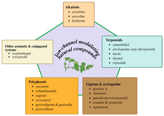
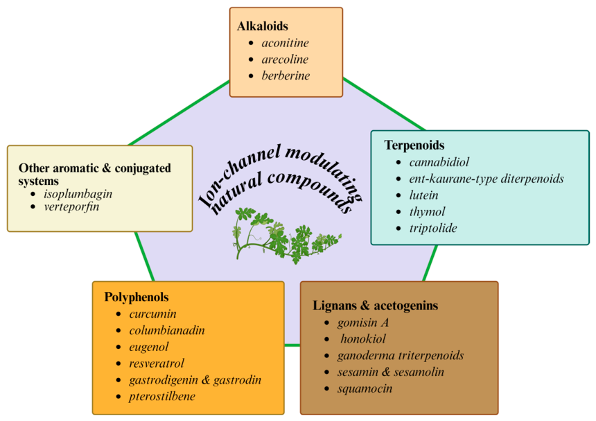
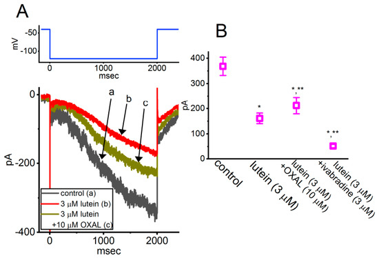
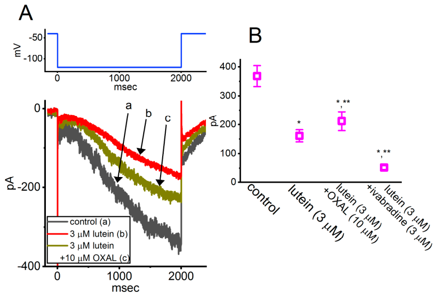
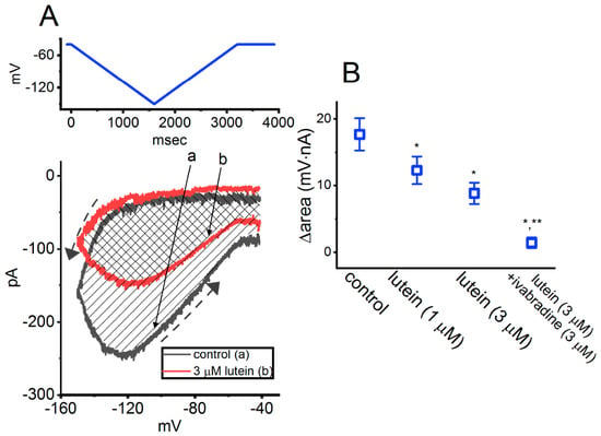
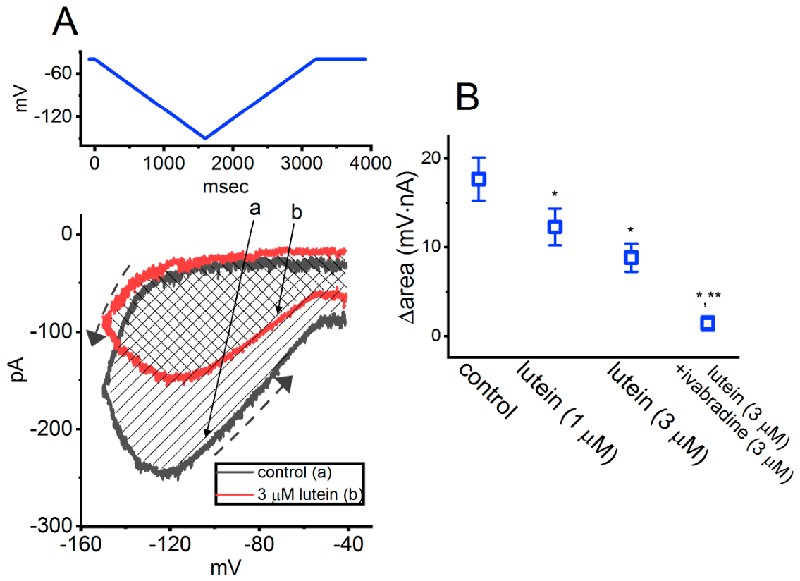
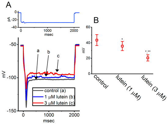
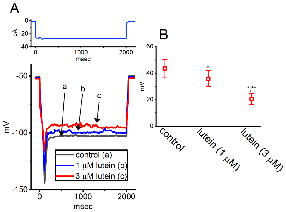
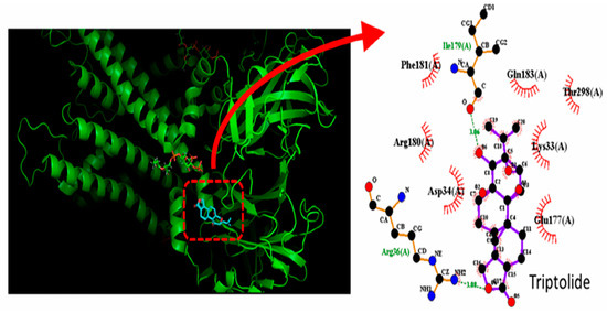
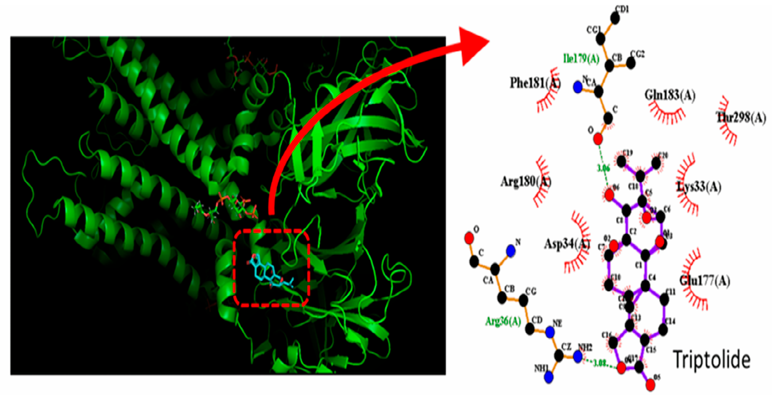
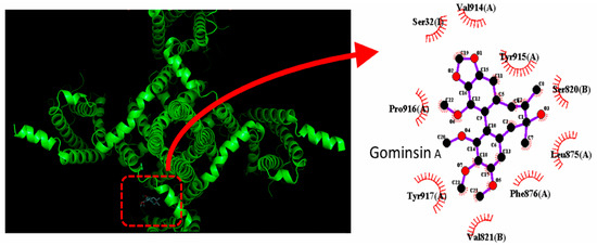
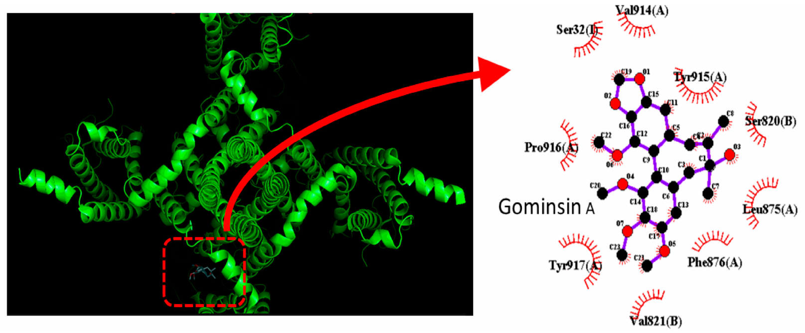
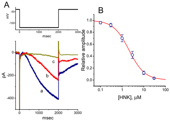
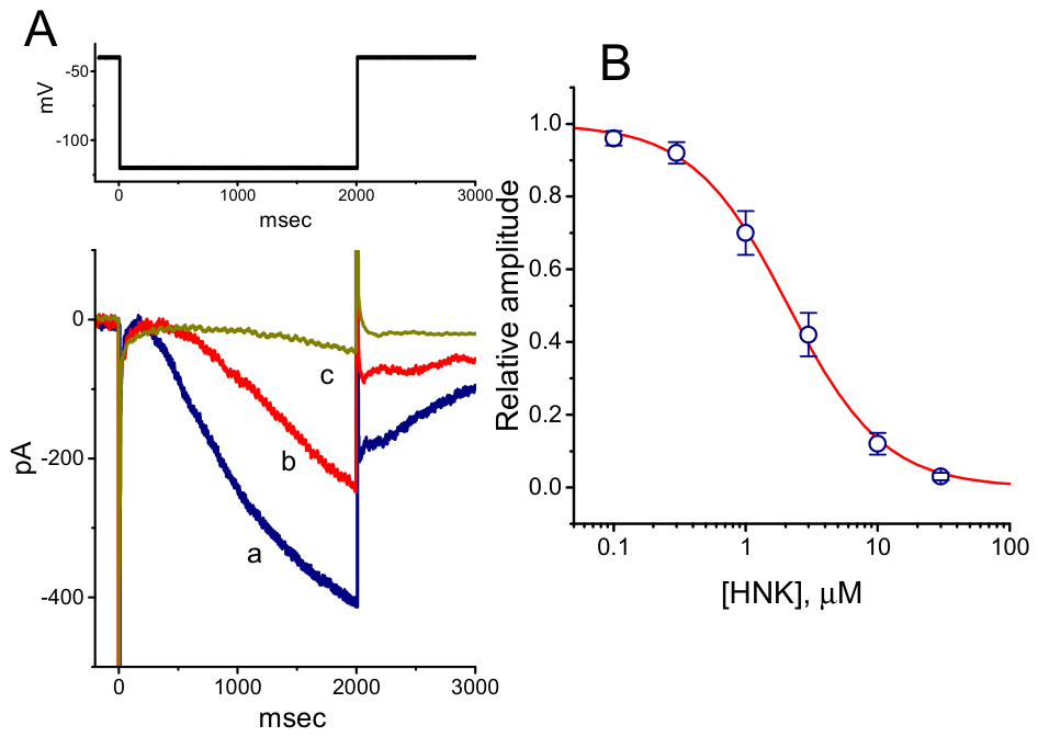
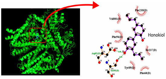
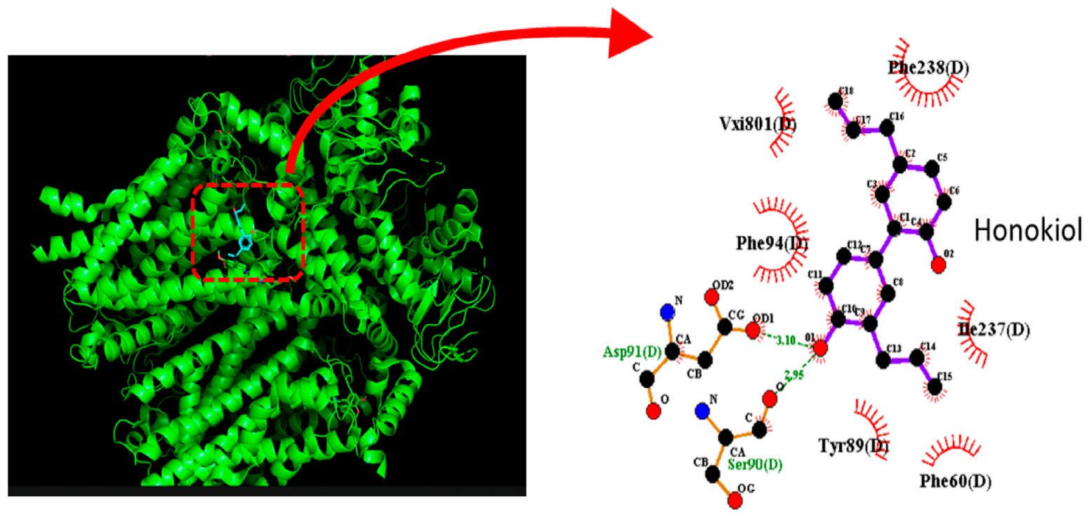
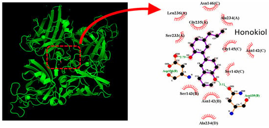
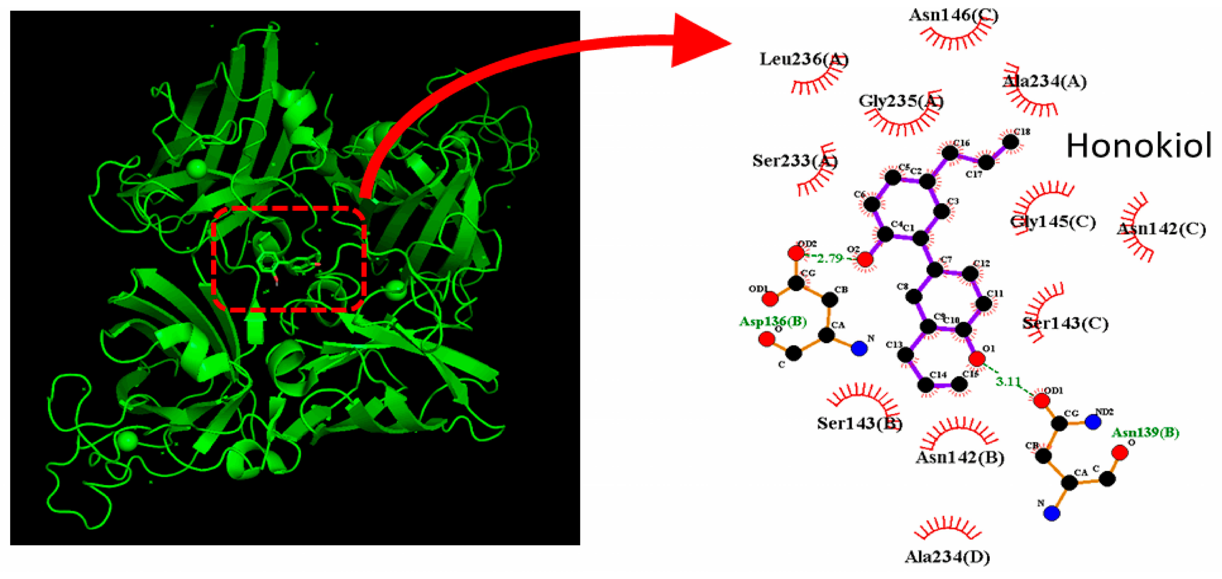
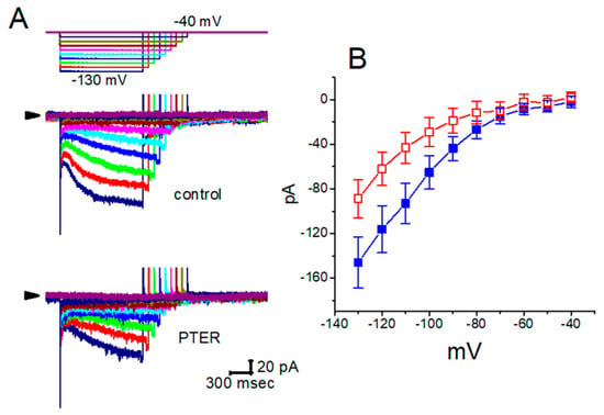
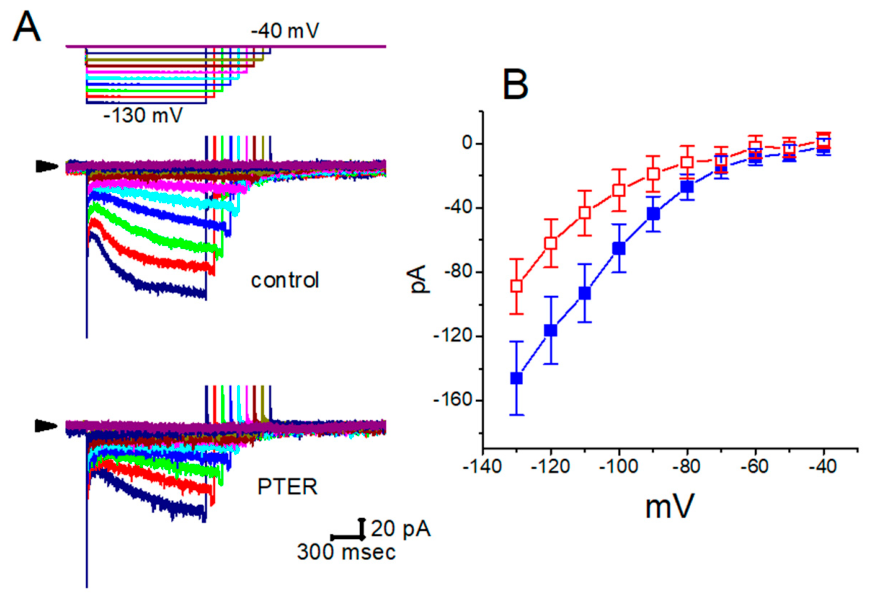
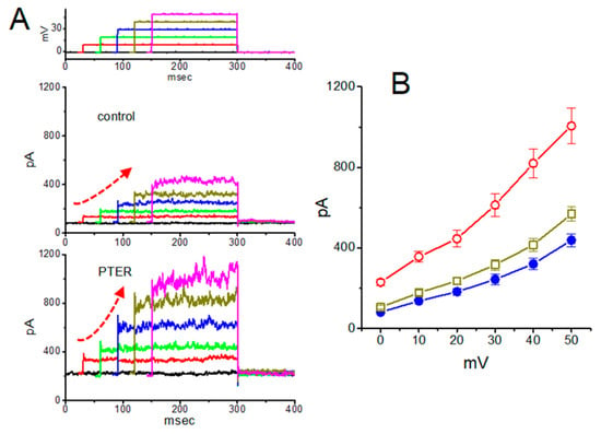
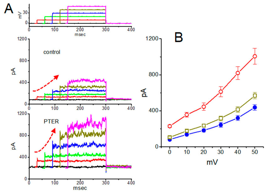
Disclaimer/Publisher’s Note: The statements, opinions and data contained in all publications are solely those of the individual author(s) and contributor(s) and not of MDPI and/or the editor(s). MDPI and/or the editor(s) disclaim responsibility for any injury to people or property resulting from any ideas, methods, instructions or products referred to in the content. |
© 2026 by the authors. Licensee MDPI, Basel, Switzerland. This article is an open access article distributed under the terms and conditions of the Creative Commons Attribution (CC BY) license.