


Abstract
Alpinia zerumbet preparations and constituents have been investigated for cardiovascular effects, but their mechanisms and exposure requirements remain incompletely defined. This narrative review evaluates evidence by preparation, model, endpoint, mechanistic intervention, and translational relevance. Isolated-vessel and animal studies support vasorelaxation and blood pressure reduction, with endothelial nitric oxide signaling and attenuation of Ca2+-dependent smooth muscle contraction contributing in preparation-dependent ways. In one spontaneously hypertensive rat study, blood pressure and remodeling improved without significant restoration of mesenteric reactivity or plasma nitrite. Endothelial protection and reduced macrophage lipid accumulation are reported repeatedly, but mostly within one research program, and remain hypothesis-generating. Pharmacological and genetic interventions implicate peroxisome proliferator-activated receptor γ (PPARγ) separately in endothelial senescence (1,8-cineole) and macrophage cholesterol handling (fruit essential oil); these parallel observations do not confirm each other, and 1,8-cineole target engagement has not been linked to effective exposure. Safety evidence is limited: antiplatelet and cardiac signals come from intra-arterial, intravenous, or isolated-tissue studies; one 30-day rat study of uncertain route reported raised lactate dehydrogenase and transaminases; and kavalactone-related hepatic risk and clinically relevant drug interactions remain unresolved. Human studies are preliminary and do not establish sustained efficacy. Development requires independently replicated standardized preparations, batch-linked characterization, exposure–response assessment, and preparation-specific safety evaluation.
1. Introduction
Endothelial dysfunction, lipid accumulation, and chronic inflammation contribute to cardiometabolic disease [1,2,3,4]. Abnormal vascular tone can raise blood pressure, whereas modified lipoprotein uptake and macrophage lipid accumulation promote atherosclerosis. Blood pressure and lipid management are therefore central to cardiovascular risk reduction [5,6].
Alpinia zerumbet (Pers.) B.L.Burtt & R.M.Sm. is a perennial herb of the family Zingiberaceae; earlier publications also use the synonym Alpinia speciosa [7,8]. Leaf tea is used in Brazilian folk medicine for diuresis and blood pressure control, and ethnomedicinal use of the mature fruits has been documented in Guizhou, China [8,9]. A. zerumbet contains terpenoids, kavalactones, flavonoids, and other polyphenols, with composition varying among plant parts and preparations [8,10,11,12,13]. Research has extended from vasorelaxation and blood pressure reduction to endothelial senescence, macrophage cholesterol efflux, and delivery systems [14,15,16,17,18]. Preliminary human studies, short-term tolerability studies, and a recent biomarker study are also available [19,20,21,22].
One earlier review cataloged the phytochemistry of A. zerumbet [8], and another summarized experimental atherosclerosis research [9]. Neither organized the cardiovascular literature primarily by preparation and level of evidence, separated functional outcomes from marker changes, or assessed whether the tested concentrations and preparations permit translation to oral products; this review adds that appraisal and extends coverage to vasorelaxation, endothelial senescence, platelet function, and cardiac electrophysiology. Recent studies of peroxisome proliferator-activated receptor γ (PPARγ) stability and delivery systems raise questions about how cellular mechanisms relate to vascular function and constituent exposure [14,15,16,17,18]. Accordingly, this narrative review first summarizes the reported chemistry of A. zerumbet (Section 2) and then evaluates cardiovascular evidence by plant part, preparation or compound, model, dose, route, duration, and endpoint. Relevant literature was retrieved from PubMed, Web of Science, Scopus, and the China National Knowledge Infrastructure (CNKI), and records were searched from inception to 31 October 2026. Records were identified with the species names “Alpinia zerumbet”, “Alpiniae zerumbet”, “Alpinia speciosa”, and “shell ginger” (and the Chinese name in CNKI) without restriction by outcome, so that efficacy, exposure, toxicity, and interaction studies were all captured. Two supplementary searches used the kavalactone names 5,6-dehydrokawain, dihydro-5,6-dehydrokawain, and desmethoxyyangonin, and combined 1,8-cineole, cineole, or eucalyptol with cardiovascular terms (hypertension, vascular, vasorelaxation, endothelium, atherosclerosis, platelet, cardiac, heart, and senescence); the reference lists of retrieved articles were also screened. The complete search strings for each database are listed in Supplementary Table S3. Primary studies in English or Chinese reporting cardiovascular, exposure, or safety outcomes for a defined A. zerumbet preparation or an isolated constituent were included. Studies without such endpoints, such as antimicrobial, cosmetic, or agricultural reports, were excluded, and studies of constituents obtained from other sources were retained where they informed mechanism, exposure, or safety relevant to A. zerumbet preparations. In total, 59 primary studies are tabulated in the evidence tables of Section 3 and Section 4. Functional outcomes are distinguished from biomarker associations, pathway interventions, and direct interaction assays, with particular attention to whether experimental concentrations and systemic exposure support translation to orally administered products.
2. Chemical Basis of the Cardiovascular Effects of Alpinia zerumbet
Reported constituents of A. zerumbet include monoterpenes and sesquiterpenes, kavalactones, flavonoids and other polyphenols, as well as diterpenes, steroids, diarylheptanoids, and neolignans [8]. Cardiovascular studies have mainly examined extracts from different plant parts, essential oils, and individual compounds such as terpinen-4-ol, 1,8-cineole, 5,6-dehydrokawain (DK), and 7,8-dihydro-5,6-dehydrokawain (DDK).
2.1. Volatile Constituents
The volatile constituents of A. zerumbet are predominantly terpenoids, including monoterpene hydrocarbons, oxygenated monoterpenes, and sesquiterpenes. Major reported monoterpene hydrocarbons include α-pinene, β-pinene, camphene, sabinene, and γ-terpinene; oxygenated monoterpenes include terpinen-4-ol, 1,8-cineole, and camphor [8,10,11,12,13]. The sesquiterpene δ-cadinene has also been reported [8] (Table 1). Essential oil composition varies with plant part and harvest period, and the relative abundance of a given constituent can differ among samples [10,11,12,13].
Terpinen-4-ol and 1,8-cineole are among the volatile constituents most frequently investigated in cardiovascular models. Acute hypotensive effects of terpinen-4-ol have been reported in normotensive and hypertensive animals [23,24], whereas 1,8-cineole studies address vasorelaxation, endothelial protection, atherosclerosis, and platelet activation [14,17,25,26,27,28,29,30]. Most of these studies used purified 1,8-cineole rather than an A. zerumbet preparation and therefore provide compound-level supportive evidence, not evidence for the effects of the plant material. A study using an orthogonal experimental design to assess combinations of α-pinene, β-pinene, camphene, and 1,8-cineole reported different 3-(4,5-dimethylthiazol-2-yl)-2,5-diphenyltetrazolium bromide (MTT) reduction and nitric oxide (NO) responses in lipopolysaccharide (LPS)-injured human umbilical vein endothelial cells (HUVECs) [31]. These screening results identify candidate constituents but do not quantify their contributions or establish synergy within the whole oil.
Table 1.
Representative volatile constituents reported in Alpinia zerumbet.
2.2. Kavalactones
Kavalactones occur in several plant parts of A. zerumbet [8] (Table 2). DK and DDK isolated from rhizomes and mature fruits have been evaluated in platelet, endothelial, and pharmacokinetic studies [33,34]. Leaf-derived compounds tested in high-glucose endothelial models include 4′-hydroxy-DDK and four cyclobutane-linked dimers, including (±)-Aniba dimer A, (±)-Aniba dimer C, and the head-to-head dimer alpingsin D (compound 5) [8,35].
Table 2.
Representative kavalactones reported in Alpinia zerumbet.
DK and DDK share a methoxy-substituted pyrone scaffold and differ mainly in saturation at positions 7 and 8 of the side chain. According to the available abstract, DK and DDK inhibited arachidonic acid-induced aggregation of rabbit platelets, with reported IC50 values of approximately 10 and 60 μg/mL, respectively [34]; the full concentration range, treatment schedule, and detailed experimental conditions could not be verified. DDK also increased MTT reduction in endothelial cells treated with the nitric oxide synthase (NOS) inhibitor Nω-nitro-L-arginine methyl ester (L-NAME) [33].
In high-glucose–exposed HUVECs, (±)-Aniba dimer A (compound 3, 12.5 μmol/L) and alpingsin D (compound 5, 25 μmol/L) increased MTT reduction to 83.12% and 75.02%, respectively, compared with 70.10% in the injury group. Compounds 1, 2, and 4 at 12.5 μmol/L produced lower signals (38.58%, 54.80%, and 58.16%) [35]. MTT reduction alone cannot distinguish cell loss, altered cellular reducing activity, or assay interference; these non-equimolar comparisons cannot define structure–activity relationships. The original study screened all five compounds across 1.5625, 3.125, 6.25, 12.5, 25, and 50 μmol/L, with the full concentration–response data presented in its Supplementary Material, but reported no half-maximal effective concentration or other concentration–response parameter; the values quoted above are the concentrations selected by the authors for each compound and therefore do not support a ranking of relative potency [35].
2.3. Flavonoids and Other Polyphenols
Ultra-high-performance liquid chromatography coupled to electrospray ionization quadrupole time-of-flight tandem mass spectrometry (UHPLC/ESI-QTOF-MS/MS) profiling of the vasoactive leaf extract AZE4 provided tentative flavonoid and polyphenol annotations through library matching [36] (Table 3). Isolation of related compounds from A. zerumbet in separate studies [8,33] supports their occurrence in the species but does not confirm every isomer or glycosidic linkage assigned in the AZE4 batch.
Table 3.
Tentative mass spectrometric annotations of flavonoids and other polyphenols in Alpinia zerumbet leaf extracts.
The polyphenol-rich 50% hydroethanolic fresh-leaf extract AZE4 relaxed isolated rat mesenteric vascular beds [36]. Among the five extracts compared, AZE4 had the highest total polyphenol content measured by the Folin–Ciocalteu method (16.30 ± 0.44 mg gallic acid equivalents per g; AZE1, AZE2, AZE3, and AZE5: 6.35 ± 0.081, 14.63 ± 0.043, 15.35 ± 0.44, and 13.46 ± 0.075 mg/g; mean ± SEM, n = 3), although its half-maximal effective dose of 7.1 ± 1.7 μg did not differ significantly from those of the other extracts. No concentration was reported for any individual flavonoid or phenolic constituent: the annotations in Table 3 rest on spectral library matching without quantification. Whether any annotated constituent is present at a vasoactive concentration in the extract therefore cannot be assessed from the available data. The functional result therefore applies to the characterized extract, while assignment of that activity to an annotated flavonoid remains unresolved. This separates evidence for a bioactive preparation from evidence identifying its active constituents.
2.4. Other Constituents
Other reported constituents include diterpenes, steroids, diarylheptanoids, and neolignans. Labdane and norlabdane diterpenes have been isolated from rhizomes and seeds [8], while diarylheptanoids and neolignans have also been isolated from rhizomes [37].
Cholestenone isolated from a mature-fruit ethyl acetate fraction increased MTT reduction in L-NAME–treated HUVECs, whereas β-sitosterol showed no significant effect [33]. A separate study compared 70% acetone extracts from six plant parts, annotated fourteen steroids by gas chromatography–mass spectrometry, and tested commercial steroid standards in low-density lipoprotein (LDL) oxidation and soybean 15-lipoxygenase assays [38].
Rhizome-derived 8′,9′-dinorneolignan enantiomers 1a, 1b, 2a, and 2b and diarylheptanoid 12 inhibited NO production in LPS-stimulated RAW264.7 macrophages, with individual half-maximal inhibitory concentrations (IC50) of 3.62, 7.63, 6.51, 5.60, and 8.33 μmol/L, respectively (n = 3). In parallel MTT assays at 10–80 μmol/L, mean signals remained approximately 95–100% of control [37]. In the same report, the 95% ethanol extract and ethyl acetate fraction of the rhizomes (80 mg/kg) inhibited croton oil-induced mouse ear edema by 20.0% and 47.6%, respectively, whereas the isolated compounds were evaluated only in vitro [37]. Inhibition of inducible NO overproduction in LPS-activated macrophages and endothelial NO-mediated vasorelaxation represent distinct cellular contexts and are not pharmacologically contradictory.
The chemical evidence supports different levels of pharmacological attribution. Terpinen-4-ol and 1,8-cineole have been tested as individual compounds in vascular or cellular models [14,17,23,24,25,26,27,28,29,30], and DK and DDK have platelet data, with additional endothelial screening for DDK [33,34]. In contrast, polyphenol annotations in a vasoactive leaf extract identify candidates without assigning activity to a particular molecule [36]. Thus, detection, structural identification, individual-compound activity, and contribution to the whole preparation are separate steps; the first three do not by themselves establish the fourth. Representative structures and plant-part-specific preparations are summarized in Scheme 1 and Figure 1. The association of particular plant parts with particular endpoints in Figure 1 reflects the distribution of research effort rather than demonstrated plant-part selectivity. Two studies compared material from different plant parts under a common protocol. One tested acetone extracts of rhizomes, stems, leaves, flowers, pericarps, and seeds in the same in vitro 15-lipoxygenase and LDL-oxidation assays, in which the seed extract was the most potent [38]; the other compared seed powder, seed/husk powder, and seed essential oil in a single in vivo dietary lipid experiment (Section 3.3.1). No study has compared preparations from different organs, such as leaf, rhizome, and fruit, in a common in vivo model, so their relative activity in vivo is unknown.
Scheme 1.
Chemical structures of four representative compounds studied in relation to the cardiovascular effects of Alpinia zerumbet. (A) Terpinen-4-ol, shown with unspecified C-4 absolute configuration; (B) 1,8-cineole (eucalyptol; 1,3,3-trimethyl-2-oxabicyclo[2.2.2]octane); (C) 5,6-dehydrokawain (DK); and (D) 7,8-dihydro-5,6-dehydrokawain (DDK). Under conventional kavalactone numbering, DK and DDK share a 4-methoxy-2-pyrone core substituted at C-6 and differ in whether the C-7/C-8 linkage of the C-6 styryl/phenylethyl side chain is unsaturated (DK) or saturated (DDK) [8,23,24,25,33,34].
Figure 1.
Distribution of reported cardiovascular research across plant parts of Alpinia zerumbet, with the representative chemical classes and primary experimental models used for each. The figure maps where evidence has been generated, not where activity resides: the pattern reflects the materials different research groups have chosen to study, and, apart from one in vitro comparison of six plant parts in lipoxygenase and LDL-oxidation assays [38] and one in vivo comparison of fruit sub-parts in a dietary lipid study [39], no study covered here compares preparations from different organs in a common in vivo model. (A) Leaf essential oils and hydroalcoholic extracts evaluated in vasorelaxation and antihypertensive models; (B) mature fruit essential oils, nanoemulsions, and ethyl acetate fractions evaluated in endothelial injury and atherosclerosis models; (C) rhizome extracts and isolated kavalactones, diarylheptanoids, and neolignans tested in antiplatelet and anti-inflammatory assays; and (D) seed and pericarp extracts and powders studied in lipid oxidation and cholesterol-handling models [8,10,11,12,13,14,15,16,17,18,23,24,25,33,34,35,36,37,38,40,41,42,43,44]. DK, 5,6-dehydrokawain; DDK, 7,8-dihydro-5,6-dehydrokawain; EtOAc, ethyl acetate; LDL, low-density lipoprotein.
3. Cardiovascular Effects and Mechanisms of Alpinia zerumbet
3.1. Vasorelaxation and Blood Pressure Reduction
Vascular studies address three related outcomes: acute relaxation, blood pressure reduction during repeated administration, and cardiovascular remodeling. Comparing these outcomes distinguishes a demonstrable vascular action from the mechanisms proposed to explain sustained antihypertensive effects (Table 4).
Table 4.
Studies of vasorelaxation and blood pressure reduction using Alpinia zerumbet preparations and reported constituents.
3.1.1. Endothelial NO Signaling and Vasorelaxation
Endothelium-derived relaxing factor was identified as NO [45]. In vascular smooth muscle, NO activates soluble guanylyl cyclase (sGC), increasing cyclic guanosine monophosphate (cGMP); cGMP-dependent protein kinase promotes myosin light-chain dephosphorylation and relaxation [46,47]. Mice lacking endothelial NOS (eNOS) exhibit impaired endothelium-dependent relaxation and elevated blood pressure [48].
A hydroalcoholic leaf extract of A. zerumbet induced long-lasting endothelium-dependent relaxation in norepinephrine-precontracted rat mesenteric vascular beds. Endothelial removal, L-NAME, and the sGC inhibitor ODQ markedly attenuated the response. The bradykinin B2 receptor antagonist HOE 140 produced a smaller but significant reduction, whereas the other tested antagonists and blockers did not alter relaxation [40] (Table 4). Mature fruit essential oil likewise produced endothelium-dependent relaxation of isolated rat thoracic aortic rings precontracted with norepinephrine or KCl, which was attenuated by endothelial removal, L-NAME, or methylene blue (an inhibitor of NO–sGC signaling), but unaffected by indomethacin [41]. These pharmacological perturbations implicate the endothelial NO–sGC pathway in the acute vasodilator actions of both preparations.
Essential oil from aerial parts and 1,8-cineole differ markedly in potency and endothelial dependence in isolated rat aorta. In endothelium-intact rings, the oil produced significant but incomplete relaxation and 1,8-cineole produced complete relaxation, with reported IC50 values of 0.29 ± 0.02 and 663.2 ± 63.8 μg/mL, respectively (mean ± SEM) [25]. Because the two concentration–response curves differ in maximal effect, these IC50 values are not directly comparable measures of potency. Endothelial removal abolished, and L-NAME almost completely abolished the whole-oil response, whereas 1,8-cineole retained a reduced relaxation, with IC50 values of 1620.6 ± 35.7 μg/mL without endothelium and 1123.9 ± 92.4 μg/mL with L-NAME. Accordingly, the potent endothelium-dependent relaxation of the aerial-part oil cannot be explained by 1,8-cineole alone. The composition cited for that oil was 1,8-cineole 33.29%, terpinen-4-ol 19.40%, p-cymene 11.04%, sabinene 6.92%, caryophyllene oxide 5.56%, β-pinene 3.54%, and γ-terpinene 3.16% of oil weight, taken from earlier analyses by the same group rather than determined for the batch tested [25]. At the whole-oil IC50 of 0.29 μg/mL, these proportions correspond to approximately 0.097 μg/mL 1,8-cineole and 0.056 μg/mL terpinen-4-ol, the former roughly 6900-fold below its own measured IC50. Constituents other than 1,8-cineole may therefore contribute, and terpinen-4-ol has independent hypotensive activity in vivo [23,24]. However, the study tested neither individual constituents at their in-oil concentrations nor a reconstituted mixture, and no isobolographic or fixed-ratio analysis was performed. The composition data are consistent with a contribution from constituents other than 1,8-cineole, but an observed difference in potency is not evidence of a demonstrated synergistic interaction. The intact-ring 1,8-cineole IC50—approximately 4.30 mmol/L—is roughly 250 times the total plasma Cmax (2.65 μg/mL, or ~17.2 μmol/L) reported after a single oral dose of unformulated 1,8-cineole at 50 mg/kg in mice [17]. Volatile loss from aerated organ baths, plasma protein binding, and the difference between total plasma and unbound target-site concentrations complicate direct numerical comparison, but this two-order-of-magnitude gap indicates that millimolar relaxation in vitro cannot be assumed to operate at systemic concentrations after oral dosing (Section 4.3).
3.1.2. Attenuation of Smooth Muscle Ca2+-Dependent Contraction and Endothelium-Independent Relaxation
Vascular smooth muscle contraction depends on extracellular Ca2+ entry and intracellular Ca2+ release; attenuation of Ca2+-dependent contraction can promote relaxation. Because some A. zerumbet essential oils retain vasorelaxant activity after endothelial removal, studies have further examined smooth muscle Ca2+-dependent responses [42,43].
A methanolic fraction of leaf essential oil relaxed phenylephrine- and KCl-precontracted rat thoracic aortic rings independently of the endothelium. It attenuated contractions induced by extracellular Ca2+ influx or by phenylephrine and caffeine in Ca2+-free medium; the other tested pathway modulators did not alter relaxation [42] (Table 4). Unfractionated leaf oil likewise inhibited Ca2+ influx- and intracellular release-dependent contractions in second-order rat mesenteric resistance arteries precontracted with KCl or U-46619 [43]. Maximal relaxation persisted after endothelial removal, although L-NAME and ODQ shifted pEC50. The shared functional finding is reduced Ca2+-dependent contraction; the contribution of NO differs between the tested vascular preparations. Because fractionation, vascular bed, and precontraction conditions changed together, these experiments do not identify which difference accounts for the NO response.
3.1.3. Blood Pressure Responses and Cardiovascular Remodeling
Sustained hypertension can alter aortic wall and left ventricular structure [44]. In addition to acute hypotensive responses, repeated-administration studies of A. zerumbet have examined blood pressure, vascular function, and cardiovascular remodeling.
Acute intravenous bolus administration of leaf essential oil (1–20 mg/kg) and terpinen-4-ol (1–10 mg/kg) elicited immediate, dose-dependent reductions in mean arterial pressure in normotensive rats [23]. Oil-induced hypotension persisted after bilateral vagotomy in anesthetized rats and after intravenous methylatropine (1 mg/kg) or hexamethonium (30 mg/kg) in conscious rats; these autonomic interventions were not tested with terpinen-4-ol [23]. Both materials also produced enhanced maximal hypotensive responses in conscious DOCA-salt hypertensive rats compared with uninephrectomized controls, and the enhancement of oil-induced hypotension was unaffected by hexamethonium or methylatropine pretreatment [24].
Repeated-dose studies report blood pressure and structural effects in hypertensive models. Oral hydroalcoholic leaf extract (50 mg/kg/day) lowered blood pressure in DOCA-salt rats [40]. In SHR, leaf essential oil (10 mg/kg/day intraperitoneally for 30 days) lowered mean arterial pressure and modestly reduced the left-ventricle/body-mass ratio, without normalization to WKY levels [32] (Table 4). A six-week drinking-water regimen of hydroalcoholic leaf extract (~50 mg/kg/day) progressively lowered tail-cuff blood pressure to WKY levels and reduced aortic and ventricular wall thickness and collagen deposition [44]. Antioxidant and immunostaining changes accompanied these effects, but mesenteric vascular reactivity, plasma nitrite, and several other measures did not significantly improve (Table 4). The authors attributed the response in part to an antioxidant action, reporting lower aortic 8-isoprostane, NOX-4, and AT1R immunostaining together with higher SOD-2 immunostaining and higher plasma catalase and glutathione peroxidase activities; circulating angiotensin II did not differ among groups, no increase in diuresis was observed, and the authors state that the extract did not reverse endothelial dysfunction in this model [44]. These are associative measurements rather than interventions: no inhibitor, antagonist, or genetic manipulation was used to test whether reduced oxidative stress or lower AT1R expression mediates the pressure reduction. The study therefore identifies candidate contributors but leaves the mechanism of the sustained antihypertensive effect unresolved.
1,8-Cineole lowered blood pressure and attenuated vascular tissue injury in L-NAME-induced hypertensive rats [29]. An ethyl acetate fraction of an ethanolic mature-fruit extract lowered blood pressure and attenuated vascular injury in an L-NNA-induced hypertension model. The fraction was administered at 83.4 or 166.7 mg/kg/day during weeks 5–8, with captopril (10 mg/kg/day) as a positive control. DDK was tested separately in the accompanying cellular assay and increased MTT reduction in L-NAME-treated HUVECs [33].
Across these studies, acute vasorelaxation and repeated-dose blood pressure reduction have functional support, but the connection between them is incomplete [23,24,25,32,40,41,42,43,44]. Endothelial removal and NOS/sGC inhibition identify an NO-associated component of acute relaxation in selected preparations. In the six-week SHR study, blood pressure and remodeling improved without statistically significant restoration of mesenteric reactivity or plasma nitrite [44]. This pattern limits attribution of the sustained response solely to the measured vascular endpoints, while leaving possible NO-dependent actions in other vascular beds and other physiological contributions unresolved. The literature supports blood pressure lowering activity in rodent models more clearly than a shared mechanism linking acute relaxation to long-term benefit (Figure 2).
Figure 2.
Preparation-specific pathways implicated in A. zerumbet-induced vasorelaxation. (A) Endothelial NO-associated relaxation. ODQ was tested with hydroalcoholic leaf extract [40] and leaf essential oil [43]; mature fruit essential oil was tested with L-NAME and methylene blue [41]. (B) Attenuation of extracellular Ca2+-dependent and intracellular Ca2+ release-related contraction by a methanolic oil fraction [42] and leaf essential oil [43]. Arrows summarize physiological relationships and proposed modulation. The vascular assays support attenuation of calcium-dependent contraction, while direct blockade of a specific channel remains unconfirmed. The lower schematic curve is illustrative. The lower strips contrast ex vivo vessel relaxation with in vivo blood pressure reduction in SHR; in the six-week study, mesenteric reactivity and plasma nitrite did not significantly recover [44]. A continuous causal link from acute relaxation to sustained pressure lowering remains unestablished [23,24,25,40,41,42,43,44]. NO, nitric oxide; eNOS, endothelial NOS; sGC, soluble guanylyl cyclase; GTP, guanosine triphosphate; cGMP, cyclic guanosine monophosphate; L-NAME, Nω-nitro-L-arginine methyl ester; ODQ, 1H-[1,2,4]oxadiazolo[4,3-a]quinoxalin-1-one; VSMC, vascular smooth muscle cell; SR, sarcoplasmic reticulum; SHR, spontaneously hypertensive rats; BP, blood pressure.
3.2. Vascular Endothelial Protection
Endothelial studies examine survival and inflammatory injury, changes associated with endothelial-to-mesenchymal transition (EndMT), and senescence. The synthesis below separates recurrent cellular outcomes from evidence identifying a necessary pathway or a direct molecular interaction. Animal vascular pathology provides an additional level of evidence, but must be linked to the interventions actually performed in each model (Table 5 and Table 6).
Table 5.
Studies of endothelial injury and acute inflammation using Alpinia zerumbet preparations and reported constituents.
Table 6.
Studies of endothelial phenotypes and vascular outcomes using Alpinia zerumbet preparations and reported constituents.
3.2.1. Attenuation of Endothelial Injury and Inflammation
The most recurrent outcome across high-glucose, oxidized low-density lipoprotein (ox-LDL), and inflammatory injury models is attenuated cellular injury, assessed using MTT reduction or viability, lactate dehydrogenase (LDH) release, and apoptosis [49,50,52,56,59]. Agreement among these endpoints provides a stronger basis for a protective phenotype than a change in an isolated signaling protein. Where protection rests mainly on MTT reduction or viability without orthogonal cell-death or functional readouts, the protective phenotype should be considered provisional.
In high-glucose-treated HUVECs, mature fruit essential oil attenuated oxidative injury and apoptosis while lowering thioredoxin-interacting protein (TXNIP) and restoring thioredoxin 1 (Trx-1) activity without altering Trx-1 protein abundance [49]. Because TXNIP binds and inhibits reduced Trx-1 [68], these changes are consistent with modulation of the TXNIP–Trx-1 redox axis. In an ox-LDL injury model with pravastatin (10 μmol/L) as a positive control, preincubation with 0.1 mg/L (100 μg/L) mature fruit essential oil improved MTT reduction and trypan blue exclusion. The same concentration increased superoxide dismutase (SOD), catalase (CAT), and glutathione peroxidase (GSH-Px) activities and intracellular reduced glutathione (GSH), and lowered malondialdehyde (MDA) and LDH release. At 0.01 mg/L the oil had no significant effect on viability, and at 10 mg/L it reduced basal HUVEC survival [52]. In ox-LDL-injured human aortic endothelial cells (HAECs), mature fruit essential oil reduced apoptosis, with lower Bax and caspase-3 protein and mRNA and higher Bcl-2 [56]. Another study reported reduced phosphorylation of c-Jun N-terminal kinase (JNK) 1/2/3 [69]. Across the high-glucose and ox-LDL models, the recurring result is reduced injury with changes in survival and apoptosis. TXNIP/Trx-1, JNK, and apoptosis-related proteins identify candidate processes accompanying that result, rather than a demonstrated common upstream target.
Mature fruit essential oil reduced nuclear factor κB (NF-κB) p65 phosphorylation, nuclear translocation, DNA binding, and inflammatory measures in high-glucose-exposed HUVECs [50]. Related LPS models showed lower Toll-like receptor 4/IκB kinase (TLR4/IKK) signaling, adhesion molecules, and aortic inflammatory infiltration [57,59], and an earlier HUVEC study reported lower LPS-induced cytokine and endothelin-1 secretion [70]. For 1,8-cineole, an initial endothelial study reported reduced LPS-induced NF-κB activation [26]. In mice, 1,8-cineole reduced LPS-associated aortic inflammatory injury. In the accompanying HUVEC experiments, GW9662 and small interfering RNA (siRNA) targeting PPARγ counteracted selected anti-inflammatory responses, supporting PPARγ involvement in the cellular effects [27]. Reduced inflammatory activation recurs across these models, whereas PPARγ intervention provides an additional causal test specifically for the 1,8-cineole cellular response, not for every oil preparation (Table 5).
Several ox-LDL injury studies report increased eNOS expression or activity. Mature fruit essential oil increased protein kinase B (Akt) phosphorylation and eNOS and sGC expression in HAECs, while reducing inducible NOS (iNOS) expression [53]. Another study, whose plant-part assignment is internally inconsistent (Table 5 note), found increased eNOS activity and mRNA and reduced iNOS activity and mRNA [55]. In HUVECs, total NOS and eNOS activities increased without a significant change in iNOS activity [54]. In high-fat-fed rats, an aqueous extract increased serum NO-related metabolites and the prostacyclin metabolite 6-keto-prostaglandin F1α (6-keto-PGF1α), while lowering endothelin-1 (ET-1) and thromboxane-related measures (Table 6 note); reductions in angiotensin II (Ang II) did not reach significance in some groups [67]. Together, these findings describe modulation of NO-related homeostasis during injury, rather than uniform activation of all NOS isoforms. They complement the acute relaxation studies, but enzyme expression and circulating metabolites alone do not establish mediation of sustained blood pressure lowering.
Autophagy studies report protection with different changes in microtubule-associated protein 1 light chain 3 (LC3) and p62. In high-glucose-treated HAECs, 1,8-cineole increased viability and reduced LDH release, with higher Beclin 1 and LC3-II/LC3-I ratio and lower p62. After chloroquine treatment, protein differences between high glucose alone and high glucose plus 1,8-cineole were no longer significant [51]. This provides a lysosomal-blockade comparison, although loss of significance alone does not separate autophagosome formation from clearance. In L-NAME–treated HUVECs, 1,8-cineole increased the phosphorylation of phosphoinositide 3-kinase (PI3K) and mechanistic target of rapamycin (mTOR), decreased the LC3-II/LC3-I ratio, and increased p62, reversing the L-NAME-induced changes; LY294002 partially counteracted these effects [29]. The studies therefore converge on reduced injury, but not on a common direction of autophagy-related marker change. A uniform mechanism of protection through increased or decreased autophagic flux is not established by these experiments.
In transforming growth factor β1 (TGF-β1)-stimulated HUVECs, mature fruit oil improved relative viability and reduced reactive oxygen species (ROS) and migration; nuclear factor erythroid 2-related factor 2 (Nrf2) increased in some treatment groups [61].
Migration and tube-like structures changed in opposite directions under different protocols. Fruit oil nanoemulsion increased both in high-glucose–exposed HUVECs and improved diabetic mouse vascular pathology [65], whereas oil reduced tube-like structures under tumor necrosis factor α (TNF-α) or high glucose [64,66]. Under TNF-α, it lowered vascular endothelial growth factor receptor 2 (VEGFR2); the mitogen-activated protein kinase kinase 1/2 (MEK1/2) inhibitor U0126 reproduced these changes [66]. Thus, increased and decreased tube formation cannot both be summarized as a general proangiogenic effect of the oil. The experiments establish context-dependent endothelial behavior; their relationship to perfused vessel formation or vascular repair remains to be demonstrated.
R8-modified fruit oil lipid microspheres improved mitochondrial membrane potential and reduced ROS and inflammatory measures in LPS-injured HUVECs, with greater effects than unmodified microspheres [58]. The absence of an unloaded R8-modified carrier limits attribution of these effects to the oil. Coumarin-6 colocalization and organ imaging with the lipophilic near-infrared dye DiR tracked dyes; their implications for constituent exposure are considered in Section 4.3.
Carboxymethyl chitosan-coated lipid nanoparticles containing eucalyptol also improved MTT reduction and reduced ROS in high-glucose-treated HUVECs. In streptozotocin-induced diabetic rats, oral treatment at a eucalyptol-equivalent dose of 18 mg/kg every other day for one month attenuated vascular injury, accompanied by higher Nrf2 and heme oxygenase 1 (HO-1) and lower Kelch-like ECH-associated protein 1 (Keap1) expression [18].
3.2.2. Endothelial-to-Mesenchymal Transition Phenotypes
EndMT is characterized by loss of endothelial markers, acquisition of mesenchymal markers, increased migratory capacity, and changes in extracellular matrix-associated phenotypes. It has been implicated in atherosclerosis [71]; A. zerumbet studies mainly use TGF-β1 or high-glucose cell models.
In TGF-β1-treated HUVECs, the higher concentration of mature fruit essential oil increased vascular endothelial cadherin (VE-cadherin) and reduced α-smooth muscle actin (α-SMA) and Snail. Changes in these markers at the lower concentration were not statistically significant. Both concentrations reduced migration, with lower p65 phosphorylation [60], supporting modulation of EndMT-associated phenotypes.
In TGF-β1-stimulated HUVECs treated with an essential oil of an unverified plant part (Table 6 note), Krüppel-like factor 4 (KLF4) knockdown increased VE-cadherin, reduced α-SMA, and attenuated migration, paralleling selected oil-associated effects. Oil treatment also reduced p65 phosphorylation and the KLF4–NF-κB co-immunoprecipitation signal [62]. Subsequent work linked oil treatment to lower KLF4, total H3 acetylation, and KLF4–acetyl-H3 co-immunoprecipitation, with lower Notch1/Snail and higher Notch3 [63]. The co-immunoprecipitation and total histone acetylation findings implicate associated protein and chromatin processes, but do not locate regulation at a particular promoter.
KLF4 has context-dependent effects: endothelial KLF4 can attenuate atherothrombosis [72], whereas elevated KLF4 contributes to EndMT in cerebral cavernous malformation models [73]. These contrasting disease settings limit generalization of KLF4 suppression as vascular protection; the findings are therefore described as KLF4-associated regulation in the tested TGF-β1 model.
In high-glucose-treated HUVECs, mature fruit oil increased CD31 (platelet endothelial cell adhesion molecule 1), reduced vimentin and tube-like structures, and improved oxidative measures. Higher Nrf2 and lower Notch1 accompanied these changes; Nrf2 overexpression modified the response, with some effects enhanced by combined oil treatment [64].
Across the TGF-β1 and high-glucose studies, shifts toward endothelial markers and away from mesenchymal markers are accompanied by altered migration or tube formation [60,62,63,64]. This convergence supports modulation of EndMT-associated phenotypes in cultured cells. KLF4 knockdown and Nrf2 overexpression add intervention evidence, but they do not show that the same regulatory sequence operates in diseased vessels (Figure 3).
Figure 3.
Endothelial protective pathways reported for A. zerumbet preparations and 1,8-cineole across cellular injury models. The panels assemble findings obtained with different test materials in separate experiments and do not represent pathways demonstrated for any single preparation in one cell population; the test material is named for each group of findings below. (A) Attenuation of high-glucose- and ox-LDL-induced oxidative stress and apoptosis with reported changes in TXNIP, Trx-1 activity, antioxidant enzymes, and apoptosis-related markers, with mature fruit essential oil except for [55], whose plant part is inconsistently reported [49,52,53,54,55,56]. The Keap1/Nrf2/HO-1 findings are from separate experiments with eucalyptol-loaded nanoparticles [18]. (B) Changes in inflammatory signaling in separate LPS and high-glucose models and endothelial adhesion molecule expression; the NF-κB and adhesion molecule findings are from mature fruit essential oil [50,57,59] and its octaarginine-modified microsphere formulation [58] and the PPARγ-dependent modulation from purified 1,8-cineole [26,27]. (C) Changes in EndMT-associated phenotypes in TGF-β1 and high-glucose models associated with KLF4, histone H3 acetylation, and Notch1/Snail downregulation, with mature fruit essential oil except for [62], whose plant part is inconsistently reported [60,62,63,64]. Arrows indicate stimulation or the sequence of reported events, blunt-ended lines indicate inhibition, and upward and downward arrows beside a marker indicate reported increases and decreases. ox-LDL, oxidized low-density lipoprotein; LPS, lipopolysaccharide; TXNIP, thioredoxin-interacting protein; Trx-1, thioredoxin 1; Nrf2, nuclear factor erythroid 2-related factor 2; HO-1, heme oxygenase 1; Keap1, Kelch-like ECH-associated protein 1; SOD, superoxide dismutase; CAT, catalase; GSH-Px, glutathione peroxidase; eNOS, endothelial nitric oxide synthase; TLR4, Toll-like receptor 4; IKK, IκB kinase; NF-κB, nuclear factor κB; ICAM-1 and VCAM-1, intercellular and vascular cell adhesion molecule 1; TGF-β1, transforming growth factor β1; EndMT, endothelial-to-mesenchymal transition; KLF4, Krüppel-like factor 4; VE-cadherin, vascular endothelial cadherin; α-SMA, α-smooth muscle actin.
3.2.3. Modulation of Endothelial Senescence
Endothelial senescence involves cell-cycle arrest, senescence-associated β-galactosidase (SA-β-gal) activity, and vascular dysfunction. Studies of human coronary tissue and HAECs associate senescence with atherosclerotic lesions, higher intercellular adhesion molecule 1 (ICAM-1), and reduced eNOS activity [74].
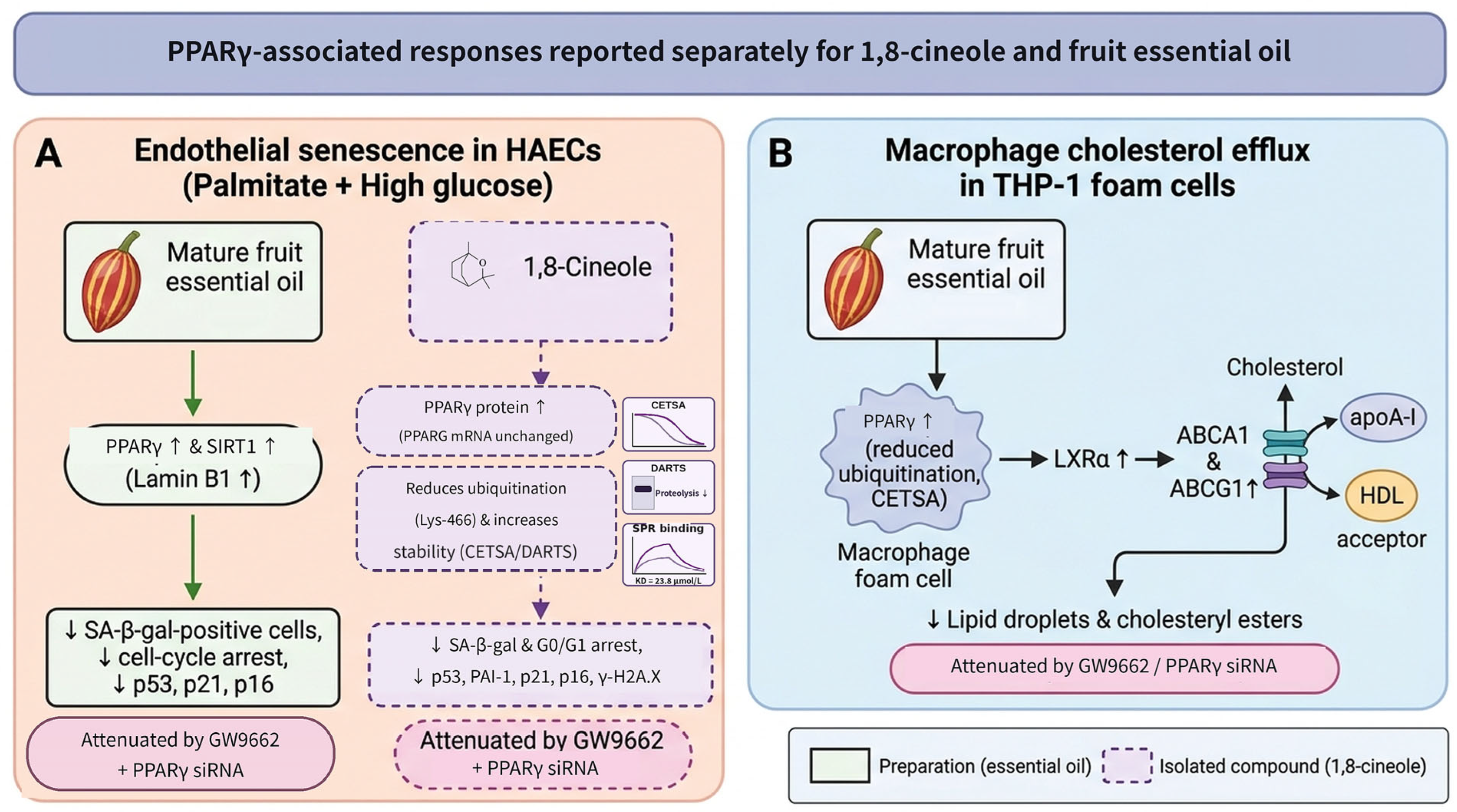
Mature fruit essential oil improved aortic pathology in diabetic mice, increasing PPARγ and sirtuin 1 (SIRT1) and reducing p53, p21, and p16. In HAECs exposed to palmitate plus high glucose, the oil reduced senescence-associated β-galactosidase-positive cells and attenuated cell-cycle arrest. GW9662 and PPARγ siRNA each weakened some protective effects [16], supporting involvement of PPARγ in the senescence response (Figure 4A).
In diabetic mice, 1,8-cineole improved aortic histopathology, blood lipids, inflammatory measures, and senescence-associated markers without significantly lowering fasting glucose [14]. In HAECs exposed to palmitate plus high glucose, 10 or 50 nmol/L reduced SA-β-gal positivity and G0/G1 arrest; GW9662 and PPARγ siRNA counteracted these responses. Rosiglitazone added no further benefit. Systemic adenoviral Pparg shRNA attenuated selected aortic and lipid effects in mice, but did not isolate endothelial-specific receptor function. Animal regimens, endpoint-specific sample sizes, and reporting limitations are detailed in Table 6.
In HAECs, 1,8-cineole increased PPARγ protein without changing PPARG mRNA. Cycloheximide-chase, MG132/chloroquine, and ubiquitination experiments supported reduced proteasomal turnover; mass spectrometry identified three candidate lysine sites, and K466R mutagenesis in HEK293T cells implicated Lys-466, although the authors note that the underlying mechanism requires further investigation [14]. Cellular thermal shift assays (CETSA) after treatment and heating of intact HAECs indicated increased PPARγ thermal stability, whereas drug affinity responsive target stability (DARTS) assays in HAEC lysates indicated greater resistance to proteolysis. Cell-free surface plasmon resonance (SPR; 3.125–100 μmol/L) yielded a dissociation constant (KD) of 23.8 μmol/L [14]. Together, these experiments support PPARγ involvement and altered PPARγ turnover in the cellular response to 1,8-cineole. However, they do not establish that direct receptor occupancy at the nominally effective nanomolar concentrations is responsible for the observed phenotype. The shared senescence phenotype of the oil and the isolated compound does not identify which constituent accounts for the oil response.
3.3. Lipid Regulation and Effects on Atherosclerosis
The lipid studies address oxidation, macrophage cholesterol handling, and endpoint plaque burden, with different implications for atherosclerosis (Table 7).
Table 7.
Studies of lipid regulation and atherosclerosis using Alpinia zerumbet preparations and reported constituents.
3.3.1. Blood Lipids and Inhibition of Lipid Oxidation
Endothelial oxidation of LDL increases macrophage uptake and apoprotein degradation, a process inhibited by antioxidant treatment [79]. This uptake-related assay differs from cellular cholesterol efflux; A. zerumbet seed studies have examined lipid-related activity through oxidation assays and animal lipid measurements.
The 70% acetone seed extract inhibited soybean 15-lipoxygenase and CuSO4-induced human LDL oxidation, with reported IC50 values of 1.26 ± 0.07 and 15.40 ± 0.86 μg/mL in Table 2 of the original study [38]. Among commercial steroid standards, cholest-4-ene-3,6-dione showed the strongest activity in these assays, although its 15-lipoxygenase IC50 was roughly 27-fold higher than that of the crude seed extract. The crude extract was therefore more active than this single tested steroid in that assay, leaving the extract activity unassigned rather than demonstrating steroid sufficiency or synergy. Soybean enzyme inhibition is an in vitro screening result, not direct evidence of inhibition of a human vascular lipoxygenase (Table 7).
Animal seed studies suggest lipid modulation without a uniform LDL-lowering response. Seed preparations increased high-density lipoprotein cholesterol (HDL-C) in one study [75], while seed oil and powder improved selected blood and hepatic lipid measures in another [39]. LDL cholesterol (LDL-C) did not decrease in the highest-dose seed oil and seed powder groups. Some seed powder and seed/pericarp powder groups also had greater fecal neutral cholesterol than the seed essential oil group [39], suggesting an intestinal contribution that may differ from the action of absorbed constituents. Inconsistent cholesterol fractions in [75] and species reporting in [39] limit numerical dose–response comparisons (Table 7 note). The evidence supports changes in lipid handling, but not a reliable, dose-dependent hypolipidemic effect shared by all seed preparations.
3.3.2. Reduction in Macrophage Lipid Accumulation
Macrophage uptake of modified LDL through CD36 and class A scavenger receptors (SR-A) promotes cholesteryl ester accumulation and foam-cell formation [80]. A. zerumbet essential oil and 1,8-cineole studies have examined these uptake-related receptors.
An early essential oil study reported fewer lipid droplets and lower cholesteryl esters in THP-1-derived macrophages, but inconsistent plant-part descriptions, ox-LDL units, and directions of ATP-binding cassette transporter A1 (ABCA1) change limit mechanistic interpretation [76] (Table 7 note). A subsequent mature fruit essential oil study found reduced lipid accumulation and fluorescently labeled ox-LDL uptake in THP-1-derived foam cells, together with lower CD36 protein and mRNA and reduced signal transducer and activator of transcription 3 (STAT3) phosphorylation, without a clear change in total STAT3. The STAT3 activator Colivelin partially reversed the reductions in phosphorylated STAT3 and CD36 [77], suggesting involvement of STAT3/CD36-associated regulation.
1,8-Cineole reduced lipid droplets and Nile Red lipid fluorescence and lowered SR-A1 mRNA in ox-LDL-induced THP-1-derived foam cells [28]. The SR-A1 immunoblot band was labeled 33 kDa, raising uncertainty about protein identity (Section 4.1); the findings primarily support lipid phenotype and transcript changes.
3.3.3. Cholesterol Efflux and Reduction in Arterial Plaque Burden
Cholesterol efflux is an important route for reducing macrophage cholesterol accumulation. PPARγ can increase ABCA1 expression through liver X receptor α (LXRα)-associated transcriptional regulation and promote efflux [81], providing a mechanistic basis for A. zerumbet lipid studies.
Mature fruit essential oil reduced macrophage lipid droplets, total cholesterol, and cholesteryl esters while increasing PPARγ and ABCA1; GW9662 reversed some of these changes [78]. Subsequent work showed increased efflux of the fluorescent cholesterol analog 3-hexanoyl-NBD cholesterol to apolipoprotein A-I (apoA-I) and high-density lipoprotein (HDL), accompanied by changes in LXRα, ABCA1, and ATP-binding cassette subfamily G member 1 (ABCG1). GW9662 or PPARγ siRNA attenuated the increased efflux and reduction in lipid droplets [15]. These interventions support PPARγ involvement in the cellular response (Figure 4B).
The macrophage study also examined PPARγ turnover [15]. At 25 μg/L, mature fruit essential oil slowed cycloheximide-chase degradation and reduced PPARγ ubiquitination, with proteasome/lysosome inhibitor experiments supporting proteasomal regulation. CETSA in PPARγ-transfected HEK293T cells showed increased thermal stability. Altered PPARγ turnover has therefore been reported in two distinct experimental settings: 1,8-cineole in senescent HAECs [14] and fruit essential oil in THP-1-derived macrophages [15]. The two findings are parallel observations rather than mutual confirmation, because they involve different preparations, cell types, and phenotypes, and the oil-associated effect has not been assigned to any individual constituent; docking of oil constituents supplied no binding constants. The finding motivates further study of receptor stability but does not establish a shared molecular mechanism.
In high-fat-fed Apoe−/− mice, mature fruit essential oil at 15 or 30 mg/kg/day for 12 weeks reduced aortic plaque burden and increased intraplaque collagen; plaque quantification used six animals per group. Lentivirus-mediated in vivo PPARγ RNA interference attenuated improvements in plaque, collagen, and some blood lipid measures [15], supporting PPARγ involvement in the animal response. The intervention did not distinguish cell types. Cellular efflux and animal plaque measurements provide functional support beyond transporter expression alone. However, systemic PPARγ knockdown does not identify whether endothelial cells, macrophages, or other tissues account for the plaque response.
Figure 4.
PPARγ-associated responses reported separately for 1,8-cineole in endothelial cells and for mature fruit essential oil in macrophages. (A) Reduced senescence-associated phenotypes in palmitate/high-glucose-exposed HAECs, shown as two parallel paths: mature fruit essential oil [16] and purified 1,8-cineole [14], each tested in its own study. The two paths are drawn separately because no experiment compared them in the same system, and the shared phenotype does not establish that the compound accounts for the oil response. In separate 1,8-cineole experiments, reduced PPARγ ubiquitination and altered protein stability were evaluated alongside intact-cell CETSA, lysate DARTS, and cell-free SPR [14]. Receptor occupancy at the effective nominal cellular concentrations remains unresolved. (B) Mature fruit essential oil-associated cholesterol efflux to apoA-I and HDL and lipid-droplet reduction in THP-1-derived macrophages associated with PPARγ–LXRα signaling and ABCA1/ABCG1 expression [15,78]. Pharmacological antagonism (GW9662) and PPARγ siRNA attenuated the cellular responses in both models. Arrows indicate the sequence of reported effects, and upward and downward arrows beside a marker indicate reported increases and decreases. HAECs, human aortic endothelial cells; SA-β-gal, senescence-associated β-galactosidase; PAI-1, plasminogen activator inhibitor 1; SIRT1, sirtuin 1; γ-H2A.X, phosphorylated histone H2A.X; CETSA, cellular thermal shift assay; DARTS, drug affinity responsive target stability; SPR, surface plasmon resonance; LXRα, liver X receptor α; ABCA1 and ABCG1, ATP-binding cassette transporters A1 and G1; apoA-I, apolipoprotein A-I; HDL, high-density lipoprotein.
Oral 1,8-cineole nanoemulsion reduced aortic plaque area and altered ABCA1, ABCG1, CD36, and SR-A expression in animal tissue and foam-cell models. At an equivalent compound dose, vitamin B12-modified nanoemulsion reduced endpoint plaque burden more than unformulated 1,8-cineole [17]. After a single oral dose of 50 mg/kg, reported areas under the plasma concentration-time curve (AUC0–t) were 31.78 ± 4.15 and 2.99 ± 0.56 h·μg/mL for the modified formulation and unformulated compound; AUC0–∞ values were 63.04 ± 24.17 and 4.68 ± 1.55 h·μg/mL, respectively (mean ± standard deviation; n = 4). The ratios of these group means are approximately 10.6 and 13.5. The reported Cmax of unformulated 1,8-cineole was 2.65 μg/mL [17].
Calculated from those group means, the extrapolated portions account for approximately 49.6% and 36.1% of AUC0–∞ for the modified formulation and unformulated compound, respectively; these are not mean individual-animal extrapolation fractions. The large extrapolated contribution makes the AUC0–∞ ratio more dependent on terminal-phase estimation than the observed AUC0–t ratio. Pharmacokinetics used C57BL/6J mice, whereas plaque efficacy was assessed in Apoe−/− mice after eight weeks of modeling followed by eight weeks of treatment. The efficacy groups lacked a matched unloaded carrier and a pretreatment sacrifice group for baseline plaque quantification. The experiments therefore support higher compound exposure and lower endpoint plaque burden in separate settings [17]. They do not show an individual exposure-response relationship, exposure-mediated efficacy, or a reduction below pretreatment plaque burden.
3.4. Inhibition of Platelet Activation and Aggregation
Platelet activation and aggregation contribute to arterial thrombosis, with different stimuli initiating responses through distinct receptors and signaling pathways [82]. Antiplatelet studies of A. zerumbet-derived DK and DDK and the reported constituent 1,8-cineole have mainly assessed aggregation, granule release, and receptor-associated signaling.
The abstract of an early study suggests stimulus- and phase-dependent platelet inhibition by kavalactones. DK and DDK inhibited arachidonic acid- and collagen-induced aggregation and adenosine triphosphate (ATP) release in rabbit platelets, without comparable inhibition of responses to adenosine diphosphate (ADP), platelet-activating factor, or thrombin. In human platelet-rich plasma, DK mainly inhibited secondary aggregation induced by ADP and epinephrine [34]. The study also reported reduced thromboxane B2 (TXB2) and attenuation of intracellular Ca2+ increases by DK. In the arachidonic acid assay, the abstract describes DK as more potent than DDK [34].
In human platelet preparations, 1,8-cineole at 6.25–100 μmol/L preferentially inhibited responses to low-dose collagen or cross-linked collagen-related peptide (CRP-XL), including aggregation, integrin activation, secretion, and Ca2+ mobilization [83]. It also attenuated thrombin-induced clot retraction in a separate assay. Lower phosphorylation of spleen tyrosine kinase (Syk), linker for activation of T cells (LAT), and Akt accompanied increased cyclic adenosine monophosphate (cAMP) and vasodilator-stimulated phosphoprotein (VASP) phosphorylation. Whole-blood perfusion showed reduced thrombus formation at 6.25–50 μmol/L, while mild LDH release occurred at 100 μmol/L [83]. A separate study using 5 mmol/L and longer pretreatment reported broader aggregation inhibition, sensitive to adenosine A2A receptor antagonism, without Annexin V or Zombie Violet cytotoxicity signals [30].
The 5 mmol/L concentration is 50–800 times the nominal concentrations in the former study. Using a molar mass of 154.25 g/mol, it corresponds to approximately 771.3 μg/mL, about 291 times the reported total plasma Cmax of unformulated 1,8-cineole after oral administration of 50 mg/kg in mice [17]. Linking these findings to systemic antiplatelet activity requires concentration–response measurements at relevant unbound exposures; the cross-species comparison does not define a human therapeutic window. The absence of conventional cytotoxicity signals at 5 mmol/L does not exclude nonspecific membrane, physicochemical, or off-target effects at such a high nominal concentration. The micromolar collagen-biased response and the broader inhibition at 5 mmol/L reflect different experimental conditions and do not imply a shared antiplatelet mechanism.
A 2025 ex vivo co-culture study similarly pretreated platelets from healthy donors with 5 mmol/L 1,8-cineole for 60 min; samples from patients with head and neck squamous cell carcinoma were used only for baseline comparison and were not treated with the compound. 1,8-Cineole reduced platelet–leukocyte aggregate formation and platelet P-selectin expression; aggregate reductions were observed across neutrophil and eosinophil subsets under several agonist conditions but not in monocytes [84]. In vivo, femoral-artery administration of 1,8-cineole in mice at doses calculated to yield estimated blood concentrations of 6.25 and 12.5 μmol/L prolonged tail-bleeding time from approximately 300 s in vehicle controls to approximately 500 s and 800 s, respectively (n = 6 per group) [83]. Functional antiplatelet activity therefore appears to carry a hemostatic cost, making benefit-risk assessment necessary before therapeutic development (Table 8). These data were obtained with the purified compound, ex vivo or after intra-arterial dosing with estimated blood concentrations [30,83,84], and cannot be extrapolated to oral A. zerumbet preparations (Section 4.3).
Table 8.
Platelet studies of compounds reported in Alpinia zerumbet.
3.5. Cardiac Effects
Cardiac studies of A. zerumbet and its reported constituents examine contractility, rhythm, and tissue and functional changes across excitation–contraction and myocardial injury models [85,86,87,88,89]. Some preparations exert negative chronotropic and inotropic effects in isolated cardiac tissues, whereas protection in isoproterenol (ISO) models varies with preparation and administration schedule (Table 9).
Table 9.
Cardiac contractility, rhythm, and myocardial injury studies of Alpinia zerumbet preparations and reported constituents.
3.5.1. Effects on Cardiac Contraction and Rhythm
Myocardial excitation–contraction coupling depends on Ca2+ entry through L-type channels and sarcoplasmic reticulum Ca2+ release [89]. Hydroalcoholic leaf extract reduced spontaneous rat atrial rate and force and lowered arterial pressure in rats and dogs [90]. Its reported K+ and Na+ contents were 132 and 51.0 mEq/L, but the contribution of these electrolytes at the tested dilutions was not resolved. Fresh leaf/flower essential oil also depressed rat atrial activity and inhibited ventricular L-type Ca2+ current by 32.6 ± 9.2% and 89.3 ± 7.4% at 25 and 250 μg/mL (mean ± SEM; n = 3 cells), with concentration-matched Cremophor EL controls and a nifedipine reference [85] (Table 9).
Separately, (−)-terpinen-4-ol produced biphasic, concentration-dependent cardiac responses in rat experiments [86]. In isolated left atria, extrasystoles occurred at concentrations of 10 μmol/L or above in four of seven preparations under 1.8 mmol/L extracellular Ca2+. Under lower extracellular Ca2+ (0.5 mmol/L), micromolar concentrations increased twitch amplitude, whereas millimolar concentrations depressed contraction; in ventricular cardiomyocytes, L-type Ca2+ current increased at 30 μmol/L and decreased at 300 μmol/L or above. Sequential intravenous bolus dosing (0.01–10 mg/kg) in anesthetized rats also induced rhythm disturbances, including supraventricular tachycardia and atrioventricular block [86]. Terpinen-4-ol therefore raises a constituent-specific proarrhythmic safety signal under the tested conditions. Sequential intravenous dosing in small cohorts (n = 4–5) does not define an oral safety threshold.
3.5.2. Effects in Isoproterenol-Induced Myocardial Injury
ISO, a β-adrenergic receptor agonist, can induce catecholamine-associated myocardial injury. A. zerumbet studies have assessed this model using 2,3,5-triphenyltetrazolium chloride (TTC) staining, myocardial enzyme assays, histopathology, and hemodynamic measurements [87,88].
Hydroalcoholic leaf extract given before and during ISO exposure reduced TTC-defined injury area from 59.0 ± 8.0% to 5.7 ± 1.1% (mean ± SEM; n = 5), although the anatomical denominator was unclear [87]. Pretreatment also reduced serum creatine kinase (CK) activity measured by the NAC-activated assay and CK-MB activity, with improvements in selected histological and hemodynamic measures.
Leaf essential oil, by contrast, did not significantly reduce ISO-associated TTC injury or ECG changes in a separate study [88] (Table 9). The oil alone group had lower R-wave amplitude, and oil-treated groups showed a lower low-frequency percentage of heart rate variability. The two studies differed in preparation, treatment duration (26–30 versus 7 days), and ISO dose (85 versus 150 mg/kg). These contrasting findings do not isolate a compositional explanation for the difference. The extract study also contains inconsistencies among kavalactone names, structures, and molecular masses, preventing confident assignment of its DK/DDK peak-area percentages [87]. The supported conclusion is narrower than general cardioprotection: one extract pretreatment regimen improved several injury measures, whereas the tested leaf oil regimen did not significantly reduce TTC injury. Both concern prevention in catecholamine-associated injury, not treatment of established myocardial infarction (Figure 5).
Figure 5.
Platelet inhibition pathways and cardiac electrophysiological effects reported for A. zerumbet preparations and constituents. (A) Inhibition of GPVI-mediated Syk/LAT/Akt phosphorylation and Ca2+ mobilization, A2A antagonist-sensitive cAMP–VASP responses at 5 mmol/L 1,8-cineole in a separate assay, and inhibition of arachidonic acid-induced TXB2 production and aggregation by DK and DDK, alongside prolonged tail-bleeding time after intra-arterial 1,8-cineole dosing in mice at estimated blood concentrations of 6.25 and 12.5 μmol/L [30,34,83,84]. (B) Cardiodepressive L-type Ca2+ current (ICa,L) blockade by leaf/flower essential oil [85], biphasic Ca2+ modulation and proarrhythmic signals with (−)-terpinen-4-ol [86], and contrasting outcomes of leaf hydroalcoholic extract versus leaf essential oil in ISO-induced myocardial injury [87,88]. GPVI, glycoprotein VI; CRP-XL, cross-linked collagen-related peptide; Syk, spleen tyrosine kinase; LAT, linker for activation of T cells; VASP, vasodilator-stimulated phosphoprotein; TXB2, thromboxane B2; SR, sarcoplasmic reticulum; SVT, supraventricular tachycardia; AV, atrioventricular; ISO, isoproterenol. The ECG is schematic. The ISO findings come from separate pretreatment studies. Arrows indicate activation or the sequence of reported effects, blunt-ended lines indicate inhibition, upward arrows indicate increases, and circled plus signs indicate stimulation. Unlabeled membrane icons are schematic.
4. Translational Challenges and Future Directions
The central translational issue is whether a pharmacologically active preparation can be developed into a reproducible product with a defined exposure and an acceptable safety profile. Leaf extracts offer repeated-dose blood pressure observations [40,44]; mature fruit oil offers cellular and animal evidence on senescence and cholesterol handling [15,16]; 1,8-cineole formulations provide measured compound pharmacokinetics [17]. These are different starting points for development, rather than evidence that one preparation is superior for every cardiovascular indication.
4.1. Functional Evidence and Mechanistic Attribution
The clearest functional support concerns isolated-vessel relaxation and blood pressure responses in animal models [23,24,25,32,40,41,42,43,44]. Endothelial removal and NOS/sGC inhibition identify a contribution of NO signaling to acute responses in selected preparations, while Ca2+-dependent contraction experiments identify a smooth muscle component. For senescence and cholesterol efflux, antagonism and genetic interventions make PPARγ involvement more than an association between protein abundance and phenotype [14,15,16]. However, systemic PPARγ antagonism or knockdown does not locate the responsible cell type. The findings support a PPARγ-associated mechanistic hypothesis across models, while external replication is needed to determine how broadly it applies.
The independence of the evidence also differs by research area. The vascular-tone, blood pressure, and cardiac studies derive from several research groups [23,24,25,32,36,40,42,43,44,85,86,87,88,90], whereas most endothelial, macrophage, and atherosclerosis studies originate from a single interconnected research network using related fruit essential oil preparations and overlapping injury models (e.g., [15,16,27,49,50,53,54,55,56,57,58,59,60,61,62,63,64,65,66,76,77,78]). The recurring endothelial and macrophage phenotypes therefore reflect internal consistency within one experimental program more than independent reproduction, and they should be regarded as hypothesis-generating until confirmed by groups using independently sourced and characterized materials. The material dimension of this limitation is specific: most of these studies share not only a research network but also a single collection area (16 of the 18 fruit oil studies report material from Zhenfeng County, Guizhou), and most do not characterize the batch they tested (Supplementary Table S2). Independent replication therefore requires both an independent group and independently sourced, separately characterized material.
Concentration reporting particularly limits comparisons of potency. Endothelial oil studies report μg/L [49,55,60,61,62,63,64], mg/L [52], or μg/mL [53,54,56]; Ref. [50] gives μg/mL in the text and legends but μg/L on the figure axes, and [58] reports 0–64 μg/L in the methods but 0–64 g/L in the results. Basal HUVEC toxicity at 10 mg/L and protection at 0.1 mg/L were reported in [52]. Injury outcomes can therefore be compared qualitatively, but not along a common concentration–response relationship without verified stock and dilution records. The discrepancies do not identify which value, if any, should be corrected, and do not justify assigning a single molar concentration to a chemically variable oil. Taken literally, the μg/L series places reproducible changes in TXNIP, NF-κB signaling, and EndMT markers at 1–4 μg/L (1–4 ng/mL) in HUVECs [49,60,61,62,63,64]. These concentrations are 25–100-fold below the protective concentration and 2500–10,000-fold below the toxic concentration reported in the same cell type [52], and even an oil consisting entirely of 1,8-cineole would, at 4 μg/L, be about five orders of magnitude below the relaxant IC50 of 1,8-cineole (663.2 μg/mL) [25]. Such potency would be exceptional for a monoterpene-rich oil, so until stock and dilution records are verified, the lowest reported concentrations should not be read as evidence of high potency.
For 1,8-cineole, nominal cellular concentrations of 10–50 nmol/L were 476–2380-fold below the cell-free SPR KD of 23.8 μmol/L [14]. SPR establishes an interaction under its assay conditions, and intact-cell CETSA provides qualitative thermal-stability evidence. Neither experiment quantified intracellular unbound concentration or receptor occupancy at the effective cellular concentrations. Thus, receptor involvement and altered turnover are supported at the tested model level, while direct binding as the quantitative explanation for the low-concentration cellular effect remains unresolved. Three explanations are compatible with the present data. First, the cellular effect may not be mediated by direct PPARγ binding at all, but by an upstream action that stabilizes the receptor indirectly; the observation that rosiglitazone, a high-affinity agonist, added no further benefit in the same cellular system is consistent with this, since an additional direct ligand would be expected to produce at least a partially additive response if occupancy were limiting, although saturation of the pathway or of the measured endpoint would also prevent additivity, so this observation does not by itself exclude direct binding. Second, the nominal medium concentration may understate the concentration at the receptor: 1,8-cineole is a small lipophilic monoterpene that partitions into membranes and lipid droplets, and intracellular accumulation was not measured. Third, the SPR KD may understate affinity in cells, since a cell-free assay with recombinant protein omits the coactivators, ligand-transfer proteins, and lipid environment of the intact receptor; a KD of 23.8 μmol/L is in any case weak for a proposed direct ligand and is of the order expected for low-affinity, partly non-specific binding of a small lipophilic molecule. These possibilities are distinguishable experimentally. Measurement of intracellular unbound 1,8-cineole concentration under the conditions producing the phenotype would test the second; a cellular target-engagement assay with an occupancy readout, and competition against a labeled reference ligand, would test the first and third; and a dose–response comparison of 1,8-cineole and rosiglitazone alone and combined, with PPARγ reporter output, would test whether their functional effects are additive, although a shared binding site would still require ligand-competition or other direct binding evidence. Until such measurements are available, PPARγ should be described as required for the response rather than as the direct molecular target of 1,8-cineole.
The immunoblot signal labeled eNOS (approximately 27 kDa) in [29] is inconsistent with the approximately 135 kDa full-length protein [91], and the SR-A1 band labeled 33 kDa in [28] requires identity clarification. These bands should not independently support protein-specific mechanisms, while the lipid phenotype, transcript measurements, and separate vascular intervention studies can still be evaluated. Likewise, inconsistent lipid fractions or species descriptions [39,75], plant-part or ABCA1 descriptions [55,62,76], and numerical reports [32,38] restrict the corresponding attribution or quantitative comparison. These recurring reporting inconsistencies (Supplementary Table S1) materially limit cross-study quantitative comparison and preclude construction of a reliable concentration–response framework (Figure 6).
Figure 6.
Quantitative concentration–affinity and systemic exposure–response gaps in 1,8-cineole cardiovascular studies. (A) Comparison between the nominal nanomolar concentrations reported to attenuate endothelial senescence in HAECs (10–50 nmol/L) and the cell-free SPR dissociation constant (KD = 23.8 μmol/L) for recombinant PPARγ, illustrating a 476- to 2380-fold gap [14]. (B) Comparison between total plasma Cmax (~17.2 μmol/L, or 2.65 μg/mL) after a 50 mg/kg oral dose of unformulated 1,8-cineole in mice [17] and the millimolar concentrations required for isolated rat aortic relaxation (IC50 ~4.30 mmol/L, or 663.2 μg/mL) [25] and high-concentration ex vivo platelet inhibition (5.0 mmol/L, or 771.3 μg/mL) [30,84], illustrating a 250- to 291-fold exposure gap. The axes are schematic. These ratios compare nominal assay values with an affinity constant or total plasma concentration, as applicable. They do not estimate receptor occupancy or unbound target-site exposure. HAECs, human aortic endothelial cells. SPR, surface plasmon resonance. Ub, ubiquitin.
4.2. Material Comparability and Standardization
Plant part, extraction, origin, and harvest conditions define the material actually tested [10,11,12,13]. They are necessary descriptors for replication, but do not by themselves explain a difference in efficacy. For example, leaf oil vascular studies also differ in fractionation, vascular bed, and precontraction conditions [42,43], while the ISO studies differ in preparation, treatment duration, and injury dose [87,88]. A comparison can identify these competing explanations without selecting composition as the cause. Botanical vouchers, processing records, and batch-linked chemical profiles are therefore needed both for replication and for testing which material differences matter.
Even leaf oils differ substantially in reported composition: terpinen-4-ol and 1,8-cineole represented 37.45% and 4.02% of chromatographic abundance in [32], compared with 17.32% and 22.40% in [88]. These profiles illustrate why the species name alone does not specify an equivalent product, but the percentages are relative chromatographic measures rather than mass fractions and cannot identify the cause of different pharmacological outcomes. In the fruit oil atherosclerosis study, the chromatogram was reproduced from earlier work and network analysis used previously identified plasma constituents [15]; this does not replace characterization of the efficacy batch. The same standard applies to the fruit oil series. Of the 18 studies tabulated under a mature-fruit essential oil label in Table 5, Table 6 and Table 7 (one of which carries an inconsistent plant-part description), four print a quantitative chromatographic profile and report the same or nearly identical constituent percentages; three of these specify material collected in Zhenfeng County, Guizhou, in October 2009 [52,53,56], and the fourth reports the same provenance without a collection date [54]. The remainder report provenance and extraction yield only, or cite composition to earlier work by the same laboratory, so preparation identity in those studies rests on the plant-part label and provenance rather than on characterization of the batch actually tested (Supplementary Table S2). Because 16 of these 18 studies report material from one collection area and most originate from one research network, the label “mature fruit essential oil” plausibly denotes broadly similar material; that similarity is, however, an inference from provenance rather than demonstrated chemical equivalence, and seasonal and site variation in Zingiberaceae oils is well documented [10]. Batch-linked marker quantification, stability, and functional comparisons should accompany quality standards [92]. Intravenous [23,24], intraperitoneal [32], and oral studies also require separate exposure assessment.
4.3. Linking Constituents and Exposure to in Vivo Effects
Individual-compound activity establishes candidates for further attribution, not their contribution at the concentrations present in an oil or extract. The aerial-oil versus 1,8-cineole comparison [25] and seed-extract versus steroid comparison [38] both show why activity cannot be assigned solely from the presence of a tested constituent. Composition-matched comparisons of the preparation, candidate compounds, and reconstructed mixtures are needed before additive, synergistic, or antagonistic interactions can be distinguished.
Existing work has compared DDK pharmacokinetics and tissue distribution after administration of the isolated compound and a fruit ethyl acetate extract [33]. Another study detected 37 parent constituents in rat plasma after oral essential oil administration, including α-pinene, β-pinene, camphene, and 1,8-cineole [93]. That study sampled blood at a single time point 1 h after dosing, selected in a preliminary experiment that compared twelve sampling times between 15 min and 24 h on the basis of chromatographic peak area, and expressed each detected constituent as a percentage of total peak area rather than as a plasma concentration; no Cmax, Tmax, AUC, or other pharmacokinetic parameter was reported for any constituent [93]. Qualitative plasma detection of this kind establishes systemic appearance only. It cannot establish that any constituent reaches a pharmacologically relevant concentration, cannot be set against the potencies measured in vitro, and cannot distinguish a constituent that is abundant in the chromatogram from one that is present at an active concentration. Inhalation studies show that organ exposure need not mirror oil composition. After male mice inhaled a leaf essential oil of A. zerumbet containing 14.9% 1,8-cineole and 3.6% α-pinene, 1,8-cineole remained below 0.1 μg/g in blood, liver, kidney, and brain, whereas α-pinene reached 1.9 ± 0.9 μg/g in the kidney and 0.7 ± 0.4 μg/g in the brain (n = 5) [94]. When the four main constituents of this oil were inhaled as pure compounds, α-pinene levels in the brain and liver after mixed inhalation were about twice those after single-compound inhalation, and α-pinene was present at about three times the level of 1,8-cineole [95]. These data concern inhalation rather than oral dosing, but they indicate that the abundance of a constituent in an oil does not predict its tissue exposure and that co-occurring constituents can alter one another’s disposition. In light of these findings and the 1,8-cineole nanoemulsion pharmacokinetic data [17], studies of intended standardized preparations should quantify concentration–time profiles and target-tissue exposure of candidate constituents and major metabolites under effective dosing regimens. Seed-powder studies also point to intestinal cholesterol handling [39], so systemic exposure alone may not capture every preparation-dependent contribution to lipid effects (Figure 7).
Delivery studies should be distinguished by the chemical species measured. The carboxymethyl chitosan-coated eucalyptol nanoparticle study measured plasma Nile Red fluorescence after an oral dye dose of 1.5 mg/kg in rats (n = 6 per group; 24 h) [18]. Fruit oil formulations similarly used Coumarin-6, Nile Red, or DiR for uptake or distribution [58,65,96]. In contrast, the vitamin B12-modified nanoemulsion study quantified plasma 1,8-cineole by gas chromatography [17], and the fruit-fraction study quantified DDK in plasma and tissues by liquid chromatography–tandem mass spectrometry (LC–MS/MS) [33]. These methods answer different questions: fluorescence maps the labeled system, whereas constituent assays quantify the chemical species relevant to pharmacokinetic interpretation. Dye dissociation and different partitioning prevent tracer signals from establishing constituent exposure. In the eucalyptol nanoparticle study, high-performance liquid chromatography was applied to the formulation to assay drug loading and in vitro release, not to plasma, so the only in vivo measurement in that work was the dye signal [18]. Of the six studies that report in vivo distribution or exposure for a formulated or fractionated preparation, two quantified an active constituent—gas chromatography for 1,8-cineole after the vitamin B12-modified nanoemulsion [17], and LC–MS/MS for DDK after the fruit ethyl acetate fraction [33]—whereas four reported fluorescent tracer signals only [18,58,65,96]. Improved carrier distribution has accordingly been demonstrated for the labeled systems, whereas enhanced delivery of the pharmacologically active constituent has been demonstrated only in the two constituent-quantifying studies. Even measured plasma exposure must be linked to target-tissue or functional responses before a delivery mechanism can explain efficacy.
Figure 7.
Research priorities and preclinical safety signals for A. zerumbet preparations. (A) Botanical authentication, plant-part specification, batch-linked chemical profiling, marker quantification, and formulation stability. (B) Parent-compound and metabolite pharmacokinetics, target-tissue exposure, and exposure–response evaluation, distinguishing constituent quantification from fluorescent dye tracking. (C) Independent functional replication, controlled clinical trials, repeated-dose toxicology, and drug–excipient interaction assessment. The lower banner summarizes preclinical safety signals across different test materials: cardiac depression [85,90], constituent-specific arrhythmias [86], prolonged tail-bleeding time after intra-arterial 1,8-cineole dosing in mice [83], and high-dose toxicological and P-gp transport findings [90,97,98,99]. PK, pharmacokinetics; P-gp, P-glycoprotein; i.a., intra-arterial. The arrows depict proposed research priorities and illustrative distribution routes. Liver enzyme changes and cellular P-gp findings are distinct endpoints.
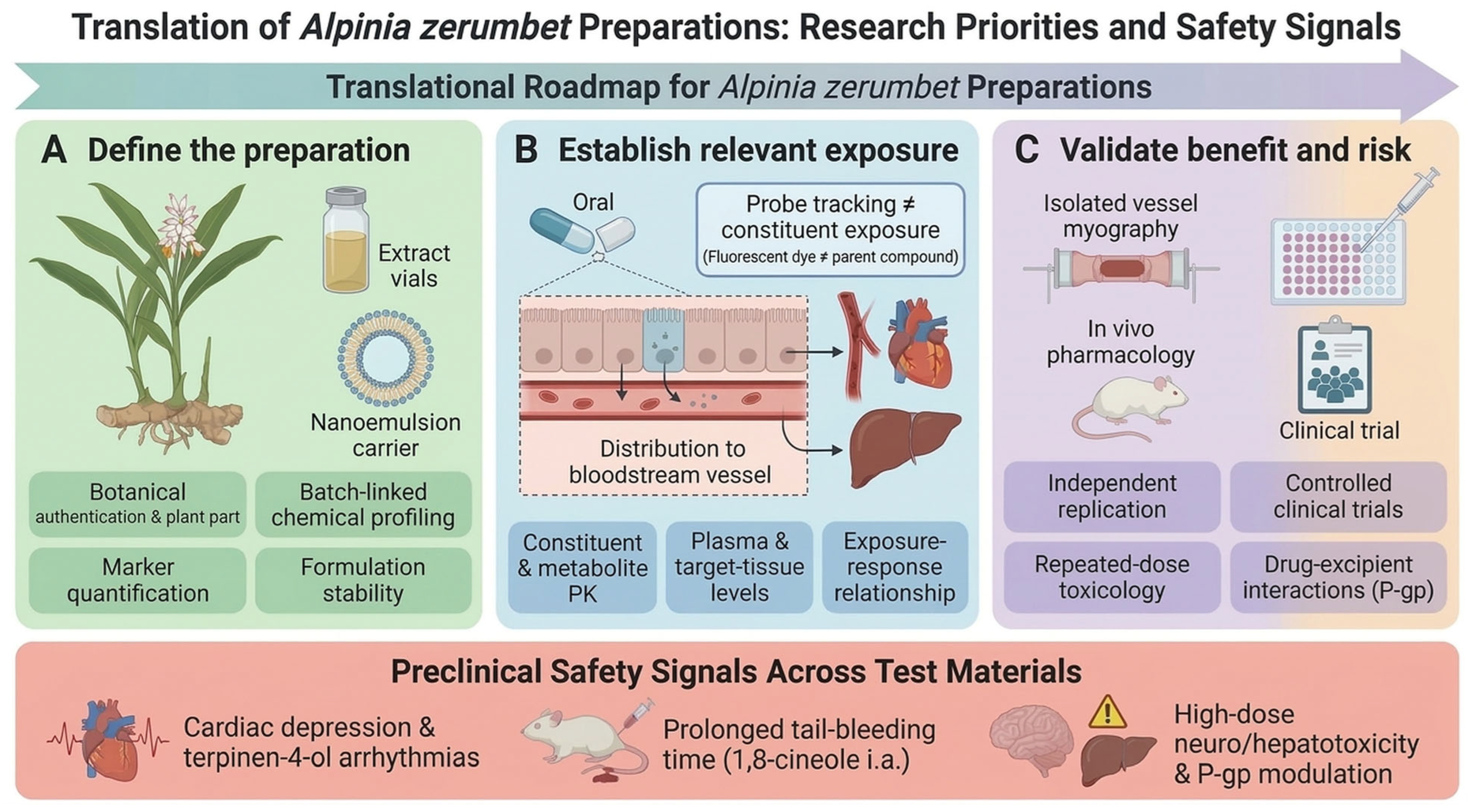
4.4. Interpretation of Human Evidence
The available human studies examined short-term blood pressure responses in small cohorts (Table 10). Ten healthy volunteers (aged 20–32 years) received A. speciosa leaf tea or placebo on separate study days 7 days apart under participant blinding [19]. A single 300 mL serving at 0.8 g/100 mL—described by the authors as five times the usual folk dose—corresponded to 2.4 g of plant material. On the A. speciosa tea day, systolic pressure changed from 105 ± 4 to 97 ± 5 mmHg and diastolic pressure from 74 ± 3 to 64 ± 4 mmHg (mean ± SE; n = 10). Mild diuresis occurred without alterations in plasma or urinary Na+, K+, calcium, phosphate, uric acid, urea, or creatinine [19]. On the placebo day, systolic pressure was 102 ± 4 before and 103 ± 3 mmHg after, and diastolic pressure 77 ± 3 and 74 ± 2 mmHg; the corresponding values for the Elephantopus scaber tea day were 102 ± 4 and 106 ± 4 mmHg systolic and 75 ± 3 and 70 ± 2 mmHg diastolic (mean ± SE; n = 10) [19]. The report marks only the A. speciosa before-and-after comparison as statistically significant; it presents no between-treatment comparison and does not describe the statistical methods used. These figures are therefore within-day before-and-after measurements, and a placebo-adjusted treatment effect cannot be derived from the published data. A Portuguese-language report by the same investigators, which we could assess only from its abstract, describes the same design (ten healthy volunteers, vanilla-flavored water as placebo, doses 7 days apart) and the same statistically significant findings for A. speciosa; it appears to be a second report of this trial rather than an independent replication [100].
Table 10.
Human studies involving Alpinia zerumbet preparations.
A second study followed 22 patients with grade I–II hypertension (off antihypertensive medication for the preceding 30 days) for 6 weeks. Patients received capsules containing 250 mg of crude hydroalcoholic leaf extract standardized to 2.5% flavonoids, titrated from 1 to 6 capsules/day (250–1500 mg/day) according to blood pressure response; it was an uncontrolled exploratory study without a concurrent control group [20]. Every blood pressure and tolerability outcome reported there is consequently a within-group change measured against each patient’s own baseline, with no comparator against which a treatment effect could be estimated.
A thesis reported a randomized, double-blind, placebo-controlled 28-day safety study in 36 healthy volunteers (24 tea, 12 placebo) [21]. Tea intake corresponded to 9.0 g dry leaves and 540 mL/day (Table 10). No clear toxicity signal emerged in the assessed clinical, laboratory, or comet-assay endpoints, although both groups reported adverse events. In the uncontrolled hypertension study, one participant withdrew because of discomfort, which investigators considered unrelated to treatment [20].
According to the publisher abstract, 55 healthy young men completed an 8-week randomized study: 28 received nine 500 mg leaf capsules daily and 27 received no supplementation [22]. The abstract describes a decrease in MDA in the supplemented group, with lower values than in controls at week 8, and within-group declines in glucose and LDL-C without significant post-intervention between-group differences. No significant changes in AST, ALT, or creatinine were reported. The labeled capsule mass was 4.5 g/day; the dry-leaf or extract equivalent, participant flow, and blinding could not be assessed from the abstract.
The human studies answer different questions: single-dose physiology in healthy volunteers [19], an uncontrolled exploratory study of short-term blood pressure in patients [20], short-term tolerability under the tested regimen in healthy volunteers [21], and biomarker changes in healthy young men [22]. The between-group MDA finding in [22] does not establish clinical cardiovascular benefit, while its nonsignificant between-group glucose and LDL-C comparisons should not be summarized as demonstrated glucose- or lipid-lowering efficacy. Across the four studies, the outcomes supported by between-group comparison are confined to the 28-day clinical, laboratory, and comet-assay endpoints of the randomized placebo-controlled thesis [21] and the week-8 malondialdehyde difference reported in [22]; the blood pressure findings of [19] and [20] rest on within-day or within-group change alone. Collectively, the studies support feasibility and short-term observations, but do not establish sustained blood pressure control or cardiovascular risk reduction for a standardized preparation. A clinical efficacy study should therefore use a defined product, an appropriate comparator, and prespecified blood pressure or other clinically relevant outcomes, with adequate follow-up.
Placing the preclinical and human doses on a common scale shows how loosely they correspond. Using body-surface-area conversion (human-equivalent dose = animal dose × animal Km/human Km, with Km = 3 for mice, 6 for rats, and 37 for adults) [101], the drinking-water dose of about 50 mg/kg/day hydroalcoholic leaf extract in rats [40,44] corresponds to about 8 mg/kg/day, or roughly 490 mg/day for a 60 kg adult, which lies within the 250–1500 mg/day of crude hydroalcoholic leaf extract given in the uncontrolled human study [20], although the equivalence of the two extracts has not been established. Fruit oil doses in mice of 15–30 mg/kg/day (route not stated) [15] and 90 mg/kg/day by gavage [16] correspond to about 1.2–2.4 and 7.3 mg/kg/day (about 70–150 and 440 mg/day), and 1,8-cineole doses of 50–200 mg/kg/day by gavage in mice [27] and 20–40 mg/kg/day by gavage in rats [29] correspond to about 3–16 mg/kg/day. No human study administered an essential oil or 1,8-cineole orally, and the leaf-tea, dried-leaf, and capsule regimens [19,21,22] cannot be converted to oil or constituent doses because their oil and 1,8-cineole contents were not measured; the 10 mg/kg/day intraperitoneal oil dose in rats [32] has no human counterpart. These conversions are allometric estimates rather than pharmacokinetic equivalence, and they show that human exposure to the oils and constituents considered active in animal models remains unknown.
Sex distribution is a further constraint shared by the preclinical and clinical literature. Where sex is reported in the animal studies summarized in Table 4, Table 5, Table 6, Table 7, Table 8 and Table 9, the animals were male in nearly every case, with rats of both sexes used only in two cardiac studies [85,86] and sex unreported in several others. The human studies include five men and five women [19], 18 men and 18 women [21], healthy young men only [22], and no stated distribution [20]. Sex differences in vascular reactivity, NO signaling, and hepatic drug metabolism are well described, and no study of A. zerumbet has analyzed outcomes by sex. The reported effects therefore apply principally to male animals, and whether they extend to females is untested.
4.5. Preclinical Safety Signals and Evidence Gaps
Preclinical toxicological findings differ by material, dose, and route. For hydroalcoholic leaf extract, an early mouse study reported an oral LD50 of 10.0 ± 2.5 g/kg and an intraperitoneal LD50 of approximately 0.76 g/kg; the latter dispersion term differs between the abstract and Table I [90]. Acute testing recorded writhing, excitation followed by hypokinesia, and pruritus. In a separate experiment, oral extract at 500–1000 mg/kg prolonged pentobarbital-induced sleeping time. The 30-day rat study compared tea (15 mL/kg, 1.5%) and extract (5 g/kg), but the route is ambiguous: the abstract describes daily injection, whereas the Table III doses suggest oral administration. Table III reports higher LDH in both groups, lower AST and no significant ALT increase with tea, and higher AST and ALT with extract. The text of reference [90] does not fully agree with its Table III. The methods state only that rats were treated for 30 days with extract, tea, or water and that the 10% lethal dose (LD10) of the material was administered daily; no route is named, so the discrepancy with the abstract cannot be resolved from the published methods. The repeated-dose findings—raised LDH and transaminases and the reported lymphocyte change—therefore cannot be attributed to oral administration. The acute median lethal dose (LD50) values and the pentobarbital sleeping-time experiment, for which the routes are stated explicitly, are unaffected by this ambiguity. No histopathological abnormalities were reported in the liver, kidneys, spleen, lungs, or heart [90].
For isolated rhizome kavalactones, oral LD50 values in mice were 5.16 g/kg (95% confidence limits, 4.72–6.03 g/kg) for DK and 6.57 g/kg (5.83–7.05 g/kg) for DDK [97]. In mice, oral DK or DDK at 30, 100, or 300 mg/kg per administration, twice daily for 14 days, produced no consistent organ toxicity in the assessed endpoints. A separate 3-month rat study used the same nominal dose levels, but dosing frequency was inconsistently reported. In a 9-week mouse study at 100 mg/kg twice daily, DDK reduced spontaneous locomotor activity at weeks 2, 4, and 9, without a consistent change throughout treatment; DK did not show this reduction. TA98/TA100 Ames tests were negative. Both compounds prolonged pentobarbital-induced sleeping time, and high acute doses produced neurological, respiratory, and cardiovascular signs [97].
For essential oil preparations, a single oral dose of a fruit essential oil self-emulsifying drug delivery system (SEDDS) in mice yielded an LD50 of 5.29 g/kg (95% confidence limits, 4.93–5.68 g/kg, expressed as total formulation mass). The formulation combined fruit essential oil, Kolliphor HS 15, and anhydrous ethanol in a 6:2:2 mass ratio, so the oil accounted for 60% of the administered mass and the LD50 corresponds to approximately 3.2 g/kg of essential oil. Lethal or near-lethal doses induced convulsions, tremor, ataxia, and respiratory depression [98]. Even expressed on an oil basis, the value is not directly comparable with the acute toxicity of unformulated oil, which was not determined in the same experiment; the authors report instead that Caco-2 cells tolerated the formulation better than free oil at matched oil content, which they attribute to reduced contact between the oil and the cell membrane. In a separate acute tail-vein toxicity screen in mice (12.5–100 mg/kg observed for 14 days), R8-modified fruit oil lipid microspheres caused no mortality or gross organ lesions [58]. In cultured human peripheral blood leukocytes, leaf essential oil showed no significant genotoxicity at 50–300 μg/mL and reduced H2O2-induced DNA damage in co- and post-treatment protocols. At 500 μg/mL, the oil reduced cell viability and increased DNA damage when tested alone; it also enhanced H2O2-associated damage in co- and post-treatment protocols. In mice, micronucleus and comet assays showed no significant genotoxic effect after a single oral dose of 400 mg/kg (n = 5 per group) [99].
Formulation excipients may also affect transport. In Caco-2 cells, free essential oil at 12 and 24 μg/mL reduced P-glycoprotein (P-gp) expression, whereas the self-emulsifying formulation at 10, 20, and 40 μg/mL—corresponding to 6, 12, and 24 μg/mL of oil—and Kolliphor HS 15 or ethanol alone increased it. In rhodamine 123 uptake assays, free oil at 6, 12, and 24 μg/mL and the formulation at 40 μg/mL raised intracellular fluorescence, indicating reduced efflux, whereas the formulation at 10 and 20 μg/mL and the excipients alone did not alter efflux function [98]. Functional inhibition therefore appeared only at the highest formulation concentration tested, and the induced P-gp expression was not accompanied by increased efflux across the range examined. These are monolayer measurements at concentrations applied directly to the apical surface, and no corresponding intestinal concentration after an oral dose of any A. zerumbet preparation has been measured. They identify a plausible transporter-mediated interaction mechanism for co-administered P-gp substrates without supporting a quantitative prediction of its clinical relevance, and they indicate that preparation and excipient effects on concomitant drugs should be assessed using actual constituent exposure together with transport measurements.
DK and DDK belong to the kavalactone class, and DK is structurally identical to desmethoxyyangonin, one of the six major kavalactones of kava (Piper methysticum). Kavalactone-containing kava preparations have been associated with clinically apparent liver injury, prompting regulatory restrictions in several countries, and kava is categorized as a well-established cause of drug-induced liver injury, although the mechanism is unresolved and isolated kavalactones have not been shown to be intrinsically hepatotoxic [102]. In human liver microsomes, kava extract inhibited the cytochrome P450 (CYP) isoforms CYP1A2, CYP2C9, CYP2C19, CYP2D6, and CYP3A4, and among individual kavalactones tested at 10 μmol/L, CYP2C9 was inhibited by desmethoxyyangonin, methysticin, and dihydromethysticin, CYP3A4 by the same three, CYP2C19 by dihydromethysticin, and CYP2D6 by methysticin, while kawain inhibited none of them [103]; inhibition constants for CYP2C9 and CYP2C19 ranged from 5 to 10 μmol/L; kava extract and kavalactones also modestly stimulated P-glycoprotein ATPase activity [104]. These findings concern kava rather than A. zerumbet, and their transferability is uncertain because the kavalactone content of A. zerumbet leaf tea and fruit preparations has not been quantified and therefore cannot be compared with that of kava root extracts. They nevertheless define a class-level question that the A. zerumbet literature has not addressed at the level of a preparation: we identified no study measuring CYP inhibition or induction by an A. zerumbet preparation or by DDK, and the only transporter data for a preparation are the Caco-2 P-gp measurements described above [98]; DK has been examined for CYP effects only as desmethoxyyangonin in the kava literature [103]. Even where an individual constituent of this species has been characterized for CYP interactions as an isolated compound in another context, such data cannot be extrapolated to a preparation whose constituent concentrations are unquantified. We likewise identified no study testing an A. zerumbet preparation together with a cardiovascular drug. A pharmacodynamic interaction is also plausible: in addition to the rabbit-platelet data for DK and DDK [34], DK isolated from Alpinia mutica fruit inhibited arachidonic acid-induced aggregation in human whole blood with an IC50 of 78.1 ± 2.8 μmol/L [105], so additive effects with antiplatelet and anticoagulant drugs need to be considered alongside pharmacokinetic interactions. Because the preparations discussed here are proposed for indications in which patients commonly receive antihypertensive, antiplatelet, and lipid-lowering therapy, and because several of those drugs are CYP2C9, CYP2C19, CYP3A4, or P-gp substrates, systematic CYP and transporter interaction assessment is a prerequisite for clinical development rather than an optional addition.
The safety findings identify different constraints rather than a common safe dose. Absence of mortality, gross lesions, or genotoxicity under one protocol does not negate effects on bleeding, cardiac rhythm, contractility, or behavior measured under another [83,85,86,97,98,99]. Conversely, injected-dose or high-concentration effects do not define an oral human toxicity threshold. Human data from outside the cardiovascular field are likewise route-specific: in a randomized, double-blind, placebo-controlled crossover trial in 57 patients with hereditary spastic paraplegia, 4 weeks of daily dermal application of an A. zerumbet essential oil product (Ziclague®) produced only mild, transient adverse events, and patient-reported blood pressure reduction occurred in 5 participants after the oil and 4 after placebo (p = 1.00; 52 participants evaluable for safety) [106]. Topical exposure of this kind does not establish the safety of oral preparations. The toxicological studies share the sex limitation noted in Section 4.4: the repeated-dose and genotoxicity experiments used male animals or did not state sex, and only the acute screen of the R8-modified microspheres [58] used equal numbers of both sexes, so sex-dependent differences in toxicity and metabolism remain unexamined. Development should link the intended product and route to effective and adverse-effect exposures, with particular attention to cardiac function, hemostasis, repeated-dose hepatic findings, kavalactone class safety, and interaction potential with concomitant drugs.
5. Conclusions
The literature supports experimental vasorelaxation and blood pressure lowering by selected A. zerumbet preparations more clearly than a single molecular explanation for cardiovascular protection. In selected preparations, endothelial NO signaling and attenuation of smooth muscle Ca2+-dependent contraction contribute to acute vascular responses; their role in sustained pressure lowering has not been established. Reduced endothelial injury and macrophage lipid accumulation have been reported repeatedly, but largely within one research program, and remain hypothesis-generating until independently replicated. Pharmacological and genetic PPARγ interventions support receptor involvement in senescence and cholesterol handling, whereas turnover and ubiquitination experiments identify altered receptor stability as a candidate molecular basis. Whether direct constituent binding explains these responses at effective cellular and in vivo exposures remains unresolved. General claims of a common autophagic mechanism, consistent angiogenic regulation, or preparation-independent cardioprotection exceed the available evidence.
Translation should proceed from the evidence available for each candidate product. Standardized leaf extracts warrant independent replication of repeated-dose oral blood pressure effects; fruit oil requires batch-linked chemistry and attribution of its cellular and animal effects; 1,8-cineole formulations require matched pharmacokinetic and pharmacodynamic evaluation. These priorities reflect different evidential gaps, not an overall ranking of preparations. Human studies have not established sustained cardiovascular efficacy, and clinical development must evaluate cardiac, hemostatic, and hepatic risks for the intended formulation and route, including the kavalactone class safety questions raised by DK and DDK, and must undertake preparation-specific CYP studies and follow-up of the in vitro P-glycoprotein findings to assess interaction risk.
Supplementary Materials
The following supporting information can be downloaded at: https://www.mdpi.com/article/10.3390/molecules31234251/s1: Table S1: Primary-study reporting inconsistencies and interpretive limitations relevant to cardiovascular evidence for Alpinia zerumbet; Table S2: Chemical characterization of the mature-fruit essential oil preparations tabulated in Table 5, Table 6 and Table 7 of the main text; Table S3: Full literature search strings.
Author Contributions
C.Z.: Investigation, software, visualization, writing—original draft, writing—review and editing. L.C.: Conceptualization, formal analysis, investigation, validation, writing—original draft, writing—review and editing. F.X.: Investigation, conceptualization, supervision, validation. L.X.: Conceptualization, supervision, validation, writing—review and editing. M.Z.: Formal analysis, project administration, supervision, validation, writing—review and editing. X.W.: Formal analysis, project administration, supervision, validation, writing—review and editing. H.Z.: Funding acquisition, project administration, resources, supervision. All authors have read and agreed to the published version of the manuscript.
Funding
This work was supported by Guangxi Key Laboratory of Zhuang and Yao Ethnic Medicine (Guike LT2600640036); University Engineering Research Center of Development and Industrialization of Zhuang and Yao Ethnic Medicinal Materials, Guangxi ([2024] No. 10); Research Center of Guangxi Zhuang Autonomous Region Medicine Resources and Application Engineering ([2020] No. 2605); Collaborative Innovation Center of Zhuang and Yao Ethnic Medicine ([2013] No. 20); Guangxi Key Discipline of Traditional Chinese Medicine (Zhuang Ethnic Pharmacy) (GZXK-Z-20-64); Guangxi First-Class Discipline: Chinese Medicine (Ethnic Pharmacy) ([2018] No. 12).
Institutional Review Board Statement
Not applicable. This review reports no newly conducted studies involving human participants or animals.
Informed Consent Statement
Not applicable.
Data Availability Statement
No new data were created or analyzed in this study. Data sharing is not applicable to this article.
Acknowledgments
During the preparation of the original manuscript, the authors used DeepSeek-V3 for translation, language editing, and manuscript formatting. All content has been verified by the authors, who take full responsibility for the content of this publication.
Conflicts of Interest
The authors declare no conflicts of interest.
References
- Ndumele, C.E.; Rangaswami, J.; Chow, S.L.; Neeland, I.J.; Tuttle, K.R.; Khan, S.S.; Coresh, J.; Mathew, R.O.; Baker-Smith, C.M.; Carnethon, M.R.; et al. Cardiovascular-kidney-metabolic health: A presidential advisory from the American Heart Association. Circulation 2023, 148, 1606–1635, Correction in Circulation 2024, 149, 13. https://doi.org/10.1161/CIR.0000000000001241. [Google Scholar] [CrossRef] [Scilit] [PubMed]
- Valenzuela, P.L.; Carrera-Bastos, P.; Castillo-García, A.; Lieberman, D.E.; Santos-Lozano, A.; Lucia, A. Obesity and the risk of cardiometabolic diseases. Nat. Rev. Cardiol. 2023, 20, 475–494. [Google Scholar] [CrossRef] [Scilit] [PubMed]
- Gallo, G.; Savoia, C. New insights into endothelial dysfunction in cardiometabolic diseases: Potential mechanisms and clinical implications. Int. J. Mol. Sci. 2024, 25, 2973. [Google Scholar] [CrossRef] [Scilit] [PubMed]
- Tamargo, I.A.; Baek, K.I.; Kim, Y.; Park, C.; Jo, H. Flow-induced reprogramming of endothelial cells in atherosclerosis. Nat. Rev. Cardiol. 2023, 20, 738–753. [Google Scholar] [CrossRef] [Scilit] [PubMed]
- Brandts, J.; Ray, K.K. Novel and future lipid-modulating therapies for the prevention of cardiovascular disease. Nat. Rev. Cardiol. 2023, 20, 600–616. [Google Scholar] [CrossRef] [Scilit] [PubMed]
- McEvoy, J.W.; McCarthy, C.P.; Bruno, R.M.; Brouwers, S.; Canavan, M.D.; Ceconi, C.; Christodorescu, R.M.; Daskalopoulou, S.S.; Ferro, C.J.; Gerdts, E.; et al. 2024 ESC Guidelines for the management of elevated blood pressure and hypertension. Eur. Heart J. 2024, 45, 3912–4018. [Google Scholar] [CrossRef] [Scilit] [PubMed]
- Royal Botanic Gardens, Kew. Plants of the World Online: Alpinia zerumbet (Pers.) B.L.Burtt & R.M.Sm. Available online: https://powo.science.kew.org/taxon/872083-1 (accessed on 27 August 2026).
- Nishidono, Y.; Tanaka, K. Phytochemicals of Alpinia zerumbet: A review. Molecules 2024, 29, 2845. [Google Scholar] [CrossRef] [Scilit] [PubMed]
- Xiao, T.; Huang, J.; Wang, X.; Wu, L.; Zhou, X.; Jiang, F.; He, Z.; Guo, Q.; Tao, L.; Shen, X. Alpinia zerumbet and its potential use as an herbal medication for atherosclerosis: Mechanistic insights from cell and rodent studies. Lifestyle Genom. 2020, 13, 138–145. [Google Scholar] [CrossRef] [Scilit] [PubMed]
- Murakami, S.; Li, W.; Matsuura, M.; Satou, T.; Hayashi, S.; Koike, K. Composition and seasonal variation of essential oil in Alpinia zerumbet from Okinawa Island. J. Nat. Med. 2009, 63, 204–208. [Google Scholar] [CrossRef] [Scilit] [PubMed]
- Victório, C.P.; Leitão, S.G.; Lage, C.L.S. Chemical composition of the leaf oils of Alpinia zerumbet (Pers.) Burtt et Smith and A. purpurata (Vieill) K. Schum. from Rio de Janeiro, Brazil. J. Essent. Oil Res. 2010, 22, 52–54. [Google Scholar] [CrossRef] [Scilit]
- Tao, L.; Shen, X.C.; Peng, J.; Zhi, N. GC-MS analysis of volatile oil constituents from whole fruits and different fruit parts of Alpinia zerumbet. Chin. Tradit. Pat. Med. 2009, 31, 909–911. (In Chinese) [Google Scholar]
- Shen, X.C.; Hu, H.S.; Xiao, H.T. GC-MS analysis of volatile oil constituents from rhizomes, stems, leaves and fruits of Alpinia zerumbet. Chin. J. Pharm. Anal. 2010, 30, 1399–1403. (In Chinese) [Google Scholar] [CrossRef]
- Fu, L.; Tai, S.; Liao, J.; Du, Y.; Zhang, G.; Guo, D.; Chen, X.; Zheng, T.; Hu, X.; Yao, W.; et al. 1,8-Cineole ameliorates vascular endothelial senescence in diabetes mellitus by directly targeting and deubiquitinating PPAR-γ in vivo and in vitro. J. Pharm. Anal. 2025, 15, 101307. [Google Scholar] [CrossRef] [Scilit] [PubMed]
- Wang, S.Q.; Xiang, J.; Zhang, G.Q.; Fu, L.Y.; Xu, Y.N.; Chen, Y.; Tao, L.; Hu, X.X.; Shen, X.C. Essential oil from Fructus Alpinia zerumbet ameliorates atherosclerosis by activating PPARγ-LXRα-ABCA1/G1 signaling pathway. Phytomedicine 2024, 123, 155227. [Google Scholar] [CrossRef] [Scilit] [PubMed]
- Liao, J.; Fu, L.Y.; Tai, S.; Xu, Y.N.; Wang, S.Q.; Guo, L.L.; Guo, D.; Du, Y.Q.; He, J.G.; Yang, H.; et al. Essential oil from Fructus Alpiniae zerumbet ameliorates vascular endothelial cell senescence in diabetes by regulating PPAR-γ signalling: A 4D label-free quantitative proteomics and network pharmacology study. J. Ethnopharmacol. 2024, 321, 117550. [Google Scholar] [CrossRef] [Scilit] [PubMed]
- Chen, Y.; Wang, J.; Xu, J.; Zhang, J.; Xu, S.; Zhang, Q.; Huang, J.; Peng, J.; Xu, H.; Du, Q.; et al. Fabrication of a polysaccharide-protein/protein complex stabilized oral nanoemulsion to facilitate the therapeutic effects of 1,8-cineole on atherosclerosis. ACS Nano 2023, 17, 9090–9109. [Google Scholar] [CrossRef] [Scilit] [PubMed]
- Peng, J.; Jiang, Z.; Wu, G.; Cai, Z.; Du, Q.; Tao, L.; Zhang, Y.; Chen, Y.; Shen, X. Improving protection effects of eucalyptol via carboxymethyl chitosan-coated lipid nanoparticles on hyperglycaemia-induced vascular endothelial injury in rats. J. Drug Target. 2021, 29, 520–530. [Google Scholar] [CrossRef] [Scilit] [PubMed]
- Laranja, S.M.R.; Bergamaschi, C.M.; Schor, N. Evaluation of acute administration of natural products with potential diuretic effects, in humans. Mem. Inst. Oswaldo Cruz 1991, 86, 237–240. [Google Scholar] [CrossRef] [Scilit] [PubMed]
- Vargas, J.H.A.; Carvalho, J.C.T. Study of the efficacy and clinical safety of Alpinia speciosa (J.C.Wendl.) K.Schum. crude extract on arterial hypertension. Int. J. Pharm. Sci. Rev. Res. 2010, 4, 27–33. [Google Scholar]
- Santana, A.P.M. Avaliação da Segurança e Genotoxicidade do Chá de Alpinia zerumbet (Pers.) Burtt & Smith em Voluntários sadios. Master’s Thesis, Universidade Federal do Ceará, Fortaleza, Brazil, 2009. Available online: https://repositorio.ufc.br/handle/riufc/2200 (accessed on 27 August 2026). (In Portuguese)
- Hoang, T.M.P.; Vu, T.T.H.; Le, D.T.; Tran, H.L.; Vuong, T.H.N.; Nguyen, T.A.N. Impact of shell ginger leaf capsule supplementation for 8 weeks on plasma malondialdehyde and selected blood biochemical parameters in healthy young men in Quang Ninh. Vietnam J. Community Med. 2026, 67, CD9, (In Vietnamese; English abstract consulted). [Google Scholar] [CrossRef] [Scilit]
- Lahlou, S.; Galindo, C.A.B.; Leal-Cardoso, J.H.; Fonteles, M.C.; Duarte, G.P. Cardiovascular effects of the essential oil of Alpinia zerumbet leaves and its main constituent, terpinen-4-ol, in rats: Role of the autonomic nervous system. Planta Medica 2002, 68, 1097–1102. [Google Scholar] [CrossRef] [Scilit] [PubMed]
- Lahlou, S.; Interaminense, L.F.L.; Leal-Cardoso, J.H.; Duarte, G.P. Antihypertensive effects of the essential oil of Alpinia zerumbet and its main constituent, terpinen-4-ol, in DOCA-salt hypertensive conscious rats. Fundam. Clin. Pharmacol. 2003, 17, 323–330. [Google Scholar] [CrossRef] [Scilit] [PubMed]
- Pinto, N.V.; Assreuy, A.M.S.; Coelho-de-Souza, A.N.; Ceccatto, V.M.; Magalhães, P.J.C.; Lahlou, S.; Leal-Cardoso, J.H. Endothelium-dependent vasorelaxant effects of the essential oil from aerial parts of Alpinia zerumbet and its main constituent 1,8-cineole in rats. Phytomedicine 2009, 16, 1151–1155. [Google Scholar] [CrossRef] [Scilit] [PubMed]
- Linghu, K.; Lin, D.; Yang, H.; Xu, Y.; Zhang, Y.; Tao, L.; Chen, Y.; Shen, X. Ameliorating effects of 1,8-cineole on LPS-induced human umbilical vein endothelial cell injury by suppressing NF-κB signaling in vitro. Eur. J. Pharmacol. 2016, 789, 195–201. [Google Scholar] [CrossRef] [Scilit] [PubMed]
- Linghu, K.G.; Wu, G.P.; Fu, L.Y.; Yang, H.; Li, H.Z.; Chen, Y.; Yu, H.; Tao, L.; Shen, X.C. 1,8-Cineole ameliorates LPS-induced vascular endothelium dysfunction in mice via PPAR-γ dependent regulation of NF-κB. Front. Pharmacol. 2019, 10, 178. [Google Scholar] [CrossRef] [Scilit] [PubMed]
- Zhu, Z.X.; Xiang, J.; Chen, X.Y.; Tao, J.L.; Pan, G.X.; Zhang, G.Q. Effects and mechanisms of 1,8-cineole on ox-LDL-induced macrophage-derived foam-cell formation. J. Guizhou Med. Univ. 2025, 50, 212–219, 239. (In Chinese) [Google Scholar] [CrossRef]
- Chen, M.; Hu, Q.; Wang, S.; Tao, L.; Hu, X.; Shen, X. 1,8-Cineole ameliorates endothelial injury and hypertension induced by L-NAME through regulation of autophagy via PI3K/mTOR signaling pathway. Eur. J. Pharmacol. 2023, 954, 175863. [Google Scholar] [CrossRef] [Scilit] [PubMed]
- Petry, J.; Weiser, T.; Griesbaum, L.; Schröder, K.; Hoch, C.C.; Bashiri Dezfouli, A.; Shoykhet, M.; Wollenberg, B. 1,8-Cineole prevents platelet activation and aggregation by activating the cAMP pathway via the adenosine A2A receptor. Life Sci. 2024, 350, 122746. [Google Scholar] [CrossRef] [Scilit] [PubMed]
- Zhang, Y.Y.; Linghu, K.G.; Chen, Y.; Tao, L.; Zhang, X.J.; Wen, B.; Shen, X.C. Orthogonal experimental study of active components of Alpinia zerumbet essential oil protecting HUVECs against lipopolysaccharide-induced injury. Chin. J. Exp. Tradit. Med. Formulae 2014, 20, 9–12. (In Chinese) [Google Scholar] [CrossRef]
- Barcelos, F.F.; Oliveira, M.L.; Giovaninni, N.P.B.; Lins, T.P.; Filomeno, C.A.; Schneider, S.Z.; Pinto, V.D.; Endringer, D.C.; Andrade, T.U. Estudo químico e da atividade biológica cardiovascular do óleo essencial de folhas de Alpinia zerumbet (Pers.) B.L.Burtt & R.M.Sm. em ratos. Rev. Bras. Plantas Med. 2010, 12, 48–56. (In Portuguese) [Google Scholar] [CrossRef] [Scilit]
- Xiao, T.; Wu, A.; Wang, X.; Guo, Z.; Huang, F.; Cheng, X.; Shen, X.; Tao, L. Anti-hypertensive and composition as well as pharmacokinetics and tissues distribution of active ingredients from Alpinia zerumbet. Fitoterapia 2024, 172, 105753. [Google Scholar] [CrossRef] [Scilit] [PubMed]
- Teng, C.M.; Hsu, S.Y.; Lin, C.H.; Yu, S.M.; Wang, K.J.; Lin, M.H.; Chen, C.F. Antiplatelet action of dehydrokawain derivatives isolated from Alpinia speciosa rhizoma. Chin. J. Physiol. 1990, 33, 41–48. [Google Scholar] [PubMed]
- You, H.; He, M.; Pan, D.; Fang, G.; Chen, Y.; Zhang, X.; Shen, X.; Zhang, N. Kavalactones isolated from Alpinia zerumbet (Pers.) Burtt. et Smith with protective effects against human umbilical vein endothelial cell damage induced by high glucose. Nat. Prod. Res. 2022, 36, 5740–5746. [Google Scholar] [CrossRef] [Scilit] [PubMed]
- da Silva, M.A.; de Carvalho, L.C.R.M.; Victório, C.P.; Ognibene, D.T.; Resende, A.C.; de Souza, M.A.V. Chemical composition and vasodilator activity of different Alpinia zerumbet leaf extracts, a potential source of bioactive flavonoids. Med. Chem. Res. 2021, 30, 2103–2113. [Google Scholar] [CrossRef] [Scilit]
- Zhang, Y.; Yu, Y.Y.; Peng, F.; Duan, W.T.; Wu, C.H.; Li, H.T.; Zhang, X.F.; Shi, Y.S. Neolignans and diarylheptanoids with anti-inflammatory activity from the rhizomes of Alpinia zerumbet. J. Agric. Food Chem. 2021, 69, 9229–9237. [Google Scholar] [CrossRef] [Scilit] [PubMed]
- Chompoo, J.; Upadhyay, A.; Gima, S.; Fukuta, M.; Tawata, S. Antiatherogenic properties of acetone extract of Alpinia zerumbet seeds. Molecules 2012, 17, 6237–6248. [Google Scholar] [CrossRef] [Scilit] [PubMed]
- Chuang, C.M.; Wang, H.E.; Peng, C.C.; Chen, K.C.; Peng, R.Y. Hypolipidemic effects of different angiocarp parts of Alpinia zerumbet. Pharm. Biol. 2011, 49, 1257–1264. [Google Scholar] [CrossRef] [Scilit] [PubMed]
- de Moura, R.S.; Emiliano, A.F.; de Carvalho, L.C.R.M.; Souza, M.A.V.; Guedes, D.C.; Tano, T.; Resende, A.C. Antihypertensive and endothelium-dependent vasodilator effects of Alpinia zerumbet, a medicinal plant. J. Cardiovasc. Pharmacol. 2005, 46, 288–294. [Google Scholar] [CrossRef] [Scilit] [PubMed]
- Tao, L.; Hu, H.S.; Shen, X.C. Endothelium-dependent vasodilatation effects of the essential oil from Fructus Alpiniae zerumbet (EOFAZ) on rat thoracic aortic rings in vitro. Phytomedicine 2013, 20, 387–393. [Google Scholar] [CrossRef] [Scilit] [PubMed]
- da Cunha, G.H.; de Moraes, M.O.; Fechine, F.V.; Bezerra, F.A.F.; Silveira, E.R.; Canuto, K.M.; de Moraes, M.E.A. Vasorelaxant and antihypertensive effects of methanolic fraction of the essential oil of Alpinia zerumbet. Vasc. Pharmacol. 2013, 58, 337–345. [Google Scholar] [CrossRef] [Scilit] [PubMed]
- Rocha, D.G.; Holanda, T.M.; Braz, H.L.B.; de Moraes, J.A.S.; Marinho, A.D.; Maia, P.H.F.; de Moraes, M.E.A.; Fechine-Jamacaru, F.V.; de Moraes Filho, M.O. Vasorelaxant effect of Alpinia zerumbet’s essential oil on rat resistance artery involves blocking of calcium mobilization. Fitoterapia 2023, 169, 105623. [Google Scholar] [CrossRef] [Scilit] [PubMed]
- Menezes, M.P.; Santos, G.P.; Nunes, D.V.Q.; Silva, D.L.B.; Victório, C.P.; Fernandes-Santos, C.; de Bem, G.F.; Costa, C.A.; Resende, A.C.; Ognibene, D.T. Alpinia zerumbet leaf extract reverses hypertension and improves adverse remodeling in the left ventricle and aorta in spontaneously hypertensive rats. Braz. J. Med. Biol. Res. 2025, 58, e14210. [Google Scholar] [CrossRef] [Scilit] [PubMed]
- Ignarro, L.J.; Buga, G.M.; Wood, K.S.; Byrns, R.E.; Chaudhuri, G. Endothelium-derived relaxing factor produced and released from artery and vein is nitric oxide. Proc. Natl. Acad. Sci. USA 1987, 84, 9265–9269. [Google Scholar] [CrossRef] [Scilit] [PubMed]
- Friebe, A.; Mergia, E.; Dangel, O.; Lange, A.; Koesling, D. Fatal gastrointestinal obstruction and hypertension in mice lacking nitric oxide-sensitive guanylyl cyclase. Proc. Natl. Acad. Sci. USA 2007, 104, 7699–7704. [Google Scholar] [CrossRef] [Scilit] [PubMed]
- Surks, H.K.; Mochizuki, N.; Kasai, Y.; Georgescu, S.P.; Tang, K.M.; Ito, M.; Lincoln, T.M.; Mendelsohn, M.E. Regulation of myosin phosphatase by a specific interaction with cGMP-dependent protein kinase Iα. Science 1999, 286, 1583–1587. [Google Scholar] [CrossRef] [Scilit] [PubMed]
- Huang, P.L.; Huang, Z.; Mashimo, H.; Bloch, K.D.; Moskowitz, M.A.; Bevan, J.A.; Fishman, M.C. Hypertension in mice lacking the gene for endothelial nitric oxide synthase. Nature 1995, 377, 239–242. [Google Scholar] [CrossRef] [Scilit] [PubMed]
- Zhang, W.; Zhang, N.L.; Zhang, Y.Y.; Chen, Y.; Fu, L.Y.; Pan, D.; Shen, X.C. Essential oil from Alpiniae Zerumbet Fructus alleviates high glucose-induced HUVEC injury via TXNIP signaling pathway. Chin. J. Exp. Tradit. Med. Formulae 2021, 27, 21–27. (In Chinese) [Google Scholar] [CrossRef]
- Huang, N.; Xu, Y.; Zhou, H.; Lin, D.; Zhang, B.; Zhang, Y.; Pan, D.; Tao, L.; Liu, X.; Shen, X. Essential oil from Fructus Alpiniae zerumbet protects human umbilical vein endothelial cells in vitro from injury induced by high glucose levels by suppressing nuclear transcription factor-kappa B signaling. Med. Sci. Monit. 2017, 23, 4760–4767. [Google Scholar] [CrossRef] [Scilit] [PubMed]
- Song, X.M.; Yang, H.; Long, Q.S.; Fang, G.Q.; Gao, G.; Gan, S.Q.; Chen, Y.; Shen, X.C. 1,8-Cineole improves endothelial cell injury induced by high glucose via regulating autophagy. Chin. Pharmacol. Bull. 2021, 37, 472–477. (In Chinese) [Google Scholar] [CrossRef]
- Shen, X.C.; Tao, L.; Li, W.K.; Zhang, Y.Y.; Luo, H.; Xia, Y.Y. Evidence-based antioxidant activity of the essential oil from Fructus A. zerumbet on cultured human umbilical vein endothelial cells’ injury induced by ox-LDL. BMC Complement. Altern. Med. 2012, 12, 174. [Google Scholar] [CrossRef] [Scilit] [PubMed]
- Xiao, T.; Zeng, Y.; Xu, Y.; Zhang, Y.; Jiang, Y.; Tao, L.; Shen, X. The endothelial protective properties of essential oil from Fructus Alpiniae zerumbet via the Akt/NOS–NO signaling pathway in vitro. Planta Medica 2014, 80, 1628–1634. [Google Scholar] [CrossRef] [Scilit] [PubMed]
- Shen, X.C.; Li, W.K.; Tao, L. Ameliorating effects of essential oil from Fructus Alpiniae zerumbet (EOFAZ) on NOS–NO system in human umbilical vein endothelial cells injury (HUVECs) induced by oxidized-low density lipoprotein (ox-LDL) in vitro. Chin. Hosp. Pharm. J. 2012, 32, 1937–1940. (In Chinese) [Google Scholar] [CrossRef]
- Xu, Y.N.; Yang, H.; Li, C.; Linghu, K.G.; Wen, B.; Shen, X.C.; Zhang, Y.Y. Protective effects of Alpinia zerumbet essential oil on ox-LDL-induced HAEC injury based on NOS signaling. Chin. J. Exp. Tradit. Med. Formulae 2017, 23, 143–147. (In Chinese) [Google Scholar] [CrossRef]
- Chen, Y.; Li, D.; Xu, Y.; Zhang, Y.; Tao, L.; Li, S.; Jiang, Y.; Shen, X. Essential oils from Fructus A. zerumbet protect human aortic endothelial cells from apoptosis induced by ox-LDL in vitro. Evid.-Based Complement. Altern. Med. 2014, 2014, 956824. [Google Scholar] [CrossRef] [Scilit] [PubMed]
- Lin, D.; Shi, T.Y.; Yang, H.; Wu, G.P.; Fu, L.Y.; Tao, L.; Shen, X.C. Effect of essential oil of Fructus Alpinia zerumber in inhibiting inflammatory injury of human aortic endothelial cells induced by lipopolysaccharide by regulating NF-κB signaling. Chin. J. Exp. Tradit. Med. Formulae 2018, 24, 139–144. (In Chinese) [Google Scholar] [CrossRef]
- Li, L.; Yang, Z.; Li, Q.; Guo, Q.; Wu, X.; Wang, Y.; Shen, X.; Chen, Y.; Tao, L. Ameliorating vascular endothelial injury for lipolysacharide-induced via mitochondrial targeting function of octaarginine-modified essential oil from Fructus Alpiniae zerumbet (EOFAZ) lipid microspheres. Chin. Herb. Med. 2025, 17, 340–351. [Google Scholar] [CrossRef] [Scilit] [PubMed]
- Ji, Y.P.; Shi, T.Y.; Zhang, Y.Y.; Lin, D.; Linghu, K.G.; Xu, Y.N.; Tao, L.; Lu, Q.; Shen, X.C. Essential oil from Fructus Alpinia zerumbet (fruit of Alpinia zerumbet (Pers.) Burtt et Smith) protected against aortic endothelial cell injury and inflammation in vitro and in vivo. J. Ethnopharmacol. 2019, 237, 149–158. [Google Scholar] [CrossRef] [Scilit] [PubMed]
- Li, C.; Zhao, S.; Xu, Y.N.; Chen, Y.; Shen, X.C.; Zhang, Y.Y. Protective effects of Alpinia zerumbet essential oil on TGF-β1-induced endothelial-to-mesenchymal transition in human umbilical vein endothelial cells. Chin. J. Exp. Tradit. Med. Formulae 2017, 23, 160–164. (In Chinese) [Google Scholar] [CrossRef]
- Zhang, Y.Y.; Zhao, S.; Fu, L.Y.; He, L.; Ran, X.; Li, J.C.; Xu, Y.N.; Shen, X.C. Protective effects of Alpinia zerumbet essential oil on TGF-β1-induced oxidative stress injury in endothelial cells. J. Southwest Minzu Univ. Nat. Sci. Ed. 2018, 44, 556–560. (In Chinese) [Google Scholar]
- Zhang, Y.Y.; Li, C.; Huang, Y.P.; Zhao, S.; Xu, Y.N.; Chen, Y.; Jiang, F.; Tao, L.; Shen, X.C. EOFAZ inhibits endothelial-to-mesenchymal transition through downregulation of KLF4. Int. J. Mol. Med. 2020, 46, 300–310. [Google Scholar] [CrossRef] [Scilit] [PubMed]
- Zhang, Y.; Zhao, S.; Tu, M.; He, L.; Xu, Y.; Gan, S.; Shen, X. Inhibitory effect of essential oil from Fructus of Alpinia zerumbet on endothelial-to-mesenchymal transformation induced by TGF-β1 and downregulation of KLF4. J. Cardiovasc. Pharmacol. 2022, 80, 82–94. [Google Scholar] [CrossRef] [Scilit] [PubMed]
- Zhao, S.; He, L.; Huang, M.; Zhang, N.L.; Jiang, F.; Shen, X.C.; Li, L.; Zhang, Y.Y. Alpinia zerumbet essential oil inhibits high-glucose-induced endothelial-to-mesenchymal transition via the Nrf2/Notch1 signaling pathway. Chin. J. Exp. Tradit. Med. Formulae 2020, 26, 99–105. (In Chinese) [Google Scholar] [CrossRef]
- Xu, J.; Jiang, Z.; Peng, J.; Sun, R.; Zhang, L.; Chen, Y.; Pan, D.; Huang, J.; Gong, Z.; Chen, Y.; et al. Fabrication of a protein-dextran conjugates formed oral nanoemulsion and its application to deliver the essential oil from Alpinia zerumbet Fructus. Int. J. Biol. Macromol. 2023, 249, 125918. [Google Scholar] [CrossRef] [Scilit] [PubMed]
- Fu, L.Y.; Yang, H.; Lin, D.; Huang, N.W.; Xu, Y.N.; Tao, L.; Shen, X.C. EOFAZ inhibits TNF-α-induced angiogenesis and inflammatory injury. Chin. Pharmacol. Bull. 2018, 34, 1594–1599. (In Chinese) [Google Scholar] [CrossRef]
- Shao, J.A.; Li, J.C.; Zeng, M.; Gu, J.; Zhang, Y.Y. Effects of ethnic medicine Alpinia zerumbet extract on vascular vasoactive factors in rats with experimental atherosclerosis. J. Southwest Minzu Univ. Nat. Sci. Ed. 2015, 41, 571–574. (In Chinese) [Google Scholar]
- Nishiyama, A.; Matsui, M.; Iwata, S.; Hirota, K.; Masutani, H.; Nakamura, H.; Takagi, Y.; Sono, H.; Gon, Y.; Yodoi, J. Identification of thioredoxin-binding protein-2/vitamin D(3) up-regulated protein 1 as a negative regulator of thioredoxin function and expression. J. Biol. Chem. 1999, 274, 21645–21650. [Google Scholar] [CrossRef] [Scilit] [PubMed]
- Wen, B.; Linghu, K.G.; Xu, Y.N.; Zhang, Y.Y.; Luo, H.; Tao, L.; Chen, Y.; Shen, X.C. Effects of Alpinia zerumbet essential oil on ox-LDL-induced HAEC injury by inhibiting JNK1/2/3 phosphorylation. Chin. J. Exp. Tradit. Med. Formulae 2015, 21, 112–115. (In Chinese) [Google Scholar] [CrossRef]
- Zhang, Y.Y.; Wen, B.; Tao, L.; Li, J.C.; Linghu, K.G.; Shen, X.C. Protective effect of Alpinia zerumbet essential oil on vascular endothelial cells injured by lipopolysaccharide. Pharmacol. Clin. Chin. Mater. Medica 2014, 30, 66–68. (In Chinese) [Google Scholar] [CrossRef]
- Chen, P.Y.; Qin, L.; Baeyens, N.; Li, G.; Afolabi, T.; Budatha, M.; Tellides, G.; Schwartz, M.A.; Simons, M. Endothelial-to-mesenchymal transition drives atherosclerosis progression. J. Clin. Investig. 2015, 125, 4514–4528. [Google Scholar] [CrossRef] [Scilit] [PubMed]
- Zhou, G.; Hamik, A.; Nayak, L.; Tian, H.; Shi, H.; Lu, Y.; Sharma, N.; Liao, X.; Hale, A.; Boerboom, L.; et al. Endothelial Kruppel-like factor 4 protects against atherothrombosis in mice. J. Clin. Investig. 2012, 122, 4727–4731. [Google Scholar] [CrossRef] [Scilit] [PubMed]
- Cuttano, R.; Rudini, N.; Bravi, L.; Corada, M.; Giampietro, C.; Papa, E.; Morini, M.F.; Maddaluno, L.; Baeyens, N.; Adams, R.H.; et al. KLF4 is a key determinant in the development and progression of cerebral cavernous malformations. EMBO Mol. Med. 2016, 8, 6–24. [Google Scholar] [CrossRef] [Scilit] [PubMed]
- Minamino, T.; Miyauchi, H.; Yoshida, T.; Ishida, Y.; Yoshida, H.; Komuro, I. Endothelial cell senescence in human atherosclerosis: Role of telomere in endothelial dysfunction. Circulation 2002, 105, 1541–1544. [Google Scholar] [CrossRef] [Scilit] [PubMed]
- Lin, L.Y.; Peng, C.C.; Liang, Y.J.; Yeh, W.T.; Wang, H.E.; Yu, T.H.; Peng, R.Y. Alpinia zerumbet potentially elevates high-density lipoprotein cholesterol level in hamsters. J. Agric. Food Chem. 2008, 56, 4435–4443. [Google Scholar] [CrossRef] [Scilit] [PubMed]
- Gan, S.Q.; Wang, S.Q.; Zhang, G.Q.; An, H.R.; Fu, L.Y.; Xiao, C.D.; Chen, Y.; Tao, L.; Shen, X.C. Essential oil from Alpinia zerumbet rhizome inhibits macrophage-derived foam-cell formation via modulating expression of CD36 and ABCA1. Chin. J. Exp. Tradit. Med. Formulae 2020, 26, 64–69. (In Chinese) [Google Scholar] [CrossRef]
- Guo, D.; Gan, S.Q.; Long, C.W.; Tai, S.D.; Chen, X.M.; Chang, C.R.; Shen, X.C. Effects and mechanisms of Alpinia zerumbet essential oil on foam-cell formation in human monocyte THP-1 cells based on network pharmacology. J. Guizhou Med. Univ. 2026, 51, 469–477, 488. (In Chinese) [Google Scholar] [CrossRef]
- Wang, S.Q.; Zhang, G.Q.; Gan, S.Q.; Xiao, C.D.; Xu, Y.N.; Chen, Y.; Tao, L.; Shen, X.C. Mechanism of essential oil from Fructus Alpinia zerumbet on inhibiting the formation of macrophage-derived foam cells via PPARγ/ABCA1 signaling. China J. Tradit. Chin. Med. Pharm. 2022, 37, 1118–1122. (In Chinese) [Google Scholar]
- Steinbrecher, U.P.; Parthasarathy, S.; Leake, D.S.; Witztum, J.L.; Steinberg, D. Modification of low density lipoprotein by endothelial cells involves lipid peroxidation and degradation of low density lipoprotein phospholipids. Proc. Natl. Acad. Sci. USA 1984, 81, 3883–3887. [Google Scholar] [CrossRef] [Scilit] [PubMed]
- Kunjathoor, V.V.; Febbraio, M.; Podrez, E.A.; Moore, K.J.; Andersson, L.; Koehn, S.; Rhee, J.S.; Silverstein, R.; Hoff, H.F.; Freeman, M.W. Scavenger receptors class A-I/II and CD36 are the principal receptors responsible for the uptake of modified low density lipoprotein leading to lipid loading in macrophages. J. Biol. Chem. 2002, 277, 49982–49988. [Google Scholar] [CrossRef] [Scilit] [PubMed]
- Chawla, A.; Boisvert, W.A.; Lee, C.H.; Laffitte, B.A.; Barak, Y.; Joseph, S.B.; Liao, D.; Nagy, L.; Edwards, P.A.; Curtiss, L.K.; et al. A PPAR gamma-LXR-ABCA1 pathway in macrophages is involved in cholesterol efflux and atherogenesis. Mol. Cell 2001, 7, 161–171. [Google Scholar] [CrossRef] [Scilit] [PubMed]
- Gawaz, M.; Geisler, T.; Borst, O. Current concepts and novel targets for antiplatelet therapy. Nat. Rev. Cardiol. 2023, 20, 583–599. [Google Scholar] [CrossRef] [Scilit] [PubMed]
- Alatawi, K.A.; Ravishankar, D.; Patra, P.H.; Bye, A.P.; Stainer, A.R.; Patel, K.; Widera, D.; Vaiyapuri, S. 1,8-Cineole affects agonists-induced platelet activation, thrombus formation and haemostasis. Cells 2021, 10, 2616. [Google Scholar] [CrossRef] [Scilit] [PubMed]
- Petry, J.; Mai, H.; Shoykhet, M.; Bashiri Dezfouli, A.; Wollenberg, B. 1,8-Cineole inhibits platelet-leukocyte aggregate formation by reducing P-selectin expression. Front. Pharmacol. 2025, 16, 1546157, Correction in Front. Pharmacol. 2025, 16, 1656591. https://doi.org/10.3389/fphar.2025.1656591. [Google Scholar] [CrossRef] [Scilit] [PubMed]
- Santos, B.A.; Roman-Campos, D.; Carvalho, M.S.; Miranda, F.M.F.; Carneiro, D.C.; Cavalcante, P.H.; Cândido, E.A.F.; Xavier Filho, L.; Cruz, J.S.; Gondim, A.N.S. Cardiodepressive effect elicited by the essential oil of Alpinia speciosa is related to L-type Ca2+ current blockade. Phytomedicine 2011, 18, 539–543. [Google Scholar] [CrossRef] [Scilit] [PubMed]
- Gondim, A.N.S.; Lara, A.; Santos-Miranda, A.; Roman-Campos, D.; Lauton-Santos, S.; Menezes-Filho, J.E.R.; de Vasconcelos, C.M.L.; Conde-Garcia, E.A.; Guatimosim, S.; Cruz, J.S. (−)-Terpinen-4-ol changes intracellular Ca2+ handling and induces pacing disturbance in rat hearts. Eur. J. Pharmacol. 2017, 807, 56–63. [Google Scholar] [CrossRef] [Scilit] [PubMed]
- Paulino, E.T.; Barros Ferreira, A.K.; Galvão da Silva, J.C.; Costa, C.D.F.; Smaniotto, S.; de Araújo-Júnior, J.X.; Silva Júnior, E.F.; Bortoluzzi, J.H.; Ribeiro, Ê.A.N. Cardioprotective effects induced by hydroalcoholic extract of leaves of Alpinia zerumbet on myocardial infarction in rats. J. Ethnopharmacol. 2019, 242, 112037. [Google Scholar] [CrossRef] [Scilit] [PubMed]
- Holanda, T.M.; Rocha, D.G.; Silveira, J.A.M.; Costa, P.P.C.; Maia, P.H.F.; Ingram, C.; de Moraes, M.E.A.; Fechine, F.V.; de Moraes Filho, M.O. Effect of essential oil of Alpinia zerumbet on cardiovascular and autonomic function in rats with isoproterenol induced acute myocardial infarction. An. Acad. Bras. Cienc. 2023, 95, e20201878. [Google Scholar] [CrossRef] [Scilit] [PubMed]
- Näbauer, M.; Callewaert, G.; Cleemann, L.; Morad, M. Regulation of calcium release is gated by calcium current, not gating charge, in cardiac myocytes. Science 1989, 244, 800–803. [Google Scholar] [CrossRef] [Scilit] [PubMed]
- Mendonça, V.L.M.; Oliveira, C.L.A.; Craveiro, A.A.; Rao, V.S.; Fonteles, M.C. Pharmacological and toxicological evaluation of Alpinia speciosa. Mem. Inst. Oswaldo Cruz 1991, 86, 93–97. [Google Scholar] [CrossRef] [Scilit] [PubMed]
- Lamas, S.; Marsden, P.A.; Li, G.K.; Tempst, P.; Michel, T. Endothelial nitric oxide synthase: Molecular cloning and characterization of a distinct constitutive enzyme isoform. Proc. Natl. Acad. Sci. USA 1992, 89, 6348–6352. [Google Scholar] [CrossRef] [Scilit] [PubMed]
- Xie, Z.B.; Hong, Y.W.; Liang, Z.N.; Lu, Y.; Luo, M.; Wei, L.C. Study on quality standards of Alpinia zerumbet fruits. Hubei Agric. Sci. 2022, 61, 142–146. (In Chinese) [Google Scholar] [CrossRef]
- Wu, L.J.; Wang, X.L.; Li, W.R.; Liu, X.; Yang, J.J.; Xiao, T.; Zhang, N.L.; Zhou, X.; Jiang, F.; Shen, X.C.; et al. Analysis of absorbed constituents in rats after oral administration of Alpinia zerumbet essential oil. J. Guizhou Med. Univ. 2019, 44, 562–567. (In Chinese) [Google Scholar] [CrossRef]
- Satou, T.; Murakami, S.; Matsuura, M.; Hayashi, S.; Koike, K. Anxiolytic effect and tissue distribution of inhaled Alpinia zerumbet essential oil in mice. Nat. Prod. Commun. 2010, 5, 143–146. [Google Scholar] [CrossRef] [Scilit]
- Satou, T.; Takahashi, M.; Kasuya, H.; Murakami, S.; Hayashi, S.; Sadamoto, K.; Koike, K. Organ accumulation in mice after inhalation of single or mixed essential oil compounds. Phytother. Res. 2013, 27, 306–311. [Google Scholar] [CrossRef] [Scilit] [PubMed]
- Xu, J.Z.; Zhang, L.L.; Zhou, Z.L.; Xu, S.; Zhou, X.; Huang, L.; Gong, Z.P.; Chen, Y.; Shen, X.C. Preparation and in vivo distribution of essential oil from Alpinia zerumbet fructus encapsulated nanoemulsions. Chin. J. Exp. Tradit. Med. Formulae 2024, 30, 126–133. (In Chinese) [Google Scholar] [CrossRef]
- Hsu, S.Y.; Lin, M.H.; Lin, L.C.; Chou, C.J. Toxicologic studies of dihydro-5,6-dehydrokawain and 5,6-dehydrokawain. Planta Medica 1994, 60, 88–90. [Google Scholar] [CrossRef] [Scilit] [PubMed]
- Wu, C.H.; He, L.; Xiao, T.; Chen, Y.; Gan, S.Q.; Shen, X.C.; Tao, L. Toxicity of Alpinia zerumbet essential oil self-emulsifying drug delivery system and its effects on paracellular transport and P-glycoprotein in Caco-2 cell monolayers. Chin. Tradit. Herb. Drugs 2023, 54, 3568–3577. (In Chinese) [Google Scholar] [CrossRef]
- Cavalcanti, B.C.; Ferreira, J.R.O.; Cabral, I.O.; Magalhães, H.I.F.; de Oliveira, C.C.; Rodrigues, F.A.R.; Rocha, D.D.; Barros, F.W.A.; da Silva, C.R.; Júnior, H.V.N.; et al. Genetic toxicology evaluation of essential oil of Alpinia zerumbet and its chemoprotective effects against H2O2-induced DNA damage in cultured human leukocytes. Food Chem. Toxicol. 2012, 50, 4051–4061. [Google Scholar] [CrossRef] [Scilit] [PubMed]
- Laranja, S.M.; Bergamaschi, C.M.; Schor, N. Avaliação de três plantas com potencial efeito diurético [Evaluation of three plants with potential diuretic effect]. Rev. Assoc. Med. Bras. 1992, 38, 13–16. (In Portuguese) [Google Scholar] [PubMed]
- Nair, A.B.; Jacob, S. A simple practice guide for dose conversion between animals and human. J. Basic Clin. Pharm. 2016, 7, 27–31. [Google Scholar] [CrossRef] [Scilit] [PubMed]
- LiverTox: Clinical and Research Information on Drug-Induced Liver Injury; Kava Kava; National Institute of Diabetes and Digestive and Kidney Diseases: Bethesda, MD, USA, 2012. Available online: https://www.ncbi.nlm.nih.gov/books/NBK548637/ (accessed on 13 November 2026).
- Mathews, J.M.; Etheridge, A.S.; Black, S.R. Inhibition of human cytochrome P450 activities by kava extract and kavalactones. Drug Metab. Dispos. 2002, 30, 1153–1157. [Google Scholar] [CrossRef] [Scilit] [PubMed]
- Mathews, J.M.; Etheridge, A.S.; Valentine, J.L.; Black, S.R.; Coleman, D.P.; Patel, P.; So, J.; Burka, L.T. Pharmacokinetics and disposition of the kavalactone kawain: Interaction with kava extract and kavalactones in vivo and in vitro. Drug Metab. Dispos. 2005, 33, 1555–1563. [Google Scholar] [CrossRef] [Scilit] [PubMed]
- Jantan, I.; Raweh, S.M.; Sirat, H.M.; Jamil, S.; Mohd Yasin, Y.H.; Jalil, J.; Jamal, J.A. Inhibitory effect of compounds from Zingiberaceae species on human platelet aggregation. Phytomedicine 2008, 15, 306–309. [Google Scholar] [CrossRef] [Scilit] [PubMed]
- de Lima, F.D.; Servelhere, K.R.; Bittar, M.F.R.; González-Salazar, C.; Martinez, A.R.M.; Benaglia, T.; de Sá Carvalho, B.; Pedroso, J.L.; Barsottini, O.G.P.; Nucci, A.; et al. Ziclague® (Alpinia zerumbet oil) in patients with hereditary spastic paraplegia—The randomized controlled ZISPAST trial. Orphanet J. Rare Dis. 2025, 20, 481. [Google Scholar] [CrossRef] [Scilit] [PubMed]
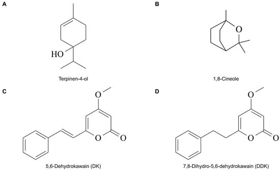
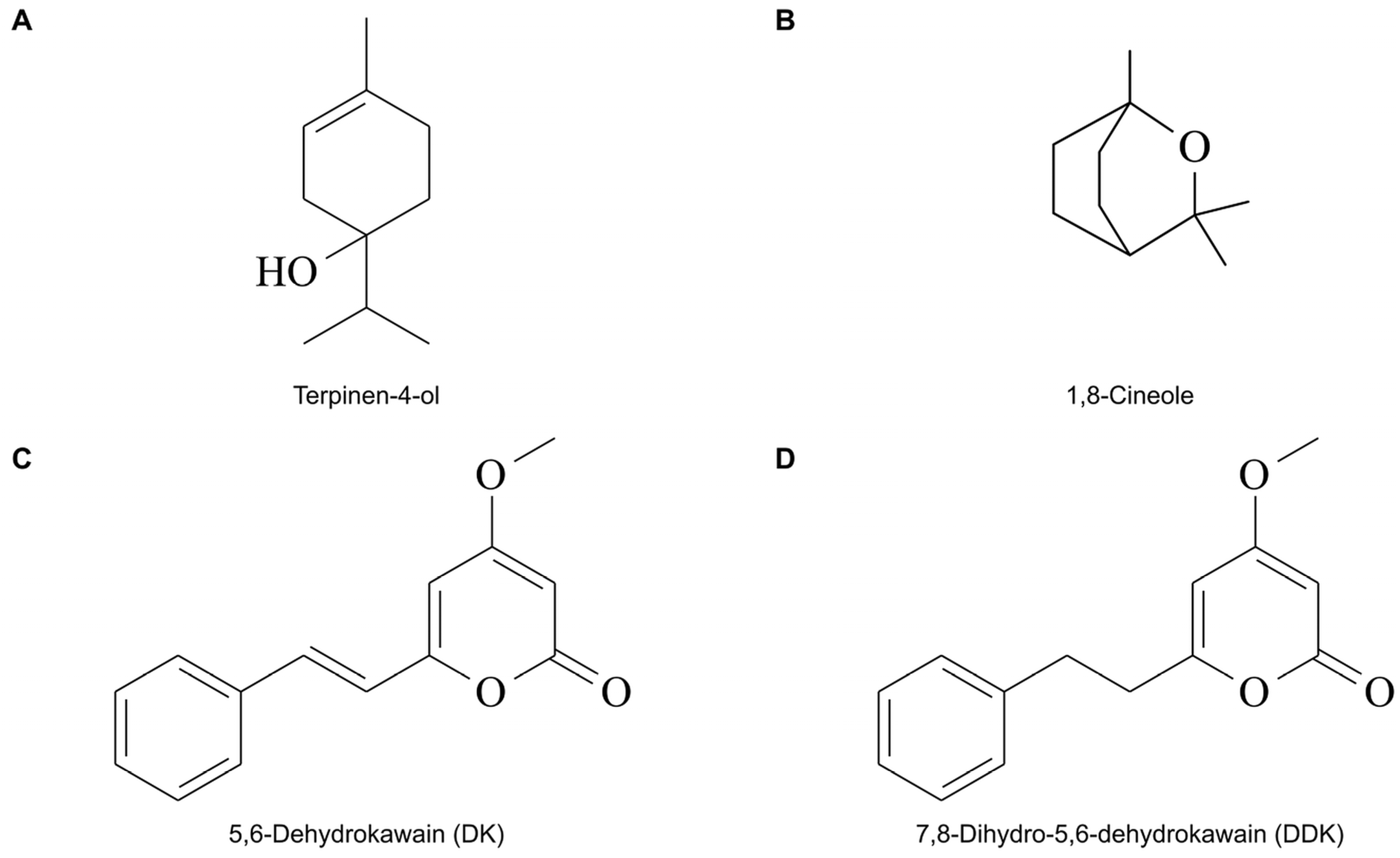
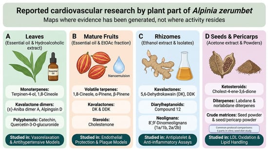
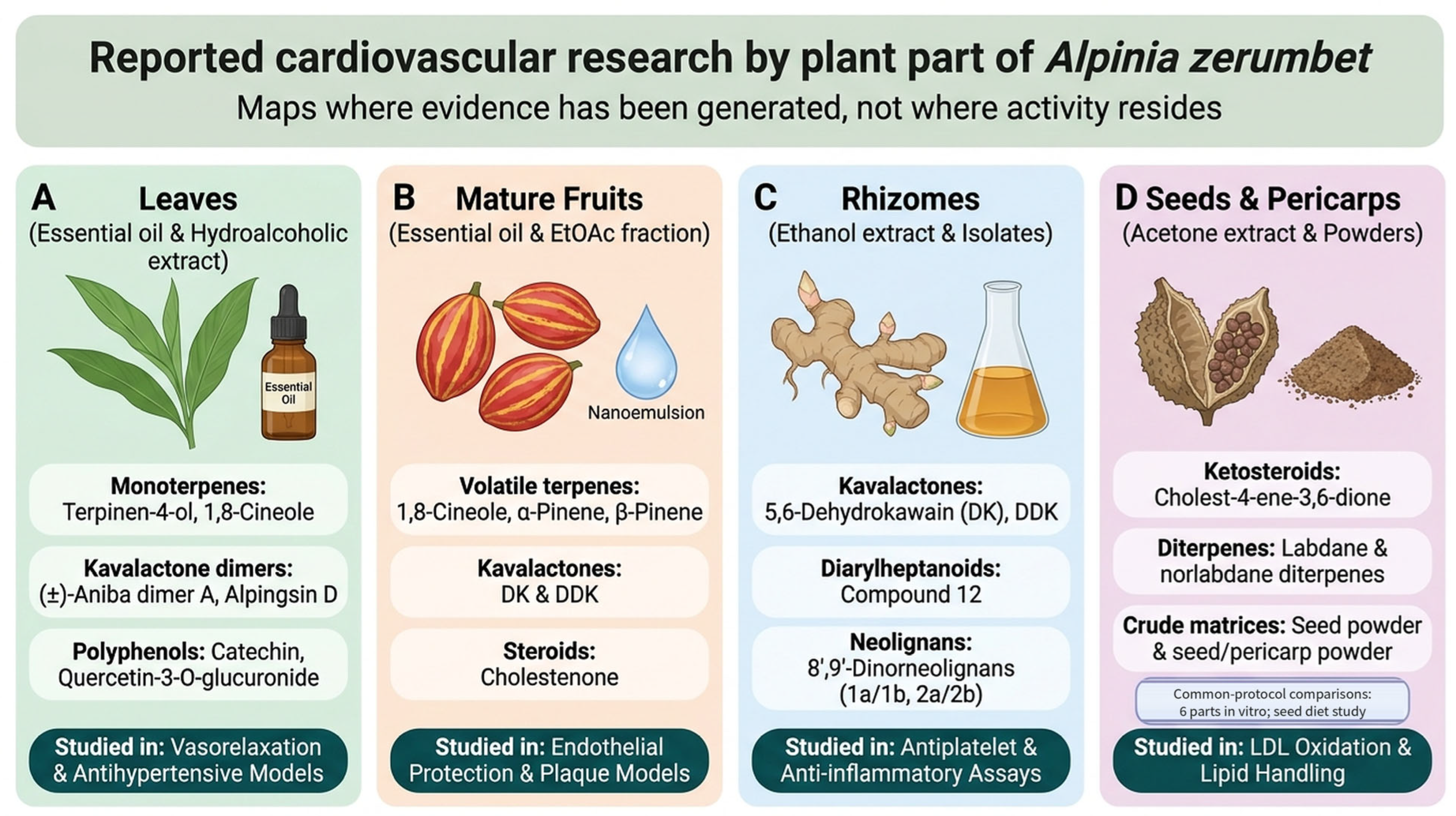
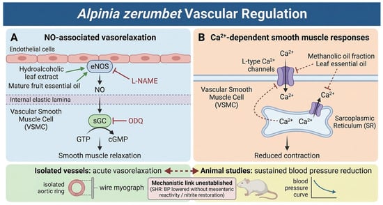
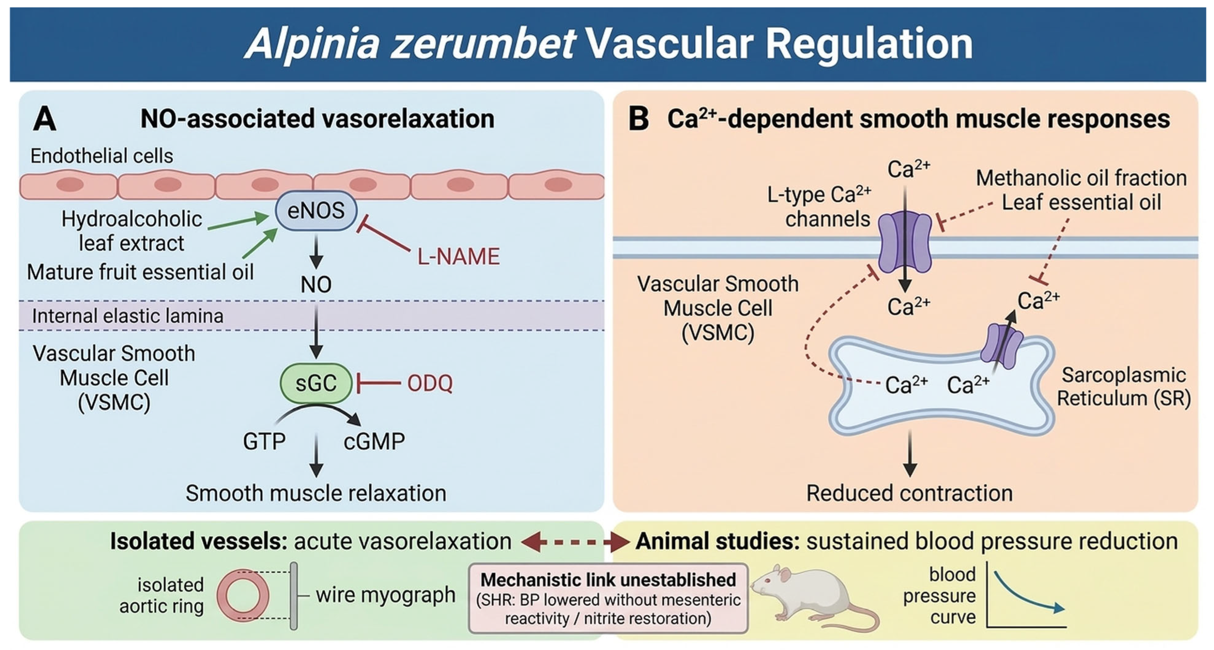
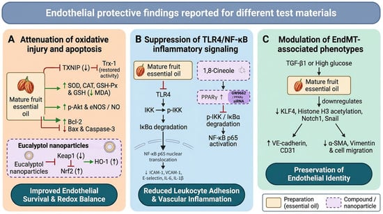
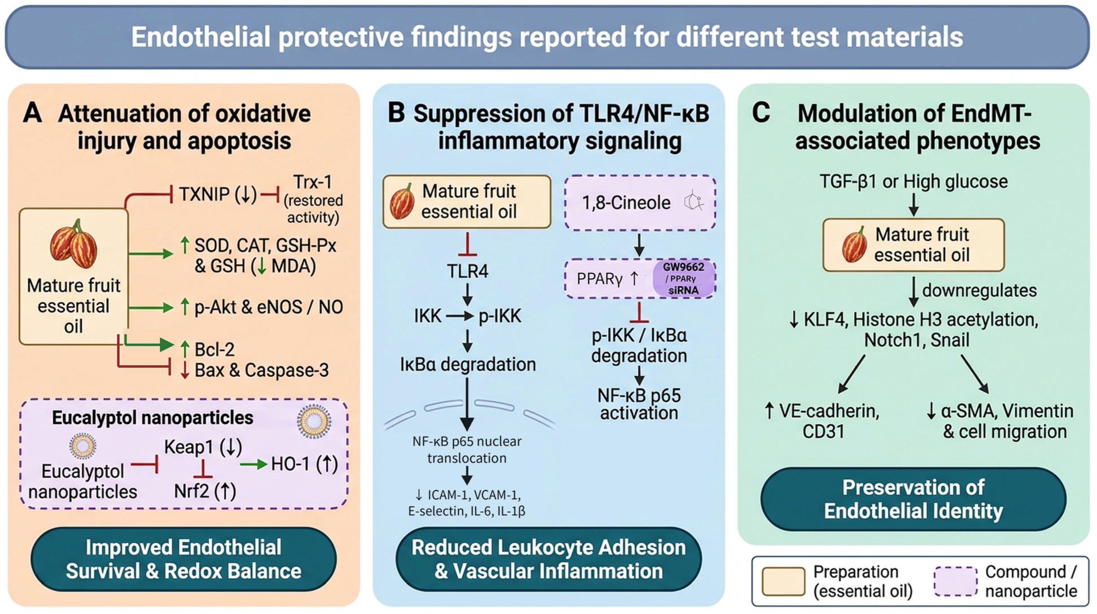
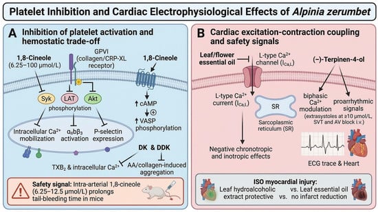
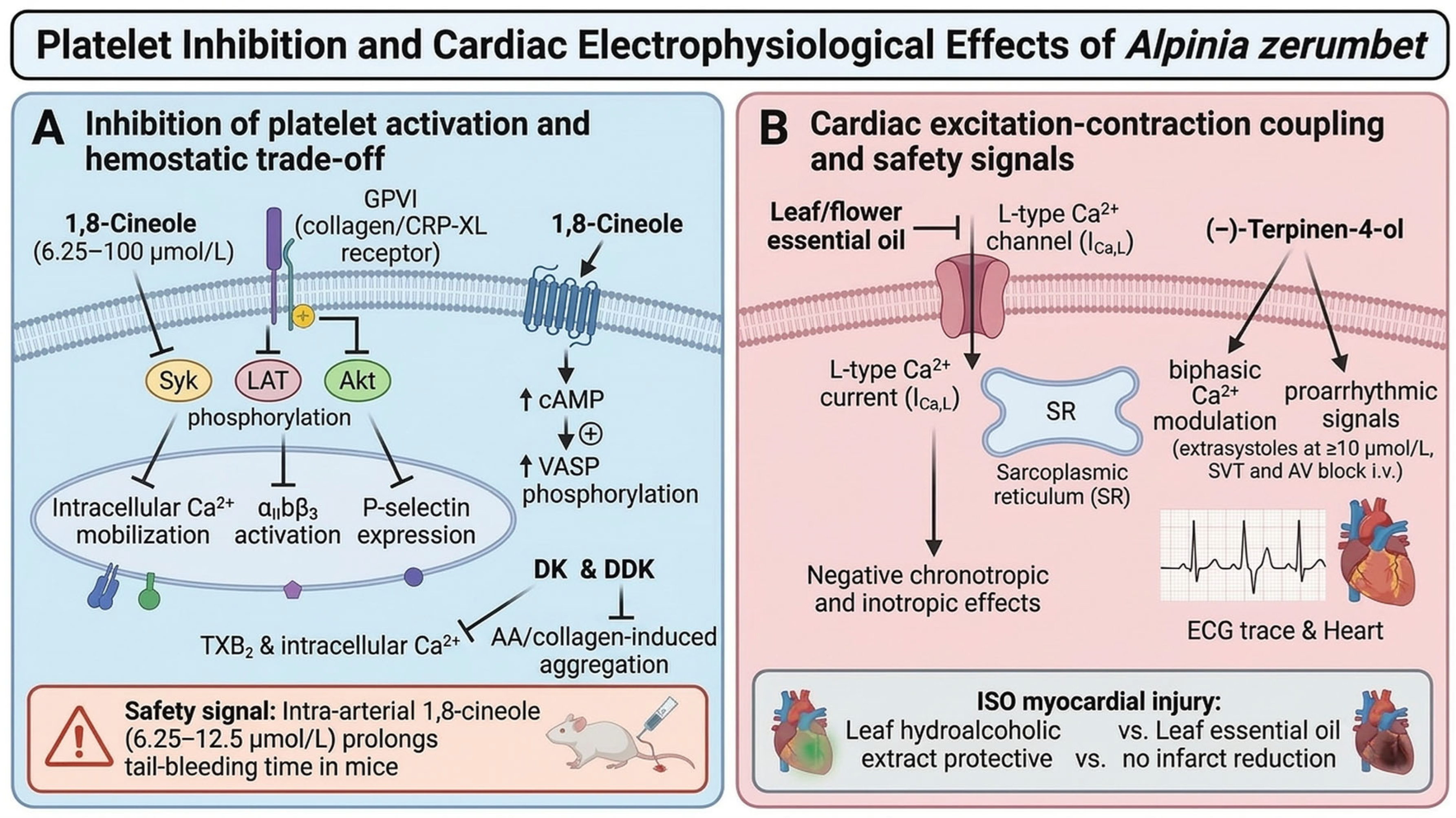
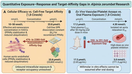
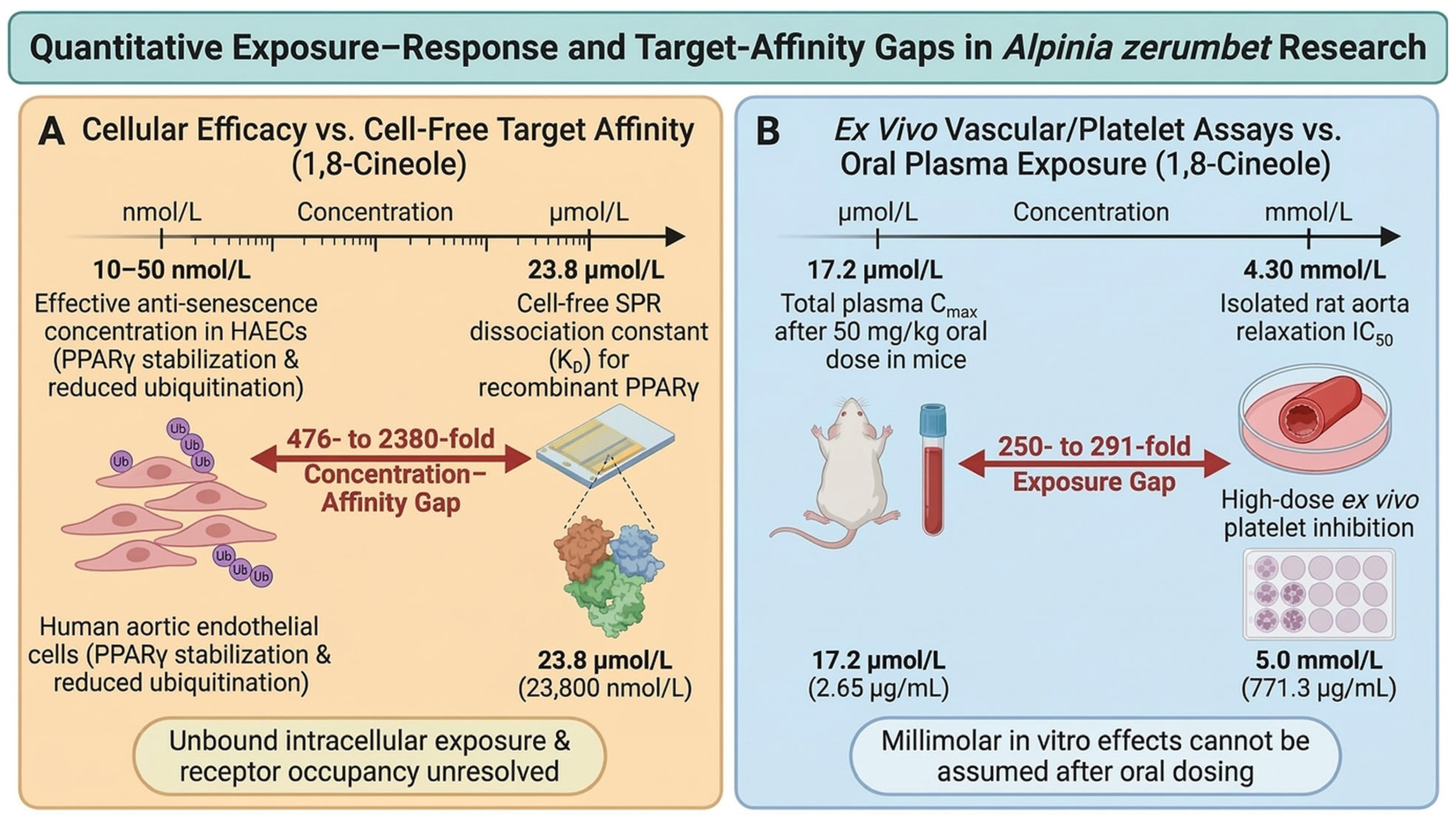

Disclaimer/Publisher’s Note: The statements, opinions and data contained in all publications are solely those of the individual author(s) and contributor(s) and not of MDPI and/or the editor(s). MDPI and/or the editor(s) disclaim responsibility for any injury to people or property resulting from any ideas, methods, instructions or products referred to in the content. |
© 2026 by the authors. Licensee MDPI, Basel, Switzerland. This article is an open access article distributed under the terms and conditions of the Creative Commons Attribution (CC BY) license.