


Abstract
Cancer cells employ multiple pro-survival strategies, including evasion of apoptosis and escape from immune surveillance. In particular, cancer cells can modulate intracellular calcium homeostasis and exploit sigma receptor (SR) activity to restore calcium balance. Furthermore, the positive correlation between sigma-1 receptor (S1R) and programmed death-ligand 1 (PD-L1) expression levels suggests that S1R may also contribute to immune evasion. Cancer cells maintain proteostasis through enhanced unfolded protein response (UPR) signaling and activation of the ubiquitin–proteasome system (UPS). The proteasome removes misfolded and ubiquitinated proteins, thereby preventing the accumulation of proteotoxic stress, endoplasmic reticulum (ER) stress, and cell death. Collectively, these adaptive mechanisms enable cancer cells to survive, proliferate, and invade, thereby promoting tumor progression. Although proteasome inhibitors (PIs) have been introduced into clinical practice for the treatment of multiple myeloma (MM), their therapeutic use is limited by several drawbacks, including the development of resistance and limited selectivity. Importantly, SRs play a pivotal role in UPR and UPS signaling; therefore, their modulation may compensate for the limitations associated with PI therapy. Consequently, a holistic strategy that simultaneously targets SRs, UPR signaling, and proteasome function may effectively disrupt key pro-survival pathways of cancer cells and represent a promising paradigm for the development of innovative anticancer therapeutics. In the present perspective, we discuss how such multi-target directed ligands (MTDLs) may offer the advantages of simultaneously hitting multiple key pathways and/or their pathogenic cross-talk with a single chemical entity.
1. Introduction
Cancer cells reside within a complex tumor microenvironment (TME) characterized by chronic endoplasmic reticulum (ER) stress, hypoxia, nutrient deprivation, oxidative stress, and an increased biosynthetic load [1,2,3]. Cellular protein homeostasis is maintained by two complementary quality-control systems: the unfolded protein response (UPR) and the ubiquitin–proteasome system (UPS) [3,4,5,6]. The UPR is regulated upstream by Binding Immunoglobulin Protein (BiP) and controls both adaptive responses and apoptosis, whereas the UPS functions as a clearance system that removes polyubiquitinated and misfolded proteins [7,8]. In cancer, both the UPR and the UPS are frequently altered to promote cell survival and stress tolerance, underscoring their potential as therapeutic targets [9,10,11,12,13]. On the other hand, the proteasome is a clinically validated target with three FDA-approved proteasome inhibitors (PIs) for multiple myeloma (MM): bortezomib, carfilzomib, and ixazomib [14,15]. However, PIs possess two main limitations: a lack of selectivity between the proteasome and its inducible isoform, the immunoproteasome, and the development of resistance [16,17,18,19]. The situation is even more critical in solid tumors. For instance, although marizomib was evaluated in a phase III clinical trial for glioblastoma (GBM), it failed to demonstrate any significant improvement in overall survival [20]. This failure is likely due to the fact that the survival pathways sustained by the UPR remain intact. Indeed, BiP upregulation confers resistance to bortezomib, whereas its downregulation restores bortezomib efficacy [21,22]. Consequently, targeting proteasomal degradation without inhibiting BiP-dependent machinery represents an incomplete therapeutic strategy, enabling cancer cells to survive. In this context, the sigma-1 receptor (S1R) plays a crucial role, as this ER chaperone directly interacts with BiP. S1R–BiP dissociation is triggered either by a reduction in ER Ca2+ levels or by S1R agonists. Once dissociated, S1R exerts a threefold effect: stabilizing inositol-requiring enzyme 1 (IRE1), facilitating ER-to-mitochondria Ca2+ transfer, and maintaining programmed death ligand-1 (PD-L1) expression, which consequently enhances immune evasion [23,24,25,26,27,28]. Notably, S1R is overexpressed in the ERs of cancer cells as an adaptive strategy for survival. Coherently, S1R knockdown and S1R antagonists are associated with reduced growth and survival of cancer cells [25,29,30,31]. Of note, the sigma-2 receptor (S2R) subtype has also been correlated with cancer: it is involved in cell proliferation, it is overexpressed in several tumor cell lines, and its agonists demonstrated anticancer activity [32,33,34,35,36,37,38,39,40,41].
Given this evidence, sigma receptor (SR) ligands may be combined with PIs within a single chemical entity to maximize their therapeutic efficacy. Indeed, S1R antagonism suppresses the adaptive response triggered by proteasome inhibition, effectively converting a proteotoxic insult from a pro-survival mechanism into lethal cellular damage. Furthermore, within a pan-SRs modulation, concurrent S2R agonism is expected to synergistically enhance the antitumor efficacy of the compounds. A first attempt to merge proteasome inhibitory activity with pan-SRs modulation within a single chemical entity has recently been undertaken by our group, leading to the identification of bivalent ligands with improved anticancer potency compared with the parent compounds.
Accordingly, this perspective is organized as follows. We first outline UPR signaling and the role of BiP as a druggable node in MM and GBM; then, we review the therapeutic potential and clinical limitations of proteasome inhibition in these two indications. We next examine the SR-BiP-UPR cascade and two axes through which SR and proteasome modulation intersect with tumor immunology/PD-L1-mediated immune evasion and the constitutive-to-immunoproteasome shift. Starting from these foundations, we propose the pharmacological rationale for dual SR–proteasome MTDLs and discuss RC-106 and its bivalent derivatives as novel chemotherapeutic agents. Successively, we addressed the medicinal chemistry challenges inherent in the structural and physicochemical requirements for engaging these two targets, including blood–brain barrier penetration for GBM. We conclude with a design roadmap and the preclinical criteria we consider necessary to test this hypothesis.
2. UPR Signaling in ER Stress and Cancer
The UPR is a network of intracellular signaling pathways that maintain the protein-folding capacity of the ER in eukaryotic cells [4,42]. UPR and ER-associated degradation (ERAD), which interacts with the UPS, represent the quality control system of misfolded proteins, preventing ER stress [4,43,44]. UPR activation is triggered by prolonged ER stress and is coordinated by three distinct ER-resident sensors: PKR-like ER kinase (PERK), inositol-requiring protein 1ɑ (IRE1ɑ), and activating transcription factor 6 (ATF6) [4,42,43]. Under physiological conditions, the three ER transmembrane stress sensors remain inactive, owing to BiP binding to their N-terminal luminal domains [6]. PERK signaling entails PERK-mediated phosphorylation of eukaryotic initiation factor 2α (eIF2α) and activation of NRF2, leading to upregulation of the CCAAT/enhancer-binding protein homologous protein (CHOP), a transcription factor that drives the apoptotic program [4,42]. IRE1α promotes apoptosis through dimerization and recruitment of TNF receptor-associated factor 2 (TRAF2), which activates ASK1 and JNK, thereby driving the apoptotic program [1]. Alternatively, IRE1 activates XBP, which in turn initiates the ERAD cascade through FBP, CULL, SKP1, Hsp40 and Hsp70 recruitment [4,45,46,47]. ATF6 is responsible for the ER-stress recovery program and activates BiP [46,47]. The TME is characterized by hypoxia, which alters ER redox homeostasis and triggers BiP dissociation, resulting in activation of PERK, ATF4, and IRE1α [7,47]. In cancer, UPR acts as a double-edged sword since it can inhibit tumor cell proliferation but also promote their survival, progression, and metastasis [45]. Cancer cells can exploit the UPR to evade chemotherapy through two complementary mechanisms: (i) induction of IRE1α- and PERK-mediated G0/G1 cell-cycle arrest and (ii) reduction in CHOP mRNA stability via PERK-dependent pathways involving ATF4, thereby escaping apoptosis [45,48]. Indeed, UPR activation has been associated with malignant progression and poor prognosis in different cancer types, including prostate and lung cancer and GBM [48,49,50,51].
3. Therapeutic Potential and Limitations of Proteasome Inhibition in Multiple Myeloma and Glioblastoma
The proteasome is responsible for the degradation of polyubiquitinated proteins and consists of two 19S regulatory particles and a 20S catalytic core. The 20S proteasome contains three catalytic sites named β5, β1, and β2, which confer chymotrypsin-like, caspase-like, and trypsin-like activities, respectively [15]. Proteins marked for degradation through polyubiquitination are recognized by the 19S regulatory particle, deubiquitinated and subsequently translocated into the 20S core, where they are degraded into peptide fragments [15]. Excessive accumulation of ubiquitinated proteins or exposure to pro-inflammatory signals triggers a shift from the constitutive proteasome toward the immunoproteasome [52]. Specifically, the immunoproteasome is constitutively expressed in cells of hematopoietic origin and can also be induced by cytokine stimulation in various tissues [53,54]. For this reason, immunoproteasome inhibition by anti-inflammatory drugs with consequent anticancer activity has been proposed [55]. PIs represent the gold standard for the treatment of MM, with three FDA-approved drugs: bortezomib, carfilzomib, and ixazomib (Table 1) [15].
Table 1.
Chemical structures of first- and second-generation proteasome inhibitors. Bortezomib is a reversible dipeptidyl boronic acid inhibitor of the 26S proteasome. It primarily targets the β5 chymotrypsin-like subunit, leading to impaired protein degradation, accumulation of misfolded proteins, and induction of apoptosis. It is clinically approved for the treatment of MM and MCL. Carfilzomib is an irreversible epoxyketone proteasome inhibitor with high selectivity for the β5 catalytic subunit, used for the treatment of relapsed or refractory MM. Marizomib is an irreversible β-lactone-γ-lactam proteasome inhibitor capable of blocking all three main catalytic activities of the 20S proteasome. Its broad inhibitory profile and ability to cross the blood–brain barrier make it a candidate for treating both systemic malignancies and brain tumors. Ixazomib is a reversible boronic acid-based proteasome inhibitor and the first orally bioavailable agent in this class. It predominantly inhibits the β5 activity of the proteasome and is approved for use in combination regimens for relapsed or refractory MM.
The efficacy of these drugs is up to the sensitivity of MM cells to inhibition of the 26S proteasome, since the UPR system is dysregulated in MM. Proteasome inhibition results in the suppression of NF-κB signaling, accumulation of misfolded and unfolded proteins, induction of ER stress, and ultimately, cell death in MM cells [15]. However, the FDA-approved drugs indiscriminately target both the constitutive proteasome and immunoproteasome, thus leading to a non-selective inhibition of protein degradation. This lack of specificity may account for the side effects and drug resistance [53,56]. Notably, proteotoxic stress and the resulting cell death are regulated not only by proteasomal activity but also by UPR-mediated survival pathways, in particular the IRE1α–XBP1 axis. Consequently, proteasome inhibition alone is frequently insufficient to elicit a robust antitumor response. In particular, following proteasome inhibition, misfolded and polyubiquitinated proteins within the ER lumen lead to BiP dissociation from the stress sensors PERK, IRE1α, and ATF6, leading to the activation of UPR [7,42]. This aspect is particularly relevant given that tumor cells frequently hyperactivate the UPR to evade apoptosis and sustain their elevated protein-synthetic burden [6,10]. BiP overexpression was associated with bortezomib resistance [21,57], highlighting the UPR as a compensatory proteostasis mechanism that sustains cancer cell survival under proteasome inhibition. Proteasome inhibitors have been investigated also for GBM treatment [20,58,59,60,61,62,63,64], showing several limitations. Specifically, carfilzomib determined cell viability, migration, and invasion reduction in-vitro [60], while marizomib showed efficacy in cell death induction both in-vitro and in-vivo [59]. Marizomib treatment was also proven to be responsible for ER-stress triggering and apoptosis induction in human GBM cells, with augmented expression levels of BiP, IRE1α, and ATF4 [64]. Preclinical studies also demonstrated that PIs potentiate temozolomide toxicity, reducing tumor volume in intracranial glioma models [65,66,67]. However, despite the favorable preclinical activity of marizomib observed in-vitro and in-vivo, clinical trials have failed to demonstrate a clinical benefit [20,68], whereas carfilzomib has not yet been evaluated in clinical trials for GBM. In a randomized phase III trial, the addition of marizomib to standard radiochemotherapy did not improve survival in newly diagnosed glioblastoma, and the accompanying analyses highlighted insufficient intratumoral target engagement and the absence of predictive biomarkers as plausible contributors. Rather than invalidating proteostasis as a target in GBM, these results delimit the conditions under which it may be exploitable: sustained and selective target engagement in the CNS and concomitant suppression of the adaptive chaperone responses that allow tumor cells to tolerate partial proteasome blockade. Both requirements motivate the multi-target approach discussed below. Combining PIs with other agents may also mitigate toxicity and overcome the limitations related to chemoresistance. However, it should be emphasized that MM and GBM do not pose the same development problem. In MM, proteasome inhibitors are approved, and the disease is systemic. In GBM, no proteasome inhibitor has improved survival, and any candidate must also reach the tumor behind the blood–brain barrier. The reason for considering both indications is a shared dependence on a high biosynthetic and proteostatic load, not an assumption that one molecule or one development path fits both.
4. BiP and the Adaptive Chaperone Response Limiting Proteasome Inhibition
At the apex of the UPR signaling cascade is BiP, an Hsp70 chaperone resident in the ER lumen that is encoded by the HSPA5 gene [47]. This protein contains two structural domains, the nucleotide-binding domain (NBD) and the substrate-binding domain (SBD), that work together to regulate BiP’s ATPase activity and its ability to bind substrates [47]. This interplay enables BiP to facilitate protein translocation into the ER, assist in the folding and retention of protein substrates within the ER lumen, initiate the UPR, and support ERAD5. Indeed, during ER stress, BiP dissociates from the three transducers PERK, IRE1ɑ, and ATF6, activating their respective UPR pathways [7,8]. The central role of BiP in regulating the UPR has been known for almost 25 years [47,69]. Interestingly, overexpression of BiP reduces UPR activation, while BiP inactivation determines ER stress and UPR activation, thus positioning BiP as a promising pharmacological target [8]. BiP is also responsible for binding misfolded or unassembled proteins, which mediates their retrograde translocation for proteasomal degradation [69]. Given this evidence, BiP plays a crucial role in maintaining ER homeostasis; moreover, because alterations in BiP activity have been associated with cancer, anti-BiP therapeutic strategies have been proposed [33]. Indeed, under basal conditions, BiP binds to the ER stress sensors IRE1α, PERK, and ATF6, maintaining them in an inactive state. Upon ER stress, BiP dissociates from these sensors, allowing their activation and the initiation of the UPR [70]. Consequently, BiP sequestration could interfere with the regulatory mechanisms governing UPR activation, potentially disrupting ER homeostasis and promoting cell death. Moreover, BiP has been associated with chemoresistance to cisplatin [71,72], doxorubicin [73], and bortezomib [57,74], and high expression of BiP is related to worse survival; conversely, reduced BiP expression is correlated with diffuse cell death in large B-cell lymphoma cell lines. BiP silencing also restored cell sensitivity to bortezomib [57]. Additionally, in 18-mantle-cell lymphoma (MCL) samples, a strong correlation between loss of sensitivity to bortezomib and up-regulation of the pro-survival chaperone BiP was demonstrated [21]. These findings align with the idea that disruption of the UPR pathway may constitute a selective strategy for leukemia therapy. Furthermore, the UPR system plays a critical role in GBM through the ubiquitin-conjugating enzyme E2T (UBE2T). UBE2T is a key participant in the ubiquitin–proteasome pathway and has been demonstrated to be significantly overexpressed in GBM [75]. Specifically, UBE2T promotes cell invasion and migration in the GBM cell lines LN229 and U25119 and binds to BiP, with which its expression has been shown to be positively correlated [75,76,77]. The central role of BiP in GBM was further confirmed by assessing the antitumor activity of BiP-targeting antibodies both in-vitro and in-vivo, supporting the potential of anti-BiP antibodies as a promising therapeutic strategy for GBM, either alone or in combination with radiotherapy [77]. Moreover, BiP silencing has been associated with reduced levels of the stemness-associated protein CD44 in GBM cells [78]. Given this evidence, BiP and the UPR system represent an innovative target for GBM treatment. BiP therefore emerges as the node whose activity determines whether proteasome inhibition is tolerated or lethal [73]. BiP itself has proven difficult to drug selectively, whereas its functional partner at the ER membrane, the S1R, is a well-characterized small-molecule target [23,24,27,31,79,80,81,82,83,84,85,86,87].
5. The Cross-Talk Between Proteasome–Sigma Receptors–UPR as Novel Target for Anticancer Therapy
Sigma receptors (SRs) exist in two subtypes, named sigma-1 (S1R) and sigma-2 (S2R), and are involved in several biological processes. Specifically, the two proteins are grouped together pharmacologically as SRs but are otherwise unrelated at the molecular level: S1R is a ligand-operated endoplasmic reticulum chaperone encoded by SIGMAR1, whereas S2R is an integral membrane protein encoded by TMEM97 [82]. S1Rs are expressed in the central nervous system, and their agonist or positive modulators are useful in the treatment of neurodegeneration, while S1R antagonists are promising for cancer treatment [79,80,81,82,83,84,85,86,87]. Indeed, pharmacological modulation of S1R in cancer cells has been correlated with the alteration of protein synthesis, post-translational modification, and degradation of cancer-promoting proteins [82]. Since resistance to proteasome inhibitors can be reversed by BiP silencing [21,57], an innovative therapeutic agent for MM and GBM should ideally exert anti-BiP activity. In this context, concomitant suppression of the UPR could potentiate proteasome inhibition by promoting the accumulation of misfolded proteins, ultimately leading to irreversible ER stress and apoptosis in malignant cells. BiP is activated by ATF4 [7], which is interestingly responsible for S1R upregulation, which in turn is correlated with BiP activity [88]. Specifically, S1R is also a ligand-operated chaperone that forms a complex with BiP, and S1R agonists can dissociate S1R from BiP, thereby promoting the activity of both proteins [79,89]. S1R is overexpressed in the ER of most cancer cells, and its knockdown or inhibition compromises cancer cell growth, proliferation, and survival [27,90]. In this context, S1R may play a protective role in cancer cell physiology, and S1R modulators can be used to control the biomass of cancer cells by regulating the UPR system [82]. In particular, S1R antagonists determined the upregulation of UPR-related markers PERK, ATF4, and XBP1 [90]. Notably, S1Rs reside at the mitochondria-associated membranes (MAMs), a key site for protein folding and synthesis. Upon the accumulation of misfolded proteins, ER stress is induced, leading to IRE1α dimerization and autophosphorylation, which activate its endoribonuclease activity. This results in the splicing and activation of XBP1 mRNA, thereby promoting the expression of several ER chaperones and components of the ERAD pathway [27]. In this context, S1R was demonstrated to stabilize IRE1ɑ, but not PERK and ATF6, promoting cellular survival by sustaining the IRE1ɑ-XBP1 axis [27]. Indeed, S1R protects IRE1α from proteasomal degradation, as S1R knockdown in stressed cells markedly reduces the half-life of IRE1α [82,89]. This effect is associated with the specific localization of S1Rs at the MAMs, where they are also involved in the regulation of Ca2+ signaling [24,91]. Upon ER Ca2+ depletion and the consequent induction of ER stress, S1R dissociates from BiP and associates with inositol 1,4,5-trisphosphate receptor type 3 (IP3R3), where it stabilizes the receptor and modulates ER-to-mitochondrial Ca2+ transfer [24]. Ca2+ balance is a critical point for cellular survival, and S1R can promote cellular resilience by altering cell electrical plasticity in a cancerous environment [24,81]. S1R also regulates mitochondrial homeostasis and interacts with proteins localized to mitochondria, including Rac1, a small GTPase involved in actin cytoskeletal dynamics, cell migration and proliferation, gene transcription, and ROS production [92]. Notably, ER Ca2+ burst determines mitochondrial Ca2+ overload, thus promoting Cytochrome C release and ultimately apoptosis [93]. Considering all this evidence, S1R roles in Ca2+ regulation were further investigated, demonstrating that rapid Ca2+ decrease induced by the ER stressor Thapsigargin induces rapid dissociation of the BiP-S1R complex, confirming the strong crosstalk between S1R and Ca2+ modulation [81]. Moreover, under conditions of chronic ER stress, S1R can translocate from MAMs to other ER compartments, where it plays a critical role in limiting the accumulation of misfolded proteins and thereby attenuating UPR activation [79,81]. In particular, under physiological Ca2+ conditions, the S1R–BiP complex remains in a quiescent state, whereas a reduction in ER Ca2+ levels promotes rapid dissociation of the complex, thereby activating S1R chaperone function and contributing to the initiation of the UPR cascade [79,81].
6. SRs-Ligands
As recapitulated by Abatematteo et al. [94], several S1R ligands have been synthesized in recent years to further investigate the role of this receptor in several pathologies. PB-190 and PRE-084 were identified as selective S1R agonists possessing neuroprotective and antidepressant activities [95,96], whereas FTC-146 has been widely utilized in positron emission tomography (PET) imaging across several animal models and in humans [97]. Conversely, S1R antagonists have been developed for the treatment of neuropathic pain. Notably, RC-752 demonstrated robust in-vivo efficacy [98], and S1RA, an S1R antagonist developed by Esteve, successfully completed Phase I clinical trials [99]. However, selective S1R antagonists also showed efficacy as anticancer agents in-vitro, such as rimcazole and IPAG [84]. On the other hand, the first highly selective S2R ligand, reported in 1995 as CB-184, paved the way for the discovery of diverse chemical classes of S2R ligands exhibiting cytotoxicity across different cell lines [84]. However, despite this compelling biology, no SR ligand has advanced as an anticancer agent. In general, several SR ligands have entered clinical trials for the treatment of various conditions, including neurodegenerative diseases, psychiatric disorders, and neuropathic pain, whereas clinical development in oncology has been far more limited (Table 2) [100].
Table 2.
Overview of anticancer SR ligands reported in the literature. S2R agonists demonstrated anticancer activity in brain and pancreatic cancers, whereas the S1R antagonist rimcazole was effective in breast and colon cancer models. Furthermore, pan-sigma modulators exhibited efficacy against glioblastoma, pancreatic, and renal cancers [37,101,102,103,104,105,106,107,108,109].
Specifically, SR31747A, a peripheral sigma ligand with documented antiproliferative activity in vitro and in xenograft models, remains the only SR ligand to have reached a randomized phase II trial in oncology [110,111]. However, interest in SR ligands has expanded significantly across various therapeutic areas, as reflected in submitted patents [112,113,114,115,116], as well as in oncology, with key patents filed in 2001 and 2014 [117,118]. Furthermore, a patent filed in 2015 disclosed the use of SRs ligands for modulating protein homeostasis [119].
Interestingly, the similarities between S1R and S2R binding sites may offer a strategic advantage for designing molecules with mixed affinity, thus leveraging the potential synergistic effects of simultaneous modulation of both targets. In fact, despite the high structural variability of known SR ligands, their binding to both receptor subtypes is mainly driven by an ionic interaction between a protonated amine and an acidic residue (Glu172 for S1R and Asp29 for S2R) and interactions with surrounding hydrophobic residues [120,121,122].
7. S1R Modulation as an Innovative Strategy to Overcome Immune Evasion
At the same time, S1R upregulation determines an increase in Programmed Death-ligand 1 (PD-L1) level [26]. Specifically, PD-L1 is a membrane glycoprotein that can be expressed on the surface of cells and binds to programmed death-1 (PD-1) receptors [123,124]. PD-L1 is an inhibitory checkpoint molecule that suppresses T-cell activation and proliferation [123,124,125]. PD-1/PD-L1 axis normally functions to regulate the host immune response, protecting against autoimmunity and limiting excessive tissue damage at sites of inflammation [124]. However, PD-L1 is also pivotal for cancer cells’ immuno-escape, thus promoting tumor growth [126,127,128]. Specifically, PD-L1 expressed on the surface of cancer cells binds to PD-1 on tumor-infiltrating cytotoxic T-lymphocytes (CTLs), leading to their functional inactivation and consequent reduction in the host antitumor immune response [123,124]. Therefore, the PD-1/PD-L1 axis acts as a double-edged sword: under physiological conditions it is pivotal in preventing autoimmunity, whereas in cancer it becomes the master regulator of immune evasion through the very same mechanisms. This duality reflects a single mechanism, since PD-1/PD-L1 engagement limits T-cell activation, which restrains autoimmunity under physiological conditions yet contributes to tumor immune escape in the cancer context. Consequently, blockade of the PD-1/PD-L1 axis represents a powerful tool to counteract immune evasion. It has already been demonstrated that S1R physically binds PD-L1 [25]. In this context, S1R antagonists could represent a powerful modulator of the immunohub in TME. Specifically, the S1R-selective agonist SA4503 led to pronounced intracellular accumulation of PD-L1 and blocked the reduction in PD-L1 protein levels induced by IPAG (a selective S1R antagonist) [25]. Moreover, IL-10 secretion is enhanced by both PD-L1 and S1R agonists and correlates with tumor growth in-vivo, whereas S1R antagonists suppress IL-10 production [23]. This may be attributable to a vicious cycle between PD-L1 and IL-10, whereby PD-L1 enhances IL-10 secretion, which, in turn, promotes PD-L1 cell-surface expression [23,25]. Since S1R agonist-induced antitumor immunity is blocked, partly by IL-10 induction [23], S1R-mediated regulation of PD-L1 may contribute to antitumor immunity by enhancing immune checkpoint inhibition while concurrently suppressing the production of anti-inflammatory cytokines [25,123,129,130]. Thus, selective S1R antagonists may represent a promising strategy for modulating the tumor immune microenvironment. Indeed, in cells characterized by high PD-L1 expression, treatment with IPAG in the presence of interferon-γ (IFN-γ) reduced both PD-L1 protein levels and its cell-surface expression [123]. Therefore, given the complexity of the TME, the involvement of S1R in PD-L1 regulation and its impact on antitumor immune responses warrant further investigation, highlighting the potential of S1R as a therapeutic target. This pathway, along with the other consequences of S1R activation and BiP dissociation, is recapitulated in Figure 1.
Figure 1.
Schematic illustration of ER-induced S1R–BiP dissociation and its downstream biological effects. Solid arrows represent activation of downstream process, while T-bars represent inhibitory interactions. Under ER stress, the S1R-BIP complex rapidly dissociates, allowing S1R and BiP to exert their distinct biological activities. S1R binds IP3R3, modulating Ca2+ signaling and promoting cellular survival and increases BiP expression, thus enhancing UPR activity. Moreover, S1R promotes cellular survival via IRE1ɑ stabilization and IRE1ɑ-XBP axis sustainment. Finally, S1R stimulates PD-L1 expression, thus suppressing T-cell infiltration, in order to limit inflammation-derived damage. This latter effect is detrimental in anticancer immunity. On the other hand, BiP activates UPR machinery by dissociating from the three ER-stress sensors PERK, IRE1ɑ and ATF6, thus promoting damaged cells apoptosis. Moreover, BiP is responsible for retrograde transport of misfolded proteins to the proteasome, and BiP upregulation results in enhanced proteasome activity.
8. Proteasome–Immunoproteasome Shift as Innovative Mechanism for Anticancer Activity
The immunoproteasome (IP) is the inflammation-induced form of proteasome, but it is constitutively expressed in hematopoietic cells [52,131]. Under non-inflammatory conditions, the IP represents only a small subset of the total proteasome pool, but it is efficiently induced by pro-inflammatory cytokines and by misfolded protein accumulation [52]. Specifically, IP is comprised of three proteolytically active subunits, β2i (MECL-1), β1i (LMP2), and β5i (LMP7), and their activity leads to the formation of peptides for major histocompatibility complex (MHC) class I presentation [131,132,133]. Apart from antigen presentation, IP is involved in protein degradation and T-cell regulation also [133,134]. Interestingly, IP expression levels differ among various cancer types and counteract tumor development [132]. In particular, IP is induced by IFN-γ and other cytokines, such as TNFɑ [135], and stimulates a rapid immune response; indeed, IP inhibition leads to reduced CTL maturation and activity [132,134,136]. Moreover, IP-induced antigen presentation promotes immune-mediated cancer cell destruction and enhances the efficacy of currently available immunotherapies [53,133,134]. IP inhibition also resulted in NLRP3 inflammasome response attenuation, with consequent cytokine secretion blockage [135]. Interestingly, under physiological conditions, the proteasome mediates the degradation of misfolded and polyubiquitinated proteins without requiring IP support, whereas under stress conditions, resulting from the accumulation of misfolded proteins or inflammatory processes, IP becomes essential for maintaining protein homeostasis [131,137]. Indeed, during inflammation, cytokines can impair proteasomal degradation capacity, resulting in the accumulation of misfolded and polyubiquitinated proteins and a subsequent induction of IP [137]. In this context, IP appears to be more efficient than the constitutive proteasome at degrading misfolded proteins, whereas IP-deficient cells are unable to efficiently clear misfolded proteins [137]. On the other hand, IP activity is also involved in cytokine release, thereby shaping the TME and influencing the balance of antitumor immune responses (Figure 2). This pleiotropic activity should be carefully considered in the development of novel IP-targeting molecules [14,17,132].
Figure 2.
Schematic illustration of IP’s double-edged role in cancer cells. Solid arrows represent downstream activation processes, while T-bars represent inhibitory interactions. IP inhibition results in attenuation of inflammation but also leads to reduced CTL maturation and activity, as well as impaired antigen presentation. On the other hand, IP inhibition reduces cytokine release, leading to attenuation of pre-existing inflammation within the TME. Conversely, IP activation enhances the antitumor immune response and cytokine production, resulting in impaired proteasome activity, while simultaneously aggravating inflammation within the TME.
Due to the limitations related to side effects and the development of drug resistance to PIs, IP has also been investigated for the development of innovative anticancer agents. In particular, the development of β5i inhibitors has gained increasing attention as a strategy to generate small molecules with improved efficacy and reduced toxicity [16,136,138]. As described by Kioon et al., DPLG3, a selective β5i inhibitor, attenuates plasmacytoid dendritic cell (pDC) responses to Toll-like receptor 9 (TLR9) stimulation and inhibits T-cell proliferation [136]. Moreover, IP inhibitors UK-101 and IPSI-001 have shown promising anticancer activity. Specifically, UK-101 induced cell death in prostate cancer cells, whereas IPSI-001 triggered apoptosis in hematological malignancies characterized by high IP expression [14,139,140]. ONX-0914, a selective IP-inhibitor, diminished pro-inflammatory cytokine production, such as TNFɑ, and induced cell death in MM cells [17,141]. However, given the critical role of IP in regulating immune responses, IP inhibition may impair T-cell activity, thereby potentially increasing the ability of cancer cells to evade immune surveillance. For this reason, IP activators could be considered as an alternative; however, data supporting their use in the literature remain scarce. Since IP can be induced by cytokines and the accumulation of misfolded proteins, a promising therapeutic strategy may be to combine proteasome inhibition with anti-inflammatory agents. Indeed, currently available anti-inflammatory drugs have already emerged as promising moieties for innovative anticancer molecules [55,142]. Interestingly, ibuprofen reduced β5i levels in male mouse models with concomitant proteasome inhibition [142]. Dihydroartemisinin, an antimalarial drug that emerged as a potent antitumor agent, was more potent when combined with loxoprofen [55], confirming the potential of non-steroidal anti-inflammatory drug (NSAID) introduction in innovative anticancer molecules.
9. Multi-Target Directed Ligands: Pharmacological Rationale
Since BiP silencing has been associated with the reduction in bortezomib resistance, and since S1R agonists have shown efficacy in releasing S1R from BiP [88,89], it is plausible to hypothesize that S1R ligands could perturb BiP function by blocking the dissociation of the BiP-S1R complex. This, in turn, may reduce BiP-mediated retrotranslocation of misfolded proteins to the proteasome. S1R antagonists have already demonstrated efficacy in inhibiting BiP-S1R dissociation [143]. Failure of complex dissociation profoundly affects cellular homeostasis. First, the inability of S1R to interact with and stabilize IRE1α compromises the pro-survival IRE1α–XBP1 axis, resulting in impaired ERAD activity and, ultimately, cell death [27,144]. In addition, the loss of S1R–IP3R3 interaction perturbs calcium homeostasis, promoting Cytochrome C release from mitochondria and triggering apoptotic signaling [24,81,93]. On the other hand, the impossibility for BiP to activate UPR machinery may reinforce the effect of PI, leading to misfolded protein accumulation and ultimately to cell death. Moreover, S1R antagonist activity also modulates antitumor immune responses by increasing the susceptibility of cancer cells to T-cell-mediated cytotoxicity [25,123]. S1R antagonist- and PI-mediated proteasome inhibition may promote a shift toward IP activity, which enhances CTL maturation and activity [132,133,134,136,145]. However, increased IP activity may also stimulate cytokine release [132,133], thereby promoting inflammation and potentially limiting the therapeutic benefit of this approach. For this reason, the development of a hybrid S1R antagonist–PI–anti-inflammatory molecule, incorporating commercially available drugs as the anti-inflammatory moiety, could potentially represent a novel therapeutic approach, although further investigation is required to validate its therapeutic relevance. Moreover, the effect of this innovative multitarget molecule could be reinforced by the presence of an S2R agonism profile. Specifically, S2R resides in the ER, and S2R agonists have already demonstrated anticancer activity [31,38,103,109].
10. A Case Study: From RC-106 to Dual SR–Proteasome MTDLs
The rationale outlined above led our interdisciplinary team to consider whether a single chemical entity can engage both SRs and the proteasome to produce an enhanced anticancer effect. To probe this hypothesis, we employed a molecular hybridization approach, which we discuss herein. Two compounds provided the starting points: RC-106 and IP24. RC-106 is a pan-SRs modulator that combines S1R antagonism with S2R agonism, induces terminal UPR, promotes apoptosis in cancer cell lines (including GBM and MM), and has the ability to reach the CNS [31,85,86]. Importantly, although RC-106 demonstrates that S1R antagonism and S2R agonism can be combined within a single molecular scaffold, it does not provide evidence of pharmacological synergy between these two activities. Moreover, CNS exposure has been reported for RC-106, but this does not extend to the bivalent derivatives, which are structurally distinct and of substantially higher molecular weight. IP24 displayed dose-dependent proteasome inhibition and matching cytotoxicity in the RPMI 8226 human MM cell line [146].
Based on this evidence, Rossino et al. recently reported a series of bivalent ligands in which the RC-106 scaffold was linked to the IP24 moiety. Specifically, the two progenitor scaffolds were conjugated at distinct attachment sites through PEG linkers of varying lengths, thereby enabling a preliminary assessment of the impact of interpharmacophore distance on the biological response (Figure 3).
Figure 3.
Chemical structures of RC-106 and IP24, and schematic representation of the putative mechanism of action of the derived bivalent compounds in cancer cells. (adapted from Rossino et al. [147]).
The four resulting bivalent prototypes (15a, 15b, 16a and 16b) were subsequently evaluated in two human GBM cell lines displaying distinct TMZ-response profiles: the TMZ-sensitive U87-MG and the TMZ-resistant T98G cell lines [148,149]. These MTDLs exhibited markedly enhanced cytotoxicity compared with the progenitor compounds RC-106 and IP24. In T98G and U87-MG cells, RC-106 displayed IC50 values at 24 h of 60.27 and 57.75, respectively, whereas the four bivalent compounds showed substantially lower IC50 values, ranging from 6.16 to 12.47 in the same cell lines, with compound 16a identified as the most active. Moreover, all four bivalent compounds inhibited proteasome activity in both T98G and U87-MG cells. In T98G cells, proteasome inhibition was observed for compounds 15a and 15b at 20 μM, 16b at 40 μM, and 16a at 10 μM, confirming the latter as the most potent among the four bivalent compounds. In U87-MG cells, all four bivalent compounds inhibited proteasome activity at 40 μM. Importantly, receptor-binding studies demonstrated that all four bivalent compounds retained affinity for Sigma receptors, although their binding profiles differed depending on the attachment site on the RC-106 scaffold, which influenced their relative preference for S1R and S2R (Table 3). These findings indicate that molecular hybridization did not abolish the ability of the RC-106-derived moiety to recognize Sigma receptors, although the functional consequences of receptor binding remain to be established.
Table 3.
Comparative overview of the pharmacological properties of parent compounds and their corresponding bivalent derivatives. The table summarizes the receptor-binding data, antiproliferative activity, and proteasome inhibition activity of the individual parent compounds and their bivalent derivatives [109,146,147].
Additional insights were provided by holotomographic live-cell imaging of T98G cells treated with 16a, which induced pronounced morphological changes consistent with proteotoxic stress. In fact, T98G cells displayed an accumulation of misfolded protein, represented by the accumulation of electron-dense aggregates, followed by the appearance of extensive cytoplasmic vacuolization. These morphological changes are compatible with a paraptosis-like phenotype, which cannot be confirmed by morphology alone [150,151,152].
Overall, these findings provide preliminary support for the feasibility of combining Sigma receptor-binding and proteasome-impairing properties within a single molecular framework. However, they do not conclusively establish a dual mechanism of action or synergistic target modulation, and further investigations, including functional receptor assays and target-specific mechanistic studies, are needed to clarify the contribution of each pharmacological component to the observed anticancer activity. Although the high molecular weight and physicochemical properties of these four bivalent compounds currently limit their potential as drug candidates, particularly in terms of oral bioavailability and CNS exposure, they provide a valuable starting point for further research and optimization, supporting the continued investigation of Sigma receptor modulation and proteasome impairment as a potential multitarget strategy in cancer therapy [147].
11. Medicinal Chemistry Challenge
From a medicinal chemistry perspective, the development of MTDLs capable of modulating both SRs and the proteasome remains a significant challenge.
Beyond achieving dual target engagement, these compounds must reconcile the markedly different structural, physicochemical, and pharmacological requirements associated with each target. SR ligands are typically lipophilic small molecules whose receptor recognition is governed by an ionic interaction between a protonated amine of the ligand and an acidic residue of the binding site, surrounded by hydrophobic residues [120,121,122]. On the other hand, clinically approved PIs are generally peptide-derived molecules bearing electrophilic warheads that form a covalent bond (either reversible or not) with the N-terminal Thr1 of the catalytically active β subunits, with the β-5 chymotrypsin-like subunit being the primary target. Due to their chemical structure, PIs often display limited membrane permeability and suboptimal central nervous system (CNS) exposure [63,153,154,155].
Consequently, balancing potency, selectivity, target engagement, and drug-like properties within a single molecular entity to target both SRs and the proteasome remains a major challenge. Moreover, the distinct binding modes required for SRs and the proteasome further complicate ligand design, demanding careful optimization of the molecular framework to preserve activity at both targets. Particular attention should also be devoted to the ADMET profile during lead optimization, as the intended therapeutic indication may impose additional constraints on physicochemical properties. For example, the development of compounds for GBM requires efficient penetration of the blood–brain barrier (BBB), a feature that may be difficult to reconcile with the physicochemical characteristics commonly associated with proteasome inhibitors. Indeed, the four prototypes described in the previous paragraph (15a–b, 16a–b) are characterized by high molecular weight (824–868 Da), which limits their drug-likeness, particularly for brain tumor applications. Indeed, the molecular weight of the present bivalent compounds is a limitation for BBB penetration, intratumoral exposure and pharmacokinetics, and the CNS exposure of these derivatives has not been shown. Nevertheless, these molecules serve as pharmacological tools to assess the viability of combining SR modulation and proteasome impairment.
RC-106 has demonstrated antiproliferative activity across a panel of human cancer cell lines and the ability to reach the CNS, supporting the feasibility of simultaneously targeting these two pathways within a single chemical scaffold. Future medicinal chemistry efforts should focus on expanding the chemical space of dual SR–proteasome ligands while optimizing the spatial arrangement of the pharmacophores, linker architecture, and target engagement at both proteins. One promising direction could involve incorporating electrophilic warheads capable of reversible covalent interaction with the proteasome, such as boronate moieties, while preserving the structural features required for SR modulation. Alternatively, the integration of pharmacophoric elements derived from non-covalent proteasome inhibitors may offer a strategy to fine-tune the hydrophilic–lipophilic balance and improve membrane permeability and BBB penetration. Moreover, before making any therapeutic claims, several additional studies are required. These should include molecular weight reduction, achieved through the use of shorter linkers or by merging rather than linking the pharmacophores; ADME profiling, including an assessment of BBB penetration; toxicity testing in non-malignant cells, which is particularly important given the ubiquitous expression and essential functions of both the UPR and the UPS system; and direct evidence of target engagement and selectivity.
Collectively, these approaches may enable the rational design of more potent and drug-like MTDLs, ultimately advancing the development of innovative anticancer agents capable of simultaneously targeting proteostasis and sigma receptor signaling.
12. Conclusions and Outlook
Given the strict relationship between SRs and the proteasome in regulating cell defense systems UPR and UPS, targeting this axis with MTDLs may represent an innovative strategy to fight cancer. Indeed, combining proteasome inhibition with SR modulation could exert multimodal antitumor effects by simultaneously targeting proteostasis and tumor immune evasion. Notably, proteasome inhibition induced by MTDLs may promote IP induction and upregulation, thereby enhancing CTL activity against cancer cells. This effect could synergize with reduced PD-L1 expression to facilitate CTL infiltration, strengthen the antitumor immune response, and ultimately promote cancer cell elimination. However, given that IP induction can promote inflammation through increased cytokine secretion, incorporation of an NSAID moiety could counteract the inflammatory consequences of IP hyperactivation while preserving its beneficial immunomodulatory effects.
Notably, single-agent therapies targeting either proteasome activity or SRs in MM and GBM have encountered major clinical bottlenecks; nevertheless, these modalities seem to exhibit complementary mechanisms of action, wherein each strategy effectively offsets the therapeutic limitations of the other. In particular, S1R antagonists prevent dissociation of the S1R–BiP complex, thereby potentially abrogating UPR activation. Moreover, when BiP remains bound to S1R, the BiP–IRE1α axis is disrupted, resulting in impaired ERAD induction. Under these conditions, however, proteasomal degradation can still be sustained by the UPS. Conversely, PIs block UPS activity, but cells can compensate through an adaptive UPR response. Given this evidence, a viable strategy to overcome the limitations of every single approach is their combination, aimed at simultaneously inhibiting both UPR and UPS. This dual blockade may lead to accumulation of misfolded proteins, overwhelming ER stress, and ultimately cancer cell death, as schematized in Figure 4.
Figure 4.
Proposed mechanism of action of MTDLs in MM and GBM. Hypothesis-driven MTDL pharmacological targeting is represented by dashed arrows; evidence-based downstream pathways are represented by T-bars (inhibitory interactions) and solid arrows (activation of downstream cascade process). S1R antagonists may act by interfering with S1R-BiP complex dissociation and their downstream biological activity (UPR and proteasome activity). This may promote misfolded protein accumulation, ER stress, Cytochrome C release, and ultimately apoptosis, reinforcing the PI activity. PI treatment may also promote IP induction, with consequent enhancement of CTL maturation and activity, and antigen presentation processes. Moreover, cancer cell death occurs also due to the impossibility for S1R to bind IP3R3, leading to calcium depletion. Moreover, S1R antagonists reduce PD-L1 expression, thus preventing immuno-escape of cancer cells, reinforcing the IP activity. Given this evidence, combining S1R antagonist activity and PI may represent an innovative anticancer strategy, reinforced by the concomitant S2R agonist profile of these innovative molecules. However, since IP activity also results in inflammation, the design of innovative molecules should consider the incorporation of commercially available NSAID fragments to manage the adverse effects associated with IP modulation, thereby selectively exploiting its beneficial effects.
The MTDLs developed by Rossino et al. [147] represent a valuable starting point for designing anticancer agents that simultaneously exploit cellular protein homeostasis systems and the complementary inhibition of the UPR and UPS. However, key limitations are represented by high molecular weight, poor drug-likeness properties, and lack of BBB penetration information. For this reason, future work should focus on lead optimization. Specifically, the molecular weight needs to be reduced, either by using shorter linkers or by merging the pharmacophoric moieties rather than connecting them via linkers. Once bivalent compounds with reduced molecular weight are obtained, a more detailed evaluation of their ADME properties must be conducted. In particular, BBB penetration needs to be assessed, alongside the toxicity profile in healthy cells, given the ubiquitous presence of the UPR and UPS. Furthermore, demonstrating specific target engagement is essential to confirm the mechanism of action. Target engagement can be evaluated by monitoring downstream biological effects, such as quantifying PD-L1 expression before and after MTDL treatment, or measuring Ca2+ efflux. Furthermore, co-immunoprecipitation followed by Western blot analysis could be used to investigate whether MTDL treatment alters the association between S1R and BiP, while a cell-free proteasome activity assay can verify the MTDLs-mediated impairment of proteasomal function. However, despite the need for further lead optimization and in-depth investigation of target engagement and mechanisms of action, preliminary studies on MTDLs exploiting UPR and UPS impairment show great promise, paving the way for the development of novel mechanism-based strategies against cancer from a new perspective.
Author Contributions
Writing—original draft preparation, M.E.V., S.S., G.R., P.L., D.R., L.M. and A.M.; writing—review and editing, M.E.V., S.S., G.R. and S.C.; supervision, M.M. and S.C.; funding acquisition, M.M. and S.C.; The manuscript was written with the contribution of all the authors. All authors have read and agreed to the published version of the manuscript.
Funding
This work was financially supported by the Italian PRIN2022 (Progetti di Ricerca di Interesse Nazionale 2022) Project “A Sustainable Approach for the Discovery of MultitaRgEt Agents against GlioblastoMa. SUS-DREAM”, project code: 20225XTNZE, CUP: F53C24000730006.
Data Availability Statement
No new data were created or analyzed in this study. Data sharing is not applicable to this article.
Acknowledgments
The authors acknowledge also the European Network for Sigma-1 Receptor as a Therapeutic Opportunity (SIGMA-1 EUROPE) COST Action CA23156.
Conflicts of Interest
The authors declare no conflicts of interest.
Abbreviations
| CTLs | Cytotoxic T-Lymphocytes |
| GBM | Glioblastoma |
| MM | Multiple Myeloma |
| PI | Proteasome Inhibitor |
| IP | Immunoproteasome |
| UPR | Unfolded Protein Response |
| ER | Endoplasmic Reticulum |
| ERAD | ER-Associated Degradation |
| MTDLs | Multi-Target Directed Ligands |
| PERK | PKR-like endoplasmic reticulum kinase |
| IRE1ɑ | Inositol-Requiring enzyme 1ɑ |
| ATF6 | Activating Transcription Factor 6 |
| ATF4 | Activating Transcription Factor 4 |
| BiP | Binding Immunoglobulin Protein |
| FBP | F-Box Protein |
| SKP1 | S-phase Kinase-associated Protein 1 |
| CUL | Cullin |
| Hsp40 | Heat shock protein 40 |
| Hsp60 | Heat shock protein 60 |
| CD44 | Cluster of Differentiation 44 |
| NF-kB | Nuclear Factor-kappa B |
| SRs | Sigma Receptors |
| MAMs | Mitochondrial-Associated Membranes |
| XBP1 | X-box-Binding Protein 1 |
| IP3R3 | Inositol 1,4,5-trisphosphate Receptor type 3 |
| PD-1 | Programmed cell Death protein-1 |
| PD-L1 | Programmed Death Ligand-1 |
| IL-10 | Interleukin-10 |
| IFN-γ | Interferon-γ |
| pDCs | plasmacytoid Dendritic Cells |
| TLR9 | Toll-Like Receptor 9 |
References
- Lebeaupin, C.; Yong, J.; Kaufman, R.J. The Impact of the ER Unfolded Protein Response on Cancer Initiation and Progression: Therapeutic Implications. In HSF1 and Molecular Chaperones in Biology and Cancer; Advances in Experimental Medicine and Biology; Mendillo, M.L., Pincus, D., Scherz-Shouval, R., Eds.; Springer International Publishing: Cham, Switzerland, 2020; Volume 1243, pp. 113–131. ISBN 978-3-030-40203-7. [Google Scholar]
- Giampietri, C.; Petrungaro, S.; Conti, S.; Facchiano, A.; Filippini, A.; Ziparo, E. Cancer Microenvironment and Endoplasmic Reticulum Stress Response. Mediat. Inflamm. 2015, 2015, 417281. [Google Scholar] [CrossRef] [Scilit] [PubMed]
- Liu, Z.; Lai, J.; Ma, Z.; Pan, J.; Yang, C.; Fu, R. Targeting the Ubiquitin–Proteasome System for Cancer. MedComm 2025, 6, e70391. [Google Scholar] [CrossRef] [Scilit] [PubMed]
- Pontisso, I.; Ornelas-Guevara, R.; Combettes, L.; Dupont, G. A Journey in UPR Modelling. Biol. Cell 2023, 115, 2200111. [Google Scholar] [CrossRef] [Scilit] [PubMed]
- Park, J.; Cho, J.; Song, E.J. Ubiquitin–Proteasome System (UPS) as a Target for Anticancer Treatment. Arch. Pharm. Res. 2020, 43, 1144–1161. [Google Scholar] [CrossRef] [Scilit] [PubMed]
- Hazari, Y.M.; Bashir, A.; Haq, E.U.; Fazili, K.M. Emerging Tale of UPR and Cancer: An Essentiality for Malignancy. Tumor Biol. 2016, 37, 14381–14390. [Google Scholar] [CrossRef] [Scilit] [PubMed]
- Kopp, M.C.; Larburu, N.; Durairaj, V.; Adams, C.J.; Ali, M.M.U. UPR Proteins IRE1 and PERK Switch BiP from Chaperone to ER Stress Sensor. Nat. Struct. Mol. Biol. 2019, 26, 1053–1062. [Google Scholar] [CrossRef] [Scilit] [PubMed]
- Vitale, M.; Bakunts, A.; Orsi, A.; Lari, F.; Tadè, L.; Danieli, A.; Rato, C.; Valetti, C.; Sitia, R.; Raimondi, A.; et al. Inadequate BiP Availability Defines Endoplasmic Reticulum Stress. eLife 2019, 8, e41168. [Google Scholar] [CrossRef] [Scilit] [PubMed]
- Madden, E.; Logue, S.E.; Healy, S.J.; Manie, S.; Samali, A. The Role of the Unfolded Protein Response in Cancer Progression: From Oncogenesis to Chemoresistance. Biol. Cell 2019, 111, 1–17. [Google Scholar] [CrossRef] [Scilit] [PubMed]
- Vandewynckel, Y.-P.; Laukens, D.; Geerts, A.; Bogaerts, E.; Paridaens, A.; Verhelst, X.; Janssens, S.; Heindryckx, F.; Vlierberghe, H.V. The Paradox of the Unfolded Protein Response in Cancer. Anticancer Res. 2013, 33, 4683–4694. [Google Scholar] [PubMed]
- Ghemrawi, R.; Kremesh, S.; Mousa, W.K.; Khair, M. The Role of ER Stress and the Unfolded Protein Response in Cancer. Cancer Genom. Proteom. 2025, 22, 363–381. [Google Scholar] [CrossRef] [Scilit] [PubMed]
- Nishchenko, D.; Antonenko, S.; Gurianov, D.; Tesliuk, M.; Telegeev, G. The Ubiquitin-Proteasome System In Cancer: Mechanisms, Targets And Therapeutic Potential. Exp. Oncol. 2026, 47, 395–407. [Google Scholar] [CrossRef] [Scilit] [PubMed]
- Chen, Y.; Zhang, Y.; Guo, X. Proteasome Dysregulation in Human Cancer: Implications for Clinical Therapies. Cancer Metastasis Rev. 2017, 36, 703–716. [Google Scholar] [CrossRef] [Scilit] [PubMed]
- Huber, E.M.; Groll, M. Inhibitors for the Immuno- and Constitutive Proteasome: Current and Future Trends in Drug Development. Angew. Chem. Int. Ed. 2012, 51, 8708–8720. [Google Scholar] [CrossRef] [Scilit] [PubMed]
- Gandolfi, S.; Laubach, J.P.; Hideshima, T.; Chauhan, D.; Anderson, K.C.; Richardson, P.G. The Proteasome and Proteasome Inhibitors in Multiple Myeloma. Cancer Metastasis Rev. 2017, 36, 561–584. [Google Scholar] [CrossRef] [Scilit] [PubMed]
- Wu, J.; Cai, S.; Su, J.; Ji, X.; Zhou, Q.; Chen, Z.; Fan, Y.; Li, X.; Liu, J.; Dong, W.; et al. Discovery of Novel Potent and Safe Immunoproteasome Inhibitors with Bridged Ring for Anti-Inflammatory Therapy. J. Med. Chem. 2025, 68, 24579–24602. [Google Scholar] [CrossRef] [Scilit] [PubMed]
- Miller, Z.; Ao, L.; Bo Kim, K.; Lee, W. Inhibitors of the Immunoproteasome: Current Status and Future Directions. Curr. Pharm. Des. 2013, 19, 4140–4151. [Google Scholar] [CrossRef] [Scilit] [PubMed]
- Kale, A.J.; Moore, B.S. The Molecular Mechanisms of Acquired Proteasome Inhibitor Resistance. J. Med. Chem. 2012, 55, 10317–10327. [Google Scholar] [CrossRef] [Scilit] [PubMed]
- Bai, Y.; Su, X. Updates to the Drug-Resistant Mechanism of Proteasome Inhibitors in Multiple Myeloma. Asia Pac. J. Clin. Oncol. 2021, 17, 29–35. [Google Scholar] [CrossRef] [Scilit] [PubMed]
- Roth, P.; Gorlia, T.; Reijneveld, J.C.; De Vos, F.; Idbaih, A.; Frenel, J.-S.; Le Rhun, E.; Sepulveda, J.M.; Perry, J.; Masucci, G.L.; et al. Marizomib for Patients with Newly Diagnosed Glioblastoma: A Randomized Phase 3 Trial. Neuro-Oncology 2024, 26, 1670–1682. [Google Scholar] [CrossRef] [Scilit] [PubMed]
- Roué, G.; Pérez-Galán, P.; Mozos, A.; López-Guerra, M.; Xargay-Torrent, S.; Rosich, L.; Saborit-Villarroya, I.; Normant, E.; Campo, E.; Colomer, D. The Hsp90 Inhibitor IPI-504 Overcomes Bortezomib Resistance in Mantle Cell Lymphoma in Vitro and in Vivo by down-Regulation of the Prosurvival ER Chaperone BiP/Grp78. Blood 2011, 117, 1270–1279. [Google Scholar] [CrossRef] [Scilit] [PubMed]
- Abdel Malek, M.A.Y.; Jagannathan, S.; Malek, E.; Sayed, D.M.; Elgammal, S.A.; Abd El-Azeem, H.G.; Thabet, N.M.; Driscoll, J.J. Molecular Chaperone GRP78 Enhances Aggresome Delivery to Autophagosomes to Promote Drug Resistance in Multiple Myeloma. Oncotarget 2015, 6, 3098–3110. [Google Scholar] [CrossRef] [Scilit] [PubMed]
- Zhu, L.X.; Sharma, S.; Gardner, B.; Escuadro, B.; Atianzar, K.; Tashkin, D.P.; Dubinett, S.M. IL-10 Mediates Sigma1 Receptor-Dependent Suppression of Antitumor Immunity. J. Immunol. 2003, 170, 3585–3591. [Google Scholar] [CrossRef] [Scilit] [PubMed]
- Pontisso, I.; Combettes, L. Role of Sigma-1 Receptor in Calcium Modulation: Possible Involvement in Cancer. Genes 2021, 12, 139. [Google Scholar] [CrossRef] [Scilit] [PubMed]
- Maher, C.M.; Thomas, J.D.; Haas, D.A.; Longen, C.G.; Oyer, H.M.; Tong, J.Y.; Kim, F.J. Small-Molecule Sigma1 Modulator Induces Autophagic Degradation of PD-L1. Mol. Cancer Res. 2018, 16, 243–255. [Google Scholar] [CrossRef] [Scilit] [PubMed]
- Chagas, P.S.; Garcia, C.B.; Sousa, L.O.; Da Silva, G.; Sousa, G.R.D.; Marcelino, R.C.; De Matos, L.L.; Kowalski, L.P.; Salles, É.; Wang, L.; et al. SIGMAR1 Knockdown Enhances Oral Cancer Cell Chemosensitivity to Cisplatin via Decreased PD-L1 Expression. Int. J. Mol. Sci. 2024, 25, 11856. [Google Scholar] [CrossRef] [Scilit] [PubMed]
- Mori, T.; Hayashi, T.; Hayashi, E.; Su, T.-P. Sigma-1 Receptor Chaperone at the ER-Mitochondrion Interface Mediates the Mitochondrion-ER-Nucleus Signaling for Cellular Survival. PLoS ONE 2013, 8, e76941. [Google Scholar] [CrossRef] [Scilit] [PubMed]
- Tesei, A.; Cortesi, M.; Zamagni, A.; Arienti, C.; Pignatta, S.; Zanoni, M.; Paolillo, M.; Curti, D.; Rui, M.; Rossi, D.; et al. Sigma Receptors as Endoplasmic Reticulum Stress “Gatekeepers” and Their Modulators as Emerging New Weapons in the Fight Against Cancer. Front. Pharmacol. 2018, 9, 711. [Google Scholar] [CrossRef] [Scilit] [PubMed]
- Mégalizzi, V.; Decaestecker, C.; Debeir, O.; Spiegl-Kreinecker, S.; Berger, W.; Lefranc, F.; Kast, R.E.; Kiss, R. Screening of Anti-Glioma Effects Induced by Sigma-1 Receptor Ligands: Potential New Use for Old Anti-Psychiatric Medicines. Eur. J. Cancer 2009, 45, 2893–2905. [Google Scholar] [CrossRef] [Scilit] [PubMed]
- Prasansuklab, A.; Safrany, S.T.; Brimson, S.; Brimson, J.M. Sigma-1 Receptor Ligands for CNS Cancer Treatment. CNS Drugs 2025, 39, 1241–1272. [Google Scholar] [CrossRef] [Scilit] [PubMed]
- Cortesi, M.; Zamagni, A.; Pignatta, S.; Zanoni, M.; Arienti, C.; Rossi, D.; Collina, S.; Tesei, A. Pan-Sigma Receptor Modulator RC-106 Induces Terminal Unfolded Protein Response In In Vitro Pancreatic Cancer Model. Int. J. Mol. Sci. 2020, 21, 9012. [Google Scholar] [CrossRef] [Scilit] [PubMed]
- Abatematteo, F.S.; Niso, M.; Lacivita, E.; Abate, C. Σ2 Receptor and Its Role in Cancer with Focus on a MultiTarget Directed Ligand (MTDL) Approach. Molecules 2021, 26, 3743. [Google Scholar] [CrossRef] [Scilit] [PubMed]
- Li, Y.; Xie, X.; Liao, S.; Zeng, Z.; Li, S.; Xie, B.; Huang, Q.; Zhou, H.; Zhou, C.; Lin, J.; et al. A011, a Novel Small-Molecule Ligand of Σ2 Receptor, Potently Suppresses Breast Cancer Progression via Endoplasmic Reticulum Stress and Autophagy. BioMed Pharmacother. 2022, 152, 113232. [Google Scholar] [CrossRef] [Scilit] [PubMed]
- Zeng, C.; Riad, A.; Mach, R.H. The Biological Function of Sigma-2 Receptor/TMEM97 and Its Utility in PET Imaging Studies in Cancer. Cancers 2020, 12, 1877. [Google Scholar] [CrossRef] [Scilit] [PubMed]
- Huang, Y.-S.; Lu, H.-L.; Zhang, L.-J.; Wu, Z. Sigma-2 Receptor Ligands and Their Perspectives in Cancer Diagnosis and Therapy. Med. Res. Rev. 2014, 34, 532–566. [Google Scholar] [CrossRef] [Scilit] [PubMed]
- Abate, C.; Niso, M.; Berardi, F. Sigma-2 Receptor: Past, Present and Perspectives on Multiple Therapeutic Exploitations. Future Med. Chem. 2018, 10, 1997–2018. [Google Scholar] [CrossRef] [Scilit] [PubMed]
- Ostenfeld, M.S.; Fehrenbacher, N.; Høyer-Hansen, M.; Thomsen, C.; Farkas, T.; Jäättelä, M. Effective Tumor Cell Death by Sigma-2 Receptor Ligand Siramesine Involves Lysosomal Leakage and Oxidative Stress. Cancer Res. 2005, 65, 8975–8983. [Google Scholar] [CrossRef] [Scilit] [PubMed]
- Crawford, K.W.; Bowen, W.D. Sigma-2 Receptor Agonists Activate a Novel Apoptotic Pathway and Potentiate Antineoplastic Drugs in Breast Tumor Cell Lines. Cancer Res. 2002, 62, 313–322. [Google Scholar] [PubMed]
- Zeng, C.; Rothfuss, J.; Zhang, J.; Chu, W.; Vangveravong, S.; Tu, Z.; Pan, F.; Chang, K.C.; Hotchkiss, R.; Mach, R.H. Sigma-2 Ligands Induce Tumour Cell Death by Multiple Signalling Pathways. Br. J. Cancer 2012, 106, 693–701. [Google Scholar] [CrossRef] [Scilit] [PubMed]
- Mach, R.H.; Zeng, C.; Hawkins, W.G. The Σ2 Receptor: A Novel Protein for the Imaging and Treatment of Cancer. J. Med. Chem. 2013, 56, 7137–7160. [Google Scholar] [CrossRef] [Scilit] [PubMed]
- Takchi, R.; Prudner, B.C.; Gong, Q.; Hagi, T.; Newcomer, K.F.; Jin, L.X.; Vangveravong, S.; Van Tine, B.A.; Hawkins, W.G.; Spitzer, D. Cytotoxic Sigma-2 Ligands Trigger Cancer Cell Death via Cholesterol-Induced-ER-Stress. Cell Death Dis. 2024, 15, 309. [Google Scholar] [CrossRef] [Scilit] [PubMed]
- Korennykh, A.; Walter, P. Structural Basis of the Unfolded Protein Response. Annu Rev. Cell Dev. Biol. 2012, 28, 251–277. [Google Scholar] [CrossRef] [Scilit] [PubMed]
- Taylor, J.P.; Hardy, J.; Fischbeck, K.H. Toxic Proteins in Neurodegenerative Disease. Science 2002, 296, 1991–1995. [Google Scholar] [CrossRef] [Scilit] [PubMed]
- Hartl, F.U. Protein Misfolding Diseases. Annu. Rev. Biochem. 2017, 86, 21–26. [Google Scholar] [CrossRef] [Scilit] [PubMed]
- Hsu, S.-K.; Chiu, C.-C.; Dahms, H.-U.; Chou, C.-K.; Cheng, C.-M.; Chang, W.-T.; Cheng, K.-C.; Wang, H.-M.D.; Lin, I.-L. Unfolded Protein Response (UPR) in Survival, Dormancy, Immunosuppression, Metastasis, and Treatments of Cancer Cells. Int. J. Mol. Sci. 2019, 20, 2518. [Google Scholar] [CrossRef] [Scilit] [PubMed]
- Ye, J.; Rawson, R.B.; Komuro, R.; Chen, X.; Davé, U.P.; Prywes, R.; Brown, M.S.; Goldstein, J.L. ER Stress Induces Cleavage of Membrane-Bound ATF6 by the Same Proteases That Process SREBPs. Mol. Cell 2000, 6, 1355–1364. [Google Scholar] [CrossRef] [Scilit] [PubMed]
- Wang, J.; Lee, J.; Liem, D.; Ping, P. HSPA5 Gene Encoding Hsp70 Chaperone BiP in the Endoplasmic Reticulum. Gene 2017, 618, 14–23. [Google Scholar] [CrossRef] [Scilit] [PubMed]
- Liu, J.; Xiao, M.; Li, J.; Wang, D.; He, Y.; He, J.; Gao, F.; Mai, L.; Li, Y.; Liang, Y.; et al. Activation of UPR Signaling Pathway Is Associated With the Malignant Progression and Poor Prognosis in Prostate Cancer. Prostate 2017, 77, 274–281. [Google Scholar] [CrossRef] [Scilit] [PubMed]
- Liu, M.-M.; Zhu, M.-L.; Dong, R.-F.; Zhang, C.; Zhang, H.; Yang, L.; Kong, L.-Y.; Xia, Y.-Z. Polyphyllin I Promotes Cell Death via Suppressing UPR-Mediated CHOP Ubiquitination and Degradation in Non-Small Cell Lung Cancer. Chin. J. Nat. Med. 2021, 19, 255–266. [Google Scholar] [CrossRef] [Scilit] [PubMed]
- Simbilyabo, L.Z.; Yang, L.; Wen, J.; Liu, Z. The Unfolded Protein Response Machinery in Glioblastoma Genesis, Chemoresistance and as a Druggable Target. CNS Neurosci. Ther. 2024, 30, e14839. [Google Scholar] [CrossRef] [Scilit] [PubMed]
- Obacz, J.; Avril, T.; Le Reste, P.-J.; Urra, H.; Quillien, V.; Hetz, C.; Chevet, E. Endoplasmic Reticulum Proteostasis in Glioblastoma—From Molecular Mechanisms to Therapeutic Perspectives. Sci. Signal. 2017, 10, eaal2323. [Google Scholar] [CrossRef] [Scilit] [PubMed]
- Marino, N.; Bedeschi, M.; Vaccari, M.E.; Cambiaghi, M.; Tesei, A. Glitches in the Brain: The Dangerous Relationship between Radiotherapy and Brain Fog. Front. Cell. Neurosci. 2024, 18, 1328361. [Google Scholar] [CrossRef] [Scilit] [PubMed]
- Tubío-Santamaría, N.; Ebstein, F.; Heidel, F.H.; Krüger, E. Immunoproteasome Function in Normal and Malignant Hematopoiesis. Cells 2021, 10, 1577. [Google Scholar] [CrossRef] [Scilit] [PubMed]
- Ebstein, F.; Kloetzel, P.-M.; Krüger, E.; Seifert, U. Emerging Roles of Immunoproteasomes beyond MHC Class I Antigen Processing. Cell. Mol. Life Sci. 2012, 69, 2543–2558. [Google Scholar] [CrossRef] [Scilit] [PubMed]
- Liu, D.; Pan, Y.; Wang, J.; Bejoma, T.; Yu, H.; Li, Y.; Zhao, Q. Design, Synthesis, and Antihematological Tumor Activity of Dihydroartemisinin–Loxoprofen Hybrids. ChemMedChem 2025, 20, e202500300. [Google Scholar] [CrossRef] [Scilit] [PubMed]
- Tripathi, S.C.; Vedpathak, D.; Ostrin, E.J. The Functional and Mechanistic Roles of Immunoproteasome Subunits in Cancer. Cells 2021, 10, 3587. [Google Scholar] [CrossRef] [Scilit] [PubMed]
- Mozos, A.; Roué, G.; López-Guillermo, A.; Jares, P.; Campo, E.; Colomer, D.; Martinez, A. The Expression of the Endoplasmic Reticulum Stress Sensor BiP/GRP78 Predicts Response to Chemotherapy and Determines the Efficacy of Proteasome Inhibitors in Diffuse Large B-Cell Lymphoma. Am. J. Pathol. 2011, 179, 2601–2610. [Google Scholar] [CrossRef] [Scilit] [PubMed]
- Roth, P.; Mason, W.P.; Richardson, P.G.; Weller, M. Proteasome Inhibition for the Treatment of Glioblastoma. Expert Opin. Investig. Drugs 2020, 29, 1133–1141. [Google Scholar] [CrossRef] [Scilit] [PubMed]
- Manton, C.A.; Johnson, B.; Singh, M.; Bailey, C.P.; Bouchier-Hayes, L.; Chandra, J. Induction of Cell Death by the Novel Proteasome Inhibitor Marizomib in Glioblastoma in Vitro and in Vivo. Sci. Rep. 2016, 6, 18953. [Google Scholar] [CrossRef] [Scilit] [PubMed]
- Areeb, Z.; Stylli, S.S.; Ware, T.M.B.; Harris, N.C.; Shukla, L.; Shayan, R.; Paradiso, L.; Li, B.; Morokoff, A.P.; Kaye, A.H.; et al. Inhibition of Glioblastoma Cell Proliferation, Migration and Invasion by the Proteasome Antagonist Carfilzomib. Med. Oncol. 2016, 33, 53. [Google Scholar] [CrossRef] [Scilit] [PubMed]
- Kong, X.-T.; Nguyen, N.T.; Choi, Y.J.; Zhang, G.; Nguyen, H.N.; Filka, E.; Green, S.; Yong, W.H.; Liau, L.M.; Green, R.M.; et al. Phase 2 Study of Bortezomib Combined with Temozolomide and Regional Radiation Therapy for Upfront Treatment of Patients With Newly Diagnosed Glioblastoma Multiforme: Safety and Efficacy Assessment. Int. J. Radiat. Oncol. 2018, 100, 1195–1203, Erratum in Int. J. Radiat. Oncol. 2018, 103, 1289. [Google Scholar] [CrossRef] [Scilit] [PubMed]
- Liu, S.J.; Raleigh, D.R.; De Groot, J.F. Proteasome Inhibition for Glioblastoma: Lessons Learned and New Opportunities. Neuro-Oncology 2024, 26, 1683–1684. [Google Scholar] [CrossRef] [Scilit] [PubMed]
- Gozdz, A. Proteasome Inhibitors against Glioblastoma-Overview of Molecular Mechanisms of Cytotoxicity, Progress in Clinical Trials, and Perspective for Use in Personalized Medicine. Curr. Oncol. 2023, 30, 9676–9688. [Google Scholar] [CrossRef] [Scilit] [PubMed]
- Kusaczuk, M.; Tyszka, N.; Krętowski, R.; Cechowska-Pasko, M. The Proteasome Inhibitor Marizomib Evokes Endoplasmic Reticulum Stress and Promotes Apoptosis in Human Glioblastoma Cells. Pharmaceuticals 2024, 17, 1089. [Google Scholar] [CrossRef] [Scilit] [PubMed]
- Gras Navarro, A.; Espedal, H.; Joseph, J.V.; Trachsel-Moncho, L.; Bahador, M.; Gjertsen, B.T.; Kristoffersen, E.K.; Simonsen, A.; Miletic, H.; Enger, P.Ø.; et al. Pretreatment of Glioblastoma with Bortezomib Potentiates Natural Killer Cell Cytotoxicity through TRAIL/DR5 Mediated Apoptosis and Prolongs Animal Survival. Cancers 2019, 11, 996. [Google Scholar] [CrossRef] [Scilit] [PubMed]
- Rahman, M.A.; Gras Navarro, A.; Brekke, J.; Engelsen, A.; Bindesbøll, C.; Sarowar, S.; Bahador, M.; Bifulco, E.; Goplen, D.; Waha, A.; et al. Bortezomib Administered Prior to Temozolomide Depletes MGMT, Chemosensitizes Glioblastoma with Unmethylated MGMT Promoter and Prolongs Animal Survival. Br. J. Cancer 2019, 121, 545–555. [Google Scholar] [CrossRef] [Scilit] [PubMed]
- Rahman, M.A.; Brekke, J.; Arnesen, V.; Hannisdal, M.H.; Navarro, A.G.; Waha, A.; Herfindal, L.; Rygh, C.B.; Bratland, E.; Brandal, P.; et al. Sequential Bortezomib and Temozolomide Treatment Promotes Immunological Responses in Glioblastoma Patients with Positive Clinical Outcomes: A Phase 1B Study. Immun. Inflamm. Dis. 2020, 8, 342–359. [Google Scholar] [CrossRef] [Scilit] [PubMed]
- Bota, D.; Desjardins, A.; Mason, W.; Kesari, S.; Magge, R.; Winograd, B.; Reich, S.D.; Levin, N.; Trikha, M. Actr-71. Full Enrollment Results From The Phase 1/2, Multicenter, Open-Label Study Of Marizomib (Mrz) ± Bevacizumab (Bev) In Recurrent Who Grade Iv Malignant Glioma (Glioblastoma, Rgbm). Neuro Oncol. 2017, 19, vi16. [Google Scholar] [CrossRef] [Scilit]
- Jin, T.; Gu, Y.; Zanusso, G.; Sy, M.; Kumar, A.; Cohen, M.; Gambetti, P.; Singh, N. The Chaperone Protein BiP Binds to a Mutant Prion Protein and Mediates Its Degradation by the Proteasome. J. Biol. Chem. 2000, 275, 38699–38704. [Google Scholar] [CrossRef] [Scilit] [PubMed]
- Gonzalez-Gronow, M.; Pizzo, S.V. Physiological Roles of the Autoantibodies to the 78-Kilodalton Glucose-Regulated Protein (GRP78) in Cancer and Autoimmune Diseases. Biomedicines 2022, 10, 1222. [Google Scholar] [CrossRef] [Scilit] [PubMed]
- Li, W.; Wang, W.; Dong, H.; Li, Y.; Li, L.; Han, L.; Han, Z.; Wang, S.; Ma, D.; Wang, H. Cisplatin-Induced Senescence in Ovarian Cancer Cells Is Mediated by GRP78. Oncol. Rep. 2014, 31, 2525–2534. [Google Scholar] [CrossRef] [Scilit] [PubMed]
- Ei, Z.Z.; Choochuay, K.; Tubsuwan, A.; Pinkaew, D.; Suksomtip, M.; Vinayanuwattikun, C.; Chanvorachote, P.; Chunhacha, P. GRP78/BiP Determines Senescence Evasion Cell Fate after Cisplatin-Based Chemotherapy. Sci. Rep. 2021, 11, 22448. [Google Scholar] [CrossRef] [Scilit] [PubMed]
- Reddy, R.K.; Mao, C.; Baumeister, P.; Austin, R.C.; Kaufman, R.J.; Lee, A.S. Endoplasmic Reticulum Chaperone Protein GRP78 Protects Cells from Apoptosis Induced by Topoisomerase Inhibitors. J. Biol. Chem. 2003, 278, 20915–20924. [Google Scholar] [CrossRef] [Scilit] [PubMed]
- Buontempo, F.; Orsini, E.; Lonetti, A.; Cappellini, A.; Chiarini, F.; Evangelisti, C.; Evangelisti, C.; Melchionda, F.; Pession, A.; Bertaina, A.; et al. Synergistic Cytotoxic Effects of Bortezomib and CK2 Inhibitor CX-4945 in Acute Lymphoblastic Leukemia: Turning off the Prosurvival ER Chaperone BIP/Grp78 and Turning on the Pro-apoptotic NF-κB. Oncotarget 2016, 7, 1323–1340. [Google Scholar] [CrossRef] [Scilit] [PubMed]
- Huang, P.; Guo, Y.; Zhao, Z.; Ning, W.; Wang, H.; Gu, C.; Zhang, M.; Qu, Y.; Zhang, H.; Song, Y. UBE2T Promotes Glioblastoma Invasion and Migration via Stabilizing GRP78 and Regulating EMT. Aging 2020, 12, 10275–10289. [Google Scholar] [CrossRef] [Scilit] [PubMed]
- Lee, H.K.; Xiang, C.; Cazacu, S.; Finniss, S.; Kazimirsky, G.; Lemke, N.; Lehman, N.L.; Rempel, S.A.; Mikkelsen, T.; Brodie, C. GRP78 Is Overexpressed in Glioblastomas and Regulates Glioma Cell Growth and Apoptosis. Neuro-Oncology 2008, 10, 236–243. [Google Scholar] [CrossRef] [Scilit] [PubMed]
- Dadey, D.Y.A.; Kapoor, V.; Hoye, K.; Khudanyan, A.; Collins, A.; Thotala, D.; Hallahan, D.E. Antibody Targeting GRP78 Enhances the Efficacy of Radiation Therapy in Human Glioblastoma and Non–Small Cell Lung Cancer Cell Lines and Tumor Models. Clin. Cancer Res. 2017, 23, 2556–2564. [Google Scholar] [CrossRef] [Scilit] [PubMed]
- Chakraborty, S.; Wei, D.; Tran, M.; Lang, F.F.; Newman, R.A.; Yang, P. PBI-05204, a Supercritical CO2 Extract of Nerium Oleander, Suppresses Glioblastoma Stem Cells by Inhibiting GRP78 and Inducing Programmed Necroptotic Cell Death. Neoplasia 2024, 54, 101008. [Google Scholar] [CrossRef] [Scilit] [PubMed]
- Hayashi, T. The Sigma-1 Receptor in Cellular Stress Signaling. Front. Neurosci. 2019, 13, 733. [Google Scholar] [CrossRef] [Scilit] [PubMed]
- Tsai, S.-Y.; Hayashi, T.; Mori, T.; Su, T.-P. Sigma-1 Receptor Chaperones and Diseases. Cent. Nerv. Syst. Agents Med. Chem. 2009, 9, 184–189. [Google Scholar] [CrossRef] [Scilit] [PubMed]
- Hayashi, T.; Su, T.-P. Sigma-1 Receptor Chaperones at the ER- Mitochondrion Interface Regulate Ca2+ Signaling and Cell Survival. Cell 2007, 131, 596–610. [Google Scholar] [CrossRef] [Scilit] [PubMed]
- Oyer, H.M.; Sanders, C.M.; Kim, F.J. Small-Molecule Modulators of Sigma1 and Sigma2/TMEM97 in the Context of Cancer: Foundational Concepts and Emerging Themes. Front. Pharmacol. 2019, 10, 1141. [Google Scholar] [CrossRef] [Scilit] [PubMed]
- Fotakopoulos, G.; Gatos, C.; Georgakopoulou, V.E.; Christodoulidis, G.; Kagkouras, I.; Trakas, N.; Foroglou, N. Exploring the Role of Sigma Receptors in the Treatment of Cancer: A Narrative Review. Cureus 2024, 16, e70946. [Google Scholar] [CrossRef] [Scilit] [PubMed]
- Fallica, A.N.; Pittalà, V.; Modica, M.N.; Salerno, L.; Romeo, G.; Marrazzo, A.; Helal, M.A.; Intagliata, S. Recent Advances in the Development of Sigma Receptor Ligands as Cytotoxic Agents: A Medicinal Chemistry Perspective. J. Med. Chem. 2021, 64, 7926–7962. [Google Scholar] [CrossRef] [Scilit] [PubMed]
- Tesei, A.; Cortesi, M.; Pignatta, S.; Arienti, C.; Dondio, G.M.; Bigogno, C.; Malacrida, A.; Miloso, M.; Meregalli, C.; Chiorazzi, A.; et al. Anti-Tumor Efficacy Assessment of the Sigma Receptor Pan Modulator RC-106. A Promising Therapeutic Tool for Pancreatic Cancer. Front. Pharmacol. 2019, 10, 490. [Google Scholar] [CrossRef] [Scilit] [PubMed]
- Marino, N.; Listro, R.; Milanese, C.; Bedeschi, M.; Cavassi, E.; Palazzi, C.; Cambiaghi, M.; Buffelli, M.R.; Nicolini, F.; Dondio, G.M.; et al. Sustainable Chemistry and Preclinical Characterization of RC-106·HCl: A Brain-Penetrant Pan Sigma Receptor Modulator for Glioblastoma. Eur. J. Pharm. Sci. 2026, 221, 107519. [Google Scholar] [CrossRef] [Scilit] [PubMed]
- Listro, R.; Marra, A.; Cavalloro, V.; Rossino, G.; Linciano, P.; Rossi, D.; Casali, E.; De Amici, M.; Mazzeo, G.; Longhi, G.; et al. Sigma Receptor and Aquaporin Modulators: Chiral Resolution, Configurational Assignment, and Preliminary Biological Profile of RC752 Enantiomers. J. Pharm. Biomed. Anal. 2024, 239, 115902. [Google Scholar] [CrossRef] [Scilit] [PubMed]
- Ortega-Roldan, J.L.; Ossa, F.; Schnell, J.R. Characterization of the Human Sigma-1 Receptor Chaperone Domain Structure and Binding Immunoglobulin Protein (BiP) Interactions. J. Biol. Chem. 2013, 288, 21448–21457. [Google Scholar] [CrossRef] [Scilit] [PubMed]
- Ha, Y.; Dun, Y.; Thangaraju, M.; Duplantier, J.; Dong, Z.; Liu, K.; Ganapathy, V.; Smith, S.B. Sigma Receptor 1 Modulates Endoplasmic Reticulum Stress in Retinal Neurons. Investig. Ophthalmol. Vis. Sci. 2011, 52, 527. [Google Scholar] [CrossRef] [Scilit] [PubMed]
- Borde, P.; Cosgrove, N.; Charmsaz, S.; Safrany, S.T.; Young, L. An Investigation of Sigma-1 Receptor Expression and Ligand-Induced Endoplasmic Reticulum Stress in Breast Cancer. Cancer Gene Ther. 2023, 30, 368–374, Correction in Cancer Gene Ther. 2023, 30, 389. [Google Scholar] [CrossRef] [Scilit] [PubMed]
- Weng, T.-Y.; Tsai, S.-Y.A.; Su, T.-P. Roles of Sigma-1 Receptors on Mitochondrial Functions Relevant to Neurodegenerative Diseases. J. BioMed Sci. 2017, 24, 74. [Google Scholar] [CrossRef] [Scilit] [PubMed]
- Zhang, G.; Li, Q.; Tao, W.; Qin, P.; Chen, J.; Yang, H.; Chen, J.; Liu, H.; Dai, Q.; Zhen, X. Sigma-1 Receptor-Regulated Efferocytosis by Infiltrating Circulating Macrophages/Microglial Cells Protects against Neuronal Impairments and Promotes Functional Recovery in Cerebral Ischemic Stroke. Theranostics 2023, 13, 543–559. [Google Scholar] [CrossRef] [Scilit] [PubMed]
- Decuypere, J.-P.; Monaco, G.; Bultynck, G.; Missiaen, L.; De Smedt, H.; Parys, J.B. The IP3 Receptor–Mitochondria Connection in Apoptosis and Autophagy. Biochim. Biophys. Acta (BBA)-Mol. Cell Res. 2011, 1813, 1003–1013. [Google Scholar] [CrossRef] [Scilit] [PubMed]
- Abatematteo, F.S.; Niso, M.; Contino, M.; Leopoldo, M.; Abate, C. Multi-Target Directed Ligands (MTDLs) Binding the Σ1 Receptor as Promising Therapeutics: State of the Art and Perspectives. Int. J. Mol. Sci. 2021, 22, 6359. [Google Scholar] [CrossRef] [Scilit] [PubMed]
- Maurice, T.; Su, T.P.; Parish, D.W.; Nabeshima, T.; Privat, A. PRE-084, a Sigma Selective PCP Derivative, Attenuates MK-801-Induced Impairment of Learning in Mice. Pharmacol. Biochem. Behav. 1994, 49, 859–869. [Google Scholar] [CrossRef] [Scilit] [PubMed]
- Skuza, G.; Sadaj, W.; Kabziński, M.; Cassano, G.; Gasparre, G.; Abate, C.; Berardi, F. The Effects of New Sigma (σ) Receptor Ligands, PB190 and PB212, in the Models Predictive of Antidepressant Activity. Pharmacol. Rep. 2014, 66, 320–324. [Google Scholar] [CrossRef] [Scilit] [PubMed]
- Shen, B.; Park, J.H.; Hjørnevik, T.; Cipriano, P.W.; Yoon, D.; Gulaka, P.K.; Holly, D.; Behera, D.; Avery, B.A.; Gambhir, S.S.; et al. Radiosynthesis and First-In-Human PET/MRI Evaluation with Clinical-Grade [18F]FTC-146. Mol. Imaging Biol. 2017, 19, 779–786. [Google Scholar] [CrossRef] [Scilit] [PubMed]
- Rossino, G.; Marra, A.; Listro, R.; Peviani, M.; Poggio, E.; Curti, D.; Pellavio, G.; Laforenza, U.; Dondio, G.; Schepmann, D.; et al. Discovery of RC-752, a Novel Sigma-1 Receptor Antagonist with Antinociceptive Activity: A Promising Tool for Fighting Neuropathic Pain. Pharmaceuticals 2023, 16, 962. [Google Scholar] [CrossRef] [Scilit] [PubMed]
- Abadias, M.; Escriche, M.; Vaqué, A.; Sust, M.; Encina, G. Safety, Tolerability and Pharmacokinetics of Single and Multiple Doses of a Novel Sigma-1 Receptor Antagonist in Three Randomized Phase I Studies. Br. J. Clin. Pharmacol. 2013, 75, 103–117. [Google Scholar] [CrossRef] [Scilit] [PubMed]
- Volz, H.-P.; Stoll, K.D. Clinical Trials with Sigma Ligands. Pharmacopsychiatry 2004, 37, 214–220. [Google Scholar] [CrossRef] [Scilit] [PubMed]
- Izzo, N.J.; Xu, J.; Zeng, C.; Kirk, M.J.; Mozzoni, K.; Silky, C.; Rehak, C.; Yurko, R.; Look, G.; Rishton, G.; et al. Alzheimer’s Therapeutics Targeting Amyloid Beta 1-42 Oligomers II: Sigma-2/PGRMC1 Receptors Mediate Abeta 42 Oligomer Binding and Synaptotoxicity. PLoS ONE 2014, 9, e111899. [Google Scholar] [CrossRef] [Scilit] [PubMed]
- Cui, X.; Yao, A.; Lv, J.; Zhang, C.; Chen, Y.; Mei, T.; Li, H.; Wu, Y.; Jia, L. Siramesine Induced Cell Death of Glioblastoma through Inactivating the STAT3-MGMT Signaling Pathway. J. Transl. Med. 2025, 23, 780. [Google Scholar] [CrossRef] [Scilit] [PubMed]
- Pati, M.L.; Hornick, J.R.; Niso, M.; Berardi, F.; Spitzer, D.; Abate, C.; Hawkins, W. Sigma-2 Receptor Agonist Derivatives of 1-Cyclohexyl-4-[3-(5-Methoxy-1,2,3,4-Tetrahydronaphthalen-1-Yl)Propyl]Piperazine (PB28) Induce Cell Death via Mitochondrial Superoxide Production and Caspase Activation in Pancreatic Cancer. BMC Cancer 2017, 17, 51. [Google Scholar] [CrossRef] [Scilit] [PubMed]
- Zhan, B.; Zhang, Z.; Piao, C.; Dong, X.; Du, Y.; Kong, C.; Jiang, Y. The Sigma-2 Receptor/TMEM97 Agonist PB28 Suppresses Cell Proliferation and Invasion by Regulating the PI3K-AKT-mTOR Signalling Pathway in Renal Cancer. J. Cell. Mol. Med. 2021, 25, 11244–11256. [Google Scholar] [CrossRef] [Scilit] [PubMed]
- Vangveravong, S.; Xu, J.; Zeng, C.; Mach, R.H. Synthesis of N-Substituted 9-Azabicyclo[3.3.1]Nonan-3α-Yl Carbamate Analogs as Σ2 Receptor Ligands. Bioorg. Med. Chem. 2006, 14, 6988–6997. [Google Scholar] [CrossRef] [Scilit] [PubMed]
- Hornick, J.R.; Xu, J.; Vangveravong, S.; Tu, Z.; Mitchem, J.B.; Spitzer, D.; Goedegebuure, P.; Mach, R.H.; Hawkins, W.G. The Novel Sigma-2 Receptor Ligand SW43 Stabilizes Pancreas Cancer Progression in Combination with Gemcitabine. Mol. Cancer 2010, 9, 298. [Google Scholar] [CrossRef] [Scilit] [PubMed]
- Ferris, R.M.; Tang, F.L.M.; Chang, K.-J.; Russell, A. Evidence That the Potential Antipsychotic Agent Rimcazole (BW 234U) Is a Specific, Competitive Antagonist of Sigma Sites in Brain. Life Sci. 1986, 38, 2329–2337. [Google Scholar] [CrossRef] [Scilit] [PubMed]
- Thomas, J.D.; Longen, C.G.; Oyer, H.M.; Chen, N.; Maher, C.M.; Salvino, J.M.; Kania, B.; Anderson, K.N.; Ostrander, W.F.; Knudsen, K.E.; et al. Sigma1 Targeting to Suppress Aberrant Androgen Receptor Signaling in Prostate Cancer. Cancer Res. 2017, 77, 2439–2452. [Google Scholar] [CrossRef] [Scilit] [PubMed]
- Rui, M.; Rossi, D.; Marra, A.; Paolillo, M.; Schinelli, S.; Curti, D.; Tesei, A.; Cortesi, M.; Zamagni, A.; Laurini, E.; et al. Synthesis and Biological Evaluation of New Aryl-Alkyl(Alkenyl)-4-Benzylpiperidines, Novel Sigma Receptor (SR) Modulators, as Potential Anticancer-Agents. Eur. J. Med. Chem. 2016, 124, 649–665. [Google Scholar] [CrossRef] [Scilit] [PubMed]
- Berthois, Y.; Bourrié, B.; Galiègue, S.; Vidal, H.; Carayon, P.; Martin, P.M.; Casellas, P. SR31747A Is a Sigma Receptor Ligand Exhibiting Antitumoural Activity Both in Vitro and in Vivo. Br. J. Cancer 2003, 88, 438–446. [Google Scholar] [CrossRef] [Scilit] [PubMed]
- Director, I.S. Evaluation of Two Doses of SR31747A (75 Mg and 125 Mg) in Non-Metastatic Androgen-Independent Prostate Cancer. Randomized, Double-Blind, Placebo Controlled Phase II Study. 2008. Available online: https://clinicaltrials.gov/study/NCT00174863 (accessed on 16 November 2026).
- Zhao, S.; Liao, S.; Yang, J.; Zhou, H.; Ding, Z.; He, Y.; Mou, X.; Ning, Y.; Zheng, X. Sigma-1 Receptor Antagonists and Their Applications. US20240317745A1, 26 September 2024. [Google Scholar]
- Zhuang, T.; Zeng, K.; Wang, Z.; Gao, W.; He, F.; Wang, S. PROTAC Compound for Targeted Degradation of Sigma-1 Receptor and Application Thereof. CN121873043A, 17 April 2026. [Google Scholar]
- Zhuang, T.; Chen, X.; Han, X.; Zeng, K.; Zhang, J.; He, F.; Liu, S.; Yu, B.; Wang, S. Compound with Sigma-1/H3 Receptor Double-Target Activity and Preparation Method and Application Thereof. CN121108035A, 12 December 2025. [Google Scholar]
- Mccurdy, C.; Burns, S. Sigma Receptor Ligand Compounds. WO2024163956A2, 8 August 2024. [Google Scholar]
- Hopkins, C.; Vadukoot, A.; Saeedi, S.; Nahid, S.; Wu, W. Heterocyclic Compounds Useful as D4 and/or Sigma1 Antagonists. WO2026174121A1, 20 August 2026. [Google Scholar]
- Crawford, K.W.; Bowen, W.D. Potentiation of Antineoplastic Agents Using Sigma-2 Ligands. AU6350201A, 20 November 2001. [Google Scholar]
- Vamvakides, A. Sigma Receptors Ligands with Anti-Apoptotic and/or Pro-Apoptotic Properties, Over Cellular Mechanisms, Exhibiting Prototypical Cytoprotective and Also Anti-Cancer Activity. US2014228375A1, 14 August 2014. [Google Scholar]
- Kim, F.J.; Salvino, J.M. Novel Sigma Receptor Ligands and Methods of Modulating Cellular Protein Homeostasis Using Same. US2015166472A1, 18 June 2015. [Google Scholar]
- Schmidt, H.R.; Kruse, A.C. The Molecular Function of σ Receptors: Past, Present, and Future. Trends Pharmacol. Sci. 2019, 40, 636–654. [Google Scholar] [CrossRef] [Scilit] [PubMed]
- Alon, A.; Lyu, J.; Braz, J.M.; Tummino, T.A.; Craik, V.; O’Meara, M.J.; Webb, C.M.; Radchenko, D.S.; Moroz, Y.S.; Huang, X.-P.; et al. Structures of the Σ2 Receptor Enable Docking for Bioactive Ligand Discovery. Nature 2021, 600, 759–764. [Google Scholar] [CrossRef] [Scilit] [PubMed]
- Pergolizzi, J.; Varrassi, G.; Coleman, M.; Breve, F.; Christo, D.K.; Christo, P.J.; Moussa, C. The Sigma Enigma: A Narrative Review of Sigma Receptors. Cureus 2023, 15, e35756. [Google Scholar] [CrossRef] [Scilit] [PubMed]
- Castagnino, P.A.; Haas, D.A.; Musante, L.; Tancler, N.A.; Tran, B.V.; Kean, R.; Steck, A.R.; Martinez, L.A.; Mostaghel, E.A.; Hooper, D.C.; et al. Sigma1 Inhibitor Suppression of Adaptive Immune Resistance Mechanisms Mediated by Cancer Cell Derived Extracellular Vesicles. Cancer Biol. Ther. 2025, 26, 2455722. [Google Scholar] [CrossRef] [Scilit] [PubMed]
- Boussiotis, V.A. Molecular and Biochemical Aspects of the PD-1 Checkpoint Pathway. N. Engl. J. Med. 2016, 375, 1767–1778. [Google Scholar] [CrossRef] [Scilit] [PubMed]
- Taube, J.M.; Young, G.D.; McMiller, T.L.; Chen, S.; Salas, J.T.; Pritchard, T.S.; Xu, H.; Meeker, A.K.; Fan, J.; Cheadle, C.; et al. Differential Expression of Immune-Regulatory Genes Associated with PD-L1 Display in Melanoma: Implications for PD-1 Pathway Blockade. Clin. Cancer Res. 2015, 21, 3969–3976. [Google Scholar] [CrossRef] [Scilit] [PubMed]
- Zhang, H.; Zhou, S.; Plewka, J.; Wu, C.; Zhu, M.; Yu, Q.; Musielak, B.; Wang, X.; Awadasseid, A.; Magiera-Mularz, K.; et al. Design, Synthesis, and Antitumor Activity Evaluation of 2-Arylmethoxy-4-(2,2′-Dihalogen-Substituted Biphenyl-3-Ylmethoxy) Benzylamine Derivatives as Potent PD-1/PD-L1 Inhibitors. J. Med. Chem. 2023, 66, 10579–10603. [Google Scholar] [CrossRef] [Scilit] [PubMed]
- Lee, D.; Cho, M.; Kim, E.; Seo, Y.; Cha, J.-H. PD-L1: From Cancer Immunotherapy to Therapeutic Implications in Multiple Disorders. Mol. Ther. 2024, 32, 4235–4255. [Google Scholar] [CrossRef] [Scilit] [PubMed]
- Feng, C.; Ge, Y.; Wang, S.; Li, M.; Chen, Q.; Dong, H.; Rui, M. Discovery of Small-Molecule PD-L1 Inhibitors via Virtual Screening and Their Immune-Mediated Anti-Tumor Effects. Pharmaceuticals 2025, 18, 1209. [Google Scholar] [CrossRef] [Scilit] [PubMed]
- Liu, L.; Zhang, H.; Hou, J.; Zhang, Y.; Wang, L.; Wang, S.; Yao, Z.; Xie, T.; Wen, X.; Xu, Q.; et al. Discovery of Novel PD-L1 Small-Molecular Inhibitors with Potent In Vivo Anti-Tumor Immune Activity. J. Med. Chem. 2024, 67, 4977–4997. [Google Scholar] [CrossRef] [Scilit] [PubMed]
- Yin, J.-Y.; Liu, H.-M.; Li, S.; Ji, X.-Q.; Wang, J.-J.; Fu, M.-J.; Liu, C.-J.; Wang, N.; Lu, G.-L.; Li, Y.; et al. Small Molecules Targeting the PD-1/PD-L1 Axis for Cancer Immunotherapy. Theranostics 2026, 16, 6051–6080. [Google Scholar] [CrossRef] [Scilit] [PubMed]
- Basler, M.; Groettrup, M. On the Role of the Immunoproteasome in Protein Homeostasis. Cells 2021, 10, 3216. [Google Scholar] [CrossRef] [Scilit] [PubMed]
- Chen, B.; Zhu, H.; Yang, B.; Cao, J. The Dichotomous Role of Immunoproteasome in Cancer: Friend or Foe? Acta Pharm. Sin. B 2023, 13, 1976–1989. [Google Scholar] [CrossRef] [Scilit] [PubMed]
- Angeles, A. Immune and Non-Immune Functions of the Immunoproteasome. Front. Biosci. 2012, 17, 1904. [Google Scholar] [CrossRef] [Scilit] [PubMed]
- Oliveri, F.; Mink, D.; Muchamuel, T.; Basler, M. Immunoproteasome Inhibition Impairs Differentiation but Not Survival of T Helper 17 Cells. Cells 2025, 14, 689. [Google Scholar] [CrossRef] [Scilit] [PubMed]
- Lee, Y.; Yoon, B.; Son, S.; Cho, E.; Kim, K.B.; Choi, E.Y.; Kim, D.-E. Inhibition of Immunoproteasome Attenuates NLRP3 Inflammasome Response by Regulating E3 Ubiquitin Ligase TRIM31. Cells 2024, 13, 675. [Google Scholar] [CrossRef] [Scilit] [PubMed]
- Ah Kioon, M.D.; Pierides, M.; Pannellini, T.; Lin, G.; Nathan, C.F.; Barrat, F.J. Noncytotoxic Inhibition of the Immunoproteasome Regulates Human Immune Cells In Vitro and Suppresses Cutaneous Inflammation in the Mouse. J. Immunol. 2021, 206, 1631–1641. [Google Scholar] [CrossRef] [Scilit] [PubMed]
- Seifert, U.; Bialy, L.P.; Ebstein, F.; Bech-Otschir, D.; Voigt, A.; Schröter, F.; Prozorovski, T.; Lange, N.; Steffen, J.; Rieger, M.; et al. Immunoproteasomes Preserve Protein Homeostasis upon Interferon-Induced Oxidative Stress. Cell 2010, 142, 613–624. [Google Scholar] [CrossRef] [Scilit] [PubMed]
- Schiffrer, E.S.; Proj, M.; Gobec, M.; Rejc, L.; Šterman, A.; Mravljak, J.; Gobec, S.; Sosič, I. Synthesis and Biochemical Evaluation of Warhead-Decorated Psoralens as (Immuno)Proteasome Inhibitors. Molecules 2021, 26, 356. [Google Scholar] [CrossRef] [Scilit] [PubMed]
- Kuhn, D.J.; Orlowski, R.Z.; Bjorklund, C.C. Second Generation Proteasome Inhibitors: Carfilzomib and Immunoproteasome-Specific Inhibitors (IPSIs). Curr. Cancer Drug Targets 2011, 11, 285–295. [Google Scholar] [CrossRef] [Scilit] [PubMed]
- Wehenkel, M.; Ban, J.-O.; Ho, Y.-K.; Carmony, K.C.; Hong, J.T.; Kim, K.B. A Selective Inhibitor of the Immunoproteasome Subunit LMP2 Induces Apoptosis in PC-3 Cells and Suppresses Tumour Growth in Nude Mice. Br. J. Cancer 2012, 107, 53–62. [Google Scholar] [CrossRef] [Scilit] [PubMed]
- Basler, M.; Lindstrom, M.M.; LaStant, J.J.; Bradshaw, J.M.; Owens, T.D.; Schmidt, C.; Maurits, E.; Tsu, C.; Overkleeft, H.S.; Kirk, C.J.; et al. Co-inhibition of Immunoproteasome Subunits LMP2 and LMP7 Is Required to Block Autoimmunity. EMBO Rep. 2018, 19, EMBR201846512. [Google Scholar] [CrossRef] [Scilit] [PubMed]
- Tiwari, S.; Mishra, M.; Salemi, M.R.; Phinney, B.S.; Newens, J.L.; Gomes, A.V. Gender-Specific Changes in Energy Metabolism and Protein Degradation as Major Pathways Affected in Livers of Mice Treated with Ibuprofen. Sci. Rep. 2020, 10, 3386. [Google Scholar] [CrossRef] [Scilit] [PubMed]
- Morató, X.; Fernández-Dueñas, V.; Pérez-Villamor, P.; Valle-León, M.; Vela, J.M.; Merlos, M.; Burgueño, J.; Ciruela, F. Development of a Novel σ1 Receptor Biosensor Based on Its Heterodimerization with Binding Immunoglobulin Protein in Living Cells. ACS Chem. Neurosci. 2023, 14, 2201–2207. [Google Scholar] [CrossRef] [Scilit] [PubMed]
- Hayashi, T. Conversion of Psychological Stress into Cellular Stress Response: Roles of the Sigma-1 Receptor in the Process. Psychiatry Clin. Neurosci. 2015, 69, 179–191. [Google Scholar] [CrossRef] [Scilit] [PubMed]
- Wang, X.; Zhang, H.; Wang, Y.; Bramasole, L.; Guo, K.; Mourtada, F.; Meul, T.; Hu, Q.; Viteri, V.; Kammerl, I.; et al. DNA Sensing via the cGAS/STING Pathway Activates the Immunoproteasome and Adaptive T-cell Immunity. EMBO J. 2023, 42, EMBJ2022110597. [Google Scholar] [CrossRef] [Scilit] [PubMed]
- Listro, R.; Malacrida, A.; Ambrosio, F.A.; Rossino, G.; Di Giacomo, M.; Cavalloro, V.; Garbagnoli, M.; Linciano, P.; Rossi, D.; Cavaletti, G.; et al. From Nature to Synthetic Compounds: Novel 1(N),2,3 Trisubstituted-5-Oxopyrrolidines Targeting Multiple Myeloma Cells. Int. J. Mol. Sci. 2022, 23, 13061. [Google Scholar] [CrossRef] [Scilit] [PubMed]
- Rossino, G.; Salvi, S.; Molteni, L.; Vaccari, M.E.; Listro, R.; Linciano, P.; Rossi, D.; Di Giacomo, M.; Aiello, L.; Tosca, E.M.; et al. First Conjugates Based on a Sigma Receptor Modulator and a Proteasome-Impairing Agent. Design, Synthesis and Exploratory Biological Evaluation in Cancer Cells. Eur. J. Med. Chem. 2027, 321, 119491. [Google Scholar] [CrossRef] [Scilit] [PubMed]
- Kanzawa, T.; Germano, I.M.; Kondo, Y.; Ito, H.; Kyo, S.; Kondo, S. Inhibition of Telomerase Activity in Malignant Glioma Cells Correlates with Their Sensitivity to Temozolomide. Br. J. Cancer 2003, 89, 922–929. [Google Scholar] [CrossRef] [Scilit] [PubMed]
- Poon, M.T.C.; Bruce, M.; Simpson, J.E.; Hannan, C.J.; Brennan, P.M. Temozolomide Sensitivity of Malignant Glioma Cell Lines—A Systematic Review Assessing Consistencies between in Vitro Studies. BMC Cancer 2021, 21, 1240. [Google Scholar] [CrossRef] [Scilit] [PubMed]
- Kunst, C.; Tümen, D.; Ernst, M.; Tews, H.C.; Müller, M.; Gülow, K. Paraptosis-A Distinct Pathway to Cell Death. Int. J. Mol. Sci. 2024, 25, 11478. [Google Scholar] [CrossRef] [Scilit] [PubMed]
- Chen, F.; Tang, H.; Cai, X.; Lin, J.; Xiang, L.; Kang, R.; Liu, J.; Tang, D. Targeting Paraptosis in Cancer: Opportunities and Challenges. Cancer Gene Ther. 2024, 31, 349–363. [Google Scholar] [CrossRef] [Scilit] [PubMed]
- Hanson, S.; Dharan, A.; PV, J.; Pal, S.; Nair, B.G.; Kar, R.; Mishra, N. Paraptosis: A Unique Cell Death Mode for Targeting Cancer. Front. Pharmacol. 2023, 14, 1159409, Corrigendum in Front. Pharmacol. 2023, 14, 1274076. [Google Scholar] [CrossRef] [Scilit] [PubMed]
- Wang, J.; Fang, Y.; Fan, R.A.; Kirk, C.J. Proteasome Inhibitors and Their Pharmacokinetics, Pharmacodynamics, and Metabolism. Int. J. Mol. Sci. 2021, 22, 11595. [Google Scholar] [CrossRef] [Scilit] [PubMed]
- Kisselev, A.F. Site-Specific Proteasome Inhibitors. Biomolecules 2021, 12, 54. [Google Scholar] [CrossRef] [Scilit] [PubMed]
- Nunes, A.T.; Annunziata, C.M. Proteasome Inhibitors: Structure and Function. Semin. Oncol. 2017, 44, 377–380. [Google Scholar] [CrossRef] [Scilit] [PubMed]
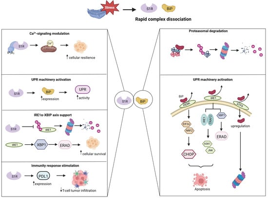
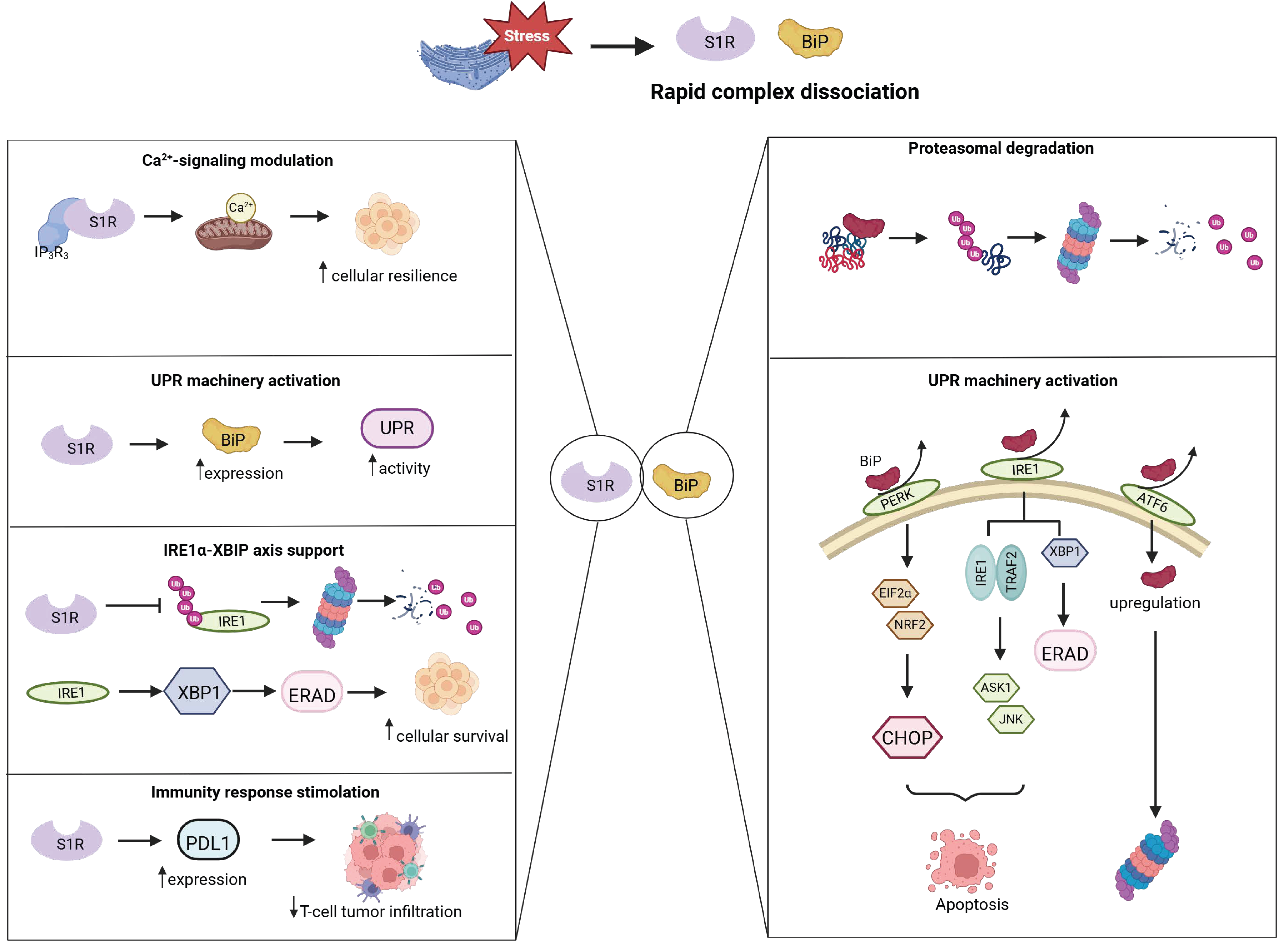
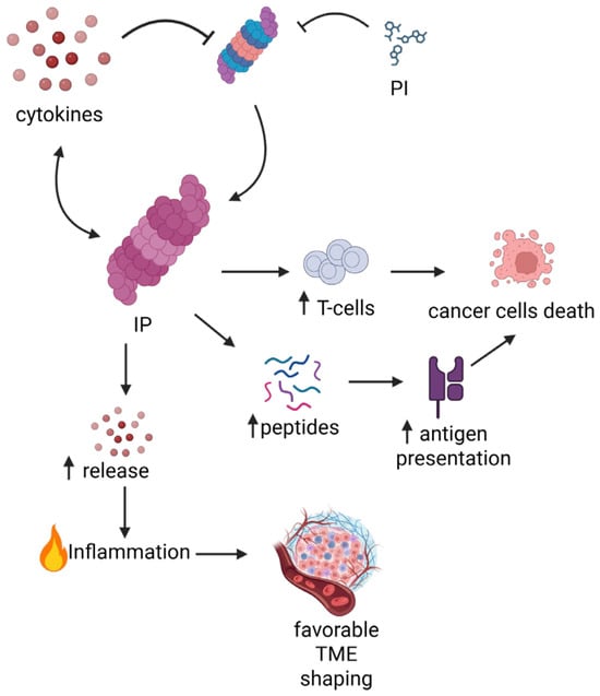
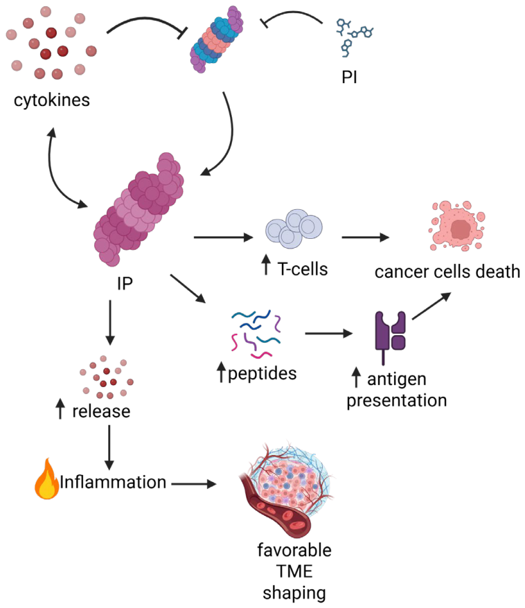
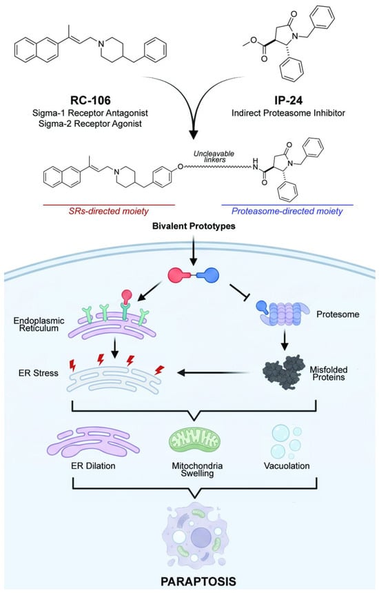
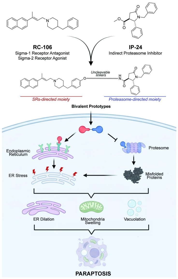
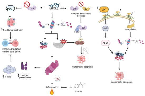
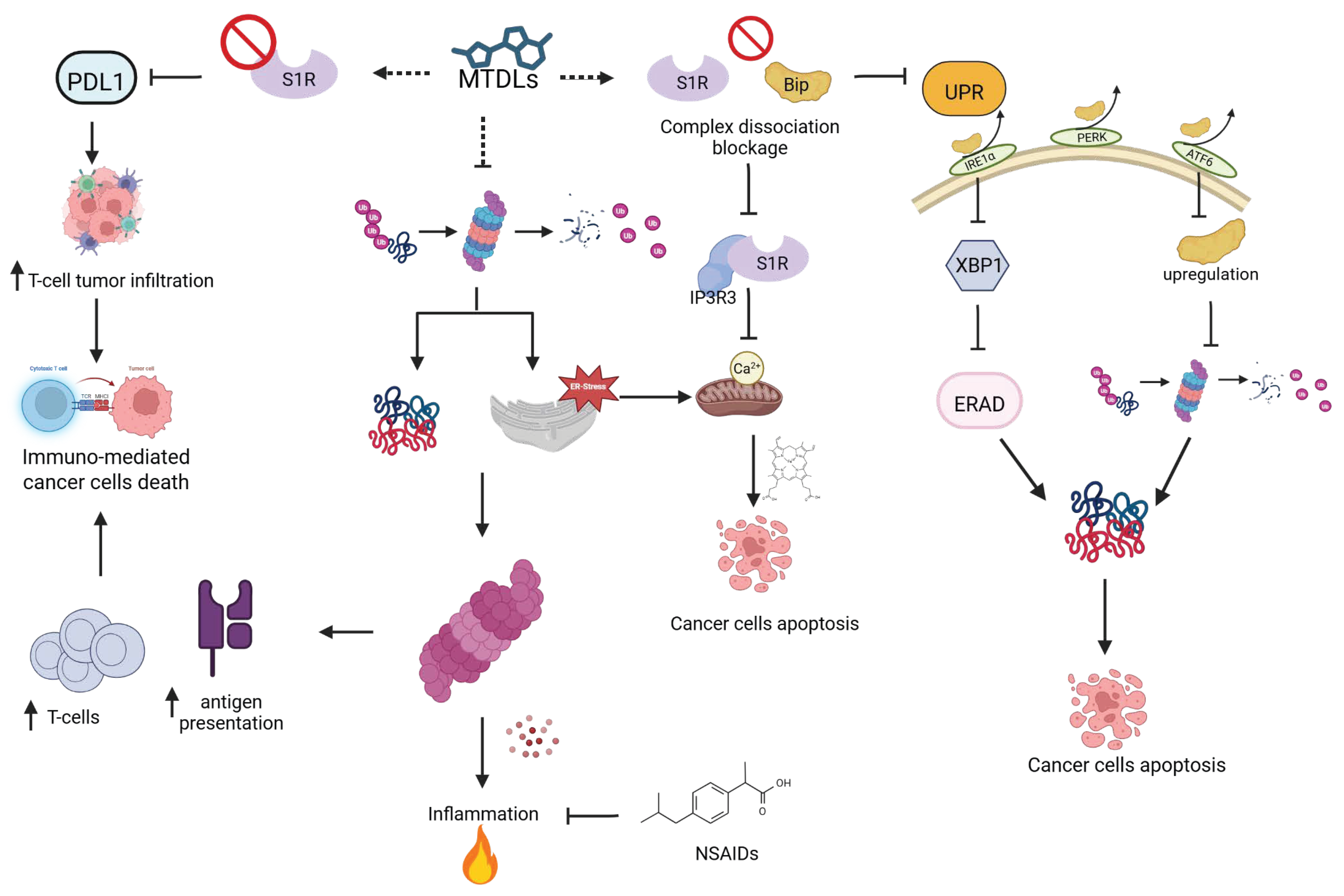

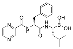
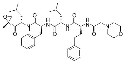
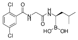
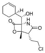
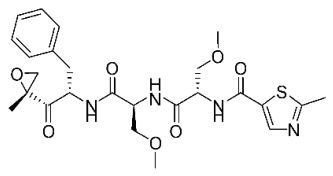
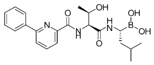
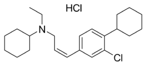
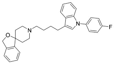
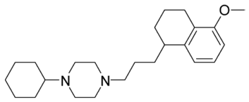
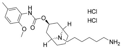
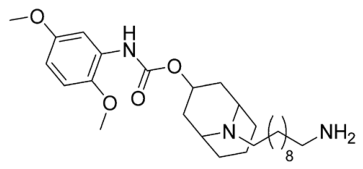
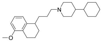
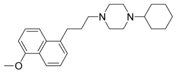
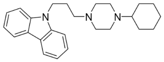
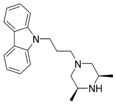
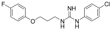
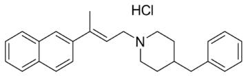
Disclaimer/Publisher’s Note: The statements, opinions and data contained in all publications are solely those of the individual author(s) and contributor(s) and not of MDPI and/or the editor(s). MDPI and/or the editor(s) disclaim responsibility for any injury to people or property resulting from any ideas, methods, instructions or products referred to in the content. |
© 2026 by the authors. Licensee MDPI, Basel, Switzerland. This article is an open access article distributed under the terms and conditions of the Creative Commons Attribution (CC BY) license.