


Abstract
Colorectal cancer (CRC) remains a major clinical challenge, and the c-Jun N-terminal kinase (JNK) signaling pathway represents a compelling yet pharmacologically complex target. Two tetracyclic ketoxime JNK inhibitors, 11H-indeno[1,2-b]quinoxalin-11-one oxime (IQ-1) and 6-(hydroxyimino)indolo[2,1-b]quinazolin-12(6H)-one (tryptanthrin-6-oxime, TRP-Ox), have previously demonstrated anti-inflammatory activity, but their direct antitumor potential against CRC has not been systematically investigated. In this study, we evaluated the in vitro and in vivo efficacy of both compounds against human SW620 colon adenocarcinoma models. Reverse pharmacophore screening of the compound’s isomers confirmed JNK1 and JNK3 as primary predicted targets alongside shared cancer-relevant effectors (including CDK2, Cyclin A2, VEGFR2, GSK3β, PTP1B, HSP90α, and MMP3), suggesting a polypharmacological mode of action. In vitro proliferation was monitored continuously via real-time impedance analysis (iCELLigence), while cell death was quantified after 24 h using annexin V/propidium iodide flow cytometry. Both compounds suppressed SW620 cell growth, driving cytotoxicity predominantly through early and late apoptosis. In vivo, immunodeficient SCID mice bearing heterotopic SW620 xenografts were treated daily for 28 days with IQ-1 (30 mg/kg) or TRP-Ox (30, 60, or 90 mg/kg) via intraperitoneal (i.p.) or peritumoral (subcutaneous, s.c.) injection, with oxaliplatin (5 mg/kg, i.p.) as a positive control. Unlike oxaliplatin, IQ-1 showed no antitumor activity by either route. Conversely, TRP-Ox induced xenograft tissue necrosis and dose-dependently reduced post-mortem tumor mass at 30 and 60 mg/kg via both administration routes. However, the 90 mg/kg dose yielded no further reduction, indicating an efficacy plateau likely associated with dose-limiting toxicity. Notably, peritumoral administration significantly outperformed delivery, indicating that the systemic efficacy of TRP-Ox is constrained by low bioavailability. Collectively, these findings identify TRP-Ox as a potent pro-apoptotic lead active against CRC cells and warrant its further preclinical development using advanced drug-delivery formulations.
1. Introduction
Colorectal cancer (CRC) remains a leading cause of cancer-related morbidity and mortality worldwide. From a clinical perspective, its epidemiology has become increasingly concerning, with diagnoses occurring at younger ages, tumors more often presenting at advanced stages, and a growing share of cases involving rectal disease [1,2]. Surgical resection combined with systemic chemotherapy, frequently including platinum-based agents such as oxaliplatin, has improved outcomes in early-stage CRC, yet relapse remains common [3]. For patients with metastatic disease, survival remains dismal, in large part because of intrinsic or acquired chemoresistance mediated by intricate molecular processes, including integrin-dependent signaling and epigenetic alterations [4,5]. These challenges highlight the urgent need to identify additional therapeutic targets and develop agents with novel mechanisms of action for refractory CRC.
The mitogen-activated protein kinase (MAPK) cascades, and the c-Jun N-terminal kinase (JNK) subfamily in particular, occupy a central position in the cellular machinery controlling proliferation, differentiation, and apoptosis in CRC [6,7]. The role of JNK signaling in CRC is highly complex and remarkably context-dependent [8,9,10]. While JNK hyperactivation generally supports intestinal tumorigenesis and drives proliferative, invasive, and drug-resistant phenotypes [11,12,13], its targeted pharmacological inhibition has successfully sensitized hypoxic colon cancer cells to DNA-damaging agents like oxaliplatin [8] and suppressed gastrointestinal cancer cell proliferation [14,15,16]. For instance, the natural JNK1 inhibitor licochalcone A has been shown to suppress colon cancer xenograft growth without apparent systemic toxicity [17]. Conversely, sustained JNK phosphorylation is a mandatory mediator of the pro-apoptotic and cytotoxic effects induced by several natural and synthetic agents in colon carcinoma cells [18,19,20,21]. On the other hand, targeting JNK has been reported to be beneficial in a variety of experimental tumor models [22,23,24,25]. For example, the JNK inhibitor, JNK-IN-8, suppresses triple-negative breast cancer growth both in vitro and in vivo [26]. Likewise, JNK inhibitor SP600125 significantly inhibited the growth of MCF7 breast cancer cells [27], stem-like glioblastoma cells [22] and spheroids from thyroid cancers [28]. This functional duality, acting as either a survival factor or a death trigger depending on the cellular context, renders JNK an attractive yet pharmacologically challenging target, thereby driving the continuous search for isoform-selective, drug-like JNK inhibitors [2,29]. JNK pathway hyperactivation significantly enhances the metastatic potential and invasiveness of CRC cells, including the SW620 line [30,31,32].
Within this context, a distinct chemotype of tetracyclic ketoximes has emerged as a scaffold for high-affinity JNK inhibition. Chemical library screening originally identified compound IQ-1 and its sodium salt IQ-1S as a potent inhibitor with nanomolar affinity for JNK isoforms and robust anti-inflammatory activity [33,34], a discovery that catalyzed subsequent structure-based optimization [33]. While IQ-1 exerts protective effects in animal models of cerebral ischemia and myocardial infarction [35,36], its therapeutic utility is hindered by unfavorable pharmacokinetics, namely low oral bioavailability and rapid systemic clearance [37,38].
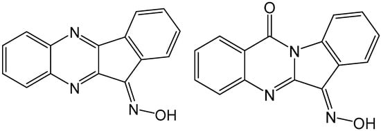
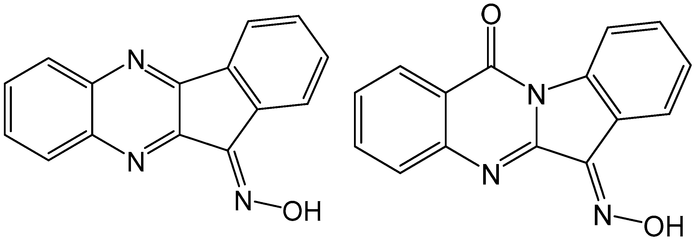
To address these challenges, attention has expanded to related scaffolds. Recently, tryptanthrin-6-oxime (TRP-Ox), an oxime derivative of the natural alkaloid tryptanthrin, was characterized as a JNK-binding agent with anti-inflammatory and neuroprotective properties [34,39,40,41,42]. Chemical structures of IQ-1 and TRP-Ox are shown in Figure 1. The parent alkaloid tryptanthrin exhibits broad antitumor efficacy, including suppressing CRC cell growth, inducing apoptosis, and modulating MAPK signaling [43,44,45]. Moreover, tryptanthrin derivatives are under active investigation as potent antiproliferative agents that target topoisomerase II [46] and inhibit CRC progression via the transforming growth factor-β (TGF-β)/SMAD and phosphoinositide 3-kinase (PI3K)/Akt/mTOR pathways [47]. Despite their well-documented anti-inflammatory and cytoprotective profiles, the direct antitumor potential of IQ-1 and TRP-Ox has not yet been systematically evaluated. However, a recent study demonstrated that the IQ-1 derivative, IQ-1S, suppresses breast cancer cell growth in both monolayer and three-dimensional spheroid cultures [48], suggesting that this chemotype possesses broader anticancer activity beyond its established indications.
Figure 1.
Chemical structures of JNK inhibitors IQ-1 (left) and TRP-Ox (right).
Within this series of derivatives, compounds IQ-1 and TRP-Ox displayed the highest binding affinities toward JNK1–3 [34,39,42]. While some analogues demonstrated superior specificity despite lower potency, IQ-1 and TRP-Ox were chosen for in vivo experiments because their straightforward synthesis allows for the large-scale production necessary for such studies. We subsequently investigated the therapeutic efficacy of both compounds using human SW620 colon adenocarcinoma models, beginning with an evaluation of their antiproliferative and cytotoxic effects in vitro. Derived from a metastatic site and harboring a TP53 mutation, the SW620 cell line serves as a clinically relevant, rigorous model of late-stage, chemoresistant CRC progression [49,50]. Growth-inhibitory profiles and cytotoxicity were characterized using real-time impedance monitoring, while the primary mode of cell death (apoptosis versus necrosis) was identified via flow cytometry using annexin V–fluorescein isothiocyanate (FITC) and propidium iodide (PI) double staining. Furthermore, the in vitro and in vivo antitumor efficacy of both compounds was investigated in immunodeficient severe combined immunodeficiency (SCID) mice bearing heterotopic SW620 xenografts across two distinct routes of administration: localized peritumoral (subcutaneous, s.c.) injection and intraperitoneal (i.p.) delivery.
2. Results and Discussion
2.1. Identification of Potential Therapeutic Targets for IQ-1 and TRP-Ox
Previously, we used kinase profiling to demonstrate that IQ-1 and TRP-Ox exhibit high binding affinity for JNK1–3 [39,42,51]. To gain deeper mechanistic insights into their broad antitumor and pro-apoptotic activities, we expanded our target identification strategy using reverse pharmacophore screening on the PharmMapper server. The Z- and E-isomers of both compounds were screened against a comprehensive database of pharmacophore patterns, and the top 300 hits for each isomer were collected. Because raw fit scores in PharmMapper are model-dependent and cannot be directly compared across different targets, the server normalizes data against a random background distribution to generate a Z-score. This metric evaluates the statistical significance of the alignment between the query molecule and a given pharmacophore model. To identify candidate anticancer targets from our hits, we filtered the results using a standard cutoff of Z-score > 1.2.
This threshold ensures that a molecule’s structural fit is at least one standard deviation above the random background noise, providing high confidence that the identified proteins represent probable binding partners for IQ-1 and TRP-Ox rather than artifactual or low-affinity hits [52].
PharmMapper ranked JNK1 and JNK3 among the top predicted targets for the Z-isomers of TRP-Ox and IQ-1 (Table 1 and Table 2). For the E-isomers, the same kinases were predicted for TRP-Ox only (Tables S1 and S2). That pattern matches the high binding affinities observed in our initial kinase screening [34], reinforcing these proteins as primary therapeutic targets and validating the rationale for these compounds as JNK inhibitors.
Table 1.
Potential anticancer protein targets of Z-TRP-Ox identified using reverse pharmacophore screening on the PharmMapper.
Table 2.
Potential anticancer protein targets of Z-IQ-1 identified using reverse pharmacophore screening on the PharmMapper.
Beyond JNKs, the profiling revealed a diverse array of effectors critical to cancer progression, survival, and extracellular matrix remodeling. These secondary targets include key cell cycle regulators (CDK2, Cyclin-A2), MAPK14/p38α, and critical proliferation drivers (EGFR, VEGFR2, FGFR1, JAK3, and BRAF) [53]. Furthermore, the predicted interactions with the anti-apoptotic proteins XIAP and BCL-XL suggest that TRP-Ox may directly antagonize apoptosis-evasion machinery. The mapping also highlighted matrix proteinases MMP3 and MMP13, enzymes pivotal for tumor invasion, and the post-translational modifier methionine aminopeptidase 2 (MetAP2), an established candidate in targeted oncology. Ultimately, this convergence of targets predicts that the aryl ketoximes IQ-1 and TRP-Ox could exert their antitumor activity through a polypharmacological mechanism, simultaneously disrupting kinase signaling, survival pathways, and metastatic cascades.
2.2. Cell Growth Inhibitory Effect
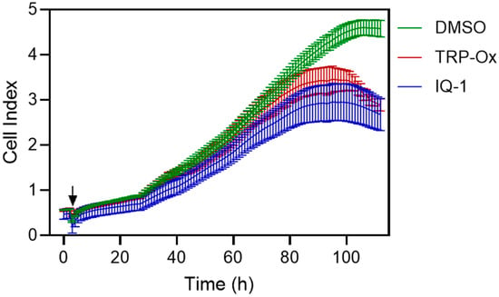
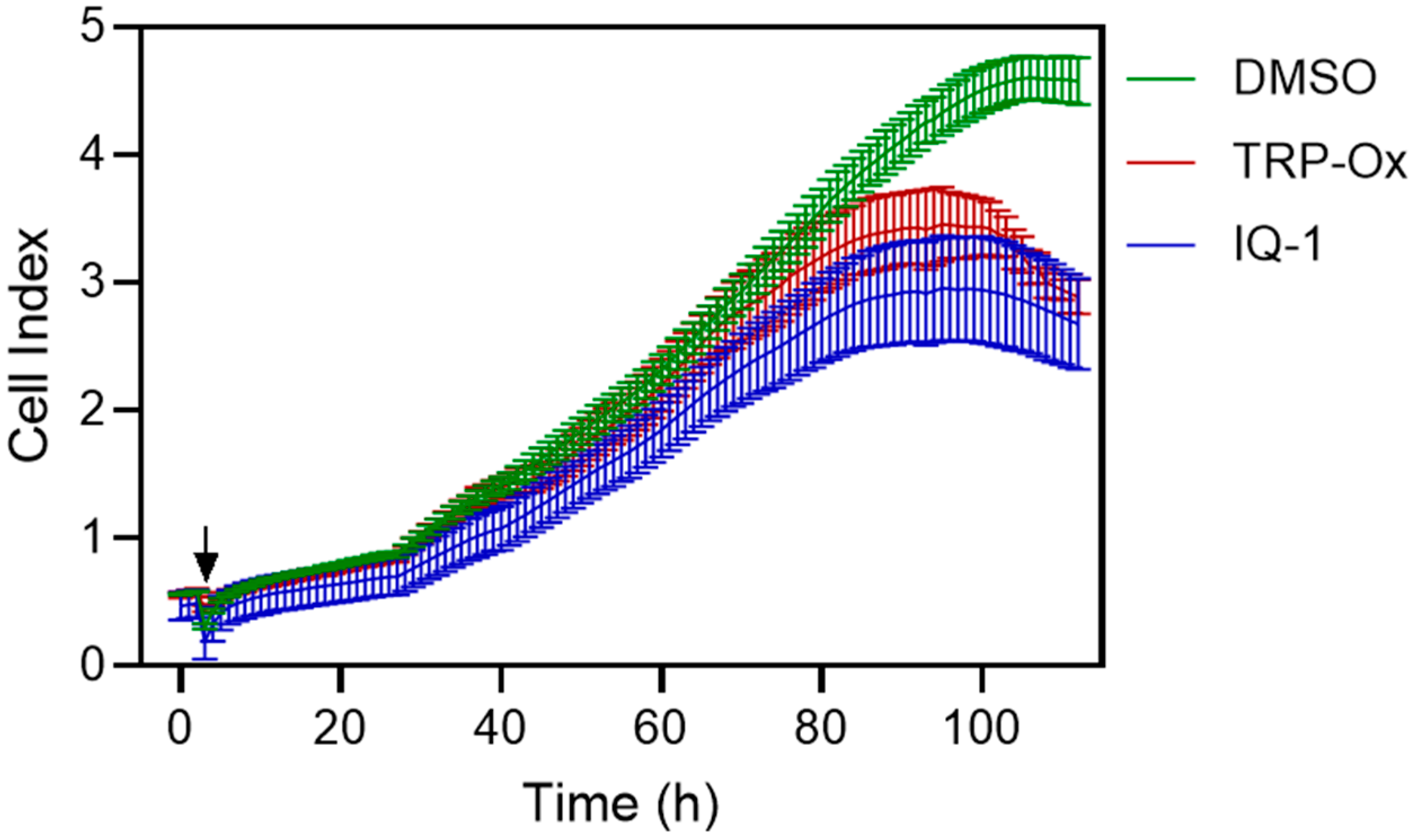
The effects of IQ-1 and TRP-Ox on the growth of SW620 cells in vitro were evaluated using the iCELLigence system. The xCELLigence is a technology that allows measurement of cellular growth in real time by measuring the net adhesion of cells on a custom-designed gold electrode following changes in electrical impedance [54,55]. Figure 2 shows the normalized cell index of SW620 cells treated with IQ-1 and TRP-Ox (both at a concentration of 20 µM) relative to the negative control (1% dimethyl sulfoxide, DMSO). Inhibition of cell growth became apparent ~80 h after treatment.
Figure 2.
Time-dependent changes in cell index values and the average cell index values for SW620 cells after treatment with TRP-Ox or IQ-1 (both at 20 µM concentration) as compared to the negative control (1% DMSO). The concentration of 20 µM for this assay was selected based on the half-maximal concentration used in our previous in vitro concentration-dependent assays for these compounds. The arrow indicates when treatment was added. The panel displays cell index values recorded at 1-h intervals. The values represent mean ± S.D., n = 3.
2.3. IQ-1 and TRP-Ox Induce Apoptosis in SW620 Cells
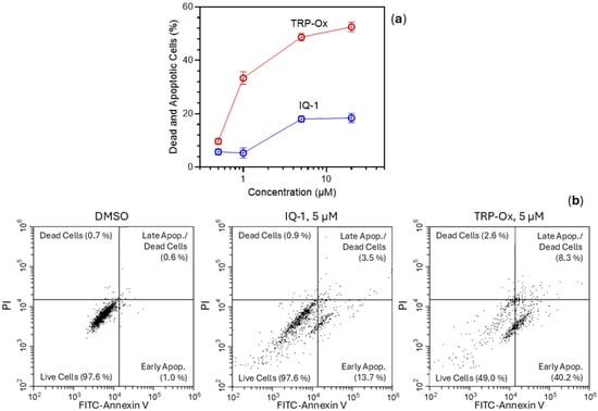
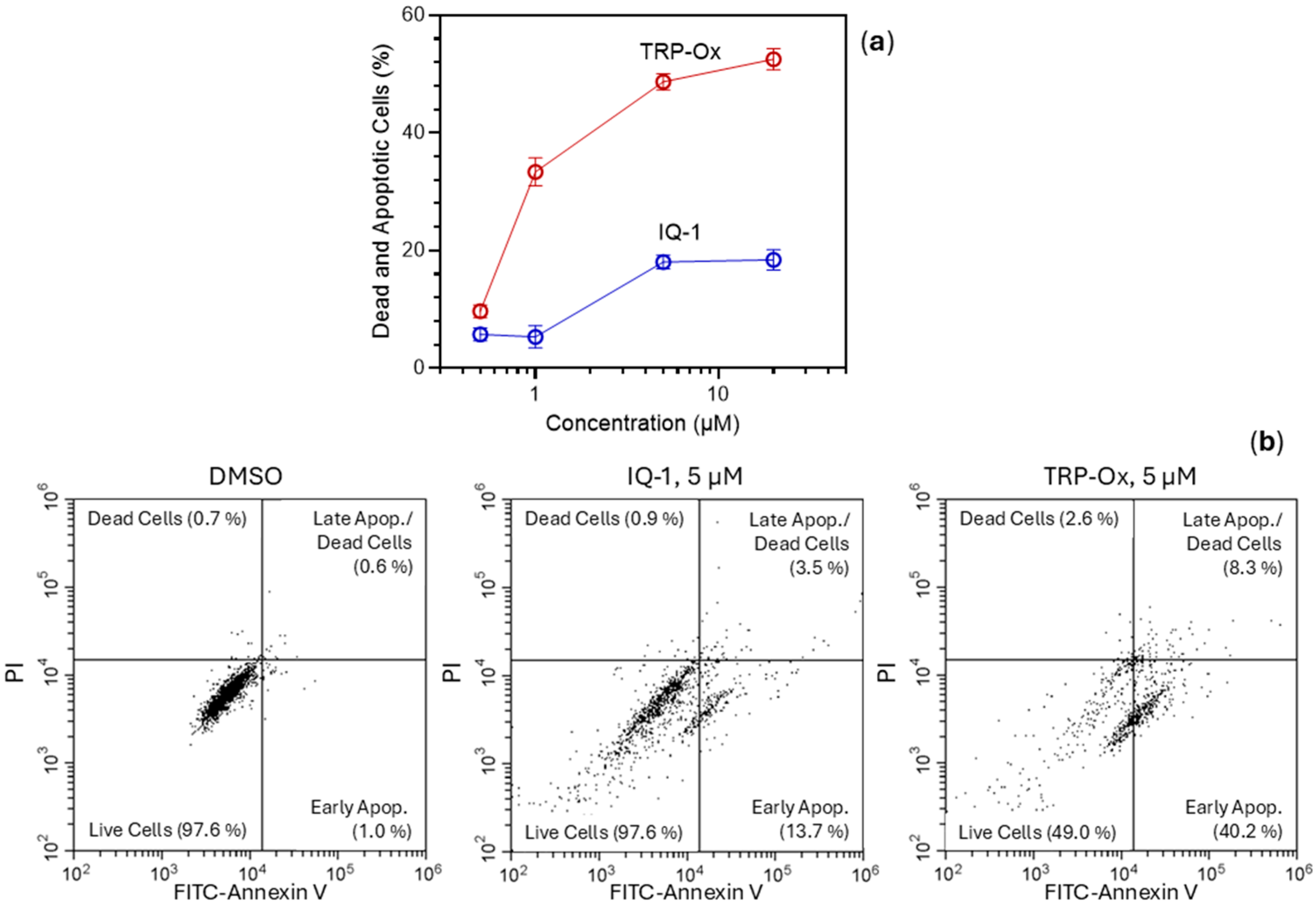
To clarify the mechanism responsible for the marked growth suppression detected by real-time impedance monitoring, we examined whether the decline in cell index reflected mainly cytostatic inhibition or overt cytotoxicity. Flow cytometric analysis via FITC–annexin V/PI staining revealed that a 24 h treatment with IQ-1 (5 and 20 µM) or TRP-Ox (1, 5, and 20 µM) triggered a significant increase across all stages of cell death, including early/late apoptosis and necrosis (Figure 3). While IQ-1 elicited only modest pro-apoptotic effects at lower concentrations (0.5 and 1 µM), becoming prominent primarily at 5 and 20 µM, TRP-Ox acted more potently at these lower doses. Notably, at 5 and 20 µM of TRP-Ox, the viable cell population fell markedly to ~50%.
Figure 3.
IQ-1 and TRP-Ox induce apoptosis of SW620 cells assessed by flow cytometry. Panel (a) displays the percentage of dead and apoptotic cells after 24 h of treatment with different concentrations of IQ-1 and TRP-Ox in comparison with the control (1% DMSO). The values represent mean ± SD. SW620 cell apoptosis measured by annexin V/PI staining is shown in Panel (b) of representative flow cytometry data for control (1% DMSO), IQ-1 (5 μM) or TRP-Ox (5 μM) treated cells at 24 h post-compound application. The bivariate plots on the panel display individual cells or events, with each event plotted according to the PI and FITC-annexin fluorescence intensity parameters. Each plot shows quadrants with dead and apoptotic cells: viable cells (lower left quadrant) were negative for both annexin V and PI, apoptotic cells were positive for annexin V staining and negative for PI, and late apoptotic/dead cells were positive for both annexin V and PI (upper right quadrant).
Taken together, these data indicate that TRP-Ox acts as a rapid and potent inducer of apoptosis, whereas IQ-1 requires higher concentrations to initiate a comparable response. The robust apoptotic activity elicited by TRP-Ox further underscores the complex, dual role of JNK signaling in CRC biology. A substantial body of literature has established that JNK/p38 pathway activation is a primary mechanism through which diverse natural and synthetic compounds induce apoptosis in colon cancer cells. Specifically, in SW620 cells, sustained JNK activation mediates the apoptotic effects of berberine, α-methyl-n-butylshikonin, and the naturally occurring thiophene derivative 2-(pro-1-ynyl)-5-(5,6-dihydroxypenta-1,3-diynyl) [18,19]. Similar mechanisms have been documented for butyrate, benzimidazole acridine, and synthetic thiazolidine derivatives in other CRC cell lines [20,21,56]. Recent evidence has extended this framework, demonstrating that novel triterpenoids and other natural products can trigger both apoptosis and ferroptosis in CRC via JNK pathways [57,58,59]. Conversely, pharmacological inhibition of JNK has also been reported to trigger apoptosis and cell-cycle arrest in gastrointestinal cancers, presumably by disrupting the hyperactivated, pro-survival JNK signaling required by tumor cells [15]. The pronounced apoptosis induced by TRP-Ox in this study aligns with this latter paradigm of JNK-targeted antitumor activity.
The apoptotic profile elicited by TRP-Ox aligns with recent literature detailing the mechanisms of its parent alkaloid, tryptanthrin, and structurally related analogs. Specifically, tryptanthrin-derived compounds exert potent cytotoxicity, induce sub-G1 cell cycle arrest, and activate executioner caspases 3 and 7 across diverse cancer cell lines [45]. Furthermore, alternative tryptanthrin derivatives act as robust topoisomerase II inhibitors to suppress cell proliferation [46]. Within the specific context of CRC, tryptanthrin drives apoptosis by upregulating the Bax/Bcl-2 ratio and accelerating caspase-3 cleavage [43]. Consequently, the ketoxime derivative TRP-Ox appears to retain, and potentially amplify, this intrinsic pro-apoptotic efficacy.
Notably, TRP-Ox effectively triggered apoptosis in SW620 cells, which harbor a p53 mutation. The evasion of apoptosis via p53 inactivation is a hallmark of late-stage, metastatic CRC that severely drives chemoresistance. However, preceding studies indicate that SW620 cells remain susceptible to apoptosis through p53-independent cascades, such as the caspase-2 and JNK signaling pathways stimulated by sulforaphane [50]. The ability of TRP-Ox to bypass p53-mediated resistance and commit SW620 cells to terminal apoptosis highlights the therapeutic potential of this chemical scaffold against advanced, refractory colorectal malignancies.
2.4. Antitumor Efficacy of TRP-Ox in Xenograft Mouse Model
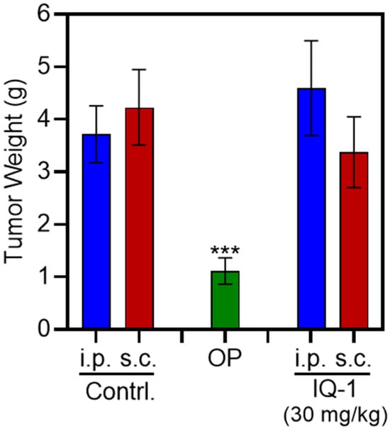
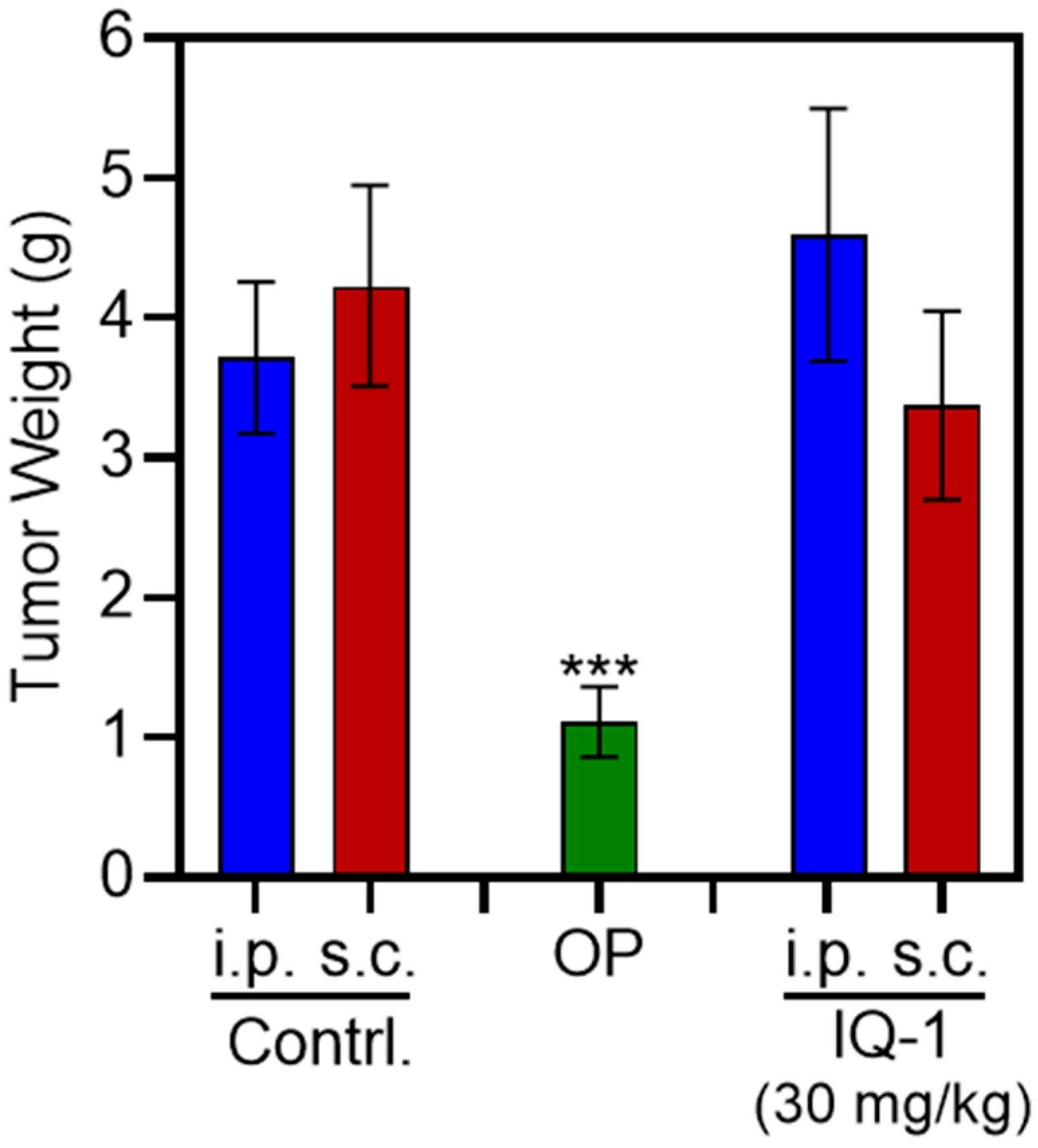
Unlike the reference drug oxaliplatin (5 mg/kg, i.p.), the compound IQ-1 showed no efficacy when administered i.p. or s.c. during preliminary testing in an SCID mouse xenograft model (Figure 4).
Figure 4.
Post-mortem tumor weights of mice treated with IQ-1 via intraperitoneal (i.p.) or subcutaneous (s.c.) administration. OP, oxaliplatin (5 mg/kg, i.p.). Differences compared to the control group are indicated by p-value (*** p < 0.001). The values represent mean ± SD.
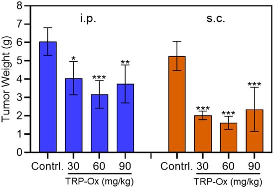
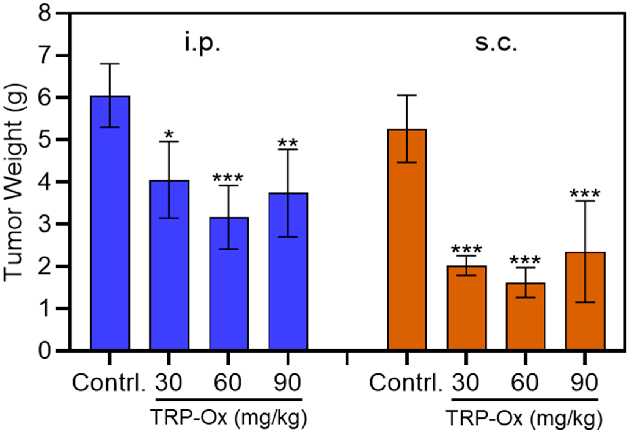
Since the IQ-1 compound proved ineffective, subsequent experiments to evaluate antitumor activity were conducted using various doses of the TRP-Ox in the same SW620 heterotopic xenograft model. When drugs are administered peritumorally (locally around the tumor area), they often trigger a localized immune or inflammatory response [60]. This causes edema (fluid accumulation) and swelling, which inflates the external dimensions of the tumor mass [61]. Because standard caliper measurements cannot differentiate between actual cancer cells and inflammatory swelling, relying solely on tumor volume is highly unreliable in these scenarios. To accurately assess the therapeutic efficacy of a drug in the presence of peritumoral inflammation, end-point weight analysis was used. Weighing the xenografts obtained at necropsy revealed a significant reduction in tumor mass following both s.c. and i.p. administration of TRP-Ox across all tested doses (30, 60, and 90 mg/kg; Figure 5). Two-way ANOVA with “treatment dose” and “route of administration” as factors demonstrated that both the dose (F3,55 = 9.784, p < 0.001, ηp2 = 0.35) and the route of administration (F1,55 = 9.412, p = 0.004, ηp2 = 0.15) exerted a statistically significant main effect on final xenograft mass, whereas the interaction between these factors was not significant (F3,55 = 0.97, p = 0.819, ηp2 = 0.05). Specifically, TRP-Ox suppressed tumor growth in a dose-dependent manner at 30 and 60 mg/kg; at the 60 mg/kg dose, mean tumor mass was reduced by approximately 48% and 69% following i.p. and s.c. administration, respectively, compared to the corresponding vehicle control groups.
Figure 5.
Post-mortem tumor weights in TRP-Ox mice following intraperitoneal (i.p.) and subcutaneous (s.c.) administration. Differences compared to the control group are indicated by p-values (* p < 0.05, ** p < 0.01, *** p < 0.001). The values represent mean ± SD.
Repeated-measures ANOVA of body weight dynamics in animals administered oxaliplatin or IQ-1 revealed significant main effects for both time (F10,140 = 2.23, p = 0.019) and treatment group (F2,14 = 4.34, p = 0.034), as well as a significant time x group interaction (F20,140 = 5.97, p < 0.001). Oxaliplatin-treated mice exhibited marked systemic toxicity, manifested by a significant reduction in body weight compared to the control group by the end of the first week (p < 0.05). This weight loss persisted throughout the treatment period, aligning with previous literature reporting cachexia and body mass reduction in mice following oxaliplatin administration [62]. Statistically significant differences compared to both the i.p. and s.c. IQ-1 groups emerged as early as two weeks post-initial administration (F2,21 = 12.76, p < 0.001; Figure S1). Notably, by the conclusion of the experiment (day 35), body weights in the oxaliplatin cohort had fully recovered, showing no significant differences from the control group. Conversely, mice treated with IQ-1 (30 mg/kg, i.p. or s.c.) or TRP-Ox at doses of 30, 60, and 90 mg/kg (i.p. or s.c.) exhibited no body weight loss in SCID mice bearing SW620 xenografts (Figures S1–S4). Collectively, these findings demonstrate that IQ-1 and TRP-Ox do not exert systemic toxicity affecting animal body weight when administered via either route; however, further comprehensive toxicological studies are required to confirm these preliminary observations.
2.5. Limitations and Perspectives
A key finding of the in vivo study was that the therapeutic efficacy depends significantly on the route of administration; specifically, peritumoral administration of TRP-Ox yielded substantially better outcomes compared to i.p. delivery. This disparity suggests potential pharmacokinetic limitations. The requirement for high local concentrations directly within the tumor site implies that systemic efficacy may be constrained by rapid clearance or low bioavailability when administered intraperitoneally. Although quantifying the exact intratumoral drug concentration was beyond the scope of the current study, determining this parameter remains a critical objective for future pharmacokinetic assessments. Notably, tryptanthrin, the parent alkaloid of TRP-Ox, has been reported to inhibit CRC tumor growth in vivo [43]. It is important to emphasize that TRP-Ox and related tryptanthrin derivatives themselves possess anti-inflammatory and neuroprotective properties [40]. Comparing these results with data obtained for IQ-1 suggests that TRP-Ox is a more promising candidate for further preclinical development.
The striking divergence in in vivo efficacy between TRP-Ox and IQ-1, two tetracyclic ketoximes with comparably high affinity for JNK1–3 [39,42,51], indicates that JNK blockade alone cannot fully account for the antitumor activity of TRP-Ox and points to the engagement of secondary targets. In other words, TRP-Ox could exert its antitumor activity through a polypharmacological mechanism. Consistent with this view, reverse pharmacophore profiling (Table 1, Table 2 and Table S2) predicted several tumor-relevant effectors of TRP-Ox that were absent from, or ranked substantially lower in, the IQ-1 target list, most notably EGFR, the anti-apoptotic proteins BCL-XL and XIAP, MetAP2, and MMP13. Direct antagonism of BCL-XL and XIAP, the latter being an endogenous inhibitor of caspases-3, -7, and -9, would lower the apoptotic threshold and provide a plausible molecular basis for the rapid, p53-independent commitment of SW620 cells to apoptosis observed in vitro. Likewise, simultaneous interference with EGFR-driven proliferative signaling, MetAP2-dependent protein maturation, and MMP13-mediated extracellular matrix remodeling is consistent with the extensive xenograft necrosis and tumor growth retardation detected in vivo. Such multi-target engagement may also rationalize the efficacy plateau observed at 90 mg/kg. Although these computational predictions await confirmation by direct biochemical and cellular target-engagement assays, they collectively position TRP-Ox as a potential polypharmacological lead whose antitumor efficacy likely reflects the coordinated disruption of kinase signaling, apoptosis-evasion machinery, and metastatic cascades rather than selective JNK inhibition alone.
Collectively, these findings suggest that the pronounced pro-apoptotic and antitumor potential of TRP-Ox is currently masked in vivo by formulation limitations. This conclusion points to a clear developmental mandate: the clinical translation of the compound requires advanced delivery engineering rather than simple dose escalation. Strategies such as transiently soluble prodrugs [63] or encapsulation within nanoparticulate and microparticulate delivery systems [64], a strategy already validated for IQ-1 using poly(lactic-co-glycolic acid) (PLGA) microparticles [65] and for tryptanthrin using ethosomes [66], represent the most rational path forward to unlock the systemic antitumor efficacy of this promising chemotype against CRC. The pronounced apoptotic response to TRP-Ox treatment aligns with the results of previous studies describing the cytotoxic and apoptosis-inducing activity of other tryptanthrin derivatives in various cancer cell lines [45].
Another limitation is that we did not confirm tumor-specific JNK inhibition by IQ-1 and TRP-Ox. Although the in vivo efficacy of these compounds has been demonstrated in other biological models via Western blotting [67,68], their effectiveness within SW620 tumors remains to be validated using complementary molecular techniques. Moreover, the lack of data on the actual concentrations of the compounds under study that are achieved within the tumor prevents us, at this stage of the research, from concluding why IQ-1 is less effective than the compound TRP-Ox.
While our previous publications extensively demonstrated the in vitro effects of IQ-1 and TRP-Ox across a diverse panel of cell lines [34,67,68], the data presented here serve as a foundational baseline. We acknowledge that this study does not constitute an exhaustive evaluation of these compounds’ efficacy. Further investigation remains essential to comprehensively delineate the precise antitumor mechanisms and cell-type selectivity of TRP-Ox. Nevertheless, the comparative data established against both negative and positive controls provide a crucial and robust benchmark for future research.
3. Materials and Methods
3.1. The Studied Compounds
The two investigational compounds, 11H-indeno[1,2-b]quinoxalin-11-one oxime (IQ-1) and 6-(hydroxyimino)indolo[2,1-b]quinazolin-12(6H)-one oxime (TRP-Ox), were synthesized as described previously ([69] and [70], respectively). The chemical structures of the compounds were confirmed by mass spectrometry and nuclear magnetic resonance. The purity of the samples was >98%. Both IQ-1 and TRP-Ox exhibit high chemical stability. We observed that they remain stable in DMSO solutions for several weeks (as confirmed by periodic NMR monitoring) and do not undergo hydrolysis upon precipitation into water from organic solvents. Also, their stability at elevated temperatures is supported by their synthesis conditions in refluxing alcohols [69,71].
To overcome the inherent hydrophobicity of these tetracyclic oximes, both compounds were suspended in 5% aqueous DMSO (Biolot, St. Petersburg, Russia) for all in vitro and in vivo experiments. Stock suspensions were aliquoted and stored at −20 °C. Immediately prior to administration, the suspensions were thawed and sonicated for 1 min using an ultrasonic homogenizer (Qsonica Q700, Newtown, CT, USA). This critical step was employed to ensure a uniform, fine dispersion of the micro-suspension and to prevent premature precipitation. A 5% aqueous DMSO solution was used as the vehicle (negative) control. Oxaliplatin (Medac GmbH, Wedel, Germany), a third-generation platinum-based antineoplastic agent, was employed as the reference chemotherapeutic agent (positive control) due to its established clinical efficacy and standard-of-care status in the treatment of advanced CRC [72,73]. For the in vitro assays, stock solutions of the compounds were prepared in DMSO and stored at −20 °C. The solutions were subsequently diluted in the cell culture medium to achieve a final DMSO concentration of 1% (v/v) during treatment.
3.2. Cell Culture
The human colon adenocarcinoma cell line SW620 (ATCC CCL-227™) was obtained from the cryobank of the Shared Research Facility “SPF Vivarium” at the Institute of Cytology and Genetics, Siberian Branch of the Russian Academy of Sciences. The SW620 line serves as a highly validated and rigorous model for studying late-stage, metastatic CRC progression [49]. Cells were thawed according to standard protocols and cultured in Dulbecco’s Modified Eagle Medium/Nutrient Mixture F-12 (DMEM/F12, 1:1; Biolot, Russia) supplemented with 10% fetal bovine serum (LT Biotech, Vilnius, Lithuania). Cultures were maintained at 37 °C in a humidified incubator with a 5% CO2 atmosphere. For passaging and experimental harvesting, cells at 70–80% confluence were detached using a 1:1 mixture of 0.25% trypsin and 0.02% versene (Biolot, Russia).
3.3. Real-Time Impedance-Based Proliferation Assay
In the real-time cell analysis (RTCA) system, cytotoxicity was evaluated by measuring cellular impedance, recorded as cell index (CI). To track dynamic changes in cell number, the experiment was performed in three independent biological replicates, each containing three technical replicates. The iCELLigence system (Agilent Technologies, Santa-Clara, CA, USA) was used according to the manufacturer’s instructions. The CI was obtained by measuring the change in the electrical impedance in the presence and absence of cells in the wells. The SW620 cell line was inoculated, keeping the cell number as 104 cells/well, on 16-well plates of the iCELLigence system. Afterward, the proliferation, attachment and spreading of the cells were monitored in each well of the e-plates every 60 min and analyzed with RTCA Software 1.2. Twenty-two hours after cell transplantation, cells in the logarithmic growth phase were treated with IQ-1 or TRP-Ox (final DMSO concentration: 1%) and monitored in real time hourly up to 120 h. Cells grown in growth medium containing 1% DMSO were used as the control. All experimental steps were conducted at 37 °C in a 5% CO2 atmosphere. For this analysis, a concentration of 20 μM was selected: half of the maximum concentration used in our previous in vitro studies, which examined the concentration dependence of the effects of these compounds [39,42,51]. Specifically, 40–50 µM represents the maximum achievable concentration due to their low solubility in aqueous buffer at physiological pH.
3.4. Flow Cytometric Analysis
To differentiate apoptotic from necrotic cell death after treatment, SW620 cells were examined by flow cytometry after double staining with annexin V–FITC and PI. Annexin V identifies phosphatidylserine externalization, an early apoptotic event in which phosphatidylserine is translocated to the outer leaflet of the plasma membrane. PI, by contrast, enters cells whose membrane integrity is compromised and intercalates into DNA, thereby marking cells in late apoptosis or necrosis. SW620 cultures were exposed to IQ-1 or TRP-Ox at concentrations of 0.5, 1, 5, and 20 µM for 24 h; vehicle-treated cultures (1% DMSO) served as controls. At the indicated time points, cells were collected, washed, stained according to the manufacturer’s instructions, and subjected to flow cytometric analysis. Cell populations were assigned quantitatively to four categories: viable cells (Annexin V−/PI−), early apoptotic cells (Annexin V+/PI−), late apoptotic cells (Annexin V+/PI+), and necrotic cells (Annexin V−/PI+) [74]. The total apoptotic fraction was defined as the combined percentage of early and late apoptotic cells.
3.5. PharmMapper Target Identification
The protein targets of IQ-1 and TRP-Ox were analyzed using PharmMapper (https://lilab-ecust.cn/pharmmapper/, accessed 20 September 2026) [52,75,76]. PharmMapper is a tool that identifies potential targets for a given small molecule using an “invert” pharmacophore mapping procedure. PharmMapper utilizes reference databases of protein drug targets encoded by sets of pharmacophore points for faster mapping. Initial structures of the test compounds were prepared using ChemDraw 16.0 software and saved in Tripos MOL2 format. The MOL2 files of Z- and E-isomers of TRP-Ox and IQ-1 were uploaded into the PharmMapper server. The top 300 potential targets were retrieved and sorted by normalized Z-score value.
3.6. Experimental Animals
The study was performed on immunodeficient male SCID specific pathogen-free (SPF)-status mice at the age of 8–10 weeks, which were kept at the Center for Genetic Resources of Laboratory Animals of the Common Use Center “SPF Vivarium” (Federal Research Center Institute of Cytology and Genetics, Siberian Division of the Russian Academy of Sciences). This model was chosen because SCID mice lack functional T and B lymphocytes, which permits efficient engraftment and growth of human tumor xenografts in the absence of immune rejection. Mice were housed in family groups of 3–5 animals in individually ventilated cages on a standard 12 h:12 h light/dark cycle. Room temperature and relative humidity were controlled at 22–24 °C and 45–70%, respectively. Animals had unrestricted access to a complete granulated rodent diet (“Prokorm”, BioPro, Novosibirsk, Russia) and reverse-osmosis purified water supplemented with the “Severyanka” mineral mix. All in vivo procedures underwent ethical review and were carried out in accordance with accepted principles for the humane treatment of laboratory animals, including 2010/63/EU and the Guide for the Care and Use of Laboratory Animals (NIH, Bethesda, MD, USA, No. 85-23, rev. 1985). The protocol was approved by the Inter-Institutional Animal Ethics Committee based on the Institute of Cytology and Genetics SB RAS (Protocol 226, dated 21 April 2025).
3.7. SW620 Xenograft Model
Heterotopic human CRC xenografts were generated by s.c. injecting 100 µL of the prepared serum-free cell suspension, containing 5 × 10⁶ SW620 cells, into the right scapular region of each SCID mouse. Tumor formation was monitored from the day of inoculation. As the xenografts developed, their linear dimensions were measured twice weekly, at 3–4-day intervals, using an electronic caliper. Tumor volume (V) was calculated according to the standard ellipsoid formula: V = (a × b2) × 0.52, where a denotes the largest tumor diameter (length) and b denotes the smallest diameter (width). When tumors reached an average volume of approximately 150 mm3, mice were randomized into experimental groups to balance baseline tumor burden across cohorts. Treatment was then initiated according to the assigned regimens. To evaluate the antitumor activity of IQ-1 and TRP-Ox, a total of 36 mice (divided into 5 groups; Table 3) and 57 mice (divided into 8 groups; Table 4) were used, respectively. The animals were randomized into groups based on their body weight.
Table 3.
Experimental groups in the IQ-1 study.
Table 4.
Experimental groups in the TRP-Ox study.
IQ-1 and TRP-Ox and 5% aqueous DMSO as a negative control were given once daily for 28 consecutive days. Injection volumes were fixed at 100 µL per mouse. Oxaliplatin was administered i.p. once every four days (each day of drug administration was followed by a break of three full days). Previously, we evaluated various dosages of IQ-1 and TRP-Ox in other animal models [67,68]. I.p. administration of the compounds in a dose range of 30–50 mg/kg was effective and well-tolerated in mice during chronic treatment (daily for up to one month). Furthermore, this dosage range is well-supported by the pharmacokinetic data of IQ-1 [37,38,77]. As a result, a dose of 30 mg/kg was selected as the lowest dose for this regimen.
To ensure rigorous welfare monitoring, the general clinical status of the animals, including physical appearance, behavioral alterations, food and water consumption, and body weight, was assessed twice weekly throughout the 28-day treatment and subsequent observation periods. Predetermined humane endpoints requiring early withdrawal or euthanasia included a tumor burden reaching ~20% of total body weight, a ≥20% loss of initial body weight, or tissue ulceration/necrosis (on the body surface or xenograft) ≥ 2 cm2. Upon meeting these endpoints or at experimental completion, mice were euthanized via CO2 asphyxiation. Tumor nodules and immediately surrounding tissues were promptly excised and weighed to determine final tumor mass.
3.8. Statistical Analysis
All statistical analyses were performed using STATISTICA 6 software. The normality of quantitative data distributions was assessed prior to analysis. Normally distributed data were compared using the unpaired Student’s t-test, whereas non-normally distributed data were evaluated using the non-parametric Mann–Whitney U-test. To evaluate the main and interactive effects of treatment dose and administration route on the final xenograft mass, a two-way analysis of variance (ANOVA) was performed, with partial eta-squared (ηp2) calculated to determine effect size. Post hoc pairwise comparisons were conducted using Tukey’s honestly significant difference (HSD) test. Variations in animal body weight over time across the experimental groups were assessed using repeated-measures ANOVA. For all statistical analyses, differences were considered statistically significant at p < 0.05.
4. Conclusions
This study provides the first evaluation of the direct antitumor potential of the JNK inhibitors IQ-1 and TRP-Ox in a preclinical model of CRC. In silico reverse pharmacophore profiling of the Z- and E-isomers corroborated our previous kinase affinity data and confirmed JNK1 and JNK3 as primary targets. The same analysis revealed a broader polypharmacological profile spanning cell-cycle regulators (CDK2, Cyclin A2), angiogenic and proliferative kinases (VEGFR2, GSK3β), and matrix-remodeling enzymes (MMP3, ADAM17). TRP-Ox was additionally predicted to target EGFR, as well as key anti-apoptotic and invasion-related proteins (BCL-XL, XIAP, MetAP2, and MMP13). That extended target set supplies a mechanistic rationale for its superior cellular and in vivo potency compared with IQ-1.
In vitro, both compounds exerted sustained antiproliferative effects on SW620 human colon carcinoma cells. Marked growth suppression became apparent after ~80 h of real-time impedance monitoring. Flow cytometric analysis at 24 h showed that this loss of viability was mediated predominantly by early and late apoptosis, alongside secondary necrosis. TRP-Ox was substantially more potent than IQ-1. It triggered significant apoptosis and reduced the viable cell fraction. In SCID mice bearing heterotopic SW620 xenografts, the two scaffolds diverged sharply. The reference drug oxaliplatin significantly inhibited tumor growth, whereas IQ-1 (30 mg/kg) was inactive by both the i.p. and the s.c. (peritumoral) routes. TRP-Ox induced tumor tissue necrosis and significantly reduced excised xenograft mass at necropsy across all tested doses (30, 60, and 90 mg/kg) after either i.p. or s.c. (peritumoral) administration. Peritumoral delivery was significantly more effective than i.p. administration. This strong dependence on the route of administration, coupled with the efficacy plateau at the highest dose, indicates that the robust intrinsic pharmacodynamic activity of TRP-Ox is restricted in vivo by low systemic bioavailability and/or rapid clearance. Translational development of TRP-Ox should therefore prioritize advanced drug-delivery strategies, such as transiently soluble prodrugs or nanoparticulate and microparticulate carriers (e.g., PLGA particles), rather than simple dose escalation. These findings establish TRP-Ox as a promising candidate for the treatment of advanced, p53-deficient CRC.
Supplementary Materials
The following supporting information can be downloaded at https://www.mdpi.com/article/10.3390/molecules31224170/s1, Table S1. Potential anticancer protein targets of E-IQ-1 identified via PharmMapper analysis. Table S2. Potential anticancer protein targets of E-TRP-Ox identified via PharmMapper analysis. Figure S1. Effects of oxaliplatin and IQ-1 on the body weight of SCID mice bearing SW620 xenografts. Mice were treated with oxaliplatin (5 mg/kg, i.p.; green line) or IQ-1 (30 mg/kg, i.p.; red line) and compared against a negative control group (vehicle, i.p.; violet line). Arrows indicate the initiation and cessation of drug administration. Data are presented as mean ± SD. Figure S2. Effects of IQ-1 on the body weight of SCID mice bearing SW620 xenografts. Mice received subcutaneous (s.c.) administration of IQ-1 (30 mg/kg; yellow line) or vehicle (negative control; violet line). Arrows indicate the initiation and cessation of treatment. Data are presented as mean ± SD. Figure S3. Effects of TRP-Ox on the body weight of SCID mice bearing SW620 xenografts. Mice received intraperitoneal (i.p.) administration of TRP-Ox at doses of 30 mg/kg (violet line), 60 mg/kg (red line), or 90 mg/kg (yellow line), and were compared against a vehicle control group (blue line). Arrows indicate the initiation and cessation of treatment. Data are presented as mean ± SD. Figure S4. Effects of TRP-Ox on the body weight of SCID mice bearing SW620 xenografts. Mice received subcutaneous (s.c.) administration of TRP-Ox at doses of 30 mg/kg (violet line), 60 mg/kg (red line), or 90 mg/kg (yellow line), and were compared against a vehicle control group (blue line). Arrows indicate the initiation and cessation of treatment. Data are presented as mean ± SD.
Author Contributions
Conceptualization, E.L.Z., I.A.S. and A.I.K.; methodology, E.L.Z., M.A.B. and A.I.K.; formal analysis, E.L.Z., O.I.K., L.D.R., I.A.R., M.A.B., K.D.V., K.S.K., A.V.U., A.R.K. and A.I.K.; writing—original draft preparation, E.L.Z., M.A.B., I.A.S. and A.I.K.; writing—review and editing, I.A.S. and A.I.K.; supervision, E.L.Z. and A.I.K. All authors have read and agreed to the published version of the manuscript.
Funding
The research was funded by the Russian Science Foundation (project No. 24-15-00334).
Institutional Review Board Statement
The animal study protocol was approved by the Inter-Institutional Animal Ethics Committee based on the Institute of Cytology and Genetics SB RAS (protocol code 226, dated 21 April 2025) for studies involving animals.
Informed Consent Statement
Not applicable.
Data Availability Statement
Data is contained within the article and Supplementary Materials.
Acknowledgments
We gratefully acknowledge the Center for Medical Genomics at the Tomsk National Research Medical Center of the Russian Academy of Sciences for their expert support and technical assistance with the use of the xCELLigence system. Their contribution was essential in the successful completion of this study.
Conflicts of Interest
The authors declare no conflicts of interest.
References
- Siegel, R.L.; Wagle, N.S.; Star, J.; Kratzer, T.B.; Smith, R.A.; Jemal, A. Colorectal cancer statistics, 2026. CA Cancer J. Clin. 2026, 76, e70067. [Google Scholar] [CrossRef] [Scilit] [PubMed]
- Zhu, Y.M.; Shuai, W.; Zhao, M.; Pan, X.L.; Pei, J.P.; Wu, Y.Y.; Bu, F.Q.; Wang, A.X.; Ouyang, L.; Wang, G. Unraveling the design and discovery of c-Jun N-terminal kinase inhibitors and their therapeutic potential in human diseases. J. Med. Chem. 2022, 65, 3758–3775. [Google Scholar] [CrossRef] [Scilit] [PubMed]
- Yu, Y.; Chen, D.D.; Wu, T.; Lin, H.Z.; Ni, L.L.; Sui, H.H.; Xiao, S.S.; Wang, C.W.; Jiang, S.P.; Pan, H.L.; et al. Dihydroartemisinin enhances the anti-tumor activity of oxaliplatin in colorectal cancer cells by altering PRDX2-reactive oxygen species-mediated multiple signaling pathways. Phytomedicine 2022, 98, 153932. [Google Scholar] [CrossRef] [Scilit] [PubMed]
- Du, Y.Y.; Rokavec, M.; Hermeking, H. RBM47-induced gasdermin A/GSDMA mediates mesenchymal-epithelial transition and pyroptosis of colorectal cancer cells. Cancers 2026, 18, 504. [Google Scholar] [CrossRef] [Scilit] [PubMed]
- Pei, M.M.; Liu, K.Y.; Qu, X.; Wang, K.R.; Chen, Q.; Zhang, Y.Y.; Wang, X.Y.; Wang, Z.; Li, X.Y.; Chen, F.; et al. Enzyme-catalyzed synthesis of selenium-doped manganese phosphate for synergistic therapy of drug-resistant colorectal cancer. J. Nanobiotechnol. 2023, 21, 72. [Google Scholar] [CrossRef] [Scilit] [PubMed]
- Fang, J.Y.; Richardson, B.C. The MAPK signalling pathways and colorectal cancer. Lancet Oncol. 2005, 6, 322–327. [Google Scholar] [CrossRef] [Scilit] [PubMed]
- Jin, Y.R.; Han, Y.Y.; Yang, S.Z.; Cao, J.Y.; Jiang, M.Z.; Liang, J. Endoplasmic reticulum-resident protein Sec62 drives colorectal cancer metastasis via MAPK/ATF2/UCA1 axis. Cell Prolif. 2022, 55, e13253. [Google Scholar] [CrossRef] [Scilit] [PubMed]
- Duan, X.F.; Xu, X.X.; Zhang, Y.M.; Gao, Y.; Zhou, J.L.; Li, J. DDR1 functions as an immune negative factor in colorectal cancer by regulating tumor-infiltrating T cells through IL-18. Cancer Sci. 2022, 113, 3672–3685. [Google Scholar] [CrossRef] [Scilit] [PubMed]
- Li, X.Q.; Chen, X.; Gong, S.Z.; Zhao, J.; Yao, C.; Zhu, H.Y.; Xiao, R.; Qin, Y.Q.; Li, R.Q.; Sun, N.; et al. Platelets promote CRC by activating the C5a/C5aR1 axis via PSGL-1/JNK/STAT1 signaling in tumor-associated macrophages. Theranostics 2023, 13, 2040–2056. [Google Scholar] [CrossRef] [Scilit] [PubMed]
- Shen, H.; Yuan, J.; Tong, D.F.; Chen, B.C.; Yu, E.D.; Chen, G.L.; Peng, C.; Chang, W.J.; Jifu, E.; Cao, F.O. Regulator of G protein signaling 16 restrains apoptosis in colorectal cancer through disrupting TRAF6-TAB2-TAK1-JNK/p38 MAPK signaling. Cell Death Dis. 2024, 15, 438. [Google Scholar] [CrossRef] [Scilit] [PubMed]
- Fujishita, T.; Aoki, M.; Taketo, M.M. JNK signaling promotes intestinal tumorigenesis through activation of mTOR complex 1 in Apc(Δ716) mice. Gastroenterology 2011, 140, 1556–1563. [Google Scholar] [CrossRef] [Scilit] [PubMed]
- Sui, H.; Cai, G.X.; Pan, S.F.; Deng, W.L.; Wang, Y.W.; Chen, Z.S.; Cai, S.J.; Zhu, H.R.; Li, Q. miR200c attenuates P-gp-mediated MDR and metastasis by targeting JNK2/c-Jun signaling pathway in colorectal cancer. Mol. Cancer Ther. 2014, 13, 3137–3151. [Google Scholar] [CrossRef] [Scilit] [PubMed]
- Zhu, M.M.; Tong, J.L.; Xu, Q.; Nie, F.; Xu, X.T.; Xiao, S.D.; Ran, Z.H. Increased JNK1 signaling pathway is responsible for ABCG2-mediated multidrug resistance in human colon cancer. PLoS ONE 2012, 7, e41763. [Google Scholar] [CrossRef] [Scilit] [PubMed]
- Vasilevskaya, I.A.; Selvakumaran, M.; Hierro, L.C.; Goldstein, S.R.; Winkler, J.D.; O’Dwyer, P.J. Inhibition of JNK sensitizes hypoxic colon cancer cells to DNA-damaging agents. Clin. Cancer Res. 2015, 21, 4143–4152. [Google Scholar] [CrossRef] [Scilit] [PubMed]
- Xia, H.H.X.; He, H.; Wang, J.D.; Gu, Q.; Lin, M.C.M.; Zou, B.; Yu, L.F.; Sun, Y.W.; Chan, A.O.O.; Kung, H.F.; et al. Induction of apoptosis and cell cycle arrest by a specific c-Jun NH2-terminal kinase (JNK) inhibitor, SP-600125, in gastrointestinal cancers. Cancer Lett. 2006, 241, 268–274. [Google Scholar] [CrossRef] [Scilit] [PubMed]
- Sun, L.; Patai, A.V.; Hogenson, T.L.; Fernandez-Zapico, M.E.; Qin, B.; Sinicrope, F.A. Irreversible JNK blockade overcomes PD-L1-mediated resistance to chemotherapy in colorectal cancer. Oncogene 2021, 40, 5105–5115. [Google Scholar] [CrossRef] [Scilit] [PubMed]
- Yao, K.; Chen, H.Y.; Lee, M.H.; Li, H.T.; Ma, W.Y.; Peng, C.; Song, N.R.; Lee, K.W.; Bode, A.M.; Dong, Z.M.; et al. Licochalcone A, a natural inhibitor of c-Jun N-terminal kinase 1. Cancer Prev. Res. 2014, 7, 139–149. [Google Scholar] [CrossRef] [Scilit] [PubMed]
- Hsu, W.H.; Hsieh, Y.S.; Kuo, H.C.; Teng, C.Y.; Huang, H.I.; Wang, C.J.; Yang, S.F.; Liou, Y.S.; Kuo, W.H. Berberine induces apoptosis in SW620 human colonic carcinoma cells through generation of reactive oxygen species and activation of JNK/p38 MAPK and FasL. Arch. Toxicol. 2007, 81, 719–728. [Google Scholar] [CrossRef] [Scilit] [PubMed]
- Wang, H.B.; Ma, X.Q. Activation of JNK/p38 pathway is responsible for α-methyl-n-butylshikonin induced mitochondria-dependent apoptosis in SW620 human colorectal cancer cells. Asian Pac. J. Cancer Prev. 2014, 15, 6321–6326. [Google Scholar] [CrossRef] [Scilit] [PubMed]
- Teraishi, F.; Wu, S.H.; Zhang, L.D.; Guo, W.; Davis, J.J.; Dong, F.Q.; Fang, B.L. Identification of a novel synthetic thiazolidin compound capable of inducing c-Jun NH2-tkinase-dependent apoptosis in human colon cancer cells. Cancer Res. 2005, 65, 6380–6387. [Google Scholar] [CrossRef] [Scilit] [PubMed]
- Zhang, Y.; Zhou, L.; Bao, Y.L.; Wu, Y.; Yu, C.L.; Huang, Y.X.; Sun, Y.; Zheng, L.H.; Li, Y.X. Butyrate induces cell apoptosis through activation of JNK MAP kinase pathway in human colon cancer RKO cells. Chem. Biol. Interact. 2010, 185, 174–181. [Google Scholar] [CrossRef] [Scilit] [PubMed]
- Matsuda, K.; Sato, A.; Okada, M.; Shibuya, K.; Seino, S.; Suzuki, K.; Watanabe, E.; Narita, Y.; Shibui, S.; Kayama, T.; et al. Targeting JNK for therapeutic depletion of stem-like glioblastoma cells. Sci. Rep. 2012, 2, 516. [Google Scholar] [CrossRef] [Scilit] [PubMed]
- Ashenden, M.; van Weverwijk, A.; Murugaesu, N.; Fearns, A.; Campbell, J.; Gao, Q.; Iravani, M.; Isacke, C.M. An in vivo functional screen identifies JNK signaling as a modulator of chemotherapeutic response in breast cancer. Mol. Cancer Ther. 2017, 16, 1967–1978. [Google Scholar] [CrossRef] [Scilit] [PubMed]
- Du, X.H.; Ke, S.B.; Liang, X.Y.; Gao, J.; Xie, X.X.; Qi, L.Z.; Liu, X.Y.; Xu, G.Y.; Zhang, X.D.; Du, R.L.; et al. USP14 promotes colorectal cancer progression by targeting JNK for stabilization. Cell Death Dis. 2023, 14, 56. [Google Scholar] [CrossRef] [Scilit] [PubMed]
- Jansen, R.A.; Mainardi, S.; Dias, M.H.; Bosma, A.; van Dijk, E.; Selig, R.; Albrecht, W.; Laufer, S.A.; Zender, L.; Bernards, R. Small-molecule inhibition of MAP2K4 is synergistic with RAS inhibitors in KRAS-mutant cancers. Proc. Natl. Acad. Sci. USA 2024, 121, e2319492121. [Google Scholar] [CrossRef] [Scilit] [PubMed]
- Soleimani, M.; Somma, A.; Kaoud, T.; Goyal, R.; Bustamante, J.; Wylie, D.C.; Holay, N.; Looney, A.; Giri, U.; Triplett, T.; et al. Covalent JNK inhibitor, JNK-IN-8, suppresses tumor growth in triple-negative breast cancer by activating TFEB- and TFE3-mediated lysosome biogenesis and autophagy. Mol. Cancer Ther. 2022, 21, 1547–1560. [Google Scholar] [CrossRef] [Scilit] [PubMed]
- Li, Q.; Ma, R.; Zhang, M. XRCC1 rs1799782 (C194T) polymorphism correlated with tumor metastasis and molecular subtypes in breast cancer. Onco Targets Ther. 2018, 11, 8435–8444. [Google Scholar] [CrossRef] [Scilit] [PubMed]
- Cirello, V.; Vaira, V.; Grassi, E.S.; Vezzoli, V.; Ricca, D.; Colombo, C.; Bosari, S.; Vicentini, L.; Persani, L.; Ferrero, S.; et al. Multicellular spheroids from normal and neoplastic thyroid tissues as a suitable model to test the effects of multikinase inhibitors. Oncotarget 2017, 8, 9752–9766. [Google Scholar] [CrossRef] [Scilit] [PubMed]
- Salh, B. c-Jun N-terminal kinases as potential therapeutic targets. Expert Opin. Ther. Targets 2007, 11, 1339–1353. [Google Scholar] [CrossRef] [Scilit] [PubMed]
- Zhou, H.M.; Zhang, Y.M.; Chen, Q.K.; Lin, Y. AKT and JNK signaling pathways increase the metastatic potential of colorectal cancer cells by altering transgelin expression. Dig. Dis. Sci. 2016, 61, 1091–1097. [Google Scholar] [CrossRef] [Scilit] [PubMed]
- Trinh, N.T.; Nguyen, T.M.N.; Yook, J.I.; Ahn, S.G.; Kim, S.A. Quercetin and quercitrin from Agrimonia pilosa Ledeb inhibit the migration and invasion of colon cancer cells through the JNK signaling pathway. Pharmaceuticals 2022, 15, 364. [Google Scholar] [CrossRef] [Scilit] [PubMed]
- Li, S.N.; Ung, T.T.; Nguyen, T.T.; Sah, D.K.; Park, S.Y.; Jung, Y.D. Cholic acid stimulates MMP-9 in human colon cancer cells via activation of MAPK, AP-1, and NF-κB activity. Int. J. Mol. Sci. 2020, 21, 3420. [Google Scholar] [CrossRef] [Scilit] [PubMed]
- Liakhov, S.A.; Schepetkin, I.A.; Karpenko, O.S.; Duma, H.; Haidarzhy, N.M.; Kirpotina, L.N.; Kovrizhina, A.R.; Khlebnikov, A.; Bagryanskaya, I.Y.; Quinn, M.T. Novel c-Jun N-terminal kinase (JNK) inhibitors with an 11H-indeno[1,2-b]quinoxalin-11-one scaffold. Molecules 2021, 26, 5688. [Google Scholar] [CrossRef] [Scilit] [PubMed]
- Schepetkin, I.A.; Kirpotina, L.N.; Khlebnikov, A.I.; Hanks, T.S.; Kochetkova, I.; Pascual, D.W.; Jutila, M.A.; Quinn, M.T. Identification and characterization of a novel class of c-Jun N-terminal kinase inhibitors. Mol. Pharmacol. 2012, 81, 832–845. [Google Scholar] [CrossRef] [Scilit] [PubMed]
- Plotnikov, M.B.; Chernysheva, G.A.; Aliev, O.I.; Smol’yakova, V.A.; Sidekhmenova, A.V.; Dunaeva, O.I.; Khlebnikov, A.I.; Plotnikova, T.M. Effect of IQ-1 on the infarct size and the parameters of cardiodynamic indicators in the acute period after myocardial ischemia/reperfusion in rats. Bull. Exp. Biol. Med. 2024, 176, 447–451. [Google Scholar] [CrossRef] [Scilit] [PubMed]
- Plotnikov, M.B.; Chernysheva, G.A.; Smol’yakova, V.I.; Aliev, O.I.; Fomina, T.I.; Sandrikina, L.A.; Sukhodolo, I.V.; Ivanova, V.V.; Osipenko, A.N.; Anfinogenova, N.D.; et al. Cardioprotective effects of a selective c-Jun N-terminal kinase inhibitor in a rat model of myocardial infarction. Biomedicines 2023, 11, 714. [Google Scholar] [CrossRef] [Scilit] [PubMed]
- Frelikh, G.A.; Yanovskaya, E.A.; Lakeev, A.P.; Chernysheva, G.A.; Smolyakova, V.I.; Kovrizhina, A.R. Dose proportionality and bioavailability of quinoxaline-based JNK inhibitor after single oral and intravenous administration in rats. Xenobiotica 2024, 54, 18–25. [Google Scholar] [CrossRef] [Scilit] [PubMed]
- Yanovskaya, E.A.; Frelikh, G.A.; Lakeev, A.P.; Chernysheva, G.A.; Smol’yakova, V.I.; Kovrizhina, A.R. Pharmacokinetics of a new neuroprotector-indenoquinoxalinone derivative after intravenous administration in rabbits and rats. Bull. Exp. Biol. Med. 2023, 175, 770–773. [Google Scholar] [CrossRef] [Scilit] [PubMed]
- Schepetkin, I.A.; Kovrizhina, A.R.; Stankevich, K.S.; Khlebnikov, A.I.; Kirpotina, L.N.; Quinn, M.T.; Cook, M.J. Design, synthesis and biological evaluation of novel O-substituted tryptanthrin oxime derivatives as c-Jun N-terminal kinase inhibitors. Front. Pharmacol. 2022, 13, 958687. [Google Scholar] [CrossRef] [Scilit] [PubMed]
- Plotnikov, M.B.; Chernysheva, G.A.; Smol’yakova, V.I.; Aliev, O.I.; Anishchenko, A.M.; Ulyakhina, O.A.; Trofimova, E.S.; Ligacheva, A.A.; Anfinogenova, N.D.; Osipenko, A.N.; et al. Neuroprotective effects of tryptanthrin-6-oxime in a rat model of transient focal cerebral ischemia. Pharmaceuticals 2023, 16, 1057. [Google Scholar] [CrossRef] [Scilit] [PubMed]
- Matveevskaya, V.V.; Pavlov, D.I.; Kovrizhina, A.R.; Sukhikh, T.S.; Sadykov, E.H.; Dorovatovskii, P.V.; Lazarenko, V.A.; Khlebnikov, A.I.; Potapov, A.S. Experimental and computational investigation of the oxime bond stereochemistry in c-Jun N-terminal kinase 3 inhibitors 11H-indeno[1,2-b]quinoxalin-11-one oxime and tryptanthrin-6-oxime. Pharmaceutics 2023, 15, 1802. [Google Scholar] [CrossRef] [Scilit] [PubMed]
- Schepetkin, I.A.; Karpenko, O.S.; Kovrizhina, A.R.; Kirpotina, L.N.; Khlebnikov, A.I.; Chekal, S.I.; Radudik, A.V.; Shybinska, M.O.; Quinn, M.T. Novel tryptanthrin derivatives with selectivity as c-Jun N-terminal kinase (JNK) 3 inhibitors. Molecules 2023, 28, 4806. [Google Scholar] [CrossRef] [Scilit] [PubMed]
- Lu, S.M.; Hou, B.L.; Wang, T.; Ma, K.Y.; Huang, A.L.; Wu, X.; Liang, Y.N.; Wang, Z. Antitumor effects of tryptanthrin on colorectal cancer by regulating the mitogen-activated protein kinase signaling pathway and targeting Topo I and IDO1. ACS Omega 2025, 10, 3206–3221. [Google Scholar] [CrossRef] [Scilit] [PubMed]
- Shabna, A.; Antony, J.; Vijayakurup, V.; Saikia, M.; Liju, V.B.; Retnakumari, A.P.; Amrutha, N.A.; Alex, V.V.; Swetha, M.; Aiswarya, S.U.; et al. Pharmacological attenuation of melanoma by tryptanthrin pertains to the suppression of MITF-M through MEK/ERK signaling. Cell. Mol. Life Sci. 2022, 79, 478. [Google Scholar] [CrossRef] [Scilit] [PubMed]
- Yu, S.T.; Chern, J.W.; Chen, T.M.; Chiu, Y.F.; Chen, H.T.; Chen, Y.H. Cytotoxicity and reversal of multidrug resistance by tryptanthrin-derived indoloquinazolines. Acta Pharmacol. Sin. 2010, 31, 259–264. [Google Scholar] [CrossRef] [Scilit] [PubMed]
- Catanzaro, E.; Betari, N.; Arencibia, J.M.; Montanari, S.; Sissi, C.; De Simone, A.; Vassura, I.; Santini, A.; Andrisano, V.; Tumiatti, V.; et al. Targeting topoisomerase II with trypthantrin derivatives: Discovery of 7-((2-(dimethylamino)ethyl)amino)indolo[2,1-b]quinazoline-6,12-dione as an antiproliferative agent and to treat cancer. Eur. J. Med. Chem. 2020, 202, 112504. [Google Scholar] [CrossRef] [Scilit] [PubMed]
- Sun, C.Y.; Cong, K.P.; Zhao, D.D.; Fan, E.G.; Guo, M.Q.; Zhang, Z.G. 8-Nitrotryptanthrin inhibits colorectal cancer progression via TGF-β/SMAD and PI3K/AKT/mTOR pathways. Front. Pharmacol. 2025, 16, 1576673. [Google Scholar] [CrossRef] [Scilit] [PubMed]
- Afrimzon, E.; Deutsch, M.; Sobolev, M.; Zurgil, N.; Khlebnikov, A.I.; Buldakov, M.A.; Schepetkin, I.A. c-Jun N-terminal kinase (JNK) inhibitor IQ-1S as a suppressor of tumor spheroid growth. Molecules 2025, 30, 4278. [Google Scholar] [CrossRef] [Scilit] [PubMed]
- Hewitt, R.E.; McMarlin, A.; Kleiner, D.; Wersto, R.; Martin, P.; Tsoskas, M.; Stamp, G.W.H.; Stetler-Stevenson, W.G. Validation of a model of colon cancer progression. J. Pathol. 2000, 192, 446–454. [Google Scholar] [CrossRef] [Scilit]
- Rudolf, E.; Andelová, H.; Cervinka, M. Activation of several concurrent proapoptic pathways by sulforaphane in human colon cancer cells SW620. Food Chem. Toxicol. 2009, 47, 2366–2373. [Google Scholar] [CrossRef] [Scilit] [PubMed]
- Schepetkin, I.A.; Khlebnikov, A.I.; Potapov, A.S.; Kovrizhina, A.R.; Matveevskaya, V.V.; Belyanin, M.L.; Atochin, D.N.; Zanoza, S.O.; Gaidarzhy, N.M.; Lyakhov, S.A.; et al. Synthesis, biological evaluation, and molecular modeling of 11H-indeno[1,2-b]quinoxalin-11-one derivatives and tryptanthrin-6-oxime as c-Jun N-terminal kinase inhibitors. Eur. J. Med. Chem. 2019, 161, 179–191. [Google Scholar] [CrossRef] [Scilit] [PubMed]
- Wang, X.; Pan, C.X.; Gong, J.Y.; Liu, X.F.; Li, H.L. Enhancing the enrichment of pharmacophore-based target prediction for the polypharmacological profiles of drugs. J. Chem. Inf. Model. 2016, 56, 1175–1183. [Google Scholar] [CrossRef] [Scilit] [PubMed]
- Wei, P.L.; Lin, J.C.; Hung, C.S.; Makondi, P.T.; Batzorig, U.; Chang, T.C.; Huang, C.Y.; Chang, Y.J. Human α-defensin 6 (HD6) suppresses CRC proliferation and metastasis through abolished EGF/EGFR signaling pathway. Int. J. Med. Sci. 2022, 19, 34–46. [Google Scholar] [CrossRef] [Scilit] [PubMed]
- Boros, L.G.; Somlyai, I.; Kovács, B.Z.; Puskás, L.G.; Nagy, L.I.; Dux, L.; Farkas, G.; Somlyai, G. Deuterium depletion inhibits cell proliferation, RNA and nuclear membrane turnover to enhance survival in pancreatic cancer. Cancer Control 2021, 28, 1073274821999655. [Google Scholar] [CrossRef] [Scilit] [PubMed]
- Ahmad, R.; Vaali-Mohammed, M.A.; Elwatidy, M.; Al-Obeed, O.; Al-Khayal, K.; Eldehna, W.M.; Abdel-Aziz, H.A.; Alafeefy, A.; Abdulla, M. Induction of ROS-mediated cell death and activation of the JNK pathway by a sulfonamide derivative. Int. J. Mol. Med. 2019, 44, 1552–1562. [Google Scholar] [CrossRef] [Scilit] [PubMed]
- Chen, K.; Chu, B.Z.; Liu, F.; Li, B.; Gao, C.M.; Li, L.L.; Sun, Q.S.; Shen, Z.F.; Jiang, Y.Y. New benzimidazole acridine derivative induces human colon cancer cell apoptosis in vitro via the ROS-JNK signaling pathway. Acta Pharmacol. Sin. 2015, 36, 1074–1084. [Google Scholar] [CrossRef] [Scilit] [PubMed]
- Wu, Q.S.; Song, M.; Luo, S.Y.; Guo, L.B.; Zhang, Q.W.; Kwok, H.F. Unveil the mechanism for EHMT–A novel triterpenoid inhibits proliferation and induces apoptosis in colon cancer through ROS-mediated JNK signaling pathway. Biomed. Pharmacother. 2024, 174, 116469. [Google Scholar] [CrossRef] [Scilit] [PubMed]
- Zhao, L.; Miao, H.; Quan, M.Q.; Wang, S.H.; Zhang, Y.; Zhou, H.K.; Zhang, X.L.; Lin, Z.H.; Piao, J.J. β-Lapachone induces ferroptosis of colorectal cancer cells via NCOA4-mediated ferritinophagy by activating JNK pathway. Chem. Biol. Interact. 2024, 389, 110866. [Google Scholar] [CrossRef] [Scilit] [PubMed]
- Wang, Q.J.; Feng, H.; Li, Z.W.; Wu, Q.B.; Li, L.; Sun, D.D.; Tan, J.N.; Fan, M.M.; Yu, C.T.; Xu, C.L.; et al. α-Hederin induces human colorectal cancer cells apoptosis through disturbing protein homeostasis. Chem. Biol. Interact. 2023, 386, 110785. [Google Scholar] [CrossRef] [Scilit] [PubMed]
- Han, C.H.; Zhang, A.L.; Liu, Z.D.; Moore, C.; Fu, Y.X. Small molecular drugs reshape tumor microenvironment to synergize with immunotherapy. Oncogene 2021, 40, 885–898. [Google Scholar] [CrossRef] [Scilit] [PubMed]
- Frelaut, M.; du Rusquec, P.; de Moura, A.; Le Tourneau, C.; Borcoman, E. Pseudoprogression and hyperprogression as new forms of response to immunotherapy. Biodrugs 2020, 34, 463–476. [Google Scholar] [CrossRef] [Scilit] [PubMed]
- Warncke, U.O.; Toma, W.; Meade, J.A.; Park, A.J.; Thompson, D.C.; Caillaud, M.; Bigbee, J.W.; Bryant, C.D.; Damaj, M.I. Impact of dose, sex, and strain on oxaliplatin-induced peripheral neuropathy in mice. Front. Pain Res. 2021, 2, 683168. [Google Scholar] [CrossRef] [Scilit] [PubMed]
- Mahato, R.; Tai, W.Y.; Cheng, K. Prodrugs for improving tumor targetability and efficiency. Adv. Drug Deliv. Rev. 2011, 63, 659–670. [Google Scholar] [CrossRef] [Scilit] [PubMed]
- Shi, Y.S.; Wang, J.Q.; Liu, J.Y.; Lin, G.; Xie, F.F.; Pang, X.; Pei, Y.H.; Cheng, Y.; Zhang, Y.; Lin, Z.N.; et al. Oxidative stress-driven DR5 upregulation restores TRAIL/Apo2L sensitivity induced by iron oxide nanoparticles in colorectal cancer. Biomaterials 2020, 233, 119753. [Google Scholar] [CrossRef] [Scilit] [PubMed]
- Kibler, E.; Lavrinenko, A.; Kolesnik, I.; Stankevich, K.; Bolbasov, E.; Kudryavtseva, V.; Leonov, A.; Schepetkin, I.; Khlebnikov, A.; Quinn, M.T.; et al. Electrosprayed poly(lactic-co-glycolic acid) particles as a promising drug delivery system for the novel JNK inhibitor IQ-1. Eur. Polym. J. 2020, 127, 109598. [Google Scholar] [CrossRef] [Scilit] [PubMed]
- Wang, P.Y.; Hong, S.H.; Cao, C.; Guo, S.J.; Wang, C.; Chen, X.; Wang, X.N.; Song, P.; Li, N.; Xu, R.D. Ethosomes-mediated tryptanthrin delivery as efficient anti-psoriatic nanotherapy by enhancing topical drug absorption and lipid homeostasis. J. Nanobiotechnol. 2024, 22, 584. [Google Scholar] [CrossRef] [Scilit] [PubMed]
- Schepetkin, I.A.; Kirpotina, L.N.; Hammaker, D.; Kochetkova, I.; Khlebnikov, A.I.; Lyakhov, S.A.; Firestein, G.S.; Quinn, M.T. Anti-inflammatory effects and joint protection in collagen-induced arthritis after treatment with IQ-1S, a selective c-Jun N-terminal kinase inhibitor. J. Pharmacol. Exp. Ther. 2015, 353, 505–516. [Google Scholar] [CrossRef] [Scilit] [PubMed]
- Kirpotina, L.N.; Schepetkin, I.A.; Hammaker, D.; Kuhs, A.; Khlebnikov, A.; Quinn, M.T. Therapeutic effects of tryptanthrin and tryptanthrin-6-oxime in models of rheumatoid arthritis. Front. Pharmacol. 2020, 11, 1145. [Google Scholar] [CrossRef] [Scilit] [PubMed]
- Tseng, C.H.; Chen, Y.R.; Tzeng, C.C.; Liu, W.T.; Chou, C.K.; Chiu, C.C.; Chen, Y.L. Discovery of indeno[1,2-b]quinoxaline derivatives as potential anticancer agents. Eur. J. Med. Chem. 2016, 108, 258–273. [Google Scholar] [CrossRef] [Scilit]
- Krivogorsky, B.; Nelson, A.C.; Douglas, K.A.; Grundt, P. Tryptanthrin derivatives as Toxoplasma gondii inhibitors-structure-activity-relationship of the 6-position. Bioorg. Med. Chem. Lett. 2013, 23, 1032–1035. [Google Scholar] [CrossRef] [Scilit] [PubMed]
- Hao, Y.N.; Guo, J.C.; Wang, Z.W.; Liu, Y.X.; Li, Y.Q.; Ma, D.J.; Wang, Q.M. Discovery of tryptanthrins as novel antiviral and anti-phytopathogenic-fungus agents. J. Agric. Food Chem. 2020, 68, 5586–5595. [Google Scholar] [CrossRef] [Scilit]
- Moser, C.; Lang, S.A.; Kainz, S.; Gaurnann, A.; Fichtner-Feig, S.; Koehl, G.E.; Schlitt, H.J.; Geissler, E.K.; Stoeltzing, O. Blocking heat shock protein-90 inhibits the invasive properties and hepatic growth of human colon cancer cells and improves the efficacy of oxaliplatin in p53-deficient colon cancer tumors in vivo. Mol. Cancer Ther. 2007, 6, 2868–2878. [Google Scholar] [CrossRef] [Scilit] [PubMed]
- Shan, J.Z.; Xuan, Y.Y.; Zhang, Q.; Zhu, C.P.; Liu, Z.; Zhang, S.Z. Ursolic acid synergistically enhances the therapeutic effects of oxaliplatin in colorectal cancer. Protein Cell 2016, 7, 571–585. [Google Scholar] [CrossRef] [Scilit] [PubMed]
- Schiavon, A.; Evaristo, C.; Saba, L.; Petiti, J.; Longo, C.; Giachino, C.; Terzolo, M.; Lo Iacono, M. Refined annexin V/PI apoptosis protocol to minimize false-positive necrotic cells. New Biotechnol. 2026, 95, 65–74. [Google Scholar] [CrossRef] [Scilit] [PubMed]
- Liu, X.F.; Ouyang, S.S.; Yu, B.A.; Liu, Y.B.; Huang, K.; Gong, J.Y.; Zheng, S.Y.; Li, Z.H.; Li, H.L.; Jiang, H.L. PharmMapper server: A web server for potential drug target identification using pharmacophore mapping approach. Nucleic Acids Res. 2010, 38, W609–W614. [Google Scholar] [CrossRef] [Scilit] [PubMed]
- Wang, X.; Shen, Y.H.; Wang, S.W.; Li, S.L.; Zhang, W.L.; Liu, X.F.; Lai, L.H.; Pei, J.F.; Li, H.L. PharmMapper 2017 update: A web server for potential drug target identification with a comprehensive target pharmacophore database. Nucleic Acids Res. 2017, 45, W356–W360. [Google Scholar] [CrossRef] [Scilit] [PubMed]
- Lakeev, A.P.; Frelikh, G.A.; Yanovskaya, E.A.; Kovrizhina, A.R.; Udut, V.V. Quantification of a promising JNK inhibitor and nitrovasodilator IQ-1 and its major metabolite in rat plasma by LC-MS/MS. Bioanalysis 2022, 14, 1423–1441. [Google Scholar] [CrossRef] [Scilit] [PubMed]

Disclaimer/Publisher’s Note: The statements, opinions and data contained in all publications are solely those of the individual author(s) and contributor(s) and not of MDPI and/or the editor(s). MDPI and/or the editor(s) disclaim responsibility for any injury to people or property resulting from any ideas, methods, instructions or products referred to in the content. |
© 2026 by the authors. Licensee MDPI, Basel, Switzerland. This article is an open access article distributed under the terms and conditions of the Creative Commons Attribution (CC BY) license.