


Abstract
The family of diimine-type copper(II) ternary complexes consists of many molecules, with more than 9000 crystallographic reports in the CCDC. Their proven anticancer properties largely explain this interest. Although most of the same type of ternary complexes share a general structure, there are cases in which this rule is fulfilled in unusual ways. As part of our study on the relationship between the structure of secondary ligands and the biological activity of Casiopeina-like complexes, we introduce four neocuproine–copper(II) ternary complexes, using the amino acids glycine, glutamine, glutamic acid and arginine, that show unique structural features compared to other mixed complexes. The compounds were synthesized, crystallized, and described using UV-Vis, infrared, and Raman spectroscopy, together with single-crystal X-ray diffraction. Computational studies were also carried out. The optimized structures were obtained using the mPW1PW91 method with the 6-311G(d) basis set for carbon, oxygen, nitrogen, and chlorine; the 6-31G basis set for hydrogen atoms; and the Watchers + f (VDZP) basis set for the copper atom in solution with the CPCM implicit model in water solvents. Hirshfeld surfaces, QTAIM, and NCI-RDG analyses were used to examine non-covalent interactions. The distorted pentacoordinate structure of the compounds alters their redox activity, making their cytotoxicity worth pursuing.
1. Introduction
Among metal-based medicines, one of the most representative is the copper(II) coordination compounds named Casiopeinas® (Cas), which are a family of more than 100 compounds with potential antitumor activity, since they have shown biological activity on numerous kinds of malignant cells, both in vitro and in vivo. The general formula of Cas is [Cu(N–N)(N–O)]NO3 and [Cu(N–N)(O–O)]NO3, with N–N being a substituted aromatic diimine (2,2′-bipyridine or 1,10-phenanthroline), and O–O as the oxygen donor or N-O, normally an aminoacidate [1]. However, of all the diamines used, complexes with neocuproine (neo), or 2,9-dimethyl-1,10-phenanthroline, have received less attention. This chelating ligand is notable for its nitrogen donor atoms and is significant in copper coordination chemistry due to the stability and optical properties of its complexes. Historically, the ligand evolved from a focus on synthesis and copper colorimetry in the 1950s. Early work in 1952 and 1954 established the ligand as a subject of coordination investigations and as a colorimetric reagent for copper [2,3]. The ligand was introduced into the coordination-chemical literature through early synthetic work and was rapidly adopted as a reagent for copper determination, establishing a connection between fundamental complex formation and practical analytical measurement [3,4]. The direct synthesis reported in 1960 uses a Skraup-type reaction between o-phenylenediamine and crotonaldehyde with either arsenic(V) oxide or sodium m-nitrobenzene sulphonate, providing a defined preparative route to neocuproine [4]. Since then, the ligand has been known in analytical chemistry for its versatility in detecting and quantifying a wide variety of metals, specifically copper, with which it forms a characteristic complex [2]. Focusing on copper analysis, its affinity for copper (I) and its ability to promote the reduction of copper (II) are particularly noteworthy. This set of oxidation states proves useful for many protocols involving the indirect detection of molecules with reducing properties, better known as cupric ion-reducing antioxidant capacity methods (CUPRAC), including assays for ascorbic acid, α-tocopherol, S-glutathione, cysteine, captopril, polyphenols, antibiotics, and other molecules [5,6,7,8,9,10]. Likewise, metal–neocuproine complexes have been studied for their ability to catalyze a wide variety of organic reactions, such as the allylation of ketones, the oxidation of olefins, the oxidation of alcohols, and the formylation of mesitol, among others [11,12,13,14,15]. In the case of copper, the enhanced activity of neocuproine complexes is largely due to the effect of the substituents at positions 2 and 9 of the structure, which, in addition to providing charge density—by being located closest to the metal center—force the complex, due to steric effects, to adopt geometries favorable for copper (I) [16,17].
Neocuproine’s high affinity for copper (I) also has biological implications; since copper is an element involved in vital processes, the formation of the Cu-neo system directly affects these processes. An example of such behavior is its ability to inhibit S-nitrosothiol-induced vasodilation in rat caudal arteries, a process dependent on copper (I) [18]. Copper complexes with neocuproine have been studied as antimicrobial and anticancer agents [19,20,21], but mechanistic evidence points to biological activity being due to copper transport, copper toxicity, reactive oxygen species, or DNA damage rather than to an intrinsic ligand effect alone. There are reports in the literature of mixed complexes of neocuproine and copper (II) whose anticancer potential has been explored [22,23], and it is fair to believe that this ligand may have some medicinal promise. The initial stage in the validation of this potential is to weigh the intrinsic hazards against worries about its efficacy. However, the use of neocuproine in model studies for metal complexes is very useful because it allows the development of appropriate control models and shows the influence of substituents. The effect of neocuproine alone was cytotoxic, e.g., in astrocytes or L1210 cells, and this effect was copper-dependent but not dependent on other metal ions [19,24]. The neocuproine-containing complex exhibits nanomolar toxicity toward bulk breast cancer cells and breast cancer stem cells, is over 50-fold more potent than cisplatin and salinomycin in monolayers, and is over 100-fold more effective in three-dimensional mammospheres. Mechanistic studies associate activity with apoptosis, elevated intracellular reactive oxygen species, and genomic DNA damage, possibly through an oxidative mechanism [20]. Neocuproine has recently been shown to effectively inhibit the growth of A549 lung cancer cells [25]. This cytotoxic effect was particularly pronounced at doses between 10 and 100 µM and was mediated by increasing oxidative stress and modulating inflammatory responses [26].
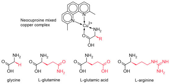
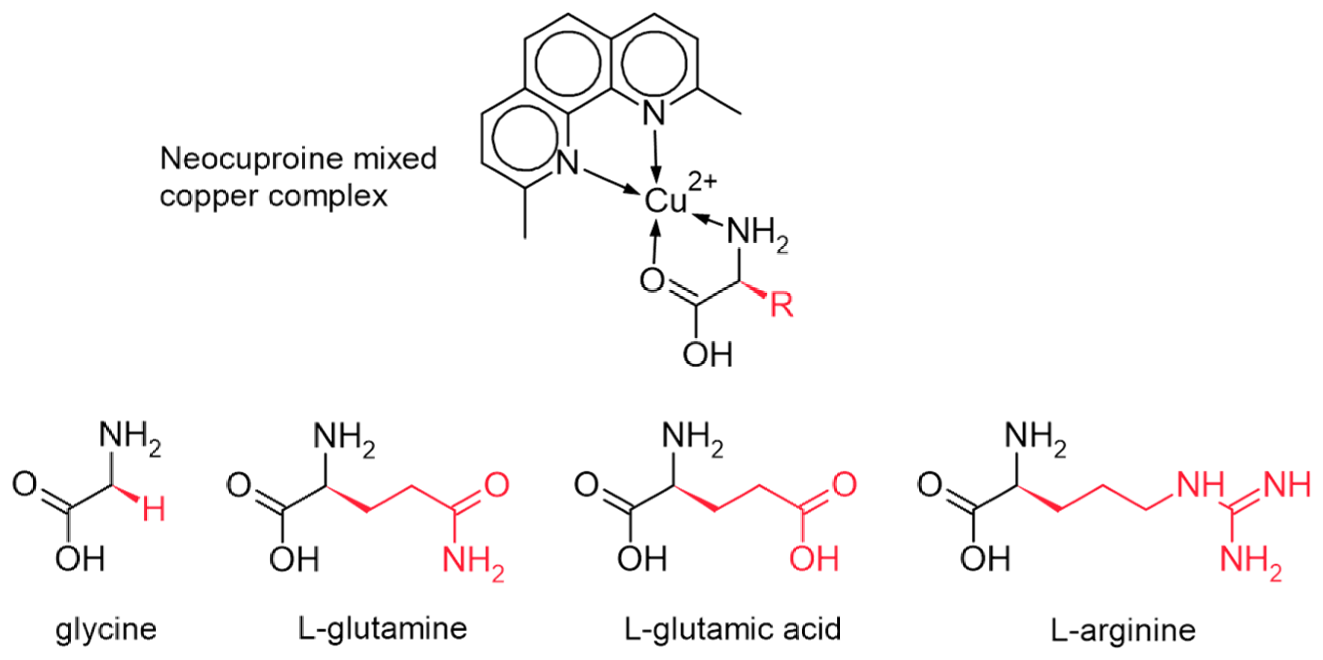
For the above-mentioned, this study presents four new reports on copper (II)–neocuproine mixed complexes with the amino acids glycine (gly), glutamine (gln), glutamic acid (glu), and arginine (arg) as secondary ligands (see Scheme 1). They were physically characterized using XRD and spectroscopic techniques. In addition, Hirshfeld surface analysis, QTAIM, and NCI-RDG computational studies allow better understanding of the structure and properties of such complexes. All of the above forms part of our study on the biological activity of copper (II) mixed complexes, with the aim of elucidating and better understanding the action of this family of compounds.
Scheme 1.
Complexes 1 to 4, indicating the amino acids employed.
2. Results
2.1. Crystal Structure
Crystal data for complexes 1 and 2 are presented in Table 1, and Table 2 summarizes crystal data for complexes 3 and 4. The full crystallographic data are presented in the CheckCIF of all the complexes in the Supplementary Materials (D1–D4).
Table 1.
Crystal data for complexes 1 and 2.
Table 2.
Crystal data for complexes 3 and 4.
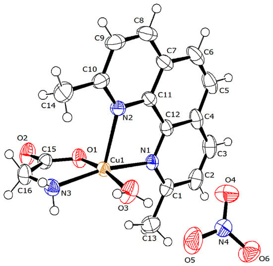
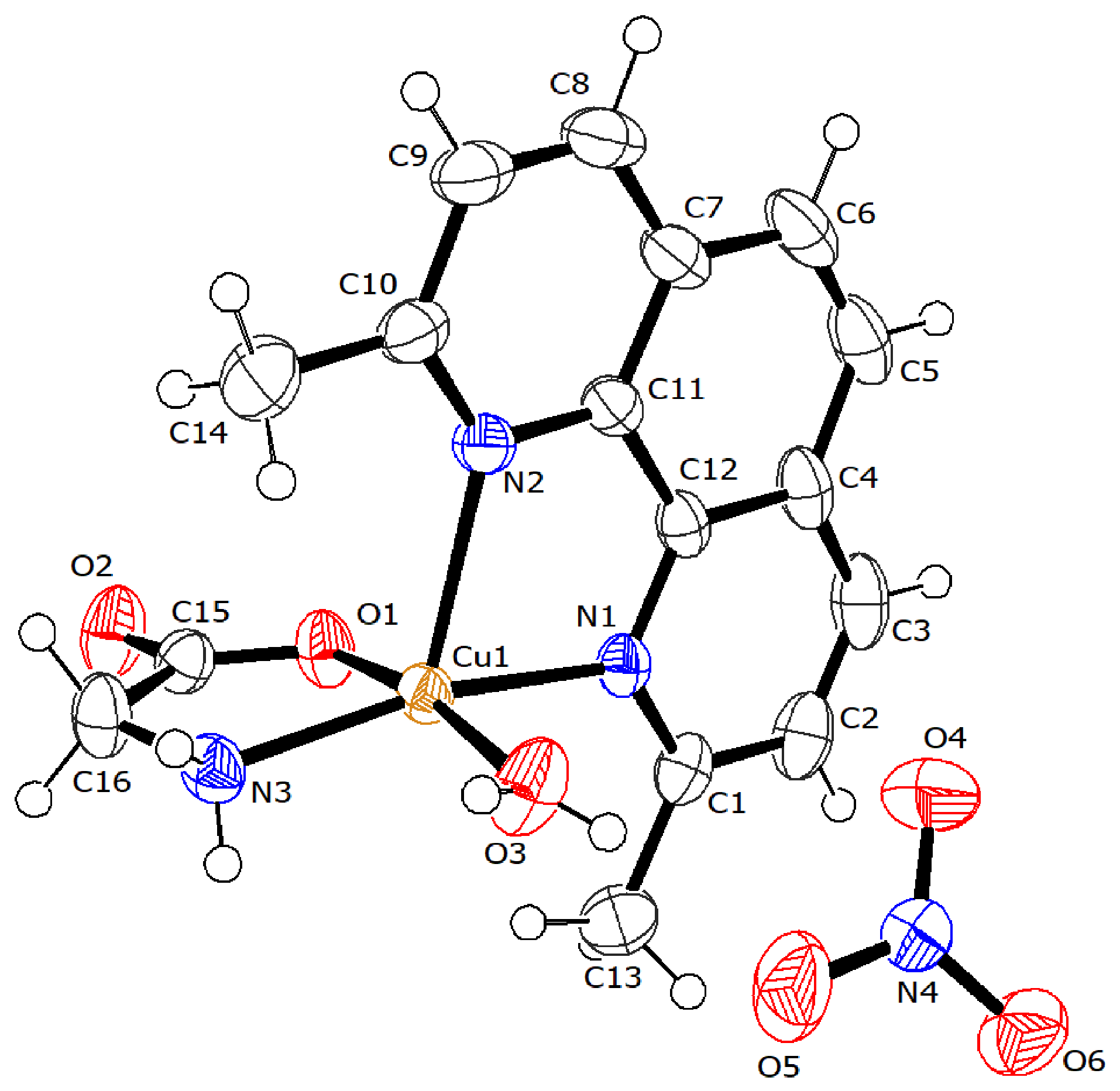
The structure of complex 1 is shown in Figure 1; it crystallizes in the triclinic P system and contains a single complex molecule in the unit cell (Z′ = 1). Despite its trigonal bipyramidal appearance, the geometry of complex 1 is better described as a distorted square pyramidal geometry (τ = 0.076) [27], with the neocuproine N2 nitrogen atom occupying the axial position (2.261 Å) and the four equatorial positions occupied by the remaining neocuproine N1 nitrogen atom (2.014 Å), the N3 atom (1.992 Å), the O1 atom (1.957 Å) of the glycine, and the O3 atom of the coordinated water molecule (1.974 Å). The resulting coordination sphere takes the form CuN3O2. The distances and angles between atoms in the coordination sphere are consistent with a distorted structure in the equatorial plane, orienting the glycine molecule closer to one of the methyl groups of neocuproine. Due to this distortion, the metal center moves closer to the other substituent. A similar compound was recently reported with another counterion [24].
Figure 1.
ORTEP diagram (50% probability) of [Cu(neo)(gly)(H2O) (complex 1).
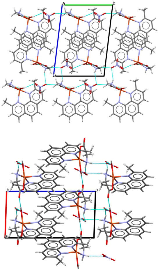
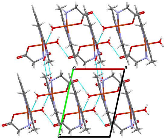
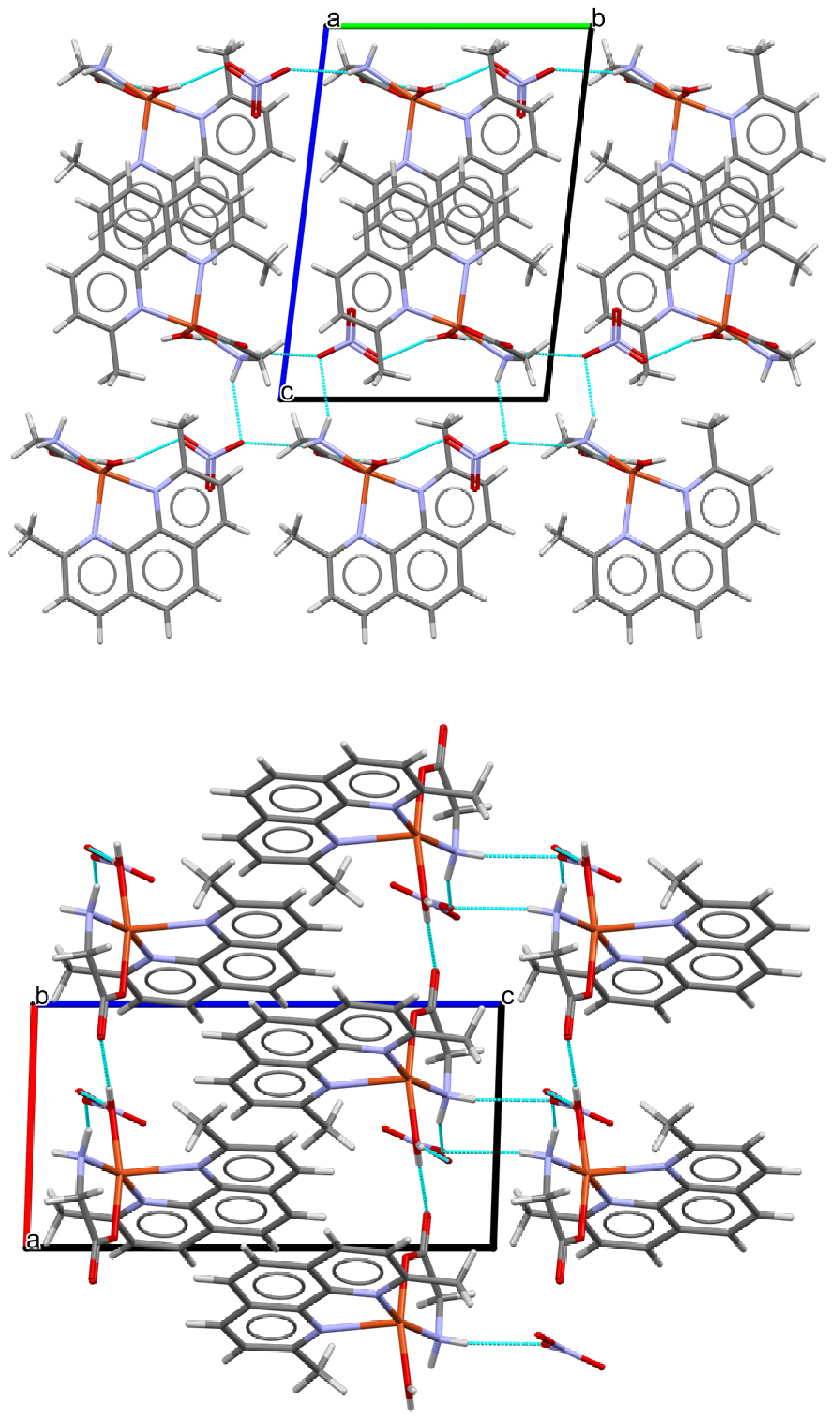
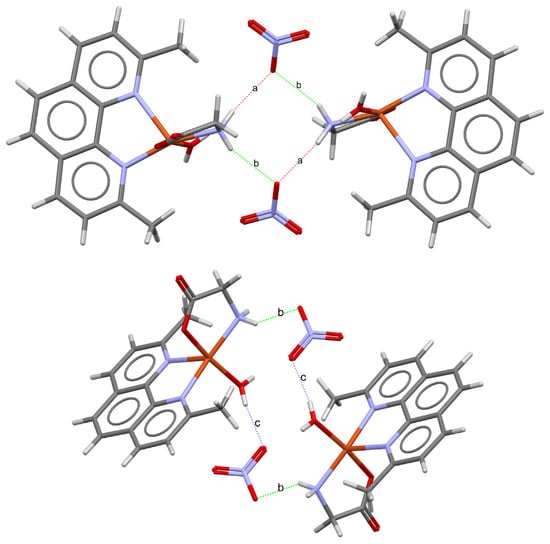
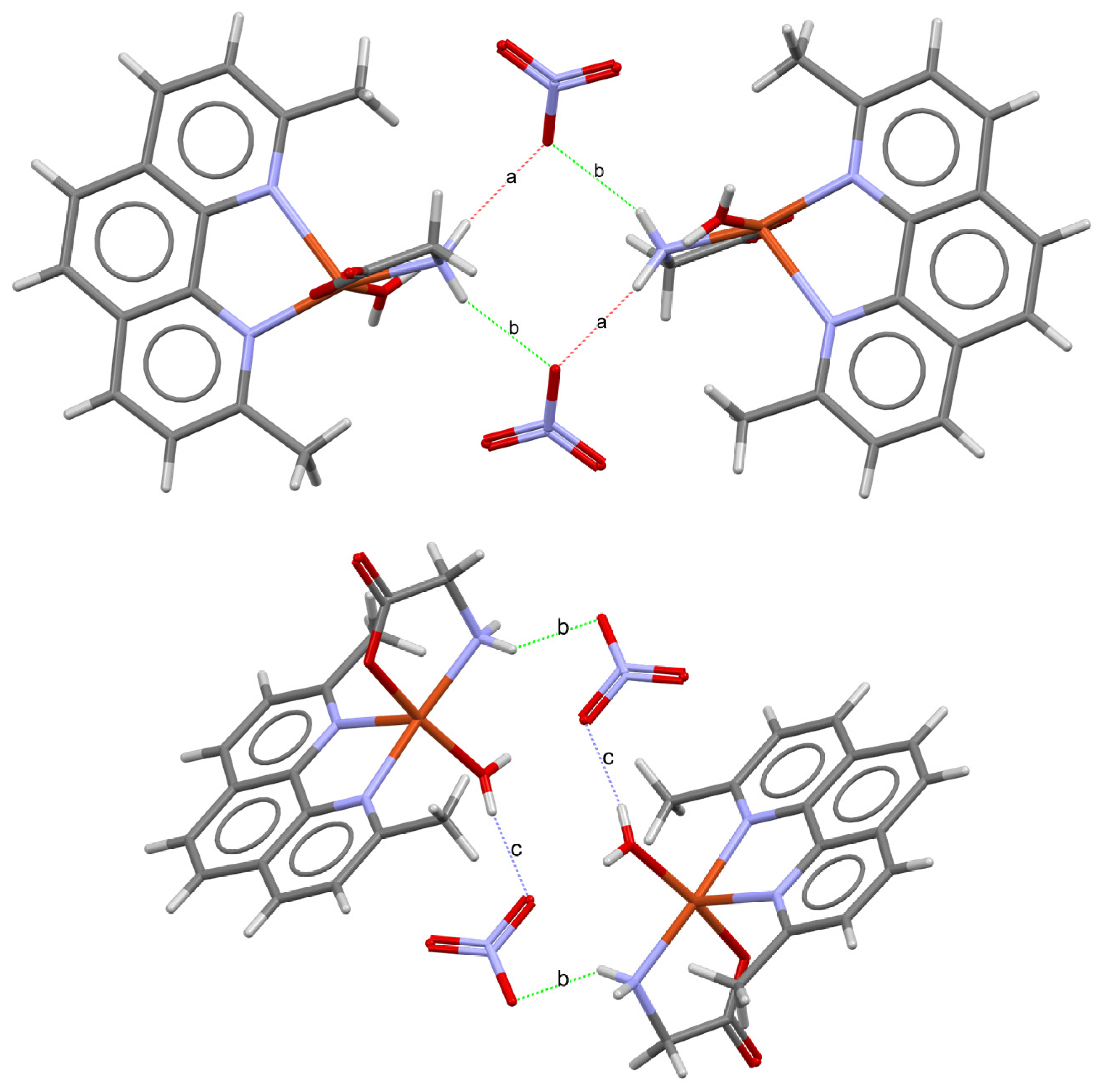
Intramolecularly, complex 1 binds the counterion via a hydrogen bond formed by coordinated water to the nitrate ion (O3-H3C∙∙∙O5; 2.002 Å). Intermolecularly, the complex forms 2D double layers that are distributed along the “a” axis (Figure 2), where, at the base of the layer, the complex molecules are arranged parallel to the a-axis via hydrogen bonds to the neighboring α-carboxyl group (O3-H3D∙∙∙O2; 1.947 Å) in a chain-like arrangement. Parallel to axis “b” (Figure 2), the chains are linked by the nitrate ion, which acts as a bridge between the α-amino group of the complex (N3-H3A∙∙∙O6; 2.226 Å) and the coordinated water molecule, resulting in a four-molecule cycle. At the top of the sheet, the complexes bind to the base by intercalating at 180° via a bond equivalent to N3-H3A∙∙∙O6, thereby forming an eight-membered ring that repeats periodically at the center of the sheet. Finally, with the configuration described, the neocuproine rings are oriented outward from the sheets, and this arrangement allows π-π stacking between neighboring rings with equivalent center-to-center distances of 3.746 Å, which enables the bonding between sheets. Figure 3 shows the rings formed with their corresponding graph set motif notation.
Figure 2.
From top to bottom, packing of complex 1: view from the “a,” “b,” and “c” axes of the unit cell. Blue lines represent the molecular contacts.
Figure 3.
Hydrogen bonds that maintained the structure through the formation of rings with the graph set motifs, top R2,2(8) and bottom R4,4(16).
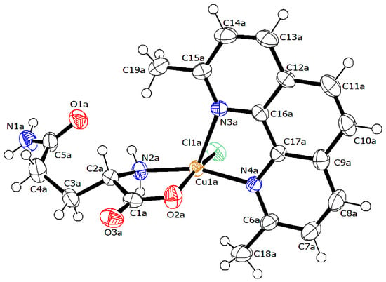
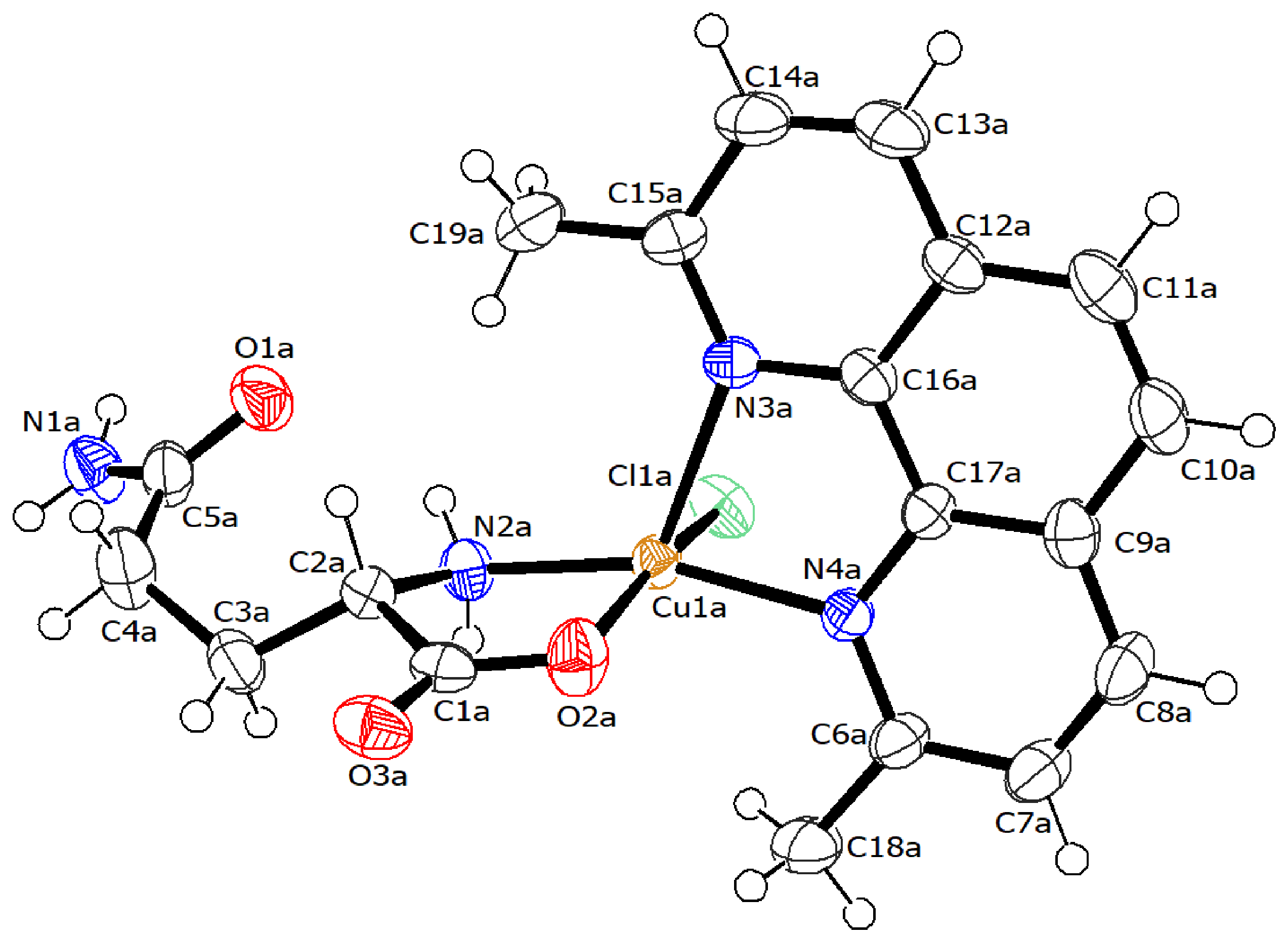
The ORTEP diagram of complex 2 is shown in Figure 4; it crystallizes in the C2 monoclinic system, and the unit cell consists of a dimer (Z′ = 2) (Figure 4) linked by the amide residues of the glutamine ligand. In the complex structure, the pentacoordinate copper metal center can be observed in a square pyramidal geometry (τ5 = 0.038–0.311) [27], with the neocuproine ligand located in a plane perpendicular to the main backbone of glutamine. The neocuproine N3 and N4 nitrogen atoms occupy both axial and equatorial positions, respectively (2.019–2.252 Å), while the glutamine N2 (1.975–1.987 Å) and O2 (1.964–1.972 Å) atoms from the main skeleton occupy the rest of the equatorial positions together with the chloride counterion (2.304–2.311 Å). The resulting coordination sphere takes the CuN3O2 form.
Figure 4.
ORTEP diagram (50% probability) of the moiety of [Cu(neo)(gln)(Cl)](H2O) of the dimer (complex 2). For the sake of simplicity, hydration molecules were omitted.
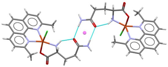
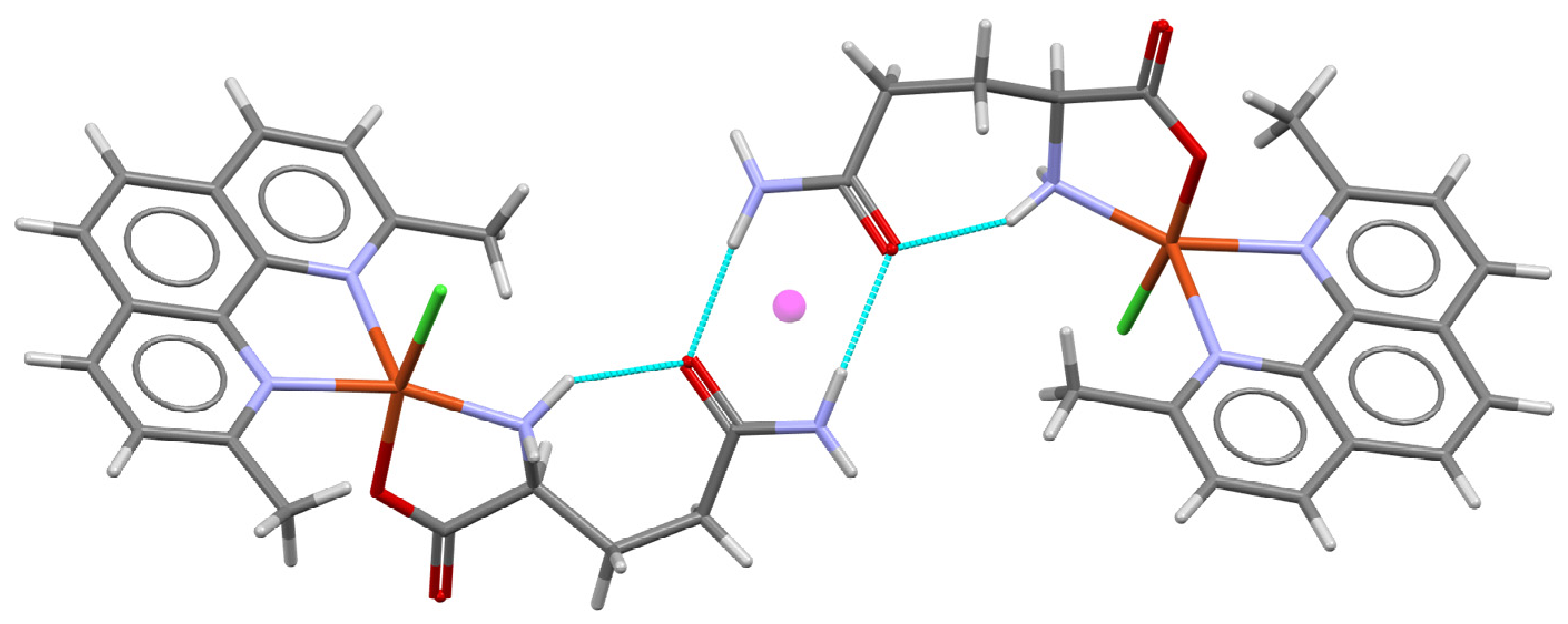
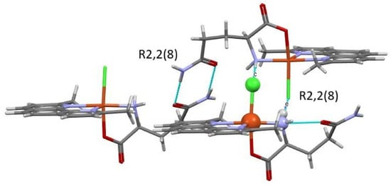
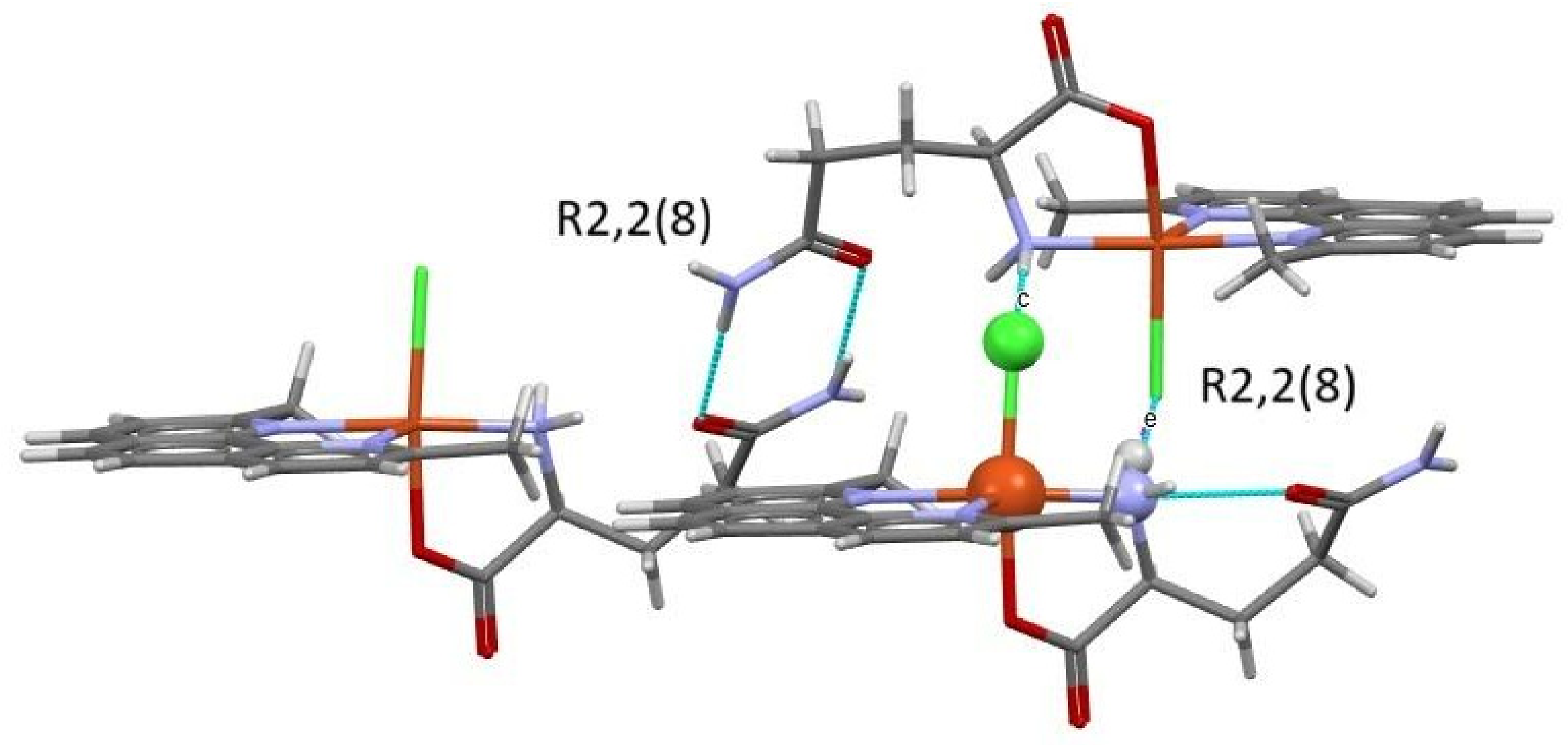
The dimer that makes up the unit cell is held together by the portion corresponding to the glutamine amide residue, where nitrogen N1 forms a hydrogen bond with the corresponding oxygen O1 of the second complex (N1-H∙∙∙O1; 1.981–2.054 Å), forming an eight-membered ring (graph set motif R2,2(8)). This interaction, reminiscent of base pairing, is the primary intramolecular interaction that maintains the stability of the complex; it is supported by a hydrogen bond between the nitrogen (N2) of the α-amino group and the oxygen (O1) of the glutamine (N2-H∙∙∙O1; 2.060–2.107 Å). At the center of the amide bond (Figure 5), a “pseudo-inversion center” is observed; however, the hydrogen on the alpha-carbon is oriented in the same position in both molecules, so it cannot be considered an element of symmetry. Figure 6 presents the rings formed in complex 2.
Figure 5.
Representation of the dimer formed by complex 2, showing the location of the “pseudo-inversion” center in purple. Blue lines represent the molecular contacts.
Figure 6.
Rings involved in the stability of complex 2, indicating the corresponding graph set motifs.
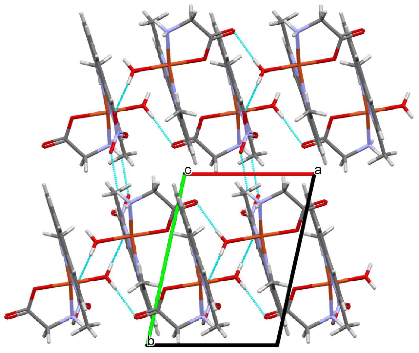
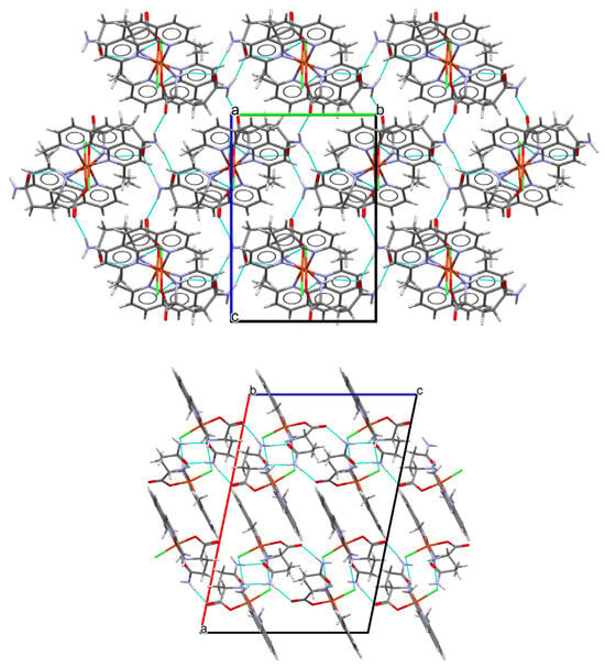
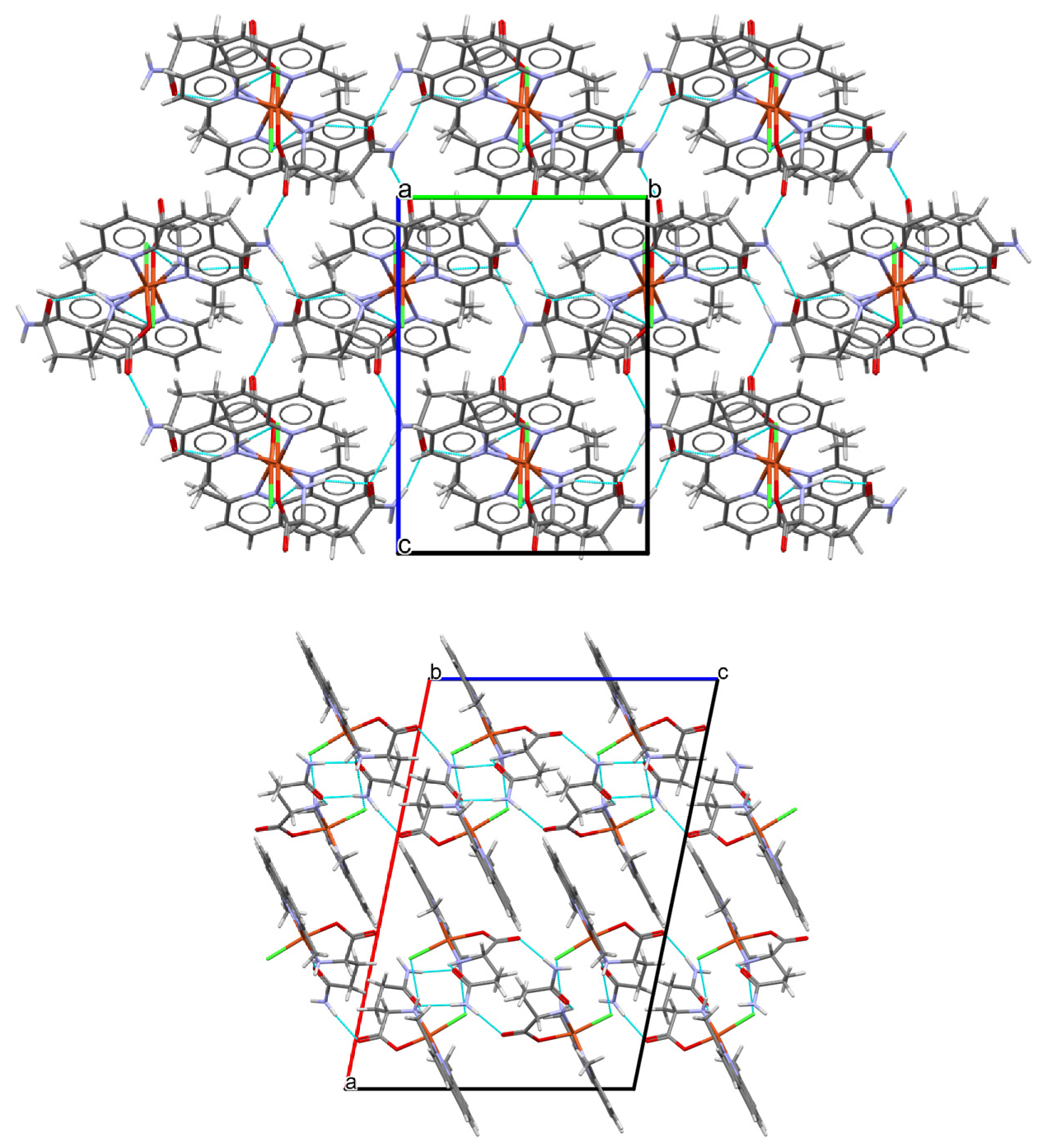
As shown in Figure 7, the intermolecular structure consists of a two-dimensional network, composed mainly of dimer chains, which are linked in parallel along the “b” axis via two hydrogen bonds between the Cl1 atoms and the N2 atoms of the α-amino group and their neighboring counterparts (N2-H∙∙∙Cl1; 2.484–2.538 Å), forming an eight-membered ring (graph set motif R2,2(8)) that intercalates with the amide ring holding the dimer together. The chains are joined perpendicular to the “b” axis via a hydrogen bond between the O3 oxygen of the α-carboxyl group and the remaining proton of an adjacent α-amino group (N2-H∙∙∙O3; 2.017–2.078 Å). The networks are ultimately linked by π–π stacking between two neocuproine rings, with a center-to-center distance of approximately 3.559–3.637 Å.
Figure 7.
From top to bottom, packing of complex 2: view from the “a” and “b” axes of the unit cell. Blue lines represent the molecular contacts.
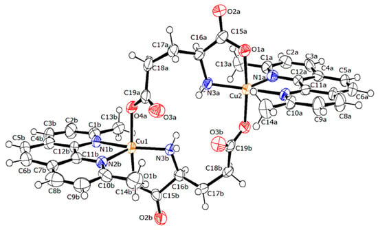
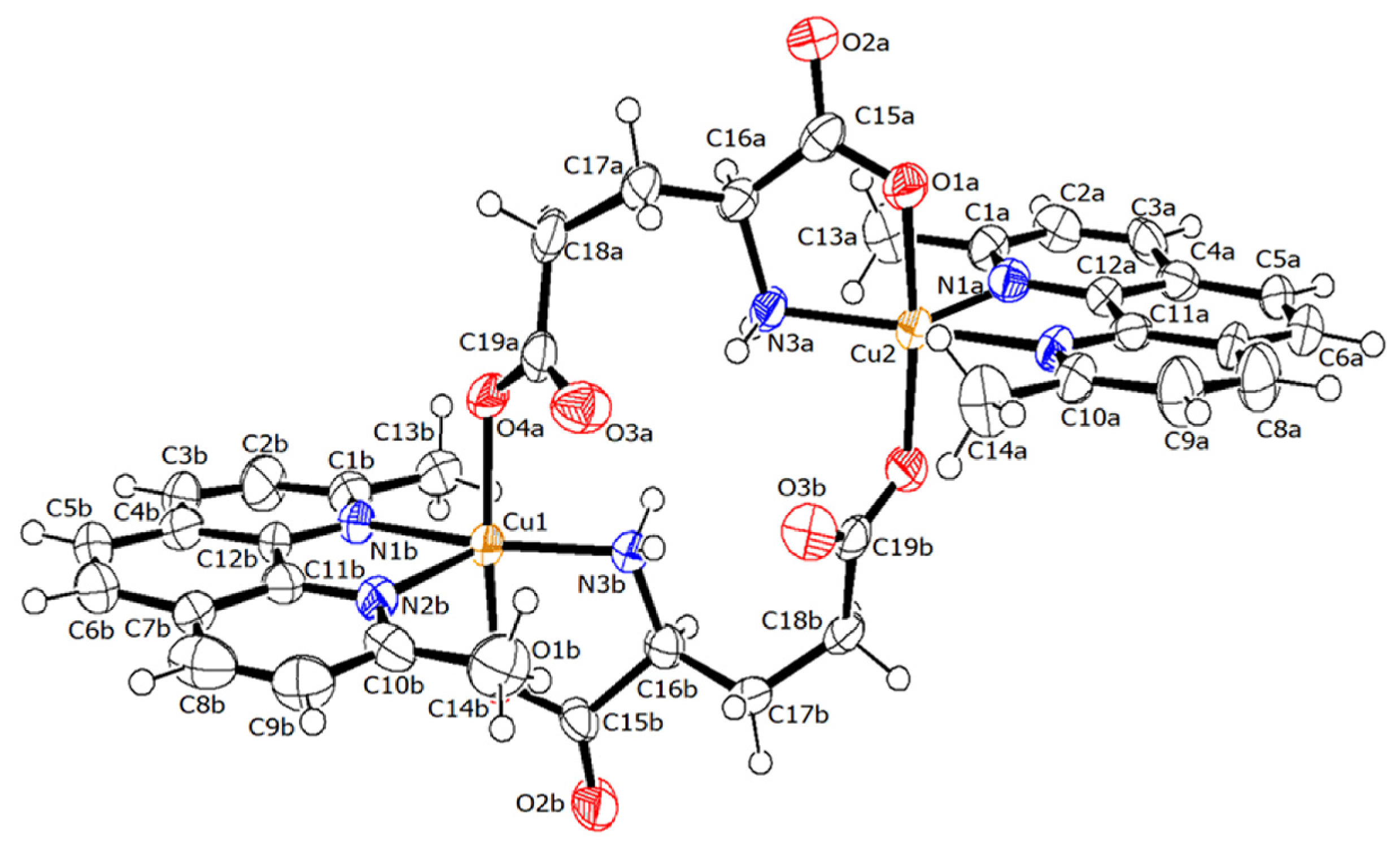
Figure 8 shows the ORTEP diagram of complex 3. It crystallizes as a methanol-solvated dihydrate in the triclinic P1 system, consisting of a dimer (Z′ = 2). This dimer is composed of two mixed-chelate units, in which glutamic acid coordinates the metal center via the amino acid’s main backbone, while the carboxylate residue also coordinates the neighboring metal center, and vice versa. Since glutamic acid is doubly deprotonated, the dimer neutralizes the positive charges of copper without the need for a counterion. At both metal centers of the dimer, a pentacoordinate square-based pyramid geometry is observed (τ = 0.210–0.258) [27]. The axial positions are occupied by atoms N1a (2.275 Å) and N2b (2.232 Å), respectively, for each sphere. The equatorial sites are occupied by the remaining nitrogen atom of neocuproine (2.043 Å N2a; 2.079 Å N1b), nitrogen N3 (1.985–1.986 Å), and oxygen O1 (1.967–1.953 Å) from the main backbone of glutamic acid, as well as oxygen O4 from the terminal carboxylate of the neighboring glutamic acid (1.974–1.969 Å). The resulting sphere takes the form of CuN3O2.
Figure 8.
ORTEP diagram (50% probability) of the bridged dimer [Cu(neo)(glu)]2•2H2O•CH4O (complex 3). For the sake of simplicity, hydration and solvate molecules were omitted for clarity.
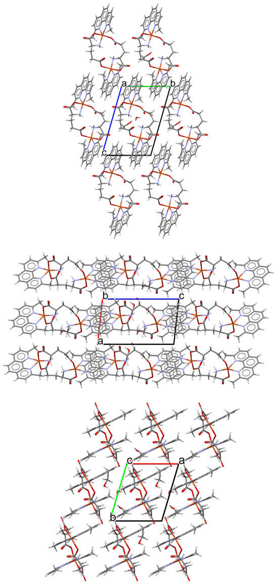
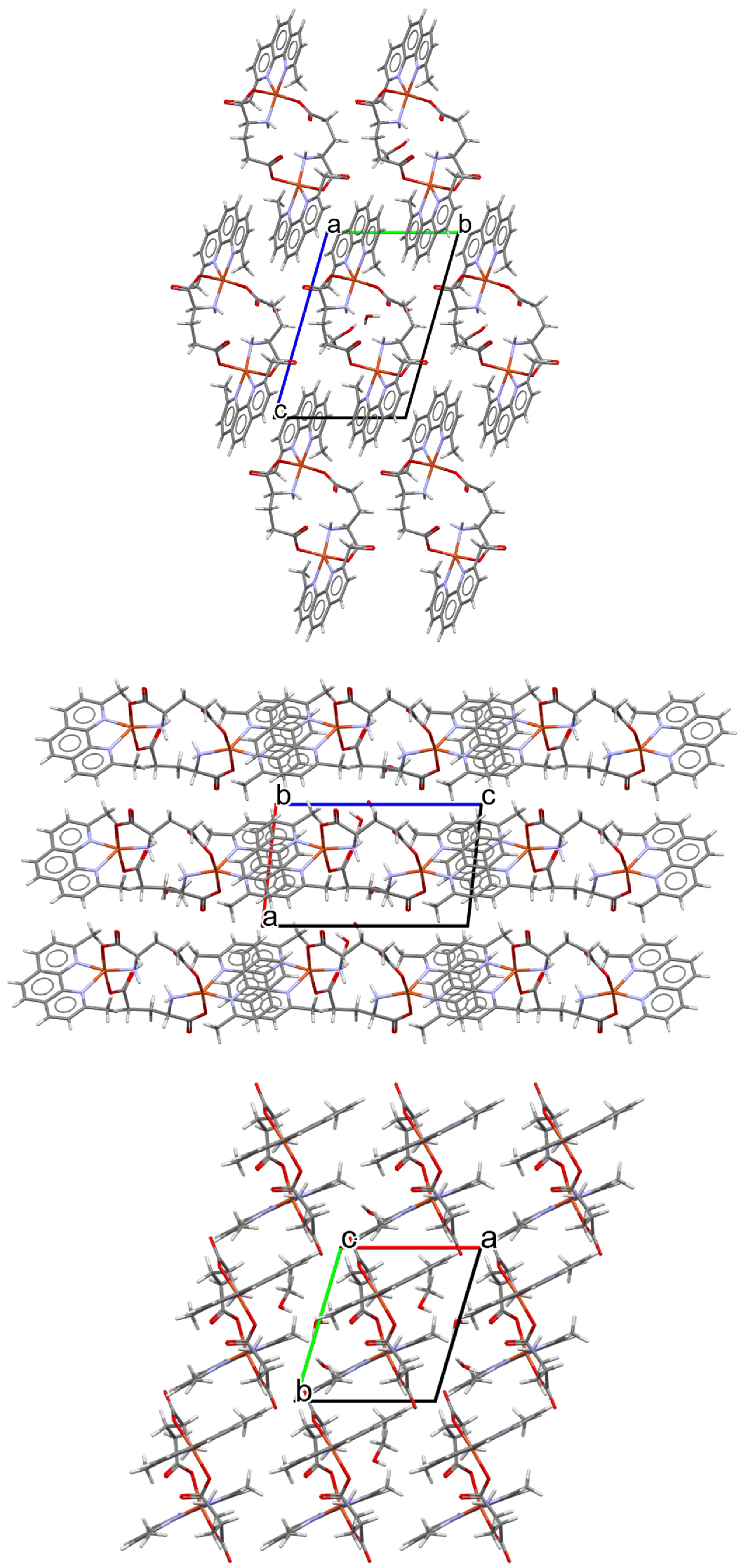
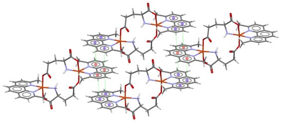
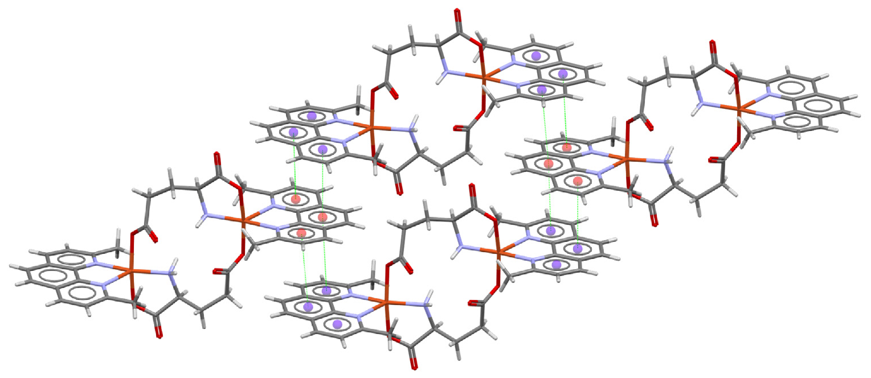
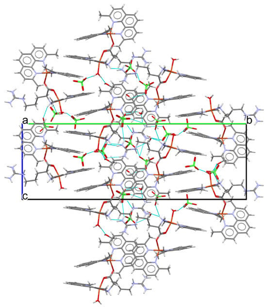
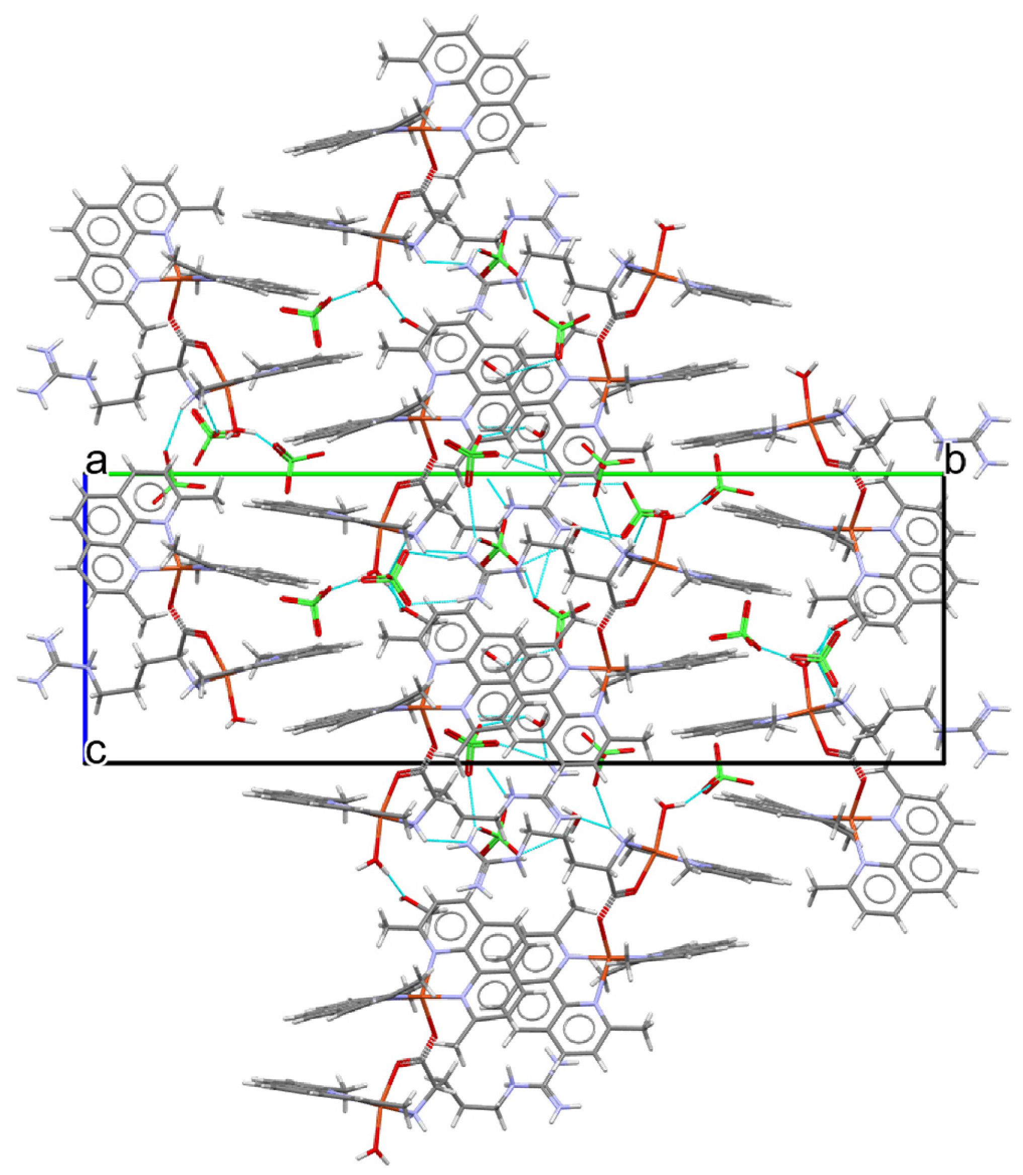
The dimer structure forms a 14-membered macrocycle that connects both metal centers via amino acid residues, which are spaced 6.552 Å apart. This structure is stabilized intramolecularly by hydrogen bonds between the α-amino group of the backbone and the carboxylate group of the same amino acid residue (N3ab-H∙∙∙O3ab; 2.305–2.349 Å). The two α-amino groups are oriented in this position, supported by a hydrogen bond toward the methanol molecule with the remaining proton (N3ab-H∙∙∙O38; 2.055–2.068 Å), and the same solvate connects to neighboring hydration molecules (O38-H∙∙∙O5; 1.825 Å). The packing of complex 3 is shown in Figure 9, and a view of the - stacking is shown in Figure 10.
Figure 9.
From top to bottom, packing of complex 3: view from the “a”, “b” and “c” axes of the unit cell. Blue lines represent the molecular contacts.
Figure 10.
π-stacking (green lines) in complex 3.
The supramolecular structure in this case consists of chains formed by dimers, arranged parallel to the “a” axis. The chains are linked by the hydrogen bonds described above, in which the solvate molecule and a hydration molecule act as a bridge, ultimately connecting to the neighboring carboxyl group (O5-H∙∙∙O3; 1.870 Å). The chains are primarily linked by π-stacking between the exposed rings of neocuproine (Figure 10), where the chains intercalate at approximately 90°. The aromatic rings of neocuproine form oblique bonds with neighboring rings, creating two grooves based on the distances between centroids.
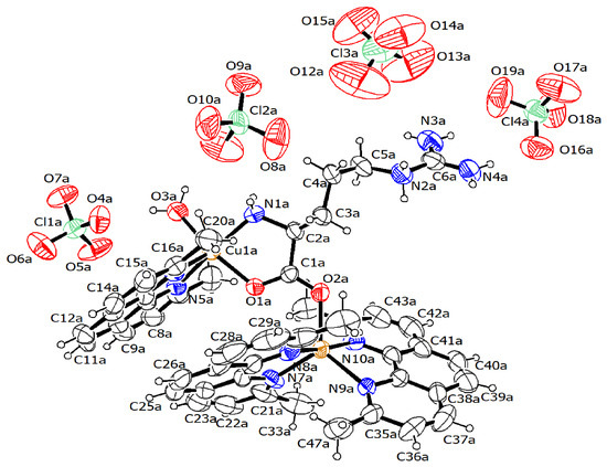
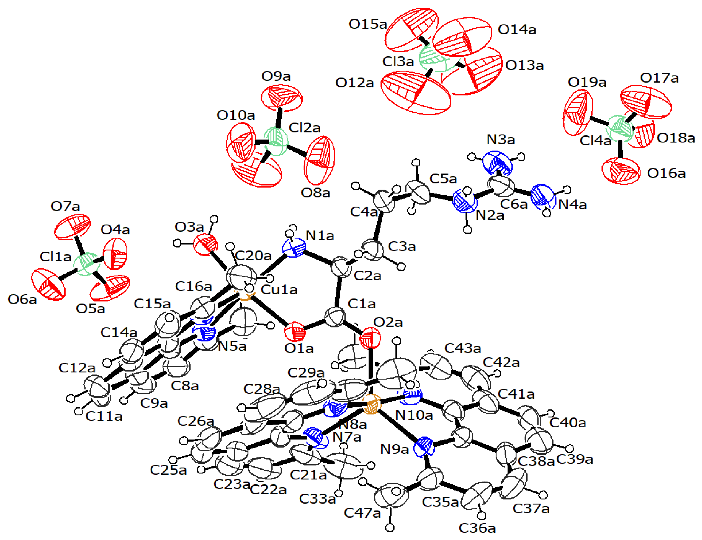
Figure 11 shows the ORTEP diagram of complex 4, which features a structure unlike any previously observed in this study. This molecule consists of a mixed neocuproine–arginine–copper(II) complex and a bis-neocuproine–copper(II) complex, in which the α-carboxyl group of arginine coordinates with the two metal centers, linking the two moieties. This complex crystallizes in the monoclinic P21 system, with two molecules in the unit cell (Z′ = 2), and despite the nature of the P21 system, no isomerism is observed in the structure. The overall conformation of the complex results in two types of coordination within the same crystal. The first consists of the mixed complex, which exhibits a square-based pyramid geometry (τ = 0.017–0.024) [27]. The nitrogen atom N6 of neocuproine occupies the axial position (2.197–2.211 Å), while the equatorial positions are occupied by atoms O1 (1.956–1.964 Å) and N1 (1.966–1.995 Å) of the arginine backbone and by oxygen O3 from the coordinated water molecule (1.974–1.999 Å). The resulting sphere in this case takes the form of CuN3O2.
Figure 11.
ORTEP diagram (30% probability) of the complex 4-cell moiety. For the sake of simplicity, hydration and solvate molecules were omitted. [Cu(neo)3(arg)](ClO4)4.
The second type of coordination corresponds to the “bis-neocuproine” moiety, which exhibits a trigonal bipyramidal geometry (τ = 0.722–0.724) [27]. Each of the two neocuproine ligands occupies both an axial and an equatorial position. The axial positions are occupied by atoms N8 (1.969–1.993 Å) and N10 (1.980–1.992 Å), while the equatorial positions are occupied by atoms N7 (2.067–2.096 Å) and N9 (2.082–2.131 Å). The remaining equatorial position is occupied by the remaining O2 oxygen atom from the adjacent α-carboxyl group (2.026–2.029 Å), resulting in a sphere with the formula CuN4O.
Due to the disorder observed in the crystal of this complex, it is difficult to determine and describe both intramolecular and intermolecular interactions, since three perchlorate counterions (ClO4−) overlap what appear to be two orientations throughout the entire crystal. What can be correctly established, however, is that the two molecules comprising the unit cell are held together by the arginine residues via hydrogen bonds with three of the perchlorate counterions in a bridging arrangement (Figure 12). The arginine in the mixed-ligand complex bonds via the α-amino group to the oxygen of the perchlorate (N1-H∙∙∙O17a/O11a; 2.211–2.488 Å), which in turn connects to the two terminal amino groups of the neighboring guanidinium (N4-H∙∙∙O16a/O9a; 2.126–2.202 Å) (N3-H∙∙∙O19a/O8a; 2.235–2.238 Å), with this bond in the middle being supported by a bridge to oxygen O12 of an intermediate perchlorate (N2b-H/N3a-H∙∙∙O12; 2.602–2.627 Å). The solvated methanol molecule also contributes to the stability of this ring, helping to orient the perchlorate via a hydrogen bond (O21a-H∙∙∙O11a; 2.283 Å) and anchoring itself toward the coordinated water via a similar interaction (O3a-H∙∙∙O21a; 1.833 Å). The disordered perchlorates also form bonds with the arginine amino groups, but since these interactions are not continuous throughout the crystal, it is difficult to say with certainty how often they recur.
Figure 12.
Packing of complex 4: view from the “a” axis of the unit cell. Blue lines represent the molecular contacts.
2.2. Spectroscopic Characterization
The four complexes presented in this study exhibit bands typical of the aminoacidate-type mixed-ligand complex family in FT-IR and Raman spectroscopy, which are patterns expected for the target structures [28,29]. FT-IR spectroscopy, in all cases, shows the typical amide bands, assignable to the aminoacidate ligands. The bands near 3000–3000 cm−1 are assignable to the asymmetric stretching of the amine (NH) groups, while the bands near 2700–2900 cm−1 are assignable to the asymmetric stretching of the methylene groups of the amino acid residue (except for complex 1 with glycine). Similarly, the bands in the 1600–1500 cm−1 range, as well as those near 1400–1300 cm−1, are assignable to amide I, II, and III signals, respectively. Neocuproine has assignments primarily in the region below 900 cm−1, where out-of-plane (CH) bending vibrations are present in the 700–800 cm−1 range, although the band assignable to the C–N stretch at 1200–1300 cm−1 can also be observed; however, it is difficult to distinguish it from the other amino acid signals. All of the above is accompanied by a large number of shoulders and overtones. Counterions are observable as high-intensity bands only in complexes 1 and 4, for nitrate and perchlorate ions, with expected values of 1300 cm−1 and 1000 cm−1, respectively. Complex 3 lacks the typical sulfate band in the 1000 cm−1 region, providing further evidence that the sulfate counterion is not present in the dimer’s structure. The Raman spectrum follows a similar pattern, particularly with the bands near 1400 cm−1, which can be assigned to symmetric vibrations of the amino acid’s main backbone.
2.3. Molecular Structure and Non-Covalent Interaction Calculations
In solution, the optimized molecular structures obtained from DFT calculations show a preference for theoretical τ5 [27] for distorted square pyramidal geometry rather than distorted trigonal bipyramidal geometry, as shown in crystal structure data in Section 2.1; see Table 3. Both angles, a and β, retain similar values in X-ray and solution structures for complexes 1–3. However, for complex 4, solution-phase calculations vary significantly due to the size and complexity of the system. Cartesian coordinates and optimized molecular structures calculated at the DFT/mPW1PW91 level of theory are shown in Table S1 and Figure S1. Non-covalent interaction analysis was performed on the optimized molecular structures of complexes 1–4, using QTAIM and NCI-RDG approaches, while Hirshfeld surface analysis was carried out on the crystal structures.
Table 3.
τ5 parameter and α and β interatomic angles (in degrees) of complexes 1–4.
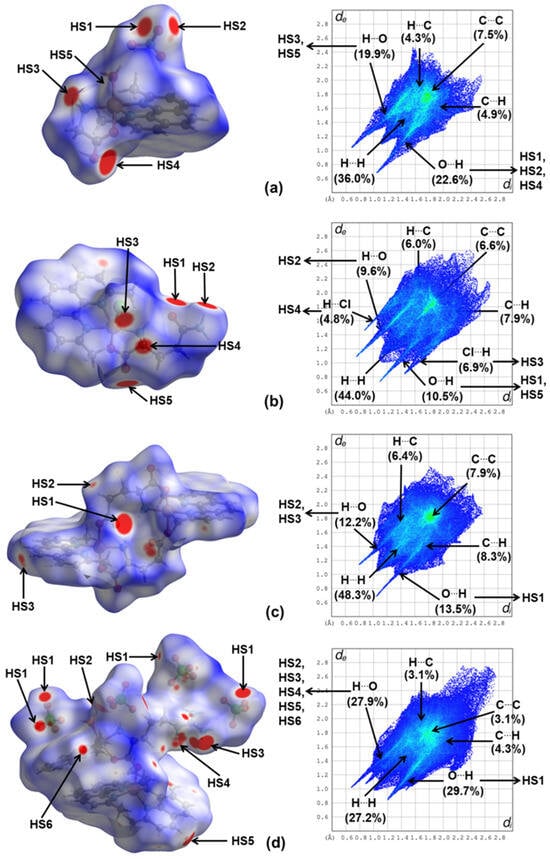
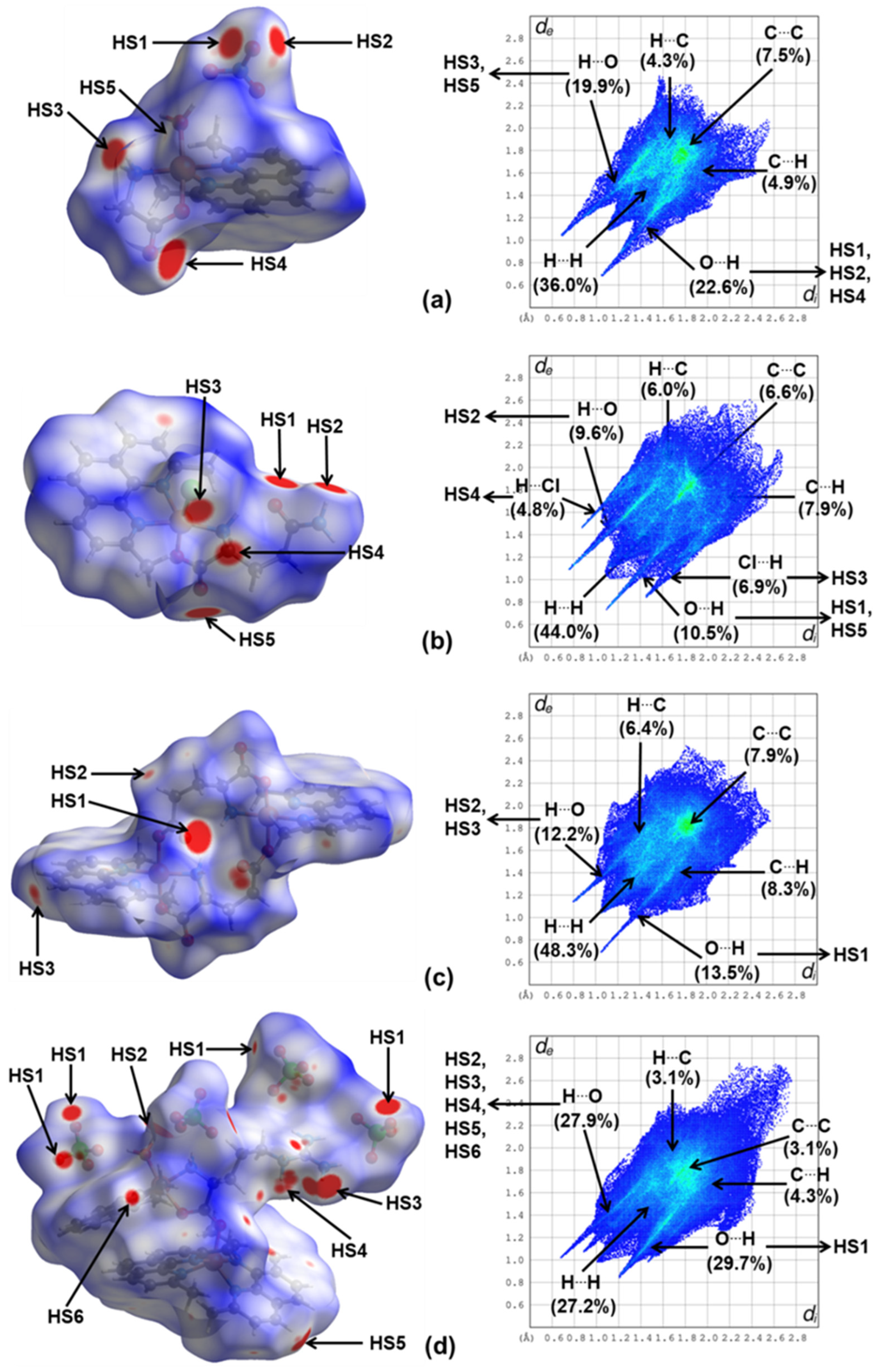
Hirshfeld surface analysis [30] was carried out to study the main contacts and interactions in the crystal packing of complexes 1–4. Figure 13 shows the Hirshfeld surfaces (HS) and the 2D-fingerprint plots of complexes 1–4 generated with the function dnorm. For complex 1, in Figure 13a, the red spots, labeled as HS1, HS2, and HS4 (with 22.6% of contribution), are due to the hydrogen bonds O…H–O between the oxygen atoms of the nitrate ion (HS1 and HS2) and glycine (HS4) inside HS, acting as acceptors of hydrogen atoms of the -NH2 moiety of glycine and the H2O molecule in the apical position outside HS of adjacent molecules in the crystal packing. HS3 and HS5 (with 19.9%) correspond to the hydrogen bonds O–H…O of hydrogen atoms of glycine and water inside HS, with the oxygen atoms of the nitrate ion and glycine outside HS. Other interactions with minor contributions are C…C (7.5%), C…H (4.9%), and H…C (4.3%). For complex 2, in Figure 13b, the hydrogen bonds O…H–O (HS1 and HS5) and O–H…O (HS2) decrease their contribution to 10.5 and 9.6%, respectively, due to the contribution of halogen bonds Cl…H–N between the Cl apical inside HS, acting as an acceptor, with hydrogens of the -NH2 moiety of glycine outside HS (HS3), and halogen bonds N–H…Cl between the -NH2 moiety of glycine inside HS, acting as a donor, with Cl of adjacent molecules outside HS (HS4). Other contributions are assigned to C…C (6.6%), C…H (7.9%), and H…C (6.0%) interactions. For complex 3, in Figure 13c, the hydrogen bonds O…H–O (HS1) contributed the majority with 13.5%, while O–H…O (HS2 and HS3) contributed with 12.2%. In this complex, interactions are observed to be almost symmetric through the carboxylate moiety of glutamic acids and neocuproine moieties. The interactions C…C (7.9%), C…H (8.3%), and H…C (6.4%) are consistent with major contacts with the neocuproine adjacent molecules outside HS. For complex 4, in Figure 13d, the major contribution of 29.7% is due to hydrogen bonds O…H–O (HS1) where the oxygen atoms of ClO4 ions inside HS act as acceptors of hydrogen atoms of outside HS molecules of the -NH2 moiety of arginine and neocuproine. Meanwhile, the hydrogen bonds O–H…O with a contribution of 27.9% (HS2-HS6) arise from the intermolecular interactions between hydrogen atoms of the -NH2 moiety of arginine and the -CH of neocuproine inside HS, acting as donors of the ClO4 ions outside HS. The major contributions of 36.0% to 48.3% for complexes 1–3 to the HS are due to the close intermolecular interactions H…H, while complex 4 has a moderate contribution of 27.2%, due to the H of neocuproine with adjacent molecules in the crystal packing. This difference is due to the increase in hydrogen bonds with the oxygens of ClO4 ions in complex 4.
Figure 13.
(a–d) Hirshfeld surfaces (HS) mapped with the dnorm parameter and 2D-fingerprint plots of non-covalent interactions of complexes 1–4.
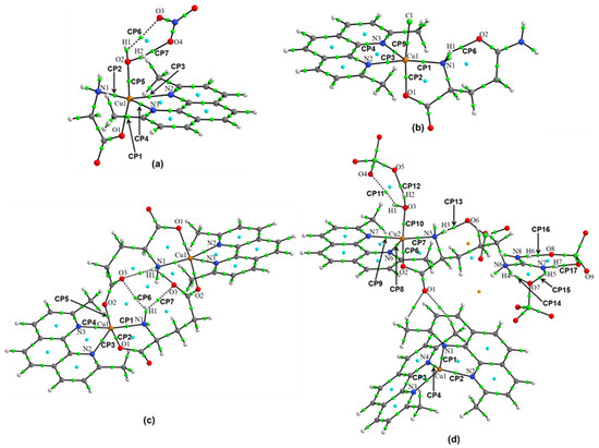
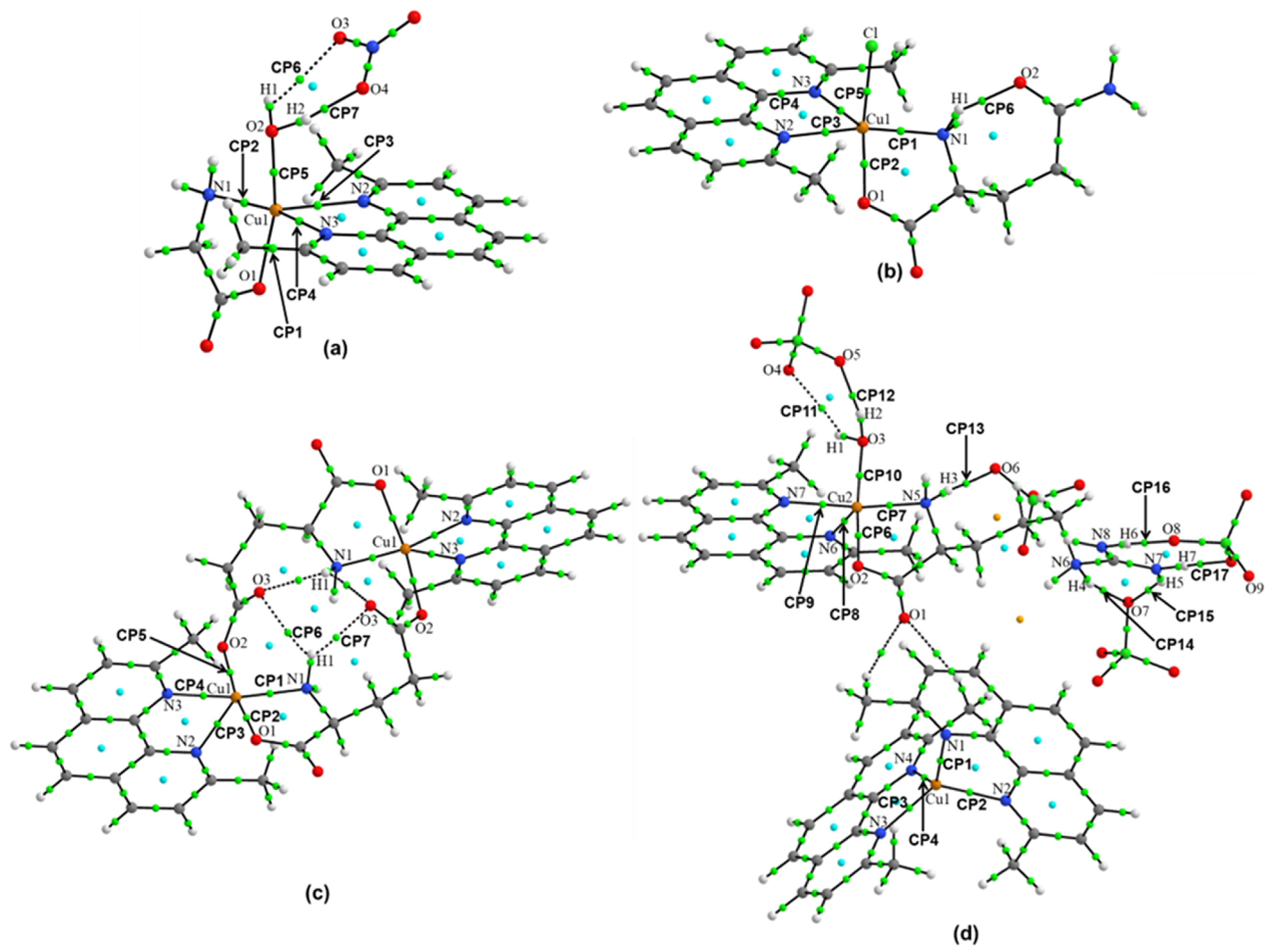
The QTAIM analysis based on the calculation of the topological parameters of the electron density on the bond critical points (BCPs) [31] was carried out to classify the non-covalent interactions with the coordinated Cu(II) atom and the hydrogen and halogen bonds in complexes 1–4. The electron density ⍴(r), the Laplacian of density ∇2⍴(r), the energy densities G(r), V(r), and H(r), the interatomic distance Dint, the interatomic angle Aint, and the energy of interaction EH···Y are shown in Table 4. Figure 12 shows the molecular graphs of complexes 1–4, where the green dots indicate bond critical points (BCPs), the blue dots indicate ring critical points (RCPs), and the orange dots indicate cage critical points. For complexes 1–3, the values of the topological parameters of ⍴(r) on the BCPs for the interactions coordinated with the Cu1 atom have values at similar ranges; for the interactions coordinated with the Cu1 atom (Cu···O and Cu···N, CP1-CP5), see Table 4. In addition, the positive values of ∇2⍴(r) in these interactions indicate the presence of metal–ligand non-covalent interactions in the coordinated Cu1 atom in the three complexes. The calculated interaction energy (EH···Y) for the BCP (labeled as CP1-CP5 in Figure 14a–c) is in the range of 16.35–42.29 kcal mol−1, with slightly higher values for complexes 1 and 2. Non-covalent interactions of type O-H···O with EH···Y of 16.22 kcal mol−1 for complex 1 (CP7), and N-H···O with EH···Y of 8.03 kcal mol−1 for complex 2 (CP6) and 4.17 kcal mol−1 for complex 3 (CP7) indicate the presence of strong hydrogen bonds in the crystal packing. For complex 3, N-H···O intramolecular interactions (CP6 and CP7) form a quasi-symmetric network in the center of the macrocycle dimer. Several RCPs form stable five-membered ring structures around Cu(II) coordinated in the three complexes (see Figure 14a–c). For complex 4, a more complex network of interactions is observed, arising from the non-symmetrical structure and the presence of four ClO4 ions. CP1-CP10 indicate metal–ligand interactions Cu···O and Cu···N between Cu1 and Cu2 atoms coordinated with neocuproine and arginine. These interactions have values of EH···Y of 18.98–37.15 kcal mol−1, except for Cu1···O1, where, in the solution-phase geometry, this distance increased to 4.8 Å. Strong hydrogen bonds of type O-H···O are found with EH···Y of 26.78 kcal mol−1 (CP12) or of type N-H···O with EH···Y of 14.6 (CP14), 10.39 (CP16), and 10.13 (CP17) kcal mol−1. All of these interactions involve ClO4 ions in the crystal packing (see Figure 14d).
Table 4.
Topological parameters (in a.u.), interatomic distances Dint (in Å), interatomic angles Aint (in degrees), and interaction energies EH···Y (in kcal/mol) of complexes 1–4.
Figure 14.
(a–d) Molecular graphs showing the main non-covalent interactions of complexes 1–4.
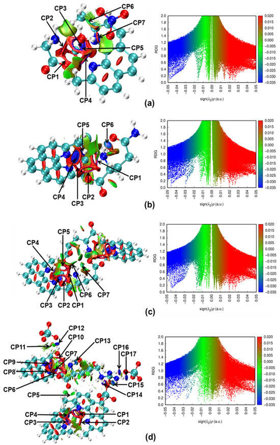
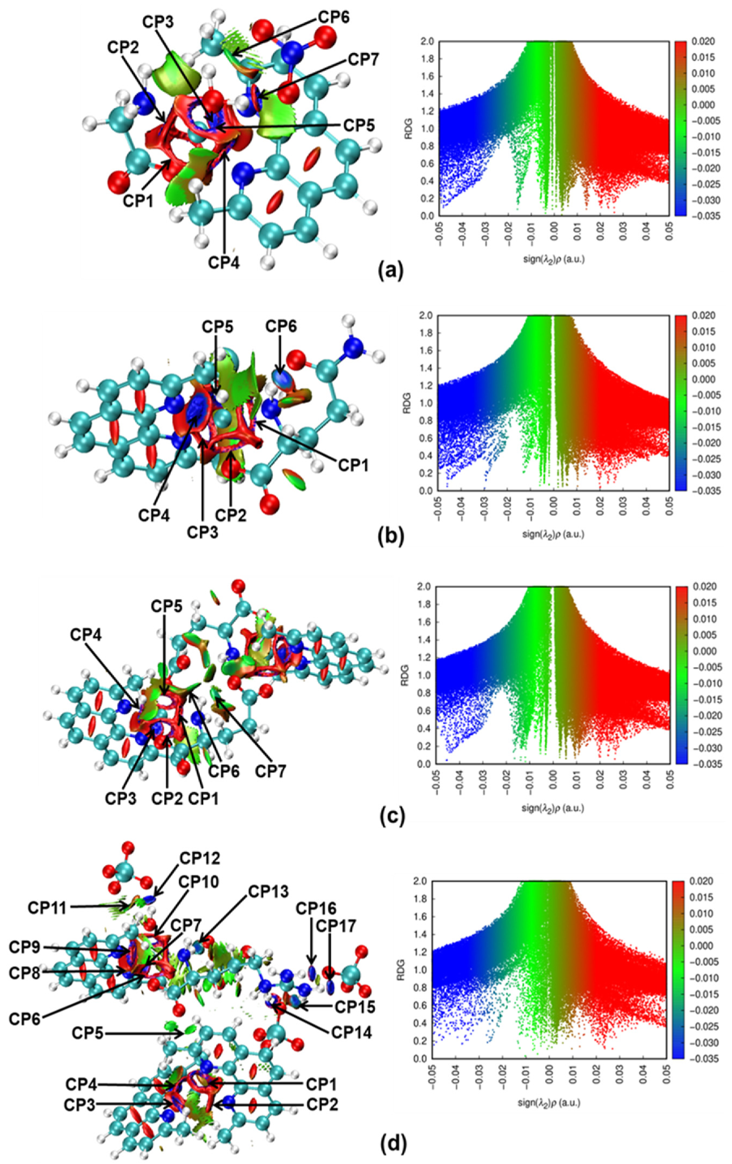
The NCI-RDG analysis was used to detect non-covalent interactions using the electron density and its reduced gradient [32] to identify strong attractive non-covalent interactions such as van der Waals, hydrogen bonds, and halogen bonds in complexes 1–4. Figure 15 shows the non-covalent interaction (NCI) surfaces and the reduced density gradient (RDG) graphs. In complexes 1–3, the metal–ligand interactions Cu···O and Cu···N between Cu1 and amino acid and neocuproine ligands are shown in blue regions labeled as CP1-CP5 in NCI surfaces. These surfaces around Cu1 atoms present some regions of repulsive interactions in red. More attractive interactions are shown for hydrogen bonds of the N-H···O type between Cu1 with a nitrogen atom of the neocuproine ligand (see CP3 and CP4 in complexes 1 and 3, and complex 2, respectively). In complex 1, the surfaces for CP6 and CP7 correspond to strong hydrogen bonds of the O-H···O type between H2O coordinated with Cu1 and the NO3 anion, while in complex 2, CP6 corresponds to a strong hydrogen bond of the N-H···O type in the backbone of glutamine. In RDG graphs, similar patterns are observed for these attractive interactions in the regions of −0.05 to −0.02 a.u. Also, in complexes 1–3, several green surfaces correspond to weak hydrogen bonds and van der Waals interactions, for example, the regions around methyl moieties of the neocuproine ligand in complexes 1–3, CP6 in complex 1, and CP6 and CP7 in the center of the macrocycle symmetric dimer in complex 2. These weak vdW interactions are observed in the regions of −0.02 to 0.005 a.u. in the RDG graphs (see Figure 13a–c). For complex 4, the attractive interactions arise mainly from the strong hydrogen bonds of N-H···O and hydrogen bonds between N-H of neocuproine and arginine ligands with the oxygen atoms of ClO4 ions (CP12-CP17) rather than the attractive metal–ligand interactions of Cu···O and Cu···N with Cu1 (CP1-CP4) and Cu2 (CP6-CP10) coordinated atoms. These attractive interactions are observed in the region of −0.05 to 0.02 on the RDG graph, with a greater dispersion than in complexes 1–3. Also, it is clearly observed that van der Waals interactions are distributed on the arginine ligand and around methyl moieties of the neocuproine ligand. Specifically, the weak interaction in CP5 between Cu1 and the O1 atom is due to the large interatomic distance of 4.8 Å, as corroborated by the null value of ⍴(r) on the corresponding BCP; see the QTAIM analysis mentioned above. The increment of vdW interactions in complex 4 is observed in the region of −0.02 to 0.01 in the RDG graph, in comparison with the RDG graphs of complexes 1–3 (see Figure 15d).
Figure 15.
(a–d). Non-covalent interaction (NCI) graph and reduced density gradient (RDG) graph of complexes 1–4.
3. Discussion
The four complexes presented in this study are part of a series of mixed copper(II)–neocuproine complexes, in which many structural characteristics are consistent with previous reports, such as the distances and angles of the coordination bonds [21,23,24,33,34,35,36,37,38,39,40,41,42,43,44,45]. The pentacoordinate system with square-based pyramid geometry found in the new reports in this study is also observed in previous reports on copper(II) complexes with bidentate and tridentate secondary ligands [24,33,34,35,36,37]. Copper(II) complexes with trigonal bipyramidal geometry are also observable, but less frequently [38]. The preference for square-based or trigonal systems appears to be a matter of the interactions between neocuproine and the secondary ligand, where steric hindrance dominates this phenomenon.
Complexes 1 and 2 in this study are direct analogs of the [Cu(neo)(ala)(H2O)](NO3)•H2O [34] complex and the [Cu(neo)(gly)(Cl)]•2H2O [24] complex, respectively, and are nearly equivalent in both bond distances and ligand arrangement. The effect of the counterion on the network of interactions is one of the main differences. In our reports, the network of the [Cu(neo)(gln)(Cl)] complex is based on interactions between neighboring glutamines, where the chloride counterion plays a secondary role, unlike [Cu(neo)(gly)(Cl)]•2H2O, where the chloride counterion is the primary linking element. In the case of the [Cu(neo)(gly)(H2O)](NO3) complex, the molecules are linked via bridges with the coordinated water, and although this same arrangement is also observed in [Cu(neo)(ala)(H2O)] (NO3)•H2O, the network in this complex also expands due to lateral interactions on the α-carboxyl side.
Complex 3 is an unusual case compared to what we have seen so far, as it consists of a neutral ditopic dimer in which the initial counterion (SO4−2) is eliminated from the final structure. Although it shares similarities with the complex [Cu2(heae)(neo)2](ClO4)•2H2O and complex [Cu2(gly)2(nphen)2(H2O)2](NO3)2 [23,44], the dimer is held together by two glutamate molecules, and it remains a predominantly ionic complex. Glutamic acid is known to have ditopic properties that may explain this phenomenon; the reported complexes [Cu (glu)(Im)], [Cu(bipy)(glu)], and [Cu(phen)(glu)(H2O)] [41] also exclude the initial counterion by neutralizing the charges with the amino acid residue while coordinating a neighboring metal center, although they do not form a ring. It is likely that the formation of this ring is driven by the methyl substituents of neocuproine, where steric hindrance prevents alternative conformations.
Complex 4 is even more unusual due to the presence of a binary complex coordinated to the mixed complex, which breaks the 1:1 ligand ratio previously observed. However, this is not an exceptional case; there are reports of copper(II)–phenanthroline complexes with ligands such as 2-hydroxybenzyl-α-glycine, (p-xylylene)-dialanine, or iminodiacetic acid with similar structures [39,40,42,43]. These structures appear to recur with ligands containing various ionizable groups, and in order to achieve stability, a ligand redistribution must occur that forces the formation of an energetically favorable structure satisfying all local interactions.
In relation to our previous work, several mixed copper (II) complexes synthesized and experimentally and theoretically characterized in our group have been analyzed for providing ideal geometries for interacting with biological molecules. These copper-based complexes have shown high interaction energies between protein and non-protein amino acids (lysine, ornithine, glycine, metformin, glutamine, citrulline, arginine, asparagine, and threonine) and nitrogen-based heterocycle ligands (mainly bipyridine, phenanthroline, imidazolyl-pyridine, and neocuproine) coordinated to Cu(II) in basal positions (between 30.0 and 50.0 kcal/mol for metal–ligand interactions), while in axial positions, molecules such as water, nitrate, or chlorine are coordinated weakly (13.0–17.0 kcal/mol), contributing to distorted geometries, such as distorted octahedral, heterobimetallic V/Cu square pyramidal bridged geometries with cyclotetravanadate, or square pyramidal geometries [46,47,48]. In addition, UV/Vis spectra have shown that in aqueous solution some complexes may change from squared pyramidal geometry to elongated tetragonal octahedral or bipyramidal trigonal geometry [49,50]. Strong intermolecular hydrogen and halogen bonds between coordinated water molecules and carboxylate or amino groups of adjacent molecules are found to generate supramolecular dimers or extensive hydrogen bond networks [46,47,48]. Water molecules of crystallization also play an important role in the network of non-covalent interactions stabilizing the complexes. These studies have guided docking calculations for their interaction with proteases, DNA, or RNA, indicating potential antiviral and anticancer properties, and cytotoxic activity studies against cancer cell lines, contributing to the study of Cu-based metallodrugs [46,48,51].
4. Materials and Methods
All reagents and solvents were purchased from Sigma-Aldrich (Merck KGaA, Darmstadt, Germany) at ACS grade or higher. The amino acids glutamine, glutamic acid and arginine were the pure l-isomers. Infrared spectroscopy (solid-state) in the range of 4000 to 650 cm−1 was performed using a Nicolet 6700 FTIR spectrometer (Thermo-Fisher, Waltham, MA, USA), coupled with the iTR accessory with a diamond tip. UV-Vis spectroscopy (in solution) in the 200–400 nm range was performed using a Varian Cary 50 spectrophotometer (Agilent Technologies, Santa Clara, CA, USA), employing a 1 cm pathlength quartz cuvette (Hellma Analytics, Müllheim, Germany). Raman spectroscopy measurements were performed using a LabRAM HR-Olympus Micro Raman (HORIBA Jobin-Yvon, Longjumeau, France) with the 633 nm line of the He-Ne laser. Single-crystal X-ray diffraction data were collected at 295 K using a Stoe-Stadivari diffractometer with an Axo microfocus source (Ag Kα radiation, λ = 0.56083 Å) and a Dectris Pilatus-100K detector.
4.1. Software
The data were processed with the CrysAlisPro suite, and SCALE3 ABSPACK was used for absorption correction (CrysAlisPro Software System, version 171.43.1a, 2022) [52]. The structure was solved by direct methods using the ShelXT program and refined by full-matrix least-squares on F2 with SHELXL-2019 [53]. The positional and anisotropic atomic displacement parameters were refined for all non-hydrogen atoms. Hydrogen atoms were located in different Fourier maps and included as fixed contributions riding their parent atoms, with isotropic thermal factors chosen as 1.2 times their carrier atoms. The OLEX2 software [54] was used as a graphical interface. Crystallographic data (excluding structure factors) for the structures reported in this paper have been deposited in the Cambridge Crystallographic Data Center (CCDC numbers 2585223, 2585242, 2586302, and 2586303). Mercury 4.0 (Version 2025.3.3) [55] was used to represent the crystallographic data.
4.2. Theoretical Calculations
The optimized structures were calculated using the mPW1PW91 functional [56] with the 6-311G(d) basis set [57] for carbon, oxygen, nitrogen, and chlorine; the 6-31G basis set [58] for hydrogen atoms; and the Watchers + f (VDZP) basis set [59] for the copper atom. CPCM [60] was used as an implicit model using water as a solvent. All calculations were performed with the Gaussian16 program package [61]. The non-covalent interaction study was performed using the Hirshfeld surface, QTAIM, and NCI-RDG analyses with the Crystal Explorer [62], AIMAll [63], Multiwfn [64], and VMD [65] programs.
4.3. General Synthesis of the Complexes
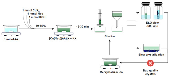
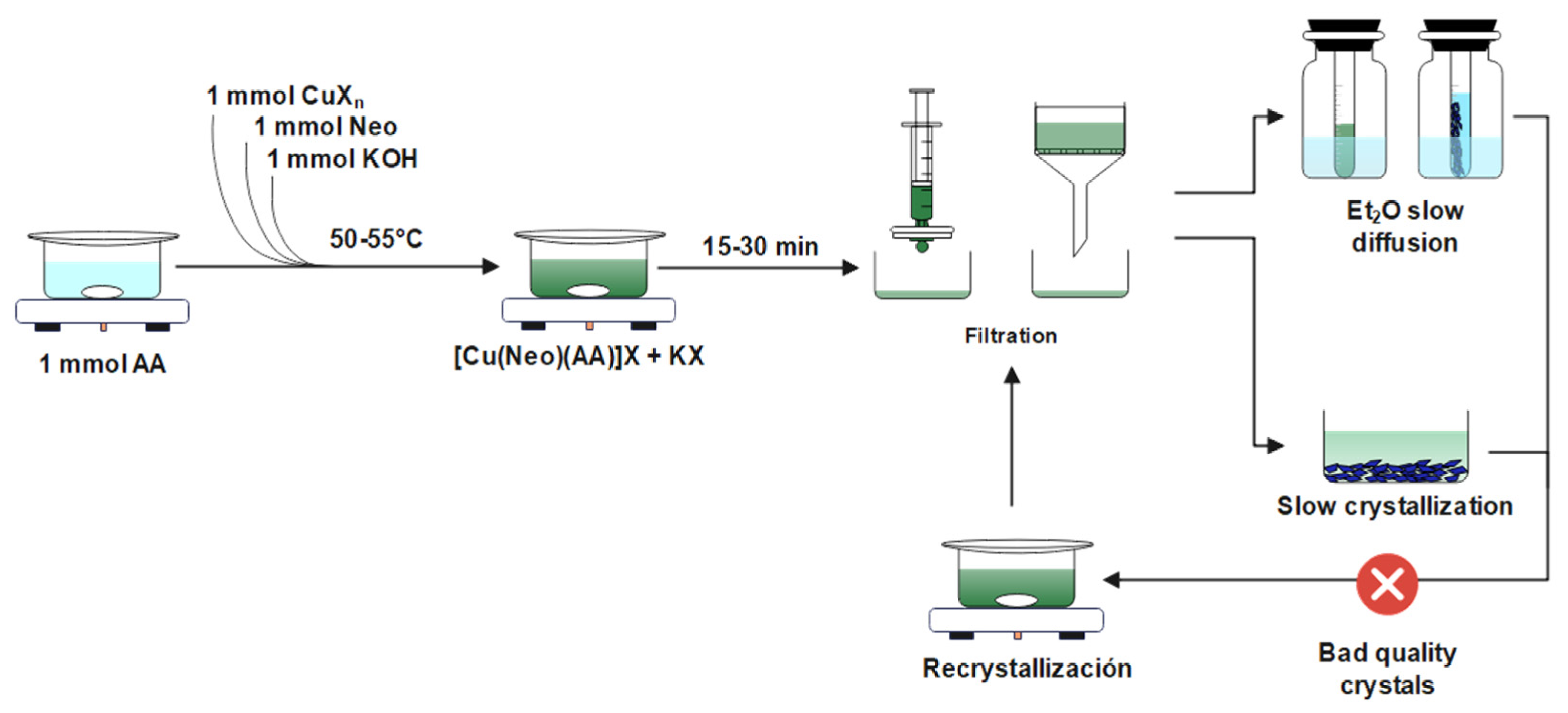
All complexes were synthesized using a direct-reaction synthesis protocol, as described in Scheme 2. For every 20 mL of suitable solvent, 1 mmol of the solid precursor amino acid was added under constant stirring at a temperature of 50–60 °C until the mixture was homogenized as much as possible. Subsequently, 1 mL of a 1 M solution of the copper(II) precursor salt was added to the mixture, maintaining the same conditions until the formation of the soluble binary complex occurred. Once all the reactants had dissolved, 1 mmol of the solid heterocyclic ligand precursor or 1 mL of a 1 M solution thereof was added, immediately forming the mixed complex with a noticeable color change. Finally, the mixture was neutralized with 1 mmol of KOH, cooled to room temperature, and filtered through Whatman® No. 1 filter paper. From the resulting solution, single crystals suitable for further characterization were obtained. The specific details of each complex obtained are described below.
Scheme 2.
Synthetic procedure for the synthesis of compounds 1 to 4.
Synthesisof [Cu(neo)(gly)(H2O)](NO3) (complex 1)
Complex 1 was synthesized using copper(II) nitrate trihydrate (Cu(NO3)2•3H2O), glycine (C2H5NO2), and neocuproine (C14H12N2) in a methanol/water mixture (1:1, v/v). After 1–2 days of slow evaporation, blue lamellar crystals were obtained. Yield: ≈60%. FT-IR (ATR, cm−1): 3305 (m), 3234 (m), 3152 (m), 1591 (s), 1362 (br), 1316 (br), 1131 (s) 855 (s), 728 (s); Raman (Raman shift, cm−1): 2926, 1616, 1501 (w), 1416 (m), 1320 (m), 1307 (sh), 1046 (m), 778 (w), 699 (w), 436 (w), 132 (s), 85 (sh); UV (H2O, λ (nm)-log ε): 276 (4.5).
Synthesis of [Cu(neo)(gln)](Cl) (complex 2)
Complex 2 was synthesized using copper chloride trihydrate (CuCl2•2H2O), glutamine (C5H10N2O3), and neocuproine (C14H12N2) in a methanol/water mixture (1:1, v/v). After 1–2 days of slow evaporation, blue rectangular crystals were obtained. Yield: ≈60%. FT-IR (ATR, cm−1): 3235 (m), 3134 (m), 2951 (w), 2808 (w), 1682 (m), 1617 (br), 1591 (sh), 1373 (s), 1126 (s), 870 (s), 715 (s); Raman (Raman shift, cm−1): 2950 (w), 1618 (w), 1419 (m), 1308 (m), 777 (w), 696 (w), 439 (w), 235 (m), 92 (s); UV (H2O, λ (nm)-log ε): 275 (4.5).
Synthesis of [Cu(neo)(glu)]2•(H2O)•(CH4O) (complex 3)
Complex 3 was synthesized using copper sulfate pentahydrate (CuSO4•5H2O), glutamic acid (C5H9NO4), and neocuproine (C14H12N2) in a methanol/water mixture (1:1, v/v). After one day of evaporation, blue rectangular microcrystals were obtained, Yield: <50%. FT-IR (ATR, cm−1): 3390, 3273 (m), 2968, 2913 (w), 1591, 1557 (br), 1378 (s), 1133 (s), 863, 733 (s); Raman (Raman shift, cm−1): 3462 (w), 3326 (w), 3221 (w), 2855 (w), 2709 (m), 1764 (w), 1697 (w), 1417 (s), 1307 (w), 131 (s), 95 (sh); UV (H2O, λ (nm)-log ε): 275 (4.6).
Synthesis of [Cu(neo)(arg)(H2O)][Cu(neo)2](ClO4)4•1.5(H2O)•(CH4O) (complex 4)
Complex 4 was synthesized using copper perchlorate hexahydrate (Cu(ClO4)2•6H2O), arginine (C6H14N4O2), and neocuproine (C14H12N2) in a methanol/water mixture (1:1, v/v) without neutralizing the mixture. After one day of evaporation, green crystals were obtained. Yield: ≈60%. FT-IR (ATR, cm−1): 3586 (m), 3276 (m), 3078 (w), 1660, 1574 (s), 1424 (s), 1046 (br), 858, 729 (s); Raman (Raman shift, cm−1): 3077 (w), 2928 (w), 1617 (w), 1499 (w), 1423 (s), 1311 (m), 932 (m), 784 (w), 700 (w), 457 (w), 411 (w), 185 (sh), 125 (sh), 86 (s); UV (H2O, λ (nm)-log ε): 276 (4.6)
Crystals and solutions of complex 1 and 2 change color over time and may be photosensitive. Although the latter has not been confirmed, it is recommended to store the samples in the dark.
For complex 4, crystallization is difficult to achieve; therefore, multiple recrystallizations may be necessary, and it may even be necessary to use alternative crystallization methods, such as slow diffusion using ethyl ether.
5. Conclusions
In this work, four new neocuproine structures were synthesized and characterized by FTIR, Raman, and UV-Vis spectroscopy, and single-crystal x-ray diffraction. Although all of the copper ions have pentacoordinate environments, they exhibit a range of geometries. They go from distorted square pyramidal to distorted trigonal bipyramidal environments. In contrast to how phenantroline complexes normally crystallize, the nitrogen atoms do not occupy equatorial positions, and unexpectedly one of the nitrogen atoms occupies the axial positions of the distorted square pyramids. Complexes 1 and 2 are relatively simple. However, complexes 3 and 4 have strange peculiarities. Complex 3 is a binuclear species with bridging glutamate ions coordinated through the carboxylate group. Complex 4 is even more unusual, since there are two different pentacoordinate species.
One is a square-based pyramid, and the other is a triangular bipyramid. In addition, the supramolecular arrangements are rather difficult to describe.
DFT calculations in aqueous solution corroborated the preferred distorted square pyramidal geometry rather than the distorted trigonal bipyramidal geometry of the complexes, as shown in the crystal structure data. Non-covalent interaction studies revealed that the crystal packing of complexes 1–4 is stabilized by O-H···O and N-H···O strong hydrogen bonds and weak attractive and van der Waals interactions, as corroborated by Hirshfeld surfaces, QTAIM, and NCI-RDG analyses. In addition, these approaches showed effective coordination of Cu(II) atoms in the complexes by means of metal–ligand interactions Cu···O and Cu···N with significant values of interaction energies. All of the above forms part of our study on the biological activity of copper (II) mixed complexes, with the aim of elucidating and better understanding the action of this family of compounds. The distorted pentacoordinate structure of the compounds will alter their redox activity, making their cytotoxicity worth pursuing.
Supplementary Materials
The following supporting information can be downloaded at: https://www.mdpi.com/article/10.3390/molecules31224119/s1: Crystallographic data D1–D4; Table S1: Cartesian coordinates of complexes the 1–4 calculated at the DFT/mPW1PW91 level of theory with CPCM implicit model using water as a solvent; Figure S1: Optimized structures of the complexes 1–4 calculated at the DFT/mPW1PW91 level of theory with CPCM implicit model using water as a solvent.
Author Contributions
D.R.-C. carried out the synthesis and experimental characterization. M.C.G. reviewed and assisted with interpreting the results. F.J.M. and M.E.C. performed DFT calculations. D.R.-C., E.G.-V., and M.C.G. wrote and revised the manuscript. D.R.-C. and E.G.-V. conceived and designed this study. L.E.S.-D.l.R. carried out the Raman measurements. All authors contributed extensively to the work presented in this paper. All authors have read and agreed to the published version of the manuscript.
Funding
Projects funded for this research include 100108444-VIEP and 100256733-VIEP; the PRODEP Academic Group BUAP-CA-263 (SEP, Mexico), and project SECIHTI-268178.
Institutional Review Board Statement
Not applicable.
Informed Consent Statement
Not applicable.
Data Availability Statement
The original contributions presented in this study are included in the article/supplementary material. Further inquiries can be directed to the corresponding author(s).
Acknowledgments
Diego Ramírez-Contreras wishes to thank SECIHTI (Mexico) Ph.D. fellowship support (number 4023482). María Eugenia Castro and Francisco J. Melendez wish to thank Laboratorio Nacional de Supercómputo del Sureste de México (LNS-BUAP, Mexico) and the SECIHTI network of national laboratories for the computer resources and support provided and the PRODEP Academic Group BUAP-CA-263 (SEP, Mexico). We are grateful to María de la Paz Elizalde González for FTIR measurements in the Laboratory of Adsorption and Chromatography of the Centro de Química. Instituto de Ciencias (BUAP, México); Brenda L. Sánchez Gaytán for reagents, laboratory equipment, and technical assistance; and Sylvain Bernes for his expertise in X-ray diffraction using the Stradivari X-ray diffractometer.
Conflicts of Interest
The authors declare no conflicts of interest.
Abbreviations
The following abbreviations are used in this manuscript:
| neo | 2,9-dimethyl-1,10-phenanthroline (neocuproine) |
| gly | Glycine |
| gln | L-glutamine |
| glu | L-glutamic acid |
| arg | L-arginine |
| heae | N,N′-bis(N-hydroxyethylaminoethyl)oxamide |
| Im | Imidazole |
| FT-IR | Fourier transform infrared spectroscopy |
| UV-Vis | Ultraviolet–visible spectroscopy |
References
- Aguilar-Jiménez, Z.; Espinoza-Guillén, A.; Resendiz-Acevedo, K.; Fuentes-Noriega, I.; Mejía, C.; Ruiz-Azuara, L. The Importance of Being Casiopeina as Polypharmacologycal Profile (Mixed Chelate–Copper (II) Complexes and Their In Vitro and In Vivo Activities). Inorganics 2023, 11, 394. [Google Scholar] [CrossRef] [Scilit]
- Smith, G.F.; McCurdy, W.H., Jr. 2, 9-Dimethyl-1, 10-Phenanthroline. Anal. Chem. 1952, 24, 371–373. [Google Scholar] [CrossRef] [Scilit]
- Gahler, A.R. Colorimetric Determination of Copper with Neo-Cuproine. Anal. Chem. 1954, 26, 577–579. [Google Scholar] [CrossRef] [Scilit]
- O’Reilly, E.J.; Plowman, R.A. Coordination Compounds of Substituted 1, 10-Phenanthrolines and Related Dipyridyls. I. Synthesis of 2, 9-Dimethyl-1, 10-Phenanthroline. Aust. J. Chem. 1960, 13, 145–149. [Google Scholar] [CrossRef] [Scilit]
- Peris-Tortajada, M.; Puchades, R.; Maquieira, A. Determination of Reducing Sugars by the Neocuproine Method Using Flow Injection Analysis. Food Chem. 1992, 43, 65–69. [Google Scholar] [CrossRef] [Scilit]
- Lee, G.; Rossi, M.V.; Coichev, N.; Moya, H.D. The Reduction of Cu (II)/Neocuproine Complexes by Some Polyphenols: Total Polyphenols Determination in Wine Samples. Food Chem. 2011, 126, 679–686. [Google Scholar] [CrossRef] [Scilit]
- Güçlü, K.; Sözgen, K.; Tütem, E.; Özyürek, M.; Apak, R. Spectrophotometric Determination of Ascorbic Acid Using Copper (II)–Neocuproine Reagent in Beverages and Pharmaceuticals. Talanta 2005, 65, 1226–1232. [Google Scholar] [CrossRef] [Scilit]
- Larsen, E.R. Spectrophotometric Determination of Copper in Fertilizer with Neocuproine. Anal. Chem. 1974, 46, 1131–1132. [Google Scholar] [CrossRef] [Scilit]
- Tütem, E.; Apak, R.; Baykut, F. Spectrophotometric Determination of Trace Amounts of Copper (I) and Reducing Agents with Neocuproine in the Presence of Copper (II). Analyst 1991, 116, 89–94. [Google Scholar] [CrossRef] [Scilit]
- Tütem, E.; Apak, R.; Günaydı, E.; Sözgen, K. Spectrophotometric Determination of Vitamin E (α-Tocopherol) Using Copper (II)-Neocuproine Reagent. Talanta 1997, 44, 249–255. [Google Scholar] [CrossRef] [Scilit]
- Ayaz, S.; Dilgin, Y.; Apak, R. Flow Injection Amperometric Determination of Hydrazine at a Cupric-Neocuproine Complex/Anionic Surfactant Modified Disposable Electrode. Microchem. J. 2020, 159, 105457. [Google Scholar] [CrossRef] [Scilit]
- Boldron, C.; Gamez, P.; Tooke, D.M.; Spek, A.L.; Reedijk, J. Copper-Mediated Selective Oxidation of a CH Bond. Angew. Chem. Int. Ed. 2005, 44, 3585–3587. [Google Scholar] [CrossRef] [Scilit]
- Arends, I.W.C.E.; ten Brink, G.-J.; Sheldon, R.A. Palladium-Neocuproine Catalyzed Aerobic Oxidation of Alcohols in Aqueous Solvents. J. Mol. Catal. A Chem. 2006, 251, 246–254. [Google Scholar] [CrossRef] [Scilit]
- Puls, F.; Linke, P.; Kataeva, O.; Knölker, H. Iron-catalyzed Wacker-type Oxidation of Olefins at Room Temperature with 1,3-diketones or Neocuproine as Ligands. Angew. Chem. 2021, 133, 14202–14209. [Google Scholar] [CrossRef] [Scilit]
- Zhang, Y.; Wang, J.; Yang, Z.; Zhang, Z.; He, X.; Chen, G.; Huang, G.; Lu, X. Hydrazine Hydrate Accelerates Neocuproine–Copper Complex Generation and Utilization in Alkyne Reduction, a Significant Supplement Method for Catalytic Hydrogenation. J. Org. Chem. 2021, 86, 17696–17709. [Google Scholar] [CrossRef] [Scilit]
- ten Brink, G.; Arends, I.W.C.E.; Hoogenraad, M.; Verspui, G.; Sheldon, R.A. Catalytic Conversions in Water. Part 23: Steric Effects and Increased Substrate Scope in the Palladium-Neocuproine Catalyzed Aerobic Oxidation of Alcohols in Aqueous Solvents#. Adv. Synth. Catal. 2003, 345, 1341–1352. [Google Scholar] [CrossRef] [Scilit]
- Lappin, A.G.; Youngblood, M.P.; Margerum, D.W. Electron-Transfer Reactions of Copper (I) and Copper (III) Complexes. Inorg. Chem. 1980, 19, 407–413. [Google Scholar] [CrossRef] [Scilit]
- Haas, K.L.; Franz, K.J. Application of Metal Coordination Chemistry to Explore and Manipulate Cell Biology. Chem. Rev. 2009, 109, 4921. [Google Scholar] [CrossRef] [Scilit]
- Al-Sa’doni, H.H.; Megson, I.L.; Bisland, S.; Butler, A.R.; Flitney, F.W. Neocuproine, a Selective Cu (I) Chelator, and the Relaxation of Rat Vascular Smooth Muscle by S-nitrosothiols. Br. J. Pharmacol. 1997, 121, 1047–1050. [Google Scholar] [CrossRef] [Scilit]
- Mohindru, A.; Fisher, J.M.; Rabinovitz, M. 2, 9-Dimethyl-1, 10-Phenanthroline (Neocuproine): A Potent, Copper-Dependent Cytotoxin with Anti-Tumor Activity. Biochem. Pharmacol. 1983, 32, 3627–3632. [Google Scholar] [CrossRef] [Scilit]
- Osei, P.B.; Northcote-Smith, J.; Fang, J.; Singh, K.; Ortu, F.; Suntharalingam, K. The Bulk Breast Cancer Cell and Breast Cancer Stem Cell Activity of Binuclear Copper (II)-phenanthroline Complexes. Chem.–A Eur. J. 2023, 29, e202301188. [Google Scholar] [CrossRef] [Scilit]
- Smit, H.; Van der Goot, H.; Nauta, W.; Timmerman, H.; De Bolster, M.W.; Stouthamer, A.H.; Vis, R.D. Mechanism of Action of the Copper (I) Complex of 2, 9-Dimethyl-1, 10-Phenanthroline on Mycoplasma Gallisepticum. Antimicrob. Agents Chemother. 1982, 21, 881–886. [Google Scholar] [CrossRef] [Scilit]
- Jiang, M.; Li, Y.-T.; Wu, Z.-Y.; Liu, Z.-Q.; Yan, C.-W. Synthesis, Crystal Structure, Cytotoxic Activities and DNA-Binding Properties of New Binuclear Copper (II) Complexes Bridged by N, N′-Bis (N-Hydroxyethylaminoethyl) Oxamide. J. Inorg. Biochem. 2009, 103, 833–844. [Google Scholar] [CrossRef] [Scilit]
- Sánchez-Lara, E.; Favela, R.; Tzian, K.; Monroy-Torres, B.; Romo-Pérez, A.; Ramírez-Apan, M.T.; Flores-Alamo, M.; Rodríguez-Diéguez, A.; Cepeda, J.; Castillo, I. Effects of the Tetravanadate [V4O12] 4− Anion on the Structural, Magnetic, and Biological Properties of Copper/Phenanthroline Complexes. JBIC J. Biol. Inorg. Chem. 2024, 29, 139–158. [Google Scholar] [CrossRef] [Scilit]
- Yesilyurt, F.; Avşaroğlu, R.; Akçay, G.; Çamur, S.; Yaman, S.; Kaya, Ö.; Uzun, S.; Hacımüftüoğlu, A. Anti-Cancer Effects of Neocuproine via Copper Chelation in A549 Lung Adenocarcinoma Cells. Experimed 2026, 16, 1–6. [Google Scholar] [CrossRef] [Scilit]
- Chen, S.-H.; Lin, J.-K.; Liu, S.-H.; Liang, Y.-C.; Lin-Shiau, S.-Y. Apoptosis of Cultured Astrocytes Induced by the Copper and Neocuproine Complex through Oxidative Stress and JNK Activation. Toxicol. Sci. 2008, 102, 138–149. [Google Scholar] [CrossRef] [Scilit]
- Addison, A.W.; Rao, T.N.; Reedijk, J.; van Rijn, J.; Verschoor, G.C. Synthesis, Structure, and Spectroscopic Properties of Copper (II) Compounds Containing Nitrogen–Sulphur Donor Ligands; the Crystal and Molecular Structure of Aqua [1,7-Bis(N-Methylbenzimidazol-2′-Yl)-2,6-Dithiaheptane] Copper (II) Perchlorate. J. Chem. Soc. Dalton Trans. 1984, 13, 1349–1356. [Google Scholar] [CrossRef] [Scilit]
- Parker, F. Applications of Infrared Spectroscopy in Biochemistry, Biology, and Medicine; Springer Science & Business Media: Berlin/Heidelberg, Germany, 2012; ISBN 1468418726. [Google Scholar]
- Farkas, E.; Sóvágó, I. Metal Complexes of Amino Acids and Peptides. In Amino Acids, Peptides and Proteins; Royal Society of Chemistry Books: Cambridge, UK, 2007; Volume 36, pp. 287–345. ISBN 9780854042524. [Google Scholar]
- Spackman, M.A.; Jayatilaka, D. Hirshfeld Surface Analysis. CrystEngComm 2009, 11, 19–32. [Google Scholar] [CrossRef] [Scilit]
- Bader, R.F.W. Atoms in Molecules: A Quantum Theory; Clarendon Press: Oxford, UK, 1990; ISBN 9780198558651. [Google Scholar]
- Johnson, E.R.; Keinan, S.; Mori-Sánchez, P.; Contreras-García, J.; Cohen, A.J.; Yang, W. Revealing Non-Covalent Interactions. J. Am. Chem. Soc. 2010, 132, 6498. [Google Scholar] [CrossRef] [Scilit]
- Simmons, C.J.; Lundeen, M.; Seff, K. Crystal and Molecular Structure of (2, 9-Dimethyl-1, 10-Phenanthroline)(Glycylglycinato) Copper (II) Pentahydrate. Inorg. Chem. 1978, 17, 1429–1435. [Google Scholar] [CrossRef] [Scilit]
- Zhang, W.; Lu, X.; Wang, G.; Cheng, Y.; Zhang, B. Methyl-Substituted Enhancement of Antitumor Activity in Square-Planar Metal Complex and Analysis of Δ E, Δ G, CV, UV-Vis and Luminescence. New J. Chem. 2015, 39, 4869–4875. [Google Scholar] [CrossRef] [Scilit]
- Alvarez, N.; Mendes, L.F.S.; Kramer, M.G.; Torre, M.H.; Costa-Filho, A.J.; Ellena, J.; Facchin, G. Development of Copper (II)-Diimine-Iminodiacetate Mixed Ligand Complexes as Potential Antitumor Agents. Inorganica Chim. Acta 2018, 483, 61–70. [Google Scholar] [CrossRef] [Scilit]
- Ganeshpandian, M.; Ramakrishnan, S.; Palaniandavar, M.; Suresh, E.; Riyasdeen, A.; Akbarsha, M.A. Mixed Ligand Copper (II) Complexes of 2,9-Dimethyl-1,10-Phenanthroline: Tridentate 3N Primary Ligands Determine DNA Binding and Cleavage and Cytotoxicity. J. Inorg. Biochem. 2014, 140, 202–212. [Google Scholar] [CrossRef] [Scilit]
- Alvarez, N.; Vina, D.; Leite, C.M.; Mendes, L.F.S.; Batista, A.A.; Ellena, J.; Costa-Filho, A.J.; Facchin, G. Synthesis and Structural Characterization of a Series of Ternary Copper (II)-L-Dipeptide-Neocuproine Complexes. Study of Their Cytotoxicity against Cancer Cells Including MDA-MB-231, Triple Negative Breast Cancer Cells. J. Inorg. Biochem. 2020, 203, 110930. [Google Scholar] [CrossRef] [Scilit]
- Kumar, R.; Guchhait, T.; Subramaniyan, V.; Mani, G. Mixed Ligand Cu (II) Complexes: Square Pyramidal vs Trigonal Bipyramidal with the Pyrrole-Based Dipodal Ligand Having Hydrogen Bond Acceptors. J. Mol. Struct. 2019, 1195, 1–9. [Google Scholar] [CrossRef] [Scilit]
- Wei, Y.-B.; Yuan, C.-X.; Yang, P. [Cu (Phen) 2 (μ-IDA) Cu (Phen)](ClO4) 2· CH3OH: Tridentate and Monodentate Binding to Two Copper (II) Species by a Bridging Ligand. Cryst. Struct. Commun. 2004, 60, m512–m514. [Google Scholar] [CrossRef] [Scilit]
- Jia, L.; Jiang, P.; Xu, J.; Hao, Z.; Xu, X.; Chen, L.; Wu, J.; Tang, N.; Wang, Q.; Vittal, J.J. Synthesis, Crystal Structures, DNA-Binding Properties, Cytotoxic and Antioxidation Activities of Several New Ternary Copper (II) Complexes of N, N′-(p-Xylylene) Di-Alanine Acid and 1, 10-Phenanthroline. Inorganica Chim. Acta 2010, 363, 855–865. [Google Scholar] [CrossRef] [Scilit]
- Antolini, L.; Marcotrigiano, G.; Menabue, L.; Pellacani, G.C.; Saladini, M.; Sola, M. Coordination Behavior of L-Glutamic Acid: Spectroscopic and Structural Properties of (L-Glutamato)(Imidazole) Copper (II),(L-Glutamato)(2, 2’-Bipyridine) Copper (II), and Aqua (L-Glutamato)(1, 10-Phenanthroline) Copper (II) Trihydrate Complexes. Inorg. Chem. 1985, 24, 3621–3626. [Google Scholar] [CrossRef] [Scilit]
- Lin, J.-G.; Qiu, L.Q.; Xu, Y.-Y. A Two-Dimensional Supramolecular Network Built through Unique π-πStacking: Synthesis and Characterization of [Cu (Phen) 2 (μ-ID A) Cu (Phen)·(NO3)](NO3)· 4 (H2O). Bull. Korean Chem. Soc. 2009, 30, 1021–1025. [Google Scholar] [CrossRef] [Scilit]
- Yang, C.-T.; Moubaraki, B.; Murray, K.S.; Vittal, J.J. Synthesis, Characterization and Properties of Ternary Copper (II) Complexes Containing Reduced Schiff Base N-(2-Hydroxybenzyl)-α-Amino Acids and 1, 10-Phenanthroline. Dalton Trans. 2003, 32, 880–889. [Google Scholar] [CrossRef] [Scilit]
- Inci, D.; Aydın, R.; Vatan, Ö.; Sevgi, T.; Yılmaz, D.; Zorlu, Y.; Yerli, Y.; Çoşut, B.; Demirkan, E.; Çinkılıç, N. Synthesis and Crystal Structures of Novel Copper (II) Complexes with Glycine and Substituted Phenanthrolines: Reactivity towards DNA/BSA and in Vitro Cytotoxic and Antimicrobial Evaluation. JBIC J. Biol. Inorg. Chem. 2017, 22, 61–85. [Google Scholar] [CrossRef] [Scilit]
- McCall, J.; Bruce, D.; Workman, S.; Cole, C.; Richter, M.M. Electrochemiluminescence of Copper (I) Bis (2,9-Dimethyl-1,10-Phenanthroline). Anal. Chem. 2001, 73, 4617–4620. [Google Scholar] [CrossRef] [Scilit]
- Ramírez-Contreras, D.; Vázquez-Rodríguez, S.; García-García, A.; Noriega, L.; Mendoza, A.; Sánchez-Gaytán, B.L.; Meléndez, F.J.; Castro, M.E.; Cárdenas-García, M.; González-Vergara, E. L-Citrullinato-Bipyridine and L-Citrullinato-Phenanthroline Mixed Copper Complexes: Synthesis, Characterization and Potential Anticancer Activity. Pharmaceutics 2024, 16, 747. [Google Scholar] [CrossRef] [Scilit]
- Martinez-Valencia, B.; Corona-Motolinia, N.D.; Sanchez-Lara, E.; Noriega, L.; Sanchez-Gaytan, B.L.; Castro, M.E.; Melendez-Bustamante, F.; Gonzalez-Vergara, E. Cyclo-Tetravanadate Bridged Copper Complexes as Potential Double Bullet pro-Metallodrugs for Cancer Treatment. J. Inorg. Biochem. 2020, 208, 111081. [Google Scholar] [CrossRef] [Scilit]
- Corona-Motolinia, N.D.; Martínez-Valencia, B.; Noriega, L.; Sánchez-Gaytán, B.L.; Méndez-Rojas, M.Á.; Melendez, F.J.; Castro, M.E.; González-Vergara, E. Synthesis, Crystal Structure, and Computational Methods of Vanadium and Copper Compounds as Potential Drugs for Cancer Treatment. Molecules 2020, 25, 4679. [Google Scholar] [CrossRef] [Scilit]
- Corona-Motolinia, N.D.; Martínez-Valencia, B.; Noriega, L.; Sánchez-Gaytán, B.L.; Mendoza, A.; Meléndez-Bustamante, F.J.; Castro, M.E.; González-Vergara, E. Ternary Copper Complex of L-Glutamine and Phenanthroline as Counterions of Cyclo-Tetravanadate Anion: Experimental–Theoretical Characterization and Potential Antineoplastic Activity. Metals 2021, 11, 1541. [Google Scholar] [CrossRef] [Scilit]
- Borges, L.J.H.; Bull, E.S.; Fernandes, C.; Horn, A., Jr.; Azeredo, N.F.; Resende, J.A.L.C.; Freitas, W.R.; Carvalho, E.C.Q.; Lemos, L.S.; Jerdy, H. In Vitro and in Vivo Studies of the Antineoplastic Activity of Copper (II) Compounds against Human Leukemia THP-1 and Murine Melanoma B16-F10 Cell Lines. Eur. J. Med. Chem. 2016, 123, 128–140. [Google Scholar] [CrossRef] [Scilit]
- Vazquez-Rodriguez, S.; Ramírez-Contreras, D.; Noriega, L.; García-García, A.; Sánchez-Gaytán, B.L.; Melendez, F.J.; Castro, M.E.; de Azevedo, W.F.; González-Vergara, E. Interaction of Copper Potential Metallodrugs with TMPRSS2: A Comparative Study of Docking Tools and Its Implications on COVID-19. Front. Chem. 2023, 11, 1128859. [Google Scholar] [CrossRef] [Scilit]
- Aligent Technologies. CrysAlis Pro, version 171.43.1a; Rigaku Oxford Diffration: Tokyo, Japan, 2013.
- Sheldrick, G.M. Crystal Structure Refinement with SHELXL. Acta Crystallogr. C Struct. Chem. 2015, 71, 8. [Google Scholar] [CrossRef] [Scilit]
- Dolomanov, O.V.; Bourhis, L.J.; Gildea, R.J.; Howard, J.A.K.; Puschmann, H. OLEX2: A Complete Structure Solution, Refinement and Analysis Program. Appl. Crystallogr. 2009, 42, 339–341. [Google Scholar] [CrossRef] [Scilit]
- Macrae, C.F.; Sovago, I.; Cottrell, S.J.; Galek, P.T.A.; McCabe, P.; Pidcock, E.; Platings, M.; Shields, G.P.; Stevens, J.S.; Towler, M. Mercury 4.0: From Visualization to Analysis, Design and Prediction. Appl. Crystallogr. 2020, 53, 226–235. [Google Scholar] [CrossRef] [Scilit]
- Adamo, C.; Barone, V. Exchange Functionals with Improved Long-Range Behavior and Adiabatic Connection Methods without Adjustable Parameters: The mPW and mPW1PW Models. J. Chem. Phys. 1998, 108, 664–675. [Google Scholar] [CrossRef] [Scilit]
- Krishnan, R.; Binkley, J.S.; Seeger, R.; Pople, J.A. Self-consistent Molecular Orbital Methods. XX. A Basis Set for Correlated Wave Functions. J. Chem. Phys. 1980, 72, 650–654. [Google Scholar] [CrossRef] [Scilit]
- Ditchfield, R.; Hehre, W.J.; Pople, J.A. Self-consistent Molecular-orbital Methods. IX. An Extended Gaussian-type Basis for Molecular-orbital Studies of Organic Molecules. J. Chem. Phys. 1971, 54, 724–728. [Google Scholar] [CrossRef] [Scilit]
- Wachters, A.J.H. Gaussian Basis Set for Molecular Wavefunctions Containing Third-row Atoms. J. Chem. Phys. 1970, 52, 1033–1036. [Google Scholar] [CrossRef] [Scilit]
- Cossi, M.; Rega, N.; Scalmani, G.; Barone, V. Energies, Structures, and Electronic Properties of Molecules in Solution with the C-PCM Solvation Model. J. Comput. Chem. 2003, 24, 669–681. [Google Scholar] [CrossRef] [Scilit]
- Frisch, M.J.; Trucks, G.W.; Schlegel, H.B.; Scuseria, G.E.; Robb, M.A.; Cheeseman, J.R. Gaussian 16, Revision B.01; Gaussian, Inc.: Wallingford, CT, USA, 2016.
- Spackman, P.R.; Turner, M.J.; McKinnon, J.J.; Wolff, S.K.; Grimwood, D.J.; Jayatilaka, D.; Spackman, M.A. CrystalExplorer: A Program for Hirshfeld Surface Analysis, Visualization and Quantitative Analysis of Molecular Crystals. J. Appl. Crystallogr. 2021, 54, 1006–1011. [Google Scholar] [CrossRef] [Scilit]
- Keith, T.A. TK Gristmill Software, Version 19.02.13; AIMAll: Overland Park, KS, USA, 2019.
- Lu, T.; Chen, F. Multiwfn: A Multifunctional Wavefunction Analyzer. J. Comput. Chem. 2012, 33, 580–592. [Google Scholar] [CrossRef] [Scilit]
- Humphrey, W.; Dalke, A.; Schulten, K. VMD: Visual Molecular Dynamics. J. Mol. Graph. 1996, 14, 33–38. [Google Scholar] [CrossRef] [Scilit]

Disclaimer/Publisher’s Note: The statements, opinions and data contained in all publications are solely those of the individual author(s) and contributor(s) and not of MDPI and/or the editor(s). MDPI and/or the editor(s) disclaim responsibility for any injury to people or property resulting from any ideas, methods, instructions or products referred to in the content. |
© 2026 by the authors. Licensee MDPI, Basel, Switzerland. This article is an open access article distributed under the terms and conditions of the Creative Commons Attribution (CC BY) license.