


Abstract
Gnetol (trans-3,5,2′,6′-tetrahydroxystilbene) is a naturally occurring tetrahydroxylated stilbenoid found mainly in Gnetum species. Despite its structural similarity to resveratrol (trans-3,5,4′-trihydroxystilbene), gnetol remains comparatively underexplored, and its therapeutic potential has yet to be clearly defined. This review critically evaluates the current knowledge of its natural occurrence, chemistry, pharmacological activities, molecular mechanisms, pharmacokinetics, metabolism, and tissue distribution, with particular attention to the relationship between biological activity and in vivo exposure. Available studies suggest that gnetol has a broad pharmacological profile involving cardiovascular protection, inflammation, metabolic regulation, melanogenesis, neuroprotection, cancer-related processes, and bone remodeling. However, cardioprotective and bone-related effects currently have stronger experimental support from animal studies, whereas most other activities remain supported primarily by biochemical or cellular evidence. Recent mechanistic studies have further implicated redox regulation, ferroptosis, iron homeostasis, and direct enzyme modulation in its biological actions, with context-dependent effects on ferroptosis in cardiomyocytes and osteoclasts. Gnetol undergoes extensive glucuronidation and exhibits low systemic bioavailability as the unchanged compound following oral administration, although the contributions of incomplete absorption, luminal degradation, and microbial metabolism remain incompletely understood. A mouse study reported relatively high concentrations in gastrointestinal samples, but residual luminal contents may have contributed to these measurements. Moreover, measured tissue concentrations do not necessarily reflect pharmacologically available unbound concentrations, and exposure at effective doses remains poorly characterized. Future studies should therefore integrate molecular activity with target-site exposure, metabolite contributions, dose–response relationships, safety, and route of administration to identify the most plausible therapeutic applications of gnetol.
1. Introduction
Stilbenoids are plant-derived phenolic compounds built around the stilbene scaffold, in which two aromatic rings are linked by a two-carbon bridge [1,2]. They occur in a variety of plant species and comprise compounds that differ mainly in hydroxylation, methylation, glycosylation, and other substitutions of the basic stilbene structure [3,4]. Among them, resveratrol (trans-3,5,4′-trihydroxystilbene, Figure 1A) has received by far the greatest attention, with extensive studies addressing its pharmacological properties, metabolism, and potential applications in cardiovascular, metabolic, inflammatory, and age-related disorders [5,6,7,8]. Other naturally occurring stilbenoids, such as piceatannol (trans-3,5,3′,4′-tetrahydroxystilbene, Figure 1B), oxyresveratrol (trans-3,5,2′,4′-tetrahydroxystilbene, Figure 1C), and gnetol (trans-3,5,2′,6′-tetrahydroxystilbene, Figure 1D), share the same basic scaffold but differ in the number, position, or substitution of hydroxyl groups. Such structural differences can influence their physicochemical properties, metabolism, and biological activity [9,10]. More broadly, the biological effects of dietary polyphenols depend not only on their chemical structures and intrinsic activities but also on their absorption, metabolism, and availability at the sites of action [11].
Figure 1.
Chemical structures of selected hydroxylated stilbenes.
Gnetol has been reported mainly in plants of the genus Gnetum (Gnetaceae) [12,13,14,15], although it has also been isolated from Ficus foveolata (Moraceae) [16]. Compared with resveratrol and several other naturally occurring stilbenoids, gnetol remains relatively understudied. Available studies have addressed its natural occurrence and chemical properties, biological activities, mechanisms of action, and fate in vivo, but these findings have yet to be brought together into a coherent picture. Early investigations focused mainly on its isolation, characterization, and biological activity, whereas more recent studies have begun to examine its molecular mechanisms and in vivo disposition. The expanding literature now provides a basis for assessing gnetol as a natural product in its own right rather than simply as a less familiar analogue of resveratrol.
To identify the studies relevant to this assessment, PubMed and Web of Science were searched using the terms “gnetol” and “trans-3,5,2′,6′-tetrahydroxystilbene”, without restrictions on publication year. The search was last updated on 5 November 2026. Additional publications were identified by examining the reference lists of relevant articles. Original studies addressing the natural occurrence, chemistry, pharmacological activities, molecular mechanisms, metabolism, pharmacokinetics, or tissue distribution of gnetol were considered. Studies involving plant extracts or mixtures of stilbenoids were also considered where relevant, but their findings were distinguished from those obtained with purified gnetol. Review articles were consulted for background information and to identify additional primary studies.
Previous reviews have discussed the biological activities of stilbenoids more broadly [9,10] or summarized the phytochemistry and pharmacology of Gnetum species [17,18]. However, these reviews have not provided a gnetol-focused critical assessment integrating its pharmacological activities with metabolism, pharmacokinetics, and tissue distribution. The present review addresses this gap by critically examining the strength of experimental evidence, the relationship between reported biological activities and in vivo exposure, and the limitations that currently hinder therapeutic translation. By distinguishing experimentally supported effects from potential therapeutic applications, this review aims to provide a more evidence-based assessment of the therapeutic potential of gnetol.
2. Natural Sources and Chemistry of Gnetol
2.1. Natural Sources and Occurrence
Gnetol is a naturally occurring stilbenoid found primarily in plants of the genus Gnetum (Gnetaceae). It was first isolated from G. ula [15] and has since been reported in several other Gnetum species and Ficus foveolata [16]. The reported botanical sources, plant materials examined, and corresponding references are summarized in Table 1. These studies examined different plant parts using various extraction and analytical methods, and the identification of gnetol in a particular plant part does not necessarily indicate its abundance or distribution throughout the plant.
Table 1.
Reported botanical sources of gnetol.
Gnetum plants contain a broad range of stilbenoids. Alongside gnetol, phytochemical investigations have identified resveratrol, isorhapontigenin, and numerous stilbene dimers and oligomers, including gnetins and gnemonosides [9,20,24]. Gnetol is therefore usually found as one component of a chemically complex extract. The presence of gnetol in an active extract alone is insufficient to attribute the observed pharmacological effects to this compound.
Among the reported sources, G. gnemon, commonly known as melinjo, is of particular interest because it is consumed as food in Southeast Asia, particularly Indonesia [24]. Remsberg et al. quantified gnetol in G. gnemon food products, reporting both aglycone and glycoside forms [14]. However, the presence of gnetol in these products does not establish its typical dietary intake or systemic exposure to unchanged gnetol. The extent of glycoside hydrolysis during digestion and the possible contributions of luminal degradation, microbial metabolism, and presystemic biotransformation remain insufficiently characterized.
2.2. Chemical and Structural Features
Gnetol is a monomeric tetrahydroxylated stilbene with the molecular formula C14H12O4 and a molecular weight of 244.24 g/mol. Like other stilbenoids, it consists of two aromatic rings connected by an ethylene bridge, forming the characteristic C6–C2–C6 framework (Figure 1). Naturally occurring gnetol is present predominantly in the trans configuration and contains four phenolic hydroxyl groups distributed between the two aromatic rings [15].
The structure of gnetol is closely related to those of several better-known natural stilbenoids (Figure 1). Resveratrol contains three hydroxyl groups, whereas gnetol, piceatannol, and oxyresveratrol each contain four. The three tetrahydroxystilbenes differ, however, in the positions of these groups on the aromatic rings. Gnetol should therefore not be regarded simply as a more highly hydroxylated form of resveratrol. Nor can its chemical or biological behavior be assumed to resemble that of other tetrahydroxystilbenes solely on the basis of the number of hydroxyl groups.
The hydroxylation pattern influences several properties relevant to the behavior of gnetol. The phenolic groups contribute to hydrogen bonding and redox chemistry and also provide sites for conjugative metabolism. Their positions can affect molecular interactions and determine which hydroxyl groups are preferentially modified by metabolic enzymes. The conjugated trans-stilbene backbone, meanwhile, contributes to the relatively planar structure and electronic properties characteristic of this class of compounds. Thus, both the substitution pattern and the integrity of the stilbene scaffold need to be considered when comparing gnetol with structurally related compounds.
Direct physicochemical data for gnetol remain limited. Free gnetol has relatively low aqueous solubility, and its stability in solution is affected by storage conditions [26]. Both properties can be improved by complexation with cyclodextrins, indicating that formulation can substantially alter its apparent solubility and stability [26]. These characteristics are relevant to experimental work with gnetol because the concentration present in a biological system may depend on how the compound is prepared and maintained during an experiment.
The presence of four free phenolic hydroxyl groups also makes gnetol susceptible to extensive conjugation after absorption. This structural feature has direct consequences for its in vivo disposition and distinguishes highly hydroxylated stilbenoids such as gnetol from more lipophilic, methoxylated members of the family. The metabolism and disposition of gnetol are considered separately in Section 5.
3. Pharmacological Activities of Gnetol
The pharmacological effects reported for gnetol cover several experimental areas, but the supporting evidence is uneven. Most studies have used biochemical assays or cultured cells, and only a limited number have extended these observations to animal models. Cardiovascular injury and bone loss currently have the strongest in vivo support, whereas evidence in other areas remains largely preclinical. Importantly, the interpretation of these findings requires consideration of the experimental concentrations or doses, appropriate controls, study replication, and whether purified gnetol or a multicomponent preparation was investigated. These factors are considered throughout this section and summarized in Table 2.
3.1. Cardiovascular and Cardioprotective Effects
The cardiovascular effects of gnetol were first examined in experimental cardiac hypertrophy. In neonatal rat cardiomyocytes, gnetol at concentrations of 1–10 μg/mL reduced endothelin-1-induced cell enlargement and protein synthesis [27]. At 5 μg/mL, the anti-hypertrophic response was associated with AMPK activation, and pharmacological inhibition or knockdown of AMPK attenuated this effect. At 50–100 μg/mL, however, cell size was also reduced in untreated cardiomyocytes, and cell viability was significantly decreased, suggesting that the effects at these concentrations may partly reflect cytotoxicity rather than specific anti-hypertrophic activity. When evaluated in spontaneously hypertensive heart failure (SHHF) rats, oral administration of gnetol at 2.5 mg/kg/day for eight weeks did not significantly lower blood pressure or reverse established cardiac hypertrophy, although an improvement in left ventricular relaxation was observed [27]. The cellular anti-hypertrophic effect therefore showed only limited translation to this animal model.
Gnetol has also shown vascular and antiplatelet effects. In a study of stilbenoids isolated from G. macrostachyum, purified gnetol inhibited arachidonic acid-induced aggregation of human platelets (IC50 = 105.9 ± 1.7 μM) and reduced platelet adhesion in vitro [23]. In SHHF rats, gnetol administered orally at 2.5 mg/kg/day for eight weeks attenuated structural alterations in peripheral and cerebral resistance arteries [28]. These findings extend its cardiovascular profile beyond cardiomyocytes. They should, however, be interpreted alongside the absence of a significant reduction in systolic blood pressure or established cardiac hypertrophy in the related SHHF study [27]. The antiplatelet results were obtained outside the body, and their relevance at attainable circulating concentrations remains to be determined.
More recent work has provided stronger evidence in doxorubicin-induced cardiac injury. In H9C2 cells, gnetol at 5–10 μM protected cardiomyocytes against oxidative damage and ferroptotic cell death and preserved cellular redox homeostasis [29]. In mice with doxorubicin-induced cardiomyopathy, intraperitoneal administration of gnetol at 3.75 or 7.5 mg/kg/day for 28 days improved ejection fraction and fractional shortening and reduced myocardial injury, fibrosis, and oxidative damage [29]. These effects were evaluated against doxorubicin-treated vehicle controls. This study provides both cellular and animal evidence for cardioprotection and currently represents one of the more substantial demonstrations of gnetol activity in vivo. Nevertheless, these findings have not yet been independently replicated, and their generalizability to other models of cardiac injury, different dosing regimens, or clinically relevant exposure conditions remains uncertain. In particular, pharmacologically available cardiac concentrations at the effective doses have not been established, limiting direct comparison between the cellular and animal findings. The molecular basis of these effects is discussed in Section 4.
3.2. Anti-Inflammatory Effects
Anti-inflammatory effects of gnetol have been demonstrated mainly in enzyme and cellular assays. Gnetol inhibited cyclooxygenase-1 (COX-1) more strongly than COX-2 [14]. Azmin et al. reported concentration-dependent inhibition of prostaglandin E2 (PGE2) production by purified gnetol in lipopolysaccharide (LPS)-stimulated human whole blood, with an IC50 of 1.84 μM, compared with 1.29 μM for the positive control indomethacin [30]. These results provide quantitative evidence of an anti-inflammatory effect under the reported assay conditions, although they do not by themselves establish the contribution of COX inhibition or predict efficacy in vivo. In LPS-stimulated macrophages, gnetol at 10–50 μM reduced nitric oxide and ROS production as well as the release of IL-1β and TNF-α [31]. Some glucuronide metabolites of gnetol also retained activity in this model [31]. The activity of these metabolites should be distinguished from that of the parent compound, because their effective concentrations and systemic exposure may differ.
These findings support direct effects on inflammatory processes, but evidence from animal models remains sparse. In particular, purified gnetol has not been extensively studied in established models of chronic inflammatory or immune-mediated disease. Its anti-inflammatory activity is therefore supported primarily at the biochemical and cellular levels. Further studies using appropriate positive controls, concentration–response analysis, and independently replicated experiments are needed to establish the reproducibility and pharmacological specificity of these effects.
3.3. Anticancer Activity
The anticancer evidence for gnetol is limited and predominantly in vitro. Purified gnetol reduced the viability of several cancer cell lines, with notable activity reported in colorectal cancer cells [14]. The reported activity was concentration-dependent, but the concentrations required for meaningful growth inhibition and their selectivity over nonmalignant cells need to be considered when interpreting these findings. Gnetol also inhibited histone deacetylase (HDAC) activity in biochemical assays [14], although the contribution of this effect to cancer-cell growth inhibition has not been established directly. In particular, biochemical HDAC inhibition does not establish intracellular target engagement or demonstrate that HDAC inhibition mediates the observed reduction in cancer-cell viability.
A recent study of G. montanum extract identified gnetol among several compounds in an F2 fraction that showed antitumor activity [22]. Because the active fraction also contained other identified constituents, this result cannot establish that gnetol was responsible for its activity. Reports involving extracts or mixed fractions should therefore be considered separately from experiments with purified gnetol. Furthermore, the contribution of gnetol to the activity of the F2 fraction cannot be determined without testing the isolated compound at concentrations corresponding to its abundance in the fraction. Direct evidence from animal tumor models using purified gnetol remains limited. Consequently, the anticancer potential of gnetol should be regarded as preliminary, pending independent confirmation, assessment of selectivity, and studies linking effective concentrations to achievable exposure in vivo.
3.4. Metabolic Effects
Several in vitro findings suggest possible effects of gnetol on metabolic processes. Gnetol inhibited α-amylase and α-glucosidase, two enzymes involved in carbohydrate digestion, and also suppressed adipocyte differentiation in a concentration-dependent manner [14]. These activities were reported for purified gnetol in biochemical or cellular assays, rather than demonstrated as glucose-lowering or weight-reducing effects in animals. These observations provide evidence of activity against metabolic targets but do not establish antidiabetic or anti-obesity efficacy.
Studies with purified gnetol in animal models of diabetes, insulin resistance, or obesity are currently lacking. The inhibition of gastrointestinal digestive enzymes may nevertheless warrant further investigation because the relevance of these targets depends on local intestinal exposure rather than systemic concentrations alone. However, the potential for a local gastrointestinal effect remains hypothetical until inhibitory concentrations are compared with experimentally measured concentrations of free gnetol in the intestinal lumen or at the relevant enzyme sites. This issue is revisited in the context of gnetol disposition and translational potential.
3.5. Neuroprotective Potential
Evidence for neuronal effects of gnetol comes from enzyme and cellular studies. Gnetol inhibited butyrylcholinesterase (BChE) with an IC50 of 1.3 μM, while showing little activity against acetylcholinesterase [16]. The biological relevance of this selectivity has not yet been tested in animal models of cognitive impairment.
In N2a neuroblastoma cells exposed to malathion, gnetol at 5, 10, and 20 μM reduced cellular injury, including lysosomal and mitochondrial dysfunction, impaired autophagy, and apoptosis, and promoted neurite outgrowth [32]. These findings suggest a protective effect under chemically induced neuronal stress, but they remain confined to a cellular model. The effective concentrations were in the micromolar range, and comparable unbound brain concentrations following systemic administration have not been demonstrated. Whether such effects are relevant in vivo will depend partly on the extent to which gnetol reaches the brain after administration.
A separate screening study identified gnetol as a potential disruptor of the interaction between neuronal nitric oxide synthase (nNOS) and postsynaptic density protein 95 (PSD-95). Its neuroprotective effect and interference with this protein interaction were examined in vitro [33]. However, identification through affinity-based screening and subsequent cellular testing does not establish selective target engagement in vivo. In another comparative study of hydroxylated stilbenes, gnetol was tested in assays involving acetylcholinesterase, amyloid-β aggregation, and neuronal cells [34]. The behavioral improvement reported in that study was obtained with piceatannol; it should not be attributed to gnetol. Together, these studies add possible neuronal mechanisms, but they do not yet establish cognitive or cerebrovascular benefit from gnetol in vivo.
3.6. Effects on Melanogenesis
Tyrosinase inhibition is one of the earliest reported biological activities of gnetol. Purified gnetol inhibited murine tyrosinase with an IC50 of 4.5 μM, compared with 139 μM for kojic acid under the same assay conditions [35]. It also reduced melanin synthesis in B16 melanoma cells, showing that the enzyme-level activity was accompanied by an effect on cellular melanogenesis.
Although this comparison indicates greater inhibitory potency than kojic acid in the reported assay, kojic acid is not necessarily the most informative comparator for assessing structure–activity relationships among stilbenoids. Oxyresveratrol, which shares a tetrahydroxystilbene scaffold and a resorcinol-type structural motif with gnetol, would provide a more structurally relevant comparison. Furthermore, inhibition measured using murine tyrosinase or murine melanoma cells cannot be assumed to predict activity against human tyrosinase, given potential species-dependent differences in enzyme properties and inhibitor sensitivity. Direct comparative studies of gnetol and oxyresveratrol using human tyrosinase and human melanocyte models are therefore warranted.
Despite the enzyme and cellular findings, the antimelanogenic effect of gnetol has not been established in reconstructed human skin, animal pigmentation models, or clinical studies. Moreover, the relationship between concentrations that inhibit melanin production and those that may impair cell viability requires further evaluation. Its relevance to topical or dermatological applications therefore remains uncertain.
3.7. Effects on Bone Metabolism
A recent study extended the pharmacology of gnetol to bone remodeling. In osteoclast cultures, gnetol at 100 μM for 48 h reduced cell viability and differentiation and promoted ferroptotic cell death [36]. In ovariectomized mice, intraperitoneal administration of gnetol at 15 mg/kg/day for eight weeks improved trabecular bone microarchitecture and reduced osteoclast activity [36]. Genetic experiments in the same study provided additional support for a specific molecular basis for these effects, which is considered in Section 4.
The combination of cellular, genetic, and animal evidence makes bone metabolism one of the better-supported areas of gnetol pharmacology. However, the reported anti-osteoporotic effects and the proposed TNFAIP3-dependent mechanism are currently supported primarily by a single study and have not been independently replicated. Moreover, the long-term consequences of promoting ferroptosis in osteoclasts, including possible effects on other cell types, remain unknown.
Peng et al. reported that gnetol affected the TNFAIP3–SLC7A11 regulatory pathway, promoting SLC7A11 ubiquitination and degradation in osteoclasts [36]. The attenuation of the protective effects in mice with myeloid-cell TNFAIP3 deficiency supports the functional involvement of this pathway. However, the genetic findings should be distinguished from direct evidence of compound–protein binding, for which the experimental method and quantitative binding affinity require separate evaluation. The effective concentration used in osteoclast cultures was also relatively high (100 μM), and it remains uncertain whether comparable pharmacologically available concentrations can be achieved in bone tissue in vivo. Whether these findings extend to other models of bone loss or translate to longer-term treatment remains unknown.
3.8. Other Reported Activities
Several other activities of gnetol have been reported but remain less extensively investigated. Gnetol increased response latency in mouse analgesia models, although the mechanism was not established [14]. It has also shown radical-scavenging and reducing activity in cell-free assays [26]. In the ABTS radical-scavenging assay, gnetol exhibited an antioxidant capacity equivalent to 13.48 μM Trolox at a gnetol concentration of 2.5 μM, while the FRAP assay yielded a Trolox-equivalent activity of 37.08 μM at 25 μM gnetol [26]. These assay-dependent measurements should not be interpreted as evidence of antioxidant efficacy in vivo.
Jinadatta et al. reported antioxidant activity and a protective effect of gnetol against CCl4-induced injury in BRL3A liver cells, with 54.3% protection observed at 200 μg/mL [19]. This concentration corresponds to approximately 819 μM, considerably above the concentrations used in several other cellular studies. However, these findings were obtained in vitro and do not establish hepatoprotective efficacy in vivo.
Thanh et al. investigated the xanthine oxidase inhibitory activity of stilbenoids isolated from Gnetum montanum [21]. Although inhibitory activity was reported for several isolated stilbenoids, the available information does not establish a quantitative xanthine oxidase inhibitory effect specifically attributable to gnetol. This highlights the importance of distinguishing the biological activities of individual stilbenoids.
More recently, gnetol inhibited SARS-CoV-2 3CLpro in a fluorescence resonance energy transfer (FRET)-based biochemical assay, with a reported IC50 of 3.11 μM [37]. However, this enzyme-level activity does not establish antiviral efficacy, and the effects of purified gnetol in infected cells or animal infection models remain insufficiently characterized.
Overall, the evidence for gnetol ranges from biochemical observations to effects demonstrated in animal models. Cardioprotection and inhibition of bone loss currently have the strongest in vivo support. Most findings concerning inflammation, cancer, metabolism, neuronal function, melanogenesis, and antiviral activity remain limited to biochemical or cellular studies. The mechanisms and tissue exposure discussed below help place these results in context. The experimental models, doses or concentrations, treatment conditions, and principal outcomes of the reported pharmacological activities are summarized in Table 2. The table also distinguishes purified gnetol from multicomponent preparations, identifies relevant controls and comparators, and separates reported sample sizes from evidence of independent replication. Where quantitative information has not been verified, this limitation is explicitly indicated rather than inferred.
Table 2.
Summary of reported pharmacological activities of gnetol.
4. Molecular Mechanisms of Gnetol
Mechanistic studies of gnetol remain limited, and the depth of evidence differs considerably among reported activities. The most detailed work has centered on redox regulation and ferroptosis, particularly in cardiomyocytes and osteoclasts. Earlier studies identified AMPK-dependent effects in cardiac hypertrophy, while several enzymes have been reported as direct or putative pharmacological targets. These findings suggest that gnetol does not act through a single conserved pathway but engages different molecular processes depending on the cellular setting. Other proposed targets include the nNOS–PSD-95 interaction in neuronal models and SARS-CoV-2 3CLpro in biochemical assays [33,37]; the in vivo relevance of either finding remains uncertain. Importantly, evidence of enzyme inhibition, cellular target engagement, pathway involvement, and therapeutic efficacy must be distinguished when evaluating these proposed mechanisms.
4.1. Redox Homeostasis and Ferroptosis
Recent studies have identified ferroptosis as one of the more extensively investigated mechanisms underlying the biological effects of gnetol, although its effects on this form of cell death are strikingly context-dependent. In doxorubicin-injured cardiomyocytes, gnetol suppresses ferroptosis, whereas in osteoclasts it promotes ferroptotic cell death [29,36]. The opposing outcomes appear to arise from distinct effects on cysteine availability, glutathione homeostasis, and iron handling.
In doxorubicin-induced cardiac injury, depletion of intracellular cysteine precedes overt cardiac dysfunction and is accompanied by loss of glutathione, iron accumulation, lipid peroxidation, and ferroptotic injury [29]. Gnetol restored intracellular cysteine and glutathione and reduced lipid peroxidation and iron accumulation. Two regulatory components were implicated. First, gnetol suppressed activation of SMAD1/5/9 and reduced hepcidin expression, thereby preserving ferroportin (FPN1) and limiting intracellular iron accumulation. Second, it maintained GPX4 activity and the glutathione-dependent defense against lipid peroxidation. The resulting reduction in ferroptosis was consistent with the cardioprotection observed in vivo. However, these findings establish the involvement of ferroptosis-related pathways rather than direct molecular binding of gnetol to SMAD proteins, GPX4, or FPN1.
In osteoclasts, Peng et al. reported that gnetol promoted ferroptosis through a TNFAIP3–SLC7A11-dependent mechanism [36]. The authors identified TNFAIP3 (A20) as a proposed molecular target and implicated its Asp70 residue in the interaction with gnetol. Their study included target-engagement approaches such as drug affinity-responsive target stability (DARTS) and cellular thermal shift assay (CETSA). These approaches provide evidence consistent with a compound–protein interaction but do not themselves quantify equilibrium binding affinity. Gnetol enhanced the association of TNFAIP3 with the cystine transporter SLC7A11, promoting K48-linked ubiquitination and proteasomal degradation of SLC7A11. This reduced cystine availability and intracellular glutathione, increased lipid peroxidation and labile iron, and promoted ferroptotic death in osteoclasts [36].
The functional importance of TNFAIP3 was further supported by the loss of gnetol-mediated bone protection in ovariectomized mice with myeloid-cell TNFAIP3 deficiency, compared with the corresponding TNFAIP3-intact model [36]. Nevertheless, genetic dependence demonstrates pathway involvement rather than independently confirming direct binding. Moreover, because TNFAIP3 deletion involves the myeloid lineage, contributions from other myeloid cell populations cannot be excluded. Thus, the proposed TNFAIP3 interaction should be distinguished from the more firmly established functional involvement of the TNFAIP3–SLC7A11 pathway. Quantitative binding measurements, additional target-engagement validation, and independent replication would strengthen the mechanistic interpretation.
These two studies reveal an unusual feature of gnetol pharmacology. In cardiomyocytes exposed to doxorubicin, gnetol preserves cysteine–glutathione homeostasis and limits ferroptosis; in osteoclasts, it disrupts cystine supply and promotes ferroptosis. Thus, gnetol cannot be classified simply as either a ferroptosis inhibitor or inducer. Its effects may depend on the molecular machinery operating in the target cell and on the pathological setting. However, differences in experimental concentrations, treatment duration, and cellular redox conditions may also contribute to these opposing outcomes. In particular, the possibility of concentration-dependent or hormetic responses, including a shift from antioxidant to pro-oxidant activity under certain conditions, has not been systematically investigated for gnetol. Furthermore, the induction of ferroptosis in osteoclasts raises an important safety concern: similar effects might occur in other susceptible cell populations if sufficient exposure is achieved. Studies comparing concentration–response relationships across different cell types, together with assessments of oxidative injury and ferroptosis in non-target tissues, are therefore needed before the therapeutic selectivity and safety of this mechanism can be established.
4.2. AMPK-Dependent Regulation
AMPK provides an earlier and mechanistically distinct example of gnetol signaling. In neonatal rat cardiomyocytes, gnetol increased phosphorylation of AMPKα at Thr172 during endothelin-1-induced hypertrophic stimulation [27]. Pharmacological inhibition of AMPK abolished the anti-hypertrophic effect, as did simultaneous knockdown of AMPKα1 and AMPKα2. These experiments support a functional requirement for AMPK rather than a simple association between AMPK phosphorylation and the cellular response. Nevertheless, the evidence does not establish AMPK as a direct binding target of gnetol.
The relevance of this mechanism in vivo is less certain. Gnetol did not reverse established cardiac hypertrophy in spontaneously hypertensive heart failure (SHHF) rats despite its AMPK-dependent effect in cultured cardiomyocytes [27]. This difference is important when interpreting pathway data obtained under controlled cellular conditions. AMPK activation appears to explain the anti-hypertrophic response in vitro, but it does not by itself establish that this pathway mediates the cardiovascular effects of gnetol in the intact animal. The available evidence therefore supports AMPK-dependent cellular activity but not established AMPK-mediated therapeutic efficacy in vivo.
4.3. Enzyme Targets and Other Molecular Actions
Several enzymes have been reported to respond to gnetol, including COX-1, COX-2, histone deacetylases (HDACs), α-amylase, α-glucosidase, butyrylcholinesterase (BChE), and tyrosinase [14,16,35]. These findings broaden the range of possible molecular targets but should be distinguished from the more extensively investigated mechanisms described above. In most cases, inhibition has been demonstrated biochemically without establishing that the enzyme mediates the corresponding biological effect in vivo. For example, Remsberg et al. [14] reported stronger inhibition of COX-1 than COX-2, together with HDAC and digestive-enzyme inhibitory activities, but the biological consequences and target specificity of these effects remain incompletely characterized. Similarly, Azmin et al. demonstrated inhibition of PGE2 production in stimulated human whole blood (IC50 = 1.84 μM), which represents a functional anti-inflammatory response rather than direct proof of binding to a particular enzyme [30].
The interpretation of these findings requires caution because gnetol is a polyhydroxylated, redox-active compound. Polyphenols can produce apparent enzyme inhibition through assay interference, colloidal aggregation, or nonspecific interactions with proteins. Appropriate validation may include detergent-based aggregation controls, counter-screening against unrelated enzymes, orthogonal assays, and direct target-engagement measurements. Where these controls have not been reported, biochemical inhibition should be regarded as preliminary evidence of a possible molecular interaction rather than definitive proof of a specific pharmacological target. Importantly, the absence of reported validation experiments does not establish that the original findings are artifacts, but it limits confidence in their specificity.
The clearest link between enzyme inhibition and a cellular phenotype is found in melanogenesis. Gnetol inhibits murine tyrosinase (IC50 = 4.5 μM) and reduces melanin production in B16 melanoma cells [35]. These findings provide complementary biochemical and cellular evidence for an antimelanogenic effect, although direct intracellular target engagement has not been established. Nevertheless, the evidence is derived from murine experimental systems, and direct confirmation using human tyrosinase and human melanocytes remains necessary. By contrast, the biological consequences of HDAC inhibition, digestive-enzyme inhibition, and selective BChE inhibition remain less firmly connected to the cellular effects attributed to gnetol.
Other proposed molecular interactions require cautious interpretation. Yao et al. identified gnetol as a candidate modulator of the neuronal nitric oxide synthase–postsynaptic density protein 95 (nNOS–PSD-95) interaction using a molecularly imprinted polymer-based screening approach [33]. Subsequent in vitro neuroprotection and co-immunoprecipitation experiments provided functional evidence consistent with disruption of this protein interaction. However, these findings do not establish direct binding of gnetol to either protein or demonstrate target engagement in vivo. Similarly, Lu et al. identified gnetol through structure-based screening and subsequently demonstrated inhibition of SARS-CoV-2 3CLpro using a fluorescence resonance energy transfer (FRET) assay (IC50 = 3.11 μM) [37]. This provides biochemical evidence of enzyme inhibition beyond virtual screening, but does not establish cellular antiviral efficacy or clinical relevance. Further validation using orthogonal assays, selectivity assessments, and appropriate disease models is warranted.
Metabolism adds another layer to this picture. Gnetol undergoes extensive glucuronidation, yet glucuronide metabolites retain measurable activity in LPS-stimulated macrophages, reducing nitric oxide, ROS, IL-1β, and TNF-α production [31]. This finding cautions against assuming that conjugation simply terminates gnetol activity. Whether these metabolites contribute materially to pharmacological effects in vivo will depend on their formation, exposure, and activity at relevant sites and is considered further in the following sections.
To distinguish the different levels of mechanistic evidence, the reported molecular targets and pathways are summarized in Table 3. The table separates biochemical inhibition and screening observations from cellular functional responses, pharmacological or genetic pathway validation, and reported target-engagement evidence. Importantly, stronger evidence for pathway involvement does not necessarily imply direct binding or greater therapeutic efficacy.
Table 3.
Molecular targets and supporting evidence of gnetol.
Taken together, current mechanistic evidence indicates that the actions of gnetol are strongly dependent on biological context. The most extensively investigated mechanisms involve redox homeostasis and ferroptosis in cardiac injury and bone loss models, although independent replication and further target validation remain necessary. AMPK dependence has been demonstrated in the cellular hypertrophy model, whereas most other proposed targets are supported mainly by biochemical or cell-based evidence. The strength and limitations of evidence for individual molecular targets and pathways are summarized in Table 3. No proposed mechanism has yet been validated as a basis for clinical efficacy in humans.
5. In Vivo Fate
The in vivo fate of gnetol is characterized by extensive conjugation, low systemic availability of the parent compound, and marked differences in exposure among tissues. These features are important when relating pharmacological activity observed in experimental systems to the concentrations that can be achieved after administration. However, the available pharmacokinetic and tissue distribution studies differ in species, dose, formulation, and sampling design, limiting direct comparisons between systemic exposure and pharmacological effects.
5.1. Metabolism
Glucuronidation is the best-characterized metabolic pathway of gnetol. Its four phenolic hydroxyl groups provide multiple sites for conjugation, and two major metabolites, trans-gnetol-2′-O-glucuronide and trans-gnetol-3-O-glucuronide, have been structurally identified [31]. Studies with hepatic preparations showed clear species differences in regioselectivity. Human enzymes preferentially formed the 2′-O-glucuronide, whereas rat enzymes favored glucuronidation at the 3-O position and showed a greater overall glucuronidation capacity. In human and rat hepatic S9 fractions, the summed apparent glucuronide formation efficiencies (Vmax/Km) were approximately 0.092 and 0.307 μL/min/mg protein, respectively [31]. However, these in vitro differences cannot be directly extrapolated to whole-body metabolic clearance, and the contributions of individual UDP-glucuronosyltransferase isoforms to gnetol disposition remain incompletely established.
Glucuronidation of gnetol should not necessarily be regarded as simple metabolic inactivation. Both the parent compound and its glucuronides showed activity in LPS-stimulated macrophages, and the 3-O-glucuronide retained appreciable effects on inflammatory mediators [31]. Whether these conjugates contribute to the pharmacological effects of gnetol in vivo is not yet clear, since their systemic and tissue exposure has not been adequately defined. The importance of conjugative metabolism has also been demonstrated for resveratrol. In a rat feeding study, Wenzel et al. identified several sulfate and glucuronide conjugates in biological samples, while most of the chemopreventive parameters examined remained unchanged despite repeated resveratrol administration [40]. These findings illustrate the importance of considering metabolite formation and actual exposure when interpreting the biological effects of stilbenoids.
Although glucuronidation is well documented, the metabolic fate of orally administered gnetol is unlikely to be explained by this pathway alone. Incomplete gastrointestinal absorption, chemical degradation within the intestinal lumen, and metabolism by intestinal microorganisms may also influence the amount of unchanged gnetol reaching the systemic circulation. In addition, intestinal as well as hepatic conjugation may contribute to presystemic metabolism. However, direct studies quantifying the contributions of luminal degradation, microbial biotransformation, and intestinal metabolism to gnetol disposition are currently lacking. These processes should therefore be considered potential contributors rather than established pathways.
This uncertainty also extends to dietary gnetol glycosides identified in Gnetum gnemon products [14]. Their gastrointestinal hydrolysis and subsequent absorption have not been adequately characterized. Consequently, systemic exposure following consumption of gnetol-containing foods cannot be reliably predicted from their total gnetol content.
Overall, the available evidence establishes glucuronidation as an important metabolic pathway but does not demonstrate that conjugation alone accounts for the low oral bioavailability of unchanged gnetol. Further studies integrating intestinal and hepatic metabolism, luminal stability, microbial transformation, and quantitative metabolite profiling are required.
5.2. Pharmacokinetics and Systemic Exposure
The pharmacokinetics of gnetol were investigated in rats by Remsberg et al. following intravenous and oral administration [14]. At intravenous and oral doses of 10 and 100 mg/kg, respectively, the absolute oral bioavailability of unchanged gnetol was approximately 6%, with a reported terminal half-life of approximately 4.2 h after oral administration. Gnetol-derived conjugates were also detected, indicating substantial metabolism after administration. Notably, glucuronide conjugates remained detectable in serum for up to 72 h following oral administration [14], suggesting more prolonged persistence of these metabolites than of the parent compound. Although enterohepatic recirculation may contribute to this observation, direct evidence remains lacking. Detectability at late sampling times should not, however, be interpreted as evidence of sustained pharmacologically effective concentrations.
The low absolute bioavailability of unchanged gnetol does not necessarily indicate poor gastrointestinal absorption. Oral bioavailability reflects the combined effects of absorption and presystemic elimination, and the relative contributions of incomplete absorption, intestinal or hepatic conjugation, luminal degradation, and microbial metabolism have not been quantified for gnetol. Furthermore, its exposure may depend on dose, formulation, and analytical methods, while available data are insufficient to establish dose proportionality or the effects of repeated administration.
A similar distinction between gastrointestinal absorption and systemic bioavailability has been observed for resveratrol. Human pharmacokinetic studies have shown that resveratrol is absorbed after oral administration but undergoes extensive presystemic metabolism, resulting in low circulating concentrations of the unchanged compound [41,42]. However, differences in chemical structure and metabolic pathways prevent direct extrapolation of these findings to gnetol.
Quantitative pharmacokinetic findings for gnetol and selected structurally related stilbenoids are summarized in Table 4. In rats, oxyresveratrol exhibited an absolute oral bioavailability of approximately 10% at 100 μmol/kg [43], whereas isorhapontigenin showed values of 17.9 ± 4.9% and 30.1 ± 6.6% at 100 and 200 μmol/kg, respectively [44]. These values provide useful quantitative context for the approximately 6% bioavailability reported for gnetol [14]. Nevertheless, the studies used different doses, formulations, and experimental designs. They therefore do not establish a definitive ranking of the intrinsic absorption or pharmacokinetic properties of these compounds.
Table 4.
Comparative pharmacokinetics of gnetol and related stilbenoids in rats.
An important consideration is that the pharmacological activity of gnetol may not be restricted to the parent compound. As discussed in Section 5.1, certain gnetol glucuronides retain biological activity in cellular models [31]. However, despite their prolonged detectability in serum, the quantitative exposure profiles of individual glucuronides, their tissue distribution, and their contribution to pharmacological effects in vivo remain poorly understood. Further studies are needed to characterize the pharmacokinetics of individual metabolites, evaluate dose proportionality and repeated-dose exposure, and establish the relationship between circulating concentrations and pharmacological activity.
An important question is whether the concentrations required for the reported pharmacological activities of gnetol can be achieved and sustained in vivo. Most cellular effects have been observed at micromolar concentrations, including 5–10 μM for cardioprotection [29], 10–50 μM for anti-inflammatory activity [31], and 100 μM for osteoclast inhibition [36]. However, the low oral bioavailability of unchanged gnetol raises concerns about whether such concentrations are attainable at target sites. Moreover, the animal efficacy studies used different dosing regimens, including oral administration of 2.5 mg/kg/day in SHHF rats [27] and intraperitoneal administration of 3.75–7.5 mg/kg/day in mice with doxorubicin-induced cardiac injury [29]. Plasma and tissue concentrations at these effective doses have not been adequately characterized.
Therefore, direct quantitative comparisons between reported in vitro effective concentrations and in vivo exposure remain premature. Total plasma concentrations cannot be equated with unbound concentrations, and tissue homogenate measurements do not necessarily represent concentrations available at intracellular molecular targets. In addition, glucuronide metabolites may contribute to pharmacological responses, but their exposure–activity relationships remain unresolved. Future studies should integrate dose-dependent pharmacokinetics, metabolite profiling, unbound target-site exposure, and pharmacodynamic outcomes under matched experimental conditions.
5.3. Tissue Distribution
We recently examined the tissue distribution of intact gnetol in mice using a validated LC–MS/MS method [45]. Following a single oral dose of 400 μmol/kg (97.7 mg/kg), plasma and tissues were collected at 5, 10, 20, 40, 60, and 80 min, with three to four mice sampled at each time point. Gnetol was detected in plasma and several extraintestinal tissues at the first sampling time of 5 min. However, this observation establishes only that measurable concentrations were present by that time; it does not independently characterize the absorption rate, absorption site, or extent of systemic bioavailability.
The administered mouse dose of 97.7 mg/kg corresponds to a human-equivalent dose (HED) of approximately 7.9 mg/kg, or about 475 mg for a 60 kg adult, based on conventional body-surface-area scaling [38,39]. This calculation is provided solely for dose-contextualization and should not be interpreted as a recommended or clinically validated human dose.
Based on the reported tissue concentration–time profiles, the approximate order of total tissue-associated exposure was stomach > small intestine > heart > lung > spleen > liver > large intestine > kidney > muscle > adipose tissue > brain [45]. Plasma exposure is considered separately because plasma and tissue concentrations are expressed in different units. The highest measured concentrations occurred in gastrointestinal samples, while the heart and lung showed comparatively greater exposure among extraintestinal organs. Brain concentrations were relatively low.
In the original study, plasma concentrations were expressed in ng/mL and tissue concentrations in ng/g, with corresponding AUC values expressed in concentration–time units. Concentration–time profiles were presented as mean ± SD (n = 3–4 per time point), whereas tissue AUC values were calculated from mean concentrations obtained from different groups of animals at successive sampling times. Consequently, these AUC estimates do not provide independent animal-level measures of variability. The numerical concentration profiles and their dispersion, rather than the rank order alone, are needed for rigorous comparisons of tissue exposure.
The tissue distribution findings may help guide further investigation of the pharmacological activities discussed earlier, but they require careful interpretation. The collected tissues were rinsed with isotonic saline before processing to reduce contamination from residual blood and surface-adhering material [45]. However, rinsing does not guarantee complete removal of gastrointestinal luminal contents or residual vascular blood. Therefore, the high concentrations measured in stomach and intestinal samples may partly reflect residual gnetol in the lumen or adhering to the mucosal surface rather than compound absorbed into gastrointestinal tissues.
This distinction is particularly important when evaluating possible gastrointestinal applications. Luminal gnetol may be relevant to digestive-enzyme targets such as α-amylase and α-glucosidase, whereas pharmacological effects requiring intracellular access in intestinal epithelial or other tissue cells depend on local target-site exposure. Thus, high total concentrations in gastrointestinal samples should not be interpreted as direct evidence of high intracellular exposure.
The relatively high cardiac concentrations indicate that intact gnetol was present in heart samples, but they do not establish its unbound intracellular concentration or engagement with molecular targets involved in cardioprotection. These observations are of interest in view of the reported cardiac effects [27,29], although a direct exposure–response relationship has not been demonstrated. Furthermore, the tissue distribution study employed a single high oral dose, whereas the cardioprotective experiments used substantially lower doses and different routes or schedules of administration. The exposure findings therefore cannot be directly extrapolated to those efficacy experiments.
Brain exposure was the lowest among the examined tissues. Detection of gnetol in brain homogenates does not by itself establish penetration across the blood–brain barrier or adequate concentrations within the brain parenchyma, because residual vascular blood may contribute to the measured signal. Consequently, the relevance of the neuroprotective effects observed in vitro remains uncertain.
Several limitations should be considered. The study used a relatively high single oral dose and a short sampling period, measuring intact gnetol rather than its major conjugated metabolites [45]. Importantly, the reported concentrations were measured in whole-tissue homogenates and represent total tissue-associated gnetol. They do not distinguish unbound drug from protein- or tissue-bound drug, nor do they provide information on distribution among cellular compartments. Furthermore, species differences in glucuronidation and transport may influence tissue exposure, limiting extrapolation from mice to rats or humans [31]. Residual vascular blood may also contribute to concentrations measured in highly perfused organs. Consequently, total tissue concentrations and AUC values cannot be directly equated with pharmacologically available concentrations at molecular targets or compared quantitatively with effective concentrations obtained in vitro.
Taken together, the available evidence demonstrates measurable distribution of intact gnetol beyond the gastrointestinal tract after oral administration, but does not establish sustained pharmacologically effective exposure at individual target sites. The apparent predominance of gastrointestinal exposure should be interpreted with particular caution because of possible luminal contributions. Future studies should quantify parent compound and metabolites, evaluate unbound target-site concentrations, and relate tissue exposure to pharmacodynamic responses at relevant doses. Longer sampling periods, improved control of luminal and vascular contamination, and cross-species comparisons will be needed to determine the translational significance of these findings.
6. Translational Perspectives and Future Directions
The pharmacological activities reported for gnetol span several biological systems, but the supporting evidence remains uneven. Cardioprotective and anti-osteoporotic activities are supported by both mechanistic and animal studies, whereas most evidence for anticancer, neuroprotective, metabolic, and antimelanogenic effects remains biochemical or cell-based. Whether these activities can be reproduced in vivo depends not only on potency but also on drug exposure at the relevant sites of action. Gnetol undergoes extensive conjugation and has low systemic bioavailability as the unchanged compound [14]. Although intact gnetol has been detected in several tissues, its pharmacologically available concentrations remain uncertain [45]. Furthermore, the reported cardioprotective and anti-osteoporotic effects were largely obtained using different doses and administration routes, including intraperitoneal administration, and have not been independently replicated [29,36]. These limitations prevent direct extrapolation to oral treatment or human efficacy.
The gastrointestinal tract represents a potential site for local pharmacological action. Following oral administration, relatively high concentrations of gnetol were measured in gastrointestinal samples, although these values may partly reflect residual luminal contents or mucosal surface-associated compounds rather than absorbed tissue exposure [45]. Such luminal exposure may be relevant to digestive-enzyme targets, including α-amylase and α-glucosidase, but effective local concentrations and pharmacological responses remain to be established. Interestingly, Patel et al. demonstrated the presence of resveratrol and its metabolites in human colorectal tissues following oral administration [46]. This provides an example of how local gastrointestinal exposure may differ from circulating parent-drug concentrations, but these findings cannot be directly extrapolated to gnetol.
Local delivery may also offer an alternative to systemic administration for selected applications. The reported antimelanogenic and anti-inflammatory activities of gnetol suggest potential dermatological applications, although its skin penetration and local pharmacokinetics remain unknown. In a recent murine burn model, topical piceatannol, a related tetrahydroxylated stilbene, accelerated wound healing [47]. However, this finding does not establish the wound-healing efficacy of gnetol, and direct studies of topical gnetol are needed before dermatological applications can be assessed.
Formulation development should be guided by the intended route of administration and target tissue. Cyclodextrin complexation has been shown to improve the aqueous solubility and stability of gnetol [26]. These improvements may facilitate formulation development, but enhanced solubility does not necessarily translate into increased oral bioavailability or pharmacological efficacy. Given the possible contributions of incomplete absorption, luminal degradation, microbial metabolism, and presystemic conjugation, formulation strategies should be evaluated using direct pharmacokinetic and pharmacodynamic measurements rather than solubility alone.
The role of conjugated metabolites also deserves greater attention. Glucuronidation is a major metabolic pathway for gnetol, and some gnetol glucuronides retain biological activity in cellular models [31]. Measurements of the parent compound alone may therefore provide an incomplete picture of exposure to potentially active gnetol-derived species. Future pharmacokinetic studies should characterize the parent compound and its identified conjugates in plasma and target tissues, preferably across different doses and following repeated administration.
Potential herb–drug interactions also require systematic investigation. Remsberg et al. directly investigated the in vitro inhibition of CYP1A2, CYP2C9, CYP2D6, and CYP3A4 by gnetol, reporting IC50 estimates of 2.26, 8.36, 25.1, and 0.425 μM, respectively [14]. However, some estimates showed substantial variability, and these findings do not establish clinically relevant drug interactions. Inhibitory potency should therefore be evaluated in relation to achievable intestinal and systemic concentrations. Potential effects on UDP-glucuronosyltransferases, sulfotransferases, and drug transporters also warrant investigation, although direct evidence for clinically relevant interactions remains insufficient.
Future research should prioritize quantitative exposure–response relationships. Plasma and tissue concentrations of parent gnetol and its metabolites should be measured under the same dosing conditions used in efficacy experiments, with unbound target-site exposure assessed where feasible. These measurements, together with dose–response studies, independent replication, and appropriate positive controls, would provide a stronger basis for evaluating therapeutic potential than additional biochemical screening alone.
Gnetol-containing foods and standardized preparations represent a separate area for investigation. Gnetol occurs naturally in edible Gnetum species, providing a dietary context for its potential development as a nutraceutical. Remsberg et al. quantified gnetol in G. gnemon food products, including its aglycone and glycoside forms [14]. However, the gastrointestinal hydrolysis of these glycosides, their contribution to systemic exposure, and variations in gnetol content among food products remain insufficiently characterized. The occurrence of gnetol in foods does not establish the safety or efficacy of concentrated extracts or purified preparations.
Safety considerations represent an additional limitation to nutraceutical development. Systematic repeated-dose toxicity, genotoxicity, and toxicokinetic data for purified gnetol remain insufficient, and a reliable no-observed-adverse-effect level (NOAEL) has not been established. Consequently, a meaningful margin of safety for human consumption cannot currently be calculated. Moreover, toxicological findings obtained with Gnetum extracts cannot be directly extrapolated to purified gnetol because of differences in chemical composition and exposure. Potential adverse effects associated with prolonged administration, including unintended ferroptosis in non-target tissues, also require investigation. These gaps must be addressed before the safety and feasibility of purified gnetol, standardized extracts, and food-based preparations can be adequately assessed.
Overall, further development of gnetol should proceed through a staged evaluation of exposure, pharmacological efficacy, and safety. Gastrointestinal and topical applications may offer opportunities for local delivery, whereas systemic applications require stronger evidence of adequate target-site exposure. At present, no proposed therapeutic or nutraceutical use of purified gnetol has been established in humans. The relationships among oral exposure, metabolism, tissue distribution, and potential applications of gnetol are summarized in Figure 2.
Figure 2.
Exposure, metabolism, tissue distribution, and potential applications of gnetol. Following oral administration, gnetol undergoes extensive glucuronidation, potentially involving intestinal and hepatic metabolism, and exhibits low systemic bioavailability as the unchanged compound. Luminal degradation and microbial metabolism may also contribute, although their roles remain uncertain. A mouse tissue distribution study detected intact gnetol in multiple organs, with relatively high measured concentrations in gastrointestinal samples that may partly reflect residual luminal contents. Total tissue concentrations do not necessarily represent pharmacologically available unbound concentrations. Potential gastrointestinal, cardiovascular, hepatic, and topical applications require further experimental validation.
7. Conclusions
Gnetol is a naturally occurring stilbene with diverse biological activities, but the strength of evidence varies considerably among these effects. Cardioprotective and bone-related effects have been reported in individual animal studies, whereas most other reported activities remain supported primarily by biochemical or cellular studies. These findings require independent replication and further validation. Gnetol undergoes extensive conjugation and exhibits low systemic bioavailability as the unchanged compound. A mouse tissue distribution study demonstrated the presence of intact gnetol in multiple organs, with high measured concentrations in gastrointestinal samples that may partly reflect residual luminal contents [45]. However, total tissue concentrations do not necessarily represent pharmacologically available exposure at molecular targets. Future investigations should therefore integrate dose–exposure–response relationships, metabolite activity, appropriate delivery strategies, and systematic safety assessments. Such evidence is essential before the therapeutic or nutraceutical potential of gnetol can be reliably established.
Author Contributions
Conceptualization, H.-S.L. and P.C.L.H.; methodology, H.-S.L., J.W., and P.C.L.H.; writing—original draft preparation, Y.Y., X.W., F.G., and J.W.; writing—review and editing, H.-S.L. and P.C.L.H.; visualization, Y.Y., X.W., F.G., and J.W.; supervision, J.W. and H.-S.L.; project administration, J.W. and H.-S.L.; funding acquisition, H.-S.L. All authors have read and agreed to the published version of the manuscript.
Funding
This work was supported by the Natural Science Foundation of Top Talent of Shenzhen Technology University (GDRC202207 to H.-S.L.). The article processing charge (APC) was covered by the personal funds of H.-S.L.
Institutional Review Board Statement
Not applicable.
Informed Consent Statement
Not applicable.
Data Availability Statement
No new data were created or analyzed in this study. Data sharing is not applicable to this article.
Acknowledgments
This work was undertaken as part of a graduate research project under the guidance of the two corresponding authors, who served as faculty supervisors. During the development of this manuscript, the authors used ChatGPT (OpenAI, San Francisco, CA, USA, GPT-6.1 Sol) to improve the English language and presentation of the text and to assist with the development of graphical materials. All AI-assisted content was subsequently reviewed and edited by the authors, who take full responsibility for the content of this publication.
Conflicts of Interest
The authors declare no conflicts of interest.
Abbreviations
| 3CLpro | 3C-like protease |
| A20 | Tumor necrosis factor alpha-induced protein 3 (TNFAIP3) |
| AA | Arachidonic acid |
| AMPK | AMP-activated protein kinase |
| AUC | Area under the concentration–time curve |
| BChE | Butyrylcholinesterase |
| BMD | Bone mineral density |
| CCl4 | Carbon tetrachloride |
| CNS | Central nervous system |
| COX-1 | Cyclooxygenase-1 |
| COX-2 | Cyclooxygenase-2 |
| DOX | Doxorubicin |
| ET-1 | Endothelin-1 |
| FPN1 | Ferroportin 1 |
| GPX4 | Glutathione peroxidase 4 |
| GSH | Glutathione |
| H9C2 | Rat cardiomyoblast cell line H9c2 |
| HDAC | Histone deacetylase |
| HED | Human equivalent dose |
| i.p. | Intraperitoneal |
| IC50 | Half-maximal inhibitory concentration |
| IL-1β | Interleukin-1 beta |
| IVRT | Isovolumic relaxation time |
| LC–MS/MS | Liquid chromatography–tandem mass spectrometry |
| LPS | Lipopolysaccharide |
| nNOS | Neuronal nitric oxide synthase |
| NO | Nitric oxide |
| PGE2 | Prostaglandin E2 |
| PSD-95 | Postsynaptic density protein 95 |
| RANKL | Receptor activator of nuclear factor-κB ligand |
| RAW 264.7 | Murine macrophage cell line RAW 264.7 |
| ROS | Reactive oxygen species |
| SARS-CoV-2 | Severe acute respiratory syndrome coronavirus 2 |
| SHHF | Spontaneously hypertensive heart failure |
| SLC7A11 | Solute carrier family 7 member 11 |
| SMAD1/5/9 | SMAD family members 1, 5, and 9 |
| TNF-α | Tumor necrosis factor alpha |
| TNFAIP3 | Tumor necrosis factor alpha-induced protein 3 |
| UGT | UDP-glucuronosyltransferase |
References
- Shen, T.; Wang, X.N.; Lou, H.X. Natural stilbenes: An overview. Nat. Prod. Rep. 2009, 26, 916–935. [Google Scholar] [CrossRef] [Scilit] [PubMed]
- Teka, T.; Zhang, L.; Ge, X.; Li, Y.; Han, L.; Yan, X. Stilbenes: Source plants, chemistry, biosynthesis, pharmacology, application and problems related to their clinical Application-A comprehensive review. Phytochemistry 2022, 197, 113128. [Google Scholar] [CrossRef] [Scilit] [PubMed]
- Riviere, C.; Pawlus, A.D.; Merillon, J.M. Natural stilbenoids: Distribution in the plant kingdom and chemotaxonomic interest in Vitaceae. Nat. Prod. Rep. 2012, 29, 1317–1333. [Google Scholar] [CrossRef] [Scilit] [PubMed]
- Zhou, L.; Cai, X.; Wang, Y.; Yang, J.; Wang, Y.; Deng, J.; Ye, D.; Zhang, L.; Liu, Y.; Ma, S. Chemistry and biology of natural stilbenes: An update. Nat. Prod. Rep. 2025, 42, 359–405. [Google Scholar] [CrossRef] [Scilit] [PubMed]
- Radeva, L.; Yoncheva, K. Resveratrol-A Promising Therapeutic Agent with Problematic Properties. Pharmaceutics 2025, 17, 134. [Google Scholar] [CrossRef] [Scilit] [PubMed]
- Prakash, V.; Bose, C.; Sunilkumar, D.; Cherian, R.M.; Thomas, S.S.; Nair, B.G. Resveratrol as a Promising Nutraceutical: Implications in Gut Microbiota Modulation, Inflammatory Disorders, and Colorectal Cancer. Int. J. Mol. Sci. 2024, 25, 3370. [Google Scholar] [CrossRef] [Scilit] [PubMed]
- Brown, K.; Theofanous, D.; Britton, R.G.; Aburido, G.; Pepper, C.; Sri Undru, S.; Howells, L. Resveratrol for the Management of Human Health: How Far Have We Come? A Systematic Review of Resveratrol Clinical Trials to Highlight Gaps and Opportunities. Int. J. Mol. Sci. 2024, 25, 747. [Google Scholar] [CrossRef] [Scilit] [PubMed]
- Farhan, M.; Rizvi, A. The Pharmacological Properties of Red Grape Polyphenol Resveratrol: Clinical Trials and Obstacles in Drug Development. Nutrients 2023, 15, 4486. [Google Scholar] [CrossRef] [Scilit] [PubMed]
- Akinwumi, B.C.; Bordun, K.M.; Anderson, H.D. Biological Activities of Stilbenoids. Int. J. Mol. Sci. 2018, 19, 792. [Google Scholar] [CrossRef] [Scilit] [PubMed]
- Dvorakova, M.; Landa, P. Anti-inflammatory activity of natural stilbenoids: A review. Pharmacol. Res. 2017, 124, 126–145. [Google Scholar] [CrossRef] [Scilit] [PubMed]
- Del Rio, D.; Rodriguez-Mateos, A.; Spencer, J.P.; Tognolini, M.; Borges, G.; Crozier, A. Dietary (poly)phenolics in human health: Structures, bioavailability, and evidence of protective effects against chronic diseases. Antioxid. Redox Signal 2013, 18, 1818–1892. [Google Scholar] [CrossRef] [Scilit] [PubMed]
- Ali, Z.; Tanaka, T.; Iliya, I.; Iinuma, M.; Furusawa, M.; Ito, T.; Nakaya, K.; Murata, J.; Darnaedi, D. Phenolic constituents of Gnetum klossii. J. Nat. Prod. 2003, 66, 558–560. [Google Scholar] [CrossRef] [Scilit] [PubMed]
- Huang, K.S.; Wang, Y.H.; Li, R.L.; Lin, M. Stilbene dimers from the lianas of Gnetum hainanense. Phytochemistry 2000, 54, 875–881. [Google Scholar] [CrossRef] [Scilit] [PubMed]
- Remsberg, C.M.; Martinez, S.E.; Akinwumi, B.C.; Anderson, H.D.; Takemoto, J.K.; Sayre, C.L.; Davies, N.M. Preclinical Pharmacokinetics and Pharmacodynamics and Content Analysis of Gnetol in Foodstuffs. Phytother. Res. 2015, 29, 1168–1179. [Google Scholar] [CrossRef] [Scilit] [PubMed]
- Zaman, A.; Prakash, S.; Wizarat, K.; Joshi, B.S.; Gawad, D.H.; Likhate, M.A. Isolation and structure of gnetol, a novel stilbene from Gnetum ula. Indian. J. Chem. 1983, 22B, 101–104. [Google Scholar]
- Sermboonpaisarn, T.; Sawasdee, P. Potent and selective butyrylcholinesterase inhibitors from Ficus foveolata. Fitoterapia 2012, 83, 780–784. [Google Scholar] [CrossRef] [Scilit] [PubMed]
- Huong, N.T.; Hop, N.Q.; Duy, D.A.; Son, N.T. The genus Gnetum: Traditional use, phytochemistry, nutritional value, biosynthesis, synthesis, pharmacology, toxicology, synthetic advance, and pharmacokinetics. Fitoterapia 2025, 182, 106461. [Google Scholar] [CrossRef] [Scilit] [PubMed]
- Shamsudin, K.J.; Rasol, N.E.; Ghani, N.A. Gnetum Genus: Review on its Traditional Uses, Phytochemistry and Pharmacological Activities. Trop. J. Nat. Prod. Res. 2025, 9, 2941–2963. [Google Scholar] [CrossRef] [Scilit]
- Jinadatta, P.; Rajshekarappa, S.; Sundera Raja Rao, K.; Pasura Subbaiah, S.G.; Shastri, S. In silico, in vitro: Antioxidant and antihepatotoxic activity of gnetol from Gnetum ula Brongn. Bioimpacts 2019, 9, 239–249. [Google Scholar] [CrossRef] [Scilit] [PubMed]
- Xiang, W.; Jiang, B.; Li, X.M.; Zhang, H.J.; Zhao, Q.S.; Li, S.H.; Sun, H.D. Constituents of Gnetum montanum. Fitoterapia 2002, 73, 40–42. [Google Scholar] [CrossRef] [Scilit] [PubMed]
- Thanh, N.T.V.; Cuong, H.D.; Tai, B.H.; Kiem, V.P. Stilbene derivatives from Gnetum montanum Markgr. with their xanthine oxidase inhibition activity. Vietnam. J. Chem. 2024, 62, 766–771. [Google Scholar] [CrossRef] [Scilit]
- Pan, X.; Hou, X.; Zhang, F.; Xie, J.; Wei, W.; Du, Z.; Deng, J.; Hao, E. Identification of chemical composition in Gnetum montanum extract and plasma components after oral administration in cynomolgus monkey by UPLC-Q-TOF-MS and its anti-tumor active components analysis. J. Pharm. Biomed. Anal. 2024, 249, 116347. [Google Scholar] [CrossRef] [Scilit] [PubMed]
- Kloypan, C.; Jeenapongsa, R.; Sri-in, P.; Chanta, S.; Dokpuang, D.; Tip-pyang, S.; Surapinit, N. Stilbenoids from Gnetum macrostachyum attenuate human platelet aggregation and adhesion. Phytother. Res. 2012, 26, 1564–1568. [Google Scholar] [CrossRef] [Scilit] [PubMed]
- Iliya, I.; Ali, Z.; Tanaka, T.; Iinuma, M.; Furusawa, M.; Nakaya, K.; Murata, J.; Darnaedi, D.; Matsuura, N.; Ubukata, M. Stilbene derivatives from Gnetum gnemon Linn. Phytochemistry 2003, 62, 601–606. [Google Scholar] [CrossRef] [Scilit] [PubMed]
- Nik Azmina, N.F.; Norizan, A.; Syah, Y.M.; Khairunissa, N.; Zawawi, N.A.; Yusof, M.I. A new stilbenoid compound from the lianas of Gnetum microcarpum. Nat. Prod. Commun. 2014, 9, 1743–1744. [Google Scholar] [CrossRef] [Scilit]
- Navarro-Orcajada, S.; Conesa, I.; Matencio, A.; Garcia-Carmona, F.; Lopez-Nicolas, J.M. Molecular encapsulation and bioactivity of gnetol, a resveratrol analogue, for use in foods. J. Sci. Food Agric. 2022, 102, 4296–4303. [Google Scholar] [CrossRef] [Scilit] [PubMed]
- Akinwumi, B.C.; Raj, P.; Lee, D.I.; Acosta, C.; Yu, L.; Thomas, S.M.; Nagabhushanam, K.; Majeed, M.; Davies, N.M.; Netticadan, T.; et al. Disparate Effects of Stilbenoid Polyphenols on Hypertrophic Cardiomyocytes In Vitro vs. in the Spontaneously Hypertensive Heart Failure Rat. Molecules 2017, 22, 204. [Google Scholar] [CrossRef] [Scilit] [PubMed]
- Lee, D.I.; Acosta, C.; Anderson, C.M.; Anderson, H.D. Peripheral and Cerebral Resistance Arteries in the Spontaneously Hypertensive Heart Failure Rat: Effects of Stilbenoid Polyphenols. Molecules 2017, 22, 380. [Google Scholar] [CrossRef] [Scilit] [PubMed]
- Chen, Y.; Zhang, B.; Wei, Y.; Dai, Y.; Zhang, B.; Li, J.; Wu, K.J.; Sun, N.; Shao, C. Cysteine imaging reveals early redox dysregulation and identifies gnetol as a ferroptosis-modulating agent in doxorubicin cardiotoxicity. Redox Biol. 2026, 92, 104131. [Google Scholar] [CrossRef] [Scilit] [PubMed]
- Azmin, N.F.N.; Ahmat, N.; Jalil, J.; Sabandar, C.W.; Zawawi, N.K.N.A.; Sazali, S.N.M.; Yusof, M.I.M.; Kamarozaman, A.S.; Tanjung, M. Separation and Isolation of a New Hydroxylated Resveratrol Trimer Together with Other Stilbenoid Compounds from the Lianas of Gnetum microcarpum Blume and Their Inhibitory Effects of Prostaglandin E2. Separations 2023, 10, 496. [Google Scholar] [CrossRef] [Scilit]
- Hornedo-Ortega, R.; Jourdes, M.; Da Costa, G.; Courtois, A.; Gabaston, J.; Teissedre, P.L.; Richard, T.; Krisa, S. Oxyresveratrol and Gnetol Glucuronide Metabolites: Chemical Production, Structural Identification, Metabolism by Human and Rat Liver Fractions, and In Vitro Anti-inflammatory Properties. J. Agric. Food Chem. 2022, 70, 13082–13092. [Google Scholar] [CrossRef] [Scilit] [PubMed]
- Venkatesan, R.; Park, Y.U.; Lee, T.H.; Kim, S.Y. Gnetol, a Resveratrol Derivative Ameliorates Malathion-Induced Neurotoxicity through Modulating Lysosomal Membrane Permeabilization in N2a Cells. J. Alzheimers Dis. Park. 2017, 7, 388. [Google Scholar]
- Yao, D.; Zhang, L.; Huang, J.; Sun, C.; Zhang, Y.; Gu, X.; Wang, C.Z.; Li, F.; Chen, L.; Yuan, C.S. A surface magnetic imprinted polymers as artificial receptors for selective and efficient capturing of new neuronal nitric oxide synthase-post synaptic density protein-95 uncouplers. J. Pharm. Biomed. Anal. 2018, 154, 180–190. [Google Scholar] [CrossRef] [Scilit] [PubMed]
- Sie, Y.Y.; Chen, L.C.; Li, C.J.; Yuan, Y.H.; Hsiao, S.H.; Lee, M.H.; Wang, C.C.; Hou, W.C. Inhibition of Acetylcholinesterase and Amyloid-beta Aggregation by Piceatannol and Analogs: Assessing In Vitro and In Vivo Impact on a Murine Model of Scopolamine-Induced Memory Impairment. Antioxidants 2023, 12, 1362. [Google Scholar] [CrossRef] [Scilit] [PubMed]
- Ohguchi, K.; Tanaka, T.; Iliya, I.; Ito, T.; Iinuma, M.; Matsumoto, K.; Akao, Y.; Nozawa, Y. Gnetol as a potent tyrosinase inhibitor from genus Gnetum. Biosci. Biotechnol. Biochem. 2003, 67, 663–665. [Google Scholar] [CrossRef] [Scilit] [PubMed]
- Peng, X.; Zhao, W.; Yang, F.; Wei, Y.; Zhang, Y.; Chen, S.; Wei, J.Y.; Huang, J.; Teng, Y.; Zeng, G.; et al. Gnetol targeting of TNFAIP3 promotes SLC7A11 ubiquitination and ferroptosis in osteoclasts to ameliorate osteoporosis. Phytomedicine 2025, 148, 157422. [Google Scholar] [CrossRef] [Scilit] [PubMed]
- Lu, J.; He, Y.; Li, Y.; Chen, X.; Li, H.; Chen, X.; Xu, J.; Chen, H.; Wang, Y.; He, X.; et al. Exploring bifunctional molecules for anti-SARS-CoV-2 and anti-inflammatory activity through structure-based virtual screening, SAR investigation, and biological evaluation. Int. J. Biol. Macromol. 2025, 287, 138529. [Google Scholar] [CrossRef] [Scilit] [PubMed]
- Nair, A.B.; Jacob, S. A simple practice guide for dose conversion between animals and human. J. Basic. Clin. Pharm. 2016, 7, 27–31. [Google Scholar] [CrossRef] [Scilit] [PubMed]
- Reagan-Shaw, S.; Nihal, M.; Ahmad, N. Dose translation from animal to human studies revisited. FASEB J. 2008, 22, 659–661. [Google Scholar] [CrossRef] [Scilit] [PubMed]
- Wenzel, E.; Soldo, T.; Erbersdobler, H.; Somoza, V. Bioactivity and metabolism of trans-resveratrol orally administered to Wistar rats. Mol. Nutr. Food Res. 2005, 49, 482–494. [Google Scholar] [CrossRef] [Scilit] [PubMed]
- Boocock, D.J.; Faust, G.E.; Patel, K.R.; Schinas, A.M.; Brown, V.A.; Ducharme, M.P.; Booth, T.D.; Crowell, J.A.; Perloff, M.; Gescher, A.J.; et al. Phase I dose escalation pharmacokinetic study in healthy volunteers of resveratrol, a potential cancer chemopreventive agent. Cancer Epidemiol. Biomark. Prev. 2007, 16, 1246–1252. [Google Scholar] [CrossRef] [Scilit] [PubMed]
- Walle, T.; Hsieh, F.; DeLegge, M.H.; Oatis, J.E., Jr.; Walle, U.K. High absorption but very low bioavailability of oral resveratrol in humans. Drug Metab. Dispos. 2004, 32, 1377–1382. [Google Scholar] [CrossRef] [Scilit] [PubMed]
- Chen, W.; Yeo, S.C.M.; Elhennawy, M.; Lin, H.S. Oxyresveratrol: A bioavailable dietary polyphenol. J. Funct. Food. 2016, 22, 122–131. [Google Scholar] [CrossRef] [Scilit]
- Dai, Y.; Yeo, S.C.M.; Barnes, P.J.; Donnelly, L.E.; Loo, L.C.; Lin, H.S. Pre-clinical Pharmacokinetic and Metabolomic Analyses of Isorhapontigenin, a Dietary Resveratrol Derivative. Front. Pharmacol. 2018, 9, 753. [Google Scholar] [CrossRef] [Scilit] [PubMed]
- Liao, B.; Jin, H.; Chen, H.; Zhang, Y.; Deng, X.; Yao, J.; Li, N.; Xu, S.; Wang, J.; Gao, M.; et al. Determination of Gnetol in Murine Biological Matrices by Liquid Chromatography-Tandem Mass Spectrometry (LC-MS/MS): Application in a Biodistribution Study. Int. J. Mol. Sci. 2025, 26, 10358. [Google Scholar] [CrossRef] [Scilit] [PubMed]
- Patel, K.R.; Brown, V.A.; Jones, D.J.; Britton, R.G.; Hemingway, D.; Miller, A.S.; West, K.P.; Booth, T.D.; Perloff, M.; Crowell, J.A.; et al. Clinical pharmacology of resveratrol and its metabolites in colorectal cancer patients. Cancer Res. 2010, 70, 7392–7399. [Google Scholar] [CrossRef] [Scilit] [PubMed]
- Wang, J.; Liao, B.; Ma, Y.; Yang, Y.; Cao, Y.; Huang, X.; Wen, T.; Lin, H.-S. Piceatannol Promotes Burn Wound Healing by Coordinately Modulating Inflammation–Oxidative Stress Crosstalk, Angiogenesis, and Fibrotic Remodeling. Biomolecules 2026, 16, 926. [Google Scholar] [CrossRef] [Scilit] [PubMed]


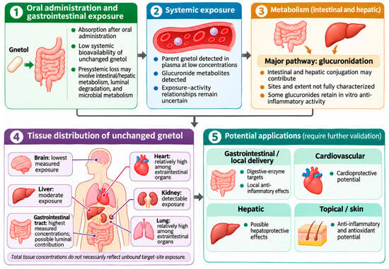
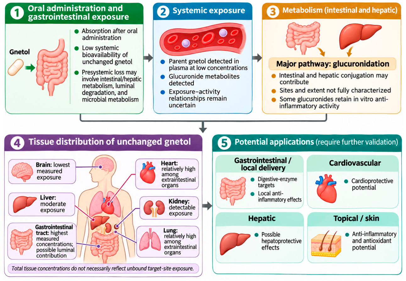

Disclaimer/Publisher’s Note: The statements, opinions and data contained in all publications are solely those of the individual author(s) and contributor(s) and not of MDPI and/or the editor(s). MDPI and/or the editor(s) disclaim responsibility for any injury to people or property resulting from any ideas, methods, instructions or products referred to in the content. |
© 2026 by the authors. Licensee MDPI, Basel, Switzerland. This article is an open access article distributed under the terms and conditions of the Creative Commons Attribution (CC BY) license.