


Abstract
This study aimed to evaluate the impact of ultrasound-assisted extraction (UAE) on the chemical composition and biological activities of gabiroba waste oil (GWO), compared to conventional Soxhlet extraction (SE). GWO was characterized for carotenoid content and analyzed by GC-MS for fatty acids (FAs) and sesquiterpenes. Total phenolic content (TPC), flavonoid content (TFC), and antioxidant (ABTS, DPPH, FRAP), antimicrobial, and antiglycemic activities were evaluated. Both methods yielded similar carotenoid content (954–1110 μg β-carotene/g). UAE enhanced phytochemical extraction, with 33% higher TPC (281 mg GAE/100 g), 50% more TFC (193 mg QE/100 g), and superior antioxidant activity (ABTS-12 μmol TE/g for hydrophilic extract, 109 μmol TE/g for lipophilic extract). UAE produces a high-quality oil with higher yield (36.6%) and lower acidity index (12.5%) compared to SE. Oleic acid was the predominant FA, followed by palmitic and linoleic acids, with β-cadinene as the major terpene. UAE oil exhibited a 21% increase in unsaturated FA and a 36% reduction in saturated FA compared to SE. UAE oil had greater antimicrobial activity for S. aureus (MIC-3.9 mg/mL) and higher α-amylase (IC50—316 μg/mL) and α-glucosidase (IC50—45 μg/mL) inhibitory potential. UAE proved to be an efficient, sustainable method to obtain phytochemical-rich oil from food residues, valorizing gabiroba waste for pharmaceutical and functional food applications.
1. Introduction
Campomanesia xanthocarpa, commonly known as gabiroba, is a native Brazilian fruit belonging to the Myrtaceae family, widely distributed across the southern and central-western regions, particularly in the Cerrado and Atlantic Forest biomes [1,2]. The fruits are small, globular berries that turn yellow and sweet upon ripening, traditionally used in the artisanal production of sweets, beverages, and jams [3,4]. Beyond their pleasant flavor, gabiroba fruits present important nutritional and functional properties, being composed of carbohydrates (7.8–17.7%), fibers (6.3–9.9%), proteins (1.1–5.5%), and lipids (0.6–3.7%). Moreover, the fruit matrix represents a rich natural source of ascorbic acid (up to 30.58 mg g−1), minerals, phenolic compounds, and carotenoids [3,5,6]. Previous investigations focusing on different anatomical parts of C. xanthocarpa, including leaves, pulp, and seeds, have demonstrated significant antioxidant, antimicrobial, anti-inflammatory, antihypercholesterolemic, antitumor, and antidiabetic activities [7,8,9,10,11]. However, despite this well-documented functional potential, the commercial exploitation and industrial processing of gabiroba remain limited [4,12]. Processing or direct consumption of gabiroba generates about 40% of agri-food waste (peels, seeds, and calyx) [4], which is increasingly recognized as a valuable source of biologically active constituents, offering strategic opportunities for sustainable practices and initiatives aligned with the principles of the circular economy.
While significant research has focused on the bioactive profile and biological activities of aqueous and alcoholic extracts from the pulp and leaves of gabiroba [4,7,8,13,14,15], comprehensive data regarding the lipid fraction recovered, particularly from fruit waste, remain underexplored. To date, a single study has investigated the oil extracted specifically from gabiroba seeds [16], reporting a profile rich in bioactive fatty acids, notably oleic, linoleic, and palmitic acids. However, the oil recovered from whole gabiroba fruit processing waste remains unexplored, leaving a clear scientific gap regarding its chemical composition and functional potential.
Researchers have investigated the chemical and physical properties of oils derived from non-conventional plant species to identify alternative lipid sources with distinct nutritional, functional, and technological characteristics, capable of expanding application possibilities and meeting the demands for new products, boosting their industrial application [17,18]. The valorization of gabiroba waste as an alternative source of oil relies heavily on the extraction method used, which directly influences yield and compound integrity. Soxhlet extraction (SE) is conventionally employed due to high yields; however, the prolonged extraction times and elevated temperatures can degrade sensitive phytochemical compounds. Additionally, other limitations of SE include high energy consumption and excessive use of organic solvents. In contrast, ultrasound-assisted extraction (UAE) has emerged as an efficient, eco-friendly alternative, offering faster extraction times, reduced solvent consumption, lower energy requirements, and better preservation of heat-sensitive compounds. This technique utilizes high-frequency sound waves to generate cavitation bubbles, enhancing mass transfer and improving the extraction of phytochemical compounds from plant materials [19,20].
Considering the nutritional potential of gabiroba, the importance of new natural sources of bioactive compounds, and the need for sustainable extraction methods, this study aims to assess the impact of ultrasound-assisted extraction on the chemical composition and biological activities of gabiroba waste oil, compared to conventional Soxhlet extraction.
2. Results and Discussion
2.1. Proximate Composition of Gabiroba Waste
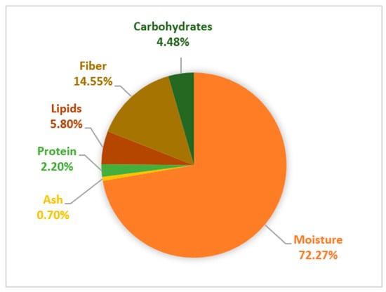
Water was the main constituent of the gabiroba waste (GW), accounting for 72.27% ± 0.1% (Figure 1). Among the pulp macronutrients, dietary fiber (14.55 ± 0.29%) and lipids (5.80 ± 0.59%) were the major components, whereas carbohydrates (4.48%) and proteins (2.20 ± 0.08%) were present in lower amounts. These results are consistent with those reported for the proximate composition of Campomanesia adamantium (green gabiroba) residue (moisture: 63.7%; ash: 0.74%; protein: 3.17%; lipids: 5.33%; fiber: 24.05%; and carbohydrates: 3%) [21]. Also, GW exhibits higher lipid, protein, and fiber contents compared to the whole fruit (1.3–1.9% of lipids, 1.1% of proteins, 6.3–9.9% of fibers) [3,6]. According to the Agência Nacional de Vigilância Sanitária (ANVISA), which establishes a daily fiber intake of 25 g in a 2000 kcal diet, GW can be classified as a high-fiber content material, since it provides 20% of the recommended daily value of fiber [22]. Fibers such as galactomannans and pectins, previously identified in the pulp of the gabiroba fruit, are promising polysaccharides due to their gelling and stabilizing properties for the food industry and pharmacological applications, such as immunoregulatory, anti-inflammatory, hypoglycemic, antioxidant and antitumor activities [7,23,24,25].
Figure 1.
Proximate composition of gabiroba waste on a wet weight basis.
2.2. Fatty Acid and Sesquiterpene Profile
GW was used to obtain oil employing ultrasound-assisted extraction (UAE) and Soxhlet (SE) techniques. Gas chromatography analysis revealed the presence of over 30 compounds (Table 1), with fatty acids accounting for more than 70% of the total identified substances. The identification of the compounds was supported by the raw total ion chromatograms (TICs) and experimental mass spectra obtained by GC-MS, which are provided in the Supplementary Materials (Figures S1–S4). In both GWO extraction methods, the predominant fatty acids were oleic acid (C18:1), followed by palmitic acid (C16:0) and linoleic acid (C18:2). These results are consistent with those reported by Santos et al. [16], who identified oleic, palmitic, and linoleic acids as the primary fatty acids in C. xanthocarpa seed oil, comprising 60.8%, 13.9%, and 14.1%, respectively.
Table 1.
Chemical composition of gabiroba waste oil obtained by Soxhlet (SE) and ultrasound-assisted extraction (UAE).
Monounsaturated fatty acids (MUFAs) constitute the predominant group of fatty acids in GWO, with the UAE oil showing a 21% increase in unsaturated fatty acid content compared to Soxhlet extraction (SE), accompanied by a 36% reduction in saturated fatty acids. The observed variation in the relative fatty acid profile between UAE and SE can be attributed to differences in extraction selectivity and thermal preservation. The milder extraction conditions and shorter extraction time employed in the UAE minimize thermal and oxidative degradation of unsaturated fatty acids, which are more susceptible to oxidation. Conversely, the prolonged exposure to high temperatures and continuous reflux inherent to SE can induce partial thermal degradation of double bonds in unsaturated species [26]. Furthermore, the energy generated by ultrasonic waves causes cavitation bubbles to form, expand, and collapse as the waves propagate through the solvent medium; this disrupts cell membranes and rapidly releases fluid, membrane-associated lipid fractions that are naturally enriched in unsaturated chains [27,28]. MUFAs and saturated fatty acids are more resistant to high temperatures and rancidity than polyunsaturated fatty acids [20]. In addition to MUFAs, the fatty acid composition of GWO also includes linoleic acid, an omega-6 (ω-6) polyunsaturated fatty acid essential for human health, as it influences the prevalence and severity of cardiovascular diseases, diabetes, cancer, and age-related functional decline [29]. Furthermore, small amounts of myristic, stearic, and eicosanoic acids were detected, and elaidic acid, a trans-isomer of oleic acid, was present at concentrations below 3%.
The biological functions of oils obtained from native Brazilian fruits have been extensively studied. Cumaru seed oil showed antimicrobial and antioxidant properties [30], and buriti oil has immunomodulatory effects against enteropathogenic Escherichia coli [31].
The volatile compounds detected in the GWO predominantly belong to the terpene class, with sesquiterpenes being the most prevalent. Among these, β-cadinene was identified as the most abundant terpene in both UAE and SE methods. Terpenes are the main components of essential oils and have been previously found in essential oils from the fruits and seeds of C. xanthocarpa [3,32]. Capeletto et al. [15] reported sesquiterpenes such as caryophyllene (17.83%), γ-cadinene (9.58%), and α-cadinol (8.83%) as the major compounds in extracts of C. xanthocarpa seeds obtained by supercritical CO2 and compressed n-butane. The terpene profile presented by the GWO is similar to that found by Cardoso et al. [33] for the hexane fractions of the fruits of C. pubescens and C. adamantium. Aromatic compounds, such as sesquiterpenes, are associated with the characteristic flavor and aroma of fruits, which supports their application as flavoring agents in foods and beverages [3].
2.3. Physicochemical Characterization of Gabiroba Waste Oil
UAE showed a slight increase (p = 0.02) in extraction yield when compared to SE (Table 2). In SE, time, temperature, solvent type, number of cycles, and solvent/solute interactions influence the extraction of biomolecules from plant matrices; that is, at its boiling point, the surface tension and viscosity of the solvent are reduced, facilitating access to the active sites of the matrix and favoring the solubilization of the compounds of interest [34]. On the other hand, the ultrasound technique was applied due to its efficiency and low cost since it does not require high temperatures and reduces extraction time, preserving the compounds present in the plant matrix.
Table 2.
Physicochemical parameters of gabiroba waste oil.
The higher oil yield observed for UAE compared to SE can be attributed to the physical and mechanical phenomena driven by acoustic cavitation, which enhance the contact between the solvent and the solid matrix and facilitate mass transfer [35]. As ultrasonic waves propagate through the sample, they generate rapid alternating compression and expansion cycles (sponge effect) within the plant tissue, producing mechanical stresses that can promote cell disruption and the formation of microchannels and micropores, favoring solvent penetration into the plant matrix and the release of intracellular oil [36]. Similar to Tan et al. [19] for avocado oil, UAE outperformed SE in both yield and extraction efficiency of GWO.
Regarding the acidity index, UAE resulted in a better-quality oil with a lower content of free fatty acids. Conversely, SE resulted in an acidity index of 14.1%, around 13% higher than UAE-based oil. High temperatures contribute to an increase in the hydrolytic rancidity of the oil, with consequent de-esterification of fatty acids and increased concentrations of free fatty acids [31]. Moreover, when the oil is exposed to high temperatures, its density can be affected due to oxidation and polymerization reactions [37]. This effect was observed for GWO, in which the density increased from 922.8 Kg m−3 for the UAE oil to 932.4 Kg m−3 for the SE oil. The refractive index (IR) for GWO obtained by UAE and SE were 1.4802 and 1.4894, respectively. IR of fats and oils is related to their average degree of unsaturation and it is helpful in observing the progress of reactions such as hydrogenation and isomerization [38]. Higher IR values indicate a greater possibility of deterioration due to oxidation [39].
2.4. Carotenoids, Phenolic Compounds and Antioxidant Capacity
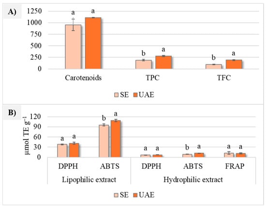
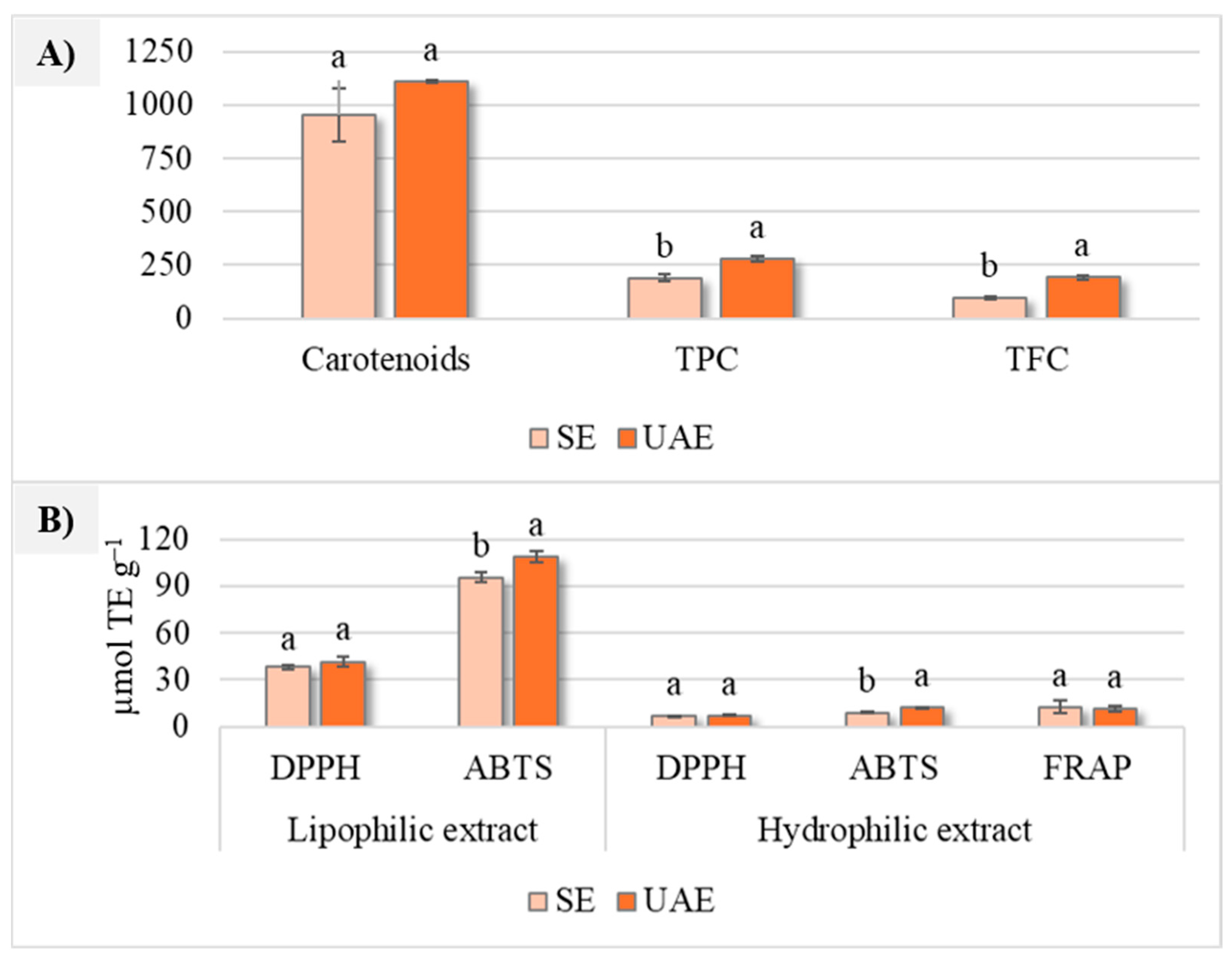
Figure 2A shows that the gabiroba waste oil (GWO) obtained exhibited high carotenoid levels; however, no significant differences (p > 0.05) were observed between UAE and SE, indicating that both extraction methods were similarly effective for carotenoid recovery. The carotenoid levels in the GWO present average values of 1032.39 ± 8.73 μg β-carotene g−1, which were comparable to that reported for buriti oil (760 μg β-carotene g−1) [31], approximately twice that reported for pequi oil (550 μg β-carotene g−1) [40], and 13-fold higher than that reported for gabiroba seed oil (79 μg β-carotene g−1) [16]. Carotenoids have important biological functions, being widely used industrially as natural colorants, in addition to providing several health benefits due to their antioxidant properties and their role as a precursor to vitamin A [14].
Figure 2.
Carotenoid content (μg of β-carotene g−1), total phenolic compounds (TPC) (mg GAE 100 g−1), total flavonoid content (TFC) (mg QE 100 g−1) (A), and antioxidant activity by DPPH, ABTS, and FRAP assays (B) of GWO. Equal letters do not differ by Tukey’s test (p > 0.05).
UAE exhibited 33% greater efficiency in obtaining total phenolic compounds (TPC: 280.79 ± 13.74 mg GAE 100 g−1) and 50% higher levels of total flavonoids (TFC: 192.83 ± 8.92 mg QE 100 g−1) compared to SE, which yielded TPC of 188.16 ± 16.42 mg GAE 100 g−1 and TFC of 97.39 ± 6.86 mg QE 100 g−1 (Figure 2A). Phenolic and flavonoid contents were compared with those of lipid fractions recovered from other non-conventional oil sources. The TPC of GWO was lower than that reported for baru seed oil (685–1315 mg GAE 100 g−1) [41]. Conversely, the TPC of GWO was higher than those reported for cumaru seed oil (22 mg GAE 100 g−1) [30], pequi oil (4.40 mg GAE 100 g−1) [40], and gabiroba seed oil (43 mg GAE 100 g−1) [16]. Similarly, GWO showed significantly higher values of TFC than those reported for pequi oil (1.10 mg QE 100 g−1).
UAE demonstrated the highest antioxidant activity as measured by the ABTS method, with values of 12.08 ± 0.14 μmol TE g−1 for the hydrophilic extract and 108.87 ± 3.64 μmol TE g−1 for the lipophilic extract (Figure 2B). The higher temperature and longer extraction time used in SE may have led to the degradation of some antioxidant compounds, which could have substantially contributed to the observed differences in antioxidant activity. However, the extraction technique did not significantly affect FRAP or DPPH values.
When comparing the antioxidant activity of lipophilic and hydrophilic extracts, lipophilic extracts showed a fivefold increase in DPPH activity and a tenfold increase in ABTS activity (Figure 2B). Assays such as DPPH, ABTS, and FRAP have different mechanisms of action, and results vary depending on the chemical composition of the extracts and the reactivity of the antioxidants in each method [31]. The observed differences in antioxidant activity across methods and extraction techniques resulted from the varying reactivity of the extract components with the radicals present in each assay.
These phytochemicals play a crucial role in lipid matrices by acting as natural antioxidants. They exert a protective effect on unsaturated fatty acids, scavenging free radicals and mitigating oxidative lipid degradation during storage and processing, and may also contribute to enhancing the biological activities associated with the oils [20].
2.5. Antimicrobial Activity
The antimicrobial activity of GWO varied according to the extraction method and target microorganism, as can be observed in Table 3. S. aureus was more susceptible to UAE oil compared to SE oil. Conversely, B. cereus was more susceptible to SE oil, whereas higher concentrations were needed for UAE oil. Evidence suggests that fatty acids act as detergents in the amphipathic structure of bacterial cell membranes, impairing energy generation processes [42]. Also, terpenes found in GWO are bioactive compounds capable of inhibiting protein and DNA synthesis and promoting cell membrane rupture [43]. Similarly, sesquiterpenes from C. xanthocarpa seed extracts have previously shown inhibitory effects against resistant bacteria like Pseudomonas aeruginosa, with MIC and MBC between 5 and 40 mg mL−1 [15].
Table 3.
Antimicrobial activity of gabiroba waste oil using microdilution method.
The antifungal activity of GWO against C. albicans was similar for both extraction methods. SE and UAE oils exhibited an MIC of 15.63 mg mL−1. However, UAE oil showed superior fungicidal activity, with an MBC of 31.25 mg mL−1 compared to 62.50 mg mL−1 for SE oil. Fatty acids, such as oleic, palmitic, and linoleic acids, found in GWO, along with lauric and stearic acids, are known for their antifungal potential [44].
GWO did not show any antimicrobial effect against S. enterica, E. coli and L. monocytogenes, probably due to structural differences in their cell walls. The outer membrane of Gram-negative bacteria, such as E. coli and S. enterica, contains a lipopolysaccharide layer that acts as a hydrophilic barrier, limiting the diffusion of hydrophobic compounds [45]. Additionally, L. monocytogenes is a Gram-positive bacterium with high resistance to environmental stress, which may also hinder the action of antimicrobial compounds [46].
Despite this selective activity, the antimicrobial potential of GWO is promising. The different antimicrobial performance between extraction methods may reflect variations in the chemical composition of the oils, which could influence their biological activity.
2.6. Antiglycemic Activity
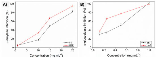
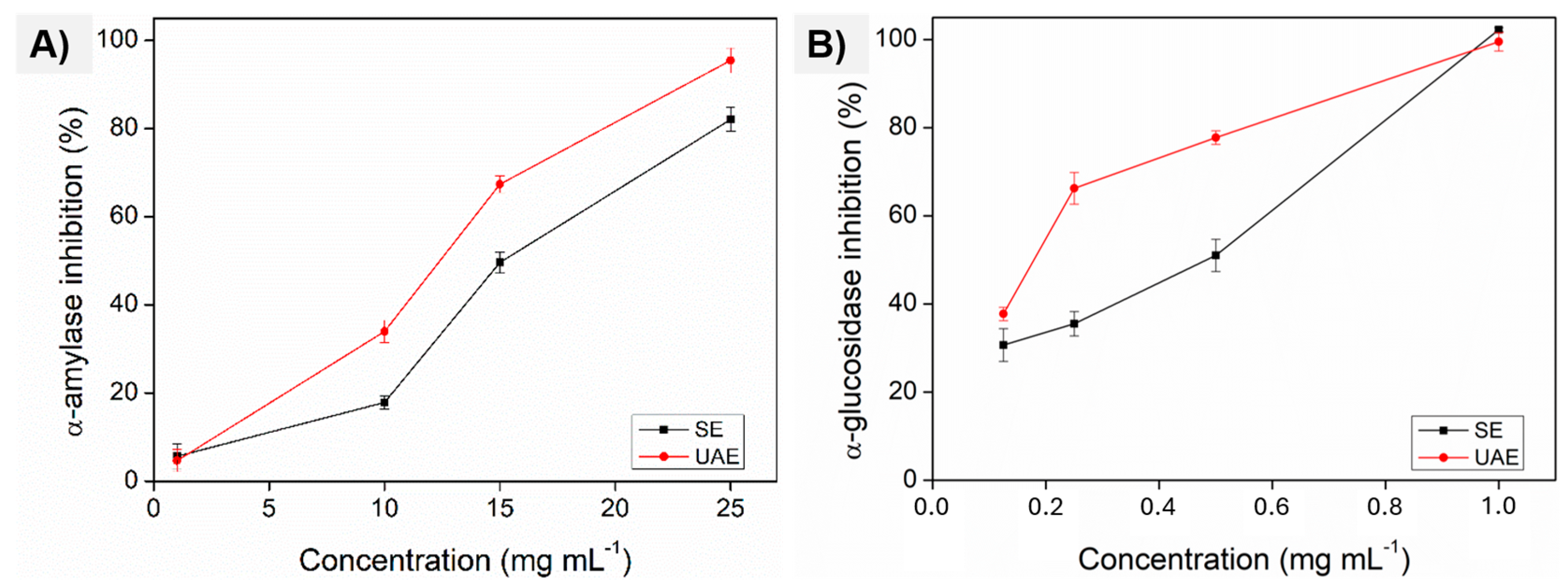
The antiglycemic activity is focused on the inhibition of α-amylase and α-glucosidase enzymes, responsible for the digestion of soluble carbohydrates. The effectiveness of GWO in inhibiting α-amylase (Figure 3A) and α-glucosidase (Figure 3B) enzymes exhibited a dose-dependent relationship. The compounds obtained by UAE potentiated the α-amylase inhibitory effect, mainly at the highest concentrations, with an inhibition of 95% for UAE and 82% for SE. The IC50 values highlighted a significant difference: 316 μg mL−1 for the UAE oil, compared to 400 μg mL−1 for the SE oil; that is, the oil obtained through ultrasound extraction is more efficient, requiring lower concentrations to achieve 50% enzyme inhibition. Methanolic extracts of gabiroba fruits rich in phenolic compounds showed inhibition of α-amylase activity with an IC50 of 26 μg mL−1 in the reaction [8], a more significant effect compared to the present work. This difference is associated with both the characteristics of the raw material and the solvent employed, which influence the chemical composition and bioactive properties of the extracts.
Figure 3.
Inhibitory activity of α-amylase (A) and α-glucosidase (B) enzymes by GWO obtained through SE and UAE.
On the other hand, both oils reached 100% inhibition of α-glucosidase at the highest concentration. However, considering the IC50 values, UAE oil (IC50: 45 μg mL−1) demonstrated superior inhibition potential compared to SE oil (IC50: 122 μg mL−1). At a concentration of 0.125 mg mL−1, the UAE oil exhibited a more significant inhibitory effect on α-glucosidase than on α-amylase, achieving 37% inhibition. Acarbose is the standard compound often used to slow carbohydrate digestion by inhibiting digestive enzymes and has IC50 values of 24 and 58 μg mL−1 for α-amylase and α-glucosidase, respectively [47]. This suggests that the UAE oil is more potent in inhibiting α-glucosidase at lower concentrations, emphasizing its potential as a more effective supplement or additive for controlling postprandial blood glucose levels.
The antiglycemic effect is a known property of aqueous and alcoholic extracts of leaves, seeds, and fruits of C. xanthocarpa [8,10,13,48]. Extracts obtained with pressurized solvent from C. xanthocarpa seeds reduced blood glucose levels in rats, with this effect being attributed to the terpenes found, such as caryophyllene, eudesmol, guaiol, α-selinene and β-cadinene [10], many of which were also identified in GWO.
The observed hypoglycemic effects of GWO, attributed to enzyme inhibition, align with findings in the literature that have linked such activities to the presence of fatty acids, reported as inhibitors of carbohydrate-hydrolyzing enzymes like α-glucosidase and α-amylase, thereby delaying glucose absorption [49]. Moreover, lipid–phenolic interactions have been shown to contribute to antioxidant and α-amylase inhibition activities [50]. Therefore, the enzymatic inhibition observed in GWO may stem from a synergistic interaction of various compounds present in the oils, further supporting its potential as an innovative natural additive for controlling glucose metabolism.
2.7. Anticancer Activity
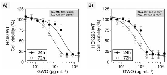
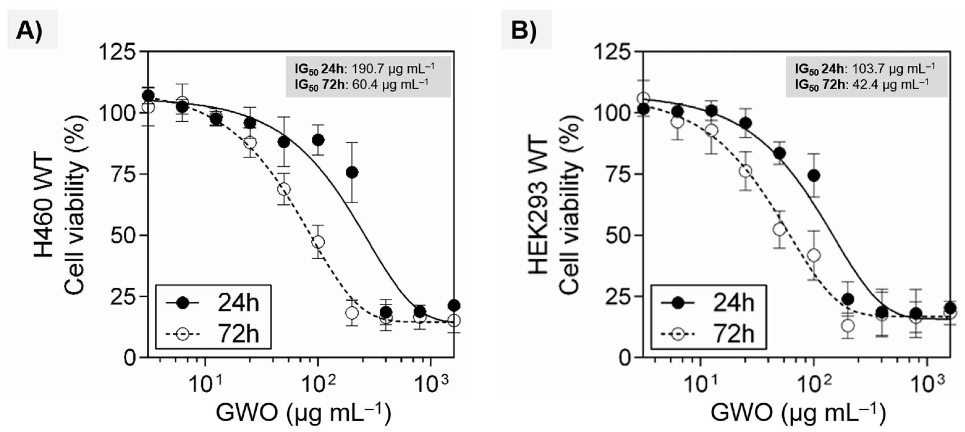
The cytotoxic effect of UAE oil was evaluated in human fibroblast (HEK293 WT) and lung cancer (H460 WT) cell lines using the MTT assay. Increasing the concentration of GWO and exposure time decreased the number of viable cells, indicating a time- and dose-dependent cytotoxic effect. As shown in Figure 4A, H460 cells responded differently depending on the incubation period when lower oil concentrations were tested. The cytotoxic effect was most severe after 72 h of exposure. However, at higher concentrations (400–1600 µg mL−1), cell viability was equally reduced to less than 25% after 24 and 72 h of exposure. According to the IG50 (concentration required to reduce 50% of cell viability), prolonged exposure to GWO was 3 times more cytotoxic.
Figure 4.
Cytotoxic effect of GWO extracted by UAE. Cell viability assay performed using the MTT method. HEK293 wild-type (A) and H460 wild-type cells (B) were exposed for 24 and 72 h with increasing concentrations of the compounds (3.125–1600 µg mL−1). Data represents mean ± SD of at least three independent experiments. The viability curves were fitted using GraphPad Prism 6 software.
Similar behavior was observed for HEK293 cells; cytotoxicity was higher after 72 h of incubation (Figure 4B). At a concentration of 100 μg mL−1, cell viability remained above 75% after 24 h of exposure and below 50% after 72 h. At this concentration and regardless of exposure time, GWO reduced cell viability to less than 25%.
The results presented by UAE oil were more significant than those presented by pectins extracted from gabiroba pulp, which reduced the metabolic activity of human glioblastoma cells by 80% at a concentration of 25 μg mL−1 [7]. It was demonstrated that fatty acids such as oleic and palmitic acid, present in GWO, have good affinity for inhibiting the myeloid cell leukemia protein 1 (Mcl-1) protein, pointing to the anticancer potential of fatty acids [51]. Additionally, unsaturated fatty acids known for inducing apoptosis in tumor cells may further enhance the generation of free radicals and lipid peroxidation, strengthening GWO anticancer effects [52]. Furthermore, flavones (5-hydroxy-7-methoxy-8-methyl) isolated from extracts of gabiroba fruits inhibited the growth of H460 cells by 27% and renal tumor cells by 46% [53]. Phytochemicals, such as phenolics and carotenoids, can act as pro-oxidants, inducing free radical production that combats tumor proliferation [54].
From a pharmacological perspective, it is important to highlight that the cytotoxicity of a compound at lower concentrations is one of the most valuable characteristics for clinical applications [7]. The IG50 value for doxorubicin in H460 cells, the same cell line used in this work, corresponds to 0.055 µM (0.03 µg mL−1) [55]. GWO at a concentration of 50 µg mL−1 reduced cell viability below 70% after 72 h of exposure. These results are promising, as a compound could be considered cytotoxic when the cell viability is reduced by more than 30% [56]. However, an important characteristic of cytotoxicity is selectivity, so the cytotoxic agent should induce a toxic effect on cancer cells, but it should not affect normal ones [57,58]. The IG50 values demonstrate that GWO was approximately 1.8 times more cytotoxic to fibroblasts than to lung cancer cells. These results reinforce the need to evaluate the selectivity of the interaction with cancer cells in the screening of anticancer drug candidates. Therefore, further investigation into the chemical composition of GWO and its cytotoxic profile is important before indicating it as a potential agent in anticancer therapy.
3. Materials and Methods
3.1. Preparation and Proximate Composition of Gabiroba Waste
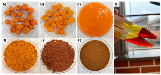
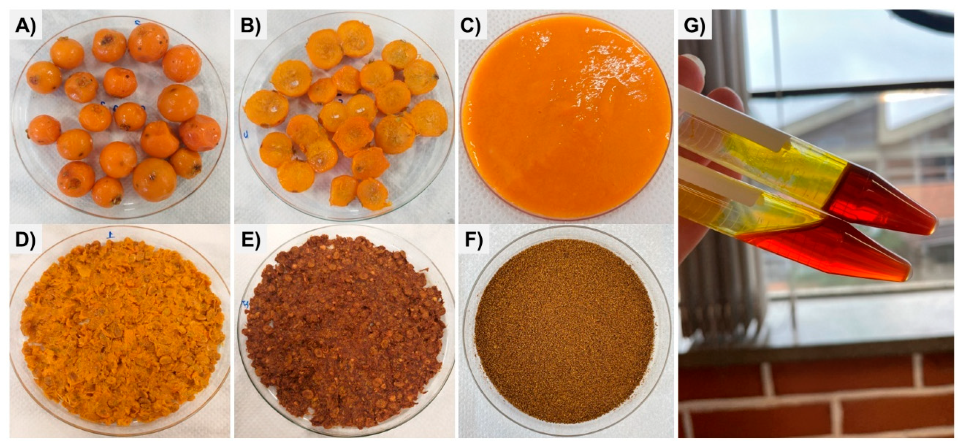
Ripe gabiroba fruits were collected in the city of Irati, Paraná state, Brazil (S25°27′56″; W50°37′51″) (Figure 5A,B). The fruits were washed, sanitized, and then stored at −18 °C. The fruits were thawed and subjected to pulping (Figure 5C) using a pulper (Braesi, model DES-20, Caxias do Sul, RS, Brazil). Gabiroba waste (GW) was collected (Figure 5D), dried at 40 °C in an oven with air circulation for 48 h (Figure 5E), and subsequently ground in a laboratory mill (Cadence, model MDR302, China). The ground material was sieved through a Tyler series 20 mesh sieve, collecting particles with a diameter smaller than 0.5 mm. The raw material (Figure 5F) was stored under vacuum at 25 ± 5 °C, protected from light, until further use.
Figure 5.
Gabiroba fruit (A), interior of the fruit (B), pulp (C), fresh residue (D), dried residue (E), residue flour (F), lipidic fractions (G).
Proximate composition analyses of GW were performed in triplicate using protocols established by AOAC [59]. Moisture content was obtained by oven drying at 105 °C to constant mass (AOAC 926.12); total proteins were determined by the Kjeldahl method, with a nitrogen-to-protein conversion factor of 6.25; fixed mineral residues were obtained by incineration in a muffle furnace at 550 °C (AOAC 945.46); and dietary fiber was obtained using the enzymatic-gravimetric method (AOAC 991.43). Lipid content was assessed by cold extraction, based on the method of Bligh and Dyer [60]. Total carbohydrate content was calculated by the difference in mass.
3.2. Extraction Procedures
The ultrasonic-assisted extraction (UAE) of gabiroba waste oil (GWO) was performed according to the methodology described by Goh et al. [61], with modifications. An ultrasound system equipped with a high-intensity probe (130 W, 20 kHz—Sonics & Materials, Inc., model Sonics Vibra-Cell VCX 130, Newtown, CT, USA) was used. The amplitude of ultrasonic vibrations was set at 100% of the nominal power. The extraction was carried out in an Erlenmeyer flask containing 20 g of sample and 50 mL of hexane for 30 min. The ultrasonic probe was directly immersed in the mixture, with the tip fully submerged in the solvent. Ultrasonication was applied in pulses of 30 s on and 30 s off to minimize overheating, and an ice bath was employed to maintain the temperature below 30 °C. After sonication, the mixture was filtered, and the raw material was subjected to exhaustive extraction with 50 mL of fresh solvent. This procedure was repeated three times, and all filtrates were pooled in a single vial. For comparison, Soxhlet extraction (SE) was performed using 20 g of sample wrapped in filter paper and placed inside a cartridge. The extraction was carried out with 150 mL of hexane recirculated for 9 h. The extraction yield (%) was calculated by determining the ratio of the mass of the extract to the mass of the raw material, multiplied by 100.
After extraction by both methods, the solvent was removed using a rotary vacuum evaporator at 35 °C under reduced pressure (5 psi). The extracted oil (Figure 5G) was stored in amber glass vials under refrigeration until further analysis. Lipophilic and hydrophilic extracts were obtained from GWO extracted by UAE and SE for the determination of carotenoids, phenolic compounds, flavonoids and antioxidant activity. Lipophilic extracts were obtained by adding 20 mL of ice-cold acetone to 1 g of oil, followed by continuous agitation in an ultrasonic bath for 10 min. After extraction, the mixture was filtered and subjected to liquid–liquid partitioning with petroleum ether and water. The ether phase was collected and used for subsequent analyses [40]. To obtain the hydrophilic extracts, 1 g of oil was extracted with 5 mL of hexane. Subsequently, 5 mL of methanol/water (60:40, v/v) was added, and the mixture was vigorously agitated for 2 min. The hydromethanolic fraction was collected and used for the analyses [62].
3.3. Physical–Chemical Parameters
Acidity index (method 325/IV) was evaluated following the methods proposed by Instituto Adolf Lutz [63]. The refractive index and density were measured at 20 °C, using an Abbe refractometer (Biobrix, Tzaneen, South Africa) and an Anton Paar vibrating-tube density meter (Graz, Austria, DMA 500 M), respectively. The acidity index was expressed as a percentage of free fatty acids, and the density as kg m−3.
3.4. Fatty Acids and Sesquiterpenes Composition by GC/MS
The identification of fatty acids and sesquiterpene compounds was performed using a gas chromatograph coupled to a triple quadrupole mass spectrometer detector (GC/MS-TQ8040, Shimadzu, Kyoto, Japan). The injection volume was 1 µL of the previously esterified samples [64]. During operation, the detector was maintained at 280 °C and the injector at 250 °C. The analysis was carried out in split mode (1:50) and separation was achieved using an SH-Rtx-5MS column (Shimadzu) (30 m × 0.25 mm × 0.25 μm). The oven was set to 50 °C for 1 min, after which it was heated at 25 °C min−1 to 175 °C and then at 4 °C min−1 to 230 °C. The final temperature was maintained for 20 min. Compounds were identified by comparing the obtained mass spectra with the NIST-11 library database. Quantification was performed using the area normalization method, with results expressed as the relative percentage of each identified compound.
3.5. Analysis of Carotenoids, Phenolic Compounds and Antioxidant Activity
Total carotenoids were determined by measuring the absorbance of the lipophilic extract at 450 nm using a spectrophotometer. The concentration was calculated using the molar extinction coefficient of β-carotene (2592 M/cm) and expressed as μg of β-carotene per gram of sample [65]. Total phenolic compounds (TPCs) and total flavonoid content (TFC) were quantified using the hydrophilic extract. TPC was determined by the Folin–Ciocalteu colorimetric method [66], and TFC was analyzed according to the methodology proposed by Prommuak et al. [67]. Quantification was performed using standard calibration curves of gallic acid for TPC and quercetin for TFC, with results expressed as mg of equivalent per 100 g of dry sample.
The antioxidant activity of lipophilic and hydrophilic extracts was evaluated using three complementary assays. The free radical scavenging capacity of DPPH was analyzed according to Brand-Williams et al. [68]. The antioxidant activity against 2,2′-azino-bis(3-ethylbenzothiazoline-6-sulfonic acid) (ABTS) radical cation was determined following the method described by Re et al. [69]. The ferric reducing antioxidant power (FRAP) test was performed according to Benzie and Strain [70]. Quantification was carried out using a standard curve of Trolox, with results expressed as µmol of Trolox equivalent (TE) per g of sample.
3.6. Antimicrobial Activity
The antimicrobial activity of GWO was tested against the pathogens Staphylococcus aureus (ATCC 25923), Bacillus cereus (ATCC 11778), Listeria monocytogenes (ATCC 19117), Escherichia coli (ATCC 25922), Salmonella enterica subsp. enterica serovar Typhimurium (ATCC 1402) and Candida albicans (ATCC 10231), using the microdilution method [71]. To determine the minimum inhibitory concentration (MIC), 50 μL of Mueller–Hinton (MH) broth was added to a 96-well plate. A volume of 50 μL of the diluted oils (500 mg mL−1) was added in the first column and serial (1:1) dilutions were made. Then, 50 μL of the bacterial suspension in MH broth (about 105 CFU mL−1) was added to the well contents. For C. albicans, the procedure consisted of adding 100 µL of the diluted oils to 100 µL of Sabouraud broth. After serial dilution, 100 µL of the yeast suspension (about 103 CFU mL−1) was added to the wells. Chloramphenicol (32 mg L−1) and nystatin (64 mg L−1) were used as positive controls. The microplates were incubated at 37 °C for 18–20 h. The MIC was defined as the lowest GWO concentration that inhibited microbial growth, indicated by the addition of resazurin (0.01%). A color change from blue to pink signaled microbial growth [72]. The minimum bactericidal concentration (MBC) was determined as the lowest GWO concentration that resulted in no bacterial growth, confirmed by inoculating an aliquot from the microplate wells onto agar plates.
3.7. Antiglycemic Activity
3.7.1. α-Amylase (From Human Saliva) Inhibition Assay
The inhibitory activity of GWO on α-amylase was evaluated as an indicator of its potential antiglycemic properties according to Silva et al. [73]. The α-amylase (EC 3.2.1.1, tipo XIII-A, Sigma-Aldrich, Saint Louis, MO, USA) enzyme solution (74 U mL−1) was incubated with GWO at concentrations of 25, 15, 10, and 1 mg mL−1, prepared in dimethyl sulfoxide (DMSO). For the inhibition test (IT), 240 µL of phosphate buffer (20 mM containing 6.7 mM NaCl, pH 6.9) was added to test tubes, followed by 25 µL of GWO and 10 µL of the α-amylase solution. The IT solutions were incubated in a water bath at 37 °C for 15 min, and after that time, 250 µL of potato starch solution (1%) was added, followed by another incubation as described above. The reaction was stopped by adding 500 μL of a solution of 3,5-dinitrosalicylic acid (DNS). The tubes were placed in a boiling bath at 100 °C for 5 min and then cooled to room temperature with the addition of 5 mL of distilled water. The reaction was monitored by measuring the absorbance at 550 nm using a UV-VIS spectrophotometer (Shimadzu, UV-1800).
3.7.2. α-Glucosidase (From Saccharomyces cerevisiae) Inhibition Assay
The test was performed with an α-glucosidase (EC 3.2.1.20, Sigma-Aldrich, Saint Louis, MO, USA) enzyme solution at 1 U mL−1, testing GWO at concentrations of 0.125, 0.250, 0.500, and 1 mg mL−1. The experiment was performed according to Souza et al. [74]. Aliquots of 10 μL of GWO, previously diluted in DMSO to the test concentrations, were added to a 96-well microplate, followed by 40 μL of potassium phosphate buffer (0.1 mol L−1, pH 6.9) and 100 μL of the α-glucosidase enzyme solution. The plate was incubated at 25 °C for 10 min and then 50 μL of the p-nitrophenyl-α-D-glucopyranoside (p-NPG, Sigma Chemical Co., Saint Louis, MI, USA) substrate solution (1.5 mg mL−1 in phosphate buffer) was added. The 1st absorbance reading (0 min) was performed in a microplate reader (TECAN—Infinite M 200, Männedorf, Switzerland) at 405 nm. Then, after 5 min incubation at 25° C, the 2nd reading (5 min) was performed.
3.8. Anticancer Activity
GWO was evaluated against the human non-small lung cancer H460 cell line and HEK293 wild-type (WT) cells provided by Dr. Attilio Di Pietro (IBCP, Lyon, France). Cell viability was evaluated by the 3-(4,5-dimethylthiazol-2-yl)-2,5-diphenyltetrazolium bromide (MTT) colorimetric assay [75]. H460 cells were seeded into 96-well culture plates at a density of 3 × 10⁴ and 1.6 × 10⁴ cells/well for 24 h and 72 h treatments, respectively. After overnight incubation, the cells were treated with increasing concentrations of the UAE oil (3.125 to 1600 µg mL−1, 2-fold serial dilution) in a volume of 200 μL for 24 and 72 h at 37 °C under 5% CO2. After the treatment, 20 μL of a 5 mg mL−1 MTT solution was added, and cells were incubated for 4 h at 37 °C under 5% CO2. After incubation, the culture medium was removed, and the formazan crystals were dissolved with 100 µL of DMSO/ethanol (1:1) solution. After obtaining a homogeneous solution, the absorbance was measured at 595 nm using an iMark™ microplate absorbance reader (Bio-Rad, Hercules, CA, USA). The results were expressed as a percentage of viable cells compared to the control condition (0.3% DMSO (v/v) taken as 100% of viable cells).
3.9. Statistical Analysis
All experiments were performed in triplicate and results were expressed as mean followed by standard deviation. Statistical analysis was performed in the software Statistica®, version 10.0 (StatSoft, Tulsa, OK, USA), using one-way analysis of variance (ANOVA). The Tukey test was applied to assess differences between means at a significance level of p ≤ 0.05.
4. Conclusions
Ultrasound-assisted extraction (UAE) proved to be an efficient method for obtaining phytochemical compounds from gabiroba waste with significant biological activities, being a faster and more effective extraction method. The gabiroba waste oil (GWO) presents a variety of compounds, with emphasis on unsaturated fatty acids, sesquiterpenes and carotenoid content, and demonstrates promising antioxidant, antimicrobial, and antihyperglycemic potential for industrial application. Further studies investigating additional cell lines, safety and the toxicological profile of the individual oil components are required to better assess the anticancer potential of GWO. The chemical profile of GWO highlights its potential for natural product formulations aimed at combating oxidative stress, microbial infections, and supporting glycemic control. The present study provides data consistent with sustainable practices, contributing to the reduction in the ecological impact of industrial waste by employing them in extraction processes, significantly reducing extraction time and energy consumption by employing the UAE technique.
Supplementary Materials
The following supporting information can be downloaded at: https://www.mdpi.com/article/10.3390/molecules31203705/s1.
Author Contributions
Conceptualization, C.L.C.d.P.; methodology, C.L.C.d.P., R.L.d.A., S.Á., G.M.M., I.d.S.Z., V.R.M. and G.V.; investigation, C.L.C.d.P., R.L.d.A. and I.d.S.Z.; resources, V.R.M., G.V., C.V.H., M.R.M. and L.I.-M.; data curation, C.L.C.d.P., R.L.d.A. and G.M.M.; writing—original draft preparation, C.L.C.d.P. and R.L.d.A.; writing—review and editing, C.L.C.d.P., R.L.d.A., S.Á., G.M.M., F.O.F., C.V.H., M.R.M. and L.I.-M.; supervision, G.M.M., F.O.F., C.V.H. and L.I.-M.; project administration, L.I.-M.; funding acquisition, G.M.M., C.V.H., M.R.M. and L.I.-M. All authors have read and agreed to the published version of the manuscript.
Funding
This study was funded by the Coordenação de Aperfeiçoamento de Pessoal de Nível Superior (CAPES, Finance Code 001) and the Conselho Nacional de Desenvolvimento Científico e Tecnológico (CNPq, Grant 315667/2021-4 and 316815/2021-7).
Institutional Review Board Statement
Not applicable.
Informed Consent Statement
Not applicable.
Data Availability Statement
The original contributions presented in the study are included in the article; further inquiries can be directed to the corresponding authors.
Conflicts of Interest
Author Cristiane Vieira Helm is employed by Brazilian Agricultural Research Corporation. The remaining authors declare that the research was conducted in the absence of any commercial or financial relationships that could be construed as a potential conflict of interest. Brazilian Agricultural Research Corporation (Embrapa Florestas) had no role in the design of the study; in the collection, analyses, or interpretation of data; in the writing of the manuscript; or in the decision to publish the results.
References
- de Oliveira Raphaelli, C.; dos Santos Pereira, E.; Camargo, T.M.; Ribeiro, J.A.; Pereira, M.C.; Vinholes, J.; Dalmazo, G.O.; Vizzotto, M.; Nora, L. Biological Activity and Chemical Composition of Fruits, Seeds and Leaves of Guabirobeira (Campomanesia xanthocarpa O. Berg-Myrtaceae): A Review. Food Biosci. 2021, 40, 100899. [Google Scholar] [CrossRef] [Scilit]
- Cardozo, C.M.L.; Inada, A.C.; Cardoso, C.A.L.; Filiú, W.F.D.O.; Farias, B.B.D.; Alves, F.M.; Tatara, M.B.; Rosa Croda, J.H.; de Cássia Avellaneda Guimarães, R.; Hiane, P.A.; et al. Effect of Supplementation with Hydroethanolic Extract of Campomanesia xanthocarpa (Berg.) Leaves and Two Isolated Substances from the Extract on Metabolic Parameters of Mice Fed a High-Fat Diet. Molecules 2020, 25, 2693. [Google Scholar] [CrossRef] [Scilit] [PubMed]
- Vallilo, M.I.; Moreno, P.R.H.; Oliveira, E.D.; Lamardo, L.C.A.; Garbelotti, M.L. Composição Química Dos Frutos de Campomanesia xanthocarpa Berg-Myrtaceae Chemical Composition of Campomanesia xanthocarpa Berg-Myrtaceae Fruit. Ciênc. Tecnol. Aliment. 2008, 28, 231–237. [Google Scholar]
- Barbieri, S.F.; de Oliveira Petkowicz, C.L.; de Godoy, R.C.B.; de Azeredo, H.C.M.; Franco, C.R.C.; Silveira, J.L.M. Pulp and Jam of Gabiroba (Campomanesia xanthocarpa Berg): Characterization and Rheological Properties. Food Chem. 2018, 263, 292–299. [Google Scholar] [CrossRef] [Scilit] [PubMed]
- Pereira, M.C.; Steffens, R.S.; Jablonski, A.; Hertz, P.F.; de O. Rios, A.; Vizzotto, M.; Flôres, S.H. Characterization and Antioxidant Potential of Brazilian Fruits from the Myrtaceae Family. J. Agric. Food Chem. 2012, 60, 3061–3067. [Google Scholar] [CrossRef] [Scilit] [PubMed]
- Santos, M.S.; Correia, C.H.; Petkowicz, C.L.O.; Cândido, L.M.B. Evaluation of the Technological Potential of Gabiroba [Campomanesia xanthocarpa Berg] Fruit. J. Nutr. Food Sci. 2012, 2, 161. [Google Scholar] [CrossRef]
- da Costa Amaral, S.; Barbieri, S.F.; Ruthes, A.C.; Bark, J.M.; Brochado Winnischofer, S.M.; Silveira, J.L.M. Cytotoxic Effect of Crude and Purified Pectins from Campomanesia xanthocarpa Berg on Human Glioblastoma Cells. Carbohydr. Polym. 2019, 224, 115140. [Google Scholar] [CrossRef] [Scilit] [PubMed]
- Grutzmann Arcari, S.; Arena, K.; Kolling, J.; Rocha, P.; Dugo, P.; Mondello, L.; Cacciola, F. Polyphenolic Compounds with Biological Activity in Guabiroba Fruits (Campomanesia xanthocarpa Berg.) by Comprehensive Two-Dimensional Liquid Chromatography. Electrophoresis 2020, 41, 1784–1792. [Google Scholar] [CrossRef] [Scilit] [PubMed]
- Klafke, J.Z.; Pereira, R.L.D.; Hirsch, G.E.; Parisi, M.M.; Porto, F.G.; de Almeida, A.S.; Rubin, F.H.; Schmidt, A.; Beutler, H.; Nascimento, S.; et al. Study of Oxidative and Inflammatory Parameters in LDLr-KO Mice Treated with a Hypercholesterolemic Diet: Comparison between the Use of Campomanesia xanthocarpa and Acetylsalicylic Acid. Phytomedicine 2016, 23, 1227–1234. [Google Scholar] [CrossRef] [Scilit] [PubMed]
- Regginato, A.; Cunico, L.; Bertoncello, K.T.; Schindler, M.S.Z.; Chitolina, R.; Marins, K.; Zanatta, A.P.; Calisto, J.F.; Oliveira, J.V.; Magro, J.D.; et al. Antidiabetic and Hypolipidemic Potential of Campomanesia xanthocarpa Seed Extract Obtained by Supercritical CO2. Braz. J. Biol. 2020, 81, 621–631. [Google Scholar] [CrossRef] [Scilit] [PubMed]
- Viecili, P.R.N.; Borges, D.O.; Kirsten, K.; Malheiros, J.; Viecili, E.; Melo, R.D.; Trevisan, G.; da Silva, M.A.; Bochi, G.V.; Moresco, R.N.; et al. Effects of Campomanesia xanthocarpa on Inflammatory Processes, Oxidative Stress, Endothelial Dysfunction and Lipid Biomarkers in Hypercholesterolemic Individuals. Atherosclerosis 2014, 234, 85–92. [Google Scholar] [CrossRef] [Scilit] [PubMed]
- Silva-Rodrigues, H.C.; Silveira, M.P.; Helm, C.V.; de Matos Jorge, L.M.; Jorge, R.M.M. Gluten Free Edible Film Based on Rice Flour Reinforced by Guabiroba (Campomanesia xanthocarpa) Pulp. J. Appl. Polym. Sci. 2020, 137, 49254. [Google Scholar] [CrossRef] [Scilit]
- Vinagre, A.S.; Rönnau, Â.D.S.R.O.; Pereira, S.F.; Silveira, L.U.D.; Wiilland, E.D.F.; Suyenaga, E.S. Anti-Diabetic Effects of Campomanesia xanthocarpa (Berg) Leaf Decoction. Artic. Braz. J. Pharm. Sci. 2010, 46, 169–177. [Google Scholar] [CrossRef] [Scilit]
- Czaikoski, K.; Mesomo, M.C.; Krüger, R.L.; Queiroga, C.L.; Corazza, M.L. Extraction of Campomanesia xanthocarpa Fruit Using Supercritical CO2 and Bioactivity Assessments. J. Supercrit. Fluids 2015, 98, 79–85. [Google Scholar] [CrossRef] [Scilit]
- Capeletto, C.; Conterato, G.; Scapinello, J.; Rodrigues, F.S.; Copini, M.S.; Kuhn, F.; Tres, M.V.; Magro, J.D.; Oliveira, J.V. Chemical Composition, Antioxidant and Antimicrobial Activity of Guavirova (Campomanesia xanthocarpa Berg) Seed Extracts Obtained by Supercritical CO2 and Compressed n-Butane. J. Supercrit. Fluids 2016, 110, 32–38. [Google Scholar] [CrossRef] [Scilit]
- Santos, M.D.S.; Miguel, O.G.; Petkowicz, C.L.O.; Cândido, L.M.B. Antioxidant and Fatty Acid Profile of Gabiroba Seed (Campomanesisa xanthocarpa Berg). Food Sci. Technol. 2012, 32, 234–238. [Google Scholar] [CrossRef] [Scilit]
- Serra, J.L.; da Cruz Rodrigues, A.M.; de Freitas, R.A.; de Almeida Meirelles, A.J.; Darnet, S.H.; da Silva, L.H.M. Alternative Sources of Oils and Fats from Amazonian Plants: Fatty Acids, Methyl Tocols, Total Carotenoids and Chemical Composition. Food Res. Int. 2019, 116, 12–19. [Google Scholar] [CrossRef] [Scilit] [PubMed]
- Nehdi, I.A. Characteristics and Composition of Washingtonia Filifera (Linden Ex André) H. Wendl. Seed and Seed Oil. Food Chem. 2011, 126, 197–202. [Google Scholar] [CrossRef] [Scilit]
- Tan, C.X.; Gun Hean, C.; Hamzah, H.; Ghazali, H.M. Optimization of Ultrasound-Assisted Aqueous Extraction to Produce Virgin Avocado Oil with Low Free Fatty Acids. J. Food Process Eng. 2018, 41, e12656. [Google Scholar] [CrossRef] [Scilit]
- Mohammadpour, H.; Sadrameli, S.M.; Eslami, F.; Asoodeh, A. Optimization of Ultrasound-Assisted Extraction of Moringa Peregrina Oil with Response Surface Methodology and Comparison with Soxhlet Method. Ind. Crops Prod. 2019, 131, 106–116. [Google Scholar] [CrossRef] [Scilit]
- Alves, A.M.; Alves, M.S.O.; Fernandes, T.O.; Naves, R.V.; Naves, M.M.V. Physical and Chemical Characterization, Total Phenolics and Antioxidant Activity of the Gabiroba Pulp and Residue. Rev. Bras. Frutic. 2013, 35, 837–844. [Google Scholar]
- ANVISA (Agência Nacional de Vigilância Sanitária). Estabelece Os Requisitos Técnicos Para Declaração Da Rotulagem Nutricional Nos Alimentos Embalados. Instrução Normativa-IN No 75. Imprensa Nacional. Diário Oficial da União, 8 October 2020.
- Dias, I.P.; Barbieri, S.F.; Fetzer, D.E.L.; Corazza, M.L.; Silveira, J.L.M. Effects of Pressurized Hot Water Extraction on the Yield and Chemical Characterization of Pectins from Campomanesia xanthocarpa Berg Fruits. Int. J. Biol. Macromol. 2020, 146, 431–443. [Google Scholar] [CrossRef] [Scilit] [PubMed]
- Barbieri, S.F.; da Costa Amaral, S.; Ruthes, A.C.; de Oliveira Petkowicz, C.L.; Kerkhoven, N.C.; da Silva, E.R.A.; Silveira, J.L.M. Pectins from the Pulp of Gabiroba (Campomanesia xanthocarpa Berg): Structural Characterization and Rheological Behavior. Carbohydr. Polym. 2019, 214, 250–258. [Google Scholar] [CrossRef] [Scilit] [PubMed]
- Barbieri, S.F.; Ruthes, A.C.; de Oliveira Petkowicz, C.L.; de Godoy, R.C.B.; Sassaki, G.L.; Santana Filho, A.P.; Silveira, J.L.M. Extraction, Purification and Structural Characterization of a Galactoglucomannan from the Gabiroba Fruit (Campomanesia xanthocarpa Berg), Myrtaceae Family. Carbohydr. Polym. 2017, 174, 887–895. [Google Scholar] [CrossRef] [Scilit] [PubMed]
- Santos, O.V.D.; Freires, S.C.V.; Palheta, H.C.D.O.; Ferreira, P.H.d.M. Impact of Use of Ultrasound-Assisted Extraction on the Quality of Brazil Nut Oil (Bertholletia excelsa HBK). Separations 2025, 12, 182. [Google Scholar] [CrossRef] [Scilit]
- Di Fraia, A.; Aulitto, M.; Limauro, D.; Paradiso, V.M.; Zarrelli, A.; Fiorentino, G. Integrated Ultrasound-Assisted Enzymatic Extraction from Grape Seeds: Impact on Oil Yield, Fatty Acid Profile, and Antimicrobial Activity. Food Biosci. 2026, 84, 109748. [Google Scholar] [CrossRef] [Scilit]
- Yu, X.; Li, S.; Peng, S.; Tao, L.; Hu, F. Optimization of Ultrasound-Assisted Extraction of Fatty Acids from Royal Jelly and Its Effect on the Structural and Antioxidant Property. Ultrason. Sonochem. 2024, 104, 106802. [Google Scholar] [CrossRef] [Scilit] [PubMed]
- FAO. Fats and Fatty Acids in Human Nutrition: Report of an Expert Consultation; FAO: Geneva, Switzerland, 2010. [Google Scholar]
- Fetzer, D.L.; Hamerski, F.; Errico, M.; Corazza, M.L. Extraction of Cumaru Seed Oil Using Compressed Propane as Solvent. J. Supercrit. Fluids 2021, 169, 105123. [Google Scholar] [CrossRef] [Scilit]
- Cruz, M.B.; da Silva Oliveira, W.; Araújo, R.L.; Honório França, A.C.; Pertuzatti, P.B. Buriti (Mauritia flexuosa L.) Pulp Oil as an Immunomodulator against Enteropathogenic Escherichia coli. Ind. Crops Prod. 2020, 149, 112330. [Google Scholar] [CrossRef] [Scilit]
- Marin, R.; Apel, M.A.; Limberger, R.P.; Raseira, M.C.; Pereira, J.F.; Zuanazzi, J.A.; Henriques, A.T. Volatile Components and Antioxidant Activity from Some Myrtaceous Fruits Cultivated in Southern Brazil. Am. J. Pharm. 2008, 27, 172–179. [Google Scholar]
- Cardoso, C.A.L.; Salmazzo, G.R.; Honda, N.K.; Prates, C.B.; Vieira, M.D.C.; Coelho, R.G. Antimicrobial Activity of the Extracts and Fractions of Hexanic Fruits of Campomanesia Species (Myrtaceae). J. Med. Food 2010, 13, 1273–1276. [Google Scholar] [CrossRef] [Scilit] [PubMed]
- Markom, M.; Hasan, M.; Daud, W.R.W.; Singh, H.; Jahim, J.M. Extraction of Hydrolysable Tannins from Phyllanthus niruri Linn.: Effects of Solvents and Extraction Methods. Sep. Purif. Technol. 2007, 52, 487–496. [Google Scholar] [CrossRef] [Scilit]
- Khodadoust, S.; Nasab, R.B.; Zeraatpisheh, F. Ultrasound-Assisted Dispersive Nano Sponge-Activated Carbon for Extraction of Propoxur and Carbaryl: Response Surface Methodology. J. Water Process Eng. 2024, 64, 105712. [Google Scholar] [CrossRef] [Scilit]
- Miano, A.C.; Ibarz, A.; Augusto, P.E.D. Mechanisms for Improving Mass Transfer in Food with Ultrasound Technology: Describing the Phenomena in Two Model Cases. Ultrason. Sonochem. 2016, 29, 413–419. [Google Scholar] [CrossRef] [Scilit] [PubMed]
- Rubalya Valantina, S. Measurement of Dielectric Constant: A Recent Trend in Quality Analysis of Vegetable Oil–A Review. Trends Food Sci. Technol. 2021, 113, 1–11. [Google Scholar] [CrossRef] [Scilit]
- Abdul-Hammed, M.; Adewale Adegboyega, S.; Abdulwahab, I.; Olakunle Jajia, A. Viscosity-Temperature Stability, Chemical Characterization, and Fatty Acid Profiles of Some Brands of Refined Vegetable Oil. Phys. Chem. Res. 2020, 8, 417–427. [Google Scholar] [CrossRef] [Scilit]
- Sarkar, A.; Pandey, J.P.; Singh, A.; Tiwari, L.; Kumar, A. A Novel Method of Using Refractive Index as a Tool for Finding the Quality of Aqueous Enzymatic Extracted Algae Oil. Adv. Appl. Sci. Res. 2015, 6, 50–60. [Google Scholar]
- Brito, R.M.; Barcia, M.T.; Farias, C.A.A.; Zambiazi, R.C.; de Marchi, P.G.F.; Fujimori, M.; Honorio-França, A.C.; França, E.L.; Pertuzatti, P.B. Bioactive Compounds of Pequi Pulp and Oil Extracts Modulate Antioxidant Activity and Antiproliferative Activity in Cocultured Blood Mononuclear Cells and Breast Cancer Cells. Food Nutr. Res. 2022, 66. [Google Scholar] [CrossRef] [Scilit] [PubMed]
- Fetzer, D.L.; Cruz, P.N.; Hamerski, F.; Corazza, M.L. Extraction of Baru (Dipteryx alata Vogel) Seed Oil Using Compressed Solvents Technology. J. Supercrit. Fluids 2018, 137, 23–33. [Google Scholar] [CrossRef] [Scilit]
- Faustino Pereira, Y.; do Socorro Costa, M.; Relison Tintino, S.; Esmeraldo Rocha, J.; Fernandes Galvão Rodrigues, F.; de Sá Barreto Feitosa, M.K.; De Menezes, I.R.A.; Melo Coutinho, H.D.; Da Costa, J.G.M.; De Sousa, E.O. Modulation of the Antibiotic Activity by the Mauritia flexuosa (Buriti) Fixed Oil against Methicillin-Resistant Staphylococcus Aureus (MRSA) and Other Multidrug-Resistant (MDR) Bacterial Strains. Pathogens 2018, 7, 98. [Google Scholar] [CrossRef] [Scilit] [PubMed]
- Masyita, A.; Sari, R.M.; Astuti, A.D.; Yasir, B.; Rumata, N.R.; Bin Emran, T.; Nainu, F.; Simal-Gandara, J. Terpenes and Terpenoids as Main Bioactive Compounds of Essential Oils, Their Roles in Human Health and Potential Application as Natural Food Preservatives. Food Chem. X 2022, 13, 100217. [Google Scholar] [CrossRef] [Scilit] [PubMed]
- Bhagavathy, S.; Sumathi, P.; Sherene Bell, I.J. Green Algae Chlorococcum humicola-a New Source of Bioactive Compounds with Antimicrobial Activity Asian Pacific Journal of Tropical Biomedicine. Asian Pac. J. Trop. Biomed. 2011, 1, 1–7. [Google Scholar]
- Bach, F.; Zielinski, A.A.F.; Helm, C.V.; Maciel, G.M.; Pedro, A.C.; Stafussa, A.P.; Ávila, S.; Haminiuk, C.W.I. Bio Compounds of Edible Mushrooms: In Vitro Antioxidant and Antimicrobial Activities. LWT 2019, 107, 214–220. [Google Scholar] [CrossRef] [Scilit]
- Garcia-Gonzalez, L.; Rajkovic, A.; Geeraerd, A.; Elst, K.; Van Ginneken, L.; Van Impe, J.; Devlieghere, F. The Development of Escherichia coli and Listeria monocytogenes Variants Resistant to High-Pressure Carbon Dioxide Inactivation. Lett. Appl. Microbiol. 2010, 50, 653–656. [Google Scholar] [CrossRef] [Scilit] [PubMed]
- Mahindrakar, K.V.; Rathod, V.K. Ultrasound-Assisted Intensified Aqueous Extraction of Phenolics from Waste Syzygium cumini Leaves: Kinetic Studies and Evaluation of Antioxidant, Antidiabetic and Anticancer Potential. Food Biosci. 2022, 46, 101547. [Google Scholar] [CrossRef] [Scilit]
- Biavatti, M.; Farias, C.; Curtius, F.; Brasil, L.; Hort, S.; Schuster, L.; Leite, S.; Prado, S. Preliminary Studies on Campomanesia xanthocarpa (Berg.) and Cuphea carthagenensis (Jacq.) J.F. Macbr. Aqueous Extract: Weight Control and Biochemical Parameters. J. Ethnopharmacol. 2004, 93, 385–389. [Google Scholar] [CrossRef] [Scilit] [PubMed]
- Teng, H.; Chen, L. α-Glucosidase and α-Amylase Inhibitors from Seed Oil: A Review of Liposoluble Substance to Treat Diabetes. Crit. Rev. Food Sci. Nutr. 2017, 57, 3438–3448. [Google Scholar] [CrossRef] [Scilit] [PubMed]
- Alu’dAtt, M.H.; Rababah, T.; Ereifej, K.; Gammoh, S.; Alhamad, M.N.; Mhaidat, N.; Kubow, S.; Johargy, A.; Alnaiemi, O.J. Investigation of Natural Lipid–Phenolic Interactions on Biological Properties of Virgin Olive Oil. J. Agric. Food Chem. 2014, 62, 11967–11975. [Google Scholar] [CrossRef] [Scilit] [PubMed]
- Allah, A.A.D.; Yousif, H.A.; Hasaballa, N.O.; Elkhawad, E.A.; Abdallah, R.B.; Ahmed, H.M.; Abdelrahman, A.M.; Hago, S.; Abdelgadir, A.A.; Alzain, A.A.; et al. Identification of Phytochemicals from Tundub Capparis decidua (Forssk) Edgew Seed Oil as Potential Anticancer Agents Using Gas Chromatography-Mass Spectroscopy Analysis, Molecular Docking, and Molecular Dynamics Studies. Sci. Afr. 2022, 19, e01517. [Google Scholar] [CrossRef] [Scilit]
- Das, U.N. Essential Fatty Acids Enhance Free Radical Generation and Lipid Peroxidation to Induce Apoptosis of Tumor Cells. Clin. Lipidol. 2011, 6, 463–489. [Google Scholar] [CrossRef] [Scilit]
- Salmazzo, G.R.; Verdan, M.H.; Silva, F.; Cicarelli, R.M.; Mota, J.d.S.; Salvador, M.J.; de Carvalho, J.E.; Cardoso, C.A.L. Chemical Composition and Antiproliferative, Antioxidant and Trypanocidal Activities of the Fruits from Campomanesia xanthocarpa (Mart.) O. Berg (Myrtaceae). Nat. Prod. Res. 2019, 35, 853–857. [Google Scholar] [CrossRef] [Scilit] [PubMed]
- Liu, Z.; Ren, Z.; Zhang, J.; Chuang, C.C.; Kandaswamy, E.; Zhou, T.; Zuo, L. Role of ROS and Nutritional Antioxidants in Human Diseases. Front. Physiol. 2018, 9, 477. [Google Scholar] [CrossRef] [Scilit] [PubMed]
- Rathos, M.J.; Khanwalkar, H.; Joshi, K.; Manohar, S.M.; Joshi, K.S. Potentiation of in Vitro and in Vivo Antitumor Efficacy of Doxorubicin by Cyclin-Dependent Kinase Inhibitor P276-00 in Human Non-Small Cell Lung Cancer Cells. BMC Cancer 2013, 13, 29. [Google Scholar] [CrossRef] [Scilit] [PubMed]
- Moura, I.O.; Santana, C.C.; Lourenço, Y.R.F.; Souza, M.F.; Silva, A.R.S.T.; Dolabella, S.S.; Silva, A.M.d.O.e.; Oliveira, T.B.; Duarte, M.C.; Faraoni, A.S. Chemical Characterization, Antioxidant Activity and Cytotoxicity of the Unconventional Food Plants: Sweet Potato (Ipomoea batatas (L.) Lam.) Leaf, Major Gomes (Talinum paniculatum (Jacq.) Gaertn.) and Caruru (Amaranthus deflexus L.). Waste Biomass Valorization 2021, 12, 2407–2431. [Google Scholar] [CrossRef] [Scilit]
- Virchea, L.I.; Frum, A.; Georgescu, C.; Pecsenye, B.; Máthé, E.; Mironescu, M.; Crăciunaș, M.-T.; Totan, M.; Tănăsescu, C.; Gligor, F.-G. An Overview of the Bioactivity of Spontaneous Medicinal Plants Suitable for the Improvement of Lung Cancer Therapies. Pharmaceutics 2025, 17, 336. [Google Scholar] [CrossRef] [Scilit] [PubMed]
- Canga, I.; Vita, P.; Oliveira, A.I.; Castro, M.Á.; Pinho, C. In Vitro Cytotoxic Activity of African Plants: A Review. Molecules 2022, 27, 4989. [Google Scholar] [CrossRef] [Scilit] [PubMed]
- AOAC. Official Methods of Analysis of The American Association of Official Analytical Chemists, 19th ed.; American Association of Official Analytical Chemists: Gaithersburg, MD, USA, 2012. [Google Scholar]
- Bligh, E.G.; Dyer, W.J. A Rapid Method of Total Lipid Extraction and Purification. Can. J. Biochem. Physiol. 1959, 37, 911–917. [Google Scholar] [CrossRef] [Scilit] [PubMed]
- Goh, B.H.H.; Ong, H.C.; Chong, C.T.; Chen, W.-H.; Leong, K.Y.; Tan, S.X.; Lee, X.J. Ultrasonic Assisted Oil Extraction and Biodiesel Synthesis of Spent Coffee Ground. Fuel 2020, 261, 116121. [Google Scholar] [CrossRef] [Scilit]
- Nakbi, A.; Issaoui, M.; Dabbou, S.; Koubaa, N.; Echbili, A.; Hammami, M.; Attia, N. Evaluation of Antioxidant Activities of Phenolic Compounds from Two Extra Virgin Olive Oils. J. Food Compos. Anal. 2010, 23, 711–715. [Google Scholar] [CrossRef] [Scilit]
- Instituto Adolf Lutz. Métodos Físico-Químicos Para Análise de Alimentos; IAL: São Paulo, Brazil, 2008. [Google Scholar]
- Ichihara, K.; Fukubayashi, Y. Preparation of Fatty Acid Methyl Esters for Gas-Liquid Chromatography. J. Lipid Res. 2010, 51, 635–640. [Google Scholar] [CrossRef] [Scilit] [PubMed]
- Rodriguez-Amaya, D.B. A Guide to Carotenoids Analyses in Foods; ILSI Press: Washington, DC, USA, 2001. [Google Scholar]
- Singleton, V.L.; Orthofer, R.; Lamuela-Ravent6s, R.M. Analysis of Total Phenols and Other Oxidation Substrates and Antioxidants by Means of Folin-Ciocalteu Reagent. Methods Enzymol. 1999, 299, 152–178. [Google Scholar] [CrossRef] [Scilit]
- Prommuak, C.; De-Eknamkul, W.; Shotipruk, A. Extraction of Flavonoids and Carotenoids from Thai Silk Waste and Antioxidant Activity of Extracts. Sep. Purif. Technol. 2008, 62, 444–448. [Google Scholar] [CrossRef] [Scilit]
- Brand-Williams, W.; Cuvelier, M.E.; Berset, C. Use of a Free Radical Method to Evaluate Antioxidant Activity. LWT Food Sci. Technol. 1995, 28, 25–30. [Google Scholar] [CrossRef] [Scilit]
- Re, R.; Pellegrini, N.; Proteggente, A.; Pannala, A.; Yang, M.; Rice-Evans, C. Antioxidant Activity Applying an Improved ABTS Radical Cation Decolorization Assay. Free Radic. Biol. Med. 1999, 26, 1231–1237. [Google Scholar] [CrossRef] [Scilit] [PubMed]
- Benzie, I.F.F.; Strain, J.J. The Ferric Reducing Ability of Plasma (FRAP) as a Measure of ‘Antioxidant Power’: The FRAP Assay. Anal. Biochem. 1996, 239, 70–76. [Google Scholar] [CrossRef] [Scilit] [PubMed]
- Wiegand, I.; Hilpert, K.; Hancock, R.E.W. Agar and Broth Dilution Methods to Determine the Minimal Inhibitory Concentration (MIC) of Antimicrobial Substances. Nat. Protoc. 2008, 3, 163–175. [Google Scholar] [CrossRef] [Scilit] [PubMed]
- Sarker, S.D.; Nahar, L.; Kumarasamy, Y. Microtitre Plate-Based Antibacterial Assay Incorporating Resazurin as an Indicator of Cell Growth, and Its Application in the in Vitro Antibacterial Screening of Phytochemicals. Methods 2007, 42, 321–324. [Google Scholar] [CrossRef] [Scilit] [PubMed]
- da Silva, S.M.; Koehnlein, E.A.; Bracht, A.; Castoldi, R.; de Morais, G.R.; Baesso, M.L.; Peralta, R.A.; de Souza, C.G.M.; de Sá-Nakanishi, A.B.; Peralta, R.M. Inhibition of Salivary and Pancreatic α-Amylases by a Pinhão Coat (Araucaria angustifolia) Extract Rich in Condensed Tannin. Food Res. Int. 2014, 56, 1–8. [Google Scholar] [CrossRef] [Scilit]
- de Souza, V.B.; Thomazini, M.; Barrientos, M.A.E.; Nalin, C.M.; Ferro-Furtado, R.; Genovese, M.I.; Favaro-Trindade, C.S. Functional Properties and Encapsulation of a Proanthocyanidin-Rich Cinnamon Extract (Cinnamomum zeylanicum) by Complex Coacervation Using Gelatin and Different Polysaccharides. Food Hydrocoll. 2018, 77, 297–306. [Google Scholar] [CrossRef] [Scilit]
- Mosmann, T. Rapid Colorimetric Assay for Cellular Growth and Survival: Application to Proliferation and Cytotoxicity Assays. J. Immunol. Methods 1983, 65, 55–63. [Google Scholar] [CrossRef] [Scilit] [PubMed]

Disclaimer/Publisher’s Note: The statements, opinions and data contained in all publications are solely those of the individual author(s) and contributor(s) and not of MDPI and/or the editor(s). MDPI and/or the editor(s) disclaim responsibility for any injury to people or property resulting from any ideas, methods, instructions or products referred to in the content. |
© 2026 by the authors. Licensee MDPI, Basel, Switzerland. This article is an open access article distributed under the terms and conditions of the Creative Commons Attribution (CC BY) license.