


Abstract
The application of magnetic resonance imaging (MRI) nano-contrast agents (nano-CAs) has increasingly attracted scholarly interest owing to their size, surface chemistry, and stability. Herein, a novel T1 nano-CA (Gd(DTPA)−GQDs) was successfully prepared through the functionalization of graphene quantum dots with poly(ethylene glycol) bis(amine) and their subsequent incorporation into Gd-DTPA. Remarkably, the resultant as-prepared nano-CA displayed an exceptionally high longitudinal proton relaxivity (r1) of 10.90 mM−1 s−1 (R2 = 0.998), which was significantly higher than that of commercial Gd-DTPA (4.18 mM−1 s−1, R2 = 0.996). The cytotoxicity studies indicated that the Gd(DTPA)−GQDs were not cytotoxic by themselves. The results of the hemolysis assay and the in vivo safety evaluation demonstrate the outstanding biocompatibility of Gd(DTPA)−GQDs. The in vivo MRI study provides evidence that Gd(DTPA)−GQDs exhibit exceptional performance as T1-CAs. This research constitutes a viable approach for the development of multiple potential nano-CAs with high-performance MR imaging capabilities.
1. Introduction
Magnetic resonance imaging is a well-established diagnostic technique renowned for its high temporal and spatial resolution and non-invasive monitoring capability [1,2]. Contrast agents (CAs) are employed to enhance the MRI effect, thereby augmenting diagnostic accuracy, specificity, and sensitivity [3,4,5]. Among the various CAs, Gd3+-diethylenetriamine-pentaacetic acid (Gd-DTPA) is an extensively adopted clinical agent due to its high thermodynamic stability [6,7]. However, Gd-DTPA has the deficits of low proton longitudinal relaxivity (r1), non-specificity, and a limited circulation time in vivo [8,9,10]. To address these limitations, Gd3+-complexed CAs, such as nanoparticles [11,12,13], liposomes [14,15], and dendrimers [16], have garnered increased attention due to their convenient surface modification potential, excellent stability, and long circulation time in vivo [17,18,19,20].
As a constituent of the nanomaterials community, graphene quantum dots (GQDs) have been deemed as a promising candidate in the realm of nano-CAs for MRI due to their salient attributes, such as low toxicity, low cost, and ease of functionalization [21]. In recent years, Gd3+-doped GQDs have elicited growing interest for their potential as dual modal magnetic resonance/fluorescence (FL) nano-CAs in biomedical imaging. For instance, Wang and colleagues developed multimodal imaging CAs by coating Gd2O3 nanoparticles with GQDs. The obtained Gd2O3/GQDs exhibited both high longitudinal relaxation (r1 = 15.995 mM−1s−1) and two-photon excitation property [22]. Another group, led by Lee, reported a straightforward hydrothermal approach for producing Gd3+-doped nitrogen-containing GQDs (NGQDs) for enhanced MR/FL dual imaging. The Gd-NGQDs displayed good biocompatibility and an increased r1 of 9.546 mM−1 s−1 [23]. Despite some progress being made in the preparation of Gd3+-doped GQDs, the preparation of Gd3+-based GQDs with an optimal balance between biosafety and high r1 values for biomedical imaging remains an ongoing challenge.
Polyethylene glycol (PEG) is a non-toxic polymer material that has been approved by the FDA [24]. This material has garnered significant attention in various biomedical applications due to its advantageous properties, such as biocompatibility, solubility, and modifiability [25]. Research has also indicated that PEG-functionalized materials can extend in vivo circulation time due to the resistance of PEGylated materials to protein biodegradation and adhesion [26].
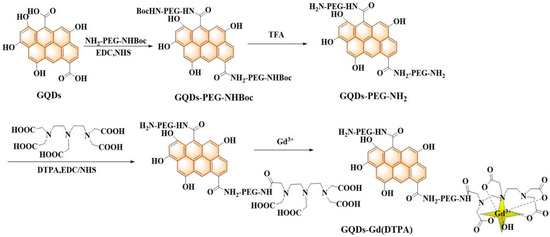
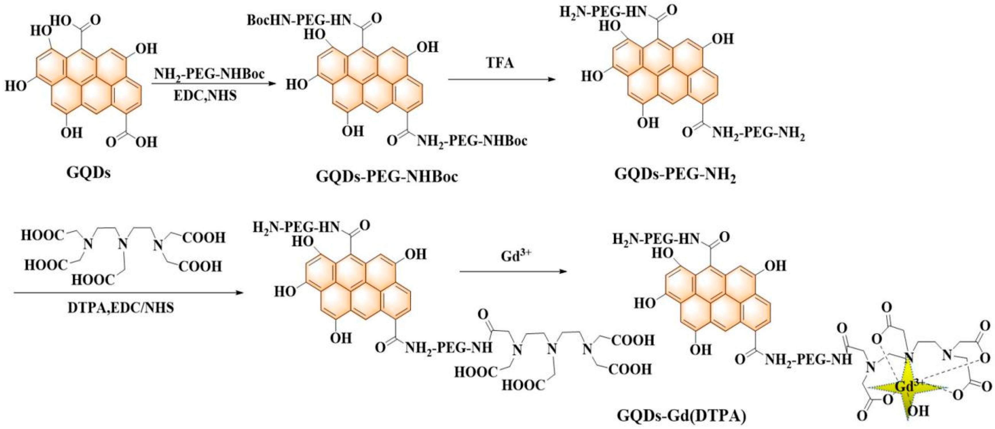
To address these challenges, the authors developed a nanocomplex composed of GQDs and Gd3+ as a new type of nano-CA (Scheme 1). In the developed nano-CAs, the GQDs were produced through a hydrothermal method and then functionalized with poly(ethylene glycol) bis(amine) to form a bridge with DTPA (diethylenetriaminepentaacetic acid dianhydride). The composite was then chelated with Gd3+ ions to form the nano-CAs, Gd(DTPA)−GQDs. The results of both in vitro and in vivo studies demonstrate that Gd(DTPA)−GQDs are novel, non-toxic, and high r1 value MRI CAs with the ability to enable FL imaging in vitro.
Scheme 1.
Schematic diagram of the preparation of GQDs and Gd(DTPA)−GQDs.
2. Results and Discussion
2.1. Synthesis and Structure of Gd(DTPA)−GQDs
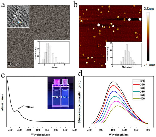
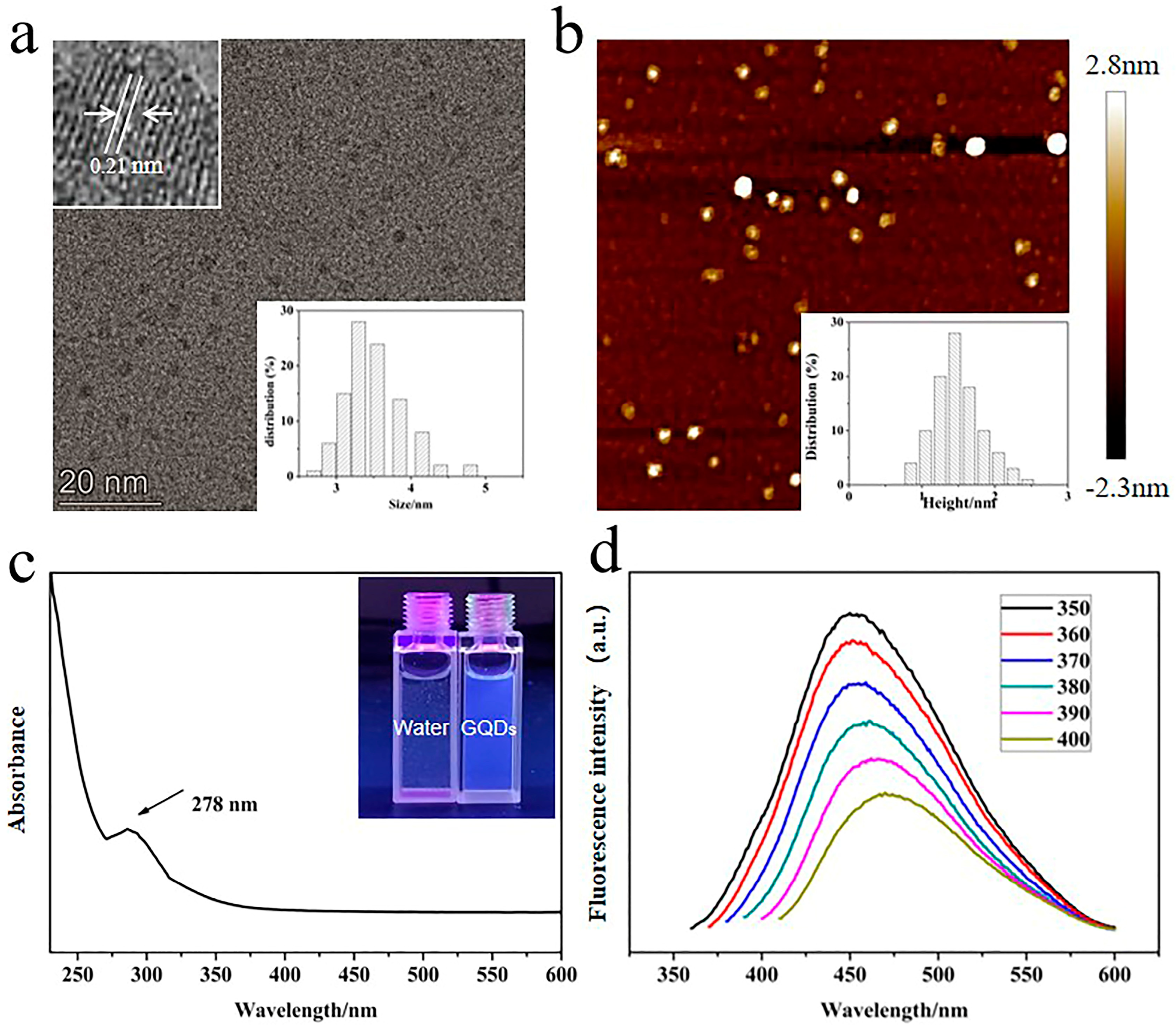
In this investigation, the synthesis of water-soluble graphene quantum dots (GQDs) of uniform size was accomplished through the chemical oxidation and cutting of GO. The subsequent linking of Gd−DTPA to the GQDs through a PEG bridge was carried out as per Scheme 1. The high-resolution transmission electron microscopy (TEM) images obtained illustrate that the average diameter of the GQDs was determined to be 3.64 ± 0.29 nm, exhibiting a highly crystalline structure with a lattice parameter of 0.21 nm (Figure 1a). Additionally, the atomic force microscopy (AFM) images depict that the thickness of the GQDs range between 0.86 and 2.54 nm (Figure 1b). After Gd(DTPA)−PEGylation, the size of the resulting Gd(DTPA)−GQDs was observed to have slightly increased to 4.15 nm, as evidenced in the TEM images (Figure S1a), which suggests that the PEG has covered the GQDs [27]. The hydrodynamic (HD) sizes of both the GQDs and the Gd(DTPA)−GQDs were analyzed using dynamic light scattering (DLS) and were found to be 5.24 ± 0.38 nm and 9.1 ± 0.79 nm, respectively (Figure S1b). Both the TEM and DLS measurements demonstrate a narrow size distribution. The zeta potential of the GQDs, as expected, was determined to be negative (−27.1mV) (Figure S1b) due to the abundance of carboxyl groups on their surface [28]. After the DTPA−PEG−NH2 was grafted onto the surface of the GQDs to form Gd(DTPA)−GQDs, the zeta potential further increased to −23.8 mV, which can be attributed to the substantial presence of amine groups in PEG [29,30,31].
Figure 1.
Characterization of the GQDs. (a) TEM image and size distribution of GQD samples (inset is the HR−TEM of GQDs). (b) AFM image and height distribution of GQDs. (c) UV−Vis spectra of GQDs. Inset is a photograph of water and GQD solutions under UV light with 365 nm excitation (1 mg/mL). (d) Photoluminescence spectra of GQD solutions under different excitation wavelengths (1 mg/mL).
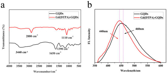
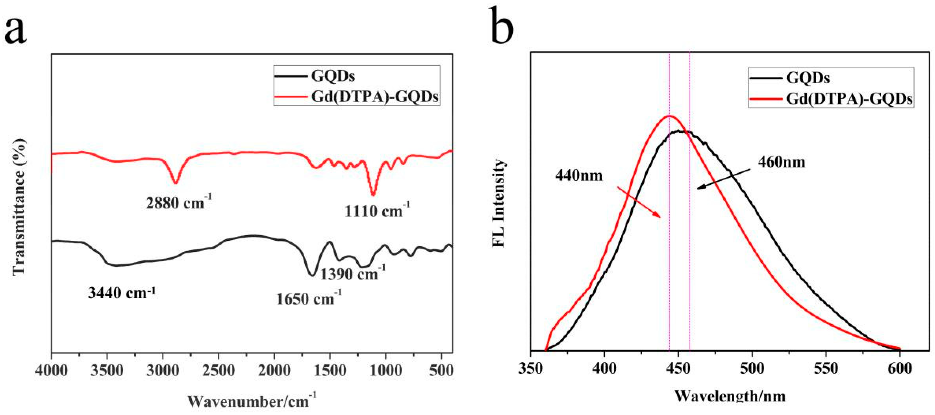
The Fourier transform infrared (FT−IR) spectra of both the GQDs and the Gd(DTPA)−GQDs are displayed in Figure 2a. The FT−IR spectrum of the initial GQDs exhibits the O−H stretching ~3440 cm−1, conjugated C=O ~1650 cm−1, and C−OH stretching ~1390 cm−1 [32]. After the surface modification process, significant decays in the peaks at 1650 and 3440 cm−1 were observed in the FT−IR spectrum of the Gd(DTPA)−GQDs, indicating the amidation of the carboxylic acid. Simultaneously, increases in the adsorption peaks at 1110 cm−1 were noted, which were attributed to the stretching of C−O−C in the PEG. A new peak at 2880 cm−1 was assigned to the N−H stretching vibration of the amine groups, providing evidence of the successful introduction of DTPA−PEG.
Figure 2.
(a) FT−IR spectra of the GQDs and Gd(DTPA)−GQDs. (b) Photoluminescence spectra of GQD and Gd(DTPA)−GQD solutions under an exciting wavelength of 350 nm (1 mg/mL).
2.2. Optical Properties
The optical properties of the GQD samples are depicted in Figure 1c,d. The samples exhibit a pronounced absorption peak at 278 nm, corresponding to the transition of aromatic C−C bonds, and exhibit blue FL under 365 nm UV light irradiation (Figure 1c and insert) [33]. The photoluminescence emission spectra of the GQDs exhibit excitation-dependent features wherein the photoluminescence emission maximum shifts to higher energy and the emission intensity decreases as the excitation wavelength increases (Figure 1d). This shift may be a result of the optical selection of various surface defect states near the surface of the GQDs [34]. As depicted in Figure 2b, after further modification, the photoluminescence emission spectra of the GQD and Gd(DTPA)−GQD solutions were blue-shifted by 20 nm (under 350 nm laser excitation) at the maximum PL position, which could be attributed to Gd3+ complexation and the resulting energy transfer between Gd3+ and GQDs [35].
2.3. Assessment of the T1 Relaxivity
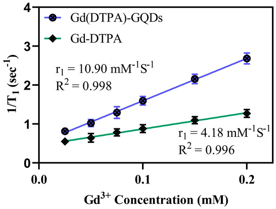
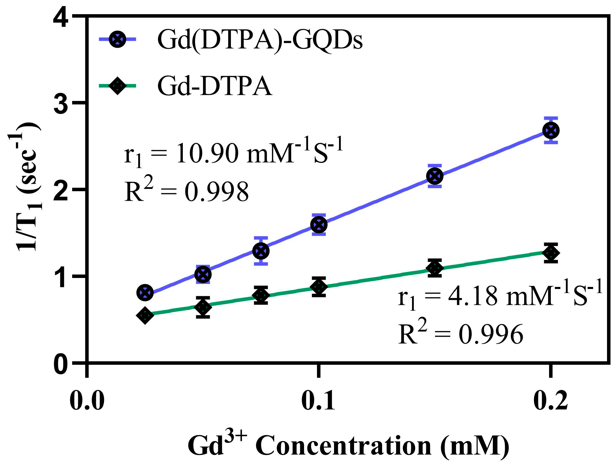
The concentration of Gd3+ in Gd(DTPA)−GQDs was quantified by the use of ICP−MS (2.75, atom. %). Subsequently, the suitability of Gd(DTPA)−GQDs as a nano−CA for MRI was evaluated by determining the relaxivity parameters [36]. The longitudinal relaxation rates (r1 values) of both Gd(DTPA)−GQD and Gd−DTPA samples were measured using the 0.5 T NMI20 Analyst NMR system at 37 °C. The results shown in Figure 3 indicate that the r1 rate of Gd(DTPA)−GQDs was 10.90 mM−1 s−1 (R2 = 0.998), which is significantly higher than that of commercial Gd−DTPA (4.18 mM−1 s−1, R2 = 0.996). The stronger relaxivity of Gd(DTPA)−GQDs compared with Gd−DTPA was tentatively attributed to the attachment of the Gd chelates to nanoparticles, prolonging the tumbling time and thus, increasing the relaxivity [37].
Figure 3.
T1 relaxation rate (r1) recorded at 0.5 T as a function of the Gd3+ concentration for the Gd−DTPA and Gd(DTPA)−GQD samples.
2.4. The Protein Adsorption and Stability of Gd(DTPA)−GQDs
Nanocarriers classified as “good” should exhibit low non-specific protein adsorption characteristic to prevent the immune system clearance and reduce the probability of phagocytic cell uptake, both of which are necessary to enhance the duration of circulation time [38,39,40]. To investigate the effect of PEG grafted onto graphene quantum dots (GQDs) on protein adsorption, an assay was performed using bovine serum albumin (BSA) as a model protein. The results, as displayed in Figure S2a, indicate that GQDs had an adsorption of BSA of up to 12.1%, which is believed to have resulted from hydrogen bonding interactions and van der Waals interactions between GQDs and BSA [41,42,43]. However, after the GQDs were functionalized with PEG, the adsorption of BSA on the surface was significantly reduced to 2.9%. This outcome demonstrates that the modification of GQDs with PEG layers effectively inhibits BSA adsorption [44,45].
To assess the stability of Gd(DTPA)−graphene quantum dots (GQDs) in biological reagents, a sample consisting of 15 mg of Gd(DTPA)−GQDs was introduced into various solutions, including deionized (DI) water, saline, DMEM, and two variations of phosphate-buffered saline (PBS 1× and PBS 5×). As demonstrated in Figure S2b, there was no noticeable alteration in the hydrodynamic size of the material throughout the observation period. The accompanying figure (Figure S2b, insert) features photographs of Gd(DTPA)−GQDs in different biological solutions (DI water, saline, DMEM, PBS 1×, and PBS 5×), showing that the material remained stable without precipitation for 72 h. These results suggest that Gd(DTPA)−GQDs exhibit excellent dispersion in physiological solutions.
2.5. Biocompatibility and Cytotocivity
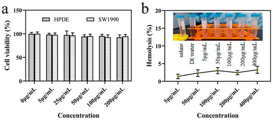
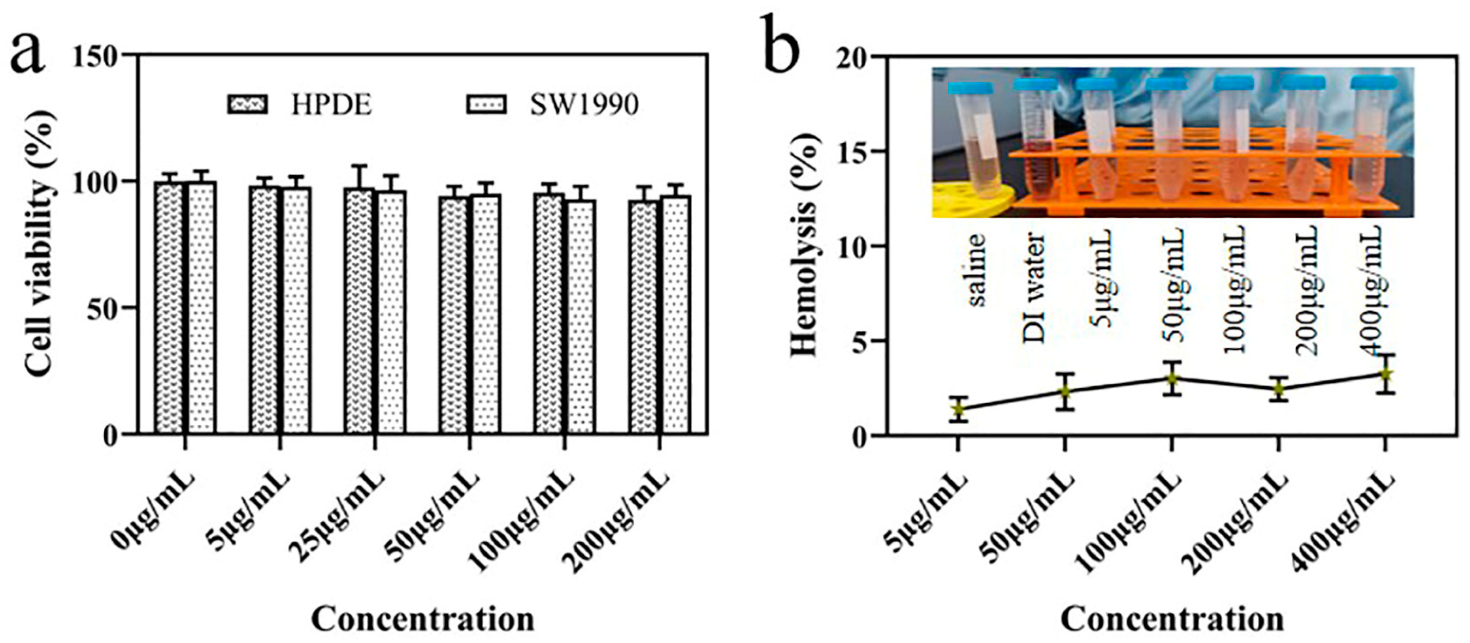
The cytotoxicity of graphene quantum dots (GQDs) and Gd(DTPA)−GQDs was evaluated on human normal pancreas (HPDE) cell lines and SW1990 cells using the CCK−8 assay. As depicted in Figure 4a and Figure S3, there was no noticeable cytotoxicity observed after 24 h of exposure to either of the nanoparticles at any of the tested concentrations. The hemolysis assay was also performed to ensure the feasibility of intravenous administration. Gd(DTPA)−GQDs of varying concentrations were incubated with red blood cells (RBCs), with saline and DI water serving as negative and positive controls, respectively. The hemolysis results, presented in Figure 4b and Figure S4, indicate that even at a high dose of 400 μg/mL, the hemolytic rate was below 5% and the RBCs remained intact, meeting the established standards for biomaterials hemolysis [46,47]. These findings demonstrate the excellent biocompatibility of Gd(DTPA)−GQDs.
Figure 4.
Biocompatibility and cytotoxicity: (a) toxicity study of HPDE cells and SW1990 cells upon treatment with Gd(DTPA)−GQDs for 24 h. (b) Hemolytic efficiency of Gd(DTPA)−GQDs to human red blood cells (inset: photos for direct observation of hemolysis).
2.6. In Vivo Safety Evaluation
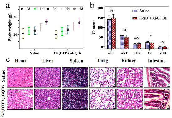
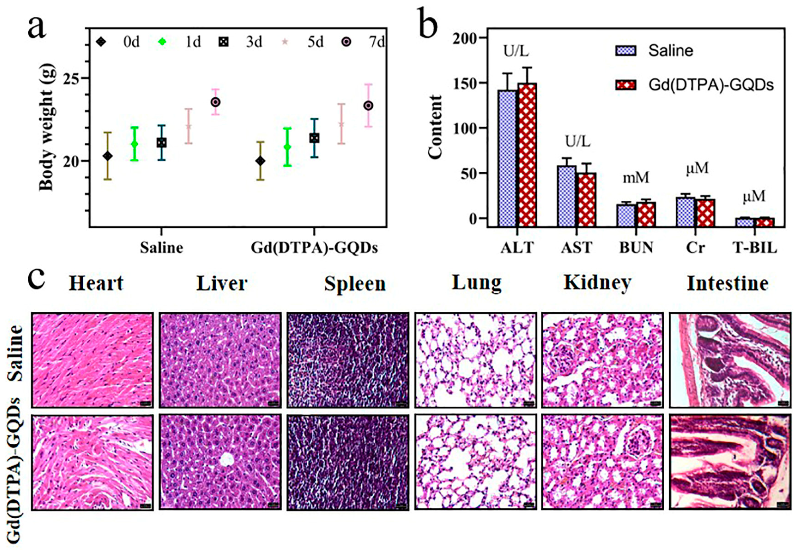
The in vivo safety was evaluated by monitoring bodyweight, conducting histological examinations, and measuring blood chemistry indices. At seven days post−treatment, the bodyweight of the Gd(DTPA)−GQDs−treated group showed no significant differences compared to the saline group (Figure 5a). Furthermore, Figure 5b indicates that the levels of liver and kidney function (liver function markers: aspartate transaminase (AST), alanine aminotransferase (ALT), and total bilirubin (T−BIL); kidney function markers: blood urea nitrogen (BUN) and creatinine (Cr)) did not significantly differ from the saline group (p > 0.05) after treatment with Gd(DTPA)−GQDs, suggesting no observable harm to the primary functions of the liver and kidney. Hematoxylin and eosin (H and E) staining images did not reveal any noticeable pathological changes in the heart, liver, spleen, lung, kidney, and intestine after treatment with either Gd(DTPA)−GQDs or saline (Figure 5c). These results demonstrate the high biocompatibility of Gd(DTPA)−GQDs, making them a suitable candidate for further in vivo bioimaging applications.
Figure 5.
In vivo safety evaluation: (a) the bodyweight changes of BALB/c−nu mice after treatment for a week. Data were means ± SD (n = 5). (b) Blood biochemistry of Gd(DTPA)−GQD−treated mice. (c) Hematoxylin- and eosin-stained images of organs dissected from mice upon treatment with saline and Gd(DTPA)−GQDs (50 mg/kg).
2.7. Cellular Uptake
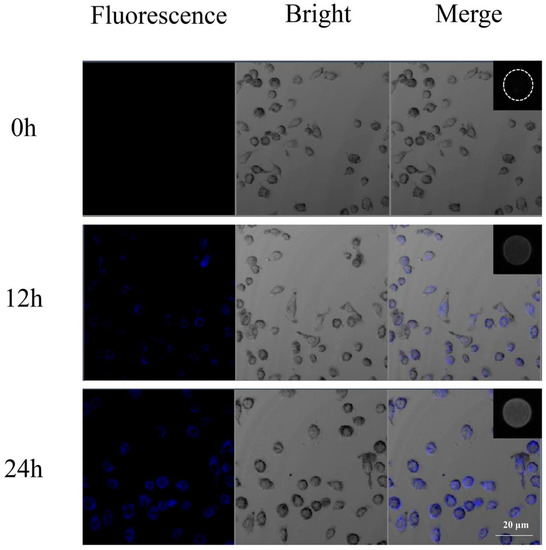
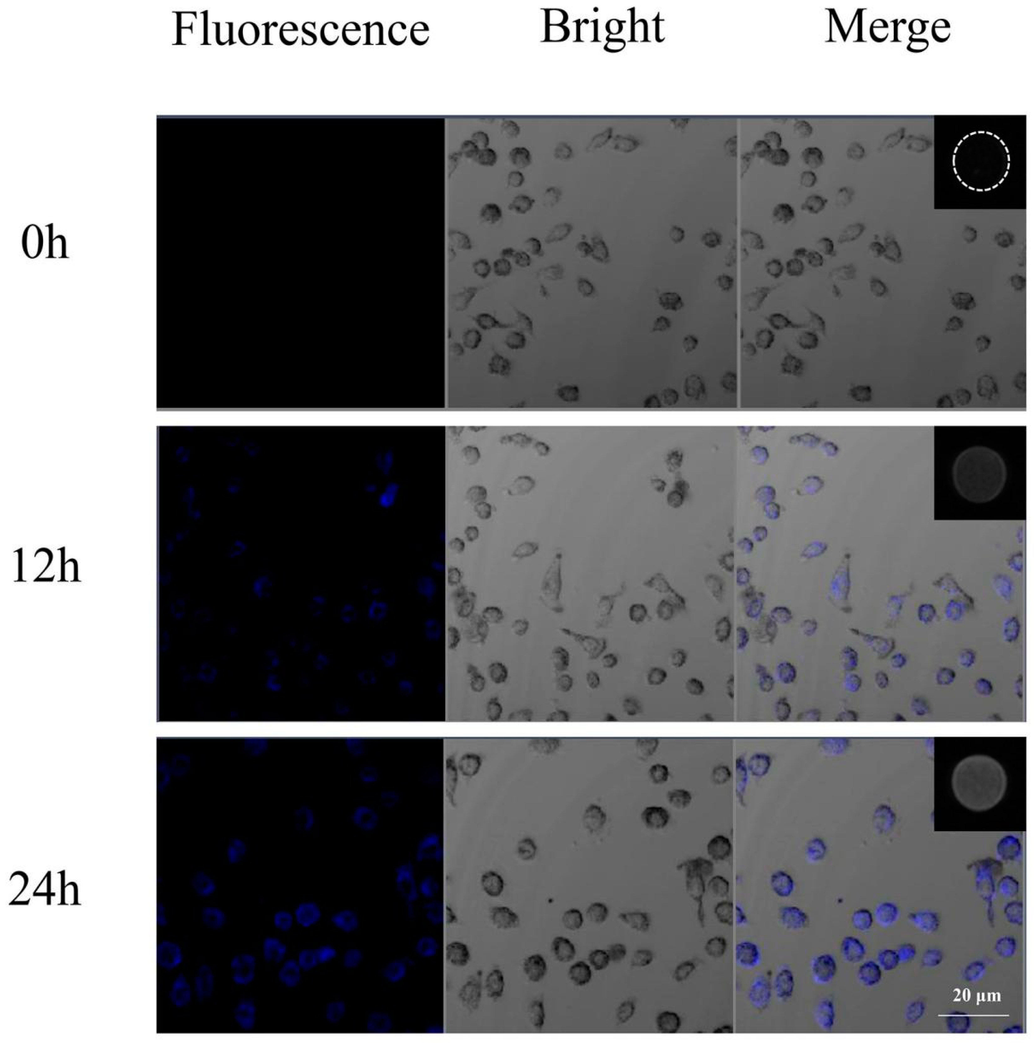
The cellular uptake efficiency of Gd(DTPA)−GQDs in SW1990 cells was evaluated utilizing fluorescence microscopy. As demonstrated in Figure 6, the Gd(DTPA)−GQDs fluorescence (represented by a blue color) accumulated within the cytoplasm of the SW1990 cells. With prolonged incubation, the blue signals within the cells increased, indicating that a greater number of Gd(DTPA)−GQDs were efficiently taken up by the cells over time (Figure S5a). To further assess the intracellular MRI enhancement effect, the nano−CA−treated SW1990 cells were collected, suspended in centrifugation tubes, and then examined using a Philips 3.0T MRI scanner (Figure 5, insert). As depicted in Figure S5b, the MR signal intensity increased with extended incubation time, demonstrating that more Gd(DTPA)−GQDs were successfully taken up by the cells over time.
Figure 6.
Brightfield/fluorescence microscopy overlay images of SW1990 cells incubated with Gd(DTPA)−GQD nanoparticles at different time points (scale bar: 20 µm). Inset is the T1−weighted MR images of a tube containing Gd(DTPA)−GQDs−labeled SW1990 cells.
2.8. Imaging in Tumor−Bearing Mice
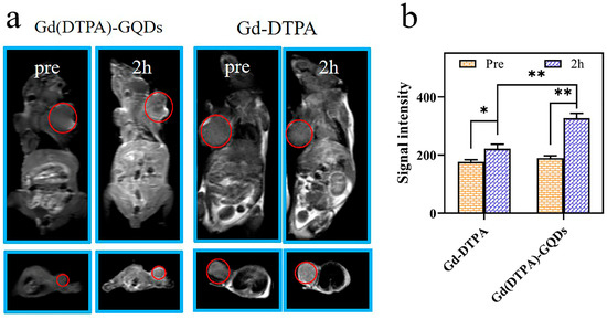
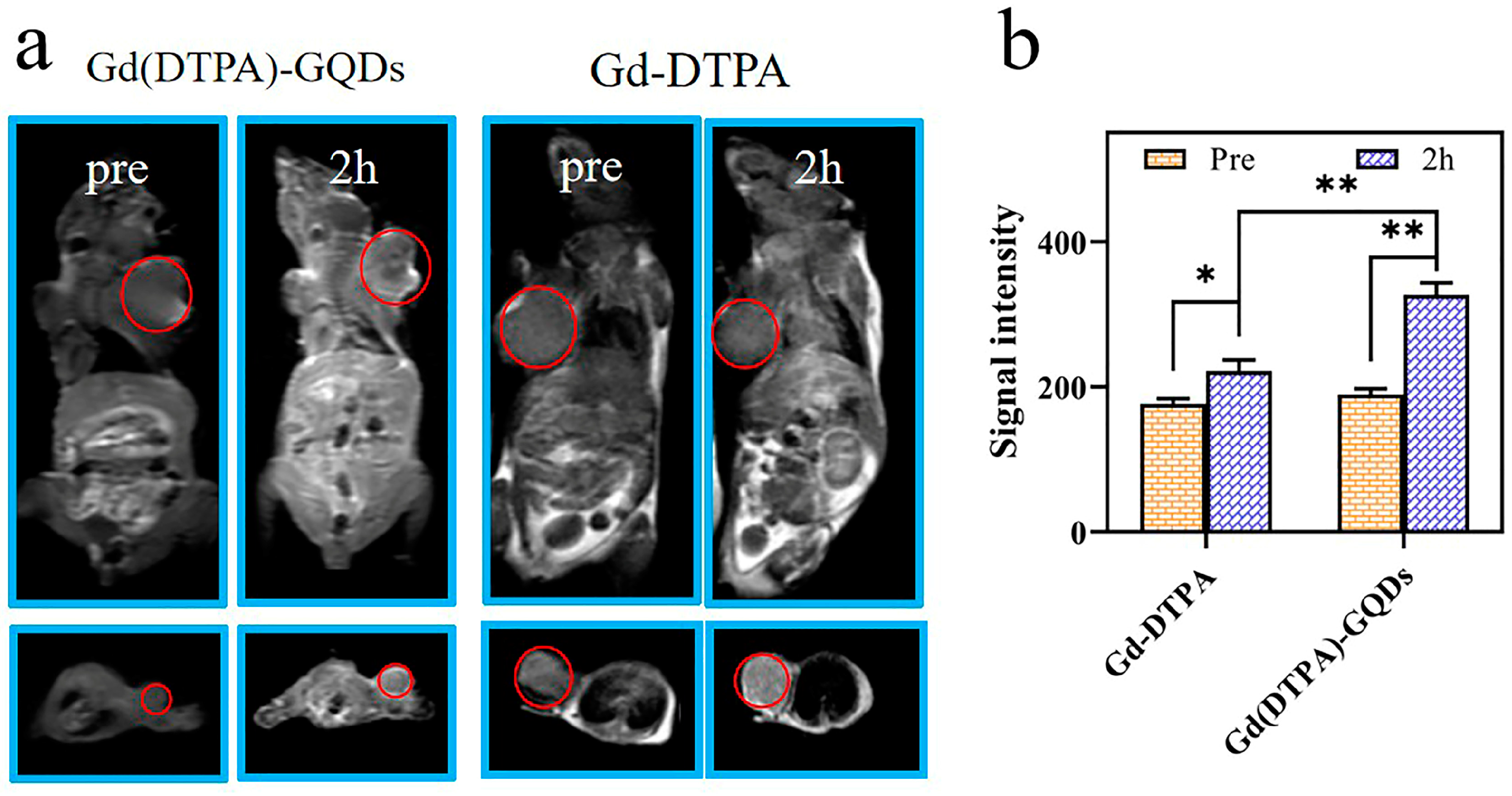
The magnetic resonance (MR) properties of Gd(DTPA)−GQDs were subjected to testing. Intravenous injections of Gd−DTPA and Gd(DTPA)−GQDs were administered to SW1990 mice with tumors, each at a dose of 1 mg (1 mg of Gd3+ per kg). As depicted in Figure 7a, the location of the tumor (red circle) was distinctly visible 2 h post−injection. An examination of the MRI signal intensity images obtained from the Gd−DTPA and the Gd(DTPA)−GQD groups revealed a marked enhancement in the latter, likely due to its elevated r1 value (Figure 7b). These results indicate that Gd(DTPA)−GQDs hold potential as a T1−positive nano−contrast agent.
Figure 7.
In vivo MRI imaging. (a) T1−weighted MRI images of BALB/c−nu (cross−sections and coronal scans) collected before and 2 h after intravenous injection with Gd−DTPA or Gd(DTPA)−GQDs (at a dose of 1 mg Gd per kg of bodyweight). (b) Signal intensity changes. Data were means ± SD (n = 3) (* p < 0.05, ** p < 0.01).
3. Materials and Methods
3.1. Materials
N-Hydroxysuccinimide (NHS, 98%), Diethylenetriaminepentaacetic acid dianhydride (DTPA, 98%), and N−(3−Dimethylaminopropyl)−N−ethylcarbodiimide hydrochloride (EDC·HCl, 99%) were offered by Macklin Biochemical Co., Ltd. (Shanghai, China). Poly(ethylene glycol) bis(amine) (PEG, M.W2000) was offered from Sigma-Aldrich (St. Louis, MO, USA). Gadolinium(III) chloride hexahydrate (GdCl3·6H2O, 99%) was provided by Aladdin Biochemical Technology Co., Ltd. (Shanghai, China). Graphene oxide (GO) was purchased from Nanjing XFNANO Materials Tech Co., Ltd. (Nanjing, China). Dulbecco’s Modified Eagle Medium (DMEM) and fetal bovine serum (FBS) were purchased from Gibco (Thermo Fisher Scientific, Waltham, MA, USA).
3.2. Synthesis of GQDs
The preparation of GQDs was based on a previously published method [48]. In brief, graphene oxide (100 mg) was combined with a mixture of 45 mL H2SO4 and 15 mL of fuming HNO3. The mixture was subjected to sonication for 30 min and then stirred at 120 °C for 1.5 h. After cooling, the solution was diluted with 200 mL of deionized water and neutralized with K2CO3 to achieve a pH range of 7–8. The resulting mixture was further purified by dialysis using a dialysis membrane (retained molecular weight: 3500 Da) for 24 h. The resulting GQDs were harvested through freeze−drying.
3.3. Synthesis of Gd(DTPA)−GQDs
The synthesis of Gd(DTPA)−GQD nanoparticles involved a series of steps. First, a solution composed of 30 mg of graphene quantum dots and 287 mg EDC·HCL in 50 mL DI water was stirred at a temperature of 37 °C for 3 h. Subsequently, 60 mg BocNH−PEG−NH2 and 172 mg NHS were introduced, and the pH was adjusted to 5.5 using 0.1 mM HCL before undergoing further agitation for 24 h at the same temperature. To eliminate the unresponsive precursors, the as-prepared solution was purified through 3500 Da molecular weight cutoff dialysis against DI water for 24 h. Then, 2 mL of trifluoroacetic acid was added to the resulting GQDs−PEG−NHBoc solution, stirred for 3 h at ambient temperature, and purified through dialysis against DI water to yield GQDs−PEG−NH2. GdCl3·6H2O (11.1 mg) was subsequently combined with an aqueous solution of DTPA (11.8 mg) for chelation and further modification to the GQDs−PEG−NH2 through reaction between the amino groups of PEG and the carboxyl groups of DTPA. The residual Gd3+ and DTPA were removed through 3500 Da cutoff dialysis for 12 h. Finally, the synthesized Gd(DTPA)−GQDs nanoparticles were subjected to freeze−drying to collect the samples.
3.4. Materials Characterization
The morphology of the GQDs was subjected to examination using transmission electron microscopy (TEM, FEI Talos F200S, New York, NY, USA) and atomic force microscopy (AFM, (Bruker Daltonics Inc. Multimode 8.0, Massachusetts, MA, USA). The Fourier transform infrared (FT−IR) spectra, UV−Vis absorption spectra, and FL spectra were obtained through utilization of FT−IR spectrometer (FT-IR 6800 JASCO, Marseille, France), Shimadzu UV−2450 spectrophotometer, and Hitachi 7000 fluorescence spectrophotometer, respectively. The hydrodynamic (HD) size and zeta potential of the samples were measured through the utilization of a nano ZS90 analyzer (Malvern Instruments Ltd., Worcestershire, UK), with the measurements being conducted at room temperature. The concentration of Gd3+ within the Gd(DTPA)−GQDs was confirmed through the application of inductively coupled plasma mass spectrometry (ICP-MS, Agilent 720 ES, Santa Clara, CA, USA).
3.5. Relaxivity Measurements
The T1 relaxivity values (r1) of Gd(DTPA)−GQDs in aqueous dispersions were determined through utilization of the NMI20 Analyst NMR System sourced from Niumag in China, with the measurements performed at 0.5 Tesla and 37 °C.
3.6. Cell Culture and Cellular Uptake Experiment
The human normal pancreas lines, HPDE, and human pancreatic cancer cell lines, SW1990, were procured from the Cell Bank of the Shanghai Institute of Cell Biology (China). They were sustained in a culture medium composed of DMEM containing 10% FBS and antibiotics (1% penicillin/streptomycin).
To measure cellular uptake, SW1990 cells were cultured in 12−well plates and allowed to grow overnight. Subsequently, Gd(DTPA)−GQDs (100 µg/mL) were introduced into designated wells for varying durations (0, 12, and 24 h). The cells were then thoroughly rinsed with phosphate-buffered saline (PBS) and fixed utilizing a 4% formaldehyde solution for 20 min. The resultant cells were observed using confocal laser scanning microscopy (CLSM).
To validate the enhancement effect of Gd(DTPA)−GQDs as MRI nano−CAs within cells, SW1990 cells were cultured in 6−well plates. Gd(DTPA)−GQDs were incorporated into the culture medium at varying intervals (0, 12, and 24 h). The cells were then harvested by centrifugation and fixed with a 1.0 % wt agarose solution. The MRI experiments were conducted utilizing a clinical 3.0 T MRI scanner (Philips, The Netherlands). The parameters for T1−weighted (T1W) imaging were set as follows: matrix = 256 × 256, layer thickness = 5 mm, FOV (field of view) = 50 × 50 mm, TR = 650 ms, TE = 10 ms, echo time (TE) = 10 ms. Raw data were transmitted to a remote computer for analysis.
3.7. BSA Adsorption and Colloidal Stability
The amount of bovine serum albumin (BSA) adsorption was prepared in accordance with a published study [49]. BSA was completely dissolved in PBS (pH 7.4, 0.3 mg/mL). Subsequently, 15 mg of both GQDs and Gd(DTPA)−GQDs were dispersed into a 10 mL solution of BSA and the mixture was stirred at 37 °C for 5 h at a shaking rate of 120 rpm. The supernatant was collected through centrifugation. Finally, the final concentration of BSA was determined through a Coomassie brilliant blue stain assay.
To assess the colloidal stability, Gd(DTPA)−GQDs were dispersed in deionized water, saline, DMEM medium, PBS 1×, and PBS 5× at a certain concentration (2 mg/mL), respectively. The samples were kept in an incubator at 37 °C and the alteration in hydrodynamic (HD) size were monitored through dynamic light scattering (DLS) measurements for a period of up to 72 h.
3.8. Biocompatibility
The cytotoxicity impact of Gd(DTPA)−GQDs was appraised against HPDE cells and SW1990 cells by CCK−8 assay. To initiate the process, the cells were cultured in separate 96−well plates at a concentration of 8 × 10³ cells/well and allowed to grow in DMEM medium overnight. Subsequently, varying concentrations of Gd(DTPA)−GQDs (0, 5, 50, 100, 150, and 200 μg/mL) were added to the wells for a 24 h duration. The viability of the cells was finally assessed through CCK−8 assay.
To delve further into the actual condition, the compatibility of Gd(DTPA)−GQDs with human red blood cells was assessed using samples procured from volunteer donors [50,51]. The red blood cells were initially collected through centrifugation and subjected to purification via three successive washes with saline. Subsequently, a mixture of 2 mL of the RBC suspension (4% v/v) was combined with: (a) 2 mL of deionized water as a reference positive control; (b) 2 mL of saline as a reference negative control; and (c) 2 mL of Gd(DTPA)−GQD suspensions in saline at various concentrations (5, 50, 100, 200, and 400 µg/mL). The resulting mixtures were then incubated at 37 °C for a duration of 3 h prior to being subjected to centrifugation. The absorbance of the supernatant layers at 541 nm was measured using a UV−Vis spectrophotometer. The hemolysis percentage was calculated using the following equation (repeated three times for each respective sample).
The biotoxicity of Gd(DTPA)−GQDs was assessed using Balb/c-nu mice (male), with approval from the Animal Ethics Committee of Qiqihar Medical University (No. QMU−AECC−2021−100). Twelve mice, weighing between (18–22g), were randomly divided into two groups (n = 6). The mice were administered intravenous injections of either 100 μL of normal saline or Gd(DTPA)−GQDs (1 mg/mL). After a seven day period, approximately 0.5 mL of blood was collected from each mouse for biochemical analysis prior to euthanasia. The main organs were then harvested and subjected to histopathological examination (Leica−DM4B digital microscope, Hesse, Germany).
3.9. In Vivo Proof-of-Concept Study
An investigation into the MRI enhancement effect of the synthesized nano−CAs was carried out in a nude mouse tumor model. Specifically, SW1990 cell (1 × 10⁷) suspensions were subcutaneously implanted into the right armpits of Balb/c-nu mice. The SW1990 tumor-bearing mice were then subjected to intravenous injection with Gd(DTPA)−GQDs for T1−weighted MRI (1 mg of Gd3+ per kg). The solutions were sterilized by filtration through membranes (pore size 0.22 μm). The MRI study was conducted using a 3.0 Tesla clinical MRI scanner (manufactured by Philips) equipped with an 8-channel carotid wall imaging special phased array coil. The Balb/c−nu mice were anesthetized using chloral hydrate through intraperitoneal injection. T1−weighted MRI scans were obtained in cross−sectional and coronal views with the following parameters: TR/TE = 650/10 milliseconds, layer thickness = 3 mm, layer spacing = 2 mm, FOV = 100 × 100 mm, matrix = 192 × 192. MR images were analyzed before injection and 2h after injection. The signal intensity (SI) of the tumor tissue was measured in approximately the same slice with a consistent diameter for each region of interest (ROI).
3.10. Statistical Analysis
The statistical data were investigated using SPSS 20.0 software with a Student’s t-test. All findings were expressed as the mean ± standard deviation. Here, a p-value of < 0.05 was considered indicative of statistical significance.
4. Conclusions
In this work, a Gd(DTPA)−GQDs−based T1−positive nano−CA (Gd(DTPA)−GQDs) was developed through a surface modification process that involved integrating Gd−DTPA and GQDs via a PEG bridge. The results showed that PEG−coated GQD nanoparticles had low nonspecific protein adsorption, high colloidal stability, and exceptional biocompatibility. Furthermore, in terms of MRI contrast agent capabilities, Gd(DTPA)−GQDs exhibited a high longitudinal relaxivity rate of 10.90 mM−1 s−1, which was significantly higher than that of the commercial Gd−DTPA (4.18 mM−1 s−1). In vivo assays confirmed that Gd(DTPA)−GQDs were suitable as a T1 contrast agent. It is anticipated that this type of nano−contrast agent possesses the potential to extend the applications of nanotechnology in non-invasive diagnosis and therapy.
Supplementary Materials
The following supporting information can be downloaded at: https://www.mdpi.com/article/10.3390/molecules28052363/s1, Figure S1: Structural characterization: (a) TEM image and size distribution of Gd(DTPA)−GQDs sample, (b) Hydrodynamic diameters and zeta potentials of GQDs and Gd(DTPA)−GQDs. Figure S2: (a) BSA adsorbance amounts of GQDs and Gd(DTPA)−GQDs, (b) Stability of Gd(DTPA)−GQDs nanocomposites in different biological solutions. Figure S3: Cytotoxicity against HPDE cells and SW1990 cells after incubation with different concentrations of various GQDs for 24 h. Figure S4: Morphology of human red blood cells upon treatment with saline, DI water, and Gd(DTPA)−GQDs for 4 h. Figure S5: Mean fluorescence intensity (a) and MRI intensity (b) of SW1990 cells after incubation with Gd(DTPA)−GQDs at different times.
Author Contributions
L.H. and Z.L. designed the research. Z.L., G.S., M.Z. and H.H. performed the experiments and analyzed the data. Z.L., L.H. and G.Q. wrote the manuscript with comments from all other authors. All authors have read and agreed to the published version of the manuscript.
Funding
This research was funded by the basic scientific research business expenses of Heilongjiang Provincial Department of Education, grant number 2019-KYYWF-1251.
Institutional Review Board Statement
The animal study protocol was approved by the Animal Ethics Committee of Qiqihar Medical University (No. QMU-AECC-2021-100).
Informed Consent Statement
Not applicable.
Data Availability Statement
Data will be made available on request.
Conflicts of Interest
The authors declare that they have no known competing financial interests or personal relationships that could have appeared to influence the work reported in this paper.
Sample Availability
Samples of the compounds are available from the authors.
References
- Bennett, K.M.; Jo, J.; Cabral, H.; Bakalova, R.; Aoki, I. MR imaging techniques for nano-pathophysiology and theranostics. Adv. Drug Deliv. Rev. 2014, 74, 75–94. [Google Scholar] [CrossRef] [Scilit] [PubMed]
- Zuo, Z.; Syrovets, T.; Genze, F.; Abaei, A.; Ma, G.; Simmet, T.; Rasche, V. High-resolution MRI analysis of breast cancer xenograft on the chick chorioallantoic membrane. NMR Biomed. 2015, 28, 440–447. [Google Scholar] [CrossRef] [Scilit] [PubMed]
- Caravan, P.; Ellison, J.J.; McMurry, T.J.; Lauffer, R.B. Gadolinium(III) Chelates as MRI Contrast Agents: Structure, Dynamics, and Applications. Chem. Rev. 1999, 99, 2293–2352. [Google Scholar] [CrossRef] [Scilit] [PubMed]
- Willke, P.; Yang, K.; Bae, Y.; Heinrich, A.J.; Lutz, C.P. Magnetic Resonance Imaging of Single Atoms on a Surface. Nat. Phys. 2019, 15, 1005–1010. [Google Scholar] [CrossRef] [Scilit]
- Guo, S.; Wang, X.; Dai, Y.; Dai, X.; Li, Z.; Luo, Q.; Zheng, X.; Gu, Z.; Zhang, H.; Gong, Q.; et al. Enhancing the Efficacy of Metal-Free MRI Contrast Agents via Conjugating Nitroxides onto PEGylated Cross-Linked Poly (Carboxylate Ester). Adv. Sci. 2020, 7, 2000467. [Google Scholar] [CrossRef] [Scilit] [PubMed]
- Jeong, S.Y.; Kim, H.J.; Kwak, B.K.; Lee, H.Y.; Seong, H.; Shin, B.C.; Yuk, S.H.; Hwang, S.J.; Cho, S.H. Biocompatible Polyhydroxyethylaspartamide-based Micelles with Gadolinium for MRI Contrast Agents. Nanoscale Res. Lett. 2010, 5, 1970–1976. [Google Scholar] [CrossRef] [Scilit]
- Ni, D.; Bu, W.; Ehlerding, E.B.; Cai, W.; Shi, J. Engineering of inorganic nanoparticles as magnetic resonance imaging contrast agents. Chem. Soc. Rev. 2017, 46, 7438–7468. [Google Scholar] [CrossRef] [Scilit]
- Gulani, V.; Calamante, F.; Shellock, F.G.; Kanal, E.; Reeder, S.B. Gadolinium Deposition in the Brain: Summary of Evidence and Recommendations. Lancet Neurol. 2017, 16, 564–570. [Google Scholar] [CrossRef] [Scilit]
- Kanda, T.; Nakai, Y.; Oba, H.; Toyoda, K.; Kitajima, K.; Furui, S. Gadolinium deposition in the brain. Magn. Reson. Imaging 2016, 34, 1346–1350. [Google Scholar] [CrossRef] [Scilit]
- Fraum, T.J.; Ludwig, D.R.; Bashir, M.R.; Fowler, K.J. Gadolinium-based contrast agents: A comprehensive risk assessment. J. Magn. Reson. Imaging 2017, 46, 338–353. [Google Scholar] [CrossRef] [Scilit]
- Zu, G.; Kuang, Y.; Dong, J.; Cao, Y.; Zhang, T.; Liu, M.; Luo, L.; Pei, R. Gadolinium(III)-based Polymeric Magnetic Resonance Imaging Agents for Tumor Imaging. Curr. Med. Chem. 2018, 25, 2910–2937. [Google Scholar] [CrossRef] [Scilit] [PubMed]
- Chen, H.; Qiu, Y.; Ding, D.; Lin, H.; Sun, W.; Wang, G.D.; Huang, W.; Zhang, W.; Lee, D.; Liu, G.; et al. Gadolinium-Encapsulated Graphene Carbon Nanotheranostics for Imaging-Guided Photodynamic Therapy. Adv. Mater. 2018, 23, e1802748. [Google Scholar] [CrossRef] [Scilit] [PubMed]
- Zhang, G.; Xie, W.; Xu, Z.; Si, Y.; Li, Q.; Qi, X.; Gan, Y.; Wu, Z.; Tian, G. CuO dot-decorated Cu@Gd2O3 core-shell hierarchical structure for Cu(i) self-supplying chemodynamic therapy in combination with MRI-guided photothermal synergistic therapy. Mater. Horiz. 2021, 8, 1017–1028. [Google Scholar] [CrossRef] [Scilit] [PubMed]
- Aryal, M.; Papademetriou, I.; Zhang, Y.Z.; Power, C.; McDannold, N.; Porter, T. MRI Monitoring and Quantification of Ultrasound-Mediated Delivery of Liposomes Dually Labeled with Gadolinium and Fluorophore through the Blood-Brain Barrier. Ultrasound Med. Biol. 2019, 45, 1733–1742. [Google Scholar] [CrossRef] [Scilit]
- Liu, Y.; Liu, J. Growing a Nucleotide/Lanthanide Coordination Polymer Shell on Liposomes. Langmuir 2019, 35, 11217–11224. [Google Scholar] [CrossRef] [Scilit]
- Ray, S.; Li, Z.; Hsu, C.H.; Hwang, L.P.; Lin, Y.C.; Chou, P.T.; Lin, Y.Y. Dendrimer-and copolymer-based nanoparticles for magnetic resonance cancer theranostics. Theranostics 2018, 8, 6322–6349. [Google Scholar] [CrossRef] [Scilit]
- Wang, X.; Guo, S.; Li, Z.; Xiao, X.; Gu, L.; Luo, Q.; Zhang, H.; Gong, Q.; Luo, K. Safe and Potent MRI Contrast Agents by Complexing Gadolinium with Enzyme/reduction Dual-sensitive Branched Polymers. Appl. Mater. Today 2019, 17, 92–103. [Google Scholar]
- Guo, S.; Xiao, X.; Wang, X.; Luo, Q.; Zhu, H.; Zhang, H.; Li, H.; Gong, Q.; Luo, K. Reductive Microenvironment Responsive Gadolinium-Based Polymers as Potential Safe MRI Contrast Agents. Biomater. Sci. 2019, 7, 1919–1932. [Google Scholar] [CrossRef] [Scilit]
- Zeng, Y.; Li, H.; Li, Z.; Luo, Q.; Zhu, H.; Gu, Z.; Zhang, H.; Gong, Q.; Luo, K. Engineered Gadolinium-Based Nanomaterials as Cancer Imaging Agents. Appl. Mater. Today 2020, 20, 100686. [Google Scholar] [CrossRef] [Scilit]
- Cai, H.; Dai, X.; Wang, X.; Tan, P.; Gu, L.; Luo, Q.; Zheng, X.; Li, Z.; Zhu, H.; Zhang, H.; et al. A Nanostrategy for Efficient Imaging-Guided Antitumor Therapy through a Stimuli-Responsive Branched Polymeric Prodrug. Adv. Sci. 2020, 7, 1903243. [Google Scholar] [CrossRef] [Scilit]
- Younis, M.R.; He, G.; Lin, J.; Huang, P. Recent Advances on Graphene Quantum Dots for Bioimaging Applications. Front. Chem. 2020, 8, 424. [Google Scholar] [CrossRef] [Scilit] [PubMed]
- Wang, F.H.; Bae, K.; Huang, Z.W.; Xue, J.M. Two-photon graphene quantum dot modified Gd2O3 nanocomposites as a dual-mode MRI contrast agent and cell labelling agent. Nanoscale 2018, 10, 5642–5649. [Google Scholar] [CrossRef] [Scilit] [PubMed]
- Lee, B.H.; Hasan, M.T.; Lichthardt, D.; Gonzalez-Rodriguez, R.; Naumov, A.V. Manganese-nitrogen and gadolinium-nitrogen Co-doped graphene quantum dots as bimodal magnetic resonance and fluorescence imaging nanoprobes. Nanotechnology 2021, 32, 095103. [Google Scholar] [CrossRef] [Scilit] [PubMed]
- Gautam, A.; Pal, K. Gefitinib conjugated PEG passivated graphene quantum dots incorporated PLA microspheres for targeted anticancer drug delivery. Heliyon 2022, 8, e12512. [Google Scholar] [CrossRef] [Scilit] [PubMed]
- Wei, W.; Zhang, X.; Chen, X.; Zhou, M.; Xu, R.; Zhang, X. Smart surface coating of drug nanoparticles with cross-linkable polyethylene glycol for bio-responsive and highly efficient drug delivery. Nanoscale 2016, 8, 8118–8125. [Google Scholar] [CrossRef] [Scilit] [PubMed]
- Suk, J.S.; Xu, Q.; Kim, N.; Hanes, J.; Ensign, L.M. PEGylation as a strategy for improving nanoparticle-based drug and gene delivery. Adv. Drug Deliv. Rev. 2016, 99, 28–51. [Google Scholar] [CrossRef] [Scilit] [PubMed]
- Ji, Y.; Li, Y.M.; Seo, J.G.; Jang, T.S.; Knowles, J.C.; Song, S.H.; Lee, J.H. Biological Potential of Polyethylene Glycol (PEG)-Functionalized Graphene Quantum Dots in In Vitro Neural Stem/Progenitor Cells. Nanomaterials 2021, 11, 1446. [Google Scholar] [CrossRef] [Scilit]
- Yuan, X.; Liu, Z.; Guo, Z.; Ji, Y.; Jin, M.; Wang, X. Cellular distribution and cytotoxicity of graphene quantum dots with different functional groups. Nanoscale Res. Lett. 2014, 9, 108. [Google Scholar] [CrossRef] [Scilit]
- Becheru, D.F.; Vlăsceanu, G.M.; Banciu, A.; Vasile, E.; Ioniţă, M.; Burns, J.S. Optical Graphene-Based Biosensor for Nucleic Acid Detection; Influence of Graphene Functionalization and Ionic Strength. Int. J. Mol. Sci. 2018, 19, 3230. [Google Scholar] [CrossRef] [Scilit]
- Wu, Z.; Pan, T.; Lin, D.; Xia, W.; Shan, J.; Cheng, R.; Yang, M.; Hu, X.; Nan, K.; Qi, L. Biocompatible tumor-targeted GQDs nanocatalyst for chemodynamic tumor therapy. J. Mater. Chem. B 2022, 10, 3567–3576. [Google Scholar] [CrossRef] [Scilit]
- Ning, Z.; Yang, L.; Yan, X.; Wang, D.; Hua, Y.; Shi, W.; Lin, J.; Meng, Z. Effect and Mechanism of the Lenvatinib@H-MnO2-FA Drug Delivery System in Targeting Intrahepatic Cholangiocarcinoma. Curr. Pharm. Des. 2022, 28, 743–750. [Google Scholar] [CrossRef] [Scilit]
- Lotfollahzadeh, S.; Hosseini, E.S.; Mahmoudi Aznaveh, H.; Nikkhah, M.; Hosseinkhani, S. TRAIL/S-layer/graphene quantum dot nanohybrid enhanced stability and anticancer activity of TRAIL on colon cancer cells. Sci. Rep. 2022, 12, 5851. [Google Scholar] [CrossRef] [Scilit] [PubMed]
- El-Hnayn, R.; Canabady-Rochelle, L.; Desmarets, C.; Balan, L.; Rinnert, H.; Joubert, O.; Medjahdi, G.; Ben Ouada, H.; Schneider, R. One-Step Synthesis of Diamine-Functionalized Graphene Quantum Dots from Graphene Oxide and Their Chelating and Antioxidant Activities. Nanomaterials 2020, 10, 104. [Google Scholar] [CrossRef] [Scilit] [PubMed]
- Habiba, K.; Makarov, V.I.; Avalos, J.; Guinel, M.J.F.; Weiner, B.R.; Morell, G. Luminescent graphene quantum dots fabricated by pulsed laser synthesis. Carbon 2013, 64, 341–350. [Google Scholar] [CrossRef] [Scilit] [PubMed]
- Ding, H.; Wang, D.; Sadat, A.; Li, Z.; Hu, X.; Xu, M.; Morais, P.C.; Ge, B.; Sun, S.; Ge, J.; et al. Single-Atom Gadolinium Anchored on Graphene Quantum Dots as a Magnetic Resonance Signal Amplifier. ACS Appl. Bio. Mater. 2021, 4, 2798–2809. [Google Scholar] [CrossRef] [Scilit]
- Ji, Z.; Ai, P.; Shao, C.; Wang, T.; Yan, C.; Ye, L.; Gu, W. Manganese-Doped Carbon Dots for Magnetic Resonance/Optical Dual-Modal Imaging of Tiny Brain Glioma. ACS Biomater. Sci. Eng. 2018, 4, 2089–2094. [Google Scholar] [CrossRef] [Scilit] [PubMed]
- Zhou, Z.; Deng, H.; Yang, W.; Wang, Z.; Lin, L.; Munasinghe, J.; Jacobson, O.; Liu, Y.; Tang, L.; Ni, Q.; et al. Early stratification of radiotherapy response by activatable inflammation magnetic resonance imaging. Nat. Commun. 2020, 11, 3032. [Google Scholar] [CrossRef] [Scilit] [PubMed]
- Chong, Y.; Ge, C.; Yang, Z.; Garate, J.A.; Gu, Z.; Weber, J.K.; Liu, J.; Zhou, R. Reduced Cytotoxicity of Graphene Nanosheets Mediated by Blood-Protein Coating. ACS Nano 2015, 9, 5713–5724. [Google Scholar] [CrossRef] [Scilit] [PubMed]
- Bai, Q.; Wang, Z.; An, Y.; Tian, J.; Li, Z.; Yang, Y.; Dong, Y.; Chen, M.; Liu, T. Chitosan-functionalized graphene oxide as adjuvant in HEV P239 vaccine. Vaccine 2022, 40, 7613–7621. [Google Scholar] [CrossRef] [Scilit]
- Jiang, W.; Fang, H.; Liu, F.; Zhou, X.; Zhao, H.; He, X.; Guo, D. PEG-coated and Gd-loaded fluorescent silica nanoparticles for targeted prostate cancer magnetic resonance imaging and fluorescence imaging. Int. J. Nanomed. 2019, 14, 5611–5622. [Google Scholar] [CrossRef] [Scilit]
- Yan, T.; Zhang, H.; Huang, D.; Feng, S.; Fujita, M.; Gao, X.D. Chitosan-Functionalized Graphene Oxide as a Potential Immunoadjuvant. Nanomaterials 2017, 7, 59. [Google Scholar] [CrossRef] [Scilit] [PubMed]
- Huang, S.; Qiu, H.; Lu, S.; Zhu, F.; Xiao, Q. Study on the molecular interaction of graphene quantum dots with human serum albumin: Combined spectroscopic and electrochemical approaches. J. Hazard. Mater. 2015, 285, 18–26. [Google Scholar] [CrossRef] [Scilit] [PubMed]
- Wei, X.Q.; Hao, L.Y.; Shao, X.R.; Zhang, Q.; Jia, X.Q.; Zhang, Z.R.; Lin, Y.F.; Peng, Q. Insight into the Interaction of Graphene Oxide with Serum Proteins and the Impact of the Degree of Reduction and Concentration. ACS Appl. Mater. Interfaces 2015, 7, 13367–13374. [Google Scholar] [CrossRef] [Scilit]
- Liu, D.; Liu, J.; Li, C.; Li, W.; Wang, W.; Liu, J. Ultrasound-Aided Targeting Nanoparticles Loaded with miR-181b for Anti-Inflammatory Treatment of TNF-α-Stimulated Endothelial Cells. ACS Omega 2020, 5, 17102–17110. [Google Scholar] [CrossRef] [Scilit] [PubMed]
- Martíne, N.M.; Russo, D.; Prévost, S.; Teixeira, J.; Morsbach, S.; Landfester, K. Poly(ethylene glycol)-Based Surfactant Reduces the Conformational Change of Adsorbed Proteins on Nanoparticles. Biomacromolecules 2022, 23, 4282–4288. [Google Scholar] [CrossRef] [Scilit]
- Huang, Y.; Ding, X.; Qi, Y.; Yu, B.; Xu, F.J. Reduction-responsive multifunctional hyperbranched polyaminoglycosides with excellent antibacterial activity, biocompatibility and gene transfection capability. Biomaterials 2016, 106, 134–143. [Google Scholar] [CrossRef] [Scilit]
- Yan, T.; Kong, S.; Ouyang, Q.; Li, C.; Hou, T.; Chen, Y.; Li, S. Chitosan-Gentamicin Conjugate Hydrogel Promoting Skin Scald Repair. Mar. Drugs. 2020, 18, 233. [Google Scholar] [CrossRef] [Scilit]
- Chong, Y.; Ma, Y.; Shen, H.; Tu, X.; Zhou, X.; Xu, J.; Dai, J.; Fan, S.; Zhang, Z. The in vitro and in vivo toxicity of graphene quantum dots. Biomaterials 2014, 35, 5041–5048. [Google Scholar] [CrossRef] [Scilit]
- Cai, D.; Han, C.; Liu, C.; Ma, X.; Qian, J.; Zhou, J.; Li, Y.; Sun, Y.; Zhang, C.; Zhu, W. Chitosan-capped enzyme-responsive hollow mesoporous silica nanoplatforms for colon-specific drug delivery. Nanoscale Res. Lett. 2020, 15, 123. [Google Scholar] [CrossRef] [Scilit]
- Zhao, H.; Zhao, H.; Jiao, Y.; Zhu, Y.; Liu, C.; Li, F.; Wang, Y.; Gu, Z.; Yang, D. Biosynthetic molecular imaging probe for tumor-targeted dual-modal fluorescence/magnetic resonance imaging. Biomaterials 2020, 256, 120220. [Google Scholar] [CrossRef] [Scilit]
- Chen, Y.; Chen, H.; Zeng, D.; Tian, Y.; Chen, F.; Feng, J.; Shi, J. Core/shell structured hollow mesoporous nanocapsules: A potential platform for simultaneous cell imaging and anticancer drug delivery. ACS Nano 2010, 4, 6001–6013. [Google Scholar] [CrossRef] [Scilit] [PubMed]
Disclaimer/Publisher’s Note: The statements, opinions and data contained in all publications are solely those of the individual author(s) and contributor(s) and not of MDPI and/or the editor(s). MDPI and/or the editor(s) disclaim responsibility for any injury to people or property resulting from any ideas, methods, instructions or products referred to in the content. |
© 2023 by the authors. Licensee MDPI, Basel, Switzerland. This article is an open access article distributed under the terms and conditions of the Creative Commons Attribution (CC BY) license (https://creativecommons.org/licenses/by/4.0/).