Skip to Content

Parasitologia

Parasitologia is an international, peer-reviewed, open access journal on parasitology published bimonthly online by MDPI.

Get Alerted

Add your email address to receive forthcoming issues of this journal.

All Articles (206)

DO11.10 CD4 T Cell Cross-Reacts with Trypanosoma cruzi Antigens

  • Fabíola Cardillo,
  • Jorge Nihei and
  • José Mengel

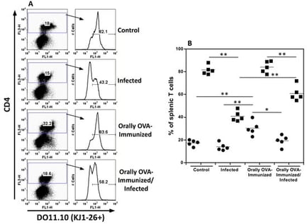
Acute Trypanosoma cruzi infection induces an exuberant immune response; however, the host is unable to clear the parasite, and the infection progresses to a chronic phase. T and B cells play a crucial role in controlling infections. Although the parasite constitutes a myriad of antigenic determinants capable of activating many T and B cell clones, some antigens trigger a large proportion of CD8 T cells, implying TCR cross-reactivity targeting these determinants. Polyclonal activation may result in an inefficient immune response against the parasite, diverting it to less critical antigenic determinants, allowing infection persistence, and increasing the risk of autoimmunity. Cross-reactivity has been demonstrated in CD8 T cells but not in CD4 T cells. Herein, we demonstrate, by cytometry, that CD4+ T cells, carrying the DO11.10 transgenic TCR, which are responsive to OVA, are activated during the T. cruzi acute infection, becoming effector memory T cells that produce cytokines such as IFN-γ, TNF-α, IL-4, and IL-10. In addition, prior oral exposure to OVA altered cytokine production by these transgenic T cells upon infection. We also demonstrate that T. cruzi induces Foxp3 expression in a sizable pool of transgenic T cells.

24 February 2026

Quantitation of splenic DO11.10 transgenic CD4+ T cells bearing the TCR clonotype, identified by the KJ1-26 mAb. (A) shows dot plots and histograms from spleen cells from uninfected control, infected, OVA-immunized, and OVA-immunized/infected transgenic mice obtained on day 25th of infection. Representative dot plots and histograms from one animal per group are depicted. (B) shows the frequencies of splenic CD4+ (closed circles) and CD4+KJ1-26+ T cells (closed squares) from individual animals in different groups. Experiments were repeated three times, on other occasions, with similar results. The Mann-Whitney test was used to compare two distinct groups of mice (* p < 0.05, ** p < 0.01).

An Eco-Tourism Farm as a Monitoring Area for the Occurrence of Tick-Borne Pathogens

  • Barbara Mangová,
  • Michal Chvostáč and
  • Veronika Rusňáková Tarageľová
  • + 4 authors

Free-ranging animals may serve as sentinels for the occurrence of ticks and tick-borne pathogens within a given habitat. Between 2020 and 2022, ticks were collected from domestic animals and vegetation on a family-operated agrotouristic farm in Western Slovakia. A total of 391 ticks were obtained (163 host-feeding and 228 questing). Ixodes ricinus predominated (94.9%), followed by Dermacentor reticulatus (4.9%) and Haemaphysalis concinna (one specimen). All ticks were screened for Borrelia burgdorferi sensu lato (s.l.), Borrelia miyamotoi, Anaplasma phagocytophilum, Babesia spp., and Rickettsia spp. by using PCR-based methods and subsequent sequencing. Borrelia burgdorferi s.l. was detected in 15.9% of I. ricinus. Six species were identified, with Borrelia afzelii dominating. Borrelia miyamotoi was detected in 1.62% of questing I. ricinus. Anaplasma phagocytophilum was identified in 20.49% of I. ricinus, mainly in host-feeding specimens. Babesia spp. were detected in I. ricinus (2.7%), represented by Babesia microti, Babesia venatorum, and Babesia capreoli. Rickettsia spp. were detected in 5.39% of I. ricinus and 31.58% of D. reticulatus. They comprised Rickettsia helvetica, Rickettsia monacensis, and Rickettsia raoultii. Co-infections involving two or three pathogens were observed in several ticks. Our results underscore the considerable diversity of tick-borne pathogens in a rural area where humans and domestic animals are in close contact to wildlife.

13 February 2026

Map of Slovakia with indication of the study site (red location marker).

Xenomas are distinctive hypertrophic host-cell lesions caused by intracellular parasites and represent a recurrent pathological finding in wild and farmed fish. Their presence has implications for fish health, diagnostic workflows, aquaculture productivity, and in some cases product quality and consumer acceptability. Despite this relevance, information on xenoma diversity, associated pathogens, and diagnostic practices remains fragmented across decades of literature. This scoping review synthesised available evidence on naturally occurring xenomas in fish, following PRISMA-ScR guidelines. Eligible sources included studies reporting clinical, pathological, or diagnostic information on xenomas in fish. Data were charted on host species and families, taxonomic identification of the aetiological agents, xenoma morphology, and diagnostic approaches. Across 114 publications published between 1968 and 2024, xenomas were reported in a wide range of teleost families and were attributed mostly to microsporidian infections, particularly species of Glugea, Loma, Spraguea, Pleistophora, and Microgemma, although myxosporean-associated cases (Kudoa, Myxidium, Nephrocystidium) were also documented. Light and electron microscopy were the most frequently applied diagnostic methods, whereas molecular techniques were used less consistently, with increasing use in the most recent decade. Macroscopic xenomas were typically described as whitish, rounded to oval, and well delimited, yet substantial morphological variation occurred across hosts and tissues. Overall, the review highlights major heterogeneity in pathogen identification and diagnostic pathways, underscoring the need for more standardised and integrative approaches.

6 February 2026

PRISMA-ScR Flow Diagram.

A supplemental description of Lamproglena barbicola Fryer, 1961 is provided based on specimens collected from the gills of Labeobarbus altianalis (Boulenger, 1900) from the Nyando River, Lake Victoria Basin, Kenya, using an integrated approach of scanning electron microscopy (SEM) and molecular analysis (18S, 28S rDNA, and cox1 gene regions). Morphologically, the specimens conform to L. barbicola and closely resemble Lamproglena hoi Dippenaar, Luus-Powell & Roux, 2001; however, SEM revealed a previously undescribed feature on the uniramous antennule in L. barbicola, namely indistinctly three-segmented, tapering from a broad base to the apex, basal segment much longer than distal, comprising 14 setae of varying sizes, ventral laterally, absence of distinctive anterior fringe of setae on the antennule, as well as several characters that differentiate L. barbicola from L. hoi, including 5 setae at the basal endopod of leg one, five cuticular protuberances in the oral region, 19 setae on the basal antennular segment, and 10 setae on the distal segment, with 1 seta on each ramus. The phylogenetic analysis confirms L. barbicola as a sister taxon of L. hoi, supporting their close relationship. The genetic divergence presented as the uncorrected genetic p-distances between L. barbicola and L. hoi are 23.1% and 0.45% for cox1 and 28S rDNA regions, respectively, with observed nucleotide differences of 145 and 3 bp between the sequences, respectively. There was no interspecific variability detected in the 18S rDNA sequences. This study provides novel molecular sequences and the first high-resolution SEM images, which reveal additional taxonomic features for L. barbicola, establishing a robust reference for future identification.

3 February 2026

Scanning electron micrographs of Lamproglena barbicola Fryer, 1961, adult female ex Labeobarbus altianalis (Boulenger, 1900). (A)—ventral view of mature adult; (B)—ventral view of cephalothorax showing antennules, antennae, oral region, maxillae, maxillipeds, and first pair of legs; (C)—detailed antennae and antennules; (D)—maxilliped; (E)—first thoracic leg; (F)—second thoracic leg; (G)—third thoracic leg; (H)—fourth thoracic leg; (I)—fifth thoracic leg; (J)—caudal rami.

News & Conferences

Issues

Open for Submission

Editor's Choice

XFacebookLinkedIn
Parasitologia - ISSN 2673-6772