
A Scoping Review of Naturally Occurring Xenomas in Fish: Clinical Features, Diagnostic Approaches, and Knowledge Gaps

 ,
,  , , , , , , and
Abstract
1. Introduction
2. Materials and Methods
2.1. Search Strategy
2.2. Study Selection
2.3. Data Extraction and Synthesis
3. Results
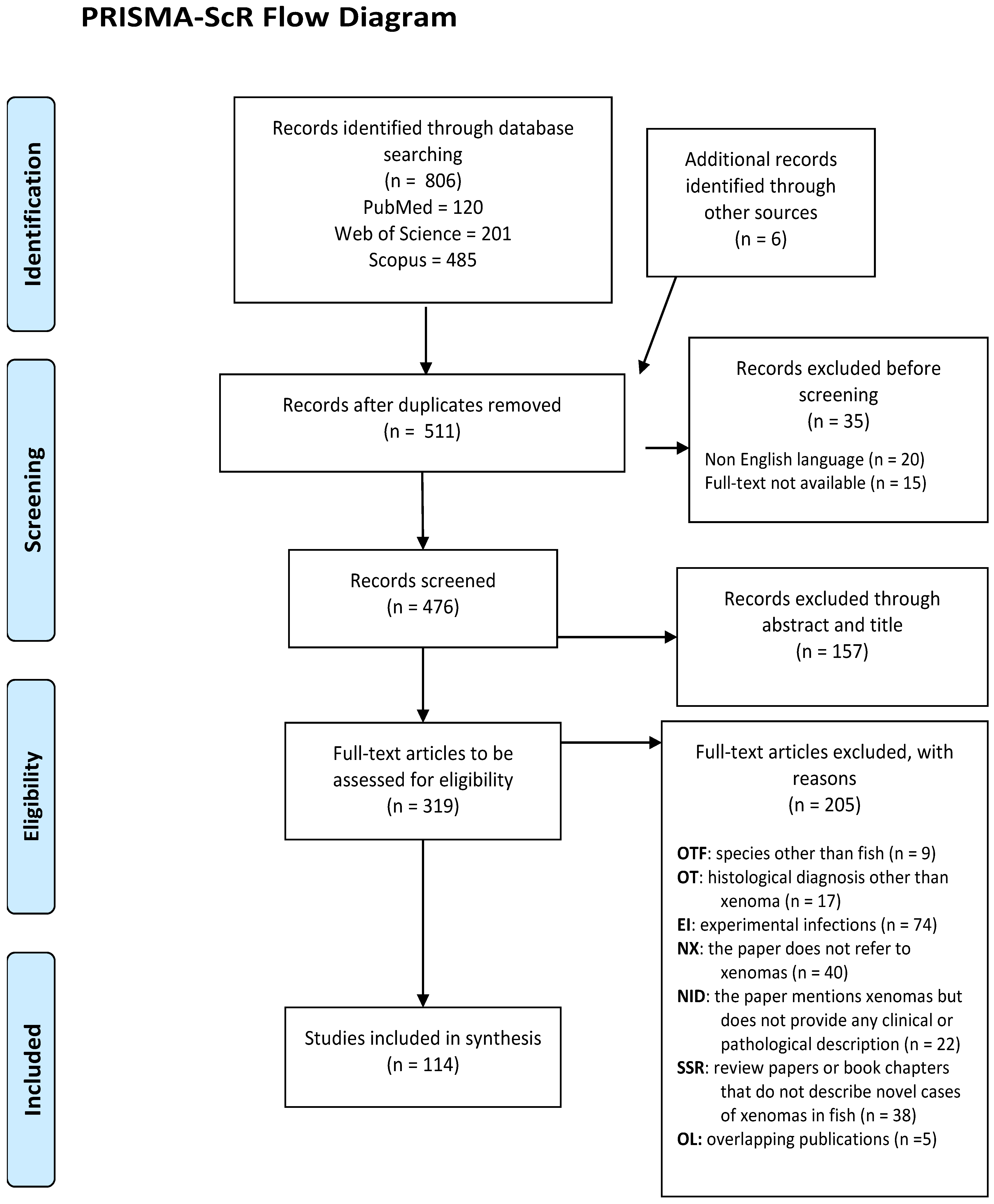
3.1. Literature Search and Study Selection
3.2. Characteristics of Included Studies and Host Fish
3.2.1. Study Characteristics
3.2.2. Host Species and Families
3.3. Quantitative Aspects (Sample Size and Prevalence)
3.4. Clinical Signs in Xenoma-Affected Fish
3.5. Anatomical Distribution and Lesion Characteristics
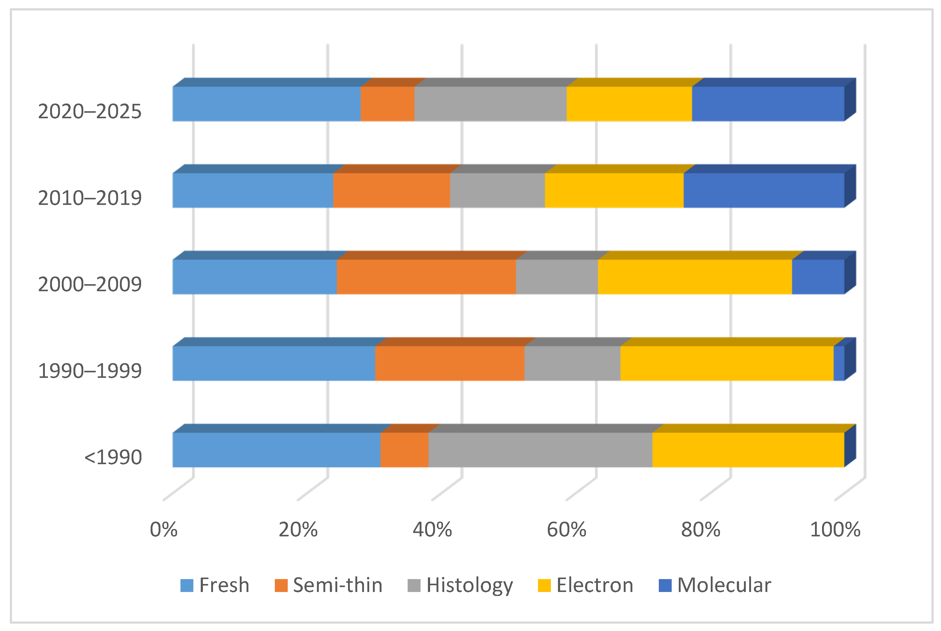
3.6. Diagnostic Methods
3.7. Aetiological Agents
3.8. Methodological and Reporting Gaps
4. Discussion
5. Conclusions
Supplementary Materials
Author Contributions
Funding
Institutional Review Board Statement
Informed Consent Statement
Data Availability Statement
Conflicts of Interest
Abbreviations
| auth | Common or vernacular fish names as provided by the original authors |
| FW | Fresh-water species |
| SW | Salt-water species |
| BW | Brackish-water species |
| C | Captive-bred specimens |
| W | Wild-caught specimens |
| F | Aquaculture farm |
| A | Aquarium |
| L | Laboratory setting |
| S | Single organ affected |
| M | Multiple organs affected |
| OTF | Species other than fish |
| EI | Experimental infections |
| NID | No clinical or pathological description provided |
| NX | Not referring to xenoma |
| OT | Histological diagnosis other than xenoma |
| SSR | Review papers or book chapters (not describing novel cases) |
| OL | Overlapping publications |
| TEM | Transmission electron microscopy |
| SEM | Scanning electron microscopy |
| FAO | Food and Agriculture Organization |
| WoRMS | World Register of Marine Species |
References
- Buchmann, K. Impact and control of protozoan parasites in maricultured fishes. Parasitology 2015, 142, 168–177. [Google Scholar] [CrossRef] [Scilit] [PubMed]
- Irwin, B.J.; Tomamichel, M.M.; Frischer, M.E.; Hall, R.J.; Davis, A.D.E.; Bliss, T.H.; Rohani, P.; Byers, J.E. Managing the threat of infectious disease in fisheries and aquaculture using structured decision making. Front. Ecol. Environ. 2024, 22, e2695. [Google Scholar] [CrossRef] [Scilit]
- Grabner, D.; Fiala, I. Biology and Life Cycles of Microsporidia and Myxozoa. In Aquatic Parasitology: Ecological and Environmental Concepts and Implications of Marine and Freshwater Parasites; Springer: Cham, Switzerland, 2025; pp. 41–69. [Google Scholar] [CrossRef] [Scilit]
- Sarker, S.; Kallert, D.M.; Hedrick, R.P.; El-Matbouli, M. Whirling disease revisited: Pathogenesis, parasite biology and disease intervention. Dis. Aquat. Org. 2015, 114, 155–175. [Google Scholar] [CrossRef] [Scilit]
- Carraro, L.; Mari, L.; Hartikainen, H.; Strepparava, N.; Wahli, T.; Jokela, J.; Gatto, M.; Rinaldo, A.; Bertuzzo, E. An epidemiological model for proliferative kidney disease in salmonid populations. Parasites Vectors 2016, 9, 487. [Google Scholar] [CrossRef] [Scilit]
- Canning, E.U.; Okamura, B. Biodiversity and evolution of the Myxozoa. Adv. Parasitol. 2004, 56, 43–131. [Google Scholar] [CrossRef] [Scilit] [PubMed]
- Lom, J.; Dyková, I. Microsporidian xenomas in fish seen in wider perspective. Folia Parasitol. 2005, 52, 69–81. [Google Scholar] [CrossRef] [Scilit]
- Vávra, J.; Lukeš, J. Microsporidia and ‘the art of living together’. Adv. Parasitol. 2013, 82, 253–319. [Google Scholar] [CrossRef] [Scilit] [PubMed]
- Ramsay, J.M.; Speare, D.J.; Dawe, S.C.; Kent, M.L. Xenoma formation during microsporidial gill disease of salmonids caused by Loma salmonae is affected by host species (Oncorhynchus tshawytscha, O. kisutch, O. mykiss) but not by salinity. Dis. Aquat. Org. 2002, 48, 125–131. [Google Scholar] [CrossRef] [Scilit]
- Ramsay, J.M.; Watral, V.; Schreck, C.B.; Kent, M.L. Pseudoloma neurophilia infections in zebrafish Danio rerio: Effects of stress on survival, growth, and reproduction. Dis. Aquat. Org. 2010, 88, 69–84. [Google Scholar] [CrossRef] [Scilit]
- Midttun, H.L.E.; Vindas, M.A.; Nadler, L.E.; Øverli, Ø.; Johansen, I.B. Behavioural effects of the common brain-infecting parasite Pseudoloma neurophilia in laboratory zebrafish (Danio rerio). Sci. Rep. 2020, 10, 8083. [Google Scholar] [CrossRef] [Scilit]
- Kent, M.L.; Shaw, R.W.; Sanders, J.L. Microsporidia in Fish; Wiley: Hoboken, NJ, USA, 2014; Volume 9781118395. [Google Scholar]
- Goater, T.M.; Goater, C.P.; Esch, G.W. Parasitism: The Diversity and Ecology of Animal Parasites, 2nd ed.; Cambridge University Press: Cambridge, UK, 2013. [Google Scholar]
- Sprengel, G.; Luchtenberg, H. Infection by endoparasites reduces maximum swimming speed of european smelt Osmerus eperlanus and European eel Anguilla anguilla. Dis. Aquat. Org. 1991, 11, 31–35. [Google Scholar] [CrossRef] [Scilit]
- Olson, R.E. Laboratory and Field Studies on Glugea stephani (Hagenmuller), a Microsporidan Parasite of Pleuronectid Flatfishes. J. Protozool. 1976, 23, 158–164. [Google Scholar] [CrossRef] [Scilit] [PubMed]
- Wiklund, T.; Lounasheimo, L.; Lom, J.; Bylund, G. Gonadal impairment in roach Rutilus rutilus from Finnish coastal areas of the northern Baltic Sea. Dis. Aquat. Org. 1996, 26, 163–171. [Google Scholar] [CrossRef] [Scilit]
- Oliviero, M.; Iaccarino, D.; Esposito, E.; Paduano, G.; D’Alessio, N.; Sgroi, G.; Lucibelli, M.G.; Auriemma, C.; degli Uberti, B.; D’Amore, M.; et al. Glugea plecoglossi (Strickland, 1911) infection in wild Sardinella aurita (Valenciennes, 1847) along the Campania coasts, in southern Italy. J. Fish Dis. 2024, 47, e13956. [Google Scholar] [CrossRef] [Scilit] [PubMed]
- Cho, H.; Lee, J.; Kim, J. Spraguea sp. (Microsporidia: Spraguidae) infection in yellow goosefish (Lophius litulon) in Korea. J. Fish Pathol. 2023, 36, 303–310. [Google Scholar]
- Curry, A. Electron microscopy and the investigation of new infectious diseases. Int. J. Infect. Dis. 2003, 7, 251–258. [Google Scholar] [CrossRef] [Scilit]
- Dezfuli, B.S.; Giari, L.; Simoni, E.; Shinn, A.P.; Bosi, G. Immunohistochemistry, histopathology and ultrastructure of Gasterosteus aculeatus tissues infected with Glugea anomala. Dis. Aquat. Org. 2004, 58, 193–202. [Google Scholar] [CrossRef] [Scilit]
- Chapman, J.M.; Kelly, L.A.; Teffer, A.K.; Miller, K.M.; Cooke, S.J. Disease ecology of wild fish: Opportunities and challenges for linking infection metrics with behaviour, condition, and survival. Can. J. Fish. Aquat. Sci. 2021, 78, 995–1007. [Google Scholar] [CrossRef] [Scilit]
- Freeman, M.A.; Fuss, J.; Kristmundsson, Á.; Bjorbækmo, M.F.M.; Mangot, J.-F.; del Campo, J.; Keeling, P.J.; Shalchian-Tabrizi, K.; Bass, D. X-Cells Are Globally Distributed, Genetically Divergent Fish Parasites Related to Perkinsids and Dinoflagellates. Curr. Biol. 2017, 27, 1645–1651.e3. [Google Scholar] [CrossRef] [Scilit]
- Tricco, A.C.; Lillie, E.; Zarin, W.; O’Brien, K.K.; Colquhoun, H.; Levac, D.; Moher, D.; Peters, M.D.J.; Horsley, T.; Weeks, L.; et al. PRISMA extension for scoping reviews (PRISMA-ScR): Checklist and explanation. Ann. Intern. Med. 2018, 169, 467–473. [Google Scholar] [CrossRef] [Scilit]
- Froese, R.; Pauly, D. (Eds.) FishBase. World Wide Web Electronic Publication. 2025. Available online: www.fishbase.org (accessed on 26 November 2025).
- WoRMS Editorial Board. World Register of Marine Species. 2026. Available online: https://www.marinespecies.org (accessed on 2 January 2026).
- Sprague, V.; Vernick, S.H. Light and Electron Microscope Study of a New Species of Glugea (Microsporida, Nosematidae) in the 4-Spined Stickleback Apeltes quadracus. J. Protozool. 1968, 15, 547–571. [Google Scholar] [CrossRef] [Scilit]
- Leiro, J.; Paramá, A.; Ortega, M.; Santamarina, M.T.; Sanmartín, M.L. Redescription of Glugea caulleryi, a microsporidian parasite of the greater sand-eel, Hyperoplus ianceolatus (Le Sauvage), (Teleostei: Ammodytidae), as Microgemma caulleryi comb. nov. J. Fish Dis. 1999, 22, 101–110. [Google Scholar] [CrossRef] [Scilit]
- Brown, A.M.V.; Kent, M.L.; Adamson, M.L. Description of five new Loma (microsporidia) species in pacific fishes with redesignation of the type species Loma morhua Morrison & Sprague, 1981, based on morphological and molecular species-boundaries tests. J. Eukaryot. Microbiol. 2010, 57, 529–553. [Google Scholar] [CrossRef] [Scilit]
- Vagelli, A.; Paramá, A.; Sanmartín, M.L.; Leiro, J. Glugea vincentiae n. sp. (Microsporidia: Glugeidae) infecting the Australian marine fish Vincentia conspersa (Teleostei: Apogonidae). J. Parasitol. 2005, 91, 152–157. [Google Scholar] [CrossRef] [Scilit] [PubMed]
- Ovcharenko, M.; Wróblewski, P.; Kvach, Y.; Drobiniak, O. Study of Loma acerinae (Microsporidia) detected from three Ponto-Caspian gobies (Gobiidae) in Ukraine. Parasitol. Res. 2017, 116, 1453–1462. [Google Scholar] [CrossRef] [Scilit] [PubMed]
- Casal, G.; Matos, E.; Teles-Grilo, M.L.; Azevedo, C. A new microsporidian parasite, Potaspora morhaphis n. gen., n. sp. (Microsporidia) infecting the Teleostean fish, Potamorhaphis guianensis from the River Amazon. Morphological, ultrastructural and molecular characterization. Parasitology 2008, 135, 1053–1064. [Google Scholar] [CrossRef] [Scilit]
- Mansour, L.; Zhang, J.Y.; Abdel-Haleem, H.M.; Darwish, A.B.; Al-Quraishy, S.; Abdel-Baki, A.-A.S. Ultrastructural description and phylogeny of a novel microsporidian, Glugea eda n. sp. from the striated fusilier, Caesio striata, in the Red Sea off Saudi Arabia. Acta Trop. 2020, 204, 105331. [Google Scholar] [CrossRef] [Scilit] [PubMed]
- Diamant, A.; Rothman, S.B.S.; Goren, M.; Galil, B.S.; Yokes, M.B.; Szitenberg, A.; Huchon, D. Biology of a new xenoma-forming gonadotropic microsporidium in the invasive blotchfin dragonet Callionymus filamentosus. Dis. Aquat. Org. 2014, 109, 35–54. [Google Scholar] [CrossRef] [Scilit]
- Baki, A.-A.S.A.; Aboelhadid, S.M.; Mansour, Q.L. Ultrastructural and molecular characterization of Glugea sp. (Microsporidia), a parasite of the Red Sea fish Carangoides bajad (Carangidae). J. Eukaryot. Microbiol. 2024, 71, e13058. [Google Scholar] [CrossRef] [Scilit]
- Casal, G.; Matos, E.; Garcia, P.; Al-Quraishy, S.; Azevedo, C. Ultrastructural and molecular studies of Microgemma carolinus n. sp. (Microsporidia), a parasite of the fish Trachinotus carolinus (Carangidae) in Southern Brazil. Parasitology 2012, 139, 1720–1728. [Google Scholar] [CrossRef] [Scilit]
- Faye, N.; Toguebaye, B.S. Microsporidian parasites in four species of carangid fishes from the Senegalese coast (West Africa). Acta Adriat. 2005, 46, 21–26. [Google Scholar]
- Kalavati, C.; Sandeep, B.V.; Narasimhamurti, C.C. Seasonal variation of the microsporidian, Glugea malabaricii parasitic in the liver of Carangoides malabaricus Bl. Acta Protozool. 1985, 24, 165–174. [Google Scholar]
- Mansour, L.; Thabet, A.; Chourabi, K.; Harrath, A.H.; Gtari, M.; Al Omar, S.Y.; Ben Hassine, O.K. Kudoa azevedoi n. sp. (Myxozoa, Multivalvulida) from the oocytes of the Atlantic horse mackerel Trachurus trachurus (Perciformes, Carangidae) in Tunisian coasts. Parasitol. Res. 2013, 112, 1737–1747. [Google Scholar] [CrossRef] [Scilit]
- Vo, H.; Butaeva, F.; Yurakhno, V. More about the biodiversity of parasites in Vietnam: A first report of microsporidia (Opistosporidia, Microsporidia) in coral fish. IOP Conf. Ser. Earth Environ. Sci. 2021, 937, 022067. [Google Scholar] [CrossRef] [Scilit]
- Narasimhamurti, C.; Kalavati, C.; Sandeep, B. A new microsporidian Glugea malabaricii sp. n. from the viscera of Carangoides malabaricus. Acta Protozool. 1985, 24, 153–164. [Google Scholar]
- Amigo, J.M.; Salvado, H.; Gracia, M.P.; Vivares, C.P. Ultrastructure and development of Microsporidium ovoideum (Thelohan, 1895) Sprague, 1977, a microsporidian parasite of the red band fish (Cepola macrophthalma L.): Redescription of the organism and reassignment to the genus Microgemma, Ralphs and Matthews. Eur. J. Protistol. 1996, 32, 532–538. [Google Scholar] [CrossRef] [Scilit]
- Amigó, J.M. The pathologic cycle of the infection of the microsporidian Microgemma ovoidea (Thèl., 1895) Amigó et al. 1996 in the liver of the Red Band Fish (Cepola macrophthalma L.). Parasitol. Res. 1997, 84, 7–12. [Google Scholar] [CrossRef] [Scilit] [PubMed]
- Fomena, A.; Coste, F.; Bouix, G. Loma camerounensis sp. nov. (Protozoa: Microsporida) a parasite of Oreochromis niloticus Linnaeus, 1757 (Teleost: Cichlidae) in fish-rearing ponds in Melen, Yaoundé, Cameroon. Parasitol. Res. 1992, 78, 201–208. [Google Scholar] [CrossRef] [Scilit]
- Videira, M.; Casal, G.; Rocha, S.; Gonçalves, E.; Azevedo, C.; Velasco, M.; Matos, E.R. Potaspora aequidens n. sp. (Microsporidia, Tetramicridae), a parasite infecting the freshwater fish Aequidens plagiozonatus (Teleostei, Cichlidae) from Brazil. Parasitol. Res. 2015, 114, 2435–2442. [Google Scholar] [CrossRef] [Scilit]
- Canning, E.U.; Curry, A. Microgemma vivaresi (Microsporidia: Tetramicridae): Host reaction to xenomas induced in sea scorpions, Taurulus bubalis (Osteichthyes: Cottidae). Folia Parasitol. 2005, 52, 95–102. [Google Scholar] [CrossRef] [Scilit]
- Homola, J.J.; Ruetz, C.R., III; Kohler, S.L.; Thum, R.A. Weak effects of a microsporidian parasite on mottled sculpin in Michigan streams. Can. J. Fish. Aquat. Sci. 2014, 71, 915–926. [Google Scholar] [CrossRef] [Scilit]
- Ryan, J.A.; Kohler, S.L. Distribution, prevalence, and pathology of a microsporidian infecting freshwater sculpins. Dis. Aquat. Org. 2016, 118, 195–206. [Google Scholar] [CrossRef] [Scilit] [PubMed]
- Scholz, F.; Fringuelli, E.; Bolton-Warberg, M.; Marcos-López, M.; Mitchell, S.; Prodhol, P.; Moffet, D.; Savage, P.; O’Sullivan, S.M.; O’Connor, I.; et al. First record of Tetramicra brevifilum in lumpfish (Cyclopterus lumpus, L.). J. Fish Dis. 2017, 40, 757–771. [Google Scholar] [CrossRef] [Scilit] [PubMed]
- Forest, J.J.H.; King, S.D.; Cone, D.K. Occurrence of Glugea pimephales in planktonic larvae of fathead minnow in Algonquin Park, Ontario. J. Aquat. Anim. Health 2009, 21, 164–166. [Google Scholar] [CrossRef] [Scilit] [PubMed]
- Liu, X.H.; Stentiford, G.D.; Voronin, V.N.; Sato, H.; Li, A.H.; Zhang, J.Y. Pseudokabatana alburnus n. gen. n. sp., (Microsporidia) from the liver of topmouth culter Culter alburnus (Actinopterygii, Cyprinidae) from China. Parasitol. Res. 2019, 118, 1689–1699. [Google Scholar] [CrossRef] [Scilit]
- Matthews, J.L.; Brown, A.M.V.; Larison, K.; Bishop-Stewart, J.K.; Rogers, P.; Kent, M.L. Pseudoloma neurophilia n. g., n. sp., a new microsporidium from the central nervous system of the zebrafish (Danio rerio). J. Eukaryot. Microbiol. 2001, 48, 227–233. [Google Scholar] [CrossRef] [Scilit]
- Weng, M.; Zhang, X.; Xin, Z.; Xue, S.; Zhang, Q.; Li, A.; Zhang, J. Intraspecific genetic diversity of the fish-infecting microsporidian parasite Pseudokabatana alburnus (Microsporidia). Front. Microbiol. 2023, 14, 1129136. [Google Scholar] [CrossRef] [Scilit] [PubMed]
- Lom, J.; Noga, E.J.; Dykova, I. Occurrence of a microsporean with characteristics of Glugea anomala in ornamental fish of the family Cyprinodontidae. Dis. Aquat. Org. 1995, 21, 239–242. [Google Scholar] [CrossRef] [Scilit]
- Azevedo, C.; Matos, E. Amazonspora hassar n. gen. and n. sp. (Phylum microsporidia, fam. Glugeidae), a parasite of the Amazonian teleost Hassar orestis (fam. Doradidae). J. Parasitol. 2003, 89, 336–341. [Google Scholar] [CrossRef] [Scilit]
- López-Verdejo, A.; Occhibove, F.; degli Uberti, B.; Montero, F.E.; Santoro, M. Mass mortality event of round sardinella Sardinella aurita Valenciennes associated with Glugea Thélohan, 1891 microsporidian infection off the southern Italian coast. J. Fish Dis. 2024, 47, e13995. [Google Scholar] [CrossRef] [Scilit]
- Mansour, L.; Thabet, A.; Harrath, A.H.; Al Omar, S.Y.; Mukhtar, A.; Sayed, S.R.; Abdel-Baki, A.-A.S. New microsporidia, Glugea sardinellensis n. sp. (Microsporea, Glugeida) found in Sardinella aurita valenciennes, 1847, collected off tunisian coasts. Acta Protozool. 2016, 55, 281–290. [Google Scholar] [CrossRef] [Scilit]
- Ramdani, S.; Ramdane, Z.; Slamovits, C.H.; Trilles, J.-P. Glugea sp. infecting Sardinella aurita in Algeria. J. Parasit. Dis. 2022, 46, 672–685. [Google Scholar] [CrossRef] [Scilit]
- Shaw, R.W.; Kent, M.L.; Docker, M.F.; Brown, A.M.V.; Devlin, R.H.; Adamson, M.L. A new species of Loma (Microsporea) in shiner perch (Cymatogaster aggregata). J. Parasitol. 1997, 83, 296–301. [Google Scholar] [CrossRef] [Scilit]
- Abdel-Ghaffar, F.; Bashtar, A.-R.; Mehlhorn, H.; Al-Rasheid, K.; Morsy, K. Microsporidian parasites: A danger facing marine fishes of the Red Sea. Parasitol. Res. 2011, 108, 219–225. [Google Scholar] [CrossRef] [Scilit]
- Abdel-Ghaffar, F.; Bashtar, A.-R.; Mehlhorn, H.; Al-Rasheid, K.; Al-Olayan, E.; Koura, E.; Morsy, K. Ultrastructure, development, and host-parasite relationship of a new species of the genus Pleistophora—A microsporidian parasite of the marine fish Epinephelus chlorostignei. Parasitol. Res. 2009, 106, 39–46. [Google Scholar] [CrossRef] [Scilit] [PubMed]
- Azevedo, C.; Abdel-Baki, A.-A.S.; Rocha, S.; Al-Quraishy, S.; Casal, G. Ultrastructure and phylogeny of Glugea arabica n. sp. (Microsporidia), infecting the marine fish Epinephelus polyphekadion from the Red Sea. Eur. J. Protistol. 2016, 52, 11–21. [Google Scholar] [CrossRef] [Scilit] [PubMed]
- Lom, J.; Dyková, I.; Feist, S. Myxosporea-induced xenoma formation in pike (Esox lucius L.) renal corpuscles associated with Myxidium lieberkuehni infection. Eur. J. Protistol. 1989, 24, 271–280. [Google Scholar] [CrossRef] [Scilit] [PubMed]
- Sokolov, S.; Volkova, E.; Kudryavtsev, A.; Parshukov, A. Nephrocystidium pickii Weissenberg, 1921 belongs to Myxozoa (Cnidaria) but is not conspecific with Myxidium lieberkuehni Bütschli, 1882 (Myxozoa: Bivalvulida: Variisporina: Myxidiidae): Molecular-genetic evidence. Syst. Parasitol. 2019, 96, 15–22. [Google Scholar] [CrossRef] [Scilit]
- Frenette, A.P.; Eydal, M.; Hansen, H.; Burt, M.D.B.; Duffy, M.S. Integrative Approach for the Reliable Detection and Specific Identification of the Microsporidium Loma morhua in Atlantic Cod (Gadus morhua). J. Eukaryot. Microbiol. 2017, 64, 67–77. [Google Scholar] [CrossRef] [Scilit] [PubMed]
- Khan, R.A. Prevalence and influence of Loma branchialis (Microspora) on growth and mortality in atlantic cod (Gadus morhua) in coastal Newfoundland. J. Parasitol. 2005, 91, 1230–1232. [Google Scholar] [CrossRef] [Scilit]
- Morrison, C.M.; Sprague, V. Light and electron microscope study of Microsporida in the gill of haddock, Melanogrammus aeglefinus (L.)*. J. Fish Dis. 1981, 4, 179–184. [Google Scholar] [CrossRef] [Scilit]
- Morrison, C.M.; Sprague, V. Electron microscopical study of a new genus and new species of Microsporida in the gills of Atlantic cod Gadus morhua L. J. Fish Dis. 1981, 4, 15–32. [Google Scholar] [CrossRef] [Scilit]
- Powell, M.D.; Gamperl, A.K. Effects of Loma morhua (Microsporidia) infection on the cardiorespiratory performance of Atlantic cod Gadus morhua (L). J. Fish Dis. 2016, 39, 189–204. [Google Scholar] [CrossRef] [Scilit]
- Rodriguez-Tovar, L.E.; Wadowska, D.W.; Wright, G.M.; Groman, D.B.; Speare, D.J.; Whelan, D.S. Ultrastructural evidence of autoinfection in the gills of Atlantic cod Gadus morhua infected with Loma sp. (phylum Microsporidia). Dis. Aquat. Org. 2003, 57, 227–230. [Google Scholar] [CrossRef] [Scilit]
- Schmahl, G.; Mehlhorn, H. Treatment of fish parasites: 6. Effects of sym. triazinone (toltrazuril) on developmental stages of Glugea anomala, Moniez, 1887 (Microsporidia): A light and electron microscopic study. Eur. J. Protistol. 1989, 24, 252–259. [Google Scholar] [CrossRef] [Scilit]
- Canning, E.; Lom, J.; Nicholas, J. Genus Glugea Thelohan 1891 (phylum Microspora): Redescription of the type species Glugea anomala (Moniez, 1887) and recognition of its sporogonic development. Protistologica 1982, 18, 193–210. [Google Scholar]
- Al-Shammari, N.A. Opportunistic Bacteria Isolated from Trypauchen Vagina Fish that Infected with Protozoan from Iraq Marine Water. Sci. J. King Faisal Univ. 2021, 22, 83–86. [Google Scholar] [CrossRef] [Scilit]
- Freeman, M.A.; Chong, W.W.; Loh, K.H. Microsporidians infecting eel gobies (Gobiidae: Amblyopinae) from Malaysia, with a description of Microgemma tilanpasiri n. sp. from the burrowing goby Trypauchen vagina. Bull. Eur. Assoc. Fish Pathol. 2015, 35, 112–119. [Google Scholar]
- Sanders, J.; Myers, M.S.; Tomanek, L.; Cali, A.; Takvorian, P.M.; Kent, M.L. Ichthyosporidium weissii n. sp. (Microsporidia) infecting the arrow goby (Clevelandia ios). J. Eukaryot. Microbiol. 2012, 59, 258–267. [Google Scholar] [CrossRef] [Scilit] [PubMed]
- Casal, G.; Matos, E.; Rocha, S.; Sindeaux-Neto, J.; Al-Quraishy, S.; Azevedo, C. Ultrastructure and phylogeny of Pleistophora beebei sp. nov. (Microsporidia) infecting the Amazonian teleostean Brachyhypopomus beebei (fam. Hypopomidae). Acta Protozool. 2016, 55, 259–268. [Google Scholar] [CrossRef]
- Matos, E.; Azevedo, C. Ultrastructural description of Microsporidium brevirostris sp. n., parasite of the teleostean Brachyhypopomus brevirostris (Hypopomidae) from the Amazon River. Acta Protozool. 2004, 43, 261–267. [Google Scholar]
- Mansour, L.; Prensier, G.; Jemaa, S.B.; Hassine, O.K.B.; Méténier, G.; Vivarès, C.P.; Cornillot, E. Description of a xenoma-inducing microsporidian, Microgemma tincae n. sp., parasite of the teleost fish Symphodus tinca from Tunisian coasts. Dis. Aquat. Org. 2005, 65, 217–226. [Google Scholar] [CrossRef] [Scilit] [PubMed]
- Freeman, M.A.; Yokoyama, H.; Osada, A.; Yoshida, T.; Yamanobe, A.; Ogawa, K. Spraguea (Microsporida: Spraguidae) infections in the nervous system of the Japanese anglerfish, Lophius litulon (Jordan), with comments on transmission routes and host pathology. J. Fish Dis. 2011, 34, 445–452. [Google Scholar] [CrossRef] [Scilit]
- Campbell, S.E.; Williams, T.A.; Yousuf, A.; Soanes, D.M.; Paszkiewicz, K.H.; Williams, B.A.P. The Genome of Spraguea lophii and the Basis of Host-Microsporidian Interactions. PLoS Genet. 2013, 9, e1003676. [Google Scholar] [CrossRef] [Scilit] [PubMed]
- Casal, G.; Clemente, S.C.S.; Matos, P.; Knoff, M.; Matos, E.; Abdel-Baki, A.A.; Azevedo, C. Redefining the genus Spraguea based on ultrastructural and phylogenetic data from Spraguea gastrophysus n. sp. (Phylum Microsporidia), a parasite found in Lophius gastrophysus (Teleostei) from Brazil. Parasitol. Res. 2012, 111, 79–88. [Google Scholar] [CrossRef] [Scilit]
- Colmenero, A.I.; Barría, C.; Feist, S.W.; Tuset, V.M. Observations on the occurrence of Spraguea lophii in Mediterranean lophiids. Parasitol. Res. 2015, 114, 1977–1983. [Google Scholar] [CrossRef] [Scilit]
- Freeman, M.A.; Yokoyama, H.; Ogawa, K. A microsporidian parasite of the genus Spraguea in the nervous tissues of the Japanese anglerfish Lophius litulon. Folia Parasitol. 2004, 51, 167–176. [Google Scholar] [CrossRef] [Scilit]
- Llarena-Reino, M.; Abollo, E.; Pascual, S. Microsporidians xenomas of anglerfish from NE Atlantic waters. J. Fish Dis. 2017, 40, 1587–1598. [Google Scholar] [CrossRef] [Scilit]
- Mansour, L.; Ben Hassine, O.K.; Vivares, C.P.; Cornillot, E. Spraguea lophii (Microsporidia) parasite of the teleost fish, Lophius piscatorius from Tunisian coasts: Evidence for an extensive chromosome length polymorphism. Parasitol. Int. 2013, 62, 66–74. [Google Scholar] [CrossRef] [Scilit]
- Jones, S.R.M.; Ahonen, H.S.; Granlund, L.; Arsiola, T.; Taskinen, J. Two novel microsporidia in skeletal muscle of pike-perch Sander lucioperca and Burbot Lota Lota in Finland. J. Parasitol. 2017, 103, 95–102. [Google Scholar] [CrossRef] [Scilit]
- Abdel-Baki, A.-A.S.; Tamihi, A.F.; Al-Qahtani, H.A.; Al-Quraishy, S.; Mansour, L. Glugea jazanensis sp. nov. infecting Lutjanus bohar in the Red Sea: Ultrastructure and phylogeny. Dis. Aquat. Org. 2015, 116, 185–190. [Google Scholar] [CrossRef] [Scilit]
- Faye, N.; Toguebaye, B.S.; Bouix, G. Microfilum lutjani N. G. N. sp. (Protozoa Microsporida), A Gill Parasite of the Golden African Snapper Lutjanus fulgens (Valenciennes, 1830) (Teleost Lutjanidae): Developmental Cycle and Infrastructure. J. Protozool. 1991, 38, 30–40. [Google Scholar] [CrossRef] [Scilit] [PubMed]
- Lom, J.; Gayevskaya, A.V.; Dyková, I. Two microsporidian parasites found in marine fishes in the Atlantic Ocean. Folia Parasitol. 1980, 27, 197–202. [Google Scholar] [PubMed]
- Caffara, M.; Quaglio, F.; Marcer, F.; Florio, D.; Fioravanti, M.L. Intestinal microsporidiosis in European seabass (Dicentrarchus labrax L.) farmed in Italy. Bull. Eur. Assoc. Fish Pathol. 2010, 30, 237–240. [Google Scholar]
- Ralphs, J.R.; Matthews, R.A. Hepatic microsporidiosis of juvenile grey mullet, Chelon labrosus (Risso), due to Microgemma hepaticus gen.nov. sp.nov. J. Fish Dis. 1986, 9, 225–242. [Google Scholar] [CrossRef] [Scilit]
- Stephens, F.J. Unidentified microsporidian in western pygmy perch Edelia vittata (Nannopercidae) from Australia. Aust. Vet. J. 2009, 87, 61–62. [Google Scholar] [CrossRef] [Scilit]
- Azevedo, C.; Matos, E. Fine structure of a new species, Loma myrophis (phylum Microsporidia), parasite of the Amazonian fish Myrophis platyrhynchus (Teleostei, Ophichthidae). Eur. J. Protistol. 2002, 37, 445–452. [Google Scholar] [CrossRef] [Scilit]
- Matos, E.; Corral, L.; Azevedo, C. Ultrastructural details of the xenoma of Loma myrophis (phylum Microsporidia) and extrusion of the polar tube during autoinfection. Dis. Aquat. Org. 2003, 54, 203–207. [Google Scholar] [CrossRef] [Scilit] [PubMed]
- Costa, G.; Melo-Moreira, E.; De Carvalho, M.A.P. Occurrence of microsporidians Glugea hertwigi and Pleistophora ladogensis, in smelt Osmerus eperlanus from two German rivers, north Sea coast. Dis. Aquat. Org. 2016, 121, 49–57. [Google Scholar] [CrossRef] [Scilit]
- Lovy, J.; Kostka, M.; Dyková, I.; Arsenault, G.; Pecková, H.; Wright, G.M.; Speare, D.J. Phylogeny and morphology of Glugea hertwigi from rainbow smelt Osmerus mordax found in Prince Edward Island, Canada. Dis. Aquat. Org. 2009, 86, 235–243. [Google Scholar] [CrossRef] [Scilit]
- Vandana, V.R.; Poojary, N.; Tripathi, G.; Kumar, P.; Sanil, N.K.; Rajendran, K.V. Hepatic microsporidiosis of mudskipper, Boleophthalmus dussumieri Valenciennes, 1837 (Perciformes: Gobiidae), due to Microgemma sp. J. Parasit. Dis. 2022, 46, 72–79. [Google Scholar] [CrossRef] [Scilit]
- Dyková, I.; Lom, J.; Egusa, S. Tissue reaction to Glugea plecoglossi infection in its natural host, Plecoglussus altivelis. Folia Parasitol. 1980, 27, 213–216. [Google Scholar]
- Khan, R.A. Effect, distribution, and prevalence of Glugea stephani (Microspora) in winter flounder (Pleuronectes americanus) living near two pulp and paper mills in Newfoundland. J. Parasitol. 2004, 90, 229–233. [Google Scholar] [CrossRef] [Scilit]
- Kuciński, M.; Złoch, I.; Trzeciak, P.; Kycko, A.; Nadolna-Ałtyn, K.; Mierzejewska, K. Infection of the European flounder (Platichthys flesus) with Glugea stephani, a possible new indicator of the weakening of the Baltic population. Fish. Res. 2023, 260, 106590. [Google Scholar] [CrossRef] [Scilit]
- Priebe, K.; Oehlenschläger, J. Alterations in skeletal muscle of witch Glyptocephalus cynoglossus from North Atlantic waters caused by a microsporidian of the genus Glugea (Order: Microsporidia Suborder: Pansporoblastina). Bull. Eur. Assoc. Fish Pathol. 1996, 16, 154–156. [Google Scholar]
- Takvorian, P.M.; Cali, A. The occurrence of Glugea stephani (Hagenmuller, 1899) in American winter flounder, Pseudopleuronects americanus (Walbaum) from the New York-New Jersey lower bay complex. J. Fish Biol. 1981, 18, 491–501. [Google Scholar] [CrossRef] [Scilit]
- Weidner, E. Ultrastructure of the Peripheral Zone of a Glugea-Induced Xenoma. J. Protozool. 1976, 23, 234–238. [Google Scholar] [CrossRef] [Scilit]
- Crandall, T.A.; Bowser, P.R. A microsporidian infection in a natural population of mosquitofish, Gambusia affinis (Baird and Girard). J. Fish Dis. 1981, 4, 317–324. [Google Scholar] [CrossRef] [Scilit]
- Reimschuessel, R.; Bennett, R.O.; May, E.B.; Lipsky, M.M. Undefined eosinophilic granular cell response to a microsporidian infection in a sergeant major fish, Abudefduf saxatilis (L.). J. Fish Dis. 1987, 10, 319–322. [Google Scholar] [CrossRef] [Scilit]
- Diggles, B.K. Some pathological abnormalities of New Zealand fishes. N. Z. J. Mar. Freshw. Res. 2003, 37, 705–713. [Google Scholar] [CrossRef] [Scilit]
- Bader, J.A.; Shotts, E.B.; Steffens, W.L.; Lom, J. Occurrence of Loma cf. salmonae in brook, brown and rainbow trout from Buford Trout Hatchery, Georgia, USA. Dis. Aquat. Org. 1998, 34, 211–216. [Google Scholar] [CrossRef] [Scilit]
- Bruno, D.W.; Collins, R.O.; Morrison, C.M. The occurrence of Loma salmonae (Protozoa: Microspora) in farmed rainbow trout, Oncorhynchus mykiss Walbaum, in Scotland. Aquaculture 1995, 133, 341–344. [Google Scholar] [CrossRef] [Scilit]
- Hauck, A.K. A mortality and associated tissue reactions of chinook salmon, Oncorhynchus tshawytscha (Walbaum), caused by the microsporidan Loma sp. J. Fish Dis. 1984, 7, 217–229. [Google Scholar] [CrossRef] [Scilit]
- Jones, S.; Prosperi-Porta, G.; Dawe, S.; Blackbourn, J.; Taylor, K.; Lowe, G.; Osborn, A. Proliferative renal myxosporidiosis in spawning coho salmon (Oncorhynchus kisutch) in British Columbia and Washington. Folia Parasitol. 2004, 51, 221–227. [Google Scholar] [CrossRef] [Scilit]
- Kent, M.L.; Elliott, D.G.; Groff, J.M.; Hedrick, R.P. Loma salmonae (Protozoa: Microspora) infections in seawater reared coho salmon Oncorhynchus kisutch. Aquaculture 1989, 80, 211–222. [Google Scholar] [CrossRef] [Scilit]
- Lovy, J.; Wright, G.M.; Speare, D.J. Ultrastructural examination of the host inflammatory response within gills of netpen reared chinook salmon (Oncorhynchus tshawytscha) with Microsporidial Gill Disease. Fish Shellfish. Immunol. 2007, 22, 131–149. [Google Scholar] [CrossRef] [Scilit]
- Markey, P.T.; Blazer, V.S.; Ewing, M.S.; Kocan, K.M. Loma sp. in salmonids from the eastern united states: Associated lesions in rainbow trout. J. Aquat. Anim. Health 1994, 6, 318–328. [Google Scholar] [CrossRef] [Scilit]
- Drinan, E.M.; Rodger, H.D.; Palmer, R. Microsporidian infection in Atlantic salmon, Salmo salar L. J. Fish Dis. 1992, 15, 211–214. [Google Scholar] [CrossRef] [Scilit]
- Sprague, V.; Hussey, K.L. Observations on Ichthyosporidium giganteum (Microsporida) with Particular Reference to the Host-Parasite Relations During Merogony. J. Protozool. 1980, 27, 169–175. [Google Scholar] [CrossRef] [Scilit]
- López-Verdejo, A.; Montero, F.E.; de la Gándara, F.; Gallego, M.A.; Ortega, A.; Raga, J.A.; Palacios-Abella, J.F. A severe microsporidian disease in cultured Atlantic Bluefin Tuna (Thunnus thynnus). IMA Fungus 2022, 13, 5. [Google Scholar] [CrossRef] [Scilit]
- Mladineo, I.; Lovy, J. A new xenoma-forming microsporidium infecting intestinal tract of Atlantic bluefin tuna (Thunnus thynnus). Acta Parasitol. 2011, 56, 339–347. [Google Scholar] [CrossRef] [Scilit]
- Matthews, R.A.; Matthews, B.F. Cell and tissue reactions of turbot Scophthalmus maximus (L.) to Tetramicra brevifilum gen. n., sp. n. (Microspora). J. Fish Dis. 1980, 3, 495–515. [Google Scholar] [CrossRef] [Scilit]
- Estévez, J.; Iglesias, R.; Leiro, J.; Ubeira, F.; Sanmartin, M. An unusual site of infection by a microsporean in the turbot Scophthalmus maximus. Dis. Aquat. Org. 1992, 13, 139–142. [Google Scholar] [CrossRef] [Scilit]
- Abdel-Baki, A.-A.S.; Al-Quraishy, S.; Rocha, S.; Dkhil, M.A.; Casal, G.; Azevedo, C. Ultrastructure and phylogeny of Glugea nagelia sp. n. (Microsporidia: Glugeidae), infecting the intestinal wall of the yellowfin hind, Cephalopholis hemistiktos (Actinopterygii: Serranidae), from the Red Sea. Folia Parasitol. 2015, 62, 2015.007. [Google Scholar] [CrossRef] [Scilit]
- Casal, G.; Rocha, S.; Costa, G.; Al-Quraishy, S.; Azevedo, C. Ultrastructural and molecular characterization of Glugea serranus n. sp., a microsporidian infecting the blacktail comber, Serranus atricauda (Teleostei: Serranidae), in the Madeira Archipelago (Portugal). Parasitol. Res. 2016, 115, 3963–3972. [Google Scholar] [CrossRef] [Scilit]
- Jithendran, K.P.; Vijayan, K.K.; Kailasam, M. Microsporidian (Glugea sp.) infection in the greasy grouper Epinephelus tauvina (Forsskal, 1775). Indian J. Fish. 2011, 58, 125–127. [Google Scholar]
- Liu, X.; Xu, L.; Hu, K.; Ren, S.; Weng, M.; Yu, J.; Li, D.; Zhang, J. New isolate of Loma psittaca (Microsporidia: Glugeidae) infecting the stomach wall of cultured hybrid grouper (Epinephelus lanceolatus♂ × Epinephelus fuscoguttatus♀) in South China. J. Eukaryot. Microbiol. 2021, 68, e12868. [Google Scholar] [CrossRef] [Scilit]
- Faye, N.; Toguebaye, B.S.; Bouix, G. First report of microsporidian infections in solefishes from Senegal coast (West Africa). Bull. Eur. Assoc. Fish Pathol. 2004, 24, 115–118. [Google Scholar]
- Faye, N.; Toguebaye, B.S.; Boiux, G. On the occurrence of microsporidian infections in the liver of four Sparid fishes species from Senegal. Bull. Eur. Assoc. Fish Pathol. 1998, 18, 84–86. [Google Scholar]
- Faye, N.; Toguebaye, B.S.; Bouix, G. On the Cytology and Development of Loma boopsi n. sp. (Microspora, Glugeidae), Parasite of Boops boops (Pisces, Teleostei, Sparidae) from the Coasts of Senegal. Arch. Für Protistenkd. 1995, 146, 85–93. [Google Scholar] [CrossRef] [Scilit]
- Faye, N.; Toguebaye, B.S.; Bouix, G. Ultrastructure and development of Pleistophora senegalensis sp. nov. (Protozoa, Microspora) from the gilt-head sea bream, Sparus aurata L. (Teleost, Sparidae) from the coast of Senegal. J. Fish Dis. 1990, 13, 179–192. [Google Scholar] [CrossRef] [Scilit]
- Morsy, K.; Bashtar, A.R.; Abdel-Ghaffar, F.; Al-Quraishy, S. Morphological and phylogenetic description of a new xenoma-inducing microsporidian, Microsporidium aurata nov. sp., parasite of the gilthead seabream Sparus aurata from the Red Sea. Parasitol. Res. 2013, 112, 3905–3915. [Google Scholar] [CrossRef] [Scilit]
- Morsy, K.; Abdel-Ghaffar, F.; Mehlhorn, H.; Bashtar, A.-R.; Abdel-Gaber, R. Ultrastructure and molecular phylogenetics of a new isolate of Pleistophora pagri sp. nov. (Microsporidia, Pleistophoridae) from Pagrus pagrus in Egypt. Parasitol. Res. 2012, 111, 1587–1597. [Google Scholar] [CrossRef] [Scilit] [PubMed]
- Morsy, K.; Dajem, S.B.; Al-Kahtani, M.; Elsaid, F.; Shati, A.; Bin-Meferij, M.; El-Kott, A. First report of microsporidian Xenomas from the red sea bream Pagrus major (Temminck & Schlegel, 1843) (Sparidae) from the red sea off Jizan, KSA: A light and transmission electron microscopic study. Acta Zool. Bulg. 2019, 71, 433–441. [Google Scholar]
- Su, Y.; Feng, J.; Sun, X.; Jiang, J.; Guo, Z.; Ye, L.; Xu, L. A new species of Glugea Thélohan, 1891 in the red sea bream Pagrus major (Temminck & Schlegel) (Teleostei: Sparidae) from China. Syst. Parasitol. 2014, 89, 175–183. [Google Scholar] [CrossRef] [Scilit]
- Abdel-Baki, A.S.; Al-Quraishy, S.; Al-Qahtani, H.; Dkhil, M.A.; Azevedo, C. Morphological and ultrastructural description of Pleistophora dammami sp. n. infecting the intestinal wall of Saurida undosquamis from the Arabian Gulf, Saudi Arabia. Parasitol. Res. 2012, 111, 413–418. [Google Scholar] [CrossRef] [Scilit]
- Abdel-Ghaffar, F.; Bashtar, A.-R.; Morsy, K.; Mehlhorn, H.; Quraishy, S.A.; Al-Rasheid, K.; Abdel-Gaber, R. Morphological and molecular biological characterization of Pleistophora aegyptiaca sp. nov. infecting the Red Sea fish Saurida tumbil. Parasitol. Res. 2012, 110, 741–752. [Google Scholar] [CrossRef] [Scilit]
- Al Quraishy, S.; Abdel-Gaber, R.; El Deeb, N.; Maher, S.; Al-Shaebi, E.; Abdel-Ghaffar, F. Ultrastructure and phylogenetic characterization of the microsporidian parasite Heterosporis lessepsianus n. sp. (Microsporidia: Glugeidae) infecting the lizardfish Saurida lessepsianus (Pisces: Synodontidae) inhabiting the Red Sea. Microb. Pathog. 2019, 130, 10–18. [Google Scholar] [CrossRef] [Scilit]
- Larki, S.; Peyghan, R.; Dezfuli, Z.T. Microscopic and Molecular Description of the Microsporidian Parasite, Heterosporis sp., Infecting Lizardfish, Saurida undosquamis in the Persian Gulf. Jentashapir J. Cell. Mol. Biol. 2021, 12, e110734. [Google Scholar] [CrossRef] [Scilit]
- Casal, G.; Matos, E.; Teles-Grilo, M.L.; Azevedo, C. Morphological and genetical description of Loma psittaca sp. n. isolated from the Amazonian fish species Colomesus psittacus. Parasitol. Res. 2009, 105, 1261–1271. [Google Scholar] [CrossRef] [Scilit]
- Franzen, C. Microsporidia: A Review of 150 Years of Research. Open Parasitol. J. 2008, 2, 1–34. [Google Scholar] [CrossRef] [Scilit]
- Lom, J. A catalogue of described genera and species of microsporidians parasitic in fish. Syst. Parasitol. 2002, 53, 81–99. [Google Scholar] [CrossRef] [Scilit]
- Bojko, J.; Reinke, A.W.; Stentiford, G.D.; Williams, B.; Rogers, M.S.J.; Bass, D. Microsporidia: A new taxonomic, evolutionary, and ecological synthesis. Trends Parasitol. 2022, 38, 642–659. [Google Scholar] [CrossRef] [Scilit]
- Taghipour, A.; Ghodsian, S.; Shajarizadeh, M.; Sharbatkhori, M.; Khazaei, S.; Mirjalali, H. Global prevalence of microsporidia infection in cats: A systematic review and meta-analysis of an emerging zoonotic pathogen. Prev. Vet. Med. 2021, 188, 105278. [Google Scholar] [CrossRef] [Scilit]
- Taghipour, A.; Bahadory, S.; Khazaei, S.; Zaki, L.; Ghaderinezhad, S.; Sherafati, J.; Abdoli, A. Global molecular epidemiology of microsporidia in pigs and wild boars with emphasis on Enterocytozoon bieneusi: A systematic review and meta-analysis. Vet. Med. Sci. 2022, 8, 1126. [Google Scholar] [CrossRef] [Scilit]
- Taghipour, A.; Bahadory, S.; Abdoli, A.; Javanmard, E. A Systematic Review and Meta-analysis on the Global Molecular Epidemiology of Microsporidia Infection Among Rodents: A Serious Threat to Public Health. Acta Parasitol. 2022, 67, 18–30. [Google Scholar] [CrossRef] [Scilit]
- Taghipour, A.; Bahadory, S.; Khazaei, S. A systematic review and meta-analysis on the global prevalence of microsporidia infection among dogs: A zoonotic concern. Trop. Med. Health 2020, 48, 75. [Google Scholar] [CrossRef] [Scilit]
- Abdoli, A.; Olfatifar, M.; Zaki, L.; Asghari, A.; Hatam-Nahavandi, K.; Nowak, O.; Pirestani, M.; Diaz, D.; Cherati, M.G.; Eslahi, A.V.; et al. The global prevalence of microsporidia infection in rabbits as a neglected public health concern: A systematic review and meta-analysis. Prev. Vet. Med. 2025, 234, 106380. [Google Scholar] [CrossRef] [Scilit]
- Ruan, Y.; Xu, X.; He, Q.; Li, L.; Guo, J.; Bao, J.; Pan, G.; Li, T.; Zhou, Z. The largest meta-analysis on the global prevalence of microsporidia in mammals, avian and water provides insights into the epidemic features of these ubiquitous pathogens. Parasites Vectors 2021, 14, 186. [Google Scholar] [CrossRef] [Scilit]
- Qiu, L.; Xia, W.; Li, W.; Ping, J.; Ding, S.; Liu, H. The prevalence of microsporidia in China: A systematic review and meta-analysis. Sci. Rep. 2019, 9, 3174. [Google Scholar] [CrossRef] [Scilit]
- Rezaeian, S.; Taghipour, A.; Bahadory, S.; Mogharab, V.; Abdoli, A. Global prevalence and genotype distribution of Microsporidia spp. in various consumables: A systematic review and meta-analysis. J. Water Health 2023, 21, 895–914. [Google Scholar] [CrossRef] [Scilit]
- Taghipour, A.; Ghodsian, S.; Jabbari, M.; Rajabpour, V.; Bahadory, S.; Malih, N.; Solhjoo, K.; Zibaei, M.; Abdoli, A. The global epidemiology of Microsporidia infection in birds: A systematic review and meta-analysis. Int. J. Environ. Health Res. 2024, 34, 2180–2196. [Google Scholar] [CrossRef] [Scilit]
- Lom, J.; Dyková, I. Myxozoan genera: Definition and notes on taxonomy, life-cycle terminology and pathogenic species. Folia Parasitol. 2006, 53, 1–36. [Google Scholar] [CrossRef] [Scilit]
- Guo, F.C.; Woo, P.T.K. Selected parasitosis in cultured and wild fish. Vet. Parasitol. 2009, 163, 207–216. [Google Scholar] [CrossRef] [Scilit]
- Dyková, I. Phylum Microspora. In Fish Diseases and Disorders; CABI Publishing: Wallingford, UK, 2006; Volume 1. [Google Scholar]
- Diamant, A.; Goren, M.; Yokeş, M.B.; Galil, B.S.; Klopman, Y.; Huchon, D.; Szitenberg, A.; Karhan, S.U. Dasyatispora levantinae gen. et sp. nov., a new microsporidian parasite from the common stingray Dasyatis pastinaca in the eastern Mediterranean. Dis. Aquat. Org. 2010, 91, 137–150. [Google Scholar] [CrossRef] [Scilit]
- Pomport-Castillon, C.; De Jonckheere, J.F.; Romestand, B. Ribosomal DNA sequences of Glugea anomala, G. stephani, G. americanus and Spraguea lophii (Microsporidia): Phylogenetic reconstruction. Dis. Aquat. Org. 2000, 40, 125–129. [Google Scholar] [CrossRef] [Scilit]
- Vossbrinck, C.R.; Debrunner-Vossbrinck, B.A. Molecular phylogeny of the Microsporidia: Ecological, ultrastructural and taxonomic considerations. Folia Parasitol. 2005, 52, 131–142. [Google Scholar] [CrossRef] [Scilit]
- Schuster, C.J.; Sanders, J.L.; Couch, C.; Kent, M.L. Recent Advances with Fish Microsporidia; Springer: Cham, Switzerland, 2022; Volume 114. [Google Scholar]
- Santana-Hernández, K.M.; Betancor, E.; Ramírez, A.S.; Acosta, B.; Rodríguez, M.; Soler-Onís, E.; Pestano, J.; Rodríguez-Ponce, E. On the Epidemiology of Xenoma-Forming Microsporidia in Wild Caught Fish from Macaronesia (FAO34). Vet. Sci. 2025, 12, 1121. [Google Scholar] [CrossRef] [Scilit]



| Main Topic | Category | Definition |
|---|---|---|
| Bibliographic Metadata | Authors | List of authors |
| Title | Title of the publication | |
| Reference | Journal, Volume, Issue, Pages | |
| Year | Year of publication | |
| Host and Biology | Family | Host taxonomy Family |
| Species | Host genus and species | |
| Common name | FAO English common name for the fish species | |
| Habitat | FW = fresh-water species, SW = salt-water species, BW = brackish-water species | |
| Origin | C = captive-bred specimens, W = wild caught specimens, Local fish market | |
| Captivity condition | Aquaculture Farm (F), Aquarium (A) or Laboratory setting (L) | |
| Temperature | Water temperature in °C | |
| Clinical signs | Signs observed at clinical examination | |
| Sample size | Number of examined fish specimens | |
| N. positive | Number of fish positive for presence of xenoma | |
| Prevalence (%) | Prevalence (N° positive for xenoma/N° examined) % If multiple categories assessed, prevalence % in different categories | |
| Xenoma described from Single (S) or Multiple (M) organs | Single (S) or Multiple (M) organs affected by xenoma formation | |
| Affected organ(s) | List of all xenoma-affected organs | |
| Diagnostic Protocol | Xenoma visible at naked eye | Possibility of detecting xenoma at naked eye, through clinical or pathologic examination (yes Y/no N) |
| Gross description of visible xenomas | Morphological features of visible xenomas | |
| Size | Size of xenomas (millimetres) | |
| Fresh smears | Applied when direct light microscopy of wet mounts was performed (yes Y/no N) | |
| Semi-thin sections | Applied when direct light microscopy of thin sections was performed (yes Y/no N) | |
| Histology | Applied when histology of xenomas or affected organs was applied on fixed, embedded, sectioned, and stained tissue samples (yes Y/no N) | |
| Electron microscopy | Applied when xenoma ultrastructure was studied by electron microscopy (yes Y/no N) | |
| Molecular diagnosis | Applied when molecular techniques were used for aetiological identification (yes Y/no N) | |
| Public sequences | Sequences deposited in public repositories (yes Y/no N) | |
| Molecular diagnostic approach | Molecular diagnostic approach (PCR, cloning, sequencing) | |
| Genes | Target genes | |
| Accession number | GenBank (GB)/European Nucleotide Archive (ENA) accession number | |
| Aetiological agent detected | An aetiological agent responsible for xenoma-formation was identified in the study (yes Y/no N) | |
| Agent Phylum | Phylum of the aetiological agent identified | |
| Agent Species | Genus and species of the aetiological agent identified |
| Decade | Number of Studies | % |
|---|---|---|
| 1968–1989 | 20 | 17.6% |
| 1990–1999 | 16 | 14% |
| 2000–2009 | 24 | 21% |
| 2010–2019 | 40 | 35.1% |
| 2020–2025 | 14 | 12.3% |
| Total | 114 |
| Fish Family | Fish Species | N° of Studies per Fish Family | Aetiological Agents Identified (Genus and Species) | Phylum of Aetiological Agents Identified | References |
|---|---|---|---|---|---|
| Ammodytidae | Hyperoplus lanceolatus | 1 | Microgemma caulleryi | Microsporidia | [27] |
| Anoplopomatidae | Anoplopoma fimbria | 1 | Loma richardi | Microsporidia | [28] |
| Apogonidae | Vincentia conspersa | 1 | Glugea vincentiae | Microsporidia | [29] |
| Atherinidae | Atherina boyeri | 1 | Loma acerinae | Microsporidia | [30] |
| Belonidae | Potamorrhaphis guianensis | 1 | Potaspora morhaphis | Microsporidia | [31] |
| Caesionidae | Caesio striata | 1 | Glugea eda | Microsporidia | [32] |
| Callionymidae | Callionymus filamentosus | 1 | Obruspora papernae | Microsporidia | [33] |
| Carangidae | Flavocaranx bajad, Trachinotus carolinus, Trachurus trachurus, Selene dorsalis, Caranx senegallus, Caranx crysos, Carangoides malabaricus, Atule mate | 7 | Glugea malabaricii, Glugea sp., Kudoa azevedoi *, Microgemma carolinus, unidentified Microsporidia | Microsporidia, Myxozoa | [34,35,36,37,38,39,40] |
| Cepolidae | Cepola macrophthalma | 2 | Microgemma ovoidea | Microsporidia | [41,42] |
| Cichlidae | Oreochromis niloticus, Aequidens plagiozonatus | 2 | Loma camerounensis, Potaspora aequidens | Microsporidia | [43,44] |
| Cottidae | Taurulus bubalis, Cottus bairdii, Cottus cognatus | 3 | Glugea sp., Microgemma vivaresi | Microsporidia | [45,46,47] |
| Cyclopteridae | Cyclopterus lumpus | 1 | Tetramicra brevifilum | Microsporidia | [48] |
| Cyprinidae | Pimephales promelas, Culter alburnus, Danio rerio, Cultrichthys erythropterus | 4 | Glugea pimephales, Pseudokabatana alburnus, Pseudoloma neurophilia | Microsporidia | [49,50,51,52] |
| Cyprinodontidae | Nothobranchius eggersi, Nothobranchius korthausae | 1 | Glugea sp. | Microsporidia | [53] |
| Doradidae | Hassar orestis | 1 | Amazonspora hassar | Microsporidia | [54] |
| Dorosomatidae | Sardinella aurita | 4 | Glugea plecoglossi, Glugea sardinellensis, Glugea sp. | Microsporidia | [17,55,56,57] |
| Embiotocidae | Cymatogaster aggregata | 1 | Loma embiotocia | Microsporidia | [58] |
| Epinephelidae | Epinephelus chlorostigma, Epinephelus polyphekadion | 3 | Glugea arabica, Pleistophora sp. | Microsporidia | [59,60,61] |
| Esocidae | Esox lucius | 2 | Myxidium lieberkuehni *, Nephrocystidium pickii * | Myxozoa | [62,63] |
| Gadidae | Gadus microcephalus, Gadus chalcogrammus, Microgadus proximus, Gadus morhua, Melanogrammus aeglefinus | 7 | Loma branchialis, Loma kenti, Loma morhua, Loma pacificodae, Loma wallae, Loma sp. | Microsporidia | [28,64,65,66,67,68,69] |
| Gasterosteidae | Gasterosteus aculeatus, Apeltes quadracus | 4 | Glugea anomala, Glugea weissenbergi | Microsporidia | [20,26,70,71] |
| Gobiidae | Trypauchen vagina, Taenioides nigrimarginatus, Neogobius melanostomus, Neogobius fluviatilis, Proterorhinus semilunaris, Clevelandia ios | 4 | Glugea sp., Ichthyosporidium weissii, Loma acerinae, Microgemma tilanpasiri, unidentified Microsporidia | [30,72,73,74] | |
| Hexagrammidae | Ophiodon elongatus | 1 | Loma lingcodae | Microsporidia | [28] |
| Hypopomidae | Brachyhypopomus beebei, Brachyhypopomus brevirostris | 2 | Microsporidium brevirostris, Pleistophora beebei | Microsporidia | [75,76] |
| Labridae | Symphodus tinca, Cheilinus chlorourus | 2 | Glugea sp., Microgemma tincae | Microsporidia | [39,77] |
| Lophiidae | Lophius litulon, Lophius piscatorius Lophius gastrophysus, Lophius budegassa | 8 | Spraguea americana, Spraguea gastrophysus, Spraguea lophii, Spraguea sp. | Microsporidia | [18,78,79,80,81,82,83,84] |
| Lotidae | Lota lota | 1 | Myosporidium spraguei | Microsporidia | [85] |
| Lutjanidae | Lutjanus bohar, Lutjanus fulgens | 2 | Glugea jazanensis, Microfilum lutjani | Microsporidia | [86,87] |
| Macruridae | Coryphaenoides nasutus | 1 | Pleistophora duodecimae | Microsporidia | [88] |
| Moronidae | Dicentrarchus labrax | 1 | Loma psittaca | Microsporidia | [89] |
| Mugilidae | Chelon labrosus | 1 | Microgemma hepaticus | Microsporidia | [90] |
| Myctophidae | Myctophum punctatum | 1 | Glugea capverdensis | Microsporidia | [88] |
| Nannopercidae | Nannoperca vittata | 1 | Unidentified Microsporidia | Microsporidia | [91] |
| Ophichthidae | Myrophis platyrhynchus | 2 | Loma myrophis | Microsporidia | [92,93] |
| Osmeridae | Osmerus eperlanus, Osmerus mordax | 2 | Glugea hertwigi | Microsporidia | [94,95] |
| Oxudercidae | Boleophthalmus dussumieri | 1 | Microgemma sp. | Microsporidia | [96] |
| Percidae | Sander lucioperca | 1 | Myosporidium spraguei | Microsporidia | [85] |
| Plecoglossidae | Plecoglossus altivelis | 1 | Glugea plecoglossi | Microsporidia | [97] |
| Pleuronectidae | Platichthys flesus, Parophrys vetulus, Platichthys stellatus, Glyptocephalus cynoglossus, Pseudopleuronectes americanus | 6 | Glugea stephani, Glugea sp. | Microsporidia | [15,98,99,100,101,102] |
| Poeciliidae | Gambusia affinis | 1 | Glugea sp. | Microsporidia | [103] |
| Pomacentridae | Abudefduf saxatilis, Abudefduf bengalensis, Pomacentrus brachialis | 2 | Glugea sp. | Microsporidia | [39,104] |
| Retropinnidae | Retropinna retropinna | 1 | Glugea sp. | [105] | |
| Salmonidae | Salvelinus fontinalis, Salmo trutta, Oncorhynchus mykiss, Salmo salar, Oncorhynchus tshawytscha, Oncorhynchus kisutch | 8 | Loma salmonae, Loma sp., unidentified Microsporidia, Unidentified Myxosporean (CKX) * | Microsporidia, Myxozoa | [106,107,108,109,110,111,112,113] |
| Sciaenidae | Leiostomus xanthurus | 1 | Ichthyosporidium giganteum | Microsporidia | [114] |
| Scombridae | Thunnus thynnus | 2 | Glugea thunni, Microsporidium milevae | Microsporidia | [115,116] |
| Scophthalmidae | Scophtalmus maximus | 2 | Tetramicra brevifilum | Microsporidia | [117,118] |
| Serranidae | Epinephelus tauvina, Cephalopholis hemistiktos, Serranus atricauda, Epinephelus lanceolatus♂ × E. fuscoguttatus♀ (hybrid) | 4 | Glugea nagelia, Glugea serranus, Glugea sp., Loma psittaca | Microsporidia | [119,120,121,122] |
| Soleidae | Dicologoglossa cuneate, Synaptura cadenati, Dagetichthys lusitanicus, Vanstraelenia chirophthalma | 1 | Microsporidium dicologoglossae, Microsporidium synapturae, Microsporidium vanstraeleniae | Microsporidia | [123] |
| Sparidae | Pagrus pagrus, Sparus caeruleostictus, Dentex maroccanus, Dentex canariensis, Boops boops, Sparus aurata, Pagrus major | 8 | Glugea pagri, Kudoa pagrusi *, Loma hoopsi, Microsporidium aurata, Microsporidium caeruleosticti, Microsporidium canariensis, Microsporidium maroccani, Microsporidium pagri, Pleistophora pagri, Pleistophora senegalensis, unidentified Microsporidia | Microsporidia, Myxozoa | [59,124,125,126,127,128,129,130] |
| Synodontidae | Saurida undosquamis, Saurida tumbil, Saurida lessepsianus | 5 | Heterosporis lessepsianus, Heterosporis sp., Pleistophora aegyptiaca, Pleistophora dammami, unidentified Microsporidia | Microsporidia | [59,131,132,133,134] |
| Tetraodontidae | Colomesus psittacus | 1 | Loma psittaca | Microsporidia | [135] |
| Xenocyprididae | Squaliobarbus curriculus, Hemiculter leucisculus, Pseudolaubuca engraulis, Toxabramis swinhonis, Elopichthys bambusa | 1 | Pseudokabatana alburnus | Microsporidia | [52] |
| Clinical Signs in Xenoma-Affected Fish | Number of Observations | % |
|---|---|---|
| Mortality | 11 | 16.9% |
| Abdominal swelling | 10 | 15.4% |
| Erratic swimming | 7 | 10.8% |
| Lethargy | 7 | 10.8% |
| Emaciation | 5 | 7.7% |
| Pale or haemorrhagic gills | 5 | 7.7% |
| Xenoma visible through body wall | 5 | 7.7% |
| Stunted growth | 4 | 6.1% |
| Signs attributable to secondary bacterial infection | 2 | 3.1% |
| Other (exophthalmos, corneal lesions, skin lesions, morphological anomalies, atypical colourations) | 9 | 13.8% |
| Genus of Microsporidia | N | % |
|---|---|---|
| Glugea | 45 | 32.8% |
| Loma | 31 | 22.6% |
| Microsporidium | 11 | 8.0% |
| Spraguea | 10 | 7.3% |
| Microgemma | 9 | 6.6% |
| Pleistophora | 9 | 6.6% |
| Others (Amazonspora, Heterosporis, Ichthyosporidium, Microfilum, Myosporidium, Obruspora, Potaspora, Pseudokabatana, Pseudoloma, and Tetramicra) | 22 | 16.1% |
Disclaimer/Publisher’s Note: The statements, opinions and data contained in all publications are solely those of the individual author(s) and contributor(s) and not of MDPI and/or the editor(s). MDPI and/or the editor(s) disclaim responsibility for any injury to people or property resulting from any ideas, methods, instructions or products referred to in the content. |
© 2026 by the authors. Licensee MDPI, Basel, Switzerland. This article is an open access article distributed under the terms and conditions of the Creative Commons Attribution (CC BY) license.
Share and Cite
Mariacher, A.; Coltraro, M.; Merlo, S.; Corradini, C.; Miranda Soares, A.I.; Cocumelli, C.; Susini, F.; Fronte, B.; Fichi, G. A Scoping Review of Naturally Occurring Xenomas in Fish: Clinical Features, Diagnostic Approaches, and Knowledge Gaps. Parasitologia 2026, 6, 10. https://doi.org/10.3390/parasitologia6010010
Mariacher A, Coltraro M, Merlo S, Corradini C, Miranda Soares AI, Cocumelli C, Susini F, Fronte B, Fichi G. A Scoping Review of Naturally Occurring Xenomas in Fish: Clinical Features, Diagnostic Approaches, and Knowledge Gaps. Parasitologia. 2026; 6(1):10. https://doi.org/10.3390/parasitologia6010010
Chicago/Turabian StyleMariacher, Alessia, Miriana Coltraro, Susanna Merlo, Carlo Corradini, Ana Isabel Miranda Soares, Cristiano Cocumelli, Francesca Susini, Baldassare Fronte, and Gianluca Fichi. 2026. "A Scoping Review of Naturally Occurring Xenomas in Fish: Clinical Features, Diagnostic Approaches, and Knowledge Gaps" Parasitologia 6, no. 1: 10. https://doi.org/10.3390/parasitologia6010010
APA StyleMariacher, A., Coltraro, M., Merlo, S., Corradini, C., Miranda Soares, A. I., Cocumelli, C., Susini, F., Fronte, B., & Fichi, G. (2026). A Scoping Review of Naturally Occurring Xenomas in Fish: Clinical Features, Diagnostic Approaches, and Knowledge Gaps. Parasitologia, 6(1), 10. https://doi.org/10.3390/parasitologia6010010