


Abstract
Free-ranging animals may serve as sentinels for the occurrence of ticks and tick-borne pathogens within a given habitat. Between 2020 and 2022, ticks were collected from domestic animals and vegetation on a family-operated agrotouristic farm in Western Slovakia. A total of 391 ticks were obtained (163 host-feeding and 228 questing). Ixodes ricinus predominated (94.9%), followed by Dermacentor reticulatus (4.9%) and Haemaphysalis concinna (one specimen). All ticks were screened for Borrelia burgdorferi sensu lato (s.l.), Borrelia miyamotoi, Anaplasma phagocytophilum, Babesia spp., and Rickettsia spp. by using PCR-based methods and subsequent sequencing. Borrelia burgdorferi s.l. was detected in 15.9% of I. ricinus. Six species were identified, with Borrelia afzelii dominating. Borrelia miyamotoi was detected in 1.62% of questing I. ricinus. Anaplasma phagocytophilum was identified in 20.49% of I. ricinus, mainly in host-feeding specimens. Babesia spp. were detected in I. ricinus (2.7%), represented by Babesia microti, Babesia venatorum, and Babesia capreoli. Rickettsia spp. were detected in 5.39% of I. ricinus and 31.58% of D. reticulatus. They comprised Rickettsia helvetica, Rickettsia monacensis, and Rickettsia raoultii. Co-infections involving two or three pathogens were observed in several ticks. Our results underscore the considerable diversity of tick-borne pathogens in a rural area where humans and domestic animals are in close contact to wildlife.
1. Introduction
In recent years, rising incidence and expansion in the geographic distribution of tick-borne zoonotic diseases have been of increasing concern in the world, including Europe [1,2,3]. Ticks and the pathogens they transmit have co-evolved in equilibrium with wildlife, which serve as a blood meal source for ticks. In addition, they can be reservoirs for pathogens, can serve as a “bridge” in cofeeding transmission, and/or feed infected ticks in case of vertical transmission of pathogens [4]. Situations of instability with high risk to humans can occur when wild reservoir hosts come into contact with domestic animals, either by the introduction of uninfested animals to infested regions or by the movement of infested animals to non-infested regions [5]. An agrotourism farm is a place where livestock, wildlife, and humans come into contact with each other and can contribute to the maintenance and spread of tick-borne diseases.
Previous studies of tick fauna on vegetation, livestock, and wild animals in different regions of Slovakia reported the dominance of the typical Central European species [6,7,8,9,10,11,12,13,14]. Among them, Ixodes ricinus, Dermacentor reticulatus, Dermacentor marginatus, and marginally Haemaphysalis concinna, Haemaphysalis inermis, and Haemaphysalis punctata have been identified as the species of public health relevance [15,16]. Ixodes ricinus is the main vector of a variety of zoonotic agents in Slovakia, i.e., tick-borne encephalitis virus (TBEV), spirochaetes of the Lyme borreliosis (LB) group (species of the Borrelia burgdorferi sensu lato (s.l.) complex), rickettsiae of the spotted fever group (SFG) (Rickettsia helvetica, Rickettsia monacensis), Coxiella burnetii and Francisella tularensis, and of emerging and neglected pathogens that pose potential risk to humans, i.e., Borrelia miyamotoi belonging to the relapsing fever (RF) group spirochaetes, Anaplasma phagocytophilum, Neoehrlichia mikurensis, Babesia microti, and Babesia venatorum [15]. Dermacentor reticulatus is recognized as the primary vector of SFG rickettsiae (Rickettsia raoultii and Rickettsia slovaca), piroplasms of veterinary importance (Babesia canis) and contribute to the circulation of F. tularensis and C. burnetii, whereas their role in transmission of TBEV is suggested [15].
Borrelia afzelii, Borrelia garinii, Borrelia burgdorferi sensu stricto (s.s.), Borrelia bavariensis, and Borrelia spielmanii are confirmed agents of human Lyme borreliosis (LB) in Europe [17,18]. Wild hosts of Borrelia spp. are not considered to develop disease symptoms, while humans and some domestic animals do [19]. So far, nine species from this complex have been confirmed in Slovakia: B. afzelii, B. garinii, Borrelia valaisiana, B. burgdorferi s.s., B. spielmanii, B. bavariensis, Borrelia bissettii, Borrelia kurtenbachii, and Borrelia lusitaniae [15,20,21,22]. Over the last years, B. miyamotoi has been detected in I. ricinus ticks in several countries of Central Europe [23,24,25,26,27], including Slovakia [12,28,29]. Although domestic ruminants are able to eliminate B. burgdorferi s.l. complex spirochetes in ticks during their feeding, B. miyamotoi spirochetes appear not to be eliminated [30].
The bacterium A. phagocytophilum has been detected in a broad range of vertebrate species in Europe and is primarily transmitted by I. ricinus [31]. Epidemiological surveys on Anaplasmataceae species infecting animals and ticks in Slovakia reported the presence of A. phagocytophilum in questing ticks, rodent tissues, and rodent-attached ticks [6,32,33,34,35,36], in dog and small ruminant (sheep and goat) blood [33], or in wild animal (carnivores, ungulates) tissue samples and feeding ticks [11,13,37].
SFG rickettsiae are responsible for serious zoonotic diseases (rickettsioses) of humans and animals [12,38,39,40]. R. helvetica and R. monacensis are transmitted by I. ricinus, whereas R. slovaca and R. raoultii are usually transmitted by Dermacentor species [41,42]. Presence of SFG rickettsiae has been confirmed in Ixodes and Dermacentor ticks in different localities of Slovakia [43,44,45,46,47,48], but also in animal tissues [46,49,50,51], or serologically in dogs and human blood [52].
Babesia species (order Piroplasmida) infect a number of vertebrate species and can cause babesiosis in animals and humans [53,54,55]. In Europe, symptomatic infections in humans and livestock are caused mainly by Babesia divergens, Babesia bovis, B. venatorum, and by B. microti [56,57]. Data on the presence of Babesia spp. and their medical and veterinary importance in Slovakia are limited to a few studies, dealing mainly with B. canis, the causative agent of canine babesiosis, which is transmitted by D. reticulatus [58,59]. Other studies focused on Babesia spp. present in I. ricinus [9,41] and Babesia spp. and Theileria spp. in blood and tissues of wild and domestic animals [8,9,11].
This is the first complex study of ticks and tick-borne pathogens in a rural area in Slovakia, with the aim to estimate the potential risk of transmission of tick-borne diseases to humans and domestic animals in a site where sylvatic and agricultural habitats overlap.
2. Materials and Methods
2.1. Characteristics of the Study Area
The farm is located on a land with an area of 14 ha, at the foot of the White Carpathians (48.82386874982039, 17.47242671391854), near the Slovak–Czech border in Myjava district (Western Slovakia) (Figure 1). Pastoral and agricultural colonization of the originally forested terrain created a mosaic of meadows and pastures, combined with agricultural land, and also various forested parts of the ravines. Irregular patches of beech, oak, and hornbeam have been preserved from the original forest ecosystems. The animals on the farm include several types of poultry, rabbits, goats, sheep, pigs (mangalitsa), cats, and dogs. Farm animals are kept in free range all year round. A paddock with horses is located in a neighbouring farm about 500 m away. Dogs and cats roam the farm freely and are treated with antiparasitic drugs on regular basis. In the surrounding area of the farm and directly on the premises, ungulates (roe deer, fallow deer, wild boar), martens, foxes, hedgehogs, wild rabbits, a large number of songbirds, birds of prey, pheasants, small mammals (rodents, shrews), and lizards have access. The farm has been offering accommodation to visitors since 2019, accommodating up to 1000 people per year, including children, with an average length of stay of 4–5 nights.
Figure 1.
Map of Slovakia with indication of the study site (red location marker).
2.2. Tick Collection and Identification
Ticks were collected directly from animals (livestock and pets) by the farm owners continually during 2020–2022. No approval of an ethical committee was necessary for the collection of ticks from pets and livestock, as the ticks were removed by their owner, who is also author of this paper (M. Derdáková). Ticks from horses were removed by their owner with her consent and under the guidance of the authors of the paper. Ticks from vegetation were collected by members of the research team by blanket dragging directly on the farm and in its close vicinity in March–June and October 2022. However, no spatial and seasonal changes in tick abundance or tick density were investigated. In 2022, ticks were also obtained from hedgehogs. All animals were alive at the time the ticks were removed and were subjected to minimal stress. The tick collections from hedgehogs were in accordance with the decision No. 8711/2022-6.3—an exemption from Act No. 543/2022 on Nature and Landscape Protection.
Ticks were identified to the species level, developmental stage, and sex by using a stereomicroscope (Olympus SZ61—Olympus Corporation, Tokyo, Japan) and standard keys [60], as well as the Bristol University Tick ID “http://www.bristoluniversitytickid.uk (accessed on 5 December 2022)”. All specimens were stored in 80% ethanol at 4 °C for further molecular analyses.
2.3. DNA Extraction
Genomic DNA was isolated individually from questing ticks by the method of alkaline hydrolysis with modifications [30,61] and from engorged ticks with the DNeasy blood and tissue kit (Qiagen, Hilden, Germany) following the manufacturer’s instructions. DNA samples were stored at −20 °C and later used as templates for the PCR amplification.
2.4. Molecular Analyses
Tick DNA samples were screened by real-time PCR to detect the presence of B. burgdorferi s.l. (target gene 23S rRNA) [62], B. miyamotoi (target gene 16S rRNA) [63], and A. phagocytophilum (target gene msp2) [62]. Nested PCR was used to identify A. phagocytophilum genotypes (target gene msp4) [64]. Conventional PCR followed by Restriction Fragment Length Polymorphism (RFLP) analysis and sequencing were used to identify B. burgdorferi s.l. genospecies (target 5S–23S (rrfA–rrlB) rRNA intergenic spacer) [32]. Conventional PCR followed by sequencing was used to identify Babesia spp. (target gene 18S rRNA) [65] and Rickettsia spp. (target gene sca4) [66]. Negative (nuclease-free water, Qiagen, Hilden, Germany) and positive (DNA from A. phagocytophilum, B. miyamotoi, Borrelia lusitaniae, Theileria capreoli, and Rickettsia slovaca—positive ticks, confirmed by sequencing) controls were used in all PCR and real-time PCR reactions. The list of primers and probes used in the PCR reactions is provided in Table 1. Detailed descriptions of the methods are included in Supplementary File S1.
Table 1.
Primers, probes, and PCR reactions used for detection of microorganisms.
Selected PCR products positive for B. burgdorferi s.l., A. phagocytophilum, Babesia spp., and Rickettsia spp. were purified using a QIAquick Spin PCR Purification Kit (Qiagen, Hilden, Germany) as described by the manufacturer. The sequencing was performed by Eurofins Genomics Europe “https://www.eurofinsgenomics.eu (accessed on 7 February 2024)”. The obtained DNA sequences were compared with those available in GenBank® database using the Basic Local Alignment Search Tool (BLAST) on “http://blast.ncbi.nlm.nih.gov/ (accessed 14 March 2024)”.
2.5. Phylogenetic Analyses
For phylogenetic comparison of the 5S–23S (rrfA–rrlB) rRNA intergenic spacer, reference sequences from GenBank were included: for B. afzelii L30135 VS461 strain isolated from I. ricinus from Switzerland, for B. bavariensis CP000013 CSF strain isolated from tissue of human patient, for B. garinii L30119 20,047 type strain from France, for B. spielmanii U76616 A14S strain isolated from human skin in Netherlands, and for B. valaisiana L30134 VS116 strain isolated from I. ricinus from Switzerland. Some sequences of the 5S–23S (rrfA-rrlB) rRNA intergenic spacer were derived from the complete genome by extracting the corresponding 5S–23S (rrfA–rrlB) rRNA intergenic spacer fragment. All selected reference sequences of the 5S–23S (rrfA–rrlB) rRNA intergenic spacer come from type strains of Borrelia spp.
For phylogenetic comparison of the msp4 gene, A. phagocytophilum “HZ” strain AY530194 was used as reference sample.
For phylogenetic comparison of the sca4 gene, R. monacensis LN794217 strain IrR/Munich isolated from questing I. ricinus from Germany was included. Gene sca4 was derived from the complete genome by extracting the corresponding sca4 gene fragment. Only this partial sequence was used in the analyses to ensure comparability with the PCR-amplified sca4 fragments generated in this study. For R. helvetica MF163040 strain AS819 isolated from questing I. ricinus from Germany was included. For R. raoultii, sequence OQ253277 isolated from Dermacentor nuttalii from China was used. This sca4 sequence clustered with R. raoultii sequences, consistent with previous reports [67].
For phylogenetic comparison of the 18S rRNA gene, the following reference samples were used: B. venatorum FJ215873 EU1 strain isolated from sheep blood from France, B. capreoli AY726009 isolated from roe deer blood, and B. microti EF413181 strain Jena isolated from Human blood in Germany.
Phylogenetic analyses were conducted with MEGA7 software (Version 7.0) [68]. The evolutionary history was inferred by using the Maximum Likelihood method based on the Tamura 3-parameter model for Babesia species and A. phagocytophylum, and Tamura 3-parameter model + Invariant sites for Rickettsia species. The models for the analysis of individual Borrelia species were: Tamura 3-parameter model for B. bavariensis and B. valaisiana, Tamura 3-parameter model + Invariant sites for B. afzelii and B. spielmanii, and Tamura 3-parameter model + Gama distributed for B. garinii. Models with the lowest Bayesian Information Criterion scores, considered to describe the substitution pattern the best, were selected using MEGA 7. The percentage of trees in which the associated taxa clustered together is shown next to the branches.
2.6. Statistical Analysis
Ninety-five percent confidence intervals (95% CI) for each proportion were calculated using an online calculator [69] at the website “http://epitools.ausvet.com.au (accessed on 5 January 2025)”. The program outputs the estimated proportion, plus upper and lower limits of the specified confidence interval, using Wilson Score interval method [70]. Statistical differences in prevalence of tick-borne pathogens between questing ticks and ticks feeding on animals were evaluated by unequal variances t-test (Welch t-test) using the updated PAST 3 system package [71]. Correlations between prevalence of pathogens (Rickettsia spp. A. phagocytophilum, Babesia spp., Borrelia spp., B. miyamotoi), tick species (D. reticulatus, I. ricinus), tick sex/developmental stage (male, female, nymph), and tick feeding stage (questing, feeding) were evaluated by Multivariate Principal component analysis (PCA) using the updated PAST 3 system package [71].
3. Results
A total of 228 questing ticks (19 females, 28 males, 181 nymphs) and 163 ticks (66 females, 27 males, 70 nymphs) removed from animals (21 from goats, 64 from cats, 21 from dogs, 3 from sheep, 13 from horses, 40 from hedgehogs, 1 from a rabbit) were collected. The collections included 371 I. ricinus (76 females/44 males/251 nymphs, 151 feeding/220 questing), 19 D. reticulatus (9 females/10 males, 12 feeding/7 questing), and one H. concinna questing male.
Prevalence of B. burgdorferi s.l. infection in I. ricinus was higher than in D. reticulatus (Table 2 and Table 3). An unidentified Borrelia species was recorded in only one D. reticulatus female feeding on horses. However, the quality of the obtained DNA sequence of the concerned Borrelia sp. was not suitable for further analysis. Nymphs of I. ricinus were similarly infected with B. burgdorferi s.l. as females and males (Table 2). Borrelia burgdorferi s.l. was detected in ticks from horses, goats, and cats (Table 4). Although the prevalence of B. burgdorferi s.l. in ticks from vegetation was higher than in ticks feeding on animals (Table 2), this difference was not significant (p > 0.05). Two species of B. burgdorferi s.l. were found in ticks from animals, and six species in ticks from vegetation. Borrelia afzelii prevailed in both cases (Figure 2).
Table 2.
Prevalence of pathogens (%) with 95% confidence intervals in Ixodes ricinus. N—number.
Table 3.
Prevalence of pathogens (%) with 95% confidence intervals in Dermacentor reticulatus. N—number.
Table 4.
Number (N) of ticks and infected ticks (I. ricinus and D. reticulatus) removed from animal hosts. Pathogen species are indicated by the abbreviations: BA—Borrelia afzelii, BG—B. garinii, BaM—Babesia microti, BaC—B. capreoli, BaV—B. venatorum, RR—Rickettsia raoultii, RH—R. helvetica, RM—R. monacensis, unsp.—unspecified Borrelia sp.
Figure 2.
Representation of Borrelia burgdorferi s.l. species in infected Ixodes ricinus ticks from vegetation and animals based on RFLP analysis.
Borrelia afzelii DNA was detected in both questing and feeding I. ricinus ticks. A total of three genotypes of B. afzelii were recorded (GNT_1_SVK, GNT_2_SVK, and GNT_3_SVK) (Figure 3). Details of the sequences, length, and source are included in https://doi.org/10.6084/m9.figshare.29994442.v1. In the phylogenetic analysis of the 5S–23S (rrfA–rrlB) rRNA intergenic spacer sequences, two well-supported clades were formed. The reference sequence was segregated in the first clade, while all three newly identified sequences were segregated in the second clade. GNT_1 and GNT_3 formed a distinct cluster, and GNT_2 clustered together with sequences originating from questing Ixodes persulcatus from Russia and from I. ricinus feeding on roe deer from Germany. Sequences of B. afzelii obtained in this study were 55.80–55.90% identical to the reference sequence L30135 from I. ricinus from Switzerland.
Figure 3.
Phylogenetic tree of 5S–23S (rrfA–rrlB) rRNA intergenic spacer sequences of Borrelia afzelii constructed by using the maximum likelihood method based on the Tamura 3-parameter model + Invariant sites. Obtained partial DNA sequences (in bold) were compared with sequences from the NCBI database. The bootstrap values (numbers in bold) based on 1000 replicates are displayed next to the branches. The reference strain (NCBI database) is marked with red dot. Partial DNA sequence of Borrelia miyamotoi (NCBI database) was used as an outgroup. Q—questing, F—feeding, N—nymph, ♀—female. In parentheses next to the identified genotypes, the hosts of the feeding ticks, the developmental stage, and the number of the analysed ticks are given.
Borrelia spielmanii DNA was detected only in questing I. ricinus ticks and only one genotype, B. spielmanii_GNT_1_SVK, was recorded (https://doi.org/10.6084/m9.figshare.29994442.v1). In the phylogenetic analysis, the reference sequence and the new sequence were segregated into two clades (Figure 4). The new sequence clustered together with sequences of German isolates of B. spielmanii from questing I. ricinus. The sequences of B. spielmanii from this study was 52.60% identical to the reference sequence U76616 isolated from human skin in the Netherlands.
Figure 4.
Phylogenetic tree of 5S–23S (rrfA–rrlB) rRNA intergenic spacer sequences of Borrelia spielmanii constructed by using the maximum likelihood method based on the Tamura 3-parameter model + Invariant sites. The obtained partial DNA sequence (in bold) was compared with sequences from the NCBI database. The bootstrap values (numbers in bold) based on 1000 replicates are displayed next to the branches. The reference strain (NCBI database) is marked with red dot. Partial DNA sequence of Borrelia miyamotoi (NCBI database) was used as outgroup. Q—questing, F—feeding, N—nymph. In parentheses next to the identified genotype, the developmental stage and the number of the analysed ticks are given.
Borrelia valaisiana DNA was detected only in questing I. ricinus ticks. Only one genotype B. valaisiana_GNT_1_SVK was recorded (https://doi.org/10.6084/m9.figshare.29994442.v1). In the phylogenetic analysis, the reference sequence and the new sequence were segregated into two clades (Figure 5). The new sequence clustered together with sequences of B. valaisiana from questing I. ricinus from Czechia. The sequences of B. valaisiana from this study was 52.40% identical to the reference sequence L30134 isolated from I. ricinus from Switzerland.
Figure 5.
Phylogenetic tree of 5S–23S (rrfA–rrlB) rRNA intergenic spacer sequences of Borrelia valaisiana constructed by using the maximum likelihood method based on the Tamura 3-parameter model. The obtained partial DNA sequence (in bold) was compared with sequences from the NCBI database. The bootstrap values (numbers in bold) based on 1000 replicates are displayed next to the branches. The reference strain (NCBI database) is marked with red dot. Partial DNA sequence of Borrelia miyamotoi (NCBI database) was used as outgroup. Q—questing, F—feeding, N—nymph, ♀—female. In parentheses next to the identified genotype, the developmental stage and the number of the analysed ticks are given.
Borrelia garinii DNA was detected in both questing and feeding I. ricinus. A total of four genotypes of B. garinii were recorded. Details of the sequences, length, and source are included in https://doi.org/10.6084/m9.figshare.29994442.v1. In the phylogenetic analysis of the 5S–23S (rrfA–rrlB) rRNA intergenic spacer sequences, two clades were formed (Figure 6). The reference sequence segregated in the second clade, while all four newly identified genotypes segregated in the first clade together with sequences obtained from questing I. ricinus from Czechia, questing Haemaphysalis longicornis from China, and a bird-feeding I. ricinus from France. Genotype B. garinii_GNT_1_SVK obtained from I. ricinus fed on a cat was identical to the B. garinii CP075425 isolate from human tissue from Germany. Sequences of B. garinii obtained in this study were 54.50–55.40% identical to the sequence L30119 of the 20,047-type strain from France.
Figure 6.
Phylogenetic tree of 5S–23S (rrfA–rrlB) rRNA intergenic spacer sequences of Borrelia garinii constructed by using the maximum likelihood method based on the Tamura 3-parameter model + Gama distributed. The obtained partial DNA sequences (in bold) were compared with sequences from the NCBI database. The bootstrap values (numbers in bold) based on 1000 replicates are displayed next to the branches. The reference strain (NCBI database) is marked with red dot. Partial DNA sequence of Borrelia miyamotoi (NCBI database) was used as an outgroup. Q—questing, F—feeding, N—nymph, ♂—male, ♀—female. In parentheses next to the identified genotypes, the host of the feeding ticks, the developmental stage, and the number of the analysed ticks are given.
Borrelia burgdorferi s.s. DNA was detected only in one questing I. ricinus. Its presence was confirmed only by the RFLP method. A closer analysis of the genotype by sequencing was not possible due to the low quality of the DNA.
Borrelia bavariensis DNA was detected only in questing I. ricinus ticks. One genotype, B. bavariensis_GNT_1_SVK, was recorded (https://doi.org/10.6084/m9.figshare.29994442.v1). In the phylogenetic analysis, the new sequence segregated in the same clade with sequences of B. bavariensis from questing I. persulcatus from China and the reference strain (Figure 7). The sequence of B. bavariensis from this study was 46.80% identical to the reference sequence CP000013 of the CSF strain isolated from tissue of a human patient in Germany.
Figure 7.
Phylogenetic tree of 5S–23S (rrfA–rrlB) rRNA intergenic spacer sequences of Borrelia bavariensis constructed by using the maximum likelihood method based on the Tamura 3-parameter model. The obtained partial DNA sequence (in bold) was compared with sequences from the NCBI database. The bootstrap values (numbers in bold) based on 1000 replicates are displayed next to the branches. The reference strain (NCBI database) is marked with red dot. Partial DNA sequence of Borrelia miyamotoi (NCBI database) was used as outgroup. Q—questing, F—feeding, N—nymph. In parentheses next to the identified genotype, the developmental stage and the number of the analysed ticks are given.
Borrelia miyamotoi was detected only in I. ricinus by real-time PCR, and its prevalence was similarly low in females and nymphs, and higher in ticks from vegetation compared to feeding ticks (Table 2), but the difference between questing and feeding ticks was not significant (p > 0.05).
Presence of A. phagocytophilum was detected only in I. ricinus (Table 2 and Table 3), and it was recorded in ticks from all host species except for horses and rabbits (Table 4). Similar prevalence (>20%) was found in nymphs and females. A three-fold higher prevalence of A. phagocytophilum was found in ticks from animals compared to ticks from vegetation (Table 2). However, the difference was not statistically significant (p > 0.05).
A total of four genotypes of A. phagocytophilum were recorded (Figure 8). Details of the sequences, length, and source are included in https://doi.org/10.6084/m9.figshare.29994442.v1. In the phylogenetic analysis of the msp4 gene sequences, two well-supported clades were formed (Figure 8). All genotypes identified in this study belonged to the first clade, which was composed of two main branches. Genotypes GNT_1 and GNT_2 from I. ricinus feeding on hedgehogs segregated in a distinct cluster together with sequences from questing I. ricinus and I. persulcatus, I. ricinus feeding on wild ruminants, blood and tissues from deer species, sheeps, dogs, and goats from Europe. Sequences of these genotypes were 99.31% and 99.66% identical to the Slovak isolates MZ945429 and MZ945436 from human blood, respectively. Genotypes GNT_3 and GNT_4 belonged to the other branch of the first clade, which is formed by clusters with sequences from blood and tissues from wild and domestic ruminants from Europe. The reference A. phagocytophilum “HZ” strain AY530194 also belonged to this cluster. However, due to the high variability of the A. phagocytophilum msp4 sequences, genotypes from this study did not show 100% identity with the reference strain.
Figure 8.
Phylogenetic tree of msp4 gene sequences of Anaplasma phagocytophilum constructed using the maximum likelihood method based on the Tamura 3-parameter model. Obtained partial DNA sequences (in bold) were compared with sequences from the NCBI database. The bootstrap values (numbers in bold) based on 1000 replicates are displayed next to the branches. Partial DNA sequence of Rickettsia helvetica obtained in this research was used as outgroup. REF with red dot indicates the reference strain. Q—questing, F—feeding, ♀—female. In parentheses next to the identified genotypes, the host of the feeding ticks, the developmental stage, and the number of the analysed ticks are given.
Prevalence of Rickettsia spp. was higher in D. reticulatus than in I. ricinus, and positive ticks prevailed among those feeding on hosts (Table 2 and Table 3). However, the difference in the prevalence of Rickettsia spp. in questing and feeding ticks was not statistically significant (p > 0.05). Rickettsia raoultii was recorded in five D. reticulatus and in one I. ricinus. Rickettsia helvetica and R. monacensis were detected only in I. ricinus. While only R. helvetica was detected in ticks from vegetation, there were three rickettsial species in ticks from animals (Figure 9). In this case, the hosts of the infected ticks were horses, goats, dogs, cats, and hedgehogs (Table 4).
Figure 9.
Representation of Rickettsia species in infected ticks from vegetation and animals.
One genotype of each recorded Rickettsia spp. was identified. Details of the sequences, length, and source are included in https://doi.org/10.6084/m9.figshare.29994442.v1. The identified sca4 gene sequences were segregated into one clade with two main branches (Figure 10). Sequence of R. helvetica _GNT_1_SVK clustered together with R. helvetica sequences derived from Ixodes spp. ticks from Europe and was identical with the sequence MF163040 of the reference strain AS819 isolated from questing I. ricinus from Germany. Genotype R_raoultii_GNT_1_SVK was clustered together with R. raoultii sequences derived from Dermacentor spp. from Russia and Ukraine and was identical with the sequence OQ253277 isolated from z D. nuttalii from China. Genotype R_monacensis_GNT_1_SVK was clustered together with sequences of Korean and Thai isolates of the species and was identical to the German isolate LN794217 from questing I. ricinus and the strain Rickettsia sp. IRS 4 (AF163010). Slovak R. monacensis isolates (PQ644890, PQ644891, PQ644892) were recorded in the past and segregated in a separate clade (Figure 10).
Figure 10.
Phylogenetic tree of sca4 gene sequences of Rickettsia species constructed using the maximum likelihood method based on Tamura 3-parameter + Invariant sites. Obtained partial DNA sequences (in bold) were compared with sequences from the NCBI database. The bootstrap values (numbers in bold) based on 1000 replicates are displayed next to the branches. Partial DNA sequence of A. marginale (NCBI database) was used as outgroup. REF with red dots indicate reference strains. Q—questing, F—feeding, N—nymph, ♂—male, ♀—female. In parentheses next to the identified genotypes, the host of the feeding ticks, the developmental stage, and the number of the analysed ticks are given.
DNA of Babesia spp. was detected only in I. ricinus ticks (Table 2 and Table 3). The overall prevalence of infection was higher in ticks from animals than in ticks from vegetation (Table 2). Nevertheless, this difference was not statistically significant (p > 0.05). Horses, goats, dogs, and hedgehogs were hosts of infected ticks (Table 4). Babesia microti and B. venatorum were detected in both ticks from vegetation and hosts, whereas B. capreoli was detected only in ticks feeding on animals (Figure 11).
Figure 11.
Representation of Babesia species in infected Ixodes ricinus ticks from vegetation and animals.
One genotype of each detected Babesia spp. was identified. Details of the sequences, length, and source are included in https://doi.org/10.6084/m9.figshare.29994442.v1. In the phylogenetic analysis of the 18S rRNA gene, the sequences obtained in the present study were segregated into two clades (Figure 12). Clade 1 was formed by two clusters, containing sequences of B. venatorum and B. capreoli. Genotype Bab_venatorum_GNT_1_SVK was clustered together with sequences obtained from questing I. ricinus and those feeding on roe and fallow deer from Slovakia and from roe deer blood from Germany and is identical with the reference sequence FJ215873 of the EU1 strain isolated from sheep blood from France. Genotype Bab_capreoli_GNT_1_SVK was clustered together with sequences derived from questing I. ricinus from Slovakia or Norway, from blood of roe deer from Czechia, Poland, and Germany, and was identical with the reference sequence AY726009 isolated from roe deer blood from France. In the second clade of the tree, containing B. microti sequences, genotype Bab_microti_GNT_1_SVK was clustered together with sequences derived from I. ricinus collected from vegetation, rodents, and roe deer from Slovakia, Germany, or Poland. The obtained genotype was identical with the B. microti EF413181 strain Jena isolated from human blood in Germany.
Figure 12.
Phylogenetic tree of 18S rRNA gene sequences of Babesia species constructed using the maximum likelihood method based on Tamura 3-parameter model. Obtained partial DNA sequences (in bold) were compared with sequences from the NCBI database. The bootstrap values (numbers in bold) based on 1000 replicates are displayed next to the branches. Partial DNA sequence of Theileria capreoli (NCBI database) was used as outgroup. REF with red dots indicate reference strains. Q—questing, F—feeding, P—parasitizing, N—nymph, ♂—male, ♀—female. In parentheses next to the identified genotypes, the host of the feeding ticks, the developmental stage, and the number of the analysed ticks are given.
The Multivariate Principal Component analysis confirmed a positive relation of the majority of the studied pathogens to I. ricinus taken from animals (Figure 13). Feeding ticks served primarily as hosts and vectors for Babesia spp., Rickettsia spp., and A. phagocytophilum. While A. phagocytophilum and Babesia spp. tended towards I. ricinus, Ricketsia spp. were dependent on the presence of D. reticulatus as expected. Questing ticks, especially I. ricinus, have been shown to be the primary hosts for Borrelia spp., including B. miyamotoi. While A. phagocytophilum and Babesia spp. positively correlated with I. ricinus females, Borrelia spp., including B. myiamotoi, positively correlated with I. ricinus nymphs. This points to the key role of I. ricinus females in the circulation of A. phagocytophilum and Babesia spp. in the studied ecosystem. Nymphs, in this case, appear to be the key in the circulation of Borrelia spp. The prevalence of Rickettsia spp. significantly correlated with D. reticulatus females, which points to the importance of this tick species presence for Rickettsia circulation in the ecosystem.
Figure 13.
Principal component analysis (PCA) plot showing the distribution of samples and the relationship between the prevalence of the studied pathogens and the tick species (I. ricinus, D. reticulatus), their feeding stage (Anim.T., Veg.T.), sex (M—male, F—female), and developmental stage (adults, N—nymphs). The first two principal components (PC1 and PC2) explain 98.93% of the total variance. Evaluated by Multivariate Principal component analysis (PCA) using updated PAST 3 system package.
Several co-infections have been detected in animal feeding ticks. In one case of I. ricinus, where nymphs fed on goat, the co-occurrence of three pathogens was recorded: B. afzelii/B. miyamotoi/B. microti. The co-occurrence of two pathogens was detected in another 6.6% of animal feeding I. ricinus (Table 5). Coinfection of unspecified Borrelia and R. raoultii was detected in one D. reticulatus female fed on horses. Co-infections have also been detected in questing I. ricinus ticks. The simultaneous occurrence of up to three pathogens (B. afzelii/R. helvetica/B. microti) was recorded in one nymph. The co-occurrence of two pathogens was detected in another 4.5% of questing I. ricinus ticks (Table 5).
Table 5.
Co-infection detected in questing and feeding I. ricinus and D. reticulatus. IR—I. ricinus, DR—D. reticulatus, S—tick sex, or developmental stage (M—male, F—female, N—nymph), host/Q—recorded on a specific host or questing, B. miyam.—B. miyamotoi, A. phag.—A. phagocytophilum, N—number of coinfecting pathogens, un.—unidentified species.
4. Discussion
The risk of tick-borne disease transmission is increasing worldwide, and tick-borne pathogens are becoming very important as they cause zoonotic diseases that affect humans, livestock, and wildlife [72]. Tick population densities are usually the highest in areas where the habitat, microclimate, and host availability are conducive to high survival of the ectoparasites [73]. These are generally areas from grasslands and pastures to pasture forests and forests with a vegetation layer. These often harbour abundant populations of wild hosts, such as rabbits, small mammals, deer, or ground-nesting birds, and they are unsuitable for crops. Thus, they can support only extensive livestock grazing [74]. Overall, the prevalence of European farms reporting tick presence was 13% for sheep farms and 6% for cattle farms, but in “hot spot” clusters, prevalence ranged between 48–100% [75]. Tick control in Europe is difficult as medically and veterinary important ticks are generally non-host specific, infesting a variety of mammals and birds and spending the majority of their life-cycle off-host in the environment [76]. The only prophylaxis remains the protection of farm animals from tick infestation, using veterinary products.
In our study, we focused on the examination of the prevalence and diversity of species from the B. burgdorferi s.l. complex, B. myiamotoi, A. phagocytophilum, Babesia spp., and Rickettsia spp. in ticks that were collected from farm animals and from the vegetation of the adjacent lands. We hypothesized that in an area where agricultural and natural habitats overlap and where wild, farmed, and domestic animals come into contact, a high diversity of tick-borne pathogens would be present.
4.1. Tick Species
Sympatric occurrence of three exophilic ticks, I. ricinus, D. reticulatus, and H. concinna, was confirmed in the study site. All ticks are epidemiologically important and vectors and also reservoirs of diverse pathogens in Slovakia [15], and, thus, employees and visitors of the farm are at risk of contracting a variety of pathogens while encountering these ticks.
4.2. Borrelia Species
During our research, six species of the B. burgdorferi s.l. complex were recorded; all except B. valaisiana cause LB. All six species were detected in ticks from vegetation, but only B. afzelii and B. garinii were found in ticks feeding on animals. Borrelia afzelii was dominant, mainly in ticks collected from animals, but also in ticks from vegetation, which corresponds to previous findings from Slovakia [20,21,22]. This is probably related to the occurrence of the main reservoir hosts at the site, which are mainly small rodents [16]. Interestingly, a high prevalence of the species B. spielmanii (24%), for which dormice are the main reservoir hosts and is more or less rare, was observed. The species has previously been found only in a few, mostly urban areas of Slovakia, accounting for up to 4% of Borrelia-positive questing ticks [77,78]. The latest data from Slovakia indicate an average prevalence of B. burgdorferi s.l. of about 19%, which increases from urban to natural habitats (13 to 22%) [21]. A similar prevalence was also recorded in this study in ticks from vegetation. In contrast to questing ticks, ticks feeding on animals showed about 50% lower prevalence. The key role of questing I. ricinus nymphs in circulation of Borrelia spp. is also supported by results of the PCA (Figure 13). There was also a lower diversity of B. burgdorferi s.l. species in feeding ticks. Domestic ruminants, including cattle, sheep, and goats, are generally incompetent reservoirs for B. burgdorferi s.l. Their serum exhibits bactericidal activity due to complement and natural antibodies, which reduces spirochetal survival in feeding ticks [79]. Consequently, these animals may contribute to a dilution effect, lowering prevalence of Borrelia spp. in tick populations, though they can maintain high tick abundance [80]. Horses, dogs, and cats appear to be incidental hosts and may serve as sentinels, reflecting the local prevalence of infected ticks. Hedgehogs are competent reservoirs for multiple Borrelia species, including B. afzelii, B. garinii, B. burgdorferi s.s., and represent a key synanthropic source of spirochetes [81,82]. However, in the present study, no Borrelia-infected ticks were found in ticks feeding on hedgehogs. Phylogenetic analyses were done for each of the detected B. burgdorferi s.l. species, except for B. burgdorferi s.s. and different genotypes were identified. This approach was chosen due to the low quality and limited number of available 5S–23S (rrfA–rrlB) rRNA intergenic spacer sequences in the GenBank database. Robust phylogenetic inference could not be reliably achieved. In this context, this region proved to be suitable primarily for species-level identification but insufficient for resolving interspecific phylogenetic relationships. For interspecific phylogenetic analysis of species within the B. burgdorferi s. l. complex, MLST-based typing is more appropriate, as it involves sequencing of eight shared housekeeping genes (pepX, rplB, clpA, clpX, recG, uvrA, pyrG, nifS) [83]. However, such a comprehensive approach was beyond the scope of the present study.
Prevalence of B. miyamotoi generally falls in the range of 1–6% [84]. In Slovakia, a prevalence of 0.75–1.7% of this pathogen in questing ticks was recorded [28,78]. A higher prevalence of B. miyamotoi was found on the farm compared to these studies, but still within the reported European average. In this study, B. miyamotoi was detected in ticks feeding on goats, as it is probably not eliminated in ticks during feeding on domestic ruminants [85]. To date, B. miyamotoi has not been demonstrated to cause illness in dogs or cats and is poorly documented in veterinary medicine [86]. Hedgehogs are reported to be competent reservoirs for B. miyamotoi [81,82], but in the present study, the pathogen was not detected in ticks derived from these animals.
4.3. Anaplasma phagocytophilum
In Slovakia, the bacterial DNA was confirmed in questing ixodid ticks, in domesticated canines, wild rodents, and several species of wild ungulates [11,37]. The prevalence of A. phagocytophilum in questing ticks collected from the farm vegetation is comparable with previous data from Slovakia, which reported 7.2% in ticks from urban/suburban areas [6]. Infection rates of A. phagocytophilum in rodent and sheep tissue were 10.75% and 8.16%, respectively [37]. However, tested wild ungulates (cervids, wild boar) from Slovakia were infected with A. phagocytophilum, and the rates of infection in cervids reached over 60.0% [11,37]. Up to 34.36% of ticks feeding on animals in our study were infected. Infected ticks were found on goats, sheep, dogs, cats, and hedgehogs. PCA also confirmed a stronger association of A. phagocytophilum infection with feeding than questing I. ricinus females. This high prevalence is likely due to the high incidence of roe deer and fallow deer in this region, which are a likely source of infection. But also, domestic ruminants, horses, and dogs are competent reservoirs for A. phagocytophilum and may develop clinical signs of anaplasmosis [87,88,89]. Cats are largely incidental hosts, and their infection is rare. Hedgehogs are also considered potential reservoirs for A. phagocytophilum [16]. Involvement of wild and domestic ruminants, dogs, and cats in the circulation of different genotypes of A. phagocytophilum in the study site is also supported by the results of the phylogenetic analysis. Presence of the four genotypes of A. phagocytophilum in ticks collected on the farm that are placed in different clusters of the phylogenetic tree for msp4 sequences indicates a high genetic variability of this pathogen. However, the identified genotypes clustered together with sequences of isolates from the blood of domestic animals and deer, not with isolates from human blood, which suggests that the A. phagocytopilum genotypes circulating in the farm may represent a risk to the livestock. To confirm this assumption, further genetic analyses involving other genetic markers of the bacterium would be necessary.
4.4. Rickettsia Species
In Slovakia, six SFG rickettsial species transmitted by ticks have been identified: Rickettsia helvetica, R. slovaca, R. raoultii, R. monacensis, R. conorii, and R. africae [48,90,91,92]. Three of the aforementioned rickettsiae were recorded in the recent study. Only R. helvetica was detected in ticks from vegetation and only in I. ricinus, which also supports the role of this tick species as its major vector and reservoir [38,91,93,94,95].
In the recent study, R. monacensis and R. raoultii were recorded only in ticks feeding on animals. DNA of R. monacensis was detected only in I. ricinus, which is in line with the assumption that this tick species is the main vector and natural reservoir of this pathogen [46,48,91,95,96]. In our study, it was detected in ticks feeding on dogs and cats. Thus, it is worth considering whether small felines and canines could also be suitable reservoirs of R. monacensis. Rickettsia raoultii was detected in D. reticulatus feeding on horses and dogs. This is consistent with previous studies, which state that D. reticulatus is primarily a tick infesting dogs and carnivores, but it can also be found on ungulates such as sheep, cattle, and horses [97]. Dermacentor reticulatus has been suggested as reservoir of R. raoultii [94,98,99]. However, we also found it in a nymph of I. ricinus feeding on a horse. This may indicate blood transmission from an infected animal. Only this one tick was recovered from this animal, so transmission through co-feeding is unlikely. Domestic ruminants and horses are primarily sentinel hosts rather than long-term reservoirs of rickettsiae [94,99]. Dogs and cats also serve as incidental hosts and sentinels for R. helvetica and R. monacensis [100,101]. Hedgehogs are likely competent reservoirs for R. helvetica in urban and peri-urban environments [102,103]. Results of the phylogenetic analysis of the Rickettsia genotypes obtained in the present study based on sequences of the sca4 gene revealed their identity with strains pathogenic to humans. Interestingly, the sequence of the R. monacensis genotype from this study did not cluster together with other sequences derived from ticks from Slovakia. Nevertheless, to further characterise the identified Rickettsia genotypes, genetic analyses including more genetic markers would be needed.
4.5. Babesia Species
Thus far, Babesia divergens, B. microti, B. odocoilei, B. canis, B. venatorum, B. gibsoni, B. capreoli, and two undescribed novel species, Babesia sp. 1 (Eurasia) and Babesia sp. 2 (Eurasia), have been found in ticks in Slovakia [9,37,104]. The presence of three Babesia species was confirmed on the farm. Babesia capreoli was detected only in one I. ricinus feeding on a hedgehog. The pathogen is associated with roe deer [105]. In the studied farm, zoonotic strains of B. microti and B. venatorum were recorded in both ticks from vegetation and ticks feeding on animals. Babesia microti is maintained in natural foci by I. ricinus and different rodent species [9,37,104]. No natural hosts of B. venatorum, besides roe deer, or any other tick vector than I. ricinus have been reliably determined [37]. To date, presence of B. venatorum was confirmed in I. ricinus ticks from a few sampling sites in Slovakia [9]. Domestic ruminants seem to be competent reservoirs for B. venatorum [106]. They support tick feeding and may occasionally contribute to pathogen maintenance, particularly in farmed animals exposed to Ixodes vectors. Hedgehogs may act as synanthropic reservoirs of zoonotic Babesia spp., sustaining local urban or peri-urban transmission cycles [107]. Dogs and cats do not appear to significantly contribute to the maintenance of Babesia species recorded in this study. PCA revealed the significant role of I. ricinus in circulation of Babesia spp. in the study site. Phylogenetic analysis of the 18S rRNA sequences obtained in this study revealed the identity of the B. venatorum and B. microti genotypes with strains pathogenic to humans.
5. Conclusions
Our investigation provides new data about distribution of ticks and prevalence of tick-borne pathogens from a rural area important as a recreation site for people in Slovakia. We found that despite the limited space of the studied area, which is located exclusively in the territory of a family farm, a high diversity of tick-borne agents circulating in the environment was recorded. We found six species of B. burgdorferi s.l., B. miyamotoi, three Rickettsia species, A. phagocytophilum, and two Babesia species, among which most of the species and genotypes, except B. valaisiana, A. phagocytophilum, and B. capreoli were confirmed to be zoonotic. The results of our study show that the selected study area has suitable conditions for maintenance of tick populations by a sufficient number of diverse vertebrate hosts and/or pathogen reservoirs, which appears to be crucial for the circulation of different tick-borne microorganisms. Based on it, we confirm the idea that free-range farms overlapping with natural habitats are a relevant monitoring system for studying the circulation of ticks and tick-borne pathogens [73,75,108,109]. In addition, we noted that the roles of domestic and companion animals in circulation of tick-borne pathogens vary considerably by pathogen. Domestic ungulates can act as dilution hosts (Borrelia), amplifiers (Anaplasma), or sentinel hosts (Babesia, Rickettsia). Horses largely mirror ruminants in their role. Dogs and cats predominantly function as indicators or occasional transient amplifiers. Hedgehogs consistently serve as competent reservoirs for multiple pathogens (Borrelia spp., A. phagocytophilum, B. microti, Rickettsia spp.). Visitors of the farm are at risk of contracting Borrelia infection mainly from questing I. ricinus nymphs, whereas ticks feeding on animals are the main source of infections with Rickettsia spp., B. venatorum, and B. microti. Understanding these host–pathogen interactions is critical for predicting infection risk and designing targeted control strategies. This is the first pilot study focusing on ticks and tick-borne pathogens in a rural area in Slovakia.
Supplementary Materials
The following supporting information can be downloaded at: https://www.mdpi.com/article/10.3390/parasitologia6010011/s1, Supplementary File S1: Methods used for molecular detection of microorganisms.
Author Contributions
Conceptualization, B.M., M.D. and M.K.; methodology, B.M., Y.M.D., D.S., M.C., M.D. and V.R.T.; investigation, B.M., Y.M.D., D.S., M.C. and M.D.; resources, B.M. and M.D.; data curation: B.M., Y.M.D., D.S. and M.C.; writing—original draft preparation: B.M. and M.K.; writing—review and editing, B.M., M.K., Y.M.D., D.S., M.C., M.D. and V.R.T.; visualisation, B.M. and M.K.; supervision, V.R.T.; project administration, V.R.T.; funding acquisition, V.R.T. and Y.M.D. All authors have read and agreed to the published version of the manuscript.
Funding
This research was funded by the Slovak Research and Development Agency (project APVV 22-0372) and Scientific Grant Agency of the Ministry of Education, Science, Research and Sport of the Slovak Republic and Slovak Academy of Sciences (projects 2/0083/25, 2/0092/26).
Institutional Review Board Statement
Approval Waived. Ticks from pets and livestock were provided by their owner, who is also author of this paper (M. Derdáková). Ticks from horses were removed by their owner with her consent and under the guidance of the authors of the paper.
Informed Consent Statement
Informed consent was obtained from the horse owner to use the provided ticks collected by her from the horses for research purposes.
Data Availability Statement
All data generated and analyzed during this study are included in the published article, its Supplementary Information File, and online resource https://doi.org/10.6084/m9.figshare.29994442.v1.
Acknowledgments
We thank Zuzana Janegová for providing ticks from horses.
Conflicts of Interest
The authors declare no conflicts of interest. The funders had no role in the design of the study; in the collection, analyses, or interpretation of data; in the writing of the manuscript; or in the decision to publish the results.
References
- Gilbert, L. The impacts of climate change on ticks and tick-borne disease risk. Annu. Rev. Entomol. 2021, 66, 373–388. [Google Scholar] [CrossRef] [Scilit] [PubMed]
- Lippi, A.A.; Ryan, S.J.; White, A.L.; Gaff, H.D.; Carlsonm, C.J. Trends and opportunities in tick-borne disease geography. J. Med. Entomol 2021, 58, 2021–2029. [Google Scholar] [CrossRef] [Scilit] [PubMed]
- Cao, B.; Bai, C.; Wu, K.; La, T.; Chen, W.; Liu, L.; Zhou, X.; Chen, C.; Li, X.; Su, Y.; et al. Ticks jump in a warmer world: Global distribution shifts of main pathogenic ticks are associated with future climate change. J. Environ. Manag. 2025, 374, 124129. [Google Scholar] [CrossRef] [Scilit] [PubMed]
- Tsao, J.I.; Hamer, S.A.; Han, S.; Sidge, J.L.; Hickling, G.J. The contribution of wildlife hosts to the rise of ticks and tick-borne diseases in North America. J. Med. Entomol. 2021, 58, 1565–1587. [Google Scholar] [CrossRef] [Scilit]
- Jongejan, F.; Uilenberg, G. The global importance of ticks. Parasitology 2004, 129, 3–14. [Google Scholar] [CrossRef] [Scilit]
- Svitálková, Z.; Haruštiaková, D.; Mahríková, L.; Berthová, L.; Slovák, M.; Kocianová, E.; Kazimírová, M. Anaplasma phagocytophilum prevalence in ticks and rodents in an urban and natural habitat in South-Western Slovakia. Parasites Vectors 2015, 8, 276. [Google Scholar] [CrossRef] [Scilit]
- Berthová, L.; Slobodník, V.; Slobodník, R.; Olekšák, M.; Sekeyová, Z.; Svitálková, Z.; Kazimírová, M.; Špitalská, E. The natural infection of birds and ticks feeding on birds with Rickettsia spp. and Coxiella burnetii in Slovakia. Exp. Appl. Acarol. 2016, 68, 299–314. [Google Scholar] [CrossRef] [Scilit]
- Blaňarová, L.; Stanko, M.; Miklisová, D.; Víchová, B.; Mošanský, L.; Kraljik, J.; Bona, M.; Derdáková, M. Presence of Candidatus Neoehrlichia mikurensis and Babesia microti in rodents and two tick species (Ixodes ricinus and Ixodes trianguliceps) in Slovakia. Ticks Tick-Borne Dis. 2016, 7, 319–326. [Google Scholar] [CrossRef] [Scilit]
- Hamšíková, Z.; Kazimírová, M.; Haruštiaková, D.; Mahríková, L.; Slovák, M.; Berthová, L.; Kocianová, E.; Schnittger, L. Babesia spp. in ticks and wildlife in different habitat types of Slovakia. Parasites Vectors 2016, 9, 292. [Google Scholar] [CrossRef] [Scilit]
- Kazimírová, M.; Hamšíková, Z.; Kocianová, E.; Marini, G.; Mojšová, M.; Mahríková, L.; Berthová, L.; Slovák, M.; Rosá, R. Relative density of host-seeking ticks in different habitat types of south-western Slovakia. Exp. Appl. Acarol. 2016, 69, 205–224. [Google Scholar] [CrossRef] [Scilit]
- Kazimírová, M.; Hamšíková, Z.; Špitalská, E.; Minichová, L.; Mahríková, L.; Caban, R.; Sprong, H.; Fonville, M.; Schnittger, L.; Kocianová, E. Diverse tick-borne microorganisms identified in free-living ungulates in Slovakia. Parasites Vectors 2018, 11, 495. [Google Scholar] [CrossRef] [Scilit] [PubMed]
- Heglasová, I.; Víchová, B.; Kraljik, J.; Mošanský, L.; Miklisová, D.; Stanko, M. Molecular evidence and diversity of the spotted-fever group Rickettsia spp. in small mammals from natural, suburban and urban areas of Eastern Slovakia. Ticks Tick-Borne Dis. 2018, 9, 1400–1406. [Google Scholar] [CrossRef] [Scilit] [PubMed]
- Víchová, B.; Bona, M.; Miterpáková, M.; Kraljik, J.; Čabanová, V.; Nemčíková, G.; Hurníková, Z.; Oravec, M. Fleas and ticks of red foxes as vectors of canine bacterial and parasitic pathogens, in Slovakia, Central Europe. Vector Borne Zoonot. Dis. 2018, 18, 611–619. [Google Scholar] [CrossRef] [Scilit]
- Mtierová, Z.; Derdáková, M.; Chvostáč, M.; Didyk, Y.M.; Mangová, B.; Rusňáková Tarageľová, V.; Selyemová, D.; Šujanová, A.; Václav, R. Local population structure and seasonal variability of Borrelia garinii genotypes in Ixodes ricinus ticks, Slovakia. Int. J. Environ. Res. Public Health 2020, 17, 3607. [Google Scholar] [CrossRef] [Scilit] [PubMed]
- Stanko, M.; Derdáková, M.; Špitalská, E.; Kazimírová, M. Ticks and their epidemiological role in Slovakia: From the past till present. Biologia 2022, 77, 1575–1610. [Google Scholar] [CrossRef] [Scilit]
- Kazimírová, M.; Mangová, B.; Chvostáč, M.; Didyk, Y.M.; de Alba, P.; Mira, A.; Purgatová, S.; Selyemová, D.; Tarageľová, V.R.; Schnittger, L. The role of wildlife in the epidemiology of tick-borne diseases in Slovakia. Curr. Res. Parasitol. Vector Borne Dis. 2024, 6, 100195. [Google Scholar] [CrossRef] [Scilit]
- Picado, R.; Baptista, C.J.; Meneses, A.; Legatti, S.; Fonseca, J.; Belas, A. Lyme disease in companion animals: An updated state-of-art and current situation in Portugal. Vet. Res. Commun. 2024, 48, 3551–3561. [Google Scholar] [CrossRef] [Scilit]
- Stanek, G.; Wormser, G.P.; Gray, J.; Strle, F. Lyme borreliosis. Lancet 2012, 379, 461–473. [Google Scholar] [CrossRef] [Scilit]
- Magnarelli, L.A.; Anderson, J.F.; Kaufmann, A.F.; Lieberman, L.L.; Whitney, G.D. Borreliosis in dogs from southern Connecticut. J. Am. Vet. Med. Assoc. 1958, 186, 955–959. [Google Scholar] [CrossRef] [Scilit]
- Rusňáková Tarageľová, V.; Mahríková, L.; Selyemová, D.; Václav, R.; Derdáková, M. Natural foci of Borrelia lusitaniae in a mountain region of Central Europe. Ticks Tick-Borne Dis. 2016, 7, 350–356. [Google Scholar] [CrossRef] [Scilit]
- Rusňáková Tarageľová, V.; Derdáková, M.; Selyemová, D.; Chvostáč, M.; Mangová, B.; Didyk, Y.M.; Koči, J.; Kolenčík, S.; Víchová, B.; Peťko, B.; et al. Two decades of research on Borrelia burgdorferi sensu lato in questing Ixodes ricinus ticks in Slovakia. Front. Cell. Infect. Microbiol. 2024, 14, 1496925. [Google Scholar] [CrossRef] [Scilit] [PubMed]
- Kazimírová, M.; Mahríková, L.; Hamšíková, Z.; Stanko, M.; Golovchenko, M.; Rudenko, N. Spatial and temporal variability in prevalence rates of members of the Borrelia burgdorferi species complex in Ixodes ricinus ticks in urban, agricultural and sylvatic habitats in Slovakia. Microorganisms 2023, 11, 1666. [Google Scholar] [CrossRef] [Scilit] [PubMed]
- Richter, D.; Schlee, D.B.; Matuschka, F.R. Relapsing fever-like spirochetes infecting European vector tick of Lyme disease agent. Emerg. Infect. Dis. 2003, 9, 697–701. [Google Scholar] [CrossRef] [Scilit] [PubMed]
- Kiewra, D.; Stańczak, J.; Richter, M. Ixodes ricinus ticks (Acari, Ixodidae) as a vector of Borrelia burgdorferi sensu lato and Borrelia miyamotoi in Lower Silesia, Poland-preliminary study. Ticks Tick-Borne Dis. 2014, 5, 892–897. [Google Scholar] [CrossRef] [Scilit]
- Michelet, L.; Delannoy, S.; Devillers, E.; Umhang, G.; Aspan, A.; Juremalm, M.; Chirico, J.; van der Wal, F.J.; Sprong, H.; Boye Pihl, T.P. High-throughput screening of tick-borne pathogens in Europe. Front. Cell. Infect. Microbiol. 2014, 4, 103. [Google Scholar] [CrossRef] [Scilit]
- Szekeres, S.; Coipan, E.C.; Rigó, K.; Majoros, G.; Jahfari, S.; Sprong, H.; Földvári, G. Eco-epidemiology of Borrelia miyamotoi and Lyme borreliosis spirochetes in a popular hunting and recreational forest area in Hungary. Parasites Vectors 2015, 8, 309. [Google Scholar] [CrossRef] [Scilit]
- Venczel, R.; Knoke, L.; Pavlovic, M.; Dzaferovic, E.; Vaculova, T.; Silaghi, C.; Overzier, E.; Konrad, R.; Kolenčík, S.; Derdakova, M.; et al. A novel duplex real-time PCR permits simultaneous detection and differentiation of Borrelia miyamotoi and Borrelia burgdorferi sensu lato. Infection 2016, 44, 47–55. [Google Scholar] [CrossRef] [Scilit]
- Hamšíková, Z.; Coipan, C.; Mahríková, L.; Minichová, L.; Sprong, H.; Kazimírová, M. Borrelia miyamotoi and co-infection with Borrelia afzelii in Ixodes ricinus ticks and rodents from Slovakia. Microb. Ecol. 2017, 73, 1000–1008. [Google Scholar] [CrossRef] [Scilit]
- Heglasová, I.; Rudenko, N.; Golovchenko, M.; Zubríková, D.; Miklisová, D.; Stanko, M. Ticks, fleas and rodent-hosts analyzed for the presence of Borrelia miyamotoi in Slovakia: The first record of Borrelia miyamotoi in a Haemaphysalis inermis tick. Ticks Tick-Borne Dis. 2020, 11, 101456. [Google Scholar] [CrossRef] [Scilit]
- Richter, D.; Matuschka, F.R. Elimination of Lyme diseases spirochetes from ticks feeding on domestic ruminants. Appl. Environ. Microbiol. 2010, 6, 7650–7652. [Google Scholar] [CrossRef] [Scilit]
- Stuen, S.; Granquist, E.G.; Silaghi, C. Anaplasma phagocytophilum a widespread multi-host pathogen with highly adaptive strategies. Front. Cell. Infect. Microbiol. 2013, 3, 31. [Google Scholar] [CrossRef] [Scilit]
- Derdáková, M.; Halanová, M.; Stanko, M.; Štefančíková, A.; Cisláková, L.; Peťko, B. Molecular evidence for Anaplasma phagocytophilum and Borrelia burgdorferi sensu lato in Ixodes ricinus ticks from eastern Slovakia. Ann. Agric. Environ. Med. 2003, 10, 269–271. [Google Scholar] [PubMed]
- Derdáková, M.; Štefančíková, A.; Špitalská, E.; Tarageľová, V.; Košťálová, T.; Hrkľová, G.; Kybicová, K.; Schánilec, P.; Majláthová, V.; Várady, M.; et al. Emergence and genetic variability of Anaplasma species in small ruminants and ticks from Central Europe. Vet. Microbiol. 2011, 153, 293–298. [Google Scholar] [CrossRef] [Scilit] [PubMed]
- Špitalská, E.; Kocianová, E. Tick-borne microorganisms in southwestern Slovakia. Ann. N. Y. Acad. Sci. 2003, 990, 196–200. [Google Scholar] [CrossRef] [Scilit] [PubMed]
- Smetanová, K.; Schwarzová, K.; Kocianová, E. Detection of Anaplasma phagocytophilum, Coxiella burnetii, Rickettsia spp., and Borrelia burgdorferi s. l. in ticks, and wild-living animals in western and middle Slovakia. Ann. N. Y. Acad. Sci. 2006, 1078, 312–315. [Google Scholar] [CrossRef] [Scilit]
- Blaňarová, L.; Stanko, M.; Carpi, G.; Miklisová, D.; Víchová, B.; Mošanský, L.; Bona, M.; Derdáková, M. Distinct Anaplasma phagocytophilum genotypes associated with Ixodes trianguliceps ticks and rodents in Central Europe. Ticks Tick-Borne Dis. 2014, 5, 928–938. [Google Scholar] [CrossRef] [Scilit]
- Víchová, B.; Majláthová, V.; Nováková, M.; Stanko, M.; Hviščová, I.; Pangrácová, L.; Chrudimský, T.; Čurlík, J.; Peťko, B. Anaplasma infections in ticks and reservoir host from Slovakia. Infect. Genet. Evol. 2014, 22, 265–272. [Google Scholar] [CrossRef] [Scilit]
- Burri, C.; Schumann, O.; Schumann, C.; Gern, L. Are Apodemus spp. mice and Myodes glareolus reservoirs for Borrelia miyamotoi, Candidatus Neoehrlichia mikurensis, Rickettsia helvetica, R. monacensis and Anaplasma phagocytophilum? Ticks Tick-Borne Dis. 2014, 5, 245–251. [Google Scholar] [CrossRef] [Scilit]
- Han, B.A.; Schmidt, J.P.; Bowden, S.E.; Drake, J.M. Rodent reservoirs of future zoonotic diseases. Proc. Natl. Acad. Sci. USA 2015, 112, 7039–7044. [Google Scholar] [CrossRef] [Scilit]
- Nicholson, W.; Paddock, C.H. Rickettsial Diseases, Health Information for International Travel. In CDC Yellow Book 2024; Oxford University Press: Oxford, UK, 2023. Available online: https://wwwnc.cdc.gov/travel/yellowbook/2024/infections-diseases/rickettsial-diseases (accessed on 15 March 2025).
- Švehlová, A.; Berthová, L.; Sallay, B.; Boldiš, V.; Sparagano, O.A.; Špitalská, E. Sympatric occurrence of Ixodes ricinus, Dermacentor reticulatus and Haemaphysalis concinna ticks and Rickettsia and Babesia species in Slovakia. Ticks Tick-Borne Dis. 2014, 5, 600–605. [Google Scholar] [CrossRef] [Scilit]
- Špitalská, E.; Sparagano, O.; Stanko, M.; Schwarzová, K.; Špitalský, Z.; Škultéty, Ľ.; Havlíková, S.F. Diversity of Coxiella-like and Francisella-like endosymbionts, and Rickettsia spp., Coxiella burnetii as pathogens in the tick populations of Slovakia, Central Europe. Ticks Tick-Borne Dis. 2018, 9, 1207–1211. [Google Scholar] [CrossRef] [Scilit] [PubMed]
- Řeháček, J.; Kováčová, E.; Kováč, P. Rickettsiae belonging to the spotted fever group from ticks in the Tribec Mountains. Folia. Parasitol. 1976, 23, 69–73. [Google Scholar] [PubMed]
- Sekeyová, Z.; Fournier, P.E.; Řeháček, J.; Raoult, D. Characterization of a new spotted fever group rickettsia detected in Ixodes ricinus (Acari: Ixodidae) collected in Slovakia. J. Med. Entomol. 2000, 37, 707–713. [Google Scholar] [CrossRef] [Scilit] [PubMed]
- Selyemová, D.; Rusňáková Tarageľová, V.; Špitalská, E.; Reichwalder, M.; Derdáková, M. Úloha psov v mestských ohniskách kliešťami prenášaných patogénov. Infovet 2013, 4, 181–184. (In Slovak) [Google Scholar]
- Špitalská, E.; Boldiš, V.; Kostanová, Z.; Kocianová, E.; Štefanidesová, K. Incidence of various tick-borne microorganisms in rodents and ticks of central Slovakia. Acta Virol. 2008, 52, 175–179. [Google Scholar]
- Špitalská, E.; Štefanidesová, K.; Kocianová, E.; Boldiš, V. Rickettsia slovaca and Rickettsia raoultii in Dermacentor marginatus and Dermacentor reticulatus ticks from Slovak Republic. Exp. Appl. Acarol. 2012, 57, 189–197. [Google Scholar] [CrossRef] [Scilit]
- Špitalská, E.; Boldiš, V.; Derdáková, M.; Selyemová, D.; Rusňáková Tarageľová, V. Rickettsial infection in Ixodes ricinus ticks in urban and natural habitats of Slovakia. Ticks Tick-Borne Dis. 2014, 5, 161–165. [Google Scholar] [CrossRef] [Scilit]
- Špitalská, E.; Literák, I.; Sparagano, O.A.; Golovchenko, M.; Kociánová, E. Ticks (Ixodidae) from passerine birds in the Carpathian region. Wien. Klin. Wochenschr. 2006, 118, 759–764. [Google Scholar] [CrossRef] [Scilit]
- Štefanidesová, K.; Kocianová, E.; Boldiš, V.; Kostanová, Z.; Kanka, P.; Nemethová, D.; Špitalská, E. Evidence of Anaplasma phagocytophilum and Rickettsia helvetica infection in free-ranging ungulates in central Slovakia. Eur. J. Wildl. Res. 2008, 54, 519–524. [Google Scholar] [CrossRef] [Scilit]
- Václav, R.; Ficová, M.; Prokop, P.; Betáková, T. Associations between coinfection prevalence of Borrelia lusitaniae, Anaplasma sp., and Rickettsia sp. in hard ticks feeding on reptile hosts. Microb. Ecol. 2011, 61, 245–253. [Google Scholar] [CrossRef] [Scilit]
- Kováčová, E.; Sekeyová, Z.; Trávniček, M.; Bhide, M.R.; Mardzinová, S.; Čurlik, J.; Španelová, D. Monitoring of humans and animals for the presence of various rickettsiae and Coxiella burnetii by serological methods. Ann. N. Y. Acad. Sci. 2006, 1078, 587–589. [Google Scholar] [CrossRef] [Scilit] [PubMed]
- Schnittger, L.; Rodriguez, A.E.; Florin-Christensen, M.; Morrison, D. Babesia: A world emerging. Infect. Genet. Evol. 2012, 12, 1788–1809. [Google Scholar] [CrossRef] [Scilit] [PubMed]
- Yabsley, M.J.; Shock, B.C. Natural history of zoonotic Babesia: Role of wildlife reservoirs. Int. J. Parasitol. Parasites Wildl. 2013, 2, 18–31. [Google Scholar] [CrossRef] [Scilit] [PubMed]
- Andersson, M.O.; Bergvall, U.A.; Chirico, J.; Christensson, M.; Lindgren, P.E.; Nordström, J.; Kjellander, P. Molecular detection of Babesia capreoli and Babesia venatorum in wild Swedish roe deer, Capreolus capreolus. Parasites Vectors 2016, 9, 221. [Google Scholar] [CrossRef] [Scilit]
- Meer-Scherrer, L.; Adelson, M.; Mordechai, E.; Lottaz, B.; Tilton, R. Babesia microti ifection in Europe. Curr. Microbiol. 2004, 48, 435–437. [Google Scholar] [CrossRef] [Scilit]
- Hildebrandt, A.; Hunfeld, K.P.; Baier, M.; Krumbholz, A.; Sachse, S.; Lorenzen, T.; Kiehntopf, M.; Fricke, H.J.; Straube, E. First confirmed autochthonous case of human Babesia microti infection in Europe. Eur. J. Clin. Microbiol. Infect. Dis. 2007, 26, 595–601. [Google Scholar] [CrossRef] [Scilit]
- Chandoga, P.; Goldová, M.; Baranová, D.; Kozák, M. First cases of canine babesiosis in the Slovak Republic. Vet. Rec. 2002, 150, 82–84. [Google Scholar] [CrossRef] [Scilit]
- Duh, D.; Slovák, M.; Saksida, A.; Strašek, K.; Petrovec, M.; Avšič-Županc, T. Molecular detection of Babesia canis in Dermacentor reticulatus ticks collected in Slovakia. Biologia 2006, 61, 231–233. [Google Scholar] [CrossRef] [Scilit]
- Estrada-Peña, A.; Mihalca, A.D.; Petney, T.N. Ticks of Europe and North Africa. A Guide to Species Identification; Springer: Berlin/Heidelberg, Germany, 2018. [Google Scholar] [CrossRef] [Scilit]
- Guy, E.C.; Stanek, G. Detection of Borrelia burgdorferi in patients with Lyme disease by the polymerase chain reaction. J. Clin. Path. 1991, 44, 610–611. [Google Scholar] [CrossRef] [Scilit]
- Courtney, J.W.; Kostelnik, L.M.; Zeidner, N.S.; Massung, R.F. Multiplex real-time PCR for detection of Anaplasma phagocytophilum and Borrelia burgdorferi. J. Clin. Microbiol. 2004, 42, 3164–3168. [Google Scholar] [CrossRef] [Scilit]
- Platonov, A.E.; Karan, L.S.; Kolyasnikova, N.M.; Makhneva, N.A.; Toporkova, M.G.; Maleev, V.V.; Fish, D.; Krause, P.J. Humans infected with relapsing fever spirochete Borrelia miyamotoi, Russia. Emerg. Infect. Dis. 2011, 17, 1816–1823. [Google Scholar] [CrossRef] [Scilit]
- Pangrácová, L.; Derdáková, M.; Pekárik, L.; Hviščová, I.; Víchová, B.; Stanko, M.; Hlavatá, H.; Peťko, B. Ixodes ricinus abundance and its infection with the tick-borne pathogens in urban and suburban areas of Eastern Slovakia. Parasites Vectors 2013, 6, 238. [Google Scholar] [CrossRef] [Scilit]
- Casati, S.; Sager, H.; Gern, L.; Piffaretti, J.C. Presence of potentially pathogenic Babesia sp. for human in Ixodes ricinus in Switzerland. Ann. Agric. Environ. Med. 2006, 13, 65–70. [Google Scholar] [PubMed]
- Sekeyová, Z.; Roux, V.; Raoult, D. Phylogeny of Rickettsia spp. inferred by comparing sequences of ‘gene D’, which encodes an intracytoplasmic protein. Int. J. Syst. Evol. Microbiol. 2001, 51, 1353–1360. [Google Scholar] [CrossRef] [Scilit] [PubMed]
- Chen, Q.; Li, Z.; Kang, M.; Hu, G.; Cai, J.; Li, J.; Han, X.; Chen, C.; He, S.; Hu, X.; et al. Molecular identification of tick (Acari: Ixodidae) and tick-borne pathogens from Przewalski’s gazelle (Procapra Przewalskii) and Tibetan sheep (Ovis aries) in Qinghai Lake National Nature Reserve, China. Heliyon 2024, 10, e40205. [Google Scholar] [CrossRef] [Scilit] [PubMed]
- Tamura, K.; Nei, M. Estimation of the number of nucleotide substitutions in the control region of mitochondrial DNA in humans and chimpanzees. Mol. Biol. Evol. 1993, 10, 512–526. [Google Scholar] [CrossRef] [Scilit]
- Sergeant, E.S.G. Epitools Epidemiological Calculators. Ausvet. Available online: http://epitools.ausvet.com.au (accessed on 5 January 2025).
- Brown, L.D.; Cai, T.T.; DasGupta, A. Confidence Intervals for a binomial proportion and asymptotic expansions. Ann. Statist. 2002, 30, 160–201. [Google Scholar] [CrossRef] [Scilit]
- Hammer, Ø.; Harper, D.A.T.; Ryan, P.D. Past: Paleontological Statistics Software Package for Education and Data Analysis. Palaeontol. Electron. 2001, 4, 4–9. [Google Scholar]
- de la Fuente, J. Controlling ticks and tick-borne diseases… looking forward. Ticks Tick-Borne Dis. 2018, 9, 1354–1357. [Google Scholar] [CrossRef] [Scilit]
- Boyard, C.; Vourc’h, G.; Barnouin, J. The relationships between Ixodes ricinus and small mammal species at the woodland–pasture interface. Exp. Appl. Acarol. 2008, 44, 61–76. [Google Scholar] [CrossRef] [Scilit]
- Pietzsch, M.; Medlock, J.; Jones, L.; Avenell, D.; Abbott, J.; Harding, P.; Leach, S. Distribution of Ixodes ricinus in the British Isles: Investigation of historical records. Med. Vet. Entomol. 2005, 9, 306–314. [Google Scholar] [CrossRef] [Scilit]
- Lihou, K.; Vineer, R.H.; Wall, R. Distribution and prevalence of ticks and tick-borne disease on sheep and cattle farms in Great Britain. Parasites Vectors 2020, 13, 406. [Google Scholar] [CrossRef] [Scilit] [PubMed]
- Randolph, S.E. Tick-borne disease systems emerge from the shadows: The beauty lies in molecular detail, the message in epidemiology. Parasitology 2009, 136, 1403–1413. [Google Scholar] [CrossRef] [Scilit] [PubMed]
- Chvostáč, M.; Špitalská, E.; Václav, R.; Vaculová, T.; Minichová, L.; Derdáková, M. Seasonal patterns in the prevalence and diversity of tick-borne Borrelia burgdorferi sensu lato, Anaplasma phagocytophilum and Rickettsia spp. in an urban temperate forest in South Western Slovakia. Int. J. Environ. Res. Public Health 2018, 15, 994. [Google Scholar] [CrossRef] [Scilit] [PubMed]
- Vaculová, T.; Derdáková, M.; Špitalská, E.; Václav, R.; Chvostáč, M.; Rusňáková Tarageľová, V. Simultaneous occurrence of Borrelia miyamotoi, Borrelia burgdorferi sensu lato, Anaplasma phagocytophilum and Rickettsia helvetica in Ixodes ricinus ticks in urban foci in Bratislava, Slovakia. Acta Parasitol. 2019, 64, 19–30. [Google Scholar] [CrossRef] [Scilit]
- Kurtenbach, K.; Sewell, H.S.; Ogden, N.H.; Randolph, S.E.; Nuttall, P.A. Serum complement sensitivity as a key factor in Lyme disease ecology. Infect Immun. 1998, 66, 1248–1251. [Google Scholar] [CrossRef] [Scilit]
- Boyard, C.; Barnouin, J.; Bord, S.; Gasqui, P.; Vourc’h, G. Reproducibility of local environmental factors for the abundance of questing Ixodes ricinus nymphs on pastures. Ticks Tick-Borne Dis. 2011, 2, 104–110. [Google Scholar] [CrossRef] [Scilit]
- Skuballa, J.; Oehme, R.; Hartelt, K.; Petney, T.; Bücher, T.; Kimmig, P.; Taraschewski, H. European hedgehogs as hosts for Borrelia spp., Germany. Emerg. Infect. Dis. 2007, 13, 952–953. [Google Scholar] [CrossRef] [Scilit]
- Skuballa, J.; Petney, T.; Pfäffle, M.; Oehme, R.; Hartelt, K.; Fingerle, V.; Kimmig, P.; Taraschewski, H. Occurrence of different Borrelia burgdorferi sensu lato genospecies including B. afzelii, B. bavariensis and B. spielmanii in hedgehogs (Erinaceus spp.) in Europe. Ticks Tick-Borne Dis. 2012, 3, 8–13. [Google Scholar] [CrossRef] [Scilit] [PubMed]
- Margos, G.; Gatewood, A.G.; Aanensen, D.M.; Hanincová, K.; Terekhova, D.; Vollmer, S.A.; Cornet, M.; Piesman, J.; Donaghy, M.; Bormane, A. MLST of housekeeping genes captures geographic population structure and suggests a European origin of Borrelia burgdorferi. Proc. Natl. Acad. Sci. USA 2008, 105, 8730–8735. [Google Scholar] [CrossRef] [Scilit] [PubMed]
- Rodino, K.G.; Pritt, B.S. When to think about other Borreliae: Hard tick relapsing fever (Borrelia miyamotoi), Borrelia mayonii, and beyond. Infect. Dis. Clin. 2022, 36, 689–701. [Google Scholar] [CrossRef] [Scilit]
- Woldehiwet, Z. The natural history of Anaplasma phagocytophilum. Vet. Parasitol. 2010, 167, 108–122. [Google Scholar] [CrossRef] [Scilit]
- Liberska, J.A.; Michalik, J.F.; Dabert, M. Exposure of dogs and cats to Borrelia miyamotoi infected Ixodes ricinus ticks in urban areas of the city of Poznań, west-central Poland. Ticks Tick-Borne Dis. 2023, 14, 102188. [Google Scholar] [CrossRef] [Scilit]
- Stuen, S.; Van de Pol, I.; Bergstrom, K.; Schouls, L.A. Identification of Anaplasma phagocytophila (formerly Ehrlichia phagocytophila) variants in blood from sheep in Norway. J. Clin. Microbiol. 2002, 40, 3192–3197. [Google Scholar] [CrossRef] [Scilit]
- Stuen, S.; Okstad, W.; Sagen, A.M. Intrauterine transmission of Anaplasma phagocytophilum in persistently infected lambs. Vet. Sci. 2018, 5, 25. [Google Scholar] [CrossRef] [Scilit]
- Carrade, D.D.; Foley, J.E.; Borjesson, D.L.; Sykes, J.E. Canine granulocytic anaplasmosis: A Review. J. Vet. Intern. Med. 2009, 23, 1129–1141. [Google Scholar] [CrossRef] [Scilit]
- Boldiš, V.; Štrus, J.; Kocianová, E.; Tušek-Žnidarič, M.; Štefanidesová, K.; Schwarzová, K.; Kúdelová, M.; Sekeyová, Z.; Špitalská, E. Life cycle of Rickettsia slovaca in L929 cell line studied by quantitative real-time PCR and transmission electron microscopy. FEMS Microbiol. Lett. 2009, 293, 102–106. [Google Scholar] [CrossRef] [Scilit]
- Sekeyová, Z.; Socolovschi, C.; Špitalská, E.; Kocianová, E.; Boldiš, V.; Diaz, M.Q.; Berthová, L.; Bohácsová, M.; Valáriková, J.; Fournier, P.E.; et al. Update on rickettsioses in Slovakia. Acta Virol. 2013, 57, 180–199. [Google Scholar] [CrossRef] [Scilit]
- Špitalská, E.; Boldiš, V.; Mošanský, L.; Sparagano, O.; Stanko, M. Rickettsia species in fleas collected from small mammals in Slovakia. Parasitol. Res. 2015, 114, 4333–4339. [Google Scholar] [CrossRef] [Scilit]
- Sprong, H.; Wielinga, P.R.; Fonville, M.; Reusken, C.; Brandenburg, A.H.; Borgsteede, F.; Gaasenbeek, C.; van der Giessen, J.W.B. Ixodes ricinus ticks are reservoir hosts for Rickettsia helvetica and potentially carry flea-borne Rickettsia species. Parasites Vectors 2009, 2, 41. [Google Scholar] [CrossRef] [Scilit]
- Minichová, L.; Hamšíková, Z.; Mahríková, L.; Slovák, M.; Kocianová, E.; Kazimírová, M.; Škultéty, Ľ.; Štefanidesová, K.; Špitalská, E. Molecular evidence of Rickettsia spp. in ixodid ticks and rodents in suburban, natural and rural habitats in Slovakia. Parasites Vectors 2017, 10, 158. [Google Scholar] [CrossRef] [Scilit]
- Špitalská, E.; Boldišová, E.; Štefanidesová, K.; Kocianová, E.; Majerčíková, Z.; Tarageľová, V.R.; Selyemová, D.; Chvostáč, M.; Derdáková, M.; Škultéty, Ľ. Pathogenic microorganisms in ticks removed from Slovakian residents over the years 2008–2018. Ticks Tick-Borne Dis. 2021, 12, 101626. [Google Scholar] [CrossRef] [Scilit] [PubMed]
- Simser, J.A.; Palmer, A.T.; Fingerle, V.; Wilske, B.; Kurtti, T.J.; Munderloh, U.G. Rickettsia monacensis sp. nov., a spotted fever group Rickettsia, from ticks (Ixodes ricinus) collected in a European city park. Appl. Environ. Microbiol. 2002, 68, 4559–4566. [Google Scholar] [CrossRef] [Scilit] [PubMed]
- Estrada-Peña, A.; Bouattour, A.; Camicas, J.L.; Walker, A.R. Ticks of Domestic Animals in the Mediterranean Region; University of Zaragoza: Zaragoza, Spain, 2004; 131p. [Google Scholar]
- Parola, P.; Rovery, C.; Rolain, J.M.; Brouqui, P.; Davoust, B.; Raoult, D. Rickettsia slovaca and R. raoultii in tick-borne rickettsioses. Emerg. Infect. Dis. 2009, 15, 1105–1108. [Google Scholar] [CrossRef] [Scilit] [PubMed]
- Vayssier-Taussat, M.; Kazimirova, M.; Hubalek, Z.; Hornok, S.; Farkas, R.; Cosson, J.F.; Bonnet, S.; Vourch, G.; Gasqui, P.; Mihalca, A.D.; et al. Emerging horizons for tick-borne pathogens: From the ‘one pathogen–one disease’ vision to the pathobiome paradigm. Future Microbiol. 2015, 10, 2033–2043. [Google Scholar] [CrossRef] [Scilit]
- Sánchez-Montes, S.; Guzmán-Cornejo, C.; Martínez-Nájera, Y.; Becker, I.; Venzal, J.M.; Labruna, M.B. Rickettsia lusitaniae associated with Ornithodoros yumatensis (Acari: Argasidae) from two caves in Yucatan, Mexico. Ticks Tick-Borne Dis. 2016, 7, 1097–1101. [Google Scholar] [CrossRef] [Scilit]
- Hornok, S.; Abichu, G.; Meli, M.L.; Tánczos, B.; Sulyok, K.M.; Gyuranecz, M.; Gönczi, E.; Farkas, R.; Hofmann-Lehmann, R. Influence of the biotope on the tick infestation of cattle and on the tick-borne pathogen repertoire of cattle ticks in Ethiopia. PLoS ONE 2014, 9, e106452. [Google Scholar] [CrossRef] [Scilit]
- Speck, S.; Perseke, L.; Petney, T.; Skuballa, J.; Pfäffle, M.; Taraschewski, H.; Bunnell, T.; Essbauer, S.; Dobler, G. Detection of Rickettsia helvetica in ticks collected from European hedgehogs (Erinaceus europaeus, Linnaeus, 1758). Ticks Tick-Borne Dis. 2013, 4, 222–226. [Google Scholar] [CrossRef] [Scilit]
- Jahfari, S.; Ruyts, S.C.; Frazer-Mendelewska, E.; Jaarsma, R.; Verheyen, K.; Sprong, H. Melting pot of tick-borne zoonoses: The European hedgehog contributes to the maintenance of various tick-borne diseases in natural cycles urban and suburban areas. Parasites Vectors 2017, 10, 134. [Google Scholar] [CrossRef] [Scilit]
- Víchová, B.; Horská, M.; Blaňarová, L.; Švihran, M.; Andersson, M.; Peťko, B. First molecular identification of Babesia gibsoni in dogs from Slovakia, central Europe. Ticks Tick-Borne Dis. 2016, 7, 54–59. [Google Scholar] [CrossRef] [Scilit]
- Fanelli, A. A historical review of Babesia spp. associated with deer in Europe: Babesia divergens/Babesia divergens-like, Babesia capreoli, Babesia venatorum, Babesia cf. odocoilei. Vet. Parasitol. 2021, 294, 109433. [Google Scholar] [CrossRef] [Scilit]
- Hildebrandt, A.; Zintl, A.; Montero, E.; Hunfeld, K.P.; Gray, J. Human babesiosis in Europe. Pathogens 2021, 10, 1165. [Google Scholar] [CrossRef] [Scilit]
- Skuballa, J.; Petney, T.; Pfäffle, M.; Taraschewski, H. Molecular detection of Anaplasma phagocytophilum in the European hedgehog (Erinaceus europaeus) and its ticks. Vector Borne Zoonotic Dis. 2010, 10, 1055–1057. [Google Scholar] [CrossRef] [Scilit]
- Milne, A. The ecology of the sheep tick Ixodes ricinus L spatial distribution. Parasitology 1950, 40, 35–45. [Google Scholar] [CrossRef] [Scilit]
- Alessandra, T.; Santo, C. Tick-borne diseases in sheep and goats: Clinical and diagnostic aspects. Small Rumin. Res. 2012, 106, 6–111. [Google Scholar] [CrossRef] [Scilit]
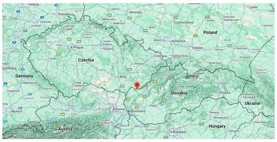
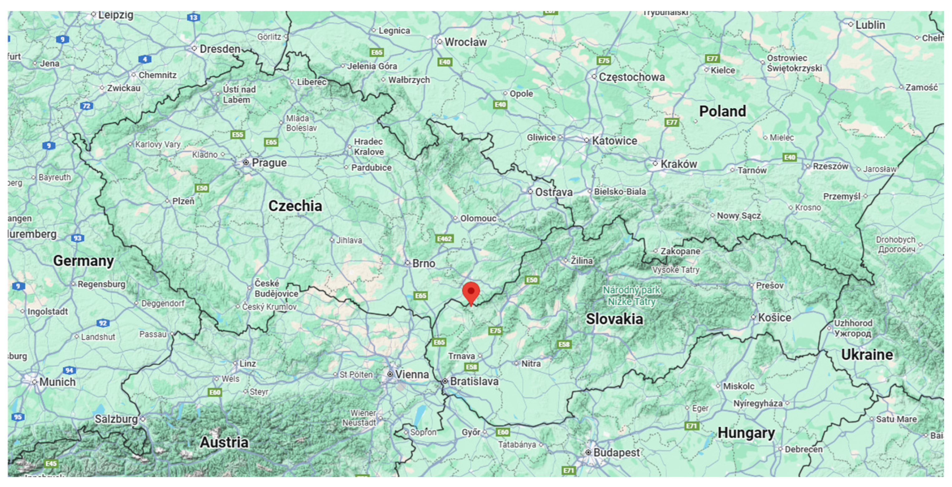
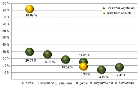
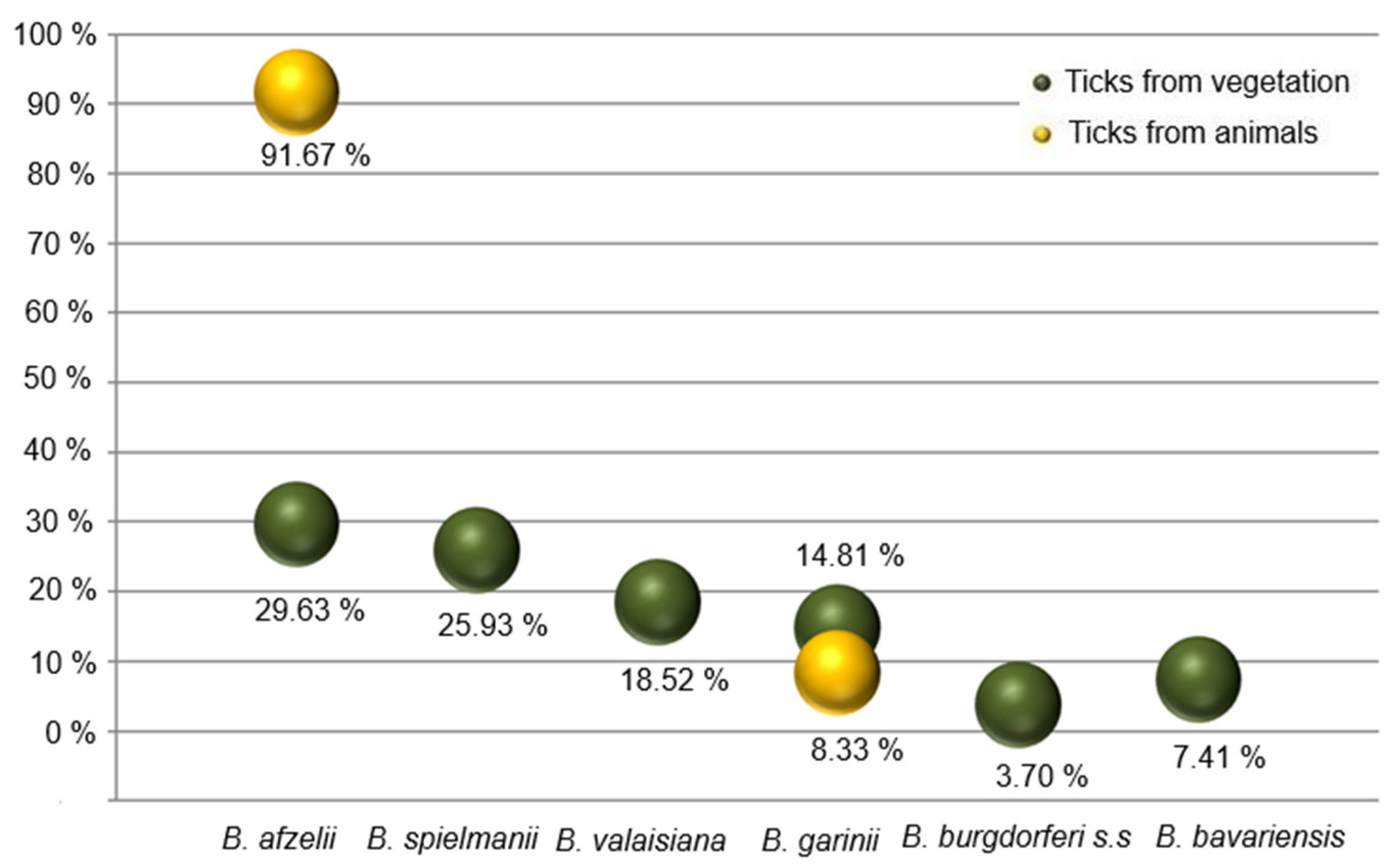
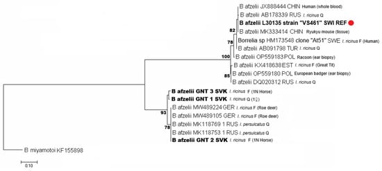
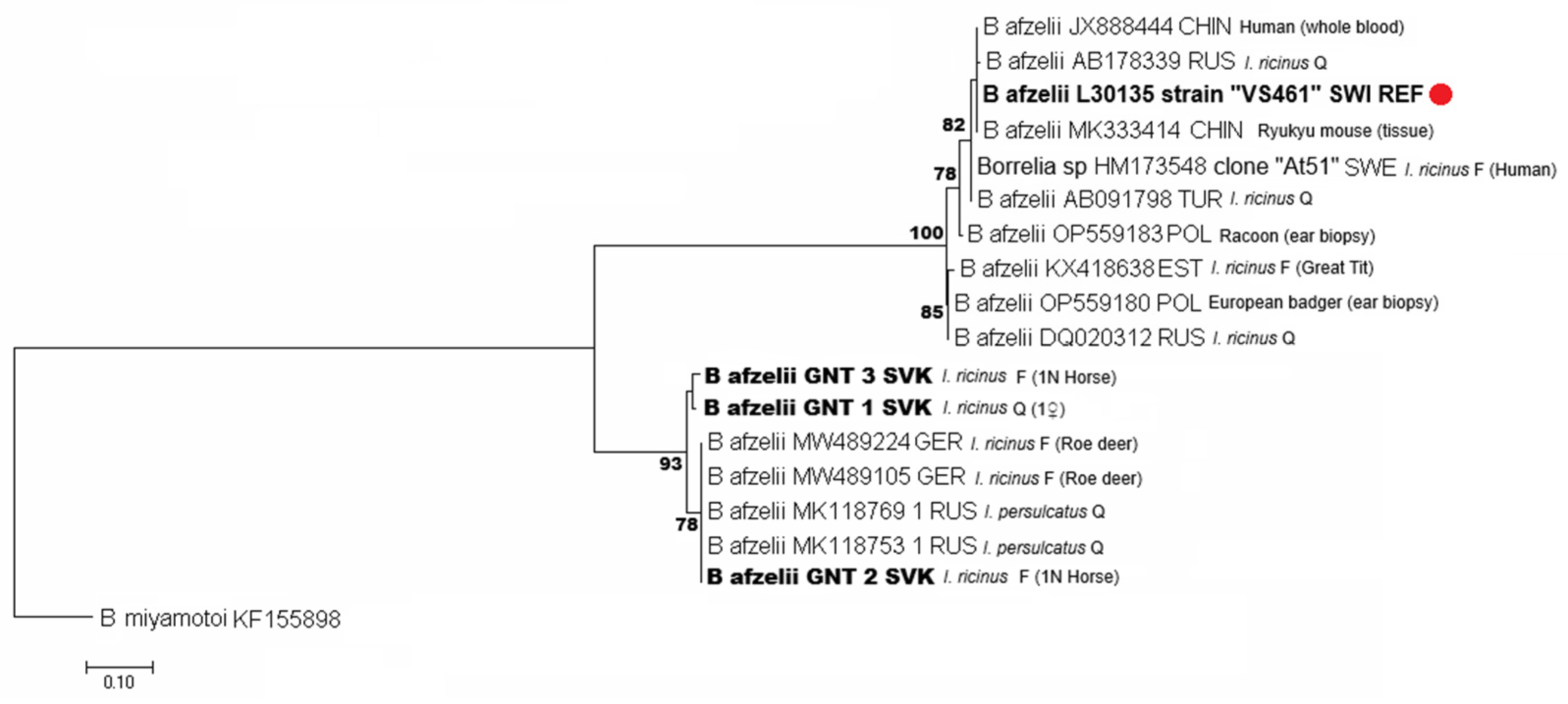
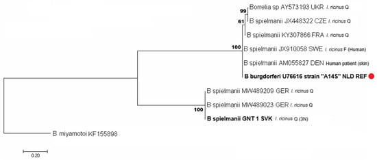
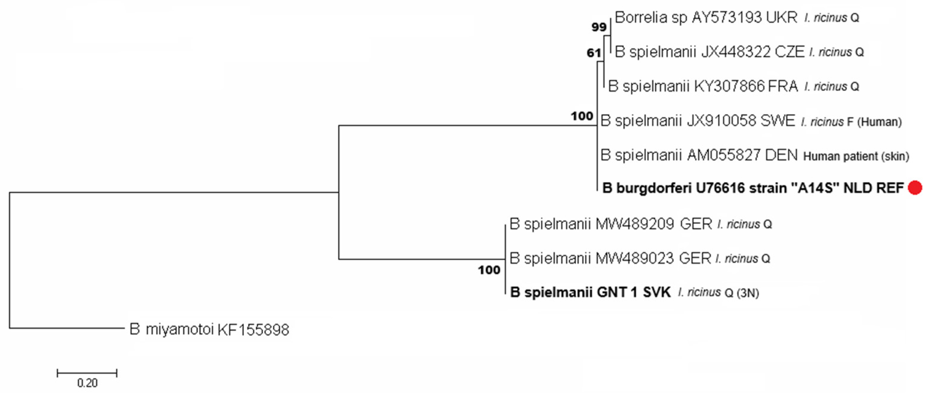
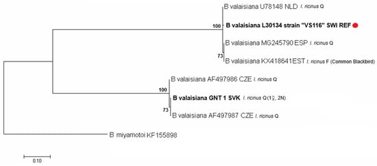
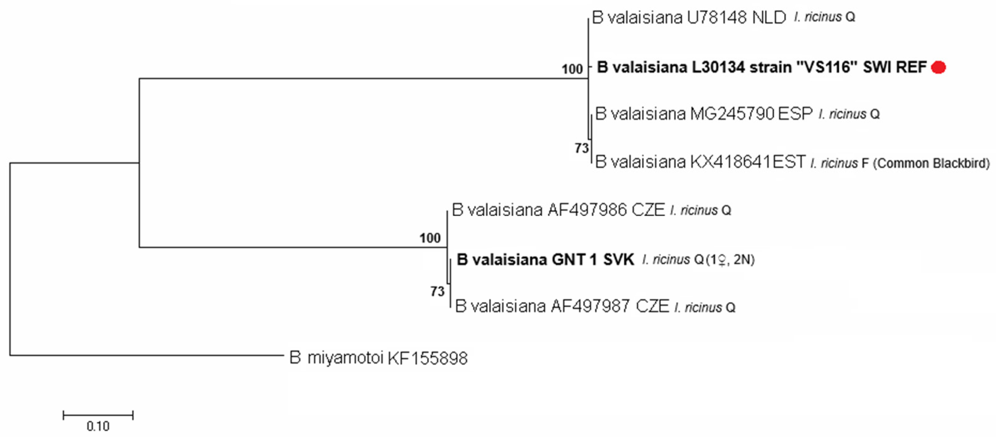
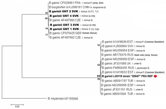
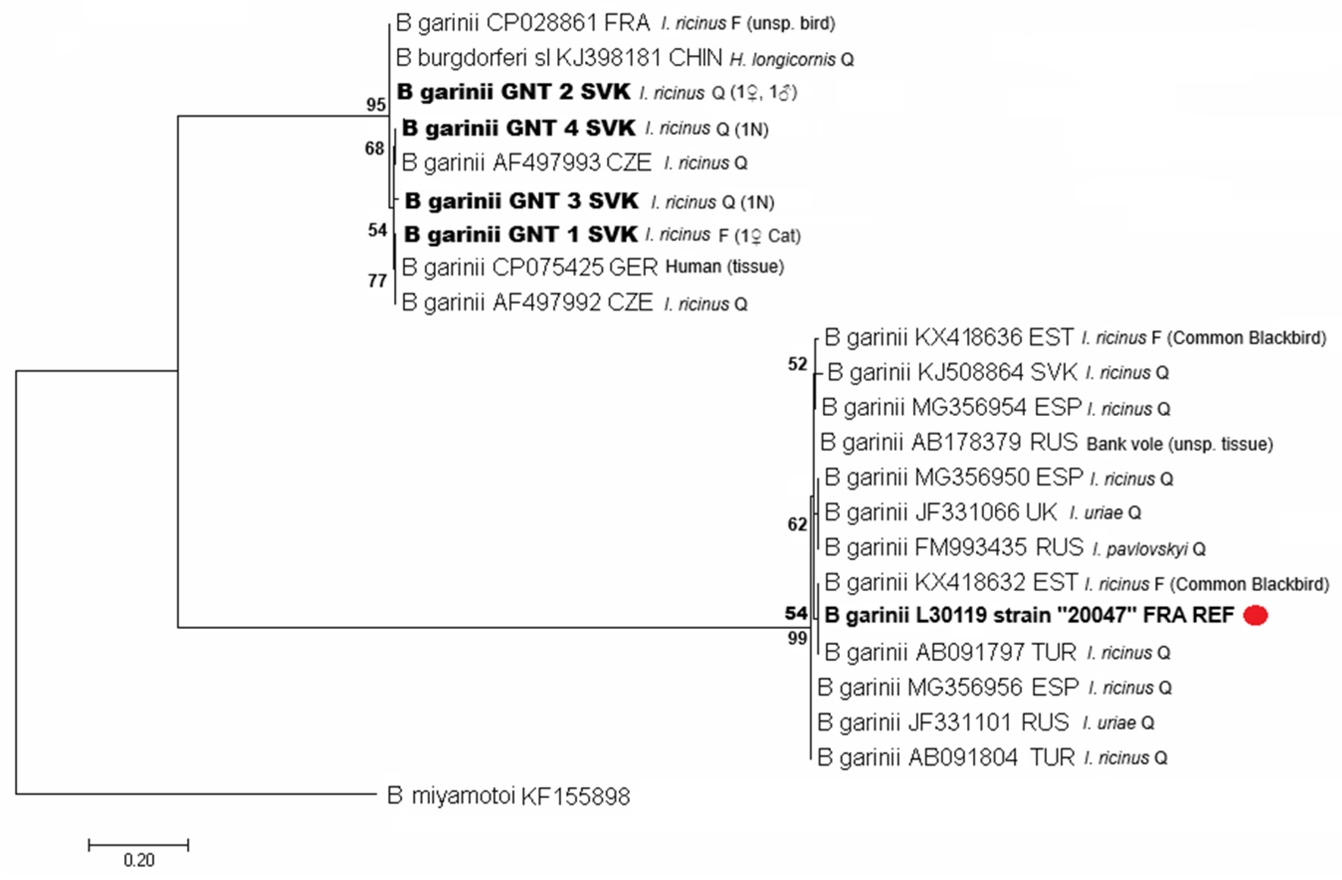
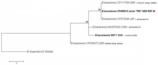
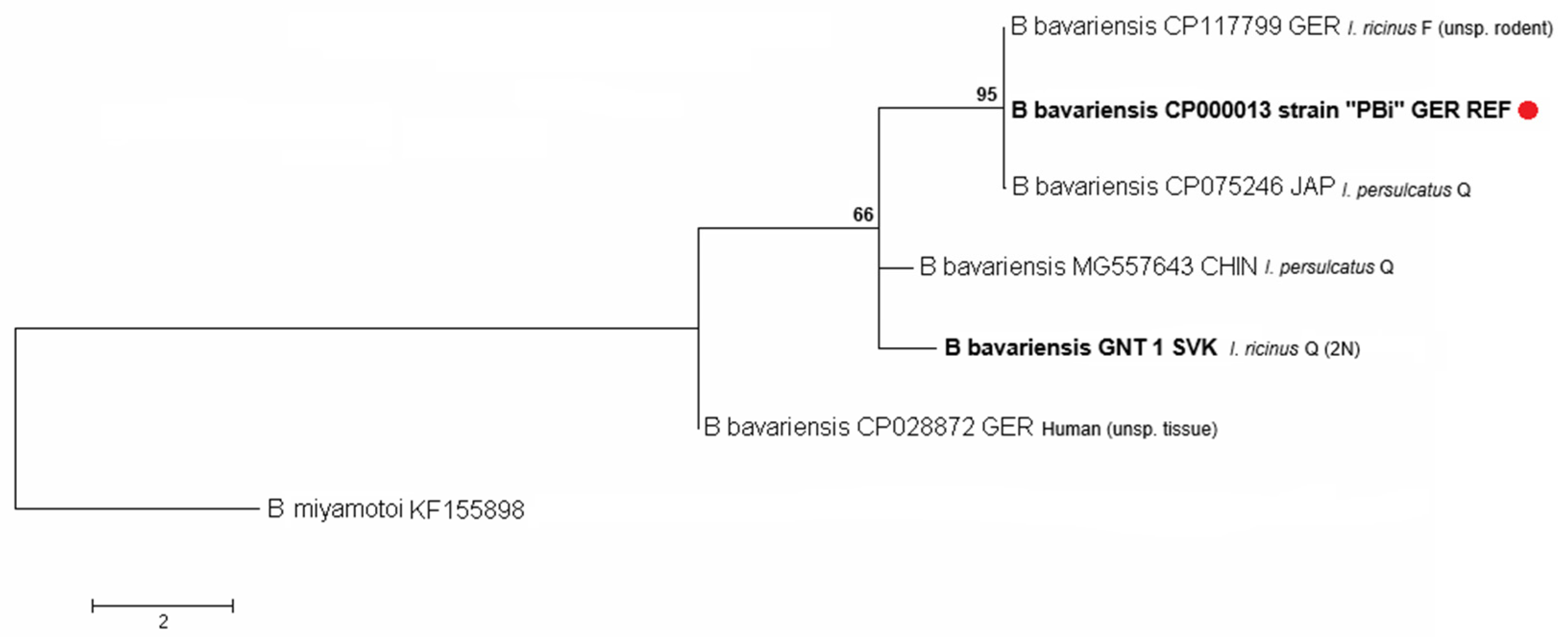
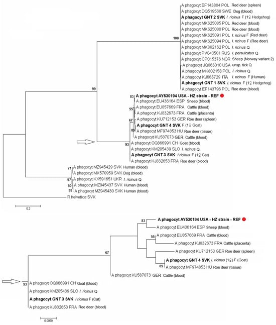
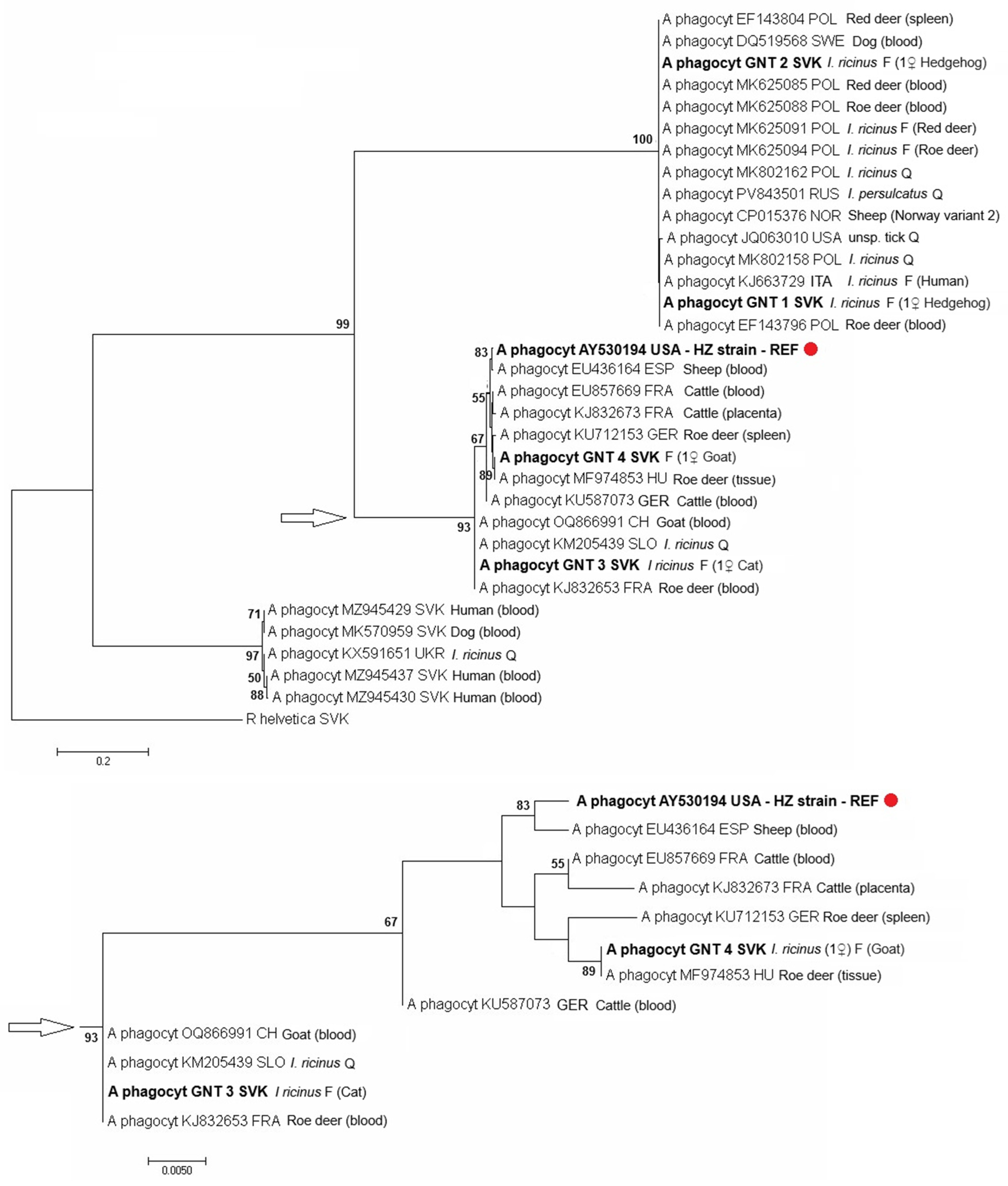
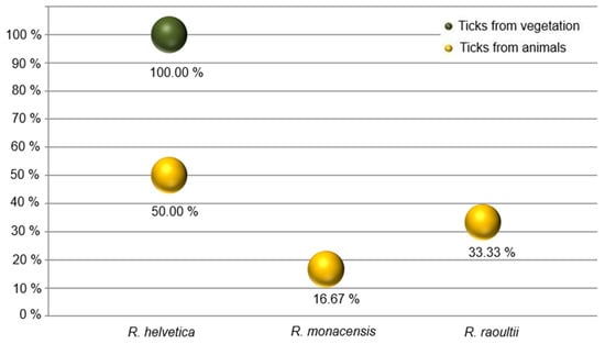
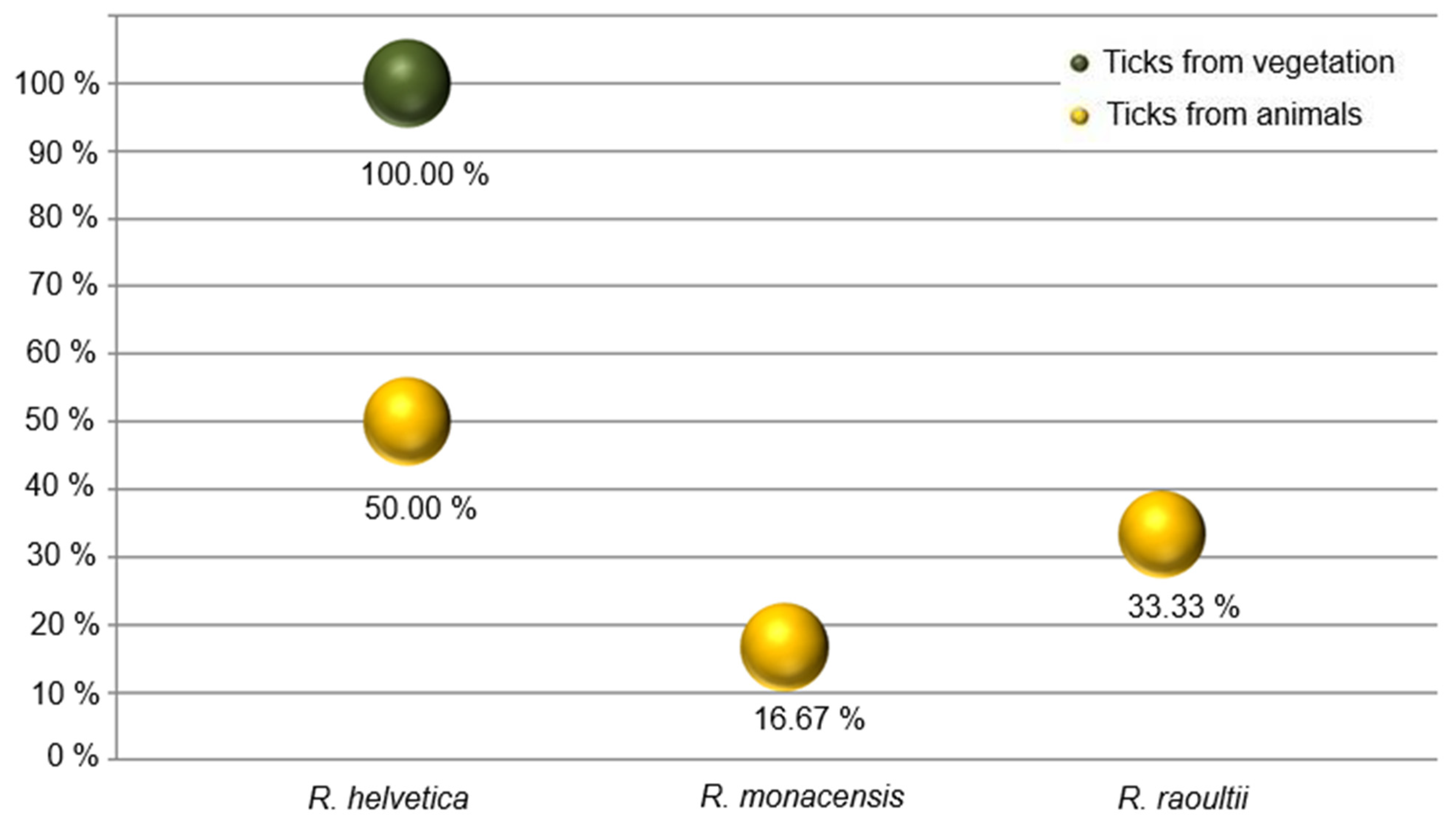
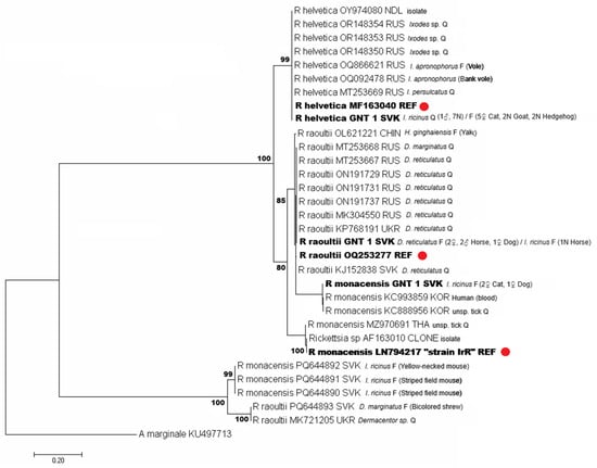
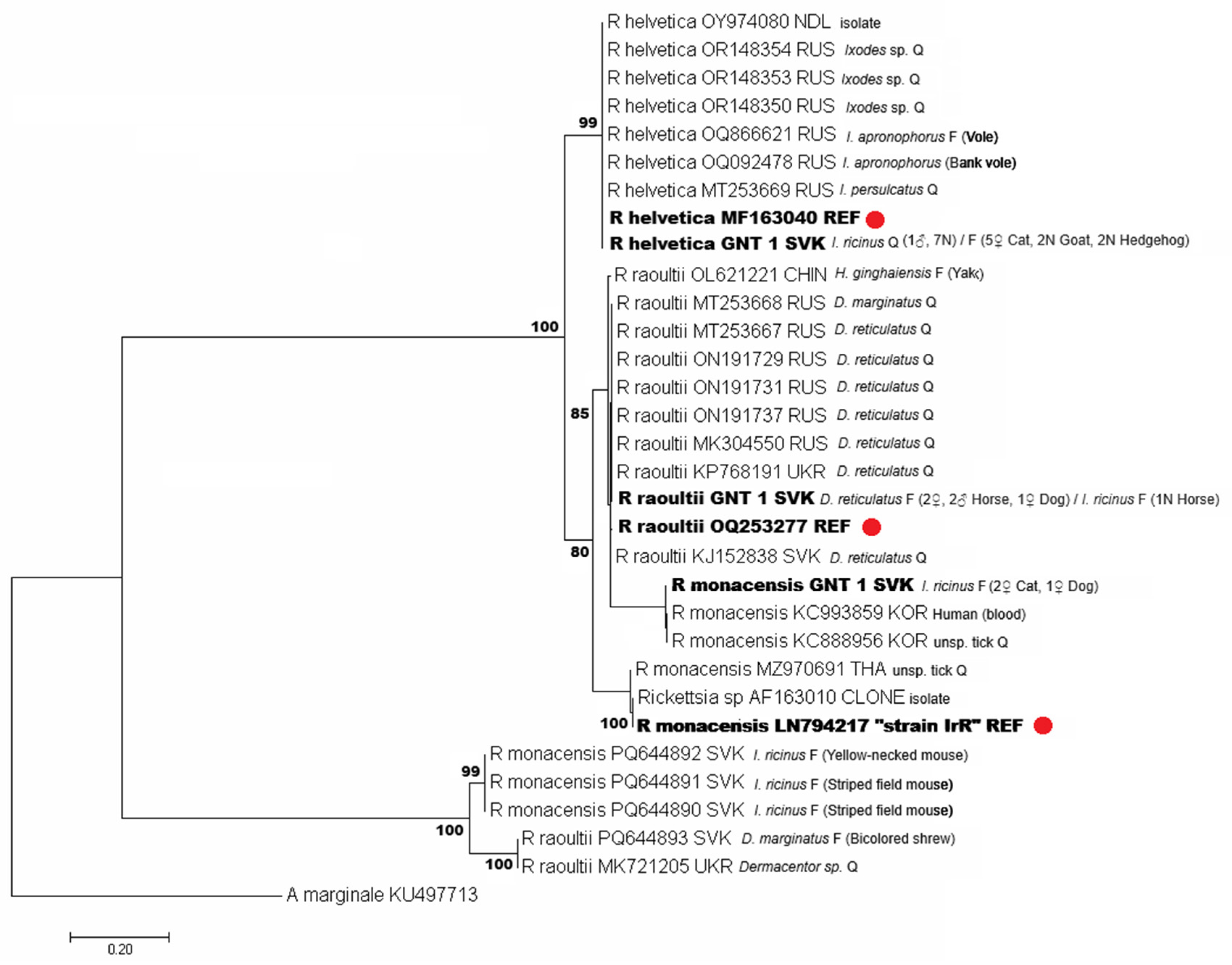
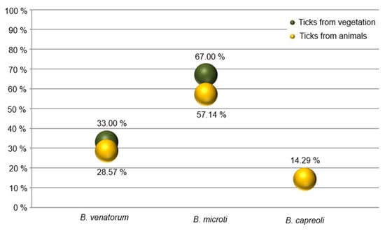
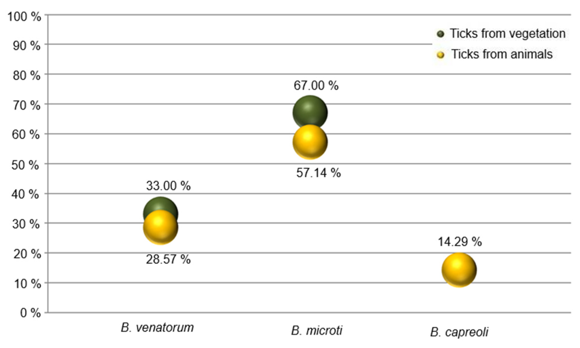
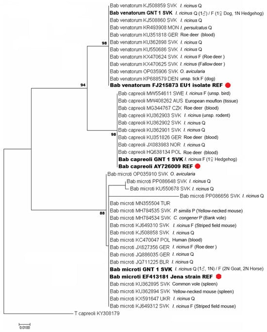
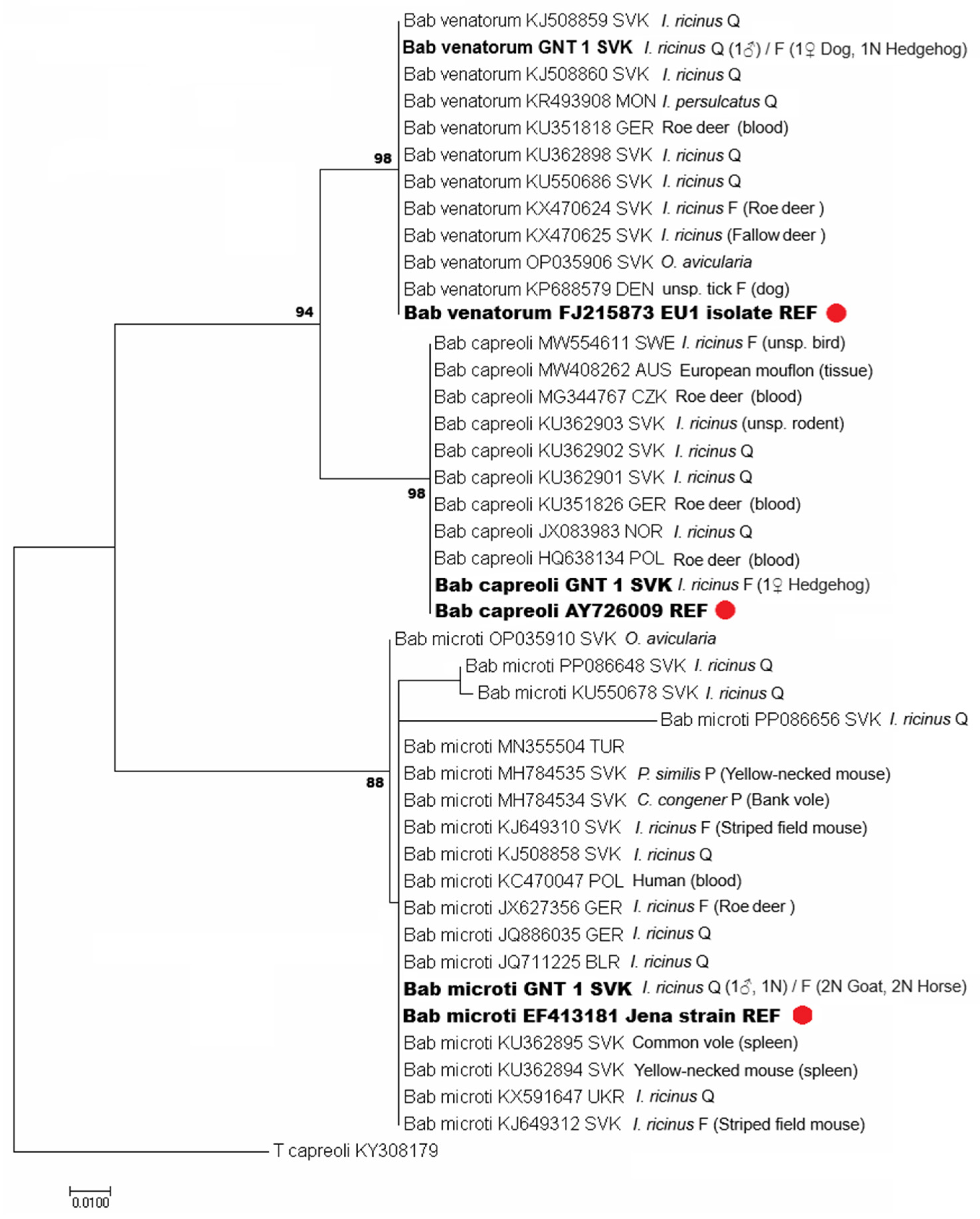
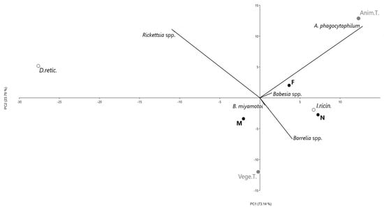
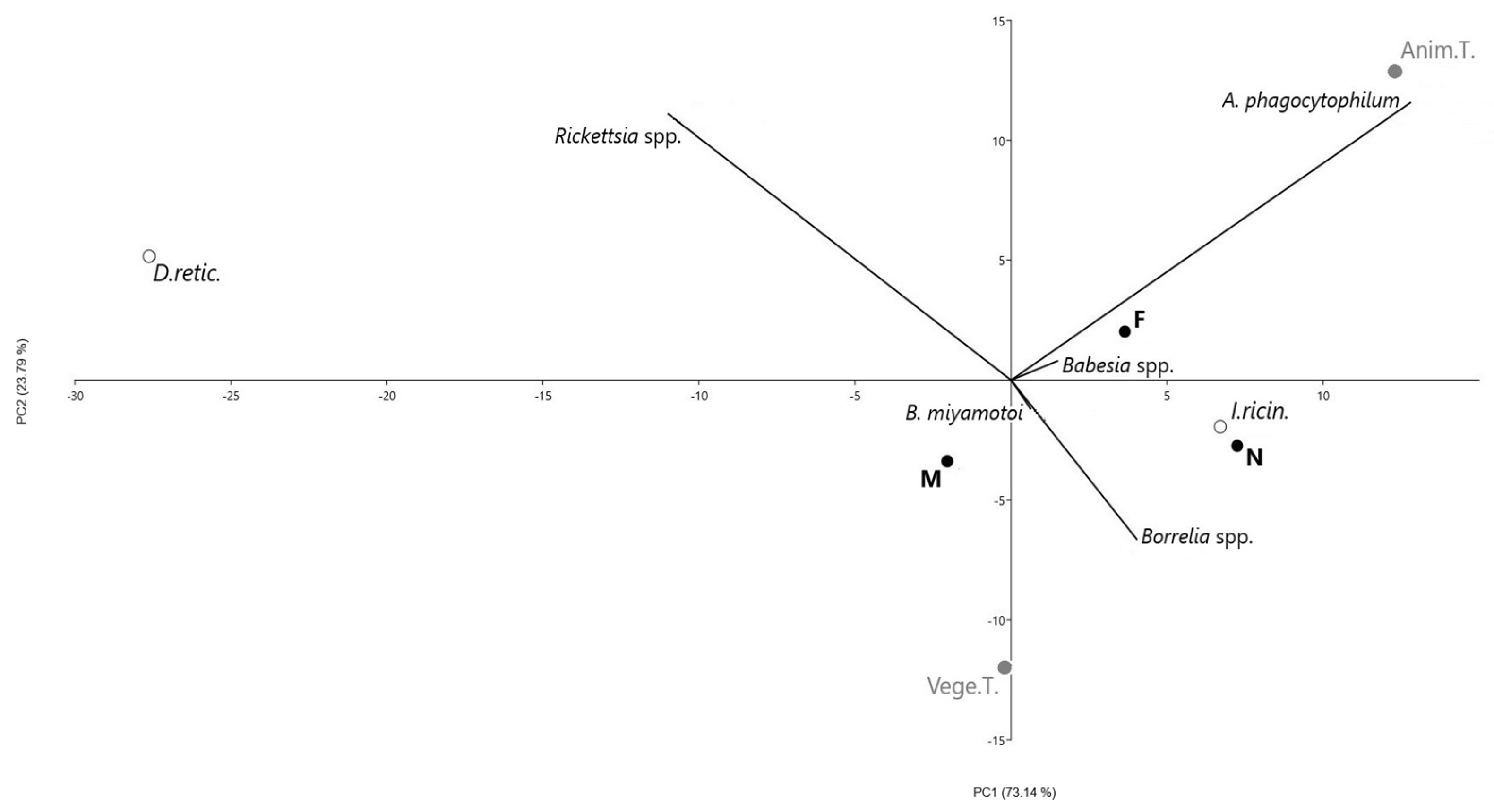

Disclaimer/Publisher’s Note: The statements, opinions and data contained in all publications are solely those of the individual author(s) and contributor(s) and not of MDPI and/or the editor(s). MDPI and/or the editor(s) disclaim responsibility for any injury to people or property resulting from any ideas, methods, instructions or products referred to in the content. |
© 2026 by the authors. Licensee MDPI, Basel, Switzerland. This article is an open access article distributed under the terms and conditions of the Creative Commons Attribution (CC BY) license.