Review

Applications of Organoids and Spheroids in Anaplastic and Papillary Thyroid Cancer Research: A Comprehensive Review

Department of Medical Oncology Laboratory, All India Institute of Medical Sciences (AIIMS), New Delhi 110029, India
*
Author to whom correspondence should be addressed.
These authors contributed equally to this work.
Organoids 2025, 4(3), 18; https://doi.org/10.3390/organoids4030018
Submission received: 15 April 2025 / Revised: 28 May 2025 / Accepted: 29 July 2025 / Published: 1 August 2025

Abstract

Organoid and spheroid technologies have rapidly become pivotal in thyroid cancer research, offering models that are more physiologically relevant than traditional two-dimensional culture. In the study of papillary and anaplastic thyroid carcinomas, two subtypes that differ both histologically and clinically, three-dimensional (3D) models offer unparalleled insights into tumor biology, therapeutic vulnerabilities, and resistance mechanisms. These models maintain essential tumor characteristics such as cellular diversity, spatial structure, and interactions with the microenvironment, making them extremely valuable for disease modeling and drug testing. This review emphasizes recent progress in the development and use of thyroid cancer organoids and spheroids, focusing on their role in replicating disease features, evaluating targeted therapies, and investigating epithelial–mesenchymal transition (EMT), cancer stem cell behavior, and treatment resistance. Patient-derived organoids have shown potential in capturing individualized drug responses, supporting precision oncology strategies for both differentiated and aggressive subtypes. Additionally, new platforms, such as thyroid organoid-on-a-chip systems, provide dynamic, high-fidelity models for functional studies and assessments of endocrine disruption. Despite ongoing challenges, such as standardization, limited inclusion of immune and stromal components, and culture reproducibility, advancements in microfluidics, biomaterials, and machine learning have enhanced the clinical and translational potential of these systems. Organoids and spheroids are expected to become essential in the future of thyroid cancer research, particularly in bridging the gap between laboratory discoveries and patient-focused therapies.

1. Introduction

Thyroid cancer stands as the most common endocrine malignancy, with its occurrence rising steadily across the globe in recent decades [1,2]. Differentiated thyroid carcinomas (DTCs), which include papillary thyroid carcinoma (PTC) and follicular thyroid carcinoma (FTC), make up about 90–95% of all cases and typically have positive outcomes when treated with surgical removal followed by radioiodine (RAI) ablation [3]. Nonetheless, despite initial treatment success, around 10–15% of PTC patients develop radioiodine–refractory PTC (RR-PTC), which is linked to decreased survival rates and limited treatment options [4]. Poorly differentiated thyroid carcinoma (PDTC) and anaplastic thyroid carcinoma (ATC), though less frequent, are highly aggressive forms characterized by swift disease progression, resistance to conventional treatments, and high mortality rates [5,6].
Traditional treatment methods for aggressive thyroid cancers, such as kinase inhibitors, radiation, and chemotherapy, have shown only limited effectiveness, partly due to tumor heterogeneity and mechanisms of therapy resistance [7]. A crucial factor in the success of RAI therapy is the expression and membrane localization of the sodium–iodide symporter (NIS); however, in RR-PTC and ATC, NIS expression is often absent or improperly localized [8]. Molecular changes like BRAFV600E mutations [9,10], RET/PTC rearrangements [11], and activation of the PI3K–AKT–mTOR pathway [12,13] are frequently involved in tumor dedifferentiation, treatment resistance, and the suppression of iodine uptake mechanisms [14,15,16]. Although targeted therapies such as BRAF and MEK inhibitors have shown potential, their effectiveness is often short-lived due to adaptive resistance and the lack of reliable preclinical models that can replicate in vivo tumor behavior [17]. These clinical and molecular challenges highlight the urgent need for improved model systems that accurately represent the pathophysiology and treatment landscape of thyroid cancer.
Despite their widespread use and accessibility, traditional two-dimensional (2D) cell culture models lack the architectural complexity, spatial heterogeneity, and dynamic cell–matrix interactions characteristic of solid tumors [18,19]. Furthermore, many therapeutic agents that demonstrate efficacy in 2D culture fail to achieve clinical success, underscoring the limitations of such models in replicating drug resistance, metastatic potential, and tumor microenvironmental cues. To address these limitations, three-dimensional (3D) culture systems—particularly organoids and spheroids—have emerged as advanced platforms that more accurately capture the in vivo biology of tumors. Organoids are self-organizing, multicellular structures derived from stem cells or patient tissues that can replicate the histological and functional features of the organ or tumor of origin [20,21]. Spheroids, although structurally simpler, are formed by forced aggregation of tumor cells and reproduce essential gradients of oxygen, nutrients, and proliferation observed in avascular tumor regions. Both systems offer significant advantages over 2D culture, including enhanced mimicry of tumor architecture, maintenance of genomic and phenotypic heterogeneity, and suitability for long-term culture and drug testing [22].
While organoids and spheroids have been extensively studied in colorectal, breast, liver cancer, etc., their application in thyroid cancer is relatively recent but rapidly expanding. Emerging studies have demonstrated the feasibility of generating PTC and ATC organoids from patient-derived tissues and cell lines, enabling functional assays for drug response, signaling pathway interrogation, and modeling of tumor progression [23]. In particular, 3D models are increasingly being utilized to evaluate drug resistance patterns, simulate epithelial–mesenchymal transition (EMT), explore cancer stemness, and characterize the tumor microenvironment in a controlled and reproducible manner [24,25].
In this review, we explore recent advances in organoid and spheroid technologies and their emerging role in thyroid cancer research. By highlighting key applications in disease modeling, therapeutic testing, and personalized medicine—particularly in papillary and anaplastic thyroid cancers—we aim to underscore the potential of 3D models to bridge the gap between laboratory research and clinical translation.

2. Limitations of Conventional Thyroid Cancer Models

2.1. 2D Cell Culture

For a long time, 2D culture systems have been essential in thyroid cancer research because they are cost-effective, easy to manipulate, and well-suited for high-throughput applications. These models have played a crucial role in advancing the understanding of signaling pathways and drug screening. Nonetheless, their lack of three-dimensional structure and microenvironmental context limits their adoption in clinical trials. Cells cultured in 2D monolayers show changes in morphology, polarity, gene expression, and drug responsiveness, which often result in discrepancies between preclinical and clinical outcomes.
Since the late 1950s, thyrocytes have been cultivated in 2D monolayer systems [26]. However, a key drawback of this method is that thyrocytes fail to organize into their natural follicular structures when grown on standard adherent plates [27]. Instead, they form a continuous epithelial sheet, where the apical side faces the culture medium, and the basal side is in contact with the dish surface. When cultured in suspension within non-adherent vessels, thyrocytes naturally assemble into follicular structures. However, their orientation results in an “inside-out” follicle, where the apical surface with microvilli is exposed to the surrounding culture medium [27]. To correct this polarity, a collagen gel-based culture system was introduced in the 1970s. In this system, embedding these follicles into a 3D matrix resembling the thyroid extracellular matrix (ECM) allows for their orientation to be reversed. This adjustment positions the microvilli inward toward the follicular lumen, restoring the structure of a functional thyroid follicle. Thus, 3D culture provides a more functionally appropriate model for studying thyroid function by closely mimicking the natural microenvironment. Additionally, they allow researchers to study thyrocyte responses to thyroid-stimulating hormone (TSH) and iodine in a more accurate and functional setting [28]. These advantages make 3D culture systems an essential tool for exploring thyroid function and disease mechanisms.

2.2. Animal Models: Challenges in Translation

Animal models, including genetically engineered mouse models (GEMMs) and patient-derived xenografts (PDXs), have played a crucial role in enhancing our understanding of the pathophysiology and treatment response of thyroid cancer. These models provide a more biologically relevant environment than 2D culture, allowing for in vivo drug testing and metastasis studies. Nonetheless, species-specific differences in tumor biology, immune interactions, and metabolism often impede the translation of animal study findings to humans. Additionally, the high cost, ethical concerns, and time-consuming nature of animal experiments restrict their extensive use in preclinical research. Mouse models have contributed greatly to thyroid cancer research by allowing for precise genetic manipulation to explore oncogenic drivers and molecular changes. They provide a clinically pertinent platform to examine tumor–immune interactions, disease progression, and metastatic potential over time. Furthermore, these models are essential for preclinical assessment of new therapeutic agents [29].
Differentiated thyroid cancers, including papillary and follicular types, comprise the majority of thyroid cancer cases. The combination of foundational research and clinical knowledge has led to the creation of genetically modified mouse models that mimic significant mutations found in human differentiated thyroid cancers, such as RET/PTC rearrangements and BRAFV600E mutations [30,31]. These models have played a crucial role in clarifying the functions of the MAPK, PI3K–AKT, and cAMP/PKA signaling pathways in the development of thyroid tumors. For example, mice with an overactive PI3K–AKT pathway have been observed to develop follicular thyroid cancer, which parallels the progression of the disease in humans. New inducible systems are being developed to represent sporadic, non-hereditary thyroid cancers more accurately, thereby increasing the relevance of preclinical research [32,33]. Moreover, progress in in vivo imaging has enhanced the ability to track tumor growth and spread, offering essential tools for preclinical assessment of therapeutic agents [34].
Despite these advantages, translational challenges persist due to interspecies differences in immune function, metabolism, and genetic complexity. The biological differences between mice and humans can impede the direct application of research findings. Moreover, the creation and maintenance of genetically modified mouse models require significant resources. Some models may not accurately represent the genetic diversity of human tumors, show unnaturally fast tumor growth, or fail to effectively simulate metastasis. Additionally, variations in immune system function between species can restrict the precision of immune-oncology research [35]. Given these constraints, there is a pressing demand for more sophisticated preclinical models that can better mimic the in vivo tumor environment.

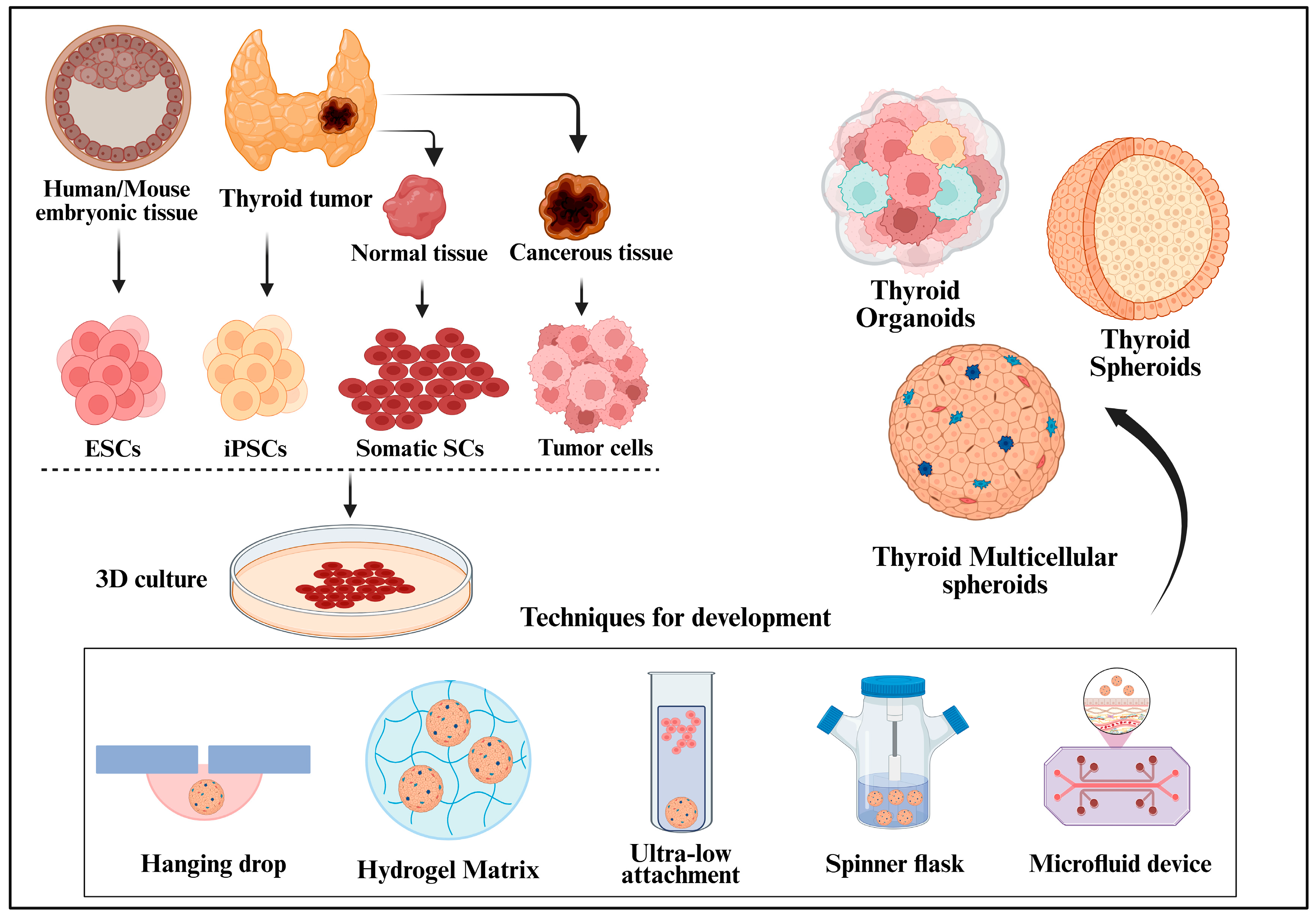
3. Organoids and Spheroids: An Emerging Paradigm in Thyroid Cancer Research

The shortcomings of traditional 2D cell culture systems and in vivo models have spurred the advancement of 3D cell culture platforms, particularly organoids and spheroids, as more biologically accurate models for cancer research. These platforms replicate the structure, diversity, and microenvironment of tumors more accurately, thereby improving the translational relevance of preclinical studies.

3.1. Organoids: Advancing Complexity and Relevance

Organoids are a sophisticated three-dimensional culture system that exhibits self-organization, multicellularity, and functional similarity to natural tissues. Originating from stem cells, adult, embryonic, or induced pluripotent organoids can replicate the histological and functional characteristics of their source organs [36]. Organoids are characterized by their ability to encompass various differentiated cell types, sustain prolonged growth, and react to physiological signals, making them excellent tools for studying organ development, disease progression, and therapeutic effects. In thyroid cancer, organoids derived from tumors preserve the genetic, epigenetic, and phenotypic diversity of the original tumor, rendering them especially useful for personalized treatment approaches. Unlike spheroids, which are created through the forced aggregation of cells, organoids naturally form from patient-derived cells following inherent developmental pathways. This ability enables them to accurately replicate tumor structure and disease-specific characteristics in vitro [37]. Furthermore, organoids are cost-efficient and ethically sound substitutes for patient-derived xenografts (PDX), offering enhanced scalability and consistency in experiments [38]. Their swift adoption in cancer research is attributed to these benefits, which include efforts to simulate treatment resistance, pinpoint actionable mutations, and evaluate new therapeutic combinations.

3.2. Spheroids: Structure, Development, and Utility

Spheroids are multicellular aggregates, typically composed of a single-cell type that self-assembles under non-adherent conditions. This model represents one of the earliest 3D culture methods, dating back to 1956, when Ehrmann and Gey observed cellular aggregates forming on rat collagen matrices [39]. In the decades that followed, especially between the 1970s and the 1980s, researchers began exploring the influence of the tumor microenvironment (TME) in 3D contexts [40].
Spheroid formation is largely driven by cell–cell and cell–matrix interactions with adhesion molecules such as integrins, cadherins, and extracellular matrix (ECM) proteins, which play critical roles in mediating compaction and stabilization. The architecture of spheroids closely recapitulates the in vivo tumor microenvironment, exhibiting three distinct zones: an outer layer of proliferating cells, a middle layer of quiescent cells, and a necrotic core resulting from limited oxygen and nutrient diffusion, mirroring the hypoxic gradient observed in solid tumors [41]. Multiple methodologies have been developed to generate spheroids and organoids (Figure 1), including the hanging drop method, low-adhesion plates, spinner flasks, and bioreactor-based suspension culture [42]. These methods facilitate the formation of spheroids of varying sizes and densities, making them suitable for use in drug testing, cancer biology research, and modeling radiotherapy resistance. The 3D culture models surpass traditional 2D culture and animal models by mimicking tumor cellular diversity and spatial arrangement. These models enable patient-specific therapeutic evaluations and bridge the gap between in vitro and in vivo research, advancing thyroid cancer studies and clinical outcomes.

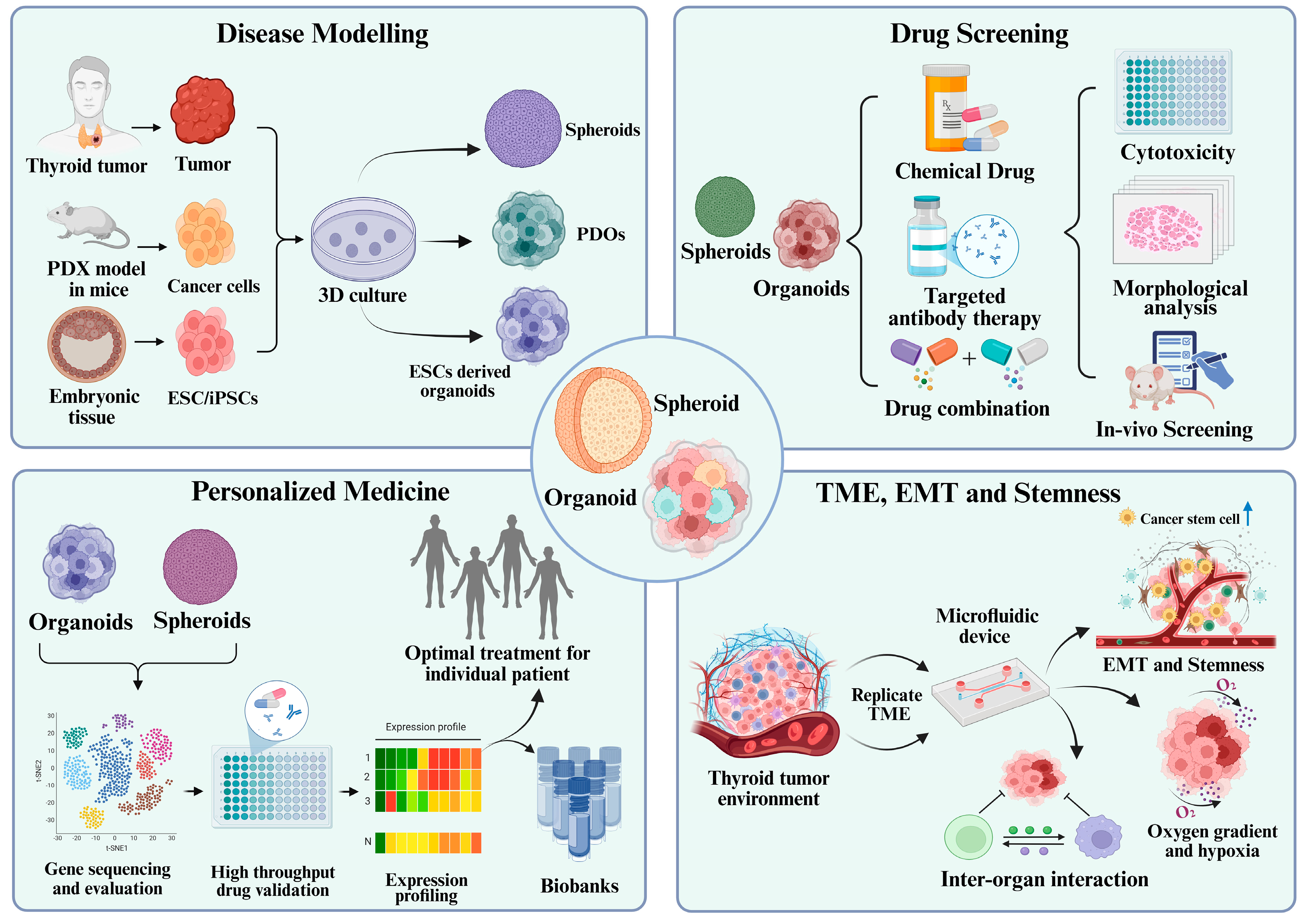
4. Applications of Organoids and Spheroids in Thyroid Cancer Research

Organoids and spheroids have quickly become sophisticated tools for studying thyroid cancer biology, offering greater physiological relevance, predictive accuracy, and translational potential than traditional 2D culture systems. These models replicate the three-dimensional structure, cellular diversity, and tumor microenvironment, enabling extensive applications in disease modeling, drug screening, personalized treatment, and microenvironmental research. Table 1 provides a comparative summary of key studies utilizing organoid and spheroid models in thyroid cancer, highlighting their applications, experimental context, and translational maturity to assist readers in evaluating their relevance and impact. Figure 1 and Figure 2 provide a visual overview of the sources, development techniques, and key translational applications of organoid and spheroid models, contextualized for their relevance in thyroid cancer research.
4a. Study selection criteria:
To compile the studies summarized in Table 1, a focused literature search was conducted using databases including PubMed, Scopus, and Google Scholar through March 2025. The search employed combinations of the terms: “thyroid cancer” AND (“organoids” OR “spheroids” OR “3D culture”) AND (“papillary” OR “anaplastic” OR “differentiated”). Eligible studies included original research articles that utilized three-dimensional (3D) culture models—specifically spheroids or organoids—derived from thyroid cancer cell lines, patient tissues, or pluripotent stem cells. This approach aimed to provide a representative and balanced overview of the current applications of 3D models in thyroid cancer research.

4.1. Disease Modelling: Mimicking Tumor Architecture and Pathophysiology

In thyroid cancer, modeling approaches differ between well-differentiated types, such as papillary thyroid carcinoma (PTC), and undifferentiated, aggressive subtypes such as anaplastic thyroid carcinoma (ATC). Patient-derived organoids and spheroids enable subtype-specific modeling by preserving histological and molecular traits, including mutational landscapes, hormone responsiveness, and microenvironmental features, making them essential tools for capturing disease heterogeneity and therapy response. Organoids from patient PTC tissues accurately mimic primary tumors’ histological and molecular features. For instance, Chen et al. demonstrated that PTC organoids retained the original tumor’s genomic and pathological features, and notably, responded to estradiol in an ERα-dependent manner. This highlights the relevance of hormone receptor signaling in certain PTC subtypes and supports the use of organoid models in exploring sex hormone influences and potential therapeutic targets in hormone-responsive thyroid tumors [49]. Similarly, Romitti et al. and Ogundipe et al. created functional thyroid organoids using embryonic stem cells and adult human tissue, respectively [48,50]. These organoids exhibited thyroid-specific markers, such as PAX8 and NKX2.1, and were able to restore hormone production in mice with hypothyroidism, highlighting their potential for regeneration and disease modeling.
Spheroid systems play a crucial role in replicating the structural intricacies of thyroid tumors. A recent study highlighted that layered ATC and PTC spheroids developed a hypoxic core, central necrosis, and reduced proliferation, characteristics that are not found in 2D culture [56]. Additionally, Ghiandai et al. revealed that 3D ATC spheroids showed varied cleavage patterns of EpCAM, indicating its potential as a biomarker for thyroid cancer dedifferentiation [51]. Kopp et al. adopted a novel method of creating multicellular spheroids in simulated microgravity, emphasizing the role of biophysical stimuli on thyroid cancer growth and angiogenesis [63]. Collectively, these models have enhanced the biological significance of in vitro thyroid cancer systems and paved the way for understanding the dynamic progression of the disease across various subtypes, including well-differentiated and anaplastic thyroid cancers.

4.2. Drug Screening and Therapeutic Evaluation

Organoid and spheroid models in thyroid cancer research enable better prediction of therapeutic outcomes and drug-resistance patterns. Unlike 2D cultures that overestimate drug effectiveness, 3D models provide a more reliable platform for preclinical drug screening by replicating tumor environments. In drug screening studies, 3D models of both ATC and PTC have been employed to evaluate the efficacy of chemotherapeutics, targeted therapies, and pathway inhibitors. Due to differences in mutation burden, differentiation status, and treatment resistance, findings from ATC and PTC models are interpreted separately, highlighting subtype-specific therapeutic vulnerabilities.
Numerous studies have demonstrated the improved predictive accuracy of spheroid models for evaluating anti-cancer agents. Bergdorf et al. performed a high-throughput screening of 1525 compounds on anaplastic thyroid cancer (ATC) cell lines using both 2D and 3D formats. Among these, 33 compounds remained effective in spheroids, and bortezomib, cabazitaxel, and YM155 were identified as strong candidates for ATC treatment. These results highlight the limitations of traditional models in identifying clinically viable compounds and emphasize the importance of 3D validation in drug development [54].
Similarly, Diaz et al. combined ATC spheroid culture with patient-derived organoids to assess the effectiveness of pyrvinium, a CK1α agonist that targets the Wnt pathway. Pyrvinium demonstrated superior performance compared to the standard treatment (dabrafenib + trametinib), even in BRAF-wild-type organoids, indicating its potential as a therapeutic option across various mutational profiles [44]. Patient-derived organoids have also been instrumental in the evaluation of targeted and combination therapies. For example, Chen et al. found that BRAFV600E-mutant PTC organoids showed a limited response to BRAF inhibitors alone, but when combined with MEK, RTK, or chemotherapeutic agents, a marked increase in cytotoxicity was observed [47]. Additionally, it has been reported that ATC spheroids derived from both cell lines and primary tumors exhibited varied responses to BRAF/MEK inhibition depending on their mutational status, effectively reflecting clinical treatment resistance [52].
Several pioneering studies have used 3D environments to investigate new treatment methods. Sekhar et al. demonstrated that RSL3, a GPX4 inhibitor, triggers ferroptosis in thyroid cancer spheroids, an effect that could be reversed by ferroptosis inhibitors [55]. Similarly, Samimi et al. evaluated the efficacy of BI-847325, a dual MEK and Aurora kinase inhibitor, using alginate-encapsulated ATC spheroids. This compound targets both the MAPK pathway and mitotic regulation, offering a potential strategy to overcome resistance in aggressive thyroid cancers. Notably, the 3D spheroids exhibited greater resistance to BI-847325 compared to 2D cultures, underscoring the importance of 3D models in uncovering drug resistance dynamics [58]. In addition to cytotoxic profiling, 3D systems enable the observation of therapy-induced phenotypic changes. For instance, it has been reported that dabrafenib alters actin cytoskeleton organization and migratory behavior in a genotype-specific manner in ATC and PTC spheroids, further supporting the functional significance of these models [59]. Together, these studies emphasize the importance of 3D organoid and spheroid platforms as vital tools in translational pharmacology for thyroid cancer, as they effectively capture complex drug–tumor interactions and aid in the development of more effective and personalized therapies.

4.3. Personalized Medicine and Patient-Specific Therapeutic Responses

The advent of organoids and spheroids as instruments for personalized medicine has transformed thyroid cancer research by providing models that maintain the unique traits of tumors in patients. These three-dimensional culture systems preserve the molecular, histopathological, and phenotypic diversity of the original tissue, allowing for ex vivo drug response testing that is directly applicable to clinical outcomes. Patient-derived organoids (PDOs) from papillary thyroid carcinoma (PTC) are particularly beneficial in this regard. Organoids from various thyroid patients revealed that their response to estradiol was specifically dependent on ERα expression, underscoring how receptor status can affect hormonal sensitivity in PTC [49]. Similarly, Sondorp et al. created organoids from both PTC and radioactive iodine-refractory differentiated (RAIRD) thyroid tumors, demonstrating varied expression of the sodium iodide symporter (NIS) and thyroid differentiation markers such as paired box 8 (PAX8) and thyroid-stimulating hormone receptor (TSHr). These results provide a promising framework for evaluating iodine avidity and predicting responses to radioiodine therapy, particularly in treatment-resistant cases [45].
Patient-derived models also support the evaluation of targeted therapies tailored to specific genetic mutations. For example, it has been documented that BRAFV600E-mutant and wild-type PTC organoids can be employed to evaluate various drug combinations, demonstrating that single-drug therapies frequently proved ineffective, whereas dual-targeting strategies exhibited patient-specific effectiveness. These organoid-based systems could eventually support real-time personalized treatment plans and customized regimens according to the tumor’s genetic and phenotypic characteristics [47]. In anaplastic thyroid carcinoma (ATC), the application of patient-derived spheroids and organoids for personalized drug response profiling is equally compelling. Scientists have created an ex vivo ATC spheroid model that retains the patient’s tumor features, including gene expression profiles and EMT markers. Their research showed differing sensitivity to dabrafenib based on BRAF mutation status, with 3D models revealing resistance patterns not detectable in 2D culture. This underscores the importance of advanced models for capturing clinical diversity [52]. A groundbreaking phase II clinical trial (NCT06482086) has recently highlighted the practical use of PDOs in directing neoadjuvant therapy for patients with locally advanced thyroid cancer (LATC). This single-arm study, which included 75 participants, utilized PDOs derived from tumor biopsies to conduct ex vivo drug sensitivity assessments. The drugs demonstrating the highest relative efficacy were chosen for tailored treatment, leading to an overall objective response rate (ORR) of 32.7%. Notably, the results were particularly promising for anaplastic thyroid carcinoma (ORR 50%) and differentiated thyroid cancer (ORR 32.6%). Additionally, a resection rate of R0/R1 was achieved in 34.5% of the participants, highlighting the potential of PDO-based approaches to transform inoperable tumors into ones that can be surgically addressed. This trial marks a significant advancement towards incorporating organoid-guided precision therapy into clinical management strategies for aggressive thyroid cancers and underscores the necessity for future randomized trials to confirm these findings [43].
These applications are supported by advancements in genomic and transcriptomic characterization, which further confirms the accuracy of these patient-derived models. Organoids can be quickly expanded, cryopreserved, and subjected to multi-omics analysis, thus providing a sustainable and reproducible system for long-term studies. To sum up, patient-derived thyroid cancer organoids and spheroids function as effective personalized models, facilitating tailored therapy evaluation, biomarker identification, and immediate clinical decision-making, thereby enhancing their role in translational medicine.

4.4. Studying Tumor Microenvironment, EMT, and Stemness

Understanding the complex interactions within the tumor microenvironment (TME), such as epithelial–mesenchymal transition (EMT), cancer stem cells (CSC) activity, and extracellular matrix (ECM) remodeling, is crucial for understanding thyroid cancer progression and resistance to treatment. The 3D culture systems, especially spheroids and organoids, provide a more functionally appropriate context for examining these processes, which are challenging to replicate in two-dimensional monolayers or traditional animal models [65]. Spheroid culture has been particularly successful in mimicking EMT and invasive traits in aggressive thyroid cancer types. For instance, thyroid cancer spheroids exhibit a layered structure with central necrosis, HIF1-α expression, and altered levels of E-cadherin and other cell adhesion markers, all of which signal EMT and dedifferentiation [56]. These 3D spheroid systems effectively recapitulate hypoxic gradients, EMT marker shifts, and stemness features—hallmarks of dedifferentiated thyroid tumors that often confer poor prognosis and therapy resistance. Likewise, a recent study revealed that EpCAM cleavage in ATC-derived spheroids was associated with dedifferentiation and resistance to BRAF inhibitors, suggesting its role in maintaining tumor-initiating cell (TIC) properties [51].
Three-dimensional models are instrumental in capturing the concept of cancer stemness. Hardin et al. successfully isolated Aldefluor-positive CSC populations from different thyroid cancer cell lines, showing that these cells could form highly tumorigenic spheroids rich in stemness markers, such as SOX2 and OCT4. When treated with agents such as resveratrol and valproic acid, these cells exhibit reduced proliferation and invasiveness during differentiation, suggesting that spheroid models are effective for evaluating CSC-targeted therapies [61].
Organoid models have also enhanced our understanding of the genetic and epigenetic factors that regulate thyroid cancer stemness and differentiation. Lasolle et al. developed organoids with inducible expression of BrafV637E, a murine equivalent of BRAFV600E, and noted significant activation of MAPK signaling, EMT, p53, and ECM-interaction pathways, offering a comprehensive system to study early thyroid tumorigenesis and dedifferentiation events [46]. Beyond classical organoid and spheroid models, researchers have also explored the impact of physical cues and biomaterials in shaping thyroid cancer behavior through TME-dependent mechanisms. Ingeson-Carlsson et al. used a double-layered collagen gel model to assess directed tumor cell invasion in BRAFV600E-mutant PTC and ATC cells. Although MAPK inhibitors reduced growth and invasion in both SW1736 (ATC) cells, they continued to migrate despite treatment, highlighting real-world challenges in managing invasive thyroid tumors [62].
Biomaterial-based scaffolds have introduced another layer for 3D modeling. Lombardo et al. cultured ATC cells on poly L-lactic acid (PLLA) scaffolds with microporous structures and observed increased CSC marker expression and chemoresistance [64]. Similarly, Melnik et al. explored the effects of simulated microgravity on spheroid formation and stress-response pathways, revealing the role of Wnt/β-catenin and TGF-β signaling in regulating adhesion, apoptosis resistance, and EMT [53,60].
Together, these models offer a comprehensive framework for exploring the cellular and molecular mechanisms underlying thyroid cancer progression, including EMT, CSC plasticity, and tumor–ECM interactions. Their ability to replicate the complex tumor environment in vitro is vital for identifying new therapeutic targets and understanding the mechanisms of resistance and metastasis.

5. Challenges and Limitations of Organoid and Spheroid Models in Thyroid Cancer Research

Although there have been encouraging developments in the use of organoid and spheroid models for thyroid cancer, numerous obstacles and limitations still hinder their widespread adoption in research and clinical applications. A major challenge is the lack of standardization and reproducibility of 3D culture. Differences in culture conditions, such as the composition of the matrix, concentrations of growth factors, and passaging techniques, can result in significant variations in experimental results, especially for organoids created from patient tissues. The use of Matrigel, an undefined animal-derived ECM substitute, introduces variability between batches and limits its clinical use because of its non-human origin [66].
Another significant issue is the cellular heterogeneity and representation. Although organoids can reflect some aspects of intertumoral diversity, they often do not fully replicate the tumor microenvironment, including immune and stromal components, which are crucial for assessing immunotherapies and stromal interactions. Although advanced co-culture systems and organoid-on-a-chip platforms have been suggested to address this, their use in thyroid cancer is still limited and technically challenging [67]. Furthermore, the stability and scalability of long-term culture remain unclear. Some organoid lines, particularly those from aggressive subtypes, such as ATC, may lose phenotypic characteristics or genetic integrity over extended passages. This temporal drift can undermine their effectiveness in longitudinal drug testing or biobanking [68]. Additionally, creating patient-derived organoids is a time-consuming and resource-intensive process, often requiring fresh surgical tissue, which limits their practicality in urgent situations, such as rapid treatment decision-making.
One significant drawback of spheroids is their basic structure and dependence on self-aggregation, which may not accurately mimic the complexity of glandular or metastatic thyroid cancers. In contrast to organoids, spheroids generally comprise a single cell type, which restricts their use in modeling multilineage differentiation or intricate tissue interactions [69]. Additionally, the absence of vascularization in both organoids and spheroids hampers the diffusion of nutrients and oxygen, often resulting in necrotic cores that may not precisely represent physiological hypoxia [41]. From a technical perspective, imaging and conducting high-throughput analyses of dense 3D structures present further challenges. The uneven penetration of antibodies or drugs into organoid cores complicates the interpretation of immunostaining results or assessment of therapeutic effectiveness. Moreover, current data analysis pipelines are primarily optimized for 2D systems, necessitating adjustments to volumetric imaging and single-cell resolution within 3D matrices [70]. Finally, ethical and regulatory issues must be considered, particularly when using stem cell-derived organoids or human tissue models. The use of embryonic stem cells, for example, is subject to stringent regulatory frameworks in many countries, and the application of organoid-based drug testing in clinical decision-making still lacks well-defined validation protocols and regulatory approvals [71,72]. In summary, while organoid and spheroid models represent significant advancements in thyroid cancer research, addressing these biological, technical, and translational challenges is crucial for their successful integration into mainstream preclinical pipelines and personalized medicine frameworks.

6. Future Perspectives and Clinical Implications

The incorporation of organoid and spheroid models in thyroid cancer research represents a pivotal advancement in the pursuit of personalized medicine and enhanced therapeutic approaches. As these three-dimensional systems progress, they are anticipated to close the translational gap between laboratory research and clinical applications, providing more precise forecasts of drug effectiveness, resistance mechanisms, and personalized treatment responses. A significant challenge lies in the integration of multicellular complexity within organoid systems. Future models are expected to encompass cancer epithelial cells along with stromal, immune, and endothelial cells to replicate the tumor microenvironment (TME). Co-culture systems featuring tumor-associated fibroblasts and immune cells could facilitate the exploration of tumor–immune interactions and effectiveness of immune checkpoint inhibitors in aggressive thyroid cancers such as anaplastic thyroid carcinoma (ATC). Recent advancements in assembloids may allow for modeling of the tumor–thyroid axis or metastatic progression [73].
Microfluidics and organoid-on-a-chip technologies present an alternative approach, enabling precise manipulation of the culture environment to mimic physiological processes, such as blood flow and drug delivery [74,75]. While there are limited studies on incorporating thyroid cancer cells into organ-on-chip models, research on normal thyroid function shows promise. Carvalho et al. created a thyroid organoid-on-chip system using thyroid organoids derived from mouse embryonic stem cells. They observed increased thyroxine production under dynamic conditions, and exposure to benzo[k]fluoranthene led to a significant decrease in thyroperoxidase and NIS expression [76]. Karwelat et al. developed a multi-organ thyroid–liver organ-on-a-chip model that combined thyroid follicle-like structures with liver 3D spheroids. Their findings showed that compounds such as methimazole and 6-propylthiouracil inhibited thyroxine synthesis without affecting hepatic conjugation, whereas liver enzyme inducers enhanced thyroxine glucuronidation [77]. These studies highlight the potential of organ-on-chip technologies for modeling thyroid function and interorgan interactions.
Artificial Intelligence (AI) and machine learning have become crucial tools for analyzing data from 3D culture systems. AI can identify biomarkers for treatment stratification by predicting drug responses to profiling single cells [78,79,80]. The integration of patient clinical data with 3D model data can lead to AI-driven decision support systems for managing thyroid cancer. The clinical translation of organoid platforms is ongoing. Organoid biobanks from patients with various thyroid cancer subtypes, such as papillary, follicular, and ATC, could facilitate both population and personalized studies [81]. These biobanks, coupled with clinical and genomic data, can inform therapeutic decisions and evaluate investigational therapies. The success of PDO-guided treatment for colorectal and pancreatic cancers suggests a similar potential for thyroid cancer [82].
The application of multi-omics technologies, including genomics, transcriptomics, proteomics, and metabolomics, is being increasingly investigated to enhance the clinical relevance of 3D culture systems in thyroid cancer studies. While many recent large-scale omics investigations have yet to directly incorporate organoid or spheroid models, their results provide a foundational basis for improving the precision and functional significance of these platforms. For example, recent multi-omics studies have enhanced the translational utility of organoid models in thyroid cancer research through molecular subtyping and treatment stratification. In a proteogenomic study of 348 PDTC and ATC samples, TP53 (48%), TERT promoter (36.5%), and BRAF (23%) were the most mutated genes, while ribosome biogenesis was a hallmark of ATC. Proteomic clustering (Pro-I, Pro-II, Pro-III) revealed signaling differences from insulin and DNA repair to immune infiltration by C5AR1+ myeloid cells, highlighting immunomodulatory targets for ATC management [83]. In papillary thyroid carcinoma (PTC), proteogenomic and metabolomic analysis of patients with varying recurrence risks revealed mutation enrichment in MUC16, TERT promoter, and gene fusions like NCOA4-RET. Multi-omics clustering identified four subtypes with different prognoses, with subtype CS2 showing worst outcomes, providing insights for risk assessment and metabolic targeting [84]. A complementary transcriptomic analysis revealed distinct somatic SNVs, notably in DCTN1 and TRRAP, along with variations in pathway enrichment related to tyrosine kinase, Jak-STAT, and Notch signaling pathways in PTC subtypes, highlighting possible mechanistic differences in tumor development [85]. Further genomic analysis of widely invasive follicular thyroid carcinomas (wiFTCs) and Hurthle cell carcinomas (HCCs) has revealed frequent mutations in FAM72D, TP53, and DGCR8. The latter is associated with changes in microRNA processing and a unique miRNA profile, underscoring its significance as a molecular classifier and a potential target for therapy [86]. Recent findings from models of EMT-driven progression in head and neck squamous cell carcinoma (HNSCC) have demonstrated translational significance, especially highlighting the EGFR-mediated EMT signature and ITGB4 as a predictive target. These insights could guide therapeutic targeting strategies in thyroid tumors enriched with EMT characteristics [87].
The adaptation of molecular signatures into 3D thyroid cancer cultures offers promise for drug screening, therapy matching, and biomarker validation. Future studies should combine real-time sequencing with PDO/spheroid assays to create predictive platforms for clinical decisions, particularly for aggressive cases. With organoid biobanking and AI analytics advancing, omics data integration will connect molecular diagnosis to personalized therapeutic response in a clinical framework. To achieve these goals, collaboration is essential to establish standardized protocols, ensure reproducibility, and define regulatory pathways for clinical applications. Integration of clinical trials and patient outcome data will validate the predictive capabilities of these models. In conclusion, organoids and spheroids hold promise in advancing thyroid cancer research and care. By refining these systems and integrating them with organ-on-a-chip, multi-omics, and AI analytics, researchers can push the boundaries of precision oncology and tailor treatments to both the tumor genotype and functional responses in thyroid cancer patients.

7. Conclusions

Organoids and spheroids have revolutionized thyroid cancer research by allowing for more precise modeling of tumor diversity, drug efficacy, and treatment resistance, particularly in aggressive forms such as anaplastic and radioiodine-resistant papillary thyroid carcinomas. Their combination with omics technologies, organ-on-a-chip systems, and AI-driven analytics presents a promising path toward personalized medicine and functional diagnostics. Although there are still current challenges, the field is progressively moving toward more standardized and clinically applicable 3D models. Ongoing refinement and collaborative validation will be crucial to fully leveraging their potential to enhance therapeutic outcomes for thyroid cancer patients. Their implementation will not only deepen biological understanding but also aid in the development of genuinely personalized medicine for patients with difficult thyroid cancers.

Author Contributions

First two authors, D.G. and N.S., equally contributed to this work. T.D.S. and D.G. contributed to this study’s conception and design. D.G., N.S., M.G. and R.G. contributed to preparing the first draft of this manuscript. T.D.S. and D.G. revised and drafted the final manuscript. All authors have read and agreed to the published version of the manuscript.

Funding

This work was supported by the Anusandhan National Research Foundation (ANRF), (CRG/2023/001373), Indian Council of Medical Research (ICMR), (IIRP-2023-0914), and All India Institute of Medical Sciences, New Delhi, Intramural grant no. (A-1028).

Institutional Review Board Statement

No human samples and animal studies were involved. All the cell line experiments were performed as per applicable institutional guidelines.

Informed Consent Statement

Not applicable.

Data Availability Statement

No data was used for the research described in this article.

Conflicts of Interest

All the authors declare no conflicts of interest or competing financial interests.

References

  1. Siegel, R.L.; Giaquinto, A.N.; Jemal, A. Cancer Statistics, 2024. CA. A Cancer J. Clin. 2024, 74, 12–49. [Google Scholar] [CrossRef]
  2. Kitahara, C.M.; Schneider, A.B. Epidemiology of Thyroid Cancer. Cancer Epidemiol. Biomark. Prev. 2022, 31, 1284–1297. [Google Scholar] [CrossRef]
  3. Ito, Y.; Miyauchi, A.; Kihara, M.; Fukushima, M.; Higashiyama, T.; Miya, A. Overall Survival of Papillary Thyroid Carcinoma Patients: A Single-Institution Long-Term Follow-Up of 5897 Patients. World J. Surg. 2018, 42, 1. [Google Scholar] [CrossRef]
  4. Durante, C.; Haddy, N.; Baudin, E.; Leboulleux, S.; Hartl, D.; Travagli, J.P.; Caillou, B.; Ricard, M.; Lumbroso, J.D.; De Vathaire, F.; et al. Long-Term Outcome of 444 Patients with Distant Metastases from Papillary and Follicular Thyroid Carcinoma: Benefits and Limits of Radioiodine Therapy. J. Clin. Endocrinol. Metab. 2006, 91, 2892–2899. [Google Scholar] [CrossRef]
  5. Maniakas, A.; Dadu, R.; Busaidy, N.L.; Wang, J.R.; Ferrarotto, R.; Lu, C.; Williams, M.D.; Gunn, G.B.; Hofmann, M.-C.; Cote, G.; et al. Evaluation of Overall Survival in Patients with Anaplastic Thyroid Carcinoma, 2000–2019. JAMA Oncol. 2020, 6, 1397–1404. [Google Scholar] [CrossRef] [PubMed]
  6. Xu, B.; Fuchs, T.; Dogan, S.; Landa, I.; Katabi, N.; Fagin, J.A.; Tuttle, R.M.; Sherman, E.; Gill, A.J.; Ghossein, R. Dissecting Anaplastic Thyroid Carcinoma: A Comprehensive Clinical, Histologic, Immunophenotypic, and Molecular Study of 360 Cases. Thyroid 2020, 30, 1505–1517. [Google Scholar] [CrossRef] [PubMed]
  7. Nguyen, Q.T.; Lee, E.J.; Huang, M.G.; Park, Y.I.; Khullar, A.; Plodkowski, R.A. Diagnosis and Treatment of Patients with Thyroid Cancer. Am. Health Drug Benefits 2015, 8, 30–40. [Google Scholar] [PubMed]
  8. Kitzberger, C.; Spellerberg, R.; Morath, V.; Schwenk, N.; Schmohl, K.A.; Schug, C.; Urnauer, S.; Tutter, M.; Eiber, M.; Schilling, F.; et al. The Sodium Iodide Symporter (NIS) as Theranostic Gene: Its Emerging Role in New Imaging Modalities and Non-Viral Gene Therapy. EJNMMI Res. 2022, 12, 25. [Google Scholar] [CrossRef]
  9. Subbiah, V.; Kreitman, R.J.; Wainberg, Z.A.; Cho, J.Y.; Schellens, J.H.M.; Soria, J.C.; Wen, P.Y.; Zielinski, C.C.; Cabanillas, M.E.; Boran, A.; et al. Dabrafenib plus Trametinib in Patients with BRAF V600E-Mutant Anaplastic Thyroid Cancer: Updated Analysis from the Phase II ROAR Basket Study. Ann. Oncol. 2022, 33, 406–415. [Google Scholar] [CrossRef]
  10. Cabanillas, M.E.; Dadu, R.; Iyer, P.; Wanland, K.B.; Busaidy, N.L.; Ying, A.; Gule-Monroe, M.; Wang, J.R.; Zafereo, M.; Hofmann, M.-C. Acquired Secondary RAS Mutation in BRAFV600E-Mutated Thyroid Cancer Patients Treated with BRAF Inhibitors. Thyroid 2020, 30, 1288–1296. [Google Scholar] [CrossRef]
  11. Vodopivec, D.M.; Hu, M.I. RET Kinase Inhibitors for RET-Altered Thyroid Cancers. Ther. Adv. Med. Oncol. 2022, 14, 17588359221101691. [Google Scholar] [CrossRef]
  12. Saji, M.; Ringel, M.D. The PI3K-Akt-mTOR Pathway in Initiation and Progression of Thyroid Tumors. Mol. Cell. Endocrinol. 2010, 321, 20–28. [Google Scholar] [CrossRef] [PubMed]
  13. Robbins, H.L.; Hague, A. The PI3K/Akt Pathway in Tumors of Endocrine Tissues. Front. Endocrinol. 2016, 6, 188. [Google Scholar] [CrossRef] [PubMed]
  14. Worden, F. Treatment Strategies for Radioactive Iodine-Refractory Differentiated Thyroid Cancer. Ther. Adv. Med. Oncol. 2014, 6, 267–279. [Google Scholar] [CrossRef]
  15. Aashiq, M.; Silverman, D.A.; Na’ara, S.; Takahashi, H.; Amit, M. Radioiodine-Refractory Thyroid Cancer: Molecular Basis of Redifferentiation Therapies, Management, and Novel Therapies. Cancers 2019, 11, 1382. [Google Scholar] [CrossRef]
  16. Liu, Y.; Wang, J.; Hu, X.; Pan, Z.; Xu, T.; Xu, J.; Jiang, L.; Huang, P.; Zhang, Y.; Ge, M. Radioiodine Therapy in Advanced Differentiated Thyroid Cancer: Resistance and Overcoming Strategy. Drug Resist. Updates 2023, 68, 100939. [Google Scholar] [CrossRef]
  17. Chew, D.; Green, V.; Riley, A.; England, R.J.; Greenman, J. The Changing Face of in Vitro Culture Models for Thyroid Cancer Research: A Systematic Literature Review. Front. Surg. 2020, 7, 43. [Google Scholar] [CrossRef]
  18. Urzì, O.; Gasparro, R.; Costanzo, E.; De Luca, A.; Giavaresi, G.; Fontana, S.; Alessandro, R. Three-Dimensional Cell Cultures: The Bridge between In Vitro and In Vivo Models. Int. J. Mol. Sci. 2023, 24, 12046. [Google Scholar] [CrossRef]
  19. Abuwatfa, W.H.; Pitt, W.G.; Husseini, G.A. Scaffold-Based 3D Cell Culture Models in Cancer Research. J. Biomed. Sci. 2024, 31, 7. [Google Scholar] [CrossRef]
  20. Qu, S.; Xu, R.; Yi, G.; Li, Z.; Zhang, H.; Qi, S.; Huang, G. Patient-Derived Organoids in Human Cancer: A Platform for Fundamental Research and Precision Medicine. Mol. Biomed. 2024, 5, 6. [Google Scholar] [CrossRef] [PubMed]
  21. Mangani, S.; Kremmydas, S.; Karamanos, N.K. Mimicking the Complexity of Solid Tumors: How Spheroids Could Advance Cancer Preclinical Transformative Approaches. Cancers 2025, 17, 1161. [Google Scholar] [CrossRef] [PubMed]
  22. Gunti, S.; Hoke, A.T.K.; Vu, K.P.; London, N.R. Organoid and Spheroid Tumor Models: Techniques and Applications. Cancers 2021, 13, 874. [Google Scholar] [CrossRef]
  23. Antonica, F.; Kasprzyk, D.F.; Opitz, R.; Iacovino, M.; Liao, X.-H.; Dumitrescu, A.M.; Refetoff, S.; Peremans, K.; Manto, M.; Kyba, M.; et al. Generation of Functional Thyroid from Embryonic Stem Cells. Nature 2012, 491, 66–71. [Google Scholar] [CrossRef]
  24. Zhao, J.; Ren, Y.; Ge, Z.; Zhao, X.; Li, W.; Wang, H.; Jiang, M. Thyroid Organoids: Advances and Applications. Endokrynol. Pol. 2023, 74, 121–127. [Google Scholar] [CrossRef]
  25. Zhang, Y.; Fu, M.; Wang, H.; Sun, H. Advances in the Construction and Application of Thyroid Organoids. Physiol. Res. 2023, 72, 557. [Google Scholar] [CrossRef]
  26. Pulvertaft, R.J.V.; Davies, J.R.; Weiss, L.; Wilkinson, J.H. Studies on Tissue Cultures of Human Pathological Thyroids. J. Pathol. Bacteriol. 1959, 77, 19–32. [Google Scholar] [CrossRef] [PubMed]
  27. Toda, S.; Aoki, S.; Uchihashi, K.; Matsunobu, A.; Yamamoto, M.; Ootani, A.; Yamasaki, F.; Koike, E.; Sugihara, H. Culture Models for Studying Thyroid Biology and Disorders. Int. Sch. Res. Not. 2011, 2011, 275782. [Google Scholar] [CrossRef] [PubMed]
  28. Kimura, T.; Van Keymeulen, A.; Golstein, J.; Fusco, A.; Dumont, J.E.; Roger, P.P. Regulation of Thyroid Cell Proliferation by TSH and Other Factors: A Critical Evaluation of in Vitro Models. Endocr. Rev. 2001, 22, 631–656. [Google Scholar] [CrossRef]
  29. Kim, C.S.; Zhu, X. Lessons from Mouse Models of Thyroid Cancer. Thyroid 2009, 19, 1317–1331. [Google Scholar] [CrossRef]
  30. Charles, R.-P.; Iezza, G.; Amendola, E.; Dankort, D.; McMahon, M. Mutationally Activated BRAFV600E Elicits Papillary Thyroid Cancer in the Adult Mouse. Cancer Res. 2011, 71, 3863–3871. [Google Scholar] [CrossRef]
  31. Landa, I.; Knauf, J.A. Mouse Models as a Tool for Understanding Progression in BrafV600E-Driven Thyroid Cancers. Endocrinol Metab. 2019, 34, 11–22. [Google Scholar] [CrossRef] [PubMed]
  32. Choi, H.R.; Kim, K. Mouse Models to Examine Differentiated Thyroid Cancer Pathogenesis: Recent Updates. Int. J. Mol. Sci. 2023, 24, 11138. [Google Scholar] [CrossRef] [PubMed]
  33. Zhang, L.; Gaskins, K.; Yu, Z.; Xiong, Y.; Merino, M.J.; Kebebew, E. An in Vivo Mouse Model of Metastatic Human Thyroid Cancer. Thyroid 2014, 24, 695–704. [Google Scholar] [CrossRef]
  34. Greco, A.; Auletta, L.; Orlandella, F.M.; Iervolino, P.L.C.; Klain, M.; Salvatore, G.; Mancini, M. Preclinical Imaging for the Study of Mouse Models of Thyroid Cancer. Int. J. Mol. Sci. 2017, 18, 2731. [Google Scholar] [CrossRef]
  35. Dutta, S.; Knauf, J.A. Development of Animal Models to Study Aggressive Thyroid Cancers. Eur. Thyroid J. 2025, 14, e240361. [Google Scholar] [CrossRef]
  36. Sato, T.; Vries, R.G.; Snippert, H.J.; van de Wetering, M.; Barker, N.; Stange, D.E.; van Es, J.H.; Abo, A.; Kujala, P.; Peters, P.J.; et al. Single Lgr5 Stem Cells Build Crypt-Villus Structures in Vitro without a Mesenchymal Niche. Nature 2009, 459, 262–265. [Google Scholar] [CrossRef]
  37. Neal, J.T.; Li, X.; Zhu, J.; Giangarra, V.; Grzeskowiak, C.L.; Ju, J.; Liu, I.H.; Chiou, S.-H.; Salahudeen, A.A.; Smith, A.R.; et al. Organoid Modeling of the Tumor Immune Microenvironment. Cell 2018, 175, 1972–1988.e16. [Google Scholar] [CrossRef] [PubMed]
  38. Clevers, H. Modeling Development and Disease with Organoids. Cell 2016, 165, 1586–1597. [Google Scholar] [CrossRef]
  39. Ehrmann, R.L.; Gey, G.O. The Growth of Cells on a Transparent Gel of Reconstituted Rat-Tail Collagen. J. Nat. Cancer Inst. 1956, 16, 1375–1403. [Google Scholar]
  40. Bissell, M.J.; Hall, H.G.; Parry, G. How Does the Extracellular Matrix Direct Gene Expression? J. Theor. Biol. 1982, 99, 31–68. [Google Scholar] [CrossRef]
  41. Friedrich, J.; Seidel, C.; Ebner, R.; Kunz-Schughart, L.A. Spheroid-Based Drug Screen: Considerations and Practical Approach. Nat. Protoc. 2009, 4, 309–324. [Google Scholar] [CrossRef]
  42. Costa, E.C.; Moreira, A.F.; de Melo-Diogo, D.; Gaspar, V.M.; Carvalho, M.P.; Correia, I.J. 3D Tumor Spheroids: An Overview on the Tools and Techniques Used for Their Analysis. Biotechnol. Adv. 2016, 34, 1427–1441. [Google Scholar] [CrossRef]
  43. Guo, Z.; Liu, J.; Zhang, X.; Ma, Y.; Wang, Y.; Li, P.; Huang, R.; Li, Z. MDT of Advanced Thyroid Cancer of West China Hospital Precision Treatment Guided by Patient-Derived Organoids-Based Drug Testing for Locally Advanced Thyroid Cancer: A Single Arm, Phase 2 Study. Endocrine, 2025; Online ahead of print. [Google Scholar] [CrossRef]
  44. Diaz, D.; Bergdorf, K.; Loberg, M.A.; Phifer, C.J.; Xu, G.J.; Sheng, Q.; Chen, S.-C.; Byrant, J.M.; Tigue, M.L.; Hartmann, H.; et al. Wnt/β-Catenin Signaling Is a Therapeutic Target in Anaplastic Thyroid Carcinoma. Endocrine 2024, 86, 114–118. [Google Scholar] [CrossRef] [PubMed]
  45. Sondorp, L.H.J.; Ogundipe, V.M.L.; Groen, A.H.; Kelder, W.; Kemper, A.; Links, T.P.; Coppes, R.P.; Kruijff, S. Patient-Derived Papillary Thyroid Cancer Organoids for Radioactive Iodine Refractory Screening. Cancers 2020, 12, 3212. [Google Scholar] [CrossRef] [PubMed]
  46. Lasolle, H.; Schiavo, A.; Tourneur, A.; Gillotay, P.; de Faria da Fonseca, B.; Ceolin, L.; Monestier, O.; Aganahi, B.; Chomette, L.; Kizys, M.M.L.; et al. Dual Targeting of MAPK and PI3K Pathways Unlocks Redifferentiation of Braf-Mutated Thyroid Cancer Organoids. Oncogene 2024, 43, 155–170. [Google Scholar] [CrossRef] [PubMed]
  47. Chen, D.; Su, X.; Zhu, L.; Jia, H.; Han, B.; Chen, H.; Liang, Q.; Hu, C.; Yang, H.; Liu, L.; et al. Papillary Thyroid Cancer Organoids Harboring BRAFV600E Mutation Reveal Potentially Beneficial Effects of BRAF Inhibitor-Based Combination Therapies. J. Transl. Med. 2023, 21, 9. [Google Scholar] [CrossRef]
  48. Romitti, M.; Tourneur, A.; De Faria Da Fonseca, B.; Doumont, G.; Gillotay, P.; Liao, X.-H.; Eski, S.E.; Van Simaeys, G.; Chomette, L.; Lasolle, H.; et al. Transplantable Human Thyroid Organoids Generated from Embryonic Stem Cells to Rescue Hypothyroidism. Nat. Commun. 2022, 13, 7057. [Google Scholar] [CrossRef]
  49. Chen, D.; Tan, Y.; Li, Z.; Li, W.; Yu, L.; Chen, W.; Liu, Y.; Liu, L.; Guo, L.; Huang, W.; et al. Organoid Cultures Derived From Patients With Papillary Thyroid Cancer. J. Clin. Endocrinol. Metab. 2021, 106, 1410–1426. [Google Scholar] [CrossRef]
  50. Ogundipe, V.M.L.; Groen, A.H.; Hosper, N.; Nagle, P.W.K.; Hess, J.; Faber, H.; Jellema, A.L.; Baanstra, M.; Links, T.P.; Unger, K.; et al. Generation and Differentiation of Adult Tissue-Derived Human Thyroid Organoids. Stem Cell Rep. 2021, 16, 913–925. [Google Scholar] [CrossRef]
  51. Ghiandai, V.; Grassi, E.S.; Gazzano, G.; Fugazzola, L.; Persani, L. Characterization of EpCAM in Thyroid Cancer Biology by Three-Dimensional Spheroids in Vitro Model. Cancer Cell Int. 2024, 24, 196. [Google Scholar] [CrossRef]
  52. Hu, J.; Liu, K.; Ghosh, C.; Khaket, T.P.; Shih, H.; Kebebew, E. Anaplastic Thyroid Cancer Spheroids as Preclinical Models to Test Therapeutics. J. Exp. Clin. Cancer Res. 2024, 43, 85. [Google Scholar] [CrossRef]
  53. Melnik, D.; Cortés-Sánchez, J.L.; Sandt, V.; Kahlert, S.; Kopp, S.; Grimm, D.; Krüger, M. Dexamethasone Selectively Inhibits Detachment of Metastatic Thyroid Cancer Cells during Random Positioning. Cancers 2023, 15, 1641. [Google Scholar] [CrossRef]
  54. Bergdorf, K.; Bauer, J.A.; Westover, D.; Phifer, C.; Murphy, B.; Tyson, D.R.; Lee, E.; Weiss, V.L. Utilizing Three-Dimensional Culture Methods to Improve High-Throughput Drug Screening in Anaplastic Thyroid Carcinoma. Cancers 2022, 14, 1855. [Google Scholar] [CrossRef] [PubMed]
  55. Sekhar, K.R.; Hanna, D.N.; Cyr, S.; Baechle, J.J.; Kuravi, S.; Balusu, R.; Rathmell, K.; Baregamian, N. Glutathione Peroxidase 4 Inhibition Induces Ferroptosis and mTOR Pathway Suppression in Thyroid Cancer. Sci. Rep. 2022, 12, 19396. [Google Scholar] [CrossRef]
  56. Oh, J.M.; Gangadaran, P.; Rajendran, R.L.; Hong, C.M.; Lee, J.; Ahn, B.-C. Different Expression of Thyroid-Specific Proteins in Thyroid Cancer Cells between 2-Dimensional (2D) and 3-Dimensional (3D) Culture Environment. Cells 2022, 11, 3559. [Google Scholar] [CrossRef] [PubMed]
  57. Caperton, C.O.; Jolly, L.A.; Massoll, N.; Bauer, A.J.; Franco, A.T. Development of Novel Follicular Thyroid Cancer Models Which Progress to Poorly Differentiated and Anaplastic Thyroid Cancer. Cancers 2021, 13, 1094. [Google Scholar] [CrossRef] [PubMed]
  58. Samimi, H.; Sohi, A.N.; Irani, S.; Arefian, E.; Mahdiannasser, M.; Fallah, P.; Haghpanah, V. Alginate-Based 3D Cell Culture Technique to Evaluate the Half-Maximal Inhibitory Concentration: An in Vitro Model of Anticancer Drug Study for Anaplastic Thyroid Carcinoma. Thyroid Res. 2021, 14, 27. [Google Scholar] [CrossRef]
  59. Lee, M.A.; Bergdorf, K.N.; Phifer, C.J.; Jones, C.Y.; Byon, S.Y.; Sawyer, L.M.; Bauer, J.A.; Weiss, V.L. Novel Three-Dimensional Cultures Provide Insights into Thyroid Cancer Behavior. Endocr. Relat. Cancer 2020, 27, 111–121. [Google Scholar] [CrossRef]
  60. Melnik, D.; Sahana, J.; Corydon, T.J.; Kopp, S.; Nassef, M.Z.; Wehland, M.; Infanger, M.; Grimm, D.; Krüger, M. Dexamethasone Inhibits Spheroid Formation of Thyroid Cancer Cells Exposed to Simulated Microgravity. Cells 2020, 9, 367. [Google Scholar] [CrossRef]
  61. Hardin, H.; Yu, X.-M.; Harrison, A.D.; Larrain, C.; Zhang, R.; Chen, J.; Chen, H.; Lloyd, R.V. Generation of Novel Thyroid Cancer Stem-Like Cell Clones: Effects of Resveratrol and Valproic Acid. Am. J. Pathol. 2016, 186, 1662–1673. [Google Scholar] [CrossRef]
  62. Ingeson-Carlsson, C.; Martinez-Monleon, A.; Nilsson, M. Differential Effects of MAPK Pathway Inhibitors on Migration and Invasiveness of BRAF(V600E) Mutant Thyroid Cancer Cells in 2D and 3D Culture. Exp. Cell Res. 2015, 338, 127–135. [Google Scholar] [CrossRef]
  63. Kopp, S.; Warnke, E.; Wehland, M.; Aleshcheva, G.; Magnusson, N.E.; Hemmersbach, R.; Corydon, T.J.; Bauer, J.; Infanger, M.; Grimm, D. Mechanisms of Three-Dimensional Growth of Thyroid Cells during Long-Term Simulated Microgravity. Sci. Rep. 2015, 5, 16691. [Google Scholar] [CrossRef] [PubMed]
  64. Lombardo, M.E.; Zito, G.; Pavia, F.C.; Pizzolanti, G.; Giordano, C.; Brucato, V.; La Carrubba, V. 3D Polymeric Supports Promote the Growth and Progression of Anaplastic Thyroid Carcinoma. Biochem. Biophys. Res. Commun. 2020, 531, 223–227. [Google Scholar] [CrossRef] [PubMed]
  65. Polak, R.; Zhang, E.T.; Kuo, C.J. Cancer Organoids 2.0: Modelling the Complexity of the Tumour Immune Microenvironment. Nat. Rev. Cancer 2024, 24, 523–539. [Google Scholar] [CrossRef] [PubMed]
  66. Aisenbrey, E.A.; Murphy, W.L. Synthetic Alternatives to Matrigel. Nat. Rev. Mater. 2020, 5, 539–551. [Google Scholar] [CrossRef]
  67. Drost, J.; Clevers, H. Organoids in Cancer Research. Nat. Rev. Cancer 2018, 18, 407–418. [Google Scholar] [CrossRef]
  68. Fatehullah, A.; Tan, S.H.; Barker, N. Organoids as an in Vitro Model of Human Development and Disease. Nat. Cell Biol. 2016, 18, 246–254. [Google Scholar] [CrossRef]
  69. Lancaster, M.A.; Knoblich, J.A. Organogenesis in a Dish: Modeling Development and Disease Using Organoid Technologies. Science 2014, 345, 1247125. [Google Scholar] [CrossRef]
  70. Rossi, G.; Manfrin, A.; Lutolf, M.P. Progress and Potential in Organoid Research. Nat. Rev. Genet. 2018, 19, 671–687. [Google Scholar] [CrossRef]
  71. de Jongh, D.; Massey, E.K.; Berishvili, E.; Fonseca, L.M.; Lebreton, F.; Bellofatto, K.; Bignard, J.; Seissler, J.; Buerck, L.W.; Honarpisheh, M.; et al. Organoids: A Systematic Review of Ethical Issues. Stem Cell Res. Ther. 2022, 13, 337. [Google Scholar] [CrossRef]
  72. Mollaki, V. Ethical Challenges in Organoid Use. BioTech 2021, 10, 12. [Google Scholar] [CrossRef]
  73. Kim, J.; Koo, B.-K.; Knoblich, J.A. Human Organoids: Model Systems for Human Biology and Medicine. Nat. Rev. Mol. Cell Biol. 2020, 21, 571–584. [Google Scholar] [CrossRef]
  74. Bhatia, S.N.; Ingber, D.E. Microfluidic Organs-on-Chips. Nat. Biotech. 2014, 32, 760–772. [Google Scholar] [CrossRef] [PubMed]
  75. Huh, D.; Hamilton, G.A.; Ingber, D.E. From 3D Cell Culture to Organs-on-Chips. Trends Cell Biol. 2011, 21, 745–754. [Google Scholar] [CrossRef] [PubMed]
  76. Carvalho, D.J.; Kip, A.M.; Romitti, M.; Nazzari, M.; Tegel, A.; Stich, M.; Krause, C.; Caiment, F.; Costagliola, S.; Moroni, L.; et al. Thyroid-on-a-Chip: An Organoid Platform for In Vitro Assessment of Endocrine Disruption. Adv. Healthc. Mater. 2023, 12, 2201555. [Google Scholar] [CrossRef] [PubMed]
  77. Karwelat, D.; Kühnlenz, J.; Steger-Hartmann, T.; Bars, R.; Tinwell, H.; Marx, U.; Bauer, S.; Born, O.; Raschke, M. A Rodent Thyroid-Liver Chip to Capture Thyroid Toxicity on Organ Function Level. ALTEX Altern. Anim. Exp. 2023, 40, 83–102. [Google Scholar] [CrossRef]
  78. Rehman, A.U.; Li, M.; Wu, B.; Ali, Y.; Rasheed, S.; Shaheen, S.; Liu, X.; Luo, R.; Zhang, J. Role of Artificial Intelligence in Revolutionizing Drug Discovery. Fundam. Res. 2024, 5, 1273–1287. [Google Scholar] [CrossRef]
  79. Dave, R.; Pandey, K.; Patel, R.; Gour, N.; Bhatia, D. Leveraging 3D Cell Culture and AI Technologies for Next-Generation Drug Discovery. Cell Biomater. 2025, 1, 100050. [Google Scholar] [CrossRef]
  80. Serrano, D.R.; Luciano, F.C.; Anaya, B.J.; Ongoren, B.; Kara, A.; Molina, G.; Ramirez, B.I.; Sánchez-Guirales, S.A.; Simon, J.A.; Tomietto, G.; et al. Artificial Intelligence (AI) Applications in Drug Discovery and Drug Delivery: Revolutionizing Personalized Medicine. Pharmaceutics 2024, 16, 1328. [Google Scholar] [CrossRef]
  81. Habchi, Y.; Himeur, Y.; Kheddar, H.; Boukabou, A.; Atalla, S.; Chouchane, A.; Ouamane, A.; Mansoor, W. AI in Thyroid Cancer Diagnosis: Techniques, Trends, and Future Directions. Systems 2023, 11, 519. [Google Scholar] [CrossRef]
  82. van de Wetering, M.; Francies, H.E.; Francis, J.M.; Bounova, G.; Iorio, F.; Pronk, A.; van Houdt, W.; van Gorp, J.; Taylor-Weiner, A.; Kester, L.; et al. Prospective Derivation of a Living Organoid Biobank of Colorectal Cancer Patients. Cell 2015, 161, 933–945. [Google Scholar] [CrossRef]
  83. Pan, Z.; Tan, Z.; Xu, N.; Yao, Z.; Zheng, C.; Shang, J.; Xie, L.; Xu, J.; Wang, J.; Jiang, L.; et al. Integrative Proteogenomic Characterization Reveals Therapeutic Targets in Poorly Differentiated and Anaplastic Thyroid Cancers. Nat. Commun. 2025, 16, 3601. [Google Scholar] [CrossRef] [PubMed]
  84. Qu, N.; Chen, D.; Ma, B.; Zhang, L.; Wang, Q.; Wang, Y.; Wang, H.; Ni, Z.; Wang, W.; Liao, T.; et al. Integrated Proteogenomic and Metabolomic Characterization of Papillary Thyroid Cancer with Different Recurrence Risks. Nat. Commun. 2024, 15, 3175. [Google Scholar] [CrossRef] [PubMed]
  85. Eng, Z.H.; Abdullah, M.I.; Ng, K.L.; Abdul Aziz, A.; Arba’ie, N.H.; Mat Rashid, N.; Mat Junit, S. Whole-Exome Sequencing and Bioinformatic Analyses Revealed Differences in Gene Mutation Profiles in Papillary Thyroid Cancer Patients with and without Benign Thyroid Goitre Background. Front. Endocrinol. 2023, 13, 1039494. [Google Scholar] [CrossRef]
  86. Paulsson, J.O.; Rafati, N.; DiLorenzo, S.; Chen, Y.; Haglund, F.; Zedenius, J.; Juhlin, C.C. Whole-Genome Sequencing of Follicular Thyroid Carcinomas Reveal Recurrent Mutations in MicroRNA Processing Subunit DGCR8. J. Clin. Endocrinol. Metab. 2021, 106, 3265–3282. [Google Scholar] [CrossRef] [PubMed]
  87. Schinke, H.; Shi, E.; Lin, Z.; Quadt, T.; Kranz, G.; Zhou, J.; Wang, H.; Hess, J.; Heuer, S.; Belka, C.; et al. A Transcriptomic Map of EGFR-Induced Epithelial-to-Mesenchymal Transition Identifies Prognostic and Therapeutic Targets for Head and Neck Cancer. Mol. Cancer 2022, 21, 178. [Google Scholar] [CrossRef]
Figure 1. Generation of 3D thyroid cancer models. A conceptual overview of developing 3D thyroid cancer models involves using embryonic tissues, patient-derived tumors, normal tissues, and stem cells to create three-dimensional culture systems by technical methods such as hanging drop culture, hydrogel matrices, ultra-low attachment plates, spinner flasks, and microfluidic devices (Created in https://BioRender.com).
Organoids 04 00018 g001
Figure 2. Overview of Organoid and Spheroid Applications in Thyroid Cancer Research. The schematic illustration highlights the primary translational uses of organoid and spheroid models in thyroid cancer studies. These three-dimensional cultures facilitate disease modeling, drug screening, and personalized medicine initiatives. In the context of drug screening, these in vitro platforms are employed to evaluate the cytotoxicity and targeted therapy effects of chemical agents. Models derived from patients are also utilized for mutational and transcriptomic analysis, high-throughput drug evaluation, and the creation of organoid biobanks. The incorporation of microfluidic systems enables the simulation of the tumor microenvironment (TME), epithelial–mesenchymal transition (EMT), and stemness characteristics [PDOs: Patient-Derived Organoids; EMT: Epithelial–Mesenchymal Transition; TME: Tumor Microenvironment] (Created in https://BioRender.com).
Organoids 04 00018 g002
Table 1. Summary of 3D culture-based studies in thyroid cancer.
3D Model FocusDevelopmental MethodsExperimentation ModelApplications and Effects of 3D Models
in Thyroid Cancer
Translational StageReferences
Patient-derived organoids (PDOs)Matrigel-based 3D culture of patient biopsy tissues grown in thyroid-specific medium used for PDO expansion and drug screening.Locally advanced thyroid cancer tissues (LATC), including DTC, MTC, and ATC.Drug sensitivity profiling and personalized neoadjuvant therapy. PDO-guided therapy achieved 32.7% ORR overall, 50% in ATC, improved R0/R1 resection to 34.5%, and validated feasibility of PDO-based treatment selection in clinical setting.Clinically relevant Single-arm phase II study (NCT06482086) [43]
Organoids and SpheroidsPatient-derived organoid culture (VWL-T5 and VWL-T60) in a 24-well low attachment plate containing 5% Matrigel and complete media.
ATC Spheroid culture in a 384-well cell-repellent plate.
Four ATC spheroid cell lines (THJ-16T, THJ-21T, THJ-29T, and THJ-11T) and two primary patient-derived ATC organoid cultures (VWL-T5 and VWL-T60).Pyrvinium, a Wnt inhibitor, suppressed growth in ATC spheroids and organoids across BRAF models. It surpassed BRAF/MEK therapy and showed additive combination effects.Clinically relevant
Patient-derived organoids tested with therapeutic comparison.
[44]
Organoids and SpheroidsSpheroid culture: Seeding and culturing.
Organoid culture: patient-derived PTC tissue mechanically and chemically digested, resuspended in DMEM/F12 medium, and combined with Basement Membrane Matrigel.
Patient-derived PTC tissues, RAIRD thyroid cancer tissues
Nthy-ori3-1, TPC-1 cell lines.
PTC organoids maintained self-renewal and gene expression, while RAIRD organoids showed dedifferentiation and NIS loss. Early NIS/TSHr upregulation in RAIRD organoids indicated failed compensation, with NIS expression correlating to treatment outcomes.Clinically relevant
Patient-derived organoids used to predict radioiodine response.
[45]
OrganoidsmESCs were genetically modified to allow for the inducible overexpression of the murine BrafV637E mutation.
Hanging drop method and embedding in Matrigel.
BrafV637E induced
mESCs.
The BrafV637E thyroid cancer organoid model mimics patient-derived PTC, showing MAPK activation and dedifferentiation. It enables studying tumor progression and therapeutics, with transcriptomes reflecting PTC pathways.Exploratory
Murine model, mechanistic tumor initiation study.
[46]
OrganoidsFreshly resected PTC tissue was mechanically and enzymatically dissociated into single cells and small clusters, which were then embedded in Matrigel and cultured in a specialized organoid growth medium.Patient-derived PTC tissues with BRAFV600E mutation or wild-type.Patient-derived PTC organoids recapitulated original tumors and enabled drug testing. While BRAFV600E inhibitors showed limited efficacy alone, combination therapies enhanced responses, demonstrating organoids’ value for treatment strategies.Clinically relevant
Drug response tied to genotype-specific outcomes.
[47]
OrganoidsModified human embryonic stem cells (hESCs) are differentiated into embryoid bodies (EBs) using the Hanging drop technique, and then embedded in Matrigel. Human embryonic stem cell line (HES3-NKX2-1WT/GFP) and derived hESC-NKX2-1-PAX8 line.Human embryonic stem cell-derived thyroid organoids produced hormones in vitro and in vivo. Transplanted organoids restored hormone levels in thyroidectomized mice and formed angiofollicular units. Single-cell sequencing showed diverse thyroid cells at varying maturation stages.Preclinical
In vivo application, but not cancer-specific.
[48]
OrganoidsDissociation of surgically resected PTC primary tissues for organoid derivation.Patient-derived PTC tissues and NTG (nodular thyroid goitre) tissues.PTC organoids mirrored tumor histopathology and genetics, enabling drug response profiling. Estradiol promoted proliferation in ERα-positive organoids, showing ERα’s role in PTC growth.Clinically relevant
Drug response relevant to proliferation.
[49]
OrganoidsMethod 1: Primary human thyroid cells digested and resuspended in culture medium or seeded in Matrigel.
Method 2: Primary sphere-forming assay in Matrigel with passaging.
Primary murine and human thyroid cells.Human thyroid organoids from adult tissue expressed thyroid-specific and stem cell markers, showing regenerative potential. Upon transplantation into hypothyroid mice, they formed functional tissue and improved survival.Preclinical
Relevance to thyroid physiology but not specific to cancer models.
[50]
SpheroidsHanging-drop technique,
poly (2-hydroxyethyl methacrylate) non-adhesive substrate.
Patient-derived tissue samples and
FRO, SW1736, HTCC3, SW579, B-CPAP, FTC133 cell lines.
EpCAM expression was elevated in poorly differentiated thyroid cancers and ATC spheres, showing resistance to BRAF inhibition and correlation with tumor-initiating traits.Preclinical
Mechanistic findings, no patient material.
[51]
SpheroidsDissociation of tumor tissue, aggregation in AggreWell plate, transfer to low-attachment plates, Matrigel embedding, and Matrigel drops.8505C, SW1736, C643, THJ-16T cell lines and ATC01 (Patient tumor sample).The ex vivo ATC spheroid model replicated patient tumors’ structure, gene expression, and drug response, showing EMT traits and varied sensitivity to BRAF/MEK inhibitors by mutation status. Preclinical
No valid clinical application provided.
[52]
SpheroidsCells rotated on a Random Positioning Machine (RPM)
and Floating spheroid formation in 96-well U-bottom plates with static force.
Nthy-ori 3-1, ML-1, WRO, FTC-133 cell linesDexamethasone inhibited spheroid formation in metastatic thyroid cancer cells through enhanced cell adhesion and stress signaling disruption, while benign cells remained unaffected. Mechanical stress and MUC1 regulation modulate DEX sensitivity, suggesting DEX’s potential as an anti-metastatic agent in thyroid cancers.Preclinical
Validated in metastatic vs. benign cell lines, but no patient-derived confirmation.
[53]
SpheroidsSeeded cells in ultra-low attachment plates to allow for spheroid formation.8505C, CAL-62, Kat-4, SW579, T238 (all human ATC cell lines).A screen of 1525 compounds in 2D and 3D ATC models identified 33 effective drugs in 3D culture. Bortezomib, cabazitaxel, and YM155 emerged as leading candidates, demonstrating 3D screening’s value for preclinical drug evaluation.Preclinical
Robust screening, no patient-derived models.
[54]
SpheroidsUltra-low-binding plate for
tumor spheroid formation.
K1 thyroid cellsRSL3, a GPX4 inhibitor, disrupted spheroid formation and reduced viability in K1 thyroid cancer cells through ferroptosis. Ferrostatin-1 co-treatment reversed these effects, showing GPX4 inhibition as a strategy against thyroid cancer growth.Preclinical
Mechanistic insight, validated in 3D culture, not patient-derived.
[55]
SpheroidsThe 1% agarose-coated plates for spheroid formation.
Cultured in 2D and formed into spheroids using the hanging drop method.
8505C, BHT101, CAL62, Hth7, SW1736 (anaplastic); and BCPAP, BHP10-3SCp, K1, and TPC-1 (papillary).
Nthy-Ori 3-1, FTC-133 (human follicular thyroid carcinoma) cell lines.
Thyroid cancer spheroids better mimic in vivo tumors than 2D culture, showing reduced proliferation and loss of thyroid markers in inner layers. Normal thyroid cells maintained differentiation in 3D, supporting spheroid models for drug testing.Preclinical
Validated for comparison between 2D and 3D, but no clinical correlation.
[56]
SpheroidsMethod 1: Cells embedded in Matrigel matrix.
Method 2: Hanging drop method for spheroid formation.
Hras1, H245T, and H340T cell lines, derived from mouse models of FTC.The 3D in vitro tumor models using HrasG12V/Pten−/−/TPO-Cre-derived thyroid cancer lines produced viable spheroids via Matrigel and hanging drop methods, providing a platform for drug screening and mechanistic research by mimicking the tumor microenvironment.Preclinical
Validated with encapsulated matrix, but lacks direct clinical correlation.
[57]
SpheroidsThe 1% Sodium alginate hydrogel encapsulation method.C643, SW1736 cell linesAlginate-based 3D culture of anaplastic thyroid carcinoma cells showed higher IC50 values for BI-847325 versus 2D culture, indicating drug resistance. This model better mimics the tumor environment for studying drug responses.Preclinical
Validated with encapsulated matrix but no clinical correlation.
[58]
SpheroidsCells grown in Matrigel discsK1, MDA-T32, MDA-T68, TPC1 (PTC); THJ-11T, THJ-16T, THJ-21T, and THJ-29T (ATC)Genetically distinct thyroid cancer spheroids showed varied morphology and E-cadherin/β-catenin expression, enabling 3D drug screening. Differential dabrafenib responses in K1 versus TPC1 spheroids demonstrated the value of 3D models for identifying drug sensitivities and cytoskeletal changes.Preclinical
Drug response modeling in 3D, not linked to clinical samples.
[59]
SpheroidsCells cultured as monolayers and then exposed to simulated microgravity using a Random Positioning Machine (RPM).FTC-133 (human follicular thyroid carcinoma).Dexamethasone inhibited FTC-133 cell spheroid formation under simulated microgravity by modulating Wnt/β-catenin and TGF-β signaling, affecting cell adhesion, EMT, and apoptosis resistance in thyroid cancer growth.Exploratory
Biophysical focus, limited translational data.
[60]
SpheroidMethod: Sorted cells (Aldefluor-positive and negative) were seeded at low density in ultra-low attachment plates with spheroid media (serum-free with growth factors and B27 supplement) to promote spheroid formation.Thyroid cancer cell lines: FRO, Kat18, NTHY-Ori-3, 8505C, BCPAP, TPC-1, THJ-16T, and THJ-21T.Cancer stem-like cells were more abundant in anaplastic thyroid cancer than well-differentiated types, with Aldefluor-positive cells showing higher stemness. CSC spheroid lines showed enriched traits, which resveratrol and valproic acid reduced, indicating potential for targeting CSCs and thyroid cell differentiation.Preclinical
CSC-focused, validated in vitro, not linked to patient treatment.
[61]
SpheroidsDouble-layered collagen gel model for analysis of directed tumor cell invasion.BCPAP (PTC) and SW1736 (ATC), both harbouring BRAFV600E mutation.BCPAP cells failed to form spheroids and were sensitive to MAPK inhibitors, while SW1736 cells formed 3D structures with reduced growth upon treatment. The 3D culture enhanced drug sensitivity and enabled tracking of tumor cell migration.Preclinical
3D co-culture, translational implication, and lab-based validation.
[62]
SpheroidsRandom Positioning Machine (RPM).Nthy-ori 3–1 (normal thyroid cells), FTC-133 (poorly differentiated follicular thyroid cancer cell line).Under simulated microgravity, normal and cancerous thyroid cells formed spheroids, with FTC-133 producing larger structures. Expression of growth factors NGAL, VEGFA, OPN, IL-6, and IL-17 indicates gravity-sensitive signaling influences spheroid formation in thyroid cancer.Exploratory
Novel model concept, limited validation.
[63]
Polymeric scaffoldCultured on polymeric Poly-L-Lactic Acid (PLLA) scaffolds produced via Thermally Induced Phase Separation (TIPS) with highly interconnected porous matrix.C643 (human ATC) cell line.PLLA scaffolds with micropores effectively modeled anaplastic thyroid carcinoma by enhancing viability and tumor-like aggregates in C643 cells. The 3D environment upregulated cancer stem cell markers and increased doxorubicin resistance, advancing ATC research and therapy development.Preclinical
Functional 3D scaffold study; no patient-derived material.
[64]
Disclaimer/Publisher’s Note: The statements, opinions and data contained in all publications are solely those of the individual author(s) and contributor(s) and not of MDPI and/or the editor(s). MDPI and/or the editor(s) disclaim responsibility for any injury to people or property resulting from any ideas, methods, instructions or products referred to in the content.

