Review

Organoids as Next-Generation Models for Tumor Heterogeneity, Personalized Therapy, and Cancer Research: Advancements, Applications, and Future Directions

1
Department of Biotechnology, School of Research & Technology, People’s University, Bhanpur, Bhopal 462037, Madhya Pradesh, India
2
Chandigarh College of Technology, Chandigarh Group of Colleges, Landran, Greater Mohali 140307, Punjab, India
3
Department of Physiology, College of Medicine, Taipei Medical University, No. 250, Wu Hsing St., Taipei 11031, Taiwan
4
Department of Pharmacology, School of Medicine, College of Medicine, Taipei Medical University, No. 250, Wu Hsing St., Taipei 11031, Taiwan
5
Department of Medical Research, Cathay General Hospital, Taipei 22174, Taiwan
6
Graduate Institute of Biomedical Materials and Tissue Engineering, College of Biomedical Engineering, Taipei Medical University, No. 301, Yuantong Rd., Zhonghe Dist., New Taipei City 235603, Taiwan
*
Author to whom correspondence should be addressed.
Organoids 2025, 4(4), 23; https://doi.org/10.3390/organoids4040023
Submission received: 27 August 2025 / Revised: 17 September 2025 / Accepted: 30 September 2025 / Published: 8 October 2025

Abstract

Organoid technology has emerged as a revolutionary tool in cancer research, offering physiologically accurate, three-dimensional models that preserve the histoarchitecture, genetic stability, and phenotypic complexity of primary tumors. These self-organizing structures, derived from adult stem cells, induced pluripotent stem cells, or patient tumor biopsies, recapitulate critical aspects of tumor heterogeneity, clonal evolution, and microenvironmental interactions. Organoids serve as powerful systems for modeling tumor progression, assessing drug sensitivity and resistance, and guiding precision oncology strategies. Recent innovations have extended organoid capabilities beyond static culture systems. Integration with microfluidic organoid-on-chip platforms, high-throughput CRISPR-based functional genomics, and AI-driven phenotypic analytics has enhanced mechanistic insight and translational relevance. Co-culture systems incorporating immune, stromal, and endothelial components now permit dynamic modeling of tumor–host interactions, immunotherapeutic responses, and metastatic behavior. Comparative analyses with conventional platforms, 2D monolayers, spheroids, and patient-derived xenografts emphasize the superior fidelity and clinical potential of organoids. Despite these advances, several challenges remain, such as protocol variability, incomplete recapitulation of systemic physiology, and limitations in scalability, standardization, and regulatory alignment. Addressing these gaps with unified workflows, synthetic matrices, vascularized and innervated co-cultures, and GMP-compliant manufacturing will be crucial for clinical integration. Proactive engagement with regulatory frameworks and ethical guidelines will be pivotal to ensuring safe, responsible, and equitable clinical translation. With the convergence of bioengineering, multi-omics, and computational modeling, organoids are poised to become indispensable tools in next-generation oncology, driving mechanistic discovery, predictive diagnostics, and personalized therapy optimization.

1. Introduction

1.1. Cancer Complexity and Model Limitations

Cancer is a profoundly heterogeneous disease characterized by dynamic genetic, epigenetic, and phenotypic variability [1]. This heterogeneity, both between patients (intertumoral) and within individual tumors (intratumoral), contributes to therapeutic resistance, disease recurrence, and poor prognosis in various malignancies [2]. Despite decades of advancement in cancer biology, a major obstacle in translating basic discoveries into effective therapies lies in the limitations of conventional preclinical models [3].
Traditionally, two-dimensional (2D) cell cultures have been the cornerstone of in vitro cancer research, as illustrated in Figure 1. While these models offer simplicity and ease of manipulation, they fail to mimic the complex spatial architecture, cellular heterogeneity, and microenvironmental interactions observed in vivo [4]. Similarly, patient-derived xenografts (PDXs) and genetically engineered mouse models (GEMMs), though more physiologically relevant, are time-consuming, costly, and often lack the scalability or patient specificity required for high-throughput applications [5]. The inability of these legacy systems to adequately represent tumor diversity and treatment responses has driven the urgent need for next-generation platforms that can more accurately recapitulate human cancer biology.
Figure 1 depicts the advancement of cancer models from 2D cell cultures to PDX models and organoids. Each stage represents increased biological relevance, with organoids offering 3D architecture, tumor heterogeneity, and personalized medicine potential.

1.2. Rise of Organoid Systems

Organoid technology has emerged as a transformative approach in cancer research, offering an intermediate model that combines the physiological relevance of in vivo systems with the scalability and control of in vitro platforms [6]. Organoids are three-dimensional (3D), self-organizing structures derived from adult stem cells, embryonic stem cells, or induced pluripotent stem cells (iPSCs) that mimic the architecture and function of their tissue of origin [7].
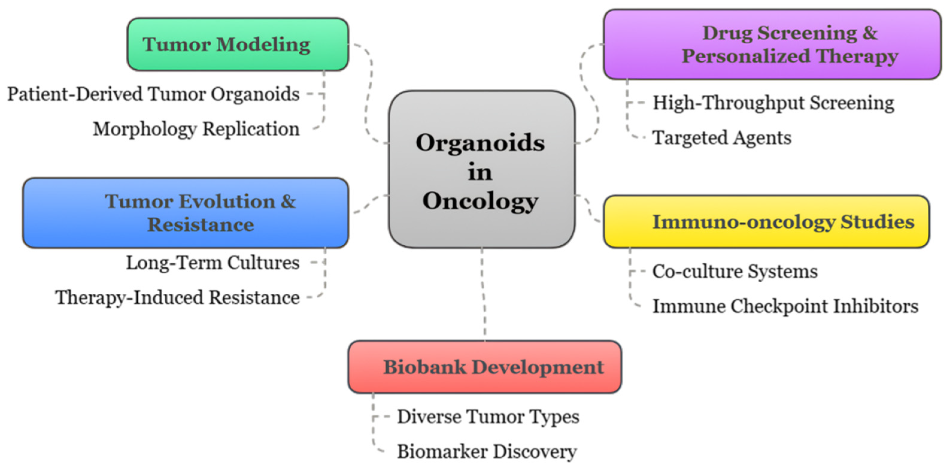
In oncology, tumor-derived organoids, commonly referred to as patient-derived organoids (PDOs), are cultivated from biopsy or surgical specimens [8]. These structures retain the genetic, epigenetic, and phenotypic features of the primary tumor, including its spatial organization, mutational landscape, and differentiation status [9]. Organoids can preserve intra- and inter-patient heterogeneity, making them highly suitable for personalized medicine approaches, as illustrated in Figure 2. Compared to traditional models, organoids demonstrate higher fidelity in modeling tumor evolution, drug resistance mechanisms, and treatment response heterogeneity [10].
Figure 2 shows the different roles of tumor organoids in oncology, including tumor modeling, drug screening, personalized therapy, immuno-oncology, resistance studies, and biobank development. Organoids bridge the gap between in vitro and in vivo models, enabling precise and patient-specific cancer research.

1.3. Integration into Cancer Research

The integration of organoid models into cancer research has significantly expanded the landscape of experimental oncology. They offer a reproducible and scalable platform for drug screening, functional genomics, microenvironment interaction studies, and real-time clinical decision support [11]. Their ability to maintain patient-specific features across multiple passages allows for longitudinal studies of tumor progression, clonal dynamics, and resistance development [12].
The organoid platform bridges the translational gap between bench and bedside by enabling individualized therapeutic testing directly on a patient’s tumor-derived material. The co-culture of organoids with immune cells or stromal components, along with the application of high-content imaging, single-cell sequencing, and CRISPR-based functional assays, has unlocked powerful strategies to explore tumor complexity in unprecedented detail [13]. These models are increasingly incorporated into clinical workflows and early phase trials to guide personalized treatment strategies, identify biomarkers of response, and evaluate novel therapeutic combinations [14]. Alongside genetic, epigenetic, and microenvironmental drivers, metabolic reprogramming is a cardinal characteristic of cancer. Nutrient uptake and bioenergetic mechanisms undergo dynamic changes during tumor growth, metabolic adjustments to hypoxic niches, and resistance to therapy. With the capacity to maintain physiologic nutrient gradients and cellular heterogeneity, organoid systems are progressively used to dissect these metabolic dependencies and vulnerabilities. A cross-disciplinary approach to cancer metabolism combined with organoid studies has the potential not only to enhance mechanistic insights but also to connect to therapeutic strategies based on metabolic plasticity.

1.4. Structure and Objectives of the Review

This review outlines the conceptual framework and technological principles of organoid development, with comparisons to conventional cancer models. Key sections address how organoids capture tumor heterogeneity and support personalized oncology through drug screening, immunotherapy modeling, and resistance profiling. Fundamental and translational applications such as modeling cancer initiation, microbiome interactions, and tumor microenvironments are discussed alongside current challenges, including standardization, scalability, and incomplete niche representation. The review also highlights recent innovations, including organoid-on-chip systems, CRISPR-based functional genomics, and AI-integrated multi-omics platforms.
The objective is to provide a focused overview of organoid applications in oncology and to emphasize their growing significance in precision medicine and translational cancer research.

2. Organoids: Concept and Technology

2.1. Definition and Biological Basis

Organoids are self-organizing three-dimensional (3D) structures derived from stem cells or organ-specific progenitors that recapitulate key architectural and functional aspects of their tissue of origin [15]. In the context of cancer research, tumor-derived organoids (also known as patient-derived organoids or PDOs) mimic not only the histopathological features of tumors but also retain the genomic, transcriptomic, and epigenetic signatures of the patient’s cancer, making them a superior platform for translational oncology [16].

2.1.1. What Defines an Organoid

An organoid is defined by its ability to (i) originate from stem or progenitor cells [17], (ii) self-organize into structures resembling in vivo tissue architecture, (iii) differentiate into multiple cell types representative of the tissue lineage [18], and (iv) exhibit long-term expansion while maintaining genomic stability. In cancer organoids, these principles are adapted to preserve tumor-specific traits, including mutational burden, molecular subtypes, and therapy resistance signatures [19].

2.1.2. Morphology, Self-Organization, and Differentiation

Tumor organoids exhibit diverse morphologies from cystic to solid masses depending on their tissue of origin and mutational landscape [20]. Their capacity for self-organization arises from intrinsic cues encoded by the tumor epithelium and modulated by the extracellular matrix (ECM) [21]. Differentiation potential may be partial in high-grade malignancies, where cellular plasticity and dedifferentiation dominate. Still, PDOs can recapitulate glandular structures in colorectal cancer or alveolar arrangements in lung adenocarcinoma, supporting their phenotypic fidelity [22].

2.1.3. Organoid Fidelity in Cancer Modeling

Organoids faithfully reflect the intra- and inter-patient heterogeneity of tumors. They conserve driver mutations (e.g., KRAS, TP53, and APC), copy number alterations, and transcriptomic signatures over long-term cultures [23]. They preserve drug response heterogeneity, thus offering superior fidelity compared to immortalized 2D lines or homogeneous spheroids [24]. Single-cell RNA-seq has further validated the retention of cancer stem cell hierarchies and clonal subpopulations in PDOs [25].

2.2. Techniques for Organoid Development in the Context of Tumor Modeling

The establishment of tumor-derived organoids represents a significant advancement in personalized oncology, offering a high-fidelity, patient-specific platform to model cancer biology [26]. The process involves a series of critical steps, including the selection of source cells, optimization of culture systems, and maintenance protocols that recapitulate the tumor microenvironment ex vivo. Recent innovations in stem cell biology, tissue engineering, and synthetic matrices have further enabled the development of highly reproducible, scalable, and clinically relevant organoid systems [27].

2.2.1. Source Cell Types

The origin and biological characteristics of the initial cell population are fundamental to the success of organoid generation, especially when modeling tumor heterogeneity, drug responsiveness, and disease progression [28]. Several stem cell sources have been employed, each with distinct advantages, technical considerations, and translational relevance in oncology research [29].
Adult Stem Cells (ASCs) from Tumor Tissues
Adult stem cells, particularly those derived from epithelial compartments of tumors, are the most common source for generating patient-derived organoids (PDOs) [30]. These cells are typically obtained from surgical resections or minimally invasive biopsies and retain key tumor-specific features, including mutational signatures, tissue-specific markers, and lineage hierarchies [31]. The dissociation and enrichment process, combining enzymatic digestion (e.g., collagenase, dispase) and mechanical disruption, yields a viable tumor epithelial fraction that can be embedded in 3D matrices such as Matrigel [32].
Organoids generated from ASCs closely mirror the patient’s tumor biology, making them suitable for personalized drug screening, resistance profiling, and real-time therapy guidance. For example, colorectal cancer organoids derived from LGR5+ stem-like cells have been shown to recapitulate adenoma-to-carcinoma progression and drug response profiles with remarkable fidelity [33].
Cancer Stem Cells (CSCs)
A subset of tumor-initiating cells known as cancer stem cells (CSCs) represents a critical source for organoid development, particularly when aiming to capture the hierarchical and functional heterogeneity of aggressive tumors [34]. CSCs exhibit self-renewal, pluripotency, and the ability to repopulate tumors upon transplantation. These cells can be isolated using surface markers (e.g., CD44high, CD133+, and ALDH1+) or functional assays such as side population analysis [35].
Organoids derived from CSC-enriched populations often demonstrate enhanced tumorigenicity, metastatic potential, and therapeutic resistance, making them ideal models for studying relapse, dormancy, and evolution under drug pressure [36]. CSC-derived organoids serve as preclinical models for testing targeted inhibitors and understanding lineage plasticity in treatment-resistant tumors [37].
Induced Pluripotent Stem Cells (iPSCs)
iPSC technology offers a versatile platform to model cancer development from its earliest cellular transitions. By reprogramming somatic cells (e.g., fibroblasts or peripheral blood mononuclear cells) into a pluripotent state and guiding their differentiation into tissue-specific organoids, researchers can recreate oncogenic transformation de novo [38]. The targeted insertion of cancer-associated mutations using CRISPR/Cas9 facilitates the creation of isogenic organoid models that allow systematic dissection of gene function, synthetic lethality, and tumor progression mechanisms [39]. This approach has been instrumental in modeling stepwise colorectal carcinogenesis (e.g., APC → TP53 → KRAS) and exploring rare or pediatric cancers with known genetic drivers. iPSC-derived organoids also support toxicology and developmental oncology applications [40].
Embryonic Stem Cells (ESCs)
Though ethically constrained and less commonly used in cancer modeling, human embryonic stem cells (ESCs) represent a powerful tool for generating highly plastic organoids with multilineage differentiation capacity [41]. ESC-derived organoids can be genetically engineered to carry tumorigenic mutations, enabling controlled modeling of cancer initiation in a developmental context.
ESC-derived systems have been used to model medulloblastoma, hepatoblastoma, and early neural tumors, especially in cases where patient-derived samples are unavailable or unsuitable [42]. These organoids serve as invaluable platforms to investigate oncogenesis during early tissue development and assess developmental toxicants.
Tissue-Resident Progenitor Cells
Certain organoid systems leverage tissue-specific progenitor populations isolated from normal or pre-malignant tissues [43]. These progenitors may carry premalignant mutations or epigenetic alterations that predispose them to transformation under defined conditions. In the context of cancer research, organoids derived from Barrett’s esophagus or inflammatory bowel disease lesions represent early transformation models that can be longitudinally monitored for neoplastic conversion [44].
Mesenchymal Stem Cells (MSCs)
MSCs are multipotent stromal cells that reside in various tissues and contribute to extracellular matrix (ECM) remodeling, immune regulation, and angiogenesis [45]. When co-cultured with tumor organoids, MSCs help replicate the tumor microenvironment (TME) more realistically [46]. These models are essential for exploring tumor-stroma interactions, immune evasion mechanisms, and for testing immunotherapy combinations in a more physiologically relevant setup.
Circulating Tumor Cells (CTCs)
CTCs are tumor cells shed into the bloodstream from primary or metastatic lesions. They can be isolated via liquid biopsy, making them a minimally invasive source for real-time tumor modeling [47]. CTC-derived organoids capture metastatic behavior, therapy resistance, and clonal evolution over time [48]. They offer a promising approach for longitudinal monitoring of cancer and personalized treatment in advanced-stage patients.
Engineered Synthetic Progenitors
These are lab-designed progenitor cells created through synthetic biology techniques, often using iPSCs or ESCs as a base [49]. They are genetically programmed with specific oncogenic circuits or pathways to model mutation–phenotype relationships. These systems support high-throughput functional genomics and CRISPR screening, and they are ideal for studying context-specific vulnerabilities in cancer, especially when patient samples are limited or unavailable [50].
Multiple stem cell sources, including adult stem cells, cancer stem cells, iPSCs, ESCs, tissue-resident progenitors, CTCs, MSCs, and engineered progenitors, are now employed in tumor organoid generation, each offering unique advantages for modeling cancer biology, drug response, and tumor evolution, as mentioned in Table 1.

2.2.2. Culture Systems

The choice of culture system is fundamental to the successful establishment and functionality of organoids, significantly influencing their morphology, viability, differentiation potential, and translational relevance. Broadly, organoid culture systems are categorized into scaffold-based systems, which provide extracellular matrix (ECM) support, and scaffold-free systems, which enable self-organization [59]. Ongoing innovations in biomaterials, bioengineering, and microfluidic technologies have further refined these systems, enabling enhanced control over the organoid microenvironment and improving reproducibility [60].
Scaffold-Based Culture Systems
Scaffold-based systems aim to replicate the native extracellular matrix, providing essential biochemical and mechanical cues that regulate cell polarization, proliferation, differentiation, and tissue architecture [61]. Matrigel, a widely used basement membrane extract rich in laminin, collagen IV, entactin, and heparan sulfate proteoglycans, has become the gold standard in organoid culture [62]. When tumor or stem cells are embedded in Matrigel droplets and cultured in niche-supplemented media, they typically form 3D structures that closely recapitulate native tissue histology [63]. The significant batch-to-batch variability and undefined composition of Matrigel have driven the exploration and development of alternative natural and synthetic scaffold materials for organoid culture. Among these, biomaterials such as collagen I, fibrin gels, hyaluronic acid, and polyethylene glycol (PEG)-based hydrogels have gained prominence due to their tunable stiffness, defined chemical properties, and enhanced biocompatibility [64]. Self-assembling peptide hydrogels, such as Puramatrix, provide nanofibrous, ECM-like networks with customizable bioactivity and mechanical features, enabling better mimicking of the native extracellular environment [65]. Recombinant ECM proteins, including laminin-111 and fibronectin-based scaffolds, offer precise control over cell-matrix interactions, supporting more consistent organoid formation [21,66]. Dynamic hydrogels, which can respond to stimuli such as pH shifts or enzymatic activity, allow for the controlled release of growth factors or real-time modulation of stiffness, thus promoting progressive tissue maturation [67].
Advances in 3D bioprinting have further enabled the spatially patterned deposition of ECM components, facilitating zonal tissue architecture and scalable, high-throughput organoid fabrication [68]. Decellularized human tissue-derived ECM provides organ-specific biochemical signals and structural complexity, demonstrating considerable potential for lineage-specific organoid development and more accurate disease modeling [69]. Together, these innovative scaffold materials are transforming the landscape of organoid culture by improving physiological relevance, reproducibility, and clinical translatability.
These next-generation scaffold materials not only improve reproducibility and scalability but also enhance the physiological relevance of organoid models, making them more suitable for clinical and pharmaceutical applications [27].
Scaffold-Free Culture Systems
Scaffold-free systems exploit the innate self-organizing capacity of cells to form spheroids or organoid-like aggregates in the absence of exogenous ECM components [70]. Common techniques include hanging drop cultures, low-attachment U- or V-bottom plates, and rotary suspension bioreactors, all of which facilitate rapid and uniform spheroid formation [71]. These models are particularly advantageous for high-throughput drug screening and toxicity assays, offering simpler handling and easier automation.
Recent innovations have significantly enhanced the functionality and versatility of scaffold-free organoid culture systems. Ultrasound-assisted and acoustic aggregation techniques now enable high-efficiency and size-controlled spheroid formation, promoting uniformity and scalability [72]. Dielectrophoresis-based cell positioning offers a non-invasive method to precisely manipulate cellular arrangements, thereby improving the structural fidelity of forming organoids [73]. Microfluidic droplet platforms further advance this approach by allowing for the parallel generation of thousands of uniform organoids while offering fine control over nutrient delivery, oxygen gradients, and spatial signaling cues [74]. Integration of these platforms into organoid-on-a-chip systems introduces dynamic perfusion, mechanical stimulation, and the ability to support multi-tissue co-cultures, closely mimicking in vivo physiological conditions [75]. Such systems are particularly valuable for investigating tissue-tissue interactions, drug pharmacokinetics, and immune-oncology responses. To bridge the gap between simplicity and biological complexity, hybrid strategies have emerged that combine scaffold-free aggregation with soft extracellular matrix coatings or engineered microtopographies to restore tissue polarity and spatial organization. The development of automated organoid culture systems employing robotic liquid handling and AI-based imaging analytics is rapidly improving standardization, scalability, and throughput, making these systems highly attractive for both basic research and translational applications [76].
The convergence of synthetic biology, biomaterials engineering, and microscale technologies is driving a paradigm shift in organoid culture methodology. As scaffold materials become increasingly defined and tunable, and as scaffold-free systems evolve to incorporate biophysical and biochemical precision, the boundary between these categories continues to blur. Emerging hybrid and modular culture systems now allow for unprecedented control over the spatiotemporal dynamics of organoid growth, supporting more accurate modeling of development, disease, and therapeutic response [77].
These advancements underscore a continued push toward standardization, personalization, and translational applicability, with the goal of establishing organoid systems as reliable platforms in precision medicine, drug discovery, and regenerative therapy.

2.2.3. Protocols and Maintenance

The successful establishment and sustained propagation of tumor organoids rely on finely tuned protocols that replicate the essential biochemical and structural features of the tumor microenvironment [78]. This includes the careful formulation of culture media, precise supplementation with niche-specific factors, and the implementation of scalable, standardized maintenance practices [79]. Recent advancements in growth media optimization, automation, and biobanking have further elevated the reproducibility, fidelity, and translational value of tumor organoid models [80]. A stepwise overview of the tumor organoid development pipeline from patient-derived tissue to mature, expandable 3D cultures is depicted in Figure 3.
Figure 3 shows the key steps from patient tumor collection through tissue dissociation, cell isolation, 3D embedding, and organoid culture, culminating in applications like drug screening and genomic profiling.
Media Components and Niche Factors
Organoid cultures require media enriched with a carefully balanced combination of growth factors, morphogens, and pathway modulators that mimic the tissue-specific stem cell niche [81]. Standard formulations typically include epidermal growth factor (EGF), Noggin, R-spondin-1, Wnt3a, and A83-01 (a selective inhibitor of the TGF-β signaling pathway), with additional support from molecules such as prostaglandin E2, nicotinamide, N-acetylcysteine, and insulin–transferrin–selenium to promote survival, stemness, and oxidative resistance [82].
These core formulations are increasingly being tailored based on tumor type, cell-of-origin, and specific genetic alterations. Consistent with previous reports, KRAS- or BRAF-mutant colorectal cancer PDOs are largely independent of p38 MAPK inhibition, whereas in APC-mutant models, Wnt3a is typically omitted to avoid excessive Wnt pathway activation [83,84]. In glioblastoma and pancreatic ductal adenocarcinoma organoids, the inclusion of FGF2, HGF, or niche-specific extracellular vesicles has been shown to improve cellular viability and mimic in vivo tumor heterogeneity [85].
Recent efforts have focused on reducing reliance on animal-derived reagents such as Matrigel or bovine serum. This has led to the development of chemically defined, xeno-free media formulations, as well as the use of recombinant or synthetic analogs of growth factors [86]. High-throughput transcriptomic and proteomic profiling of organoids and their tissue counterparts are increasingly guiding the rational design of customized media that better preserve lineage-specific differentiation and maintain the clonal diversity of tumor subpopulations over long-term culture [87]. Media formulations are now being optimized to support co-culture systems that include immune, stromal, or endothelial components, making tumor organoids more physiologically reflective of the native tumor microenvironment.
Growth Timeline and Batch Scaling
The initial establishment of tumor organoids typically occurs over 7 to 21 days, depending on variables such as tissue source, tumor type, and cellular viability [88]. Following successful initiation, organoids can be expanded through passaging every one to two weeks, using mechanical shearing or enzymatic dissociation, depending on the organoid structure and fragility [89]. Importantly, long-term expansion has been demonstrated for a wide range of malignancies, including breast, colorectal, prostate, and lung cancers, while maintaining genomic, phenotypic, and functional fidelity.
As the field advances toward clinical and pharmaceutical applications, the need for scalable and high-throughput culture systems has led to the adoption of automated and semi-automated workflows [90]. Robotic handling platforms, combined with machine vision and real-time growth monitoring, are now routinely used in large-scale organoid production for drug screening and biomarker discovery [11]. Microfluidic systems, organoid-on-a-chip technologies, and spinning bioreactors have been deployed to improve nutrient diffusion, gas exchange, and uniformity of organoid size and shape, particularly important for drug penetration studies and modeling dynamic tumor behavior [91].
Recent innovations have also enhanced the standardization of cryopreservation protocols. Optimized freezing and thawing methods utilizing cryoprotectants such as DMSO in defined formulations have led to reliable recovery of viable, functional organoids post-thaw [92]. Both slow-freezing and vitrification methods have been adapted for tumor organoids, with protocol modifications specific to tissue type and organoid morphology. This has facilitated the development of living organoid biobanks, which are now widely used for retrospective analysis, patient-specific drug testing, and longitudinal studies of tumor evolution.
To ensure consistency and translational validity, routine quality control measures are increasingly embedded into the organoid workflow [93]. These include genomic profiling to assess mutational stability, phenotypic validation using immunohistochemistry and transcriptomics, and functional assays such as drug response testing and tumorigenicity assessment. These practices are crucial not only for research reproducibility but also for potential regulatory compliance in clinical applications.
The refinement of protocols and maintenance strategies for tumor organoids has significantly advanced over the past decade, with a strong emphasis on standardization, scalability, and physiological relevance [94]. Continued integration of omics-guided media design, bioengineering platforms, and automated workflows is expected to further enhance the fidelity and clinical applicability of tumor organoid systems. The variation in organoid platforms based on input cells, matrices, and media compositions is highlighted in Table 2.

2.3. Comparison with Other Preclinical Models

While organoid technology has revolutionized cancer research, its full potential is best appreciated through direct comparison with other conventional preclinical models. Organoids uniquely balance biological fidelity, experimental manipulability, and scalability, making them a powerful intermediate between reductionist in vitro systems and complex in vivo models [105]. Recent technological advances continue to widen the gap in translational relevance between organoids and traditional models, especially in the context of personalized oncology and high-throughput functional assays.

2.3.1. Two-Dimensional (2D) Cell Lines

For decades, two-dimensional monolayer cell cultures have been the backbone of in vitro cancer research due to their cost-effectiveness, ease of genetic manipulation, and compatibility with high-throughput screening platforms [106]. Their utility is increasingly limited by critical biological shortcomings. The lack of spatial architecture in 2D cultures results in abnormal cell polarity, unregulated growth dynamics, and artificial cell–cell and cell-matrix interactions [107]. Prolonged passaging and artificial selection pressures often lead to genetic drift, loss of heterogeneity, and altered drug responses.
Recent transcriptomic and proteomic analyses have confirmed that 2D cell lines diverge significantly from the primary tumors they are meant to model, particularly in terms of pathway activation, epigenetic regulation, and response to targeted therapies [108]. As a result, drug screening in 2D cultures frequently yields false positives or false negatives, diminishing their translational accuracy. Their inability to model microenvironmental factors such as stromal interactions, hypoxia, and immune infiltration further limits their predictive utility, especially for therapies involving tumor–stroma crosstalk or immune checkpoint modulation [109].

2.3.2. Three-Dimensional (3D) Spheroids

Three-dimensional multicellular spheroids represent an improvement over 2D models by recreating the spatial and metabolic gradients present in solid tumors [110]. Spheroids exhibit central hypoxia, nutrient deprivation, and outer proliferative zones, features more reflective of in vivo tumor physiology [111]. They are widely used for studying radiotherapy sensitivity, drug diffusion, and tumor metabolism.
Spheroids are typically derived from established cancer cell lines, which inherently lack the genomic and phenotypic heterogeneity of patient tumors. They fail to recapitulate tissue-specific architecture, tumor–stroma interactions, and immune components, limiting their ability to model complex disease states such as invasion, metastasis, and immune evasion [112]. While some efforts have been made to generate patient-derived spheroids, they often lack long-term stability and clonal representation, making them less reliable for precision medicine applications.
Recent advances in spheroid technologies, including scaffold-assisted aggregation, micro-patterned culture surfaces, and incorporation of stromal cells, have marginally improved their biological complexity [113]. These improvements remain limited compared to the tissue-mimicking and patient-specific capabilities of organoid systems.

2.3.3. Patient-Derived Xenografts (PDXs)

Patient-derived xenografts, established by implanting fresh human tumor tissue into immunodeficient mice, are among the most clinically reflective cancer models available [114]. PDXs preserve tumor histology, mutational landscape, and clonal architecture, making them valuable tools for evaluating drug efficacy, resistance mechanisms, and biomarker discovery [115]. Importantly, PDXs have been used in co-clinical trials to guide therapeutic decision-making, offering a powerful bridge between preclinical research and patient care.
Despite these advantages, PDX models are constrained by several limitations. They require 3–6 months for engraftment and expansion, involve substantial financial and ethical costs, and are inherently low-throughput [40]. The lack of a functional human immune system in traditional PDX mice restricts their application in immuno-oncology. Although humanized PDX models with reconstituted immune systems have shown promise, they are technically complex, costly, and often lack full immunocompetence [116]. Murine stromal replacement over serial passages can result in altered tumor–stroma dynamics and adaptive changes that deviate from the patient’s original tumor biology [117].
Recent concerns about murine-specific selection pressures, genomic evolution during passaging, and failure to capture early resistance phenotypes have further emphasized the need for more tractable and patient-specific in vitro systems, such as organoids [118].

2.3.4. Organoids: A New Paradigm in Preclinical Modeling

Organoids represent a transformative platform in cancer research by capturing the patient-specific genotype, 3D tissue architecture, and functional heterogeneity within a controllable in vitro system. Derived from primary or metastatic tumor tissues, organoids retain the histological and molecular fidelity of their source tumors, including clonal diversity, mutational profiles, and epigenetic landscapes [119]. They are amenable to genetic engineering, high-throughput drug screening, and multi-omics profiling, making them uniquely suited for personalized therapy testing and functional genomics.
Recent innovations have enhanced the versatility of organoid systems, including co-culture with immune and stromal cells, incorporation into microfluidic devices (organoids-on-chip), and integration with CRISPR/Cas9-based gene editing, enabling the study of tumor-immune interactions, metastatic behavior, and resistance evolution [120]. Standardized protocols for biobanking, cryopreservation, and longitudinal culturing have made organoids a scalable and reproducible model for translational research.
Unlike PDX models, organoids offer rapid generation timelines (within 1–3 weeks), lower cost, and the ability to model patient tumors in a high-throughput format. Unlike 2D and spheroid models, they maintain a tissue-like architecture and microenvironmental signaling fidelity essential for accurate therapeutic assessment.
As organoid technology continues to evolve, especially with advances in AI-driven image analysis, dynamic perfusion systems, and spatial transcriptomics, it is poised to become the gold standard for preclinical cancer modeling, offering an optimal balance of biological relevance, scalability, and clinical applicability [6]. A comprehensive comparison of major cancer models is summarized in Table 3.

3. Tumor Heterogeneity and Organoids

Tumor heterogeneity is increasingly recognized as a central hallmark of cancer progression, therapeutic failure, and clinical relapse. It encompasses a broad spectrum of biological variability, not only between tumors of the same histological subtype (intertumoral heterogeneity) but also within a single tumor mass (intratumoral heterogeneity). This multidimensional complexity arises through the interplay of genetic alterations, epigenetic remodeling, microenvironmental pressures, and cellular plasticity. Accurate modeling of this heterogeneity is imperative for understanding cancer biology and for developing personalized therapeutic strategies. Organoids, as three-dimensional culture systems derived from patient tumors, represent an advanced preclinical platform that preserves the cellular diversity and evolutionary dynamics of the tumor, thus enabling in-depth interrogation of heterogeneity in vitro.

3.1. Dimensions of Tumor Heterogeneity

3.1.1. Genetic and Epigenetic Variability

Genetic heterogeneity in tumors arises due to the accumulation of somatic mutations, gene amplifications, chromosomal rearrangements, and loss of heterozygosity across evolving clones [127]. Epigenetic variability, including DNA methylation changes, histone modification patterns, and chromatin accessibility shifts, further contributes to transcriptomic reprogramming, cell-state transitions, and resistance phenotypes [128]. Single-cell whole-genome sequencing has revealed that tumors can harbor dozens of coexisting subclones, each with distinct mutational profiles. Similarly, single-nucleus epigenomic profiling has uncovered regional epigenetic heterogeneity that correlates with transcriptional plasticity and metastatic potential. Organoids derived from primary tumors retain both genomic and epigenetic features of their source tissue, including driver mutations (e.g., TP53, APC, KRAS), copy number landscapes, and non-coding regulatory marks [129]. These models enable longitudinal tracking of epigenomic reprogramming and the identification of epigenetic vulnerabilities that may not be evident in bulk tissue analyses.

3.1.2. Spatial and Temporal Heterogeneity

Spatial heterogeneity reflects the variation in cellular phenotypes, genotypes, and functional states across different regions of the same tumor. This can result from localized microenvironmental gradients such as hypoxia, mechanical stiffness, and immune infiltration [130]. Organoids derived from spatially distinct regions of a tumor can retain this topographic information, allowing comparative analysis of clone-specific behavior and therapeutic responsiveness. Temporal heterogeneity, on the other hand, develops through the course of disease progression and in response to external pressures, including therapy. The ability to generate organoids from serial patient samples, such as those obtained pre-treatment, during relapse, or from metastases, offers a powerful method to model clonal evolution and therapy-induced selection [131]. Advances in spatial transcriptomics, combined with digital pathology and machine learning, now allow organoid-derived tissue to be mapped back to its in situ topography, providing insights into the spatial origins of resistance clones [132].

3.1.3. Functional Heterogeneity in Drug Resistance

Functional heterogeneity refers to the differences in proliferative potential, differentiation state, drug sensitivity, and immune evasion capacity among tumor subpopulations. This form of heterogeneity is particularly challenging to capture using traditional assays, as it may not always correspond to genetic divergence [133]. Organoids provide a dynamic and scalable platform for functional assays such as high-content imaging, apoptosis induction profiling, and mitochondrial activity measurement under drug treatment. Functional pharmacotyping using organoids has revealed intratumoral variation in response to standard-of-care and targeted agents, even within organoids derived from a single biopsy [134]. Coupling these functional assays with single-cell RNA sequencing and proteomic barcoding enables the identification of rare drug-tolerant persister cells and lineage trajectories that underlie acquired resistance.

3.2. Organoids as Models for Tumor Heterogeneity

3.2.1. Preservation of Tumor Architecture and Clonal Diversity

Unlike conventional cell lines, which often lose structural and genomic fidelity after serial passaging, organoids preserve the three-dimensional organization and clonal diversity of the parental tumor. Histopathological analyses have confirmed that organoids derived from colorectal, gastric, and breast tumors maintain key architectural features such as glandular formation, lumen polarity, and mucin secretion [40]. The use of defined extracellular matrices (e.g., Matrigel, collagen I) supports the maintenance of cellular polarity and basement membrane attachment, while co-culture with fibroblasts, endothelial cells, or tumor-associated macrophages further enhances niche fidelity. Multi-regional organoid generation allows researchers to study the architectural and molecular heterogeneity between spatial clones, providing mechanistic insights into localized therapeutic resistance and recurrence.

3.2.2. Clonal Evolution and Single-Cell Resolution Studies

Recent advances in single-cell technologies, including single-cell RNA sequencing (scRNA-seq), single-cell ATAC sequencing, and high-throughput lineage barcoding, have facilitated clonal tracing and phenotypic mapping within organoid cultures [135]. By introducing unique molecular barcodes or CRISPR-based lineage tags, investigators can reconstruct clonal dynamics during tumor expansion, differentiation, or drug exposure. Organoids serve as an ideal platform for such studies because they enable longitudinal tracking under defined experimental conditions. Studies have revealed that treatment with tyrosine kinase inhibitors or chemotherapeutic agents induces the selection of pre-existing resistant clones and can also drive epigenetic reprogramming of initially sensitive populations [136]. Combined with computational modeling, organoid-based lineage tracing is now being used to delineate evolutionary bottlenecks and identify early markers of therapeutic failure.

3.2.3. Subtype Stratification and Plasticity Assessment

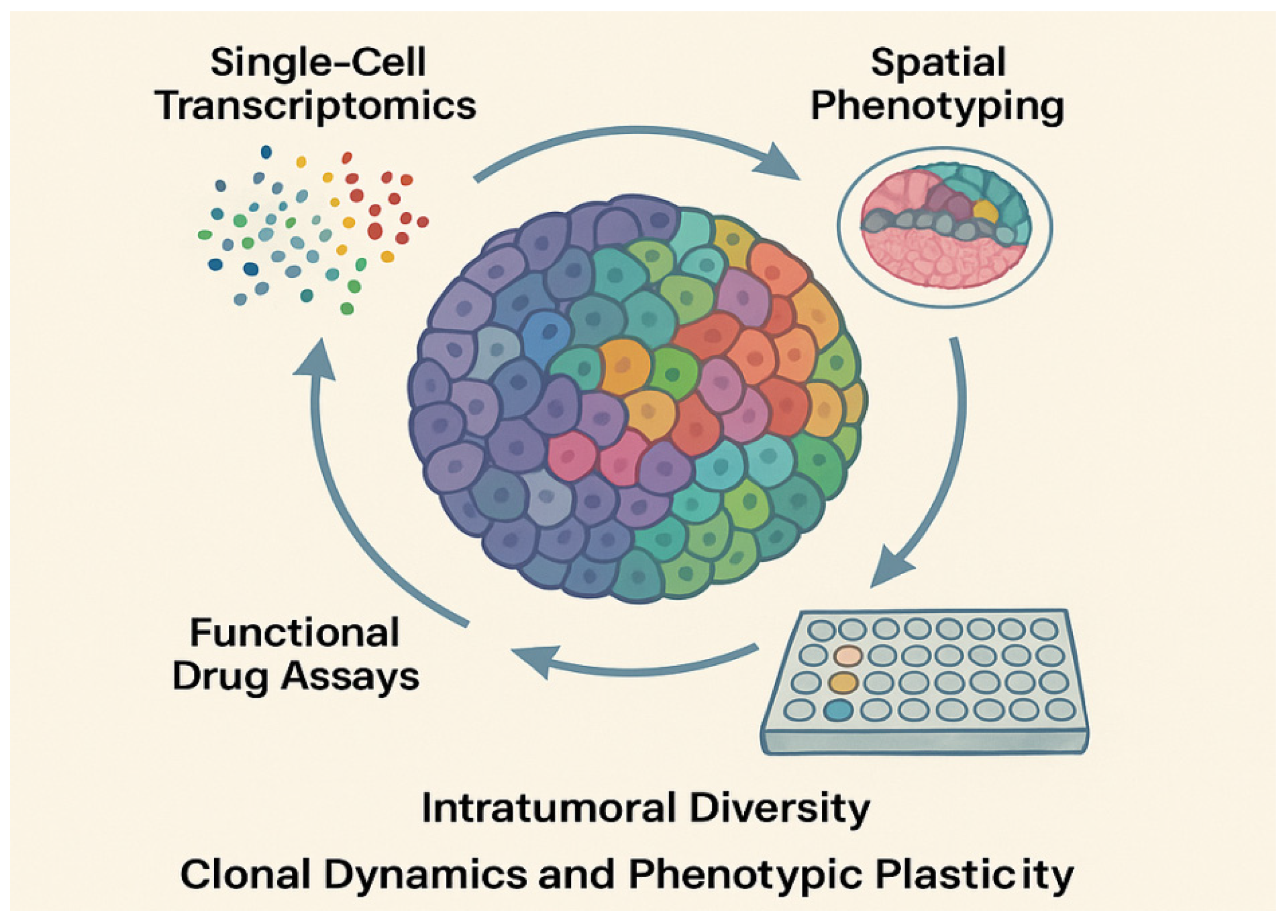
Molecular subtyping of cancers has informed prognosis and guided targeted therapy strategies. Organoid cultures preserve these subtype identities, making them suitable for functional subtype validation [137]. Beyond static subtyping, organoids allow real-time observation of phenotypic plasticity and cell-state transitions. For instance, organoids subjected to androgen deprivation or anti-estrogen therapy undergo subtype switching, recapitulating clinical patterns of resistance in prostate and breast cancers, respectively [138]. Using scRNA-seq and pseudotime analysis, researchers have mapped lineage trajectories during epithelial–mesenchymal transition (EMT), neuroendocrine differentiation, and stemness reactivation, highlighting the utility of organoids in modeling therapy-induced plasticity [139]. Patient-derived organoids have emerged as powerful ex vivo systems for modeling tumor heterogeneity, enabling integration of single-cell transcriptomics, spatial profiling, and drug response assays to reveal clonal diversity and phenotypic plasticity, as illustrated in Figure 4.
Figure 4 shows the Patient-derived tumor organoids as integrative platforms for modeling intratumoral heterogeneity. The combination of single-cell transcriptomics, spatial phenotyping, and functional drug screening enables high-resolution dissection of clonal diversity, lineage plasticity, and therapeutic response within individual tumors. The different colors in the organoid represent distinct cell populations/clusters identified through single-cell analyses, emphasizing intratumoral diversity.

3.3. Patient-Derived Organoid Biobanks

3.3.1. International Consortia and Standardization Efforts

The development of large-scale patient-derived organoid biobanks has significantly advanced the systematic collection, standardization, and functional annotation of tumor models across various cancer types [140]. International initiatives such as the Human Cancer Models Initiative (HCMI), a collaborative effort involving the National Cancer Institute (NCI), Cancer Research UK, the Wellcome Sanger Institute, and Hubrecht Organoid Technology, aim to generate over 1000 next-generation cancer models using standardized operating procedures (SOPs) and unified clinical and molecular data annotation [141]. The Hubrecht Organoid Biobank currently holds the world’s most extensive repository of adult stem cell-derived organoids, facilitating global academic and commercial research with well-characterized models.
The Wellcome Sanger Institute has been instrumental in establishing hundreds of organoid lines derived from a broad spectrum of tumor types, complete with genomic profiling and pharmacological response data [142]. Many of these models are now accessible through international repositories, including searchable platforms and data commons. These consortia emphasize reproducibility, open access, and inclusivity, with dedicated efforts to involve low- and middle-income countries to ensure global tumor heterogeneity is adequately represented.

3.3.2. Cancer Type Representation and Sample Diversity

Contemporary organoid biobanks reflect a broad oncologic spectrum, encompassing prevalent cancers such as colorectal, breast, lung, prostate, pancreatic, and gastric cancers, as well as rare malignancies including sarcomas, pediatric cancers, thymoma, cholangiocarcinoma, and hematologic neoplasms [143]. These organoids are derived not only from surgical resections but also from minimally invasive procedures, including core biopsies, ascitic fluid, and even circulating tumor cells (CTCs). This has enabled longitudinal sampling and the construction of dynamic disease models. Many biobanks also feature matched tumor-normal organoid pairs, which are invaluable for comparative genomic and functional studies.
For instance, a patient-derived organoid (PDO) biobank focusing on high-risk colorectal adenomas in China has been developed, encompassing 37 organoid lines from 33 patients [144]. High-throughput screening of these models uncovered substantial intrapatient heterogeneity and identified potential therapeutic agents such as metformin. Organoid biobanks are increasingly extending beyond oncology. Inflammatory diseases, such as Crohn’s disease, have been modeled using PDOs, revealing subtype-specific molecular signatures and broadening the scope of organoid-based precision medicine.

3.3.3. Integration with Clinical Data and Predictive Platforms

The integration of detailed clinical metadata, including treatment regimens, imaging outcomes, histopathological features, and longitudinal survival data, has transformed organoid biobanks from static model repositories into dynamic platforms for predictive and translational research [145]. Functional assays using PDOs have shown strong concordance with real-world patient responses, supporting their use in functional precision medicine. Emerging machine learning frameworks trained on multi-omics and drug response data from organoid libraries are demonstrating promise in forecasting individualized therapeutic outcomes [146].
Organoids are also being incorporated into adaptive clinical trial designs, such as umbrella and basket trials, where they serve as biomarker-driven stratification tools. Collaborative efforts among global institutions, including the Wellcome Sanger Institute, Helmholtz Munich, and others, are currently conducting large-scale perturbation screening and single-cell multiomic analysis to construct comprehensive resources such as the “Cancer Plasticity Atlas”. These initiatives are expected to enable virtual drug screening and personalized treatment simulations, marking a new frontier in cancer systems biology [141].

4. Organoids in Personalized Cancer Therapy

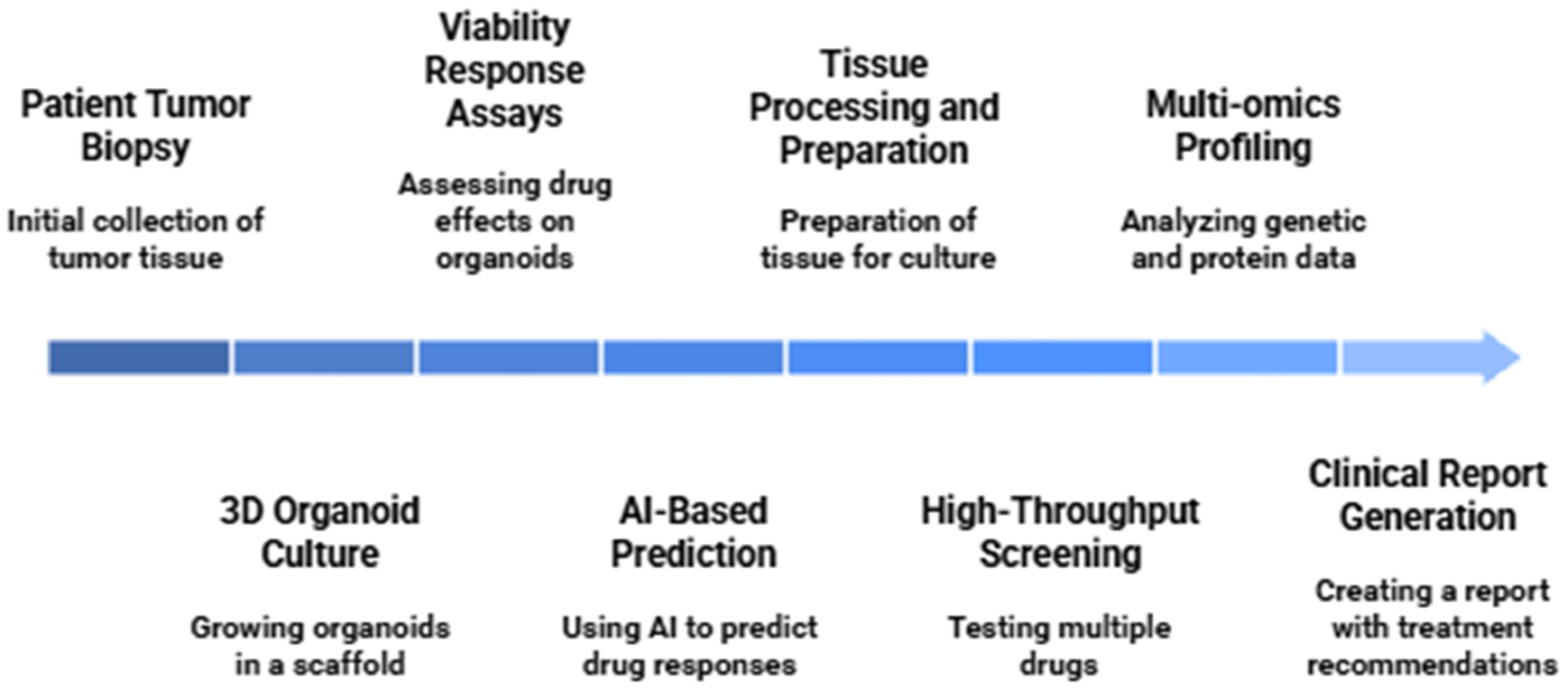
Organoids have emerged as a transformative platform in the realm of personalized oncology. By preserving the molecular and phenotypic features of the primary tumor, patient-derived organoids (PDOs) provide a robust, scalable, and clinically relevant system to predict drug responses, assess resistance mechanisms, and explore immune interactions [16]. Their use has expanded from preclinical validation into real-time therapeutic decision-making, underscoring their relevance in functional precision medicine. The integration of PDO-based platforms into drug development pipelines enables rapid, patient-specific therapeutic screening. A representative workflow from biopsy to high-throughput drug testing and response assessment is illustrated in Figure 5.
Figure 5 shows a patient-derived organoid-based drug testing workflow, from tumor dissociation to high-throughput screening and therapeutic response profiling for personalized treatment selection.

4.1. Drug Screening and Predictive Testing

4.1.1. High-Throughput Pharmacotyping Platforms

High-throughput pharmacotyping platforms using PDOs have redefined the landscape of preclinical drug testing. Organoids are derived from patient biopsies, expanded in three-dimensional matrices, and subjected to systematic drug exposure in miniaturized formats such as 96- or 384-well plates [147]. Using automated liquid handling and high-content imaging, drug efficacy is assessed through viability assays, apoptosis induction, and morphological analysis.
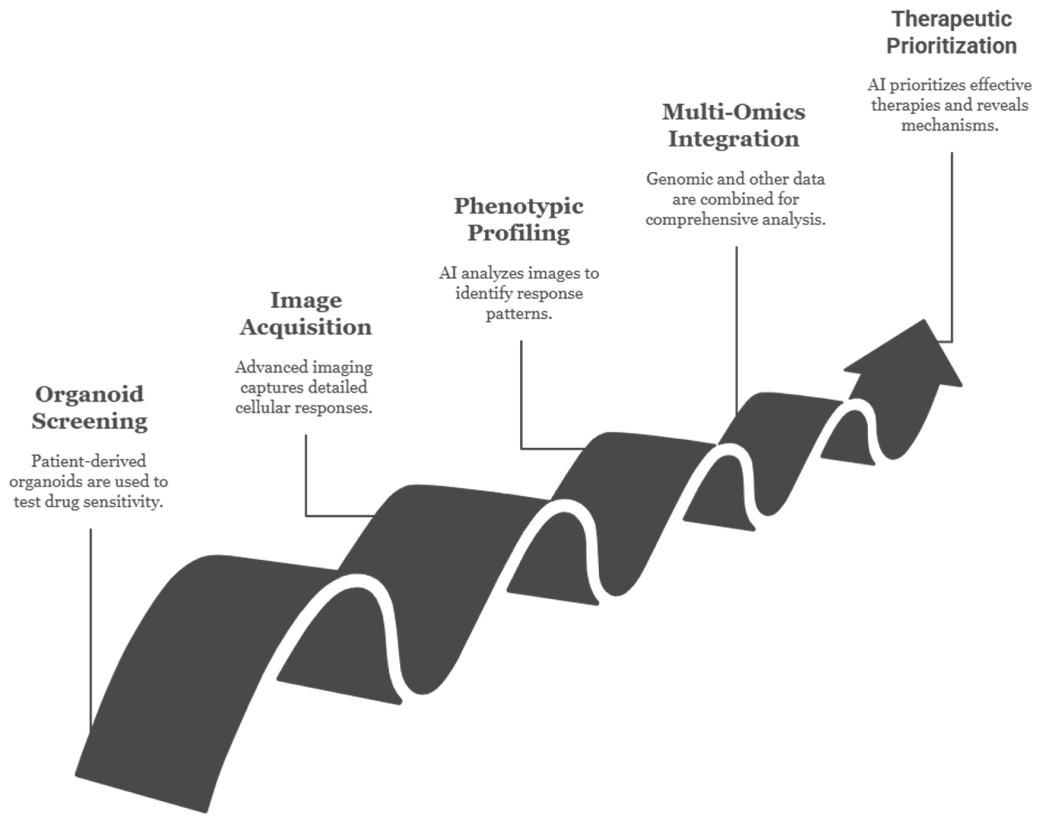
Recent advancements include the integration of AI-driven phenotypic readouts, barcoded drug libraries for pooled screening, and dynamic live-cell monitoring tools. These platforms enable rapid screening of hundreds of compounds, including standard chemotherapies, targeted therapies, epigenetic drugs, and emerging experimental agents [148]. The ability to tailor drug panels based on patient-specific genomic features enhances the functional stratification of tumors and supports personalized regimen development.

4.1.2. Organoid–Drug Response Correlation with Clinical Data

Multiple clinical studies have demonstrated that PDO-derived drug sensitivity correlates strongly with patient response. In colorectal cancer, PDO pharmacotyping accurately predicted responsiveness to FOLFOX (folinic acid, fluorouracil, and oxaliplatin), irinotecan, and EGFR inhibitors [149]. In breast cancer, particularly in BRCA1/2-mutated cases, PDOs recapitulated clinical responses to PARP (Poly (ADP-ribose) polymerase) inhibitors [150]. Similarly, in non-small cell lung cancer, PDOs reflecting diverse EGFR mutations, including uncommon exon 20 insertions, have guided the selection of tyrosine kinase inhibitors [151].
The predictive value of PDOs is further enhanced by combining pharmacotyping data with transcriptomic and genomic analyses, allowing for the development of integrated biomarkers and therapeutic algorithms. These platforms not only mirror baseline sensitivity but also serve as a tool for prospective therapy selection in real-world clinical settings.

4.1.3. Resistance Profiling and Secondary Screening

Resistance profiling using organoids enables the exploration of both intrinsic and acquired resistance mechanisms. PDOs derived from matched samples before and after treatment capture clonal evolution under therapeutic pressure. In vitro modeling of resistance through chronic low-dose exposure or cyclic drug administration has revealed mechanisms such as bypass signaling activation, transcriptional reprogramming, and epigenetic alterations [152].
Secondary screening of resistant organoids facilitates the identification of alternative therapeutic agents, combination regimens, or re-sensitization strategies. For example, colorectal cancer organoids resistant to anti-EGFR therapy have been shown to regain sensitivity upon MEK inhibition, highlighting the potential of PDOs in guiding salvage therapy design [153]. These findings underscore the role of organoids not only in initial therapy prediction but also in longitudinal treatment planning.

4.2. Immune-Oncology Applications

4.2.1. Organoid–Immune Cell Co-Culture

The integration of immune components into patient-derived organoid (PDO) systems has expanded their translational utility in immune-oncology research. Co-culture platforms combining PDOs with autologous or allogeneic immune cells, including CD8+ and CD4+ T cells, natural killer (NK) cells, dendritic cells (DCs), and peripheral blood mononuclear cells (PBMCs), now provide sophisticated models to investigate immune infiltration, cytotoxicity, and tumor-immune interactions in a physiologically relevant three-dimensional microenvironment [46].
These co-culture systems utilize either direct embedding, transwell inserts, or organoid-on-chip technologies. More advanced approaches incorporate air–liquid interface (ALI) and microfluidic platforms, preserving spatial orientation and enabling long-term viability [154]. Functional assays assess immune cell proliferation, immune synapse formation, cytokine production (e.g., IFN-γ, TNF-α), target cell apoptosis, and antigen presentation. Such models closely replicate in vivo immune dynamics and are instrumental for dissecting mechanisms of immune evasion, exhaustion, and stromal modulation [155].

4.2.2. Immunotherapy Biomarker Testing

PDOs have emerged as dynamic platforms for the functional evaluation of biomarkers predictive of immunotherapy response [156]. Unlike static genomic or transcriptomic analyses, organoid-based systems allow for real-time measurement of biomarker changes in response to immunologic stimuli. Critical parameters such as PD-L1 expression, HLA class I presentation, interferon-gamma sensitivity, and tumor mutational burden can be interrogated under various conditions, including IFN-γ stimulation or co-culture with immune effector cells [157].
This functional testing approach is particularly advantageous in tumors with ambiguous or heterogeneous immune profiles, where conventional biomarkers often fail to stratify responders. PDOs can simulate the tumor’s immune contexture, enabling personalized classification of patients into likely responders or non-responders to immune checkpoint inhibitors (ICIs). These assays are increasingly considered for companion diagnostic development and immunotherapy regimen optimization.

4.2.3. Checkpoint Inhibitors and CAR-T Cell Testing in PDOs

PDOs provide a robust preclinical platform for evaluating immune-based therapies, notably immune checkpoint blockade and chimeric antigen receptor (CAR)-T cell therapies. In CAR-T and organoid co-cultures, tumor-specific cytotoxicity, cytokine production, and antigen-dependent responses can be measured in a context that reflects patient-specific tumor biology [158]. Recent studies using glioblastoma, pancreatic, and colorectal PDOs have revealed that CAR-T cell efficacy is contingent upon antigen density, tumor subtype, and immunosuppressive mechanisms such as PD-L1 upregulation [159].
PDO-based co-cultures allow detailed modeling of resistance mechanisms, including T cell exhaustion, antigen escape, and stromal shielding. These models support combination strategies involving checkpoint inhibitors, cytokine modulation, or metabolic reprogramming to enhance CAR-T efficacy [160]. Organoid systems have been instrumental in designing antigen selection thresholds, improving safety and specificity in CAR-T development.

4.2.4. Future Directions

  • Emerging innovations continue to enhance the predictive and translational potential of PDO–immune co-culture platforms.
  • High-content imaging and AI integration are enabling real-time tracking of immune–tumor interactions, immune synapse dynamics, and cytokine signaling within live organoids, facilitating quantitative immune response profiling [161].
  • Complex tumor microenvironment (TME) modeling through the addition of stromal fibroblasts, endothelial cells, and extracellular matrix components is refining the physiological relevance of organoid-based immune assays [162].
  • Mathematical modeling and systems immunology approaches are being employed to simulate CAR-T dynamics, antigen heterogeneity, and immunosuppressive gradients, guiding rational design of immunotherapies and dosing strategies [163].
  • Clinical trial integration of PDO-based immune stratification tools is underway, with organoid response data being incorporated into biomarker-driven, umbrella, and basket trial designs.

4.3. Clinical Case Studies: Real-World Translation of PDO-Guided Therapy

Patient-derived organoids (PDOs) have transitioned from preclinical research models to clinically actionable tools, with mounting evidence demonstrating their predictive accuracy in guiding therapeutic decisions across a variety of cancer types. The following case studies underscore the translational relevance of PDO-based pharmacotyping in personalizing treatment strategies:

4.3.1. Predicting FOLFOX Response in Colorectal Cancer: A Landmark Prospective Study

In a landmark prospective study involving 54 metastatic colorectal cancer (mCRC) patients enrolled in early-phase trials, Vlachogiannis et al. generated PDOs from biopsy specimens and subjected them to ex vivo drug testing. Notably, PDO responses to FOLFOX (5-fluorouracil + oxaliplatin) closely mirrored patient outcomes: organoid sensitivity was associated with significantly prolonged progression-free survival (PFS). The study reported a sensitivity of 88% and specificity of 100% for predicting clinical response, establishing PDOs as powerful functional correlates of therapeutic efficacy in CRC [96].

4.3.2. Cetuximab Efficacy in RAS-Wild-Type CRC: Beyond Genomic Predictors

Ooft and colleagues evaluated PDOs from 21 patients with wild-type RAS mCRC undergoing cetuximab-based therapy. PDO response correlated with clinical benefit in most cases, with one patient showing resistance to 5-FU but strong organoid sensitivity to cetuximab, ultimately achieving partial remission on cetuximab–FOLFOX. The study highlighted the advantage of PDOs in refining therapeutic selection, especially when molecular testing is inconclusive or insufficient for drug stratification [164].

4.3.3. PARP Inhibitor Sensitivity in BRCA-Deficient and BRCA-Wild-Type TNBC

To delineate the therapeutic response to PARP inhibition in triple-negative breast cancer (TNBC), Bruna et al. (2016) [165] established a living biobank of breast cancer patient-derived organoids (PDOs), including BRCA1-mutant and BRCA-wild-type subtypes. Notably, PDOs with BRCA1 mutations exhibited marked sensitivity to olaparib, consistent with clinical expectations and validating their homologous recombination deficiency (HRD) status. Intriguingly, the study also identified a subset of BRCA-wild-type TNBC PDOs that responded to PARP inhibitors, particularly when harboring additional genomic aberrations such as TP53 mutations or replication stress signatures. This highlights the potential of PDOs to uncover non-canonical PARP inhibitor sensitivities and stratify patients for combination therapies based on functional vulnerabilities rather than solely BRCA status [165].

4.3.4. Dissecting EGFR-TKI Sensitivity in NSCLC: From Canonical to Atypical EGFR Mutations

In a cohort of seven non-small cell lung cancer (NSCLC) patients, PDOs were generated from tumor biopsies and evaluated for response to tyrosine kinase inhibitors (TKIs). A PDO derived from an L858R-mutated tumor showed sensitivity to gefitinib, while another with an EGFR exon 20 insertion, typically resistant to first-generation TKIs, demonstrated resistance to gefitinib but responded robustly to osimertinib. These results were recapitulated in patient responses, affirming the predictive accuracy of PDO pharmacotyping in EGFR-mutant NSCLC, particularly for non-canonical variants [166].

4.3.5. Functional TKI Sensitivity in EGFR-Wild-Type NSCLC Revealed by PDO Profiling

A 2023 study reported a case of advanced non-small cell lung cancer (NSCLC) lacking canonical epidermal growth factor receptor (EGFR) mutations, including exon 19 deletion, L858R, and T790M, as confirmed by next-generation sequencing (NGS). Despite this, patient-derived organoids (PDOs) generated from the tumor exhibited marked in vitro sensitivity to the EGFR tyrosine kinase inhibitor (TKI) gefitinib, with >75% reduction in viability. Guided by PDO screening, the patient received gefitinib and showed notable clinical improvement. This case highlights how functional profiling can uncover therapeutic vulnerabilities beyond standard genomic biomarkers, offering a complementary strategy for personalized therapy in biomarker-negative NSCLC [167].

4.4. Real-Time Therapy Guidance Using PDOs

The integration of PDOs into real-time clinical decision-making is progressing rapidly, driven by the feasibility of deriving, expanding, and pharmacotyping organoids within clinically actionable timeframes [168]. Across multiple cancer centers, organoid-based testing has been successfully embedded into workflows for therapy selection in patients with advanced or refractory disease.
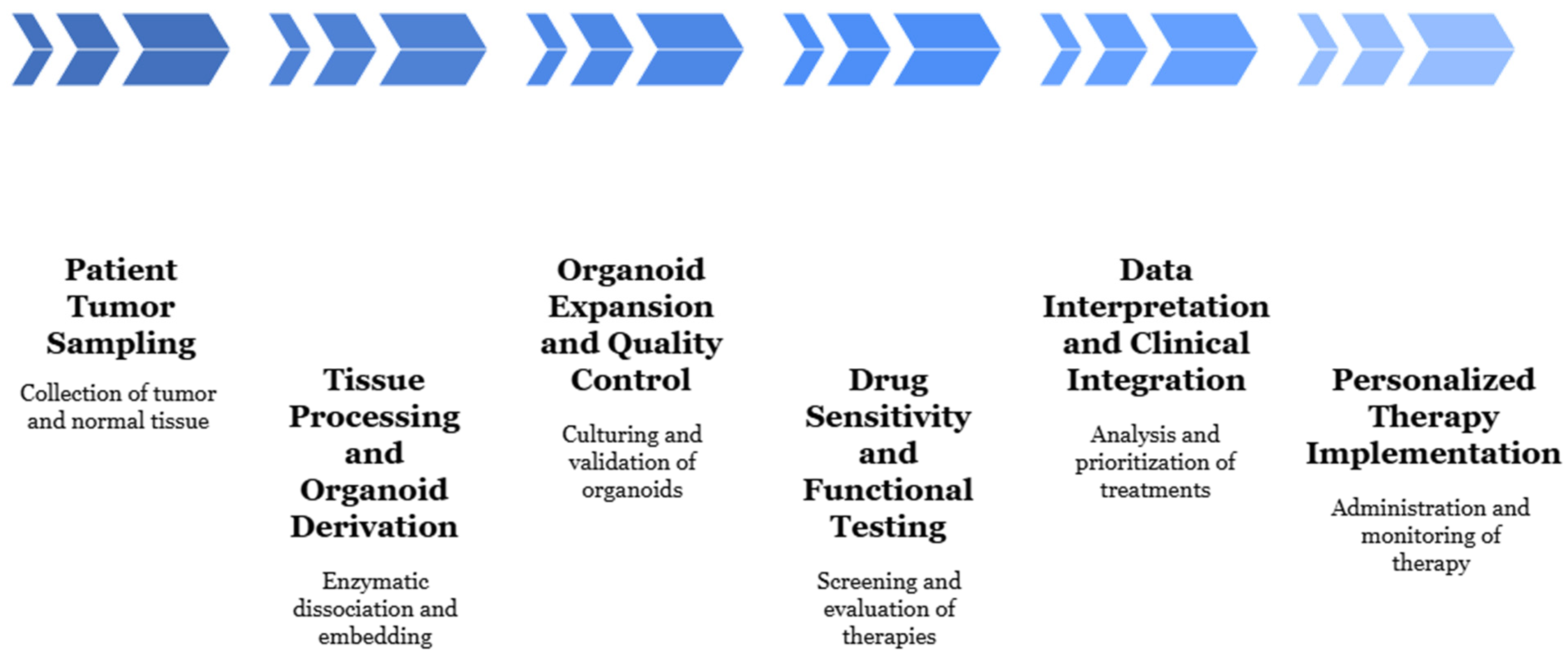
Living PDO biobanks have enabled clinicians to assess resistance evolution over time by comparing drug responses from primary and metastatic lesions or pre- and post-treatment samples. This longitudinal analysis has supported therapy adaptation in real time, such as shifting from monotherapy to combinatorial regimens in response to acquired resistance detected in PDOs. These studies are moving PDOs from bench to bedside, providing critical evidence for their integration into precision oncology pipelines, as mentioned in Figure 6.
Figure 6 illustrates the clinical workflow of incorporating patient-derived organoids, from tumor biopsy to organoid-based drug testing and individualized therapy selection.

5. Fundamental and Translational Applications

Organoids have revolutionized both fundamental and translational cancer research by offering a physiologically relevant but experimentally tractable model system. These three-dimensional structures have enabled the modeling of early tumorigenic events, host–microbiome interactions, and complex tumor microenvironments, with applications that span from mechanistic dissection to preclinical therapeutic validation [169].

5.1. Modeling Cancer Initiation and Progression

5.1.1. Gene Editing (CRISPR/Cas9) in Organoids

The application of CRISPR/Cas9 genome editing in organoids has enabled functional interrogation of cancer-driver genes in a patient-relevant context. Organoids derived from healthy epithelial tissues can be sequentially edited to introduce oncogenic mutations, allowing researchers to reconstruct stepwise tumor evolution in vitro [39]. This approach provides temporal and spatial resolution of mutation-driven transformation, in comparison to static cell line models or complex in vivo systems. For example, in intestinal organoids, researchers have introduced combinatorial mutations in APC, TP53, KRAS, and SMAD4 to recapitulate the adenoma-to-carcinoma sequence characteristic of colorectal cancer [40]. These engineered models exhibit progressive loss of polarity, epithelial integrity, and differentiation, along with enhanced Wnt and MAPK signaling, mimicking early neoplastic transformation. Recent innovations have also integrated base editors and prime editing systems into organoids to generate precise point mutations and study isoform-specific effects of cancer genes [170].

5.1.2. APC, TP53, and KRAS Mutations in CRC Modeling

The modeling of colorectal cancer using sequential genetic perturbations in normal colon organoids has become a benchmark in cancer biology [40]. Loss-of-function mutations in APC trigger hyperactivation of Wnt signaling, leading to increased proliferation. Subsequent introduction of activating mutations in KRAS enhances MAPK signaling and metabolic rewiring, while TP53 inactivation disrupts genomic integrity and apoptosis pathways [171]. Studies using such models have demonstrated that the combination of these mutations not only alters transcriptional and metabolic profiles but also confers resistance to chemotherapeutic agents and facilitates invasion into extracellular matrix analogs [172]. When orthotopically implanted into mice, these genetically engineered organoids form tumors that histologically and molecularly resemble patient-derived colorectal carcinomas. These models have thus proven instrumental in dissecting early oncogenic cooperation and validating therapeutic targets involved in epithelial transformation.

5.1.3. Phenotype Tracking Through Lineage Tracing

Lineage tracing in organoids, facilitated by CRISPR-based barcoding or inducible fluorescent reporters, enables the tracking of individual clones during transformation, therapy exposure, or metastatic progression [173]. In this context, organoids serve as platforms to map clonal dynamics and phenotypic plasticity under selective pressures. For example, tracking of barcoded subclones has revealed that rare minor populations within organoids may expand under drug pressure, giving rise to therapy-resistant phenotypes, an observation directly relevant to tumor relapse and acquired resistance in patients [174].

5.2. Microbiome–Tumor–Host Axis

5.2.1. GI Organoids and Microbiota Co-Culture

The gastrointestinal epithelium is a primary site of microbial interaction, and recent efforts have focused on integrating microbiota into GI-derived organoid models. Co-culturing intestinal or colonic organoids with bacterial species, including Fusobacterium nucleatum, Bacteroides fragilis, and Escherichia coli (pks+ strains) has enabled mechanistic studies of host–microbe interactions during tumorigenesis [175].
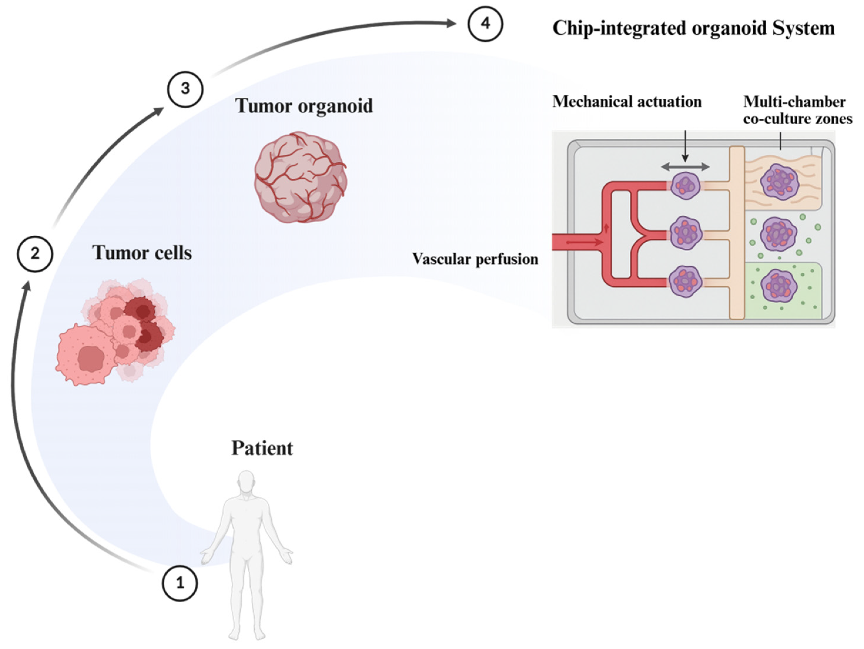
These bacteria induce a range of cellular responses in organoids, from DNA damage and genomic instability to inflammatory cytokine secretion and epigenetic remodeling. For instance, F. nucleatum has been shown to activate β-catenin signaling in colon organoids via FadA adhesin, promoting oncogenic transformation [176]. Such studies elucidate the microbiota’s role not just as a passive modulator but as an active participant in cancer initiation and progression. Several critical questions remain unanswered. How do different microbial communities modulate tumorigenesis across varying host genetic backgrounds? To what extent do organoid models capture the spatial and temporal dynamics of microbial colonization observed in vivo? Comparative studies using germ-free animal models and ex vivo explants are still needed to validate microbiota-induced phenotypes seen in organoid systems [177]. Limitations such as a lack of immune components, absence of mucus-secreting layers, and reduced oxygen tension gradients constrain the physiological accuracy of these co-cultures. Advancements in anaerobic chamber integration and multi-compartment chip-based models may offer more faithful reconstruction of the gut–microbiota–tumor axis [178].

5.2.2. Role of Short-Chain Fatty Acids and Pathogens

Short-chain fatty acids (SCFAs), such as butyrate and propionate, produced by commensal bacteria, exert both tumor-suppressive and tumor-promoting effects depending on context. In normal colon organoids, butyrate promotes differentiation and inhibits proliferation through HDAC inhibition and PPARγ activation [179]. In organoids harboring oncogenic mutations, SCFAs may paradoxically fuel metabolic reprogramming and support tumor growth. Conversely, genotoxins such as colibactin, secreted by pks+ E. coli, induce double-strand breaks and chromosomal rearrangements in colonic organoids, modeling microbial-driven mutagenesis [180]. The ability to manipulate microbiota composition and metabolite exposure in organoid cultures has opened new avenues to study microbe–epithelial crosstalk in a controlled and patient-relevant manner.

5.3. Tumor Microenvironment Integration

5.3.1. Fibroblasts, Endothelial Cells, ECM Modeling

Recreating the tumor microenvironment (TME) within organoid systems has been a major advance in bridging in vitro and in vivo cancer models. Co-cultures of tumor organoids with cancer-associated fibroblasts (CAFs), endothelial cells, and immune components have enabled the study of stromal–epithelial interactions, matrix remodeling, and angiogenic dynamics [181].
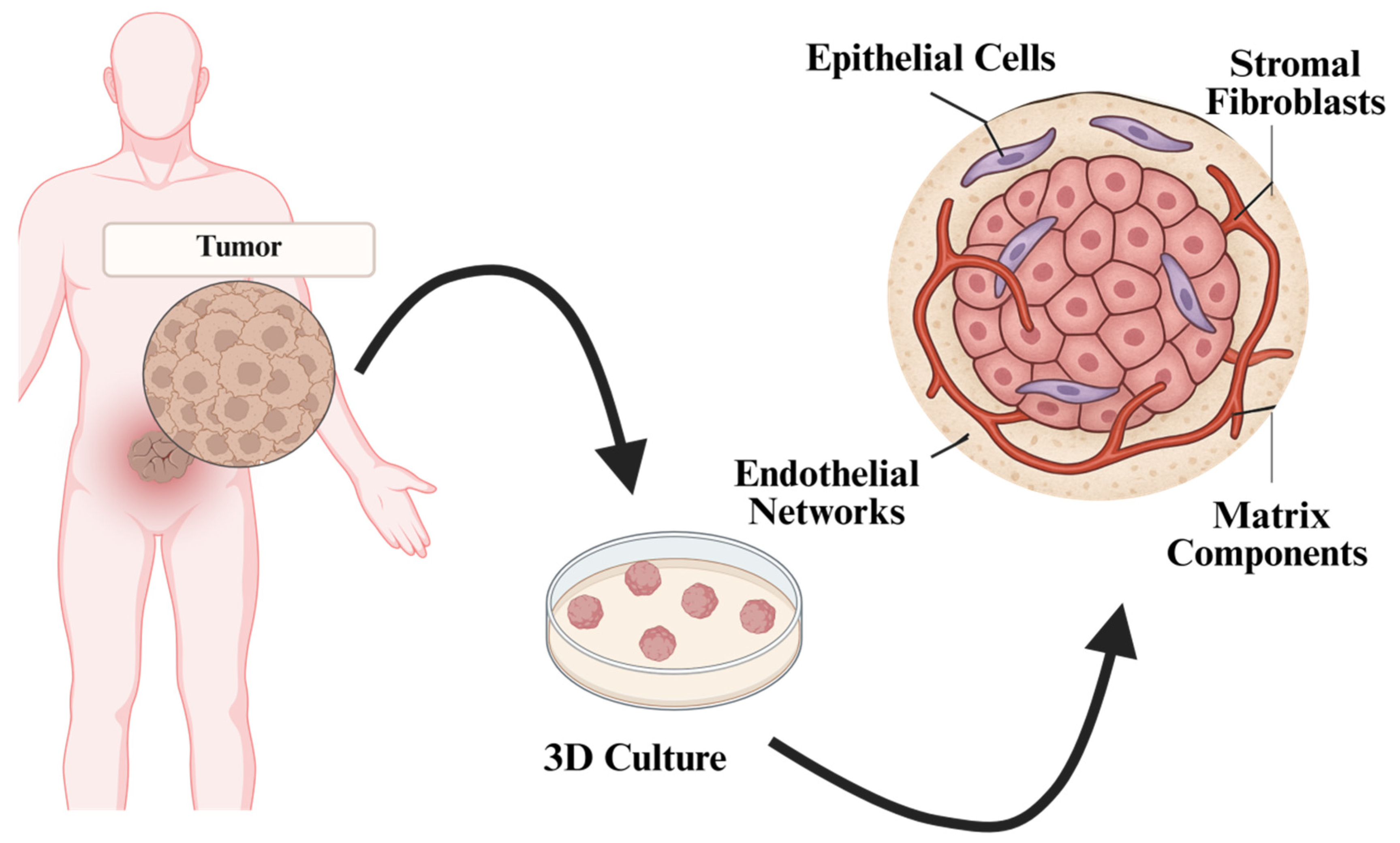
For example, co-culture of oral squamous carcinoma organoids with patient-matched CAFs revealed CAF-mediated secretion of VEGF-A and nicotinamide N-methyltransferase (NNMT), which modulated vascular signaling and enhanced tumor invasiveness [182]. In liver cancer organoids, the integration of endothelial progenitor cells in a three-dimensional ECM scaffold has been used to simulate vasculogenesis and assess anti-angiogenic therapies. Synthetic hydrogels and tunable extracellular matrices are increasingly employed to mimic stiffness gradients and integrin-binding profiles observed in native tumors [183]. These platforms facilitate studies on mechano-sensing, migration, and invasion, further enriching organoid utility in modeling tumor biophysics. A representative overview of such tumor microenvironment-integrated organoid systems is depicted in Figure 7, illustrating the spatial organization of epithelial tumor cells, stromal fibroblasts, endothelial elements, and extracellular matrix components.
Figure 7 shows a tumor organoid model incorporating key elements of the tumor microenvironment, including epithelial cells, stromal fibroblasts, endothelial networks, and extracellular matrix (ECM). This integrated system mimics cellular interactions and structural organization observed in vivo.

5.3.2. Paracrine Signaling and Angiogenesis Assays

Organoid–stroma co-culture systems have proven valuable for dissecting paracrine signaling pathways that regulate tumor progression. Cytokines such as IL-6, IL-8, CXCL12, and TGF-β secreted by stromal or immune cells modulate organoid proliferation, EMT, and therapeutic resistance [184]. Conversely, tumor-derived signals influence stromal activation and extracellular matrix remodeling. Functional angiogenesis assays within organoid cultures have demonstrated the formation of vascular networks in response to tumor-secreted factors, such as VEGF and FGF2 [185]. In perfused microfluidic organoid-on-chip platforms, these networks support nutrient exchange and simulate the vascularized tumor niche. Such integrated systems are now being used to test anti-angiogenic drugs and assess vascular normalization strategies in real time. Representative co-culture models incorporating stromal and immune cell types are summarized in Table 4.

5.4. Organoids in Cancer Metabolism Studies

5.4.1. Modeling Metabolic Reprogramming

A characteristic feature of cancer is metabolic rewiring, which allows tumor cells to maintain their growth, to adapt to nutrient changes, and to be resistant to therapy. To explore these events, organoids present a physiologically relevant platform, which maintains nutrient gradients, hypoxic niches, and stromal forces unattainable in traditional 2D systems. Recent reviews indicated that organoid models are coming into more and more common use to decompose metabolic heterogeneity, nutrient fluxes, and therapeutic vulnerabilities [196].

5.4.2. Metabolic Dependencies and Therapy Resistance

The nutrient dependencies and metabolic plasticity behind drug resistance have been demonstrated by the use of patient-derived organoids (PDOs). Indicatively, metabolic restructuring in favor of glycolysis has been demonstrated to sustain resistance to the neoadjuvant chemotherapy in triple-negative breast cancer, which can be recapitulated and functionally validated in organoid models [197]. Such models emphasize that metabolic transitions do not just provide a survival advantage, but they also provide an exploitable therapeutic target.

5.4.3. Hypoxia and Microenvironmental Influence

Cancer metabolism is highly influenced by the tumor microenvironment. Organoids of glioblastoma reproduced hypoxic gradients and heterogeneity of cancer stem cells with fidelity, recapitulating metabolic stress in vivo and enabling mechanistic studies of therapy resistance [198]. These characteristics render organoids the optimal choice to study the effect of oxygen and nutrient availability on clonal dynamics and invasive potential.

5.4.4. Epigenetic Regulation and Plasticity

The metabolites themselves can be epigenetic controllers of the tumor cell fate. Recently, lactate was found to facilitate cancer stemness and plasticity by modifying its epigenome, which offers a mechanistic connection between metabolism and chemoresistance [199]. By integrating these insights into PDO platforms, it is possible to longitudinally monitor metabolic-epigenetic crosstalk and to detect vulnerabilities in resistant subclones.

5.4.5. Translational Perspectives

These studies combined make organoids an inseparable part of cancer metabolism research. Combining metabolomics, imaging, and functional assays, organoids have the potential to reveal metabolic dependencies, predict therapeutic outcomes, and discover new drug targets. By connecting the metabolic biology to drug resistance, heterogeneity, and microenvironmental cues, organoid platforms are positioned at the center of precision oncology and next-generation cancer treatment.

6. Challenges and Limitations

Organoid technology has demonstrated unparalleled potential in preclinical modeling and functional precision oncology. Several limitations continue to impede its seamless integration into routine clinical and pharmaceutical pipelines.

6.1. Standardization Issues

6.1.1. Inter-Laboratory Variability

A persistent challenge in organoid research is the lack of standardized protocols across laboratories, leading to significant variability in organoid morphology, growth kinetics, and functional readouts [200]. Differences in tissue dissociation techniques, matrix composition, passage number, and culture media formulations can produce divergent results, even from genetically identical samples. These inconsistencies compromise the reproducibility and translational fidelity of organoid-based assays.
Harmonization initiatives led by consortia such as the Human Cancer Models Initiative (HCMI) and the Organoid Standardization Network are working toward universally accepted standard operating procedures (SOPs), unified data annotations, and quality control metrics [201]. The incorporation of automated platforms, machine vision algorithms, and digital image-based analytics is expected to improve inter-laboratory consistency and assay robustness.

6.1.2. Matrix and Media Inconsistencies

Matrigel and other basement membrane extracts, though widely used, suffer from batch-to-batch variability, xenogeneic origin, and undefined biochemical composition, impacting organoid reproducibility and clinical translatability [202]. The dependency on animal-derived growth factors and supplements raises regulatory concerns for good manufacturing practice (GMP) compliance. Bioengineered matrices, including polyethylene glycol (PEG)-based hydrogels, hyaluronic acid composites, and recombinant ECM proteins, are being actively developed to provide defined, tunable, and xeno-free scaffolds [203]. Parallel efforts are focused on chemically defined, serum-free media formulations tailored to tumor-specific genetic and phenotypic contexts.

6.2. Incomplete Physiological Representation

6.2.1. Absence of Vascularization

Most current organoid systems lack functional vasculature, which restricts nutrient and oxygen diffusion, limits organoid size and viability, and impairs modeling of angiogenesis, hypoxia, and drug pharmacokinetics. The incorporation of endothelial progenitor cells into scaffold matrices, along with perfusable microfluidic platforms (organoid-on-chip), has shown promise in simulating vascularized microenvironments [204]. Advances in 3D bioprinting and dynamic hydrogel systems are being leveraged to promote vessel formation and flow-based modeling of angiogenic signaling.

6.2.2. Limited Neural and Hormonal Inputs

Neurotrophic factors and hormonal signaling pathways play critical roles in tumor progression, invasion, and therapeutic resistance, but are largely absent in conventional organoid cultures. The lack of neural components restricts the study of perineural invasion, neuroendocrine tumors, and psychoneuroimmunological interactions [205]. Integration of iPSC-derived neurons and ganglia, along with neural–epithelial co-culture systems, is beginning to illuminate cancer–neuron interactions. These are further supported by electrophysiological chip-based systems that can simulate neurotransmitter signaling in real time.

6.2.3. Incomplete Immune Representation

Although autologous immune-organoid co-culture models have made significant strides, they often lack systemic immune components, such as naive lymphocytes, myeloid progenitors, and antigen-presenting cell networks, limiting their capacity to fully recapitulate tumor–immune dynamics. Microfluidic multi-organ chips and iPSC-derived hematopoietic precursors are being used to simulate immune cell infiltration, circulation, and checkpoint activation [206]. These platforms are enabling the study of immune evasion, exhaustion, and response to checkpoint inhibitors in a physiologically relevant context.

6.3. Scalability, Cost, and Clinical Translation

6.3.1. Limited Scalability and GMP Compliance

Manual handling, reliance on costly reagents, and labor-intensive protocols render large-scale organoid production challenging. Most existing platforms lack validation for GMP conditions required for clinical deployment in diagnostics or personalized therapy. Modular bioreactors, robotic handling systems, and closed-loop culture devices are now being developed for high-throughput, GMP-compatible organoid manufacturing [207]. These systems integrate real-time monitoring, automated passaging, and AI-based image analysis to ensure consistency and reduce operator bias.

6.3.2. High Operational Costs

The cost associated with long-term culture, especially growth factors, including R-spondin, Wnt3a, and FGF2, and matrix components, such as Matrigel, remains prohibitive for routine clinical use and widespread industrial scaling [208]. Cost-effective alternatives include the use of recombinant analogs, pooled culture platforms, and media recycling technologies. Public–private partnerships and centralized organoid facilities are being explored to offset infrastructure costs and facilitate equitable access.

6.4. Regulatory and Ethical Considerations

6.4.1. Lack of Regulatory Frameworks

The absence of formal validation pipelines by regulatory authorities such as the FDA or EMA for organoid-based assays creates ambiguity around their use in clinical decision-making and drug approval pathways. Ongoing efforts to integrate organoid pharmacotyping into umbrella and basket trials, as seen in the Selective Precision Experimental Combination Therapy Umbrella Trial (SPECTRUM) and Drug Rediscovery Protocol (DRUP) extensions, are helping to establish clinical correlations and assay validation benchmarks [209,210]. Dialogue between organoid consortia and regulators is also fostering the creation of translational pipelines and companion diagnostics.

6.4.2. Ethical and Data Governance Issues

The generation and storage of patient-derived organoids, often coupled with genomic, transcriptomic, and pharmacologic data, raise ethical concerns around patient consent, data security, and biobank governance [211]. Dynamic consent frameworks, blockchain-based data tracking, and decentralized identity verification protocols are being adopted to enhance patient autonomy and data traceability. These models promote ethical compliance while supporting collaborative multi-center research.

7. Future Perspectives and Innovations

Organoid technology is entering a new era, defined by cross-disciplinary innovations that extend beyond conventional 3D culture systems. Microfluidic engineering, functional genomics, artificial intelligence, and regulatory integration are rapidly enhancing organoids’ predictive capacity, standardization, and clinical relevance. This section explores the next frontier in organoid research and application, offering a forward-looking roadmap grounded in recent technological and biomedical advances.

7.1. Organoid-on-a-Chip

7.1.1. Microfluidic Perfusion Systems

The integration of microfluidic systems with organoid cultures, termed “organoid-on-a-chip”, represents a significant leap toward recapitulating physiological microenvironments. These platforms support continuous perfusion of culture media, maintaining stable nutrient and oxygen gradients, and simulating hemodynamic forces encountered in vivo [212]. Perfused tumor organoid chips have demonstrated enhanced viability, improved barrier function, and more accurate pharmacodynamic responses compared to static culture, as shown in Figure 8. Recent studies in pancreatic and colorectal cancer have shown that vascularized, chip-based organoids respond differentially to chemotherapy and anti-angiogenic agents, mirroring patient-specific responses in xenografts and clinical settings [213].
Figure 8 shows a dynamic organoid-on-a-chip platform integrating vascular perfusion, mechanical forces, and multi-cell co-culture zones. Enables real-time analysis of drug response, tumor–stroma interactions, and microenvironmental cues.

7.1.2. Spatiotemporal Simulation of Organ-Level Cues

Advanced microengineered platforms now incorporate organ-level mechanical and chemical cues, enabling a higher fidelity of tissue simulation. For example, lung tumor organoids subjected to cyclic stretching replicate airway dynamics and show improved alveolar-like differentiation [22]. In intestinal cancer models, devices that emulate peristalsis and pH gradients have enabled the study of biomechanical regulation of stemness, tumor growth, and epithelial barrier function [214]. These dynamic systems also facilitate real-time imaging, enabling high-resolution analysis of drug penetration, tumor–stroma interactions, and migration phenotypes under physiologically relevant flow conditions.

7.2. Organoid Libraries and CRISPR Screens

7.2.1. Isogenic Organoid Banks

The generation of large, isogenic organoid libraries using CRISPR-based editing has enabled systematic dissection of cancer gene function in a controlled 3D context. These libraries consist of organoids derived from a single genetic background, engineered to carry distinct driver mutations (e.g., TP53, KRAS, PIK3CA, IDH1), allowing rigorous genotype–phenotype mapping [39,40].

7.2.2. Functional Genomic Screens (Synthetic Lethality)

High-throughput CRISPR knockout and base-editing screens in organoid systems are identifying context-specific genetic dependencies and synthetic lethal interactions. Recent pooled CRISPR screens in microsatellite instability-high (MSI-H) colorectal cancer organoids identified novel vulnerabilities, such as WRN helicase inhibition, leading to selective cell death [215].

7.3. Organoid Image-to-Response Prediction

Artificial intelligence (AI), particularly deep learning, is transforming the analysis of organoid-based drug screening. Convolutional neural networks (CNNs) trained on thousands of annotated organoid images can now predict drug response, treatment toxicity, and resistance mechanisms with high accuracy [76]. A recent model trained on ovarian cancer organoids achieved over 90% concordance with clinical outcomes by identifying early morphological signatures predictive of platinum sensitivity [216]. These models allow real-time, non-invasive assessment of treatment efficacy, reducing reliance on endpoint assays and enabling adaptive drug screening workflows. To enhance predictive accuracy and mechanistic insights, integrated AI–multi-omics pipelines have been developed that combine organoid-based drug screening with image-driven phenotypic profiling and multi-omic data integration, as illustrated in Figure 9.
Figure 9 shows an integrated pipeline combining organoid pharmacotyping, high-content imaging, deep learning–based phenotypic analysis, and multi-omic mapping. This system enables mechanistic interpretation and prioritization of personalized therapies in cancer models.

7.4. Strategic Gaps and Translational Priorities in Organoid Oncology

Despite rapid progress in organoid-based cancer modeling, several critical barriers remain that must be addressed to realize organoids’ full clinical and translational potential. This section outlines key challenges and forward-looking strategies to enhance organoid platforms for precision medicine. A comprehensive SWOT analysis, shown in Figure 10, further illustrates the strategic strengths and translational gaps that will define the next phase of organoid-based oncology research.
Closing these gaps will require collaborative efforts to standardize protocols, validate assays, and integrate organoids ethically into clinical workflows. By addressing weaknesses and leveraging opportunities, organoid technologies can move from experimental tools to reliable precision oncology platforms, ultimately improving patient outcomes.

8. Conclusions

Organoid technology has emerged as a transformative platform in cancer research, enabling the development of three-dimensional, patient-specific models that recapitulate tumor architecture, genetic heterogeneity, and microenvironmental complexity. Compared to traditional models, organoids offer superior fidelity, scalability, and translational relevance, bridging the gap between laboratory findings and clinical decision-making. Their integration into functional precision oncology now allows real-time pharmacotyping, resistance profiling, and immunotherapy modeling using autologous tumor material. Clinical studies increasingly demonstrate concordance between organoid-based drug responses and patient outcomes, underscoring their potential in individualized treatment planning. Despite these advances, significant challenges remain. The lack of standardized protocols, limited vascular and neural integration, and the absence of dynamic immune interactions constrain their full physiological representation. Scalability under GMP conditions and regulatory approval pathways also remain key bottlenecks for clinical implementation. Ongoing innovations, including vascularized organoid-on-chip systems, iPSC-derived immune co-cultures, AI-driven drug response analytics, and CRISPR-based functional genomics, are actively addressing these gaps. As these technologies converge, organoids are evolving into dynamic platforms capable of modeling tumor evolution, predicting therapy combinations, and supporting virtual clinical trial simulations. Organoids are poised to transition from advanced research tools to essential components of precision oncology. Organoid models now also provide unique opportunities to study cancer metabolism, capturing nutrient dependencies and metabolic plasticity. Integrating these insights enhances their translational power in linking heterogeneity, therapy resistance, and precision oncology. Their continued development will redefine how to model cancer, identify actionable vulnerabilities, and tailor treatments in real time.

Author Contributions

M.K.S. conceptualized and outlined the whole manuscript. A.M. and R.S. performed the initial literature survey and prepared the first draft of the manuscript. R.S. and N.D. assisted in the collection and organization of relevant literature, preparation of figures/tables, and refinement of the manuscript. S.-H.J. provided critical insights, reviewed the manuscript, and offered intellectual inputs to strengthen the scientific depth. M.K.S. finally supervised the review paper, contributed to the overall design of the manuscript, revised and edited the final version, and approved the submission. All authors have read and agreed to the published version of the manuscript.

Funding

This research received no external funding.

Institutional Review Board Statement

Not applicable.

Informed Consent Statement

Not applicable.

Data Availability Statement

No new data were created or analyzed in this study. Data sharing is not applicable to this article.

Conflicts of Interest

The authors declare no conflict of interest.

References

  1. Biswas, A.; De, S. Drivers of dynamic intratumor heterogeneity and phenotypic plasticity. Am. J. Physiol. Cell Physiol. 2021, 320, C750–C760. [Google Scholar] [CrossRef]
  2. Zhang, A.; Miao, K.; Sun, H.; Deng, C.X. Tumor heterogeneity reshapes the tumor microenvironment to influence drug resistance. Int. J. Biol. Sci. 2022, 18, 3019. [Google Scholar] [CrossRef]
  3. Sajjad, H.; Imtiaz, S.; Noor, T.; Siddiqui, Y.H.; Sajjad, A.; Zia, M. Cancer models in preclinical research: A chronicle review of advancement in effective cancer research. Anim. Model. Exp. Med. 2021, 4, 87–103. [Google Scholar] [CrossRef] [PubMed]
  4. Habanjar, O.; Diab-Assaf, M.; Caldefie-Chezet, F.; Delort, L. 3D cell culture systems: Tumor application, advantages, and disadvantages. Int. J. Mol. Sci. 2021, 22, 12200. [Google Scholar] [CrossRef] [PubMed]
  5. Liu, Y.; Wu, W.; Cai, C.; Zhang, H.; Shen, H.; Han, Y. Patient-derived xenograft models in cancer therapy: Technologies and applications. Signal Transduct. Target. Ther. 2023, 8, 160. [Google Scholar] [CrossRef] [PubMed]
  6. Han, X.; Cai, C.; Deng, W.; Shi, Y.; Li, L.; Wang, C.; Zhang, J.; Rong, M.; Liu, J.; Fang, B.; et al. Landscape of human organoids: Ideal model in clinics and research. Innov. 2024, 5, 100620. [Google Scholar] [CrossRef]
  7. Shao, Y.; Fu, J. Engineering multiscale structural orders for high-fidelity embryoids and organoids. Cell Stem Cell 2022, 29, 722–743. [Google Scholar] [CrossRef]
  8. Furbo, S.; Urbano, P.C.; Raskov, H.H.; Troelsen, J.T.; Kanstrup Fiehn, A.M.; Gögenur, I. Use of patient-derived organoids as a treatment selection model for colorectal cancer: A narrative review. Cancers 2022, 14, 1069. [Google Scholar] [CrossRef]
  9. Qu, S.; Xu, R.; Yi, G.; Li, Z.; Zhang, H.; Qi, S.; Huang, G. Patient-derived organoids in human cancer: A platform for fundamental research and precision medicine. Mol. Biomed. 2024, 5, 6. [Google Scholar] [CrossRef]
  10. Xian, L.; Zhao, P.; Chen, X.; Wei, Z.; Ji, H.; Zhao, J.; Liu, W.; Li, Z.; Liu, D.; Han, X.; et al. Heterogeneity, inherent and acquired drug resistance in patient-derived organoid models of primary liver cancer. Cell. Oncol. 2022, 45, 1019–1036. [Google Scholar] [CrossRef]
  11. Singh, D.; Thakur, A.; Rakesh kumar, A. Advancements in Organoid-Based Drug Discovery: Revolutionizing Precision Medicine and Pharmacology. Drug Dev. Res. 2025, 86, e70121. [Google Scholar] [CrossRef] [PubMed]
  12. Bolck, H.A.; Corrò, C.; Kahraman, A.; von Teichman, A.; Toussaint, N.C.; Kuipers, J.; Chiovaro, F.; Koelzer, V.H.; Pauli, C.; Moritz, W.; et al. Tracing clonal dynamics reveals that two-and three-dimensional patient-derived cell models capture tumor heterogeneity of clear cell renal cell carcinoma. Eur. Urol. Focus 2021, 7, 152–162. [Google Scholar] [CrossRef] [PubMed]
  13. Arana, Á.J.; González-Llera, L.; Barreiro-Iglesias, A.; Sánchez, L. Emerging Frontiers in Zebrafish Embryonic and Adult-Derived Cell Lines. Int. J. Mol. Sci. 2025, 26, 4351. [Google Scholar] [CrossRef] [PubMed]
  14. Serelli-Lee, V.; Ito, K.; Koibuchi, A.; Tanigawa, T.; Ueno, T.; Matsushima, N.; Imai, Y. A state-of-the-art roadmap for biomarker-driven drug development in the era of personalized therapies. J. Pers. Med. 2022, 12, 669. [Google Scholar] [CrossRef]
  15. Brassard, J.A.; Nikolaev, M.; Hübscher, T.; Hofer, M.; Lutolf, M.P. Recapitulating macro-scale tissue self-organization through organoid bioprinting. Nat. Mater. 2021, 20, 22–29. [Google Scholar] [CrossRef]
  16. Spagnol, G.; Sensi, F.; De Tommasi, O.; Marchetti, M.; Bonaldo, G.; Xhindoli, L.; Noventa, M.; Agostini, M.; Tozzi, R.; Saccardi, C. Patient derived organoids (PDOs), extracellular matrix (ECM), tumor microenvironment (TME) and drug screening: State of the art and clinical implications of ovarian cancer organoids in the era of precision medicine. Cancers 2023, 15, 2059. [Google Scholar] [CrossRef]
  17. Nikokiraki, C.; Psaraki, A.; Roubelakis, M.G. The potential clinical use of stem/progenitor cells and organoids in liver diseases. Cells 2022, 11, 1410. [Google Scholar] [CrossRef]
  18. Elmentaite, R.; Domínguez Conde, C.; Yang, L.; Teichmann, S.A. Single-cell atlases: Shared and tissue-specific cell types across human organs. Nat. Rev. Genet. 2022, 23, 395–410. [Google Scholar] [CrossRef]
  19. Kang, D.H.; Lee, J.; Im, S.; Chung, C. Navigating the complexity of resistance in lung cancer therapy: Mechanisms, organoid models, and strategies for overcoming treatment failure. Cancers 2024, 16, 3996. [Google Scholar] [CrossRef]
  20. Ng, A.S.; Chan, D.K. Commonalities and differences in the mutational signature and somatic driver mutation landscape across solid and hollow viscus organs. Oncogene 2023, 42, 2713–2724. [Google Scholar] [CrossRef]
  21. Bhattacharya, A.; Alam, K.; Roy, N.S.; Kaur, K.; Kaity, S.; Ravichandiran, V.; Roy, S. Exploring the interaction between extracellular matrix components in a 3D organoid disease model to replicate the pathophysiology of breast cancer. J. Exp. Clin. Cancer Res. 2023, 42, 343. [Google Scholar] [CrossRef]
  22. Ekanger, C.T.; Ramnefjell, M.P.; Guttormsen, M.S.; Hekland, J.; Dahl-Michelsen, K.; Lotsberg, M.L.; Lu, N.; Stuhr, L.E.; Hoareau, L.; Salminen, P.R.; et al. An Organoid Model for Translational Cancer Research Recapitulates Histoarchitecture and Molecular Hallmarks of Non-Small-Cell Lung Cancer. Cancers 2025, 17, 1873. [Google Scholar] [CrossRef] [PubMed]
  23. Nam, C.; Ziman, B.; Sheth, M.; Zhao, H.; Lin, D.C. Genomic and epigenomic characterization of tumor organoid models. Cancers 2022, 14, 4090. [Google Scholar] [CrossRef] [PubMed]
  24. El Harane, S.; Zidi, B.; El Harane, N.; Krause, K.H.; Matthes, T.; Preynat-Seauve, O. Cancer spheroids and organoids as novel tools for research and therapy: State of the art and challenges to guide precision medicine. Cells 2023, 12, 1001. [Google Scholar] [CrossRef] [PubMed]
  25. Monberg, M. Leveraging Single Cell Technologies for the Characterization and Treatment of Refractory Pancreatic Cancer. Ph.D. Thesis, The University of Texas, Houston, TX, USA, 2022. [Google Scholar]
  26. Xiao, Y.; Li, Y.; Jing, X.; Weng, L.; Liu, X.; Liu, Q.; Chen, K. Organoid models in oncology: Advancing precision cancer therapy and vaccine development. Cancer Biol. Med. 2025, 22, 903–927. [Google Scholar] [CrossRef]
  27. Marchini, A.; Gelain, F. Synthetic scaffolds for 3D cell cultures and organoids: Applications in regenerative medicine. Crit. Rev. Biotechnol. 2022, 42, 468–486. [Google Scholar] [CrossRef]
  28. Xu, H.; Jiao, D.; Liu, A.; Wu, K. Tumor organoids: Applications in cancer modeling and potentials in precision medicine. J. Hematol. Oncol. 2022, 15, 58. [Google Scholar] [CrossRef]
  29. Tu, S.M.; Singh, S.R.; Arnaoutakis, K.; Malapati, S.; Bhatti, S.A.; Joon, A.Y.; Atiq, O.T.; Pisters, L.L. Stem cell theory of cancer: Implications for translational research from bedside to bench. Cancers 2022, 14, 3345. [Google Scholar] [CrossRef]
  30. Pamarthy, S.; Sabaawy, H.E. Patient derived organoids in prostate cancer: Improving therapeutic efficacy in precision medicine. Mol. Cancer 2021, 20, 125. [Google Scholar] [CrossRef]
  31. Bisht, S.; Nigam, M.; Kunjwal, S.S.; Sergey, P.; Mishra, A.P.; Sharifi-Rad, J. Cancer stem cells: From an insight into the basics to recent advances and therapeutic targeting. Stem Cells Int. 2022, 2022, 9653244. [Google Scholar] [CrossRef]
  32. Ren, J.; Liu, M.; Rong, M.; Zhang, X.; Wang, G.; Liu, Y.; Li, H.; Duan, S. The pros and cons of mechanical dissociation and enzymatic digestion in patient-derived organoid cultures for solid tumor. Cell Organoid 2024, 1, 9410009. [Google Scholar] [CrossRef]
  33. Pang, X.; Hu, Y.; Dai, Z.; Lou, Q.; Xu, W.; Chen, L. Precision medicine research progress based on colorectal cancer organoids. Discov. Oncol. 2025, 16, 1181. [Google Scholar] [CrossRef] [PubMed]
  34. Alnasser, S.M. Advances and Challenges in Cancer Stem Cells for Onco-Therapeutics. Stem Cells Int. 2023, 2023, 8722803. [Google Scholar] [CrossRef] [PubMed]
  35. Izycka, N.; Rucinski, M.; Andrzejewska, M.; Szubert, S.; Nowak-Markwitz, E.; Sterzynska, K. The prognostic value of Cancer Stem Cell Markers (CSCs) expression—ALDH1A1, CD133, CD44—For survival and long-term follow-up of ovarian cancer patients. Int. J. Mol. Sci. 2023, 24, 2400. [Google Scholar] [CrossRef] [PubMed]
  36. Shah, S.; D’Souza, G.G. Modeling tumor microenvironment complexity in vitro: Spheroids as physiologically relevant tumor models and strategies for their analysis. Cells 2025, 14, 732. [Google Scholar] [CrossRef]
  37. Aldoghachi, A.F.; Chong, Z.X.; Yeap, S.K.; Cheong, S.K.; Ho, W.Y.; Ong, A.H. Stem cells for cancer therapy: Translating the uncertainties and possibilities of stem cell properties into opportunities for effective cancer therapy. Int. J. Mol. Sci. 2023, 24, 1012. [Google Scholar] [CrossRef]
  38. Cerneckis, J.; Cai, H.; Shi, Y. Induced pluripotent stem cells (iPSCs): Molecular mechanisms of induction and applications. Signal Transduct. Target. Ther. 2024, 9, 112. [Google Scholar] [CrossRef]
  39. Lo, Y.H.; Kolahi, K.S.; Du, Y.; Chang, C.Y.; Krokhotin, A.; Nair, A.; Sobba, W.D.; Karlsson, K.; Jones, S.J.; Longacre, T.A.; et al. A CRISPR/Cas9-engineered ARID1A-deficient human gastric cancer organoid model reveals essential and nonessential modes of oncogenic transformation. Cancer Discov. 2021, 11, 1562–1581. [Google Scholar] [CrossRef]
  40. Al-Kabani, A.; Huda, B.; Haddad, J.; Yousuf, M.; Bhurka, F.; Ajaz, F.; Patnaik, R.; Jannati, S.; Banerjee, Y. Exploring Experimental Models of Colorectal Cancer: A Critical Appraisal from 2D Cell Systems to Organoids, Humanized Mouse Avatars, Organ-on-Chip, CRISPR Engineering, and AI-Driven Platforms—Challenges and Opportunities for Translational Precision Oncology. Cancers 2025, 17, 2163. [Google Scholar]
  41. Miloradovic, D.; Pavlovic, D.; Jankovic, M.G.; Nikolic, S.; Papic, M.; Milivojevic, N.; Stojkovic, M.; Ljujic, B. Human embryos, induced pluripotent stem cells, and organoids: Models to assess the effects of environmental plastic pollution. Front. Cell Dev. Biol. 2021, 9, 709183. [Google Scholar] [CrossRef]
  42. Dave, B.; Tailor, J. Human stem cell models to unravel brain cancer. BMC Cancer 2024, 24, 1465. [Google Scholar] [CrossRef]
  43. Yehya, A.; Youssef, J.; Hachem, S.; Ismael, J.; Abou-Kheir, W. Tissue-specific cancer stem/progenitor cells: Therapeutic implications. World J. Stem Cells 2023, 15, 323. [Google Scholar] [CrossRef]
  44. Milne, J.V.; Mustafa, E.H.; Clemons, N.J. Modelling esophageal adenocarcinoma and Barrett’s esophagus with patient-derived organoids. Front. Mol. Biosci. 2024, 11, 1382070. [Google Scholar] [CrossRef]
  45. Zhou, J.; Shi, Y. Mesenchymal stem/stromal cells (MSCs): Origin, immune regulation, and clinical applications. Cell. Mol. Immunol. 2023, 20, 555–557. [Google Scholar] [CrossRef] [PubMed]
  46. Gu, Z.; Wu, Q.; Shang, B.; Zhang, K.; Zhang, W. Organoid co-culture models of the tumor microenvironment promote precision medicine. Cancer Innov. 2024, 3, e101. [Google Scholar] [CrossRef] [PubMed]
  47. Allen, T.A. The role of circulating tumor cells as a liquid biopsy for cancer: Advances, biology, technical challenges, and clinical relevance. Cancers 2024, 16, 1377. [Google Scholar] [CrossRef] [PubMed]
  48. Pan, C.; Wang, X.; Yang, C.; Fu, K.; Wang, F.; Fu, L. The culture and application of circulating tumor cell-derived organoids. Trends Cell Biol. 2025, 35, 364–380. [Google Scholar] [CrossRef]
  49. Marei, H.E. Stem Cell and Synthetic Embryo Models: Advances, Applications, and Ethical Considerations. Stem Cell Rev. Rep. 2025, 21, 1648–1668. [Google Scholar] [CrossRef]
  50. Azeez, S.S.; Hamad, R.S.; Hamad, B.K.; Shekha, M.S.; Bergsten, P. Advances in CRISPR-Cas technology and its applications: Revolutionising precision medicine. Front. Genome Ed. 2024, 6, 1509924. [Google Scholar] [CrossRef]
  51. Yan, H.H.N.; Chan, A.S.; Lai, F.P.; Leung, S.Y. Organoid cultures for cancer modeling. Cell Stem Cell 2023, 30, 917–937. [Google Scholar] [CrossRef] [PubMed]
  52. Verstegen, M.M.A.; Coppes, R.P.; Beghin, A.; De Coppi, P.; Gerli, M.F.M.; de Graeff, N.; Pan, Q.; Saito, Y.; Shi, S.; Zadpoor, A.A.; et al. Clinical applications of human organoids. Nat. Med. 2025, 31, 409–421. [Google Scholar] [CrossRef] [PubMed]
  53. Chen, L.; Luo, X.; Zhang, J.; Zhang, J.; Yang, C.; Zhao, Y. Harnessing Organoid Platforms for Nanoparticle Drug Development. Drug Des. Dev. Ther. 2025, 19, 6125–6143. [Google Scholar] [CrossRef] [PubMed] [PubMed Central]
  54. He, R.; Weng, Z.; Liu, Y.; Li, B.; Wang, W.; Meng, W.; Li, B.; Li, L. Application of Induced Pluripotent Stem Cells in Malignant Solid Tumors. Stem Cell Rev. Rep. 2023, 19, 2557–2575. [Google Scholar] [CrossRef] [PubMed] [PubMed Central]
  55. De Visser, K.E.; Joyce, J.A. The evolving tumor microenvironment: From cancer initiation to metastatic outgrowth. Cancer Cell 2023, 41, 374–403. [Google Scholar] [CrossRef] [PubMed]
  56. Wang, J.; Tao, X.; Zhu, J.; Dai, Z.; Du, Y.; Xie, Y.; Chu, X.; Fu, G.; Lei, Z. Tumor organoid-immune co-culture models: Exploring a new perspective of tumor immunity. Cell Death Discov. 2025, 11, 195. [Google Scholar] [CrossRef] [PubMed] [PubMed Central]
  57. Policastro, B.; Nissen, N.; Alves, C.L. Deciphering Breast Tumor Heterogeneity Through Patient-Derived Organoids and Circulating Tumor Cells. J. Pers. Med. 2025, 15, 271. [Google Scholar] [CrossRef] [PubMed] [PubMed Central]
  58. Mukhare, R.; Gandhi, K.A.; Kadam, A.; Raja, A.; Singh, A.; Madhav, M.; Chaubal, R.; Pandey, S.; Gupta, S. Integration of Organoids With CRISPR Screens: A Narrative Review. Biol. Cell 2025, 117, e70006. [Google Scholar] [CrossRef] [PubMed] [PubMed Central]
  59. Badekila, A.K.; Kini, S.; Jaiswal, A.K. Fabrication techniques of biomimetic scaffolds in three-dimensional cell culture: A review. J. Cell. Physiol. 2021, 236, 741–762. [Google Scholar] [CrossRef]
  60. Yi, S.A.; Zhang, Y.; Rathnam, C.; Pongkulapa, T.; Lee, K.B. Bioengineering approaches for the advanced organoid research. Adv. Mater. 2021, 33, 2007949. [Google Scholar] [CrossRef]
  61. Valdoz, J.C.; Johnson, B.C.; Jacobs, D.J.; Franks, N.A.; Dodson, E.L.; Sanders, C.; Cribbs, C.G.; Van Ry, P.M. The ECM: To scaffold, or not to scaffold, that is the question. Int. J. Mol. Sci. 2021, 22, 12690. [Google Scholar] [CrossRef]
  62. Kozlowski, M.T.; Crook, C.J.; Ku, H.T. Towards organoid culture without Matrigel. Commun. Biol. 2021, 4, 1387. [Google Scholar] [CrossRef]
  63. Zhao, Z.; Chen, X.; Dowbaj, A.M.; Sljukic, A.; Bratlie, K.; Lin, L.; Fong, E.L.S.; Balachander, G.M.; Chen, Z.; Soragni, A.; et al. Organoids. Nat. Rev. Methods Prim. 2022, 2, 94. [Google Scholar] [CrossRef] [PubMed] [PubMed Central]
  64. Rana, M.M.; De la Hoz Siegler, H. Evolution of hybrid hydrogels: Next-generation biomaterials for drug delivery and tissue engineering. Gels 2024, 10, 216. [Google Scholar] [CrossRef]
  65. Sharma, P.; Pal, V.K.; Roy, S. An overview of latest advances in exploring bioactive peptide hydrogels for neural tissue engineering. Biomater. Sci. 2021, 9, 3911–3938. [Google Scholar] [CrossRef]
  66. Thakuri, P.S.; Liu, C.; Luker, G.D.; Tavana, H. Biomaterials-Based Approaches to Tumor Spheroid and Organoid Modeling. Adv. Healthc. Mater. 2018, 7, e1700980. [Google Scholar] [CrossRef] [PubMed] [PubMed Central]
  67. Shou, Y.; Teo, X.Y.; Wu, K.Z.; Bai, B.; Kumar, A.R.; Low, J.; Le, Z.; Tay, A. Dynamic stimulations with bioengineered extracellular matrix-mimicking hydrogels for mechano cell reprogramming and therapy. Adv. Sci. 2023, 10, 2300670. [Google Scholar] [CrossRef] [PubMed]
  68. Hu, Y.; Zhu, T.; Cui, H.; Cui, H. Integrating 3D bioprinting and organoids to better recapitulate the complexity of cellular microenvironments for tissue engineering. Adv. Healthc. Mater. 2025, 14, 2403762. [Google Scholar] [CrossRef] [PubMed]
  69. Smandri, A.; Al-Masawa, M.E.; Hwei, N.M.; Fauzi, M.B. ECM-derived biomaterials for regulating tissue multicellularity and maturation. iScience 2024, 27, 109141. [Google Scholar] [CrossRef]
  70. Scalise, M.; Marino, F.; Salerno, L.; Cianflone, E.; Molinaro, C.; Salerno, N.; De Angelis, A.; Viglietto, G.; Urbanek, K.; Torella, D. From spheroids to organoids: The next generation of model systems of human cardiac regeneration in a dish. Int. J. Mol. Sci. 2021, 22, 13180. [Google Scholar] [CrossRef]
  71. Liu, D.; Chen, S.; Win Naing, M. A review of manufacturing capabilities of cell spheroid generation technologies and future development. Biotechnol. Bioeng. 2021, 118, 542–554. [Google Scholar] [CrossRef]
  72. Arora, S.; Singh, S.; Mittal, A.; Desai, N.; Khatri, D.K.; Gugulothu, D.; Lather, V.; Pandita, D.; Vora, L.K. Spheroids in cancer research: Recent advances and opportunities. J. Drug Deliv. Sci. Technol. 2024, 100, 106033. [Google Scholar] [CrossRef]
  73. Li, Y.; Wang, Y.; Wan, K.; Wu, M.; Guo, L.; Liu, X.; Wei, G. On the design, functions, and biomedical applications of high-throughput dielectrophoretic micro-/nanoplatforms: A review. Nanoscale 2021, 13, 4330–4358. [Google Scholar] [CrossRef]
  74. Garcia Garcia, F.C. Microfluidic Control of Oxygen for Long-Term Maintenance of Tissues on Chip. Ph.D. Thesis, University of Southampton, Southampton, UK, 2024. [Google Scholar]
  75. Picollet-D’hahan, N.; Zuchowska, A.; Lemeunier, I.; Le Gac, S. Multiorgan-on-a-chip: A systemic approach to model and decipher inter-organ communication. Trends Biotechnol. 2021, 39, 788–810. [Google Scholar] [CrossRef]
  76. Maramraju, S.; Kowalczewski, A.; Kaza, A.; Liu, X.; Singaraju, J.P.; Albert, M.V.; Ma, Z.; Yang, H. AI-organoid integrated systems for biomedical studies and applications. Bioeng. Transl. Med. 2024, 9, e10641. [Google Scholar] [CrossRef] [PubMed]
  77. Silva-Pedrosa, R.; Salgado, A.J.; Ferreira, P.E. Revolutionizing disease modeling: The emergence of organoids in cellular systems. Cells 2023, 12, 930. [Google Scholar] [CrossRef] [PubMed]
  78. Hofer, M.; Lutolf, M.P. Engineering organoids. Nat. Rev. Mater. 2021, 6, 402–420. [Google Scholar] [CrossRef] [PubMed]
  79. Giron-Michel, J.; Padelli, M.; Oberlin, E.; Guenou, H.; Duclos-Vallée, J.C. State-of-the-art liver cancer organoids: Modeling cancer stem cell heterogeneity for personalized treatment. BioDrugs 2025, 39, 237–260. [Google Scholar] [CrossRef]
  80. Huang, W.; Xu, Z.; Li, S.; Zhou, J.; Zhao, B. Living biobanks of organoids: Valuable resource for translational research. Biopreserv. Biobank. 2024, 22, 543–549. [Google Scholar] [CrossRef]
  81. Feng, Q.S.; Shan, X.F.; Yau, V.; Cai, Z.G.; Xie, S. Facilitation of Tumor Stroma-Targeted Therapy: Model Difficulty and Co-Culture Organoid Method. Pharmaceuticals 2025, 18, 62. [Google Scholar] [CrossRef]
  82. Gabriel, V.; Zdyrski, C.; Sahoo, D.K.; Ralston, A.; Wickham, H.; Bourgois-Mochel, A.; Ahmed, B.; Merodio, M.M.; Paukner, K.; Piñeyro, P.; et al. Adult animal stem cell-derived organoids in biomedical research and the one health paradigm. Int. J. Mol. Sci. 2024, 25, 701. [Google Scholar] [CrossRef]
  83. Fujii, M.; Shimokawa, M.; Date, S.; Takano, A.; Matano, M.; Nanki, K.; Ohta, Y.; Toshimitsu, K.; Nakazato, Y.; Kawasaki, K.; et al. A colorectal tumor organoid library demonstrates progressive loss of niche factor requirements during tumorigenesis. Cell Stem Cell 2016, 18, 827–838. [Google Scholar] [CrossRef] [PubMed]
  84. Tufail, M.; Wu, C. Wnt3a is a promising target in colorectal cancer. Med. Oncol. 2023, 40, 86. [Google Scholar] [CrossRef] [PubMed]
  85. Indira Chandran, V.; Gopala, S.; Venkat, E.H.; Kjolby, M.; Nejsum, P. Extracellular vesicles in glioblastoma: A challenge and an opportunity. npj Precis. Oncol. 2024, 8, 103. [Google Scholar] [CrossRef] [PubMed] [PubMed Central]
  86. Ornelas-González, A.; González-González, M.; Rito-Palomares, M. Microcarrier-based stem cell bioprocessing: GMP-grade culture challenges and future trends for regenerative medicine. Crit. Rev. Biotechnol. 2021, 41, 1081–1095. [Google Scholar] [CrossRef]
  87. Liu, X.; Zhou, Z.; Zhang, Y.; Zhong, H.; Cai, X.; Guan, R. Recent progress on the organoids: Techniques, advantages and applications. Biomed. Pharmacother. 2025, 185, 117942. [Google Scholar] [CrossRef]
  88. Lv, J.; Du, X.; Wang, M.; Su, J.; Wei, Y.; Xu, C. Construction of tumor organoids and their application to cancer research and therapy. Theranostics 2024, 14, 1101–1125. [Google Scholar] [CrossRef]
  89. Guzmán Herrera, A. Mechanics of Organoid Formation and Repair. Ph.D. Thesis, University College London, London, UK, 2023. [Google Scholar]
  90. Ulhaq, A. Organoid Computer Vision: A Survey and Outlook. engrXiv 2024. preprint. [Google Scholar]
  91. Man, Y.; Liu, Y.; Chen, Q.; Zhang, Z.; Li, M.; Xu, L.; Tan, Y.; Liu, Z. Organoids-On-a-Chip for Personalized Precision Medicine. Adv. Healthc. Mater. 2024, 13, 2401843. [Google Scholar] [CrossRef]
  92. Uhrig, M.; Ezquer, F.; Ezquer, M. Improving cell recovery: Freezing and thawing optimization of induced pluripotent stem cells. Cells 2022, 11, 799. [Google Scholar] [CrossRef]
  93. Ahn, S.J. Standards for organoids. Int. J. Stem Cells 2024, 17, 99–101. [Google Scholar] [CrossRef]
  94. Veninga, V.; Voest, E.E. Tumor organoids: Opportunities and challenges to guide precision medicine. Cancer Cell 2021, 39, 1190–1201. [Google Scholar] [CrossRef]
  95. Van de Wetering, M.; Francies, H.E.; Francis, J.M.; Bounova, G.; Iorio, F.; Pronk, A.; van Houdt, W.; van Gorp, J.; Taylor-Weiner, A.; Kester, L.; et al. Prospective derivation of a living organoid biobank of colorectal cancer patients. Cell 2015, 161, 933–945. [Google Scholar] [CrossRef] [PubMed] [PubMed Central]
  96. Vlachogiannis, G.; Hedayat, S.; Vatsiou, A.; Jamin, Y.; Fernández-Mateos, J.; Khan, K.; Lampis, A.; Eason, K.; Huntingford, I.; Burke, R.; et al. Patient-derived organoids model treatment response of metastatic gastrointestinal cancers. Science 2018, 359, 920–926. [Google Scholar] [CrossRef] [PubMed] [PubMed Central]
  97. Drost, J.; Clevers, H. Organoids in cancer research. Nat. Rev. Cancer 2018, 18, 407–418. [Google Scholar] [CrossRef] [PubMed]
  98. Cho, Y.W.; Min, D.W.; Kim, H.P.; An, Y.; Kim, S.; Youk, J.; Chun, J.; Im, J.P.; Song, S.H.; Ju, Y.S.; et al. Patient-derived organoids as a preclinical platform for precision medicine in colorectal cancer. Mol. Oncol. 2022, 16, 2396–2412. [Google Scholar] [CrossRef] [PubMed] [PubMed Central]
  99. Ren, H.; Jia, X.; Yu, L. The building blocks of embryo models: Embryonic and extraembryonic stem cells. Cell Discov. 2025, 11, 40. [Google Scholar] [CrossRef] [PubMed]
  100. Romanesco, R. The Added Value of Organoid Models in the Context of Pediatric Cancer Research. Master’s Thesis, Utrecht University, Utrecht, The Netherlands, 2025. [Google Scholar]
  101. Hsiung, N.; Ju, Y.; Yang, K.; Yang, P.; Zeng, W.; Zhao, H.; Zou, P.; Ye, J.; Yi, K.; Wang, X. Organoid-based tissue engineering for advanced tissue repair and reconstruction. Mater. Today Bio 2025, 33, 102093. [Google Scholar] [CrossRef] [PubMed] [PubMed Central]
  102. Seidlitz, T.; Stange, D.E. Gastrointestinal cancer organoids-applications in basic and translational cancer research. Exp. Mol. Med. 2021, 53, 1459–1470. [Google Scholar] [CrossRef] [PubMed] [PubMed Central]
  103. Li, W.; Zhou, Z.; Zhou, X.; Khoo, B.L.; Gunawan, R.; Chin, Y.R.; Zhang, L.; Yi, C.; Guan, X.; Yang, M. 3D biomimetic models to reconstitute tumor microenvironment in vitro: Spheroids, organoids, and tumor-on-a-chip. Adv. Healthc. Mater. 2023, 12, 2202609. [Google Scholar] [CrossRef]
  104. Praharaj, P.P.; Bhutia, S.K.; Nagrath, S.; Bitting, R.L.; Deep, G. Circulating tumor cell-derived organoids: Current challenges and promises in medical research and precision medicine. Biochim. Biophys. Acta Rev. Cancer 2018, 1869, 117–127. [Google Scholar] [CrossRef] [PubMed] [PubMed Central]
  105. Bhattacharya, R.; Bose, D.; Kaur, T.; Patel, R.; Renuka, O.; Rodriguez, R.V. Model organoids: Integrated frameworks for the next frontier of healthcare advancements. Stem Cell Rev. Rep. 2025, 21, 319–336. [Google Scholar] [CrossRef]
  106. Ballav, S.; Deshmukh, A.J.; Siddiqui, S.; Aich, J.; Basu, S. Two-dimensional and three-dimensional cell culture and their applications. In Cell Culture-Advanced Technology and Applications in Medical and Life Sciences; IntechOpen: Rijeka, Croatia, 2021. [Google Scholar]
  107. Ćosić, M.; Petelinec, A. The role of 3D cell cultures in understanding mitosis and tissue architecture. Period. Biol. 2024, 126, 1–4. [Google Scholar] [CrossRef]
  108. Abbas, Z.N.; Al-Saffar, A.Z.; Jasim, S.M.; Sulaiman, G.M. Comparative analysis between 2D and 3D colorectal cancer culture models for insights into cellular morphological and transcriptomic variations. Sci. Rep. 2023, 13, 18380. [Google Scholar] [CrossRef]
  109. Valkenburg, K.C.; de Groot, A.E.; Pienta, K.J. Targeting the tumour stroma to improve cancer therapy. Nat. Rev. Clin. Oncol. 2018, 15, 366–381. [Google Scholar] [CrossRef] [PubMed] [PubMed Central]
  110. Colombo, E.; Cattaneo, M.G. Multicellular 3D models to study tumour-stroma interactions. Int. J. Mol. Sci. 2021, 22, 1633. [Google Scholar] [CrossRef]
  111. Gayan, S.; Teli, A.; Nair, A.; Tomar, G.; Dey, T. Macro-and micro-nutrient–based multiplex stress conditions modulate in vitro tumorigenesis and aggressive behavior of breast cancer spheroids. Vitr. Model. 2022, 1, 85–101. [Google Scholar] [CrossRef]
  112. Rodrigues, J.; Heinrich, M.A.; Teixeira, L.M.; Prakash, J. 3D in vitro model (R) evolution: Unveiling tumor–stroma interactions. Trends Cancer 2021, 7, 249–264. [Google Scholar] [CrossRef] [PubMed]
  113. Yoo, S.; Lee, H.J. Spheroid-Hydrogel-Integrated Biomimetic System: A New Frontier in Advanced Three-Dimensional Cell Culture Technology. Cells Tissues Organs 2025, 214, 128–147. [Google Scholar] [CrossRef] [PubMed] [PubMed Central]
  114. Jin, K.; Teng, L.; Shen, Y.; He, K.; Xu, Z.; Li, G. Patient-derived human tumour tissue xenografts in immunodeficient mice: A systematic review. Clin. Transl. Oncol. 2010, 12, 473–480. [Google Scholar] [CrossRef] [PubMed]
  115. Blanchard, Z.; Brown, E.A.; Ghazaryan, A.; Welm, A.L. PDX models for functional precision oncology and discovery science. Nat. Rev. Cancer 2025, 25, 153–166. [Google Scholar] [CrossRef]
  116. Chiorazzi, M.; Martinek, J.; Krasnick, B.; Zheng, Y.; Robbins, K.J.; Qu, R.; Kaufmann, G.; Skidmore, Z.; Juric, M.; Henze, L.A.; et al. Autologous humanized PDX modeling for immuno-oncology recapitulates features of the human tumor microenvironment. J. Immunother. Cancer 2023, 11, e006921. [Google Scholar] [CrossRef]
  117. Cammarota, F.; Laukkanen, M.O. Mesenchymal stem/stromal cells in stromal evolution and cancer progression. Stem Cells Int. 2016, 2016, 4824573. [Google Scholar] [CrossRef]
  118. Wang, Q.; Guo, F.; Jin, Y.; Ma, Y. Applications of human organoids in the personalized treatment for digestive diseases. Signal Transduct. Target. Ther. 2022, 7, 336. [Google Scholar] [CrossRef] [PubMed] [PubMed Central]
  119. Li, X.; Francies, H.E.; Secrier, M.; Perner, J.; Miremadi, A.; Galeano-Dalmau, N.; Barendt, W.J.; Letchford, L.; Leyden, G.M.; Goffin, E.K.; et al. Organoid cultures recapitulate esophageal adenocarcinoma heterogeneity providing a model for clonality studies and precision therapeutics. Nat. Commun. 2018, 9, 2983. [Google Scholar] [CrossRef] [PubMed]
  120. Gunti, S.; Hoke, A.T.; Vu, K.P.; London, N.R., Jr. Organoid and spheroid tumor models: Techniques and applications. Cancers 2021, 13, 874. [Google Scholar] [CrossRef] [PubMed]
  121. Ronzetti, M.; Simeonov, A. A comprehensive update on the application of high-throughput fluorescence imaging for novel drug discovery. Expert Opin. Drug Discov. 2025, 20, 785–797. [Google Scholar] [CrossRef] [PubMed]
  122. Fedi, A.; Vitale, C.; Giannoni, P.; Caluori, G.; Marrella, A. Biosensors to monitor cell activity in 3D hydrogel-based tissue models. Sensors 2022, 22, 1517. [Google Scholar] [CrossRef]
  123. de la Rocha, A.M.; Berlow, N.E.; Azzam, D.J. Functional precision medicine: The future of cancer care. Trends Mol. Med. 2025, 31, 404–408. [Google Scholar] [CrossRef]
  124. Aksoy, S.A.; Earl, J.; Grahovac, J.; Karakas, D.; Lencioni, G.; Sığırlı, S.; Bijlsma, M.F. Organoids, tissue slices and organotypic cultures: Advancing our understanding of pancreatic ductal adenocarcinoma through in vitro and ex vivo models. Semin. Cancer Biol. 2025, 109, 10–24. [Google Scholar] [CrossRef]
  125. Liu, F.; Li, G.; Zheng, Y.; Liu, Y.; Liu, K. Multiplex imaging analysis of the tumor immune microenvironment for guiding precision immunotherapy. Front. Immunol. 2025, 16, 1617906. [Google Scholar] [CrossRef]
  126. Wang, X.; Wang, L.; Lin, H.; Zhu, Y.; Huang, D.; Lai, M.; Xi, X.; Huang, J.; Zhang, W.; Zhong, T. Research progress of CTC, ctDNA, and EVs in cancer liquid biopsy. Front. Oncol. 2024, 14, 1303335. [Google Scholar] [CrossRef]
  127. McGranahan, N.; Swanton, C. Clonal heterogeneity and tumor evolution: Past, present, and the future. Cell 2017, 168, 613–628. [Google Scholar] [CrossRef]
  128. Beyes, S.; Bediaga, N.G.; Zippo, A. An epigenetic perspective on intra-tumour heterogeneity: Novel insights and new challenges from multiple fields. Cancers 2021, 13, 4969. [Google Scholar] [CrossRef] [PubMed]
  129. Testa, U.; Pelosi, E.; Castelli, G. Colorectal cancer: Genetic abnormalities, tumor progression, tumor heterogeneity, clonal evolution and tumor-initiating cells. Med. Sci. 2018, 6, 31. [Google Scholar] [CrossRef] [PubMed]
  130. Mierke, C.T. Phenotypic heterogeneity, bidirectionality, universal cues, plasticity, mechanics, and the tumor microenvironment drive cancer metastasis. Biomolecules 2024, 14, 184. [Google Scholar] [CrossRef] [PubMed]
  131. Zhu, L.; Jiang, M.; Wang, H.; Sun, H.; Zhu, J.; Zhao, W.; Fang, Q.; Yu, J.; Chen, P.; Wu, S.; et al. A narrative review of tumor heterogeneity and challenges to tumor drug therapy. Ann. Transl. Med. 2021, 9, 1351. [Google Scholar] [CrossRef]
  132. Wang, H.; Cheng, P.; Wang, J.; Lv, H.; Han, J.; Hou, Z.; Xu, R.; Chen, W. Advances in spatial transcriptomics and its application in the musculoskeletal system. Bone Res. 2025, 13, 54. [Google Scholar] [CrossRef] [PubMed] [PubMed Central]
  133. Fu, Y.C.; Liang, S.B.; Luo, M.; Wang, X.P. Intratumoral heterogeneity and drug resistance in cancer. Cancer Cell Int. 2025, 25, 103. [Google Scholar] [CrossRef]
  134. Seppälä, T.T.; Zimmerman, J.W.; Sereni, E.; Plenker, D.; Suri, R.; Rozich, N.; Blair, A.; Thomas, D.L.; Teinor, J.; Javed, A.; et al. Patient-derived organoid pharmacotyping is a clinically tractable strategy for precision medicine in pancreatic cancer. Ann. Surg. 2020, 272, 427–435. [Google Scholar] [CrossRef]
  135. Wagner, D.E.; Klein, A.M. Lineage tracing meets single-cell omics: Opportunities and challenges. Nat. Rev. Genet. 2020, 21, 410–427. [Google Scholar] [CrossRef]
  136. Yang, Y.; Li, S.; Wang, Y.; Zhao, Y.; Li, Q. Protein tyrosine kinase inhibitor resistance in malignant tumors: Molecular mechanisms and future perspective. Signal Transduct. Target. Ther. 2022, 7, 329. [Google Scholar] [CrossRef]
  137. Wang, R.C.; Wang, Z. Precision medicine: Disease subtyping and tailored treatment. Cancers 2023, 15, 3837. [Google Scholar] [CrossRef]
  138. James, C.; Whitehead, A.; Plummer, J.T.; Thompson, R.; Badal, S. Failure to progress: Breast and prostate cancer cell lines in developing targeted therapies. Cancer Metastasis Rev. 2024, 43, 1529–1548. [Google Scholar] [CrossRef] [PubMed]
  139. Sar, F.; Chung, H.C.; Lin, Y.Y.; Lin, D.; Morova, T.; Haegert, A.; Volik, S.; Bell, R.; LeBihan, S.; Ozturan, D.; et al. Longitudinal single-cell RNA sequencing of a neuroendocrine transdifferentiation model reveals transcriptional reprogramming in treatment-induced neuroendocrine prostate cancer. bioRxiv 2025. preprint. [Google Scholar]
  140. Zhou, Z.; Cong, L.; Cong, X. Patient-derived organoids in precision medicine: Drug screening, organoid-on-a-chip and living organoid biobank. Front. Oncol. 2021, 11, 762184. [Google Scholar] [CrossRef] [PubMed]
  141. Tonsing-Carter, E.; Agarwal, R.; Kyi, C.W.; Perez-Mayoral, J.; Soria, C.T.; Zenklusen, J.C. Human cancer models initiative (HCMI): A community resource of next-generation cancer models and associated data. Cancer Res. 2023, 83 (Suppl. S7), 4681. [Google Scholar] [CrossRef]
  142. Sanger Institute. Organoid Cell Lines—Cancer Models; Wellcome Trust Sanger Institute: Cambridge, UK, 2023; Available online: https://www.sanger.ac.uk/technology/organoid-cell-lines/ (accessed on 4 February 2019).
  143. Li, H.; Liu, H.; Chen, K. Living biobank-based cancer organoids: Prospects and challenges in cancer research. Cancer Biol. Med. 2022, 19, 965–982. [Google Scholar] [CrossRef]
  144. Luo, Z.; Wang, B.; Luo, F.; Guo, Y.; Jiang, N.; Wei, J.; Wang, X.; Tseng, Y.; Chen, J.; Zhao, B.; et al. Establishment of a large-scale patient-derived high-risk colorectal adenoma organoid biobank for high-throughput and high-content drug screening. BMC Med. 2023, 21, 336. [Google Scholar] [CrossRef]
  145. Brancato, V.; Esposito, G.; Coppola, L.; Cavaliere, C.; Mirabelli, P.; Scapicchio, C.; Borgheresi, R.; Neri, E.; Salvatore, M.; Aiello, M. Standardizing digital biobanks: Integrating imaging, genomic, and clinical data for precision medicine. J. Transl. Med. 2024, 22, 136. [Google Scholar] [CrossRef]
  146. Mukherjee, A.; Abraham, S.; Singh, A.; Balaji, S.; Mukunthan, K.S. From data to cure: A comprehensive exploration of multi-omics data analysis for targeted therapies. Mol. Biotechnol. 2025, 67, 1269–1289. [Google Scholar] [CrossRef]
  147. Driehuis, E.; Kretzschmar, K.; Clevers, H. Establishment of patient-derived cancer organoids for drug-screening applications. Nat. Protoc. 2020, 15, 3380–3409. [Google Scholar] [CrossRef]
  148. Grüner, B.M.; Schulze, C.J.; Yang, D.; Ogasawara, D.; Dix, M.M.; Rogers, Z.N.; Chuang, C.H.; McFarland, C.D.; Chiou, S.H.; Brown, J.M.; et al. An in vivo multiplexed small-molecule screening platform. Nat. Methods 2016, 13, 883–889. [Google Scholar] [CrossRef] [PubMed] [PubMed Central]
  149. Tong, L.; Cui, W.; Zhang, B.; Fonseca, P.; Zhao, Q.; Zhang, P.; Xu, B.; Zhang, Q.; Li, Z.; Seashore-Ludlow, B.; et al. Patient-derived organoids in precision cancer medicine. Med 2024, 5, 1351–1377. [Google Scholar] [CrossRef]
  150. Dilmac, S.; Ozpolat, B. Mechanisms of PARP-Inhibitor-Resistance in BRCA-Mutated Breast Cancer and New Therapeutic Approaches. Cancers 2023, 15, 3642. [Google Scholar] [CrossRef] [PubMed] [PubMed Central]
  151. Bironzo, P.; Reale, M.L.; Sperone, T.; Tabbò, F.; Caglio, A.; Listì, A.; Passiglia, F.; Di Maio, M.; Righi, L.; Bussolino, F.; et al. Clinical and molecular features of epidermal growth factor receptor (EGFR) mutation positive non-small-cell lung cancer (NSCLC) patients treated with tyrosine kinase inhibitors (TKIs): Predictive and prognostic role of co-mutations. Cancers 2021, 13, 2425. [Google Scholar] [CrossRef] [PubMed]
  152. Morel, D.; Jeffery, D.; Aspeslagh, S.; Almouzni, G.; Postel-Vinay, S. Combining epigenetic drugs with other therapies for solid tumours—Past lessons and future promise. Nat. Rev. Clin. Oncol. 2020, 17, 91–107. [Google Scholar] [CrossRef] [PubMed]
  153. Tardito, S.; Matis, S.; Zocchi, M.R.; Benelli, R.; Poggi, A. Epidermal growth factor receptor targeting in colorectal carcinoma: Antibodies and patient-derived organoids as a smart model to study therapy resistance. Int. J. Mol. Sci. 2024, 25, 7131. [Google Scholar] [CrossRef] [PubMed]
  154. Cicolini, M. Development of a Microfluidic Alveolus-on-Chip Supporting Epithelial-Endothelial Cells Co-Culture and Air-Liquid Interface Implementation for the Modelling of the Physiological Alveolar-Capillary Barrier. Ph.D. Thesis, Politecnico di Torino, Torino, Italy, 2022. [Google Scholar]
  155. Parseh, B.; Khosravi, A.; Fazel, A.; Ai, J.; Ebrahimi-Barough, S.; Verdi, J.; Shahbazi, M. 3-Dimensional model to study apoptosis induction of activated natural killer cells conditioned medium using patient-derived colorectal cancer organoids. Front. Cell Dev. Biol. 2022, 10, 895284. [Google Scholar] [CrossRef]
  156. Li, P.; Huang, M.; Li, M.; Li, G.; Ma, Y.; Zhao, Y.; Wang, X.; Zhang, Y.; Shi, C. Combining molecular characteristics and therapeutic analysis of PDOs predict clinical responses and guide PDAC personalized treatment. J. Exp. Clin. Cancer Res. 2025, 44, 72. [Google Scholar] [CrossRef]
  157. Dijkstra, K.K.; Cattaneo, C.M.; Weeber, F.; Chalabi, M.; van de Haar, J.; Fanchi, L.F.; Slagter, M.; van der Velden, D.L.; Kaing, S.; Kelderman, S.; et al. Generation of tumor-reactive T cells by co-culture of peripheral blood lymphocytes and tumor organoids. Cell 2018, 174, 1586–1598.e12. [Google Scholar] [CrossRef]
  158. Guedan, S.; Luu, M.; Ammar, D.; Barbao, P.; Bonini, C.; Bousso, P.; Buchholz, C.J.; Casucci, M.; De Angelis, B.; Donnadieu, E.; et al. Time 2EVOLVE: Predicting efficacy of engineered T-cells–how far is the bench from the bedside? J. Immunother. Cancer 2022, 10, e003487. [Google Scholar] [CrossRef]
  159. D’Accardo, C. The Use of CD44v6-CAR T Cells in Combination with PD-1/PD-L1 and PI3K/AKT Pathway Inhibitors as Therapeutic Strategies for Advanced Cancer. Ph.D. Thesis, University of Palermo, Palermo, Italy, 2024. [Google Scholar]
  160. Palacios, P.A.; Flores, I.; Cereceda, L.; Otero, F.F.; Müller, M.; Brebi, P.; Contreras, H.R.; Carreño, L.J. Patient-Derived Organoid Models for NKT Cell-Based Cancer Immunotherapy. Cancers 2025, 17, 406. [Google Scholar] [CrossRef]
  161. Lee, R.Y.; Wu, Y.; Goh, D.; Tan, V.; Ng, C.W.; Lim, J.C.; Lau, M.C.; Yeong, J.P. Application of artificial intelligence to in vitro tumor modeling and characterization of the tumor microenvironment. Adv. Healthc. Mater. 2023, 12, 2202457. [Google Scholar] [CrossRef]
  162. Polak, R.; Zhang, E.T.; Kuo, C.J. Cancer organoids 2.0: Modelling the complexity of the tumour immune microenvironment. Nat. Rev. Cancer 2024, 24, 523–539. [Google Scholar] [CrossRef] [PubMed]
  163. Murias-Closas, A.; Prats, C.; Calvo, G.; López-Codina, D.; Olesti, E. Computational modelling of CAR T-cell therapy: From cellular kinetics to patient-level predictions. eBioMedicine 2025, 113, 105597. [Google Scholar] [CrossRef] [PubMed]
  164. Ooft, S.N.; Weeber, F.; Dijkstra, K.K.; McLean, C.M.; Kaing, S.; van Werkhoven, E.; Schipper, L.; Hoes, L.; Vis, D.J.; van de Haar, J.; et al. Patient-derived organoids can predict response to chemotherapy in metastatic colorectal cancer patients. Sci. Transl. Med. 2019, 11, eaay2574. [Google Scholar] [CrossRef] [PubMed]
  165. Bruna, A.; Rueda, O.M.; Greenwood, W.; Batra, A.S.; Callari, M.; Batra, R.N.; Pogrebniak, K.; Sandoval, J.; Cassidy, J.W.; Tufegdzic-Vidakovic, A.; et al. A biobank of breast cancer explants with preserved intra-tumor heterogeneity to screen anticancer compounds. Cell 2016, 167, 260–274.e22. [Google Scholar] [CrossRef]
  166. Liu, Y.; Zhou, Y.; Chen, P. Lung cancer organoids: Models for preclinical research and precision medicine. Front. Oncol. 2023, 13, 1293441. [Google Scholar] [CrossRef] [PubMed] [PubMed Central]
  167. Pan, Y.; Cui, H.; Song, Y. Organoid drug screening report for a non-small cell lung cancer patient with EGFR gene mutation negativity: A case report and review of the literature. Front. Oncol. 2023, 13, 1109274. [Google Scholar] [CrossRef]
  168. Napoli, G.C.; Figg, W.D.; Chau, C.H. Functional drug screening in the era of precision medicine. Front. Med. 2022, 9, 912641. [Google Scholar] [CrossRef]
  169. Miserocchi, G.; Bocchini, M.; Cortesi, M.; Arienti, C.; De Vita, A.; Liverani, C.; Mercatali, L.; Bravaccini, S.; Ulivi, P.; Zanoni, M. Combining preclinical tools and models to unravel tumor complexity: Jump into the next dimension. Front. Immunol. 2023, 14, 1171141. [Google Scholar] [CrossRef]
  170. Jeong, Y.K.; Song, B.; Bae, S. Current status and challenges of DNA base editing tools. Mol. Ther. 2020, 28, 1938–1952. [Google Scholar] [CrossRef]
  171. Ferreira, A.; Pereira, F.; Reis, C.; Oliveira, M.J.; Sousa, M.J.; Preto, A. Crucial role of oncogenic KRAS mutations in apoptosis and autophagy regulation: Therapeutic implications. Cells 2022, 11, 2183. [Google Scholar] [CrossRef]
  172. Wang, D.; Li, Y.; Ge, H.; Ghadban, T.; Reeh, M.; Güngör, C. The extracellular matrix: A key accomplice of cancer stem cell migration, metastasis formation, and drug resistance in PDAC. Cancers 2022, 14, 3998. [Google Scholar] [CrossRef] [PubMed]
  173. Cao, J.; Guo, Z.; Xu, X.; Li, P.; Fang, Y.; Deng, S. Advances in CRISPR-Cas9 in lineage tracing of model animals. Anim. Model. Exp. Med. 2025, 8, 1004–1022. [Google Scholar] [CrossRef] [PubMed]
  174. Soragni, A.; Knudsen, E.S.; O’Connor, T.N.; Tognon, C.E.; Tyner, J.W.; Gini, B.; Kim, D.; Bivona, T.G.; Zang, X.; Witkiewicz, A.K.; et al. Acquired resistance in cancer: Towards targeted therapeutic strategies. Nat. Rev. Cancer 2025, 25, 613–633. [Google Scholar] [CrossRef] [PubMed]
  175. Al-Qadami, G.; Raposo, A.; Chien, C.C.; Ma, C.; Priebe, I.; Hor, M.; Fung, K. Intestinal organoid coculture systems: Current approaches, challenges, and future directions. Am. J. Physiol. Gastrointest. Liver Physiol. 2025, 328, G252–G276. [Google Scholar] [CrossRef]
  176. Rubinstein, M.R.; Wang, X.; Liu, W.; Hao, Y.; Cai, G.; Han, Y.W. Fusobacterium nucleatum promotes colorectal carcinogenesis by modulating E-cadherin/β-catenin signaling via its FadA adhesin. Cell Host Microbe 2013, 14, 195–206. [Google Scholar] [CrossRef]
  177. Levy, M.; Thaiss, C.A.; Zeevi, D.; Dohnalová, L.; Zilberman-Schapira, G.; Mahdi, J.A.; David, E.; Savidor, A.; Korem, T.; Herzig, Y.; et al. Microbiota-modulated metabolites shape the intestinal microenvironment by regulating NLRP6 inflammasome signaling. Cell 2015, 163, 1428–1443. [Google Scholar] [CrossRef]
  178. Monteiro, C.F.; Deus, I.A.; Custódio, C.A.; Mano, J.F. Biomaterials meet organ-on-chips–a perspective on tumor modeling. Int. Mater. Rev. 2025, 70, 31–68. [Google Scholar] [CrossRef]
  179. Schilderink, R.; Verseijden, C.; Seppen, J.; Muncan, V.; Van Den Brink, G.R.; Lambers, T.T.; van Tol, E.A.; de Jonge, W.J. The SCFA butyrate stimulates the epithelial production of retinoic acid via inhibition of epithelial HDAC. Am. J. Physiol. Gastrointest. Liver Physiol. 2016, 310, G1138–G1146. [Google Scholar] [CrossRef]
  180. Iftekhar, A.; Berger, H.; Bouznad, N.; Heuberger, J.; Boccellato, F.; Dobrindt, U.; Hermeking, H.; Sigal, M.; Meyer, T.F. Genomic aberrations after short-term exposure to colibactin-producing E. coli transform primary colon epithelial cells. Nat. Commun. 2021, 12, 1003. [Google Scholar] [CrossRef] [PubMed] [PubMed Central]
  181. Strating, E.; Verhagen, M.P.; Wensink, E.; Dünnebach, E.; Wijler, L.; Aranguren, I.; De la Cruz, A.S.; Peters, N.A.; Hageman, J.H.; van der Net, M.M.; et al. Co-cultures of colon cancer cells and cancer-associated fibroblasts recapitulate the aggressive features of mesenchymal-like colon cancer. Front. Immunol. 2023, 14, 1053920. [Google Scholar] [CrossRef]
  182. Wang, X.; Zhao, H.; Luo, X.; Chen, Y.; Shi, C.; Wang, Y.; Bai, J.; Shao, Z.; Shang, Z. NNMT switches the proangiogenic phenotype of cancer-associated fibroblasts via epigenetically regulating ETS2/VEGFA axis. Oncogene 2024, 43, 2647–2660. [Google Scholar] [CrossRef] [PubMed]
  183. Rosales, A.M.; Anseth, K.S. The design of reversible hydrogels to capture extracellular matrix dynamics. Nat. Rev. Mater. 2016, 1, 15012. [Google Scholar] [CrossRef] [PubMed]
  184. Jain, S.M.; Deka, D.; Das, A.; Paul, S.; Pathak, S.; Banerjee, A. Role of interleukins in inflammation-mediated tumor immune microenvironment modulation in colorectal cancer pathogenesis. Dig. Dis. Sci. 2023, 68, 3220–3236. [Google Scholar] [CrossRef] [PubMed]
  185. Atiya, H.; Frisbie, L.; Pressimone, C.; Coffman, L. Mesenchymal stem cells in the tumor microenvironment. Adv. Exp. Med. Biol. 2020, 1234, 31–42. [Google Scholar]
  186. Fernando, K.; Kwang, L.G.; Lim, J.T.; Fong, E.L. Hydrogels to engineer tumor microenvironments in vitro. Biomater. Sci. 2021, 9, 2362–2383. [Google Scholar] [CrossRef]
  187. Jia, H.; Chen, X.; Zhang, L.; Chen, M. Cancer associated fibroblasts in cancer development and therapy. J. Hematol. Oncol. 2025, 18, 36. [Google Scholar] [CrossRef]
  188. Esparza, A.; Jimenez, N.; Borrego, E.A.; Browne, S.; Natividad-Diaz, S.L. Review: Human stem cell-based 3D in vitro angiogenesis models for preclinical drug screening applications. Mol. Biol. Rep. 2024, 51, 260. [Google Scholar] [CrossRef] [PubMed] [PubMed Central]
  189. Zhou, R.; Brislinger, D.; Fuchs, J.; Lyons, A.; Langthaler, S.; Hauser, C.A.E.; Baumgartner, C. Vascularised organoids: Recent advances and applications in cancer research. Clin. Transl. Med. 2025, 15, e70258. [Google Scholar] [CrossRef] [PubMed] [PubMed Central]
  190. Dong, E.; Yue, X.Z.; Shui, L.; Liu, B.R.; Li, Q.Q.; Yang, Y.; Luo, H.; Wang, W.; Yang, H.S. IFN-γ surmounts PD-L1/PD1 inhibition to CAR-T cell therapy by upregulating ICAM-1 on tumor cells. Signal Transduct. Target. Ther. 2021, 6, 20. [Google Scholar] [CrossRef]
  191. Djomehri, S.I.; Burman, B.; Gonzalez, M.E.; Takayama, S.; Kleer, C.G. A reproducible scaffold-free 3D organoid model to study neoplastic progression in breast cancer. J. Cell Commun. Signal. 2019, 13, 129–143. [Google Scholar] [CrossRef] [PubMed] [PubMed Central]
  192. Li, Z.; Yue, M.; Liu, Y.; Zhang, P.; Qing, J.; Liu, H.; Zhou, Y. Advances of Engineered Hydrogel Organoids within the Stem Cell Field: A Systematic Review. Gels 2022, 8, 379. [Google Scholar] [CrossRef] [PubMed] [PubMed Central]
  193. Liu, K.; Chen, X.; Fan, Z.; Ren, F.; Liu, J.; Hu, B. From organoids to organoids-on-a-chip: Current applications and challenges in biomedical research. Chin. Med. J. 2025, 138, 792–807. [Google Scholar] [CrossRef] [PubMed] [PubMed Central]
  194. Subtil, B.; Iyer, K.K.; Poel, D.; Bakkerus, L.; Gorris, M.A.J.; Escalona, J.C.; van den Dries, K.; Cambi, A.; Verheul, H.M.W.; de Vries, I.J.M.; et al. Dendritic cell phenotype and function in a 3D co-culture model of patient-derived metastatic colorectal cancer organoids. Front. Immunol. 2023, 14, 1105244. [Google Scholar] [CrossRef] [PubMed] [PubMed Central]
  195. Gaebler, D.; Hachey, S.J.; Hughes, C.C.W. Improving tumor microenvironment assessment in chip systems through next-generation technology integration. Front. Bioeng. Biotechnol. 2024, 12, 1462293. [Google Scholar] [CrossRef] [PubMed] [PubMed Central]
  196. Richiardone, E.; Van den Bossche, V.; Corbet, C. Metabolic studies in organoids: Current applications, opportunities and challenges. Organoids 2022, 1, 85–105. [Google Scholar] [CrossRef]
  197. Derouane, F.; Desgres, M.; Moroni, C.; Ambroise, J.; Berlière, M.; Van Bockstal, M.R.; Galant, C.; van Marcke, C.; Vara-Messler, M.; Hutten, S.J.; et al. Metabolic adaptation towards glycolysis supports resistance to neoadjuvant chemotherapy in early triple negative breast cancers. Breast Cancer Res. 2024, 26, 29. [Google Scholar] [CrossRef]
  198. Hubert, C.G.; Rivera, M.; Spangler, L.C.; Wu, Q.; Mack, S.C.; Prager, B.C.; Couce, M.; McLendon, R.E.; Sloan, A.E.; Rich, J.N. A three-dimensional organoid culture system derived from human glioblastomas recapitulates the hypoxic gradients and cancer stem cell heterogeneity of tumors found in vivo. Cancer Res. 2016, 76, 2465–2477. [Google Scholar] [CrossRef]
  199. Nguyen, N.T.; Gevers, S.; Kok, R.N.; Burgering, L.M.; Neikes, H.; Akkerman, N.; Betjes, M.A.; Ludikhuize, M.C.; Gulersonmez, C.; Stigter, E.C.; et al. Lactate controls cancer stemness and plasticity through epigenetic regulation. Cell Metab. 2025, 37, 903–919.e10. [Google Scholar] [CrossRef]
  200. Andrews, M.G.; Kriegstein, A.R. Challenges of organoid research. Annu. Rev. Neurosci. 2022, 45, 23–39. [Google Scholar] [CrossRef]
  201. ElHarouni, D.; Al-Jazrawe, M.; Choi, S.; Dede, M.; Hinoue, T.; Misek, S.A.; Noh, H.; Zanella, L.; Tseng, M.; Francies, H.E.; et al. Abstract B025: Integrative clinical and molecular analysis of 665 next-generation in vitro cancer models generated by the the Human Cancer Models Initiative (HCMI) for advancing precision medicine and functional drug discovery. Cancer Res. 2025, 85 (Suppl. S5), B025. [Google Scholar] [CrossRef]
  202. Li, K.; He, Y.; Jin, X.; Jin, K.; Qian, J. Reproducible extracellular matrices for tumor organoid culture: Challenges and opportunities. J. Transl. Med. 2025, 23, 497. [Google Scholar] [CrossRef] [PubMed]
  203. Magno, V.; Meinhardt, A.; Werner, C. Polymer hydrogels to guide organotypic and organoid cultures. Adv. Funct. Mater. 2020, 30, 2000097. [Google Scholar] [CrossRef]
  204. Cheruku, G.R.; Wilson, C.V.; Raviendran, S.; Xiao, Q. Recent Advances and Future Perspectives in Vascular Organoids and Vessel-on-Chip. Organoids 2024, 3, 203–246. [Google Scholar] [CrossRef]
  205. Goenka, A.; Khan, F.; Verma, B.; Sinha, P.; Dmello, C.C.; Jogalekar, M.P.; Gangadaran, P.; Ahn, B.C. Tumor microenvironment signaling and therapeutics in cancer progression. Cancer Commun. 2023, 43, 525–561. [Google Scholar] [CrossRef] [PubMed] [PubMed Central]
  206. Morrison, A.I.; Sjoerds, M.J.; Vonk, L.A.; Gibbs, S.; Koning, J.J. In vitro immunity: An overview of immunocompetent organ-on-chip models. Front. Immunol. 2024, 15, 1373186. [Google Scholar] [CrossRef]
  207. Louey, A.; Hernández, D.; Pébay, A.; Daniszewski, M. Automation of organoid cultures: Current protocols and applications. SLAS Discov. Adv. Sci. Drug Discov. 2021, 26, 1138–1147. [Google Scholar] [CrossRef]
  208. Pridgeon, C.S. Development of Stem Cell-Derived Hepatocyte Models. Ph.D. Thesis, The University of Liverpool, Liverpool, UK, 2019. [Google Scholar]
  209. van der Velden, D.L.; Hoes, L.R.; van der Wijngaart, H.; van Berge Henegouwen, J.M.; van Werkhoven, E.; Roepman, P.; Schilsky, R.L.; de Leng, W.W.; Huitema, A.D.; Nuijen, B.; et al. The Drug Rediscovery protocol facilitates the expanded use of existing anticancer drugs. Nature 2019, 574, 127–131. [Google Scholar] [CrossRef]
  210. Weeber, F.; Ooft, S.N.; Dijkstra, K.K.; Voest, E.E. Tumor organoids as a pre-clinical cancer model for drug discovery. Cell Chem. Biol. 2017, 24, 1092–1100. [Google Scholar] [CrossRef]
  211. De Jongh, D.; Massey, E.K.; Bunnik, E.M. Organoids: A systematic review of ethical issues. Stem Cell Res. Ther. 2022, 13, 337. [Google Scholar] [CrossRef]
  212. Yu, F.; Hunziker, W.; Choudhury, D. Engineering microfluidic organoid-on-a-chip platforms. Micromachines 2019, 10, 165. [Google Scholar] [CrossRef]
  213. Baptista, L.S.; Porrini, C.; Kronemberger, G.S.; Kelly, D.J.; Perrault, C.M. 3D organ-on-a-chip: The convergence of microphysiological systems and organoids. Front. Cell Dev. Biol. 2022, 10, 1043117. [Google Scholar] [CrossRef]
  214. Clevenger, A.J.; McFarlin, M.K.; Collier, C.A.; Sheshadri, V.S.; Madyastha, A.K.; Gorley, J.P.; Solberg, S.C.; Stratman, A.N.; Raghavan, S.A. Peristalsis-associated mechanotransduction drives malignant progression of colorectal cancer. Cell. Mol. Bioeng. 2023, 16, 261–281. [Google Scholar] [CrossRef]
  215. Picco, G.; Rao, Y.; Al Saedi, A.; Lee, Y.; Vieira, S.F.; Bhosle, S.; May, K.; Herranz-Ors, C.; Walker, S.J.; Shenje, R.; et al. Novel WRN helicase inhibitors selectively target microsatellite-unstable cancer cells. Cancer Discov. 2024, 14, 1457–1475. [Google Scholar] [CrossRef]
  216. Psilopatis, I.; Sykaras, A.G.; Mandrakis, G.; Vrettou, K.; Theocharis, S. Patient-derived organoids: The beginning of a new era in ovarian cancer disease modeling and drug sensitivity testing. Biomedicines 2022, 11, 1. [Google Scholar] [CrossRef]
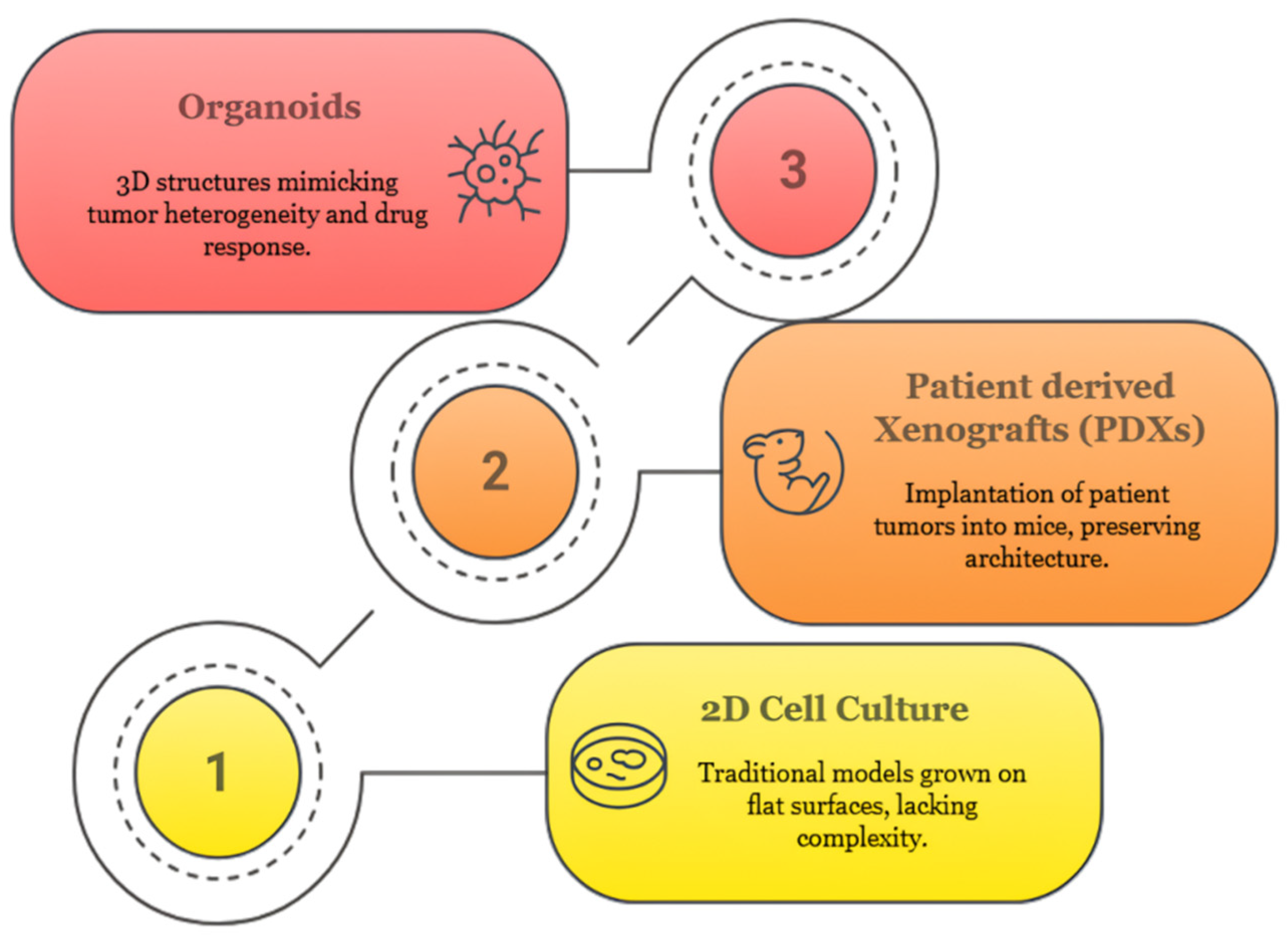
Figure 1. Evolution of cancer models: from 2D cultures to organoids.
Organoids 04 00023 g001
Figure 2. Multifaceted roles of organoids in cancer research and precision oncology.
Organoids 04 00023 g002
Figure 3. Stepwise workflow illustrating tumor organoid development from patient-derived tissue.
Organoids 04 00023 g003
Figure 4. Patient-derived tumor organoids as functional models of tumor heterogeneity.
Organoids 04 00023 g004
Figure 5. Drug testing pipeline using patient-derived organoids.
Organoids 04 00023 g005
Figure 6. Clinical workflow for personalized therapy using patient-derived tumor organoids.
Organoids 04 00023 g006
Figure 7. Tumor organoid model integrating epithelial, stromal, endothelial, and ECM components.
Organoids 04 00023 g007
Figure 8. Organoid-on-a-chip platform mimicking tumor microenvironmental dynamics.
Organoids 04 00023 g008
Figure 9. AI-driven multi-omics workflow for organoid-based drug response and mechanistic profiling.
Organoids 04 00023 g009
Figure 10. Comprehensive SWOT analysis of an accurate and integrated platform.
Organoids 04 00023 g010
Table 1. Comparative overview of stem cell sources for tumor organoid generation: biological attributes, applications, and recent innovations.
Stem Cell SourceBiological CharacteristicsEmerging Oncological ApplicationsRecent Innovations and DevelopmentsSource
Adult Stem Cells (ASCs)Tissue-resident, lineage-restricted; retain tumor-specific mutations and architecture.Generation of patient-derived organoids (PDOs), drug screening, and therapy response predictionImproved culture media for rare cancers; biobanking and automation for personalized oncology platforms[30,51,52]
Cancer Stem Cells (CSCs)Tumor-initiating, chemoresistant, and self-renewing; key drivers of heterogeneity and relapseModeling tumor recurrence, metastasis, and drug resistance evolutionSingle-cell CSC organoid derivation; metabolic and immune-resistance profiling for precision immunotherapy[34,53]
Induced Pluripotent Stem Cells (iPSCs)Reprogrammed from somatic cells; pluripotent with epigenetic memory in some cases.Controlled tumor initiation models, gene-function studies, and rare/pediatric cancer modelingCRISPR-edited isogenic iPSC organoids are used in synthetic lethality screens and mutation-specific drug discovery.[38,54]
Embryonic Stem Cells (ESCs)Pluripotent, high differentiation potential; unrestricted developmental capacityModeling congenital tumors, developmental toxicology, pediatric brain and liver cancersESC-derived tumor organoids used in modeling syndromic mutations (e.g., DICER1); new ethical protocols in development[41,42]
Tissue-Resident ProgenitorsCommitted progenitors from pre-malignant or inflamed tissues; transformation-proneModeling early-stage carcinogenesis (e.g., IBD-associated CRC, Barrett’s esophagus)Used to study field cancerization, inflammation-driven transformation, and chromosomal instability (CIN)[44,55]
Mesenchymal Stem Cells (MSCs)Multipotent stromal cells with immunomodulatory and ECM remodeling capabilitiesTumor–stromal co-culture systems, angiogenesis, and immune evasion modelingMSC–tumor organoid co-cultures for immune checkpoint therapy prediction; MSCs as vehicles for cytokine delivery in chips[45,46,56]
Circulating Tumor Cells (CTCs)Shed from primary tumors; highly metastatic; accessible via liquid biopsyReal-time tumor modeling, tracking clonal evolution, and non-invasive resistance profilingCTC-derived organoids used in breast, prostate, and lung cancer for longitudinal therapy monitoring[47,48,57]
Engineered Synthetic ProgenitorsCustom-designed using genetic circuits or modular engineering from iPSCs/ESCsFunctional genomics, mutation–phenotype correlation, scalable cancer modelingSynthetic organoid libraries created for multiplexed CRISPR screening; ideal for identifying context-specific vulnerabilities[49,50,58]
Table 2. Diverse organoid generation platforms: source cells, scaffold strategies, media requirements, and translational utility.
Organoid System TypePrimary Cellular SourceScaffold ConfigurationMedia Composition and Microenvironmental FactorsScalability and Throughput PotentialCancer Models AddressedEmerging Applications and Technological InnovationsPrincipal Limitations and ChallengesSource
ASC-Derived OrganoidsTumor-resident adult stem cells isolated from patient biopsiesNatural ECM hydrogels (e.g., Matrigel, collagen I)Defined media enriched with EGF, Wnt3a, R-spondin, Noggin, A83-01 to sustain stemnessModerate; supports batch production and cryopreservationColorectal, gastric, pancreatic, breast, prostate, lungLarge-scale patient-derived organoid (PDO) biobanking, AI-integrated drug sensitivity profiling, personalized therapy developmentVariability in ECM batches; limited integration of stromal and immune components[95,96]
CSC-Enriched OrganoidsPurified cancer stem cells (e.g., CD44+, ALDH1+ subpopulations)Suspension cultures or ECM-based matricesStemness-maintaining media with minimized differentiation cues to preserve CSC phenotypesLow to moderate; challenging to expand in large quantitiesGlioblastoma, triple-negative breast cancer, hepatocellular carcinoma, ovarianMapping of drug resistance pathways, CSC lineage tracing, and relapse modelingTechnical difficulty in isolation; risk of phenotypic drift during expansion[96,97]
iPSC-Derived Tumor OrganoidsInduced pluripotent stem cells (iPSCs) reprogrammed from patient somatic cells with engineered oncogenic alterationsSynthetic, chemically defined, or tunable hydrogelsLineage-specific differentiation media coupled with CRISPR-based oncogenic mutation inductionHigh; amenable to automation and pooled genetic screensPediatric tumors, gliomas, colorectal, and pancreatic cancersModeling mutation-specific tumorigenesis, synthetic lethality studies, and organoid-based CRISPR screeningComplex and time-intensive differentiation protocols; potential for incomplete recapitulation of tumor heterogeneity[78,98]
ESC-Derived OrganoidsPluripotent embryonic stem cells derived pre-implantationDefined synthetic matrices or MatrigelDevelopmental stage-specific media containing BMP4, FGF2, Activin A to induce tumor-relevant lineagesLow to moderate; ethical and regulatory limitationsCongenital tumors (hepatoblastoma, medulloblastoma, neuroblastoma)Investigation of developmental origins of childhood tumors, modeling early oncogenic eventsRestricted availability due to ethical constraints; limited clinical relevance for adult cancers[99,100]
Progenitor-Derived OrganoidsPre-malignant progenitors from dysplastic or inflamed tissuesCollagen I or natural ECM-based scaffoldsMedia mimicking inflammatory or pre-neoplastic microenvironmentsModerate; dependent on availability of early lesionsBarrett’s esophagus, IBD-associated CRC, gastric intestinal metaplasiaModeling progression from inflammation to malignancy; studying chromosomal instability (CIN) and field cancerizationLimited to pre-invasive disease; often not reflective of invasive carcinoma biology[101,102]
CTC-Derived OrganoidsCirculating tumor cells isolated from patient blood samplesHybrid systems combining ECM embedding with microfluidic capture or hanging-drop spheroidsMinimal survival-supporting media with ROCK inhibitor (ROCKi), B27 supplement, and antioxidantsLow; hindered by scarcity and fragility of CTCsMetastatic breast, prostate, and non-small cell lung cancer (NSCLC)Liquid biopsy-based real-time modeling of metastatic progression and treatment resistance evolutionLow cell yield; high culture failure rates; genetic drift possible during expansion[103,104]
Table 3. Evaluation of cancer modeling platforms based on tumor fidelity, scalability, cost, microenvironment integration, and clinical relevance.
Model TypeTumor FidelityScalabilityMicroenvironmental IntegrationTranslational UtilityEmerging InnovationsKey LimitationsSource
2D Cell LinesLow, artificial monolayer growth, loss of heterogeneityVery high, cost-efficient, and automation-compatibleAbsent; lacks ECM, immune, or stromal signalsRoutine molecular biology and initial drug screensAI-driven image-based functional screening (e.g., Cell Painting, DeepCell), Lineage barcoding, and synthetic lethal drug screeningLack of clinical correlation; poorly modeled in vivo responses[106,107,108,109,121]
3D SpheroidsModerate; partial 3D cell–cell interaction, but clonal originModerate; suitable for rapid testingLimited; lacks immune or stromal cell typesStudies on drug penetration, metabolic gradientsReal-time metabolic imaging via biosensor dyes, used in combination with perfused scaffolds for dynamic testingLacks patient specificity and tissue context[111,112,113,122]
Patient-Derived Xenografts (PDXs)High; preserves tumor histology, subtype, and molecular profileLow; requires months to establish and propagateNative murine microenvironment; no functional human immunity unless humanizedIn vivo efficacy, biomarker validation, and co-clinical trialsHumanized PDXs for immune checkpoint modeling, PDX transcriptomic–proteomic atlases (PDXNet, EurOPDX)Costly, low throughput; species mismatch; limited to select cancer types[5,115,116,117,118]
Organoids (PDOs)High; retains tumor architecture, genetic identity, and intratumoral heterogeneityModerate to high; suitable for biobanking and automationPartial; improved with TME co-cultures and microfluidicsFunctional precision oncology, resistance profiling, and real-time clinical matchingAI-integrated phenotypic drug response prediction, Single-cell sequencing + spatial omics overlays, Organoid-on-chip for vasculature and flow simulationStill lacks systemic inputs (e.g., circulation, hormones); needs standardization[119,120,123]
Organotypic Tumor Slices (Live Tissue Cultures)Very high; native tumor microenvironment retained for the short termLow; viable for 5–10 days post-excisionFull microenvironment (fibroblasts, vessels, immune cells) intactShort-term ex vivo drug response studies; immune profilingUsed for multiplexed immunotherapy testing (e.g., PD-1, TIL dynamics), Time-lapse imaging for tumor–immune interaction analysisLimited lifespan, not scalable, variable reproducibility[124,125]
Liquid Biopsy-Integrated Models (CTC/DNA-Coupled Organoids)Patient-specific; reflects real-time mutational statusLow (nascent field); limited sample materialMinimal unless co-cultured; under developmentLongitudinal tracking of resistance evolution; real-time personalizationCTC-derived PDOs for drug screening in metastatic settings, Cell-free DNA used to guide organoid mutation editingTechnically challenging, low cell recovery; requires enrichment tools[57,126]
Table 4. Selected organoid co-culture platforms recapitulating tumor–stroma–immune interactions, highlighting matrix designs, cellular components, and assay outputs.
Co-Culture TypeIntegrated Cell TypesMatrix or Platform UsedExperimental ApplicationsKey Functional InsightsReference
Organoid–CAF Co-CultureTumor organoids + Cancer-associated fibroblasts (CAFs)Matrigel, collagen I, or PEG-based synthetic ECMECM remodeling, tumor invasion, TME-driven resistanceFibroblast-secreted factors (IL-6, VEGF-A); enhanced invasion; EMT; stromal-mediated drug resistance[186,187]
Organoid–Endothelial Cell Co-CultureTumor organoids + HUVECs/EPCsFibrin or hyaluronic acid hydrogels; microfluidic chipModeling angiogenesis, perfusable vasculatureVessel formation, endothelial barrier function, response to anti-angiogenics (e.g., bevacizumab)[188,189]
Organoid–Immune Cell Co-CultureTumor organoids + PBMCs/TILs/NK cellsAir–liquid interface; transwell inserts; ALI chipsImmune activation, checkpoint response, cytotoxicity assaysT cell infiltration, IFN-γ secretion, PD-L1 upregulation, immune synapse formation, CAR-T specificity[46,190]
Organoid–MSC Co-CultureTumor organoids + Mesenchymal stem/stromal cellsDual-compartment ECM (Matrigel + collagen I)Tumor–stromal crosstalk, cytokine delivery, immunomodulationCytokine gradients (TGF-β, IL-8), matrix stiffness modulation, and immune suppression modeling[56,191]
Organoid–Neural Cell Co-CultureTumor organoids + Sensory ganglia or iPSC-derived neuronsLaminin-rich hydrogel, chip-based neural nichesPerineural invasion, neurotrophic signaling in tumorsNeurite extension, neurotransmitter effects (e.g., norepinephrine), and tumor migration toward nerve projections[192,193]
Organoid–Dendritic Cell Co-CultureTumor organoids + moDCs or primary DCsECM dome + immune-compatible matrix (collagen IV)Antigen presentation, neoantigen response predictionDC maturation (CD83, CD86), T cell priming, neoantigen-specific responses, cytokine profiling[194]
Multi-lineage Tumor–Immune–Stroma ChipOrganoids + fibroblasts + immune cells (T, NK, macrophages)Perfused microfluidic chip with ECM scaffoldingIntegrated TME simulationSpatial phenotyping, immune evasion tracking, multi-cell interaction mapping, live-cell imaging[195]
Disclaimer/Publisher’s Note: The statements, opinions and data contained in all publications are solely those of the individual author(s) and contributor(s) and not of MDPI and/or the editor(s). MDPI and/or the editor(s) disclaim responsibility for any injury to people or property resulting from any ideas, methods, instructions or products referred to in the content.