Communication

Resistance to MAPK Pathway Inhibition in BRAF-V600E Mutant Colorectal Cancer Can Be Overcome with Insulin Receptor/Insulin-like Growth Factor-1 Receptor Inhibitors

by
Layla El Bouazzaoui
1,2,3,†,
Daniëlle A. E. Raats
1,3,†,
André Verheem
1,
Inne H. M. Borel Rinkes
1,
Hugo J. G. Snippert
2,
Madelon M. Maurice
2,3 and
Onno Kranenburg
1,3,*
1
Division of Imaging and Cancer, Laboratory Translational Oncology, UMC Utrecht, 3584 CX Utrecht, The Netherlands
2
Oncode Institute and Center for Molecular Medicine, UMC Utrecht, 3584 CX Utrecht, The Netherlands
3
Utrecht Platform for Organoid Technology (U-PORT), Utrecht University, 3584 CX Utrecht, The Netherlands
*
Author to whom correspondence should be addressed.
These authors contributed equally to this work.
Organoids 2025, 4(2), 14; https://doi.org/10.3390/organoids4020014
Submission received: 18 March 2025 / Revised: 2 May 2025 / Accepted: 9 June 2025 / Published: 12 June 2025

Abstract

:
The current treatment for refractory BRAF-V600E mutant metastatic colorectal cancer (mCRC) involves combined inhibition of BRAF and the epidermal growth factor receptor (EGFR). However, tumour responses are often short-lived due to a rebound in mitogen-activated protein kinase (MAPK) activity. In this study, we combined short-term cell viability assays with long-term regrowth assays following drug removal over a period of three weeks. This allowed assessment of regrowth after therapy discontinuation. We tested the effect of combined BRAF inhibition (encorafenib) and EGFR inhibition (afatinib) on BRAF-V600E mutant CRC patient-derived organoids (PDOs). Combined EGFR/BRAF inhibition initially caused a major reduction in PDO growth capacity in BRAF-V600E mutant PDOs. This was followed by rapid regrowth after drug removal, mirroring clinical outcomes. EGFR inhibition in BRAF-V600E mutant PDOs led to activation of the insulin receptor (IR) and insulin-like growth factor-1 receptor (IGF1R). The IGF1R/IR inhibitor linsitinib prevented the rebound in MAPK activity following removal of afatinib and encorafenib, prevented regrowth of CRC PDOs, and improved the anti-tumour response in an in vivo model. PDO regrowth assays allow the identification of pathways driving tumour recurrence. IR/IGF1R-inhibition prevents regrowth following golden standard MAPK pathway-targeted therapy and provides a strategy to improve the treatment of BRAF-V600E mutant CRC

1. Introduction

The mitogen-activated protein kinase (MAPK) pathway plays an important role in the pathogenesis of colorectal cancer (CRC) [1]. In CRC, activation of the MAPK pathway is typically the result of activating mutations in KRAS (~40%) or BRAF (~10%). BRAF-V600E mutant CRCs are associated with poor prognosis in the metastatic setting [2,3]. Multiple anti-cancer drugs that target components of the MAPK pathway have been successful in the treatment of BRAF-V600E mutant cancers, such as BRAF inhibitors in melanoma [4]. However, treatment with BRAF inhibitors is less successful in CRC, caused by feedback activation of the epidermal growth factor receptor (EGFR) [5]. The results of the BEACON trial recently showed that the best targeted treatment option for patients with refractory BRAF-V600E mutant CRC is a combination of the BRAF inhibitor encorafenib and the EGFR-targeting antibody cetuximab [6]. However, the overall clinical benefit is still limited (~9 months median overall survival) [7].
A limitation of essentially all targeted anti-cancer therapies is the treatment-induced emergence of compensatory signalling pathways to mitigate the deleterious consequences of target inhibition. Tumour regrowth after cessation of BRAF- and EGFR-targeted therapy in CRC patients is often observed, whilst these patients may initially have responded well to treatment [5,8]. The effects of drug treatment on tumour cell viability are usually measured on the last day of a 3–5-day in vitro drug test platform, thus focusing on immediate responses. However, pathways driving tumour recurrence following therapy discontinuation cannot be studied using this experimental setup. To address this, we combined short- and long-term assays documenting the immediate response to treatment, as well as regrowth capacity, for a period of 3 weeks.
Using this approach, we tested the effect of encorafenib (a BRAF inhibitor) and afatinib (an EGFR inhibitor) on a panel of BRAF-V600E mutant CRC patient-derived organoids (PDOs). Afatinib was chosen over cetuximab, the clinical standard, because cetuximab’s in vitro effects depend heavily on the EGF concentration in the medium. We recapitulate the clinically observed rebound in MAPK activity and tumour regrowth, and show that the insulin receptor (IR) and insulin-like growth factor-1 receptor (IGF1R) are key drivers of this recurrence. The results suggest that the addition of IR/IGF1R inhibitors may improve and prolong the response of BRAF-V600E mutant CRC to standard-of-care BRAF/EGFR-targeting therapy.

2. Materials and Methods

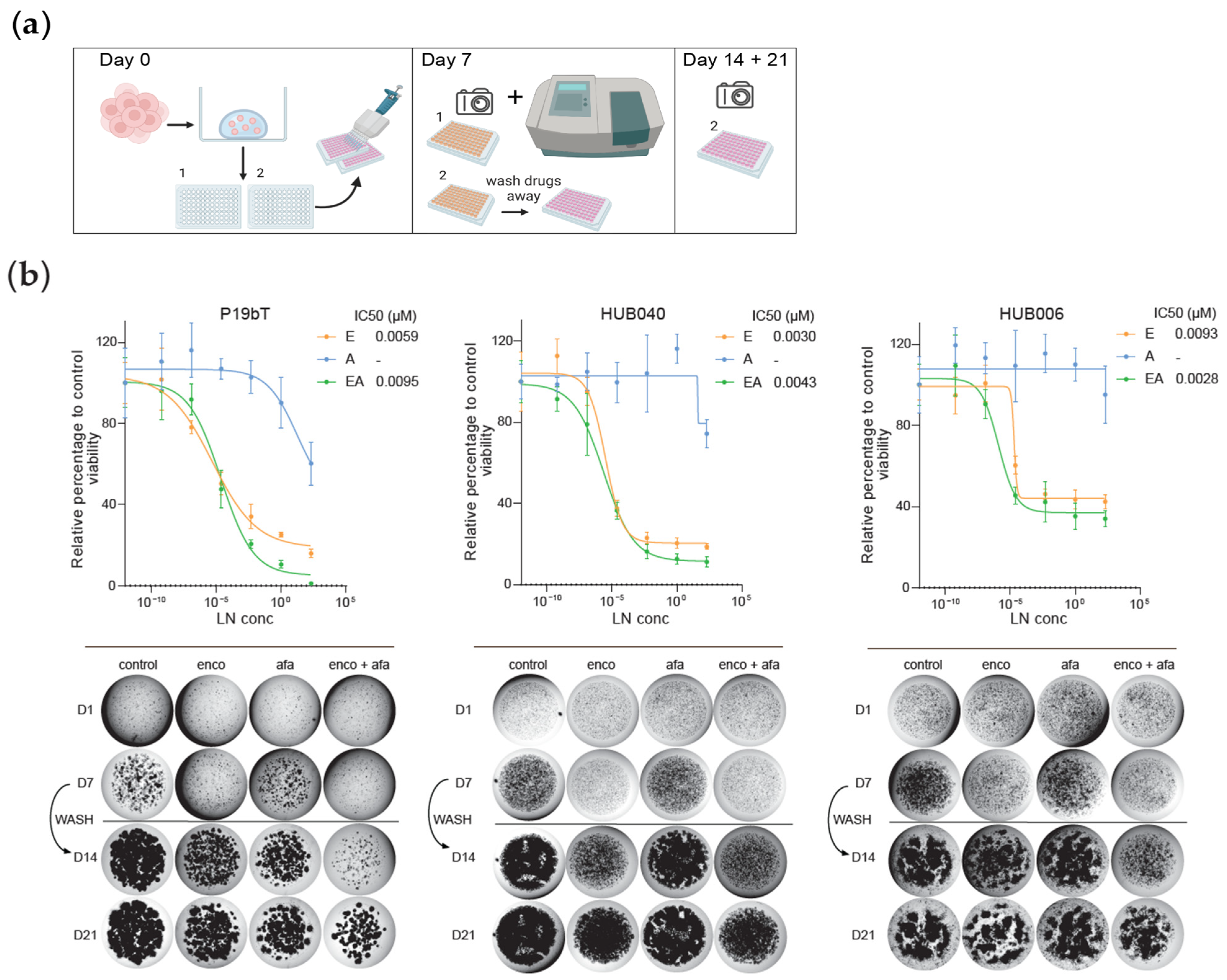
2.1. Organoid Culture

CRC PDOs P19bT, P9T, and P26T used in this study were previously established and characterized [9]. Organoids identified by HUB codes HUB-02-B2-040 (HUB040), and HUB-02-B2-006 (HUB006) are catalogued at HUB Organoids B.V. (Utrecht, The Netherlands). The basal culture medium for colorectal cancer organoids was as follows: Advanced DMEM/F12 (Gibco, Waltham, MA, USA, 12634-010), supplemented with 2 mM Glutamax (Gibco, 35050-038), 10 mM HEPES (Lonza, Basel, Switzerland, 17737E), and 1× Penicillin/Streptomycin (Gibco, 15070-063). For the final CRC organoid growth medium, the basal medium was further supplemented with 1% Noggin conditioned medium (home-made), 1× B27 Supplement (Gibco, 17504-044), 10 mM Nicotinamide (Sigma-Aldrich, St Louis, MO, USA, N0636), 1.25 mM N-acetylcysteine (Sigma-Aldrich, A9165), 500 nM A83-01 (Tocris, Bristol, UK, 2939), and 10 µM SB202190 (Sigma-Aldrich, S7067). Growth medium was refreshed every 3–4 days. Organoids were passaged every 7 days by TrypLE Express (Gibco, 12604021) treatment for 3–5 min at 37 °C and re-embedded in a mixture of basal medium and Basement Membrane Extract (R&D Systems, Minneapolis, MN, USA, 3432-010-01) (ratio 1:3). Culture medium after splitting was supplemented with 10 µM Y-27632 dihydrochloride (Sigma-Aldrich, Y0503). The absence of mycoplasms was regularly controlled by a PCR-based test.

2.2. In Vitro Drug Screens and Cell Viability Assays

PDOs were dissociated into single cells using TrypLE Express (Gibco, 12604021), washed in basal medium, re-suspended in CRC organoid growth medium, and plated in Basement Membrane Extract (R&D Systems, 3432-010-01). In total, 4 µL droplets of organoid–matrix suspension were plated in 96-wells plates, after which CRC organoid growth medium was added to the wells. Drugs were immediately dispensed using the TECAN D300e Digital Dispenser (TECAN, Männedorf, Switzerland). Conditions and drug concentrations were tested in quadruplicate. Organoid medium and tested drugs were refreshed after 3–4 days and cell viability was measured after 7 days using CellTiter-Glo® 3D Cell Viability Assays (Promega, Madison, WI, USA, G9681) in accordance with manufacturer’s instructions. Luminescence levels were measured using a SpectraMax M5 microplate reader (Molecular Devices, Sunnyvale, CA, USA).

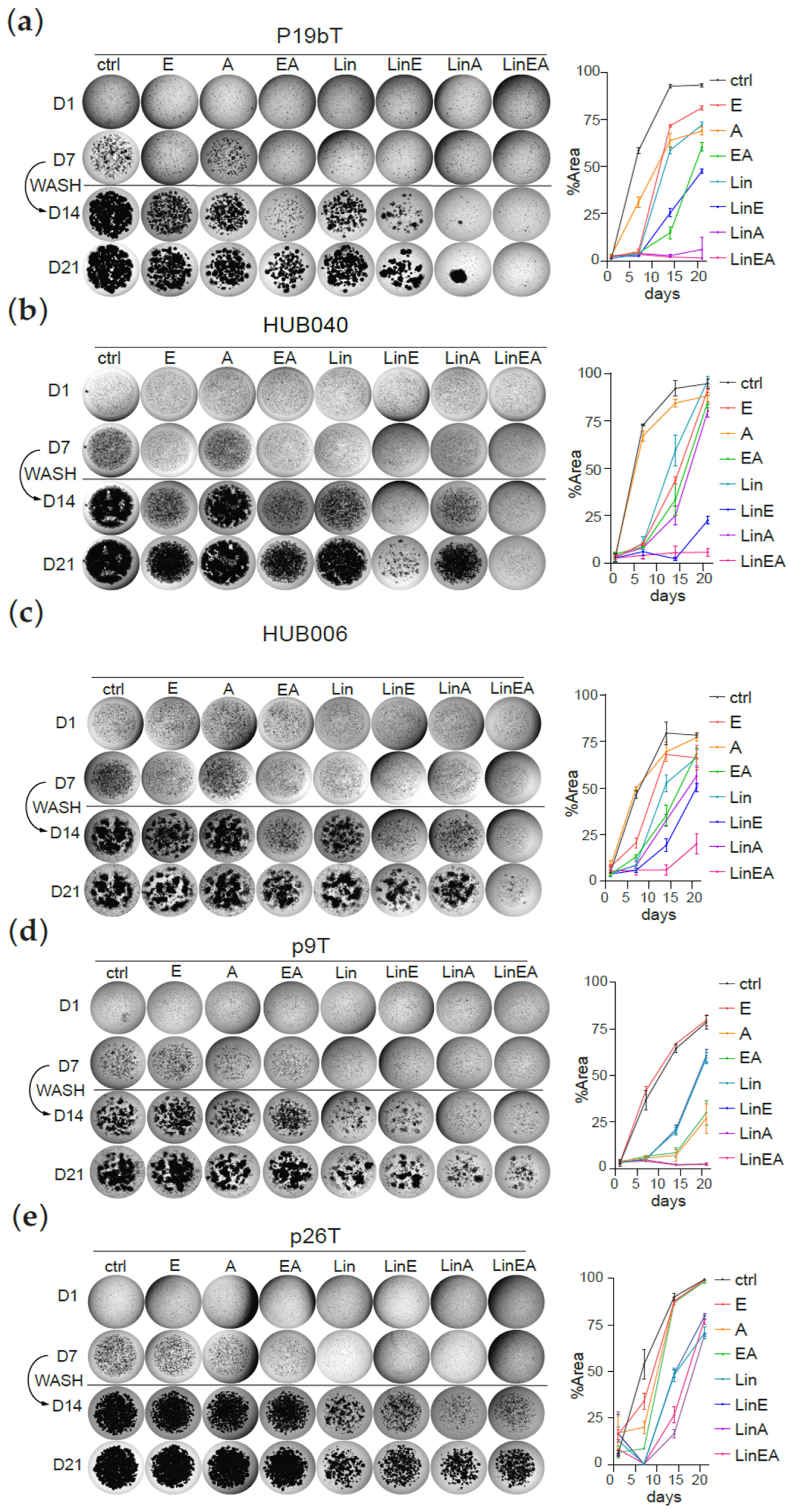
2.3. Regrowth Assays

96-wells plates of PDOs treated with the indicated drugs for 7 days were washed twice with basal medium, after which CRC organoid growth medium was added to the wells. Medium was refreshed every 3–4 days and regrowth of PDOs was assessed for 2 more weeks. Area percentage of organoid outgrowth was calculated using Fiji (Image J, version 1.53t) software. Values are the mean of 3 technical replicates, all derived from a single preparation of organoids.

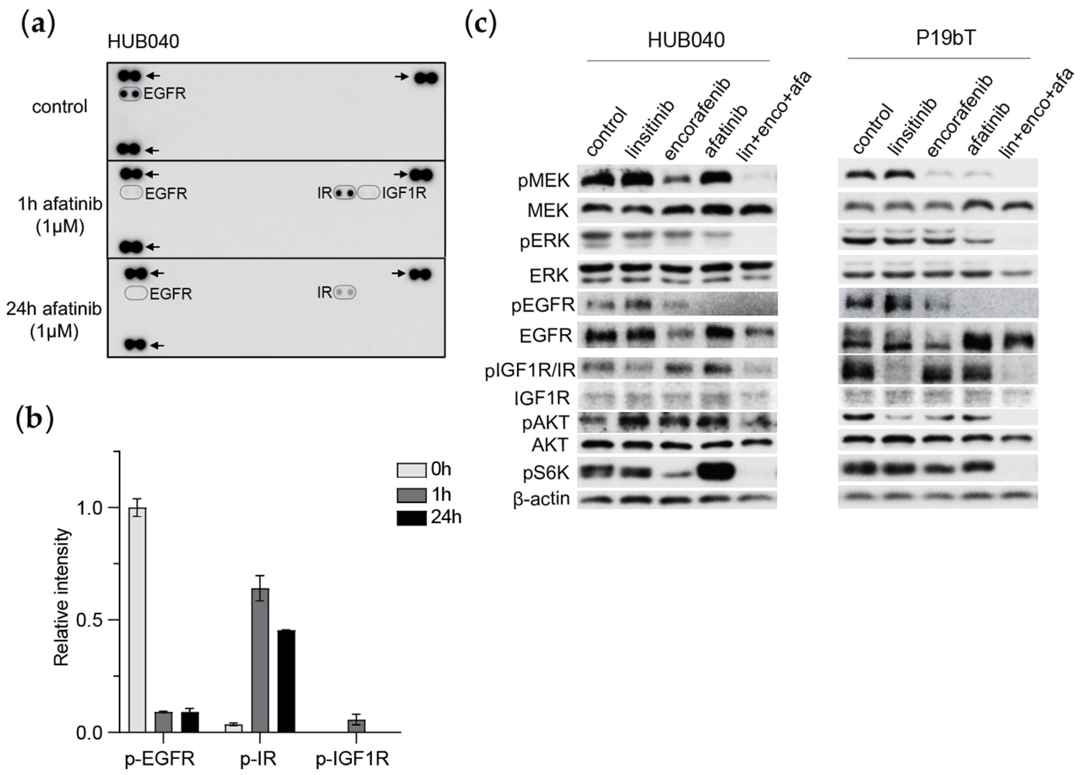
2.4. Phospho-RTK Array

HUB040 organoids treated with DMSO or afatinib (1 µM) were lysed, and equal amounts of lysate (400 µg) were run on the Human Phospho-RTK Array (R&D systems-ARY001B) according to manufacturer’s protocol.

2.5. Western Blot

PDOs were treated with linsitinib (1 µM), encorafenib (1 µM), afatinib (1 µM), or a combination of all drugs (1 µM) for 24 h, after which PDOs were harvested using 1 mg/mL dispase (Gibco), washed with PBS, and lysed in Laemmli buffer in the presence of protease inhibitor cocktail (Cell Signaling Technology, Danvers, MA, USA, CST5871) and 1 mM phenylmethylsulfonyl fluoride. In total, 10–20 μg of total protein were run on a 10% SDS-PAGE gel at 120 V until loading dye reached the bottom of the gel. The proteins were then transferred to a nitrocellulose membrane (Bio-Rad, Hercules, CA, USA) using the BioRad Transblot Turbo Transfer System (BioRad, 1704155) and blocked with 5% BSA diluted in TBS-T (50 mM Tris-HCl/150 mM NaCl (pH 7.6)/0.1% Tween-20) for 1 h at room temperature. Membranes were rocked overnight at 4 °C with primary antibodies diluted 1:1000 in blocking solution. Membranes were probed with antibodies directed against phospho-MEK1/2 (CST9121), MEK1/2 (CST9122), phospho-ERK1/2 (CST9101), ERK1/2 (CST4695), phospho-EGFR (CST2234), EGFR (CST2646), phospho-IGF1R/IR (CST3021), IGF1R (sc-81667), phospho-AKT (CST9271), AKT (CST9272), and phospho-S6 ribosomal protein (CST2211). β-actin (Novus Biologicals, Littleton, CO, USA, NB600-501) was used as an internal control. Primary antibodies were revealed using goat anti-mouse HRP (Dako, Glostrup, Denmark, P0447, 1:2000), goat anti-rabbit HRP (Dako, P0448, 1:1000), and detected with WesternBright ECL (Advansta, San Jose, CA, USA, K-12045). For quantification, FIJI software was used.

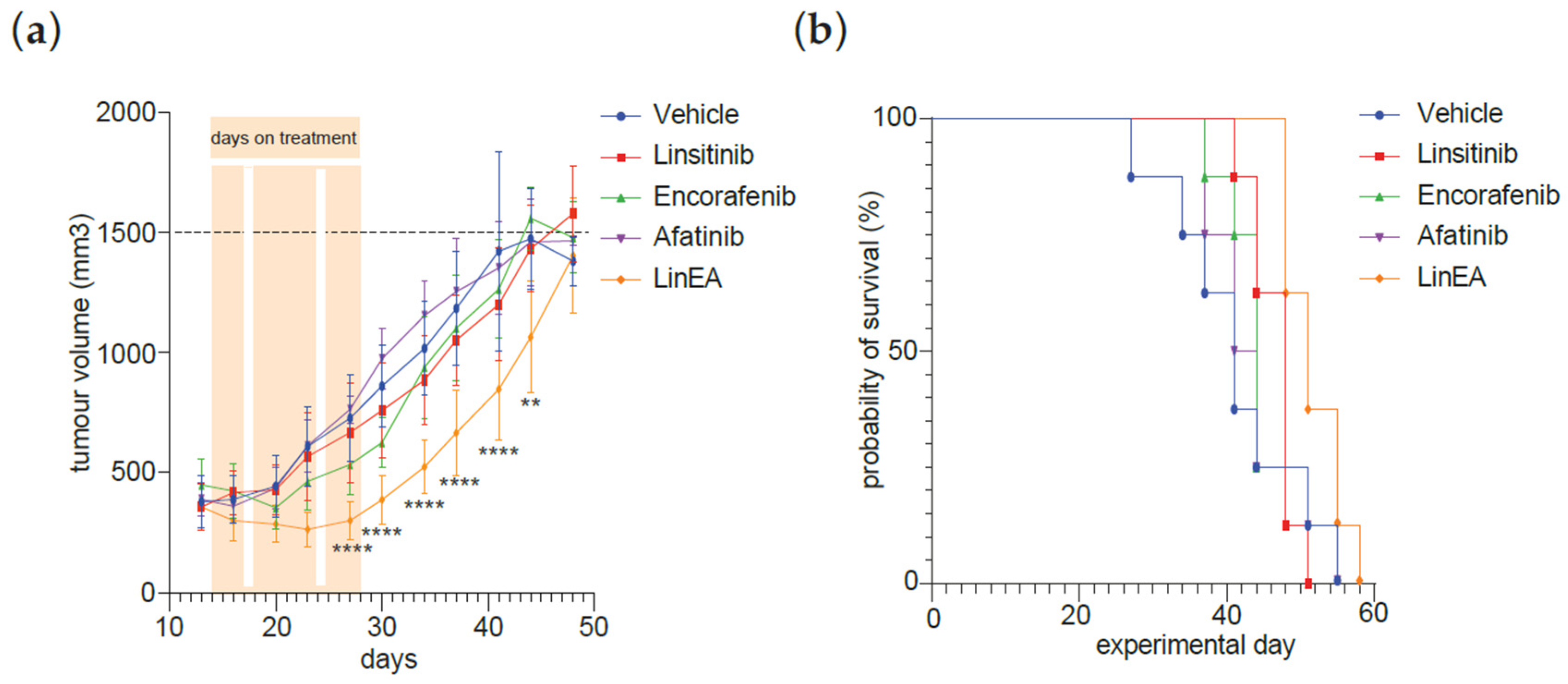
2.6. Xenograft Tumour Growth

Animal experiments were approved by the Competent Authority, The Netherlands (License number AVD11500202115055), which is advised by the Animal Ethics Committee. Animal work protocol (15055-1-17) was approved by the Animal Welfare Body and was performed in accordance with the Dutch Law on Animal Experiments and the European Directive 2010/63/EU. P19bT organoids were dissociated 7 days after passaging into single cells by TrypLE Express (Gibco, 12604021), mixed with 30% growth medium/70% Matrigel (Corning, Corning, NY, USA, CLS354234), and plated in 6-well plates. Organoids were harvested using 1 mg/mL dispase (Gibco, 17105041) 24 h after plating and collected in growth medium. A total of 150.000 cells were mixed with Matrigel at a 1:1 ratio for a total volume of 100 µL. The cell–Matrigel mixture was then subcutaneously injected into 11-week old male NOD.Cg-Prkdcscid Il2rgtm1Wjl/SzJ (NSG) mice (8 animals per treatment, 40 animals in total), supplied by Charles River. Male mice were chosen for this study to minimize variability due to sex differences, ensuring consistency with previous experiments conducted in our lab. When the tumours grew to an average of ±400 mm3, afatinib (10 mg/kg), encorafenib (20 mg/kg), linsitinib (40 mg/kg), or a combination treatment of all drugs was administered by oral gavage for 11 days over a time span of 3 weeks. The control group was injected with only vehicle. All drugs were obtained from Selleckchem (Houston, TX, USA). Tumour growth was determined by measurement of the short and long diameters of the tumour with a calliper, and tumour volumes were determined according to the following formula: A × B2 × 0.5236, where A indicates the largest diameter and B the diameter perpendicular to A. When mice reached a tumour volume > 1500 mm3, they were taken out of the experiment.

2.7. Data Analysis and Statistical Analysis

Quantitative data are presented as mean ± SD, if not stated otherwise. The statistical analyses were performed using GraphPad Prism 9. p-values < 0.05 were considered statistically significant (* p < 0.05; ** p < 0.01; *** p < 0.001; **** p < 0.0001).

3. Results

3.1. Inhibition of BRAF and EGFR in BRAF-V600E Mutant CRC PDOs Causes Potent Growth Inhibition, Followed by Rapid Regrowth After Drug Removal

To model tumour regrowth following BRAF/EGFR inhibition, we employed three BRAF-V600E mutant CRC PDOs and assessed their sensitivity to EGFR inhibition (afatinib) and/or BRAF inhibition (encorafenib) in short-term assays (direct response) and long-term assays (regrowth) (Figure 1a).
PDOs seeded as single cells were first exposed to a logarithmic range of drug concentrations (0.1 nM to 10 μM) for 7 days, after which regrowth following treatment was assessed for 14 more days (Figure 1a). Viability of organoids was measured using CellTiter-Glo 3D assays after short-term assays, and by measurement of area after both short-term and long-term assays. At day 7 the PDOs P19bT and HUB040 were highly sensitive to treatment with encorafenib, either alone or in combination with afatinib, while showing minimal sensitivity to afatinib alone [10] (Figure 1b). However, after drug wash-out, these organoids displayed rapid regrowth (Figure 1b). The BRAF-V600E mutant CRC PDO HUB006 was resistant to afatinib and less sensitive to encorafenib treatment compared to the other two PDOs after 7 days of drug treatment (Figure 1b), and showed unabated growth 2 weeks after drug withdrawal.

3.2. EGFR Inhibition Induces Compensatory Activation of IR and IGF1-R

The rapid regrowth of afatinib and/or encorafenib-treated organoids is most likely caused by the compensatory activation of other signalling pathway(s) [11,12]. To investigate which pathways in BRAF-V600E mutant CRC PDOs are stimulated in response to EGFR inhibition, the activation of 49 different receptor tyrosine kinases (RTKs) was assessed using phospho-RTK arrays. Treatment of the PDO HUB040 with afatinib potently decreased the levels of phospho-EGFR within an hour (Figure 2a,b). However, in parallel, afatinib treatment caused increased phosphorylation of the insulin receptor (IR) and insulin-like growth factor-1 receptor (IGF1R) (Figure 2a,b). Phospho-EGFR levels were still strongly diminished 24 h after afatinib treatment, while phospho-IR levels remained as high as 1 h post-afatinib treatment.
Immunoblot analysis revealed that 24 h treatment with encorafenib or afatinib barely altered the high phospho-MEK, phospho-ERK, phospho-AKT, and phospho-S6K levels in HUB040 and P19bT organoids (Figure 2c). Notably, addition of the IR/IGF1R inhibitor linsitinib to encorafenib and afatinib caused a strong inhibition of the phosphorylation of MEK, ERK, AKT, and S6K (Figure 2c, Supplementary Figures S1 and S2). Most importantly, this triple combination also effectively inhibited IGF1R/IR phosphorylation, an effect that was not observed in organoids treated with encorafenib and afatinib alone (Supplementary Figure S1).

3.3. IR/IGF1R Inhibition Prevents Regrowth of CRC PDOs

The current standard-of-care treatment for refractory BRAF-V600E mCRC is aimed at the inhibition of MAPK signalling. Based on the results above, we hypothesized that the addition of the IGF1R/IR inhibitor linsitinib could potentially enhance the long-term anti-tumorigenic effect of MAPK pathway inhibition in BRAF-V600E mutant CRC PDOs. Therefore, linsitinib was combined with afatinib and/or encorafenib to assess potential synergistic effects of this treatment combination. A concentration of 10 μM was used for all drugs, as this was the highest concentration in our initial drug sensitivity assays at which organoids were still able to grow following drug exposure. Interestingly, the combination treatment of linsitinib with afatinib and encorafenib consistently limited regrowth in all three BRAF-V600E mutant CRC PDOs (Figure 3a–c). Among KRAS mutant PDOs, P9T responded similarly to the BRAF-V600E mutant organoids, whereas P26T showed marked resistance to the combination treatment (Figure 3d,e), indicating variability in response among KRAS mutant models.

3.4. Combined Treatment MAPK Inhibitors and Linsitinib Decreases Tumour Growth in CRC PDO Xenograft

Based on the above data, the triple combination treatment (linsitinib 40 mg kg−1/encorafenib 20 mg kg−1/afatinib~10 mg kg−1) was used to treat P19bT tumour xenografts in vivo. After ± 2 weeks following implantation, the tumours grew to an average volume of 400–500 mm3, after which treatments were started. Drugs were administered orally for 11 days starting 14 days post-implantation. There was no difference in body weight between treatment groups. However, mice treated with the triple combination treatment displayed a significant reduction in tumour volume when compared to mice treated with encorafenib or afatinib alone (Figure 4a). Of note, mice treated with the triple therapy reached the humane endpoint (tumour volume of 1500 mm3) notably later than all other treatment groups (Figure 4b and Figure S3). Mice tolerated the triple combination treatment well.

4. Discussion

Acquired resistance to EGFR/BRAF inhibitors occurs in virtually all BRAF-V600E mutant CRC patients, and the prognosis of this subgroup of patients remains very poor [6,13]. In vitro drug response assays are generally based on measurements of cell viability parameters directly post-treatment. Such assays are inadequate for assessing regrowth potential following drug treatment. In this study, we show that long-term regrowth assays provide a valuable means to assess long-term effects following targeted treatment. Despite strong declines in cell viability measured directly post treatment, several CRC PDO models showed regrowth in long-term assays where PDO growth was assessed over a time course of two weeks following drug treatment.
The identification of compensatory pathways that are induced following targeted drug treatment is essential for rationally developing more effective treatment strategies. Previously, it has been reported that IGF1R and IR are activated in response to inhibition of different components of the RAS/RAF/MEK/ERK cascade, such as after BRAF inhibition in melanoma [14,15], and after EGFR inhibition in epithelial cells [16] and in cholangiocarcinoma [17]. Similarly, we show in our study that dysregulation of the IGF1R/IR axis drives regrowth of BRAF-V600E mutant PDOs following EGFR inhibition. Thus, simultaneous targeting of compensatory signalling pathways (IR/IGF1R) strongly improves the response to EGFR/BRAF inhibition. In general, novel combination treatment strategies using drugs that target compensatory signalling pathways should be evaluated in both short- and long-term assays.
We have shown that the addition of IGF1R/IR inhibitor linsitinib to combined EGFR/BRAF inhibition resulted in durable treatment responses in in vitro studies and greater tumour growth inhibition in an in vivo model. Linsitinib is a well-tolerated drug in different cancer patient populations [18,19], and is therefore a good candidate to be combined with regular targeted treatment. Unexpectedly, despite IR/IGF1R being strong AKT activators, their inhibition alone did not fully suppress pS6K. This suggests that S6 phosphorylation in BRAF-V600E mutant CRC involves complex crosstalk beyond the IGF1R/IR-AKT axis. Notably, only combined BRAF/EGFR/IR inhibition fully suppressed S6 phosphorylation in two independent models, highlighting the need for multi-target strategies. Previously, it has also been suggested that combined inhibition of EGFR/BRAF and the PI3K/AKT pathway may be a viable treatment strategy for BRAF-V600E mutant CRCs [20,21]. However, our results show that inhibition of EGFR directly results in increased phosphorylation of IR/IGF1R, which provides a rationale to target these upstream receptors.
The present study has several limitations. First, we acknowledge that afatinib is an irreversible pan-ErbB inhibitor, targeting not only EGFR but also HER2 (ERBB2) and HER4 (ERBB4). While we primarily referred to its EGFR-inhibitory function, its broader activity could contribute to the observed effects. However, in BRAF-V600E mutant CRC, EGFR reactivation is a well-established resistance mechanism to BRAF inhibition [5], making EGFR the most relevant target in this context. Second, in the in vivo study, only single agent and triple combination therapy were tested, while double combination therapies were not tested. Nevertheless, the study shows that linsitinib can potentially be used in a triple combination therapy strategy concomitantly targeting EGFR, BRAF, and IR/IGF1R. Third, the concentrations used in the in vivo assays are higher than what is typically achievable in patients. These doses were chosen to explore efficacy and potential synergy in preclinical models, highlighting the need for future dose optimization and pharmacokinetic studies. Finally, while the triple combination therapy was able to significantly reduce tumour volume compared to single agent treatments, tumour volumes eventually resumed to levels seen in the single treatment groups. This may be due to the short treatment period of 11 days, and/or to the fact that the tumour sizes at the start of treatment were relatively large (±400–500 mm3). Despite the limitations of this in vivo study, we identified linsitinib as a candidate drug to improve and prolong the response of BRAF-V600E mutant mCRC to the BRAF/EGFR-targeting standard-of-care regimen. Taken together, this study provides a rationale for therapeutic strategies targeting IR/IGF1R/EGFR/BRAF in BRAF-V600E mutant mCRC, similar as in BRAF-mutant melanoma [15].

5. Conclusions

This study demonstrates that despite initial sensitivity to combined BRAF and EGFR inhibition, BRAF-V600E mutant colorectal cancer organoids rapidly resume growth following treatment withdrawal. Using long-term regrowth assays, we identified activation of the insulin receptor (IR) and IGF1 receptor (IGF1R) as key contributors to this rebound. Co-treatment with the IR/IGF1R inhibitor linsitinib effectively suppressed MAPK reactivation, limited organoid regrowth, and improved treatment efficacy in vivo. These findings identify IR/IGF1R inhibition as a potential strategy to improve the efficacy of MAPK pathway-targeted therapy for BRAF-V600E mutant colorectal cancer.

Supplementary Materials

The following supporting information can be downloaded at: https://www.mdpi.com/article/10.3390/organoids4020014/s1, Figure S1: Immunoblot analysis of HUB040 and P19bT organoids treated for 24 h with indicated drugs (1 µM each drug); Figure S2: Western blot raw data; Figure S3: Statistical analysis of Kaplan–Meier survival data.

Author Contributions

Conceptualization, L.E.B., D.A.E.R., and O.K.; methodology, L.E.B., D.A.E.R., and A.V.; validation, L.E.B. and D.A.E.R.; writing—original draft preparation, L.E.B. and O.K.; writing—review and editing, L.E.B. and O.K.; supervision, I.H.M.B.R., H.J.G.S., M.M.M., and O.K.; funding acquisition, H.J.G.S., M.M.M., and O.K. All authors have read and agreed to the published version of the manuscript.

Funding

This research was funded by Utrecht Life Sciences as part of the Utrecht Platform for Organoid Technology facility and by the Dutch Organization for Scientific Research (ZonMW TOP Grant 91218050).

Institutional Review Board Statement

The collection and processing of human colorectal cancer tissues (HUB-Cancer TCBio protocol ID number 12-093) was approved by the Biobank Research Ethics Committee of the University Medical Center Utrecht (Utrecht, The Netherlands). Animal experiments were approved by the Competent Authority, The Netherlands (License number AVD11500202115055), which is advised by the Animal Ethics Committee. Animal work protocol (15055-1-17) was approved on February 28, 2023 by the Animal Welfare Body and was performed in accordance with the Dutch Law on Animal Experiments and the European Directive 2010/63/EU.

Informed Consent Statement

Not applicable.

Data Availability Statement

The data underlying this article are available in the article and its online Supplementary Material. If further details are required, this information may be shared upon reasonable request to the corresponding author.

Conflicts of Interest

The authors declare no conflicts of interest.

References

  1. Fang, J.Y.; Richardson, B.C. The MAPK signalling pathways and colorectal cancer. Lancet Oncol. 2005, 6, 322–327. [Google Scholar] [CrossRef] [PubMed]
  2. Clarke, C.N.; Kopetz, E.S. BRAF mutant colorectal cancer as a distinct subset of colorectal cancer: Clinical characteristics, clinical behavior, and response to targeted therapies. J. Gastrointest. Oncol. 2015, 6, 660–667. [Google Scholar] [CrossRef]
  3. Morris, V.; Overman, M.J.; Jiang, Z.Q.; Garrett, C.; Agarwal, S.; Eng, C.; Kee, B.; Fogelman, D.; Dasari, A.; Wolff, R.; et al. Progression-free survival remains poor over sequential lines of systemic therapy in patients with BRAF-mutated colorectal cancer. Clin. Color. Cancer 2014, 13, 164–171. [Google Scholar] [CrossRef]
  4. Flaherty, K.T.; Puzanov, I.; Kim, K.B.; Ribas, A.; McArthur, G.A.; Sosman, J.A.; O’Dwyer, P.J.; Lee, R.J.; Grippo, J.F.; Nolop, K.; et al. Inhibition of mutated, activated BRAF in metastatic melanoma. N. Engl. J. Med. 2010, 363, 809–819. [Google Scholar] [CrossRef]
  5. Prahallad, A.; Sun, C.; Huang, S.; Di Nicolantonio, F.; Salazar, R.; Zecchin, D.; Beijersbergen, R.L.; Bardelli, A.; Bernards, R. Unresponsiveness of colon cancer to BRAF(V600E) inhibition through feedback activation of EGFR. Nature 2012, 483, 100–104. [Google Scholar] [CrossRef] [PubMed]
  6. Tabernero, J.; Grothey, A.; Van Cutsem, E.; Yaeger, R.; Wasan, H.; Yoshino, T.; Desai, J.; Ciardiello, F.; Loupakis, F.; Hong, Y.S.; et al. Encorafenib Plus Cetuximab as a New Standard of Care for Previously Treated BRAF V600E–Mutant Metastatic Colorectal Cancer: Updated Survival Results and Subgroup Analyses from the BEACON Study. J. Clin. Oncol. 2021, 39, 273–284. [Google Scholar] [CrossRef]
  7. Kopetz, S.; Grothey, A.; Yaeger, R.; Van Cutsem, E.; Desai, J.; Yoshino, T.; Wasan, H.; Ciardiello, F.; Loupakis, F.; Hong, Y.S.; et al. Encorafenib, binimetinib, and cetuximab in BRAF V600E–mutated colorectal cancer. N. Engl. J. Med. 2019, 381, 1632–1643. [Google Scholar] [CrossRef] [PubMed]
  8. Kopetz, S.; Desai, J.; Chan, E.; Hecht, J.R.; O’Dwyer, P.J.; Lee, R.J.; Nolop, K.B.; Saltz, L. PLX4032 in metastatic colorectal cancer patients with mutant BRAF tumors. J. Clin. Oncol. 2010, 28, 3534. [Google Scholar] [CrossRef]
  9. Van De Wetering, M.; Francies, H.E.; Francis, J.M.; Bounova, G.; Iorio, F.; Pronk, A.; Van Houdt, W.; Van Gorp, J.; Taylor-Weiner, A.; Kester, L.; et al. Prospective derivation of a living organoid biobank of colorectal cancer patients. Cell 2015, 161, 933–945. [Google Scholar] [CrossRef]
  10. Verissimo, C.S.; Overmeer, R.M.; Ponsioen, B.; Drost, J.; Mertens, S.; Verlaan-Klink, I.; Gerwen, B.V.; van der Ven, M.; Wetering, M.V.; Egan, D.A.; et al. Targeting mutant RAS in patient-derived colorectal cancer organoids by combinatorial drug screening. Elife 2016, 5, e18489. [Google Scholar] [CrossRef]
  11. Lackner, M.R.; Wilson, T.R.; Settleman, J. Mechanisms of acquired resistance to targeted cancer therapies. Future Oncol. 2012, 8, 999–1014. [Google Scholar] [CrossRef] [PubMed]
  12. Saunders, N.A.; Simpson, F.; Thompson, E.W.; Hill, M.M.; Endo-Munoz, L.; Leggatt, G.; Minchin, R.F.; Guminski, A. Role of intratumoural heterogeneity in cancer drug resistance: Molecular and clinical perspectives. EMBO Mol. Med. 2012, 4, 675–684. [Google Scholar] [CrossRef] [PubMed]
  13. Ahronian, L.G.; Sennott, E.M.; Van Allen, E.M.; Wagle, N.; Kwak, E.L.; Faris, J.E.; Godfrey, J.T.; Nishimura, K.; Lynch, K.D.; Mermel, C.H.; et al. Clinical Acquired Resistance to RAF Inhibitor Combinations in BRAF-Mutant Colorectal Cancer through MAPK Pathway Alterations. Cancer Discov. 2015, 5, 358–367. [Google Scholar] [CrossRef]
  14. Villanueva, J.; Vultur, A.; Lee, J.T.; Somasundaram, R.; Fukunaga-Kalabis, M.; Cipolla, A.K.; Wubbenhorst, B.; Xu, X.; Gimotty, P.A.; Kee, D.; et al. Acquired resistance to BRAF inhibitors mediated by a RAF kinase switch in melanoma can be overcome by cotargeting MEK and IGF-1R/PI3K. Cancer Cell 2010, 18, 683–695. [Google Scholar] [CrossRef]
  15. Patel, H.; Mishra, R.; Yacoub, N.; Alanazi, S.; Kilroy, M.K.; Garrett, J.T. IGF1R/IR Mediates Resistance to BRAF and MEK Inhibitors in BRAF-Mutant Melanoma. Cancers 2021, 13, 5863. [Google Scholar] [CrossRef]
  16. Buck, E.; Eyzaguirre, A.; Rosenfeld-Franklin, M.; Thomson, S.; Mulvihill, M.; Barr, S.; Brown, E.; O’Connor, M.; Yao, Y.; Pachter, J.; et al. Feedback mechanisms promote cooperativity for small molecule inhibitors of epidermal and insulin-like growth factor receptors. Cancer Res. 2008, 68, 8322–8332. [Google Scholar] [CrossRef] [PubMed]
  17. Vaquero, J.; Lobe, C.; Tahraoui, S.; Claperon, A.; Mergey, M.; Merabtene, F.; Wendum, D.; Coulouarn, C.; Housset, C.; Desbois-Mouthon, C.; et al. The IGF2/IR/IGF1R Pathway in Tumor Cells and Myofibroblasts Mediates Resistance to EGFR Inhibition in Cholangiocarcinoma. Clin. Cancer Res. 2018, 24, 4282–4296. [Google Scholar] [CrossRef]
  18. Puzanov, I.; Lindsay, C.R.; Goff, L.; Sosman, J.; Gilbert, J.; Berlin, J.; Poondru, S.; Simantov, R.; Gedrich, R.; Stephens, A.; et al. A phase I study of continuous oral dosing of OSI-906, a dual inhibitor of insulin-like growth factor-1 and insulin receptors, in patients with advanced solid tumors. Clin. Cancer Res. 2015, 21, 701–711. [Google Scholar] [CrossRef]
  19. Fassnacht, M.; Berruti, A.; Baudin, E.; Demeure, M.J.; Gilbert, J.; Haak, H.; Kroiss, M.; Quinn, D.I.; Hesseltine, E.; Ronchi, C.L.; et al. Linsitinib (OSI-906) versus placebo for patients with locally advanced or metastatic adrenocortical carcinoma: A double-blind, randomised, phase 3 study. Lancet Oncol. 2015, 16, 426–435. [Google Scholar] [CrossRef]
  20. Mao, M.; Tian, F.; Mariadason, J.M.; Tsao, C.C.; Lemos, R., Jr.; Dayyani, F.; Gopal, Y.N.; Jiang, Z.Q.; Wistuba, I.I.; Tang, X.M.; et al. Resistance to BRAF inhibition in BRAF-mutant colon cancer can be overcome with PI3K inhibition or demethylating agents. Clin. Cancer Res. 2013, 19, 657–667. [Google Scholar] [CrossRef]
  21. van Geel, R.; Tabernero, J.; Elez, E.; Bendell, J.C.; Spreafico, A.; Schuler, M.; Yoshino, T.; Delord, J.P.; Yamada, Y.; Lolkema, M.P.; et al. A Phase Ib Dose-Escalation Study of Encorafenib and Cetuximab with or without Alpelisib in Metastatic BRAF-Mutant Colorectal Cancer. Cancer Discov. 2017, 7, 610–619. [Google Scholar] [CrossRef] [PubMed]
Figure 1. Inhibition of BRAF and EGFR in BRAF-V600E mutant CRC PDOs causes potent growth inhibition, followed by rapid regrowth after drug removal. (a) Overview of experimental setup. BRAF-V600E mutant CRC PDOs were dissociated into single cells, seeded in basement membrane extract in two 96-well plates, one plate for a short-term assay, and one plate for a long-term regrowth assay. Medium containing a logarithmic range of drug concentrations (0.1 nM to 10 μM) was added and cells were incubated for 7 days, after which one plate was used to measure cell viability, and one plate was used to assess regrowth after drug withdrawal. (b) Graphs depicting cell viability curves of drug-treated organoids measured with CellTiter-Glo 3D assays 7 days after the start of the experiment. Pictures were taken of the wells treated with the highest combination (10 μM) of indicated drugs at indicated time points. Values are the mean of 4 technical replicates. A, afatinib; E, encorafenib; EA, encorafenib and afatinib.
Organoids 04 00014 g001
Figure 2. EGFR inhibition induces compensatory activation of the insulin receptor (IR) and IGF-1 receptor (IGF1R). (a) The phosphorylation status of RTKs in afatinib-treated HUB040 organoids was assessed by Human phospho-RTK array. Representative dot images from the phospho-RTK array, arrows indicate the reference spots on the array blot. (b) Relative phospho-EGFR, phopho-IR, and phospho-IGF1R levels of HUB040 treated with afatinib. (c) Immunoblot analysis of HUB040 and P19bT organoids treated for 24 h with indicated drugs (1 µM each drug).
Organoids 04 00014 g002
Figure 3. Triple combination therapy of encorafenib, afatinib, and linsitinib results in durable treatment responses in vitro and greater tumour growth inhibition in vivo. (ae) Representative images of BRAF-V600E CRC organoids treated with indicated drugs (10 μM) for 7 days, followed with a re-growth period of 2 weeks after drug wash-out. Values are the mean of three technical replicates.
Organoids 04 00014 g003
Figure 4. Combined treatment MAPK inhibitors and linsitinib decreases tumour growth in CRC PDO xenograft. (a) Growth rate of subcutaneous organoid allografts, mean tumour volumes ± SD are shown. Drugs were administered for 11 days, which started on day 14 post-implantation. Tumour volume was recorded two times a week. Two-way ANOVA with Tukey’s multiple comparison test was performed for statistical significance determination. Statistical significance scores are depicted as Vehicle group vs. LinEA group, 8 animals per treatment group, ** p < 0.01, **** p < 0.0001. (b) Kaplan–Meier curve with survival data which were plotted as % of animals surviving in each group using a cut-off tumour volume of 1500 mm3.
Organoids 04 00014 g004
Disclaimer/Publisher’s Note: The statements, opinions and data contained in all publications are solely those of the individual author(s) and contributor(s) and not of MDPI and/or the editor(s). MDPI and/or the editor(s) disclaim responsibility for any injury to people or property resulting from any ideas, methods, instructions or products referred to in the content.

