Review

A Concise Review of Organoid Tissue Engineering: Regenerative Applications and Precision Medicine

by
Karnika Yogeswari Makesh
1,
Abilash Navaneethan
1,
Mrithika Ajay
1,
Ganesh Munuswamy-Ramanujam
2,
Arulvasu Chinnasamy
3,
Dhanavathy Gnanasampanthapandian
1,* and
Kanagaraj Palaniyandi
1,*
1
Cancer Science Laboratory, Department of Biotechnology, School of Bioengineering, SRM Institute of Science and Technology, Kattankulathur 603203, Tamil Nadu, India
2
Interdisciplinary Institute of Indian System of Medicine, SRM Institute of Science and Technology, Kattankulathur 603203, Tamil Nadu, India
3
Department of Zoology, School of Lifesciences, University of Madras, Guindy Campus, Chennai 600025, Tamil Nadu, India
*
Authors to whom correspondence should be addressed.
Organoids 2025, 4(3), 16; https://doi.org/10.3390/organoids4030016
Submission received: 16 April 2025 / Revised: 3 June 2025 / Accepted: 1 July 2025 / Published: 4 July 2025
(This article belongs to the Special Issue The Current Applications and Potential of Stem Cell-Derived Organoids)

Abstract

Organoids are three-dimensional tissue culture models derived from stem cells, and they have become one of the most valuable tools in biomedical research. These self-organizing miniature organs mimic the structure−function properties of their in vivo counterparts and offer an exceptional prospective for disease modeling, drug discovery, and regenerative medicine. By replicating the complexity of human tissue, organoids enable the study of disease pathophysiology, tissue development, and cellular interactions in a highly controlled and manipulable environment. Recent developments in organoid technology have enabled the production of functional organoids of various tissues. These systems have proven to be highly promising tools for personalized medicine. In addition, organoids have also raised hopes for the development of functional transplantable organs, transforming the study of regenerative medicine. This review provides an overview of the current state of organoid technology and its application and prospects and focuses on the transformative impact of organoid technology on biomedical research and its contribution to human health.

1. Introduction

The first known attempt to generate organs in vitro took place in 1907, when Wilson showed that dissociated sponge cells can self-assemble and reorganize into a complete organism [1]. The term “organoid,” which refers to something that resembles an organ, was first documented in the literature in 1946, when Smith and Cochrane described a cystic teratoma [2]. Organoids are three-dimensional (3D) in vitro cultures of stem or progenitor cells that mimic the cellular diversity, structural organization, and functions of the corresponding tissues in vivo [3]. They are characterized by three main features: self-organization, multicellularity, and functional similarity to native tissues [4]. Organoids are miniaturized, laboratory-grown versions of organs that develop from embryonic stem cells, induced pluripotent stem cells (ESCs or iPSCs), or neonatal/adult stem cells. They have the remarkable ability to self-organize into structures that mimic real organs and perform essential functions, making them valuable for research and medical applications [5]. Organoids provide a more advanced platform for research by creating a 3D environment that better mimics real tissue, unlike traditional 2D cultures that are often unable to replicate tissue-level physiology [6]. Three-dimensional cultures provide greater precision in controlling cell and cell−matrix interactions, tissue stiffness, biochemical signaling, and overall tissue density. This allows the extracellular matrix (ECM) to be customized to closely resemble the organ of interest [7].
Organoids are divided into PSCs and adult stem cells (AdSCs). PSC organoids are derived from ESCs or iPSCs and developed by suspension culture in a defined medium to promote cell aggregation and targeted differentiation [8]. In contrast to PSC organoids, which rely on directed differentiation, AdSC organoids develop from tissue-derived adult stem cells that are first isolated by tissue dissociation and then cultured in a specialized medium containing growth factors to support stem-cell activity and organoid formation [9]. The formation of three germ layers—endoderm, mesoderm, and ectoderm—during human development gives rise to all organs, and the successful generation of organoids from tissues derived from each layer highlights the potential of the technology to model a wide range of organs [10]. By retaining the 3D structure and genetic diversity of the original tissue, organoids serve as a powerful platform for highly efficient drug screening and as a valuable tool for the advancement of precision and personalized medicine [11]. For more than a century, animal models and 2D cell lines have played a crucial role in biomedical research. They help us understand cellular signaling pathways, guide the development of drug candidates, identify drug targets, and uncover the pathological mechanisms of diseases [12]. Organoids have revolutionized biomedical research by bridging the gap between traditional cell cultures and animal models, providing a powerful platform for disease modeling and personalized medicine [13].

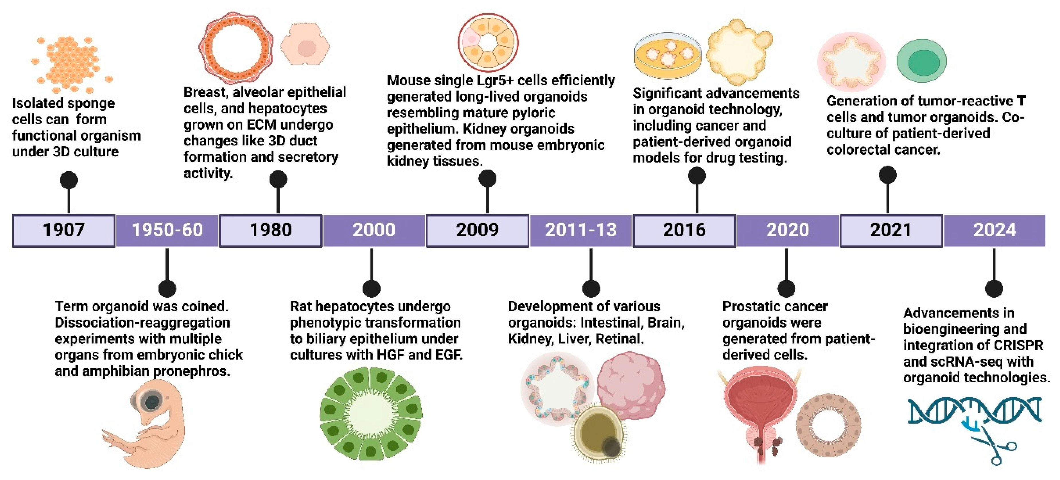
2. History of Organoids

The history of organoids began in 1907, when Wilson et al. demonstrated that dissociated sponge cells could self-assemble into a complete organism, and this was the first successful attempt to generate organs in vitro [1]. Ross Granville Harrison developed the first ex vivo tissue culture technique that demonstrated the ability of embryonic nerve fibers to develop outside the body, laying the foundation for 3D tissue modeling [14]. In the 1950s and 1960s, the term “organoid” predominantly referred to intracellular structures rather than complex tissue models, as researchers focused on organelles and cellular architecture rather than stem cell-derived 3D cultures [15]. In 1980, a significant change occurred with the introduction of collagen- and laminin-rich matrices, which enabled cells to create spatially structured 3D structures and laid the foundation for advanced tissue engineering [14]. Despite this progress, the focus decreased after 1985, primarily due to terminological uncertainties, as some studies defined the developing structures as “organs” rather than “organoids”, leading to different research priorities [15].
The return of organoid research in the 2000s was driven by advances in PSC technology, which enabled researchers to differentiate stem cells into many tissue types, greatly improving in vitro modeling of human development and disease [16]. In 2009, Clevers et al. made the crucial discovery that intestinal stem cells containing leucine-rich repeats and the G-protein-coupled receptor 5 (Lgr5+) can self-organize into long-term, self-renewing intestinal organoids, laying the foundation for today’s organoid technology [15]. This approach was extended to cancer research in 2011 by producing colon cancer organoids that allowed researchers to simulate tumors with greater precision and improve preclinical drug testing [16]. In 2013, Lancaster et al. produced cerebral organoids from human pluripotent stem cells, providing an unparalleled platform for the study of brain development and neurodegenerative diseases, including microcephaly and Alzheimer’s disease [17]. Between 2016 and 2020, significant progress was made in organoid culture techniques, leading to the successful derivation of organoids from the esophagus, stomach, liver, pancreas, kidney, lung, and retina, expanding their potential applications in disease modeling and regenerative medicine [18,19].
A significant achievement in 2020 was the creation of beating heart organoids, which provide a functional model for the study of cardiovascular disease, heart failure, and drug-induced cardiotoxicity [16,20]. The advent of patient-derived organoids (PDOs) has significantly transformed cancer research and precision medicine, as these models accurately mimic patient-specific genomic and phenotypic characteristics, surpassing the capabilities of traditional 2D monolayer cultures and patient-derived xenografts (PDXs) in preclinical contexts [21]. The ability to preserve genetic integrity and facilitate long-term proliferation in PDOs has increased their therapeutic importance for personalized treatment approaches [22]. Since 2021, advances in gene editing (CRISPR-Cas9), single-cell sequencing, and 3D bioprinting have significantly improved the functionality, scalability, and reproducibility of organoid cultures, increasing their applicability for high-throughput drug screening and therapeutic validation [23,24]. The integration of microfluidic organ-on-a-chip platforms has effectively reduced the discrepancy between static in vitro systems and dynamic physiological environments, facilitating real-time observation of organoid responses to stimuli [25,26].
By 2024, the advancement of organoid-immune co-culture systems enabled the study of immune−tumor interactions and provided a robust tool to study tumor microenvironments, immunotherapies, and checkpoint inhibitor responses [27]. The evolution of organoid technology from 1907 to 2024 is a testament to the growing importance of organoids and organoid technologies in biomedical research, regenerative medicine, and personalized medicine in cancer (Figure 1). With all these advances, we still face major challenges, such as vascularization, functional maturity, and large-scale production to enable clinical applications in organ transplantation, disease modeling, and precision medicine [28].

3. Cell Culture Approaches for Organoids

The formation of organoids requires the use of high-tech culture techniques that enable the growth, differentiation and functional maturation of organoids, making them indispensable for biomedical research [29]. The various existing technologies include bioreactors, suspension cultures, co-culture techniques, and microfluidic systems aimed at optimizing the culture conditions of organoids [30]. Microfluidic bioreactors (MFBs) have recently gained attention as they provide nutrient delivery, oxygenation, and waste removal in a precise manner, improving organoid viability and reproducibility [31]. Such bioreactors reduce variability in organoid formation and are better suited to promote targeted differentiation strategies [32].
Another system that has been used extensively is the stirred bioreactor (SBR), in which mass transfer is improved, and the environment for organoid growth is made dynamic [29]. However, despite their simplicity and scalability, these SBRs do not allow continuous perfusion and do not adequately replicate physiological conditions [30]. Furthermore, rotating wall vessel (RWV) bioreactors allow low-shear conditions that reduce almost all types of mechanical stress, favoring the maintenance of organoids in which some delicate structural features are to be preserved [33]. Electrically stimulating (ES) bioreactors combine electrical signals to promote maturation, especially in neural and cardiac organoids, where it is crucial to study electrophysiological activity [31]. Suspension cultures with low concentrations of the widely used extracellular matrix (ECM) would be a more cost-effective means for the expansion of organoids [32]. Such cultures provide long-term stability and reduce the costs otherwise associated with ECM-rich conditions, which is particularly beneficial for high-throughput drug screening and CRISPR-Cas9-based studies [34]. In addition, different air−liquid interface (ALI) cultures are used to develop skin organoids from iPSCs, with a clear preference for better stratification and keratinocyte differentiation [35]. The ALI format has a better potential to promote hair follicle morphogenesis than conventional floating cultures, allowing for greater consideration of the microenvironment during organoid development [33,36].
Recent developments in the integration of co-culture techniques have enabled novel applications of these organoid models to study host−microbe interactions under controlled conditions [31]. Organoid-derived monolayers and trans-well systems provide more access to the lumen of the organoid, which promotes microbial colonization and improves sampling for host−pathogen studies [31]. The Intestinal Hemi-anaerobic Co-culture System (IHACS) maintains hypoxic and normoxic conditions in its two-chamber design, allowing microbial and epithelial components to survive together and increasing the physiological relevance of intestinal organoid studies [33]. In addition, perfusion bioreactor systems provide fine-tuning of microenvironmental aspects, making them a valuable tool for chemotherapy testing in colorectal cancer organoids [30,37]. In this way, researchers can assess drug efficacy and resistance mechanisms under conditions that are as close as possible to the actual physiological microenvironment of the tumor [38]. Organoids arise from the 3D culture method by seeding cell suspensions from ESC, iPSC, somatic stem cells, or cancer cells in media differentiation to obtain spheroids that serve as a transitional phase to full organoid maturation [29]. Standard suspension cultures, so-called mini-bioreactors or rolling flasks, prepare the spheroids, which are then encapsulated in ECM-based hydrogels such as Matrigel, which then polymerize to form a stable 3D scaffold [39]. In this context, perfused bioreactors allow the modification of culture conditions, leading to enhanced differentiation and maturation of organoids, as they are better supplied with nutrients and mechanical stimulation, which is essential for the functional development of such complex organoid structures [40] (Figure 2).
Microfluidics-based organoids-on-chip models are emerging as a major innovation in organoid culture, enabling real-time monitoring with improved environmental controls [33,41]. These systems allow more realistic mimicking of physiological conditions by integrating vascularization and perfusion dynamics and are therefore excellent candidates for drug testing and disease modeling [42]. The refinement of organoid cultures has far-reaching implications for basic research and clinical applications [29].

4. Human Organoids as an Upcoming Model for Research

Organoids have revolutionized biomedical research by using a human-relevant system to study development, disease, and therapeutics. Organoids are small, self-organized 3D tissues made from stem cells that mimic the structure and function of natural organs, such as intestinal organoids, which form a cryptovillus architecture and respond to stimuli such as prostaglandin E2 or Wnt agonists in a manner similar to in vivo intestinal tissue [43]. A major turning point in this field was the development of methods to generate organoids from both pluripotent stem cells (iPSCs/ESCs) and adult stem cells, extending their versatility to different tissue types, such as brain, intestine, and kidney [44,45].
One of the strengths of organoids is their applicability to humans. Since they are developed from human stem cells—either PSC or adult stem cells (ASC)—organoids retain the genetic and phenotypic characteristics of the donor [46]. This makes them more physiologically representative than animal models, which generally do not mimic human-specific diseases due to interspecies variations [46]. The ability of organoids to mimic organogenesis and mirror in vivo development makes them invaluable for the study of congenital defects and developmental biology [47]. For example, patient-derived kidney organoids from people with polycystic kidney disease (PKD) have shown cyst formation that mimics the pathology of the disease in vivo [48]. Brain organoids are of central importance for research into neurodevelopmental diseases, such as microcephaly and autism [49]. These models have proven useful in elucidating how genetic mutations affect brain development and function, leading to a better understanding of such diseases that are difficult to study in vivo [49].
Organoids allow researchers to study the progression of diseases such as colorectal cancer, cystic fibrosis, and infectious diseases under manipulated conditions, providing new opportunities for disease modeling [46,50,51]. For example, colorectal cancer organoids have shown that heterogeneity and resistance mechanisms, such as Wnt signaling mutations and APC loss, contribute to resistance to EGFR inhibitors [51]. Patient-derived organoids can also predict how a patient would respond to treatment, opening new possibilities for personalized medicine. By applying drugs to an individual patient’s organoids, clinicians can identify the optimal therapies with minimal side effects [50].
In drug development, organoids provide a high-throughput platform for drug screening. Their ability to model the response of human tissue reduces the number of animal models and increases the relevance of preclinical studies [52]. For example, liver organoids have been used to screen drug toxicity. Bile duct-like liver organoids exposed to acetaminophen showed dose-dependent hepatotoxicity, confirming their use in preclinical screening and providing an excellent model for determining the effects of drugs on the human liver. Intestinal organoids have also been used to study the effect of new drugs on intestinal function and gain insights into drug absorption and metabolism [52]. Although they have numerous advantages, organoids are not without drawbacks. Challenges such as morphological variability, batch-to-batch inconsistency, and the lack of standardized culture protocols often hinder the reproducibility and interpretation of data [53,54]. Without an adequate blood supply, organoids cannot accurately model organ-level processes, which limits their ability to model systemic disease [55]. Organoids are also less mature and complex than mature organs, which may make them less suitable for modeling adult tissue processes or late-stage diseases [52]. For example, while brain organoids can model early neurodevelopment, they cannot model the complexity of the adult human brain.
Ethical questions are also linked to brain organoids. Recent debates about electrical oscillations, which resemble the EEG of premature babies, in forebrain organoids raise the question of the potential for consciousness or sensation. The more advanced the models become, the more researchers and ethicists will have to deal with the ethical issues of creating tissues with potentially human-like neural activity [56]. These issues need to be addressed as the technology advances to enable responsible and transparent research practices [57].
Further research is attempting to overcome these challenges through improved vascularization and maturation. Techniques such as 3D bioprinting and microfluidic devices are currently being developed. For example, 3D-printed liver organoids with permeable vessels showed increased survival and metabolic function in vitro, creating more realistic organoid models. For example, researchers are attempting to incorporate blood vessels into organoids, which could make them more functional and viable [57]. In addition, the integration of multiple organoids into a single system, known as “organ-on-a-chip,” would allow the study of inter-organ communication and systemic diseases. These multi-organ models would be able to mimic the complex interactions among organs and would provide a more comprehensive model for the study of diseases affecting multiple systems [46,51,52,58].
To improve structural integrity and functional maturation, biomaterials such as Matrigel, collagen I, alginate, and fibrin hydrogels are often used as extracellular matrix (ECM) mimics to support 3D organoid culture [59,60]. While Matrigel remains the gold standard, its batch-to-batch variability and tumor-derived origin pose limitations for clinical application [61]. Recent advances in synthetic hydrogels, particularly polyethylene glycol (PEG)-based scaffolds, have enabled more reproducible, chemically defined platforms with tunable mechanical properties and better physiological relevance [62].
In parallel, 3D bioprinting has emerged as a powerful strategy to spatially arrange cells and bioinks with ECM components to create structurally complex organoid architectures [63,64]. In particular, gelatin methacryloyl (GelMA) has been used for bioprinting liver and heart organoids, which exhibit enhanced viability and function due to their photocrosslinkable properties and ECM-like structure [65]. Fibrin-based bioinks and decellularized ECM materials have also shown promise for creating vascularized constructs that support endothelial cell integration and perfusion in vitro [66].
These engineered microenvironments not only enhance the architectural fidelity of organoids but also allow precise modulation of growth factor gradients, oxygen content, and matrix stiffness, which are critical for mimicking developmental factors in vivo and promoting organoid maturation [4,67].
The combination of organoids and CRISPR-Cas9 gene-editing technology also holds the potential to unravel the mysteries of gene function and disease mechanisms. By introducing specific gene mutations into organoids, scientists can study the effects of these mutations on the onset and progression of disease [68]. These methods have already been used to study disease mechanisms such as cystic fibrosis by editing CFTR mutations in intestinal organoids and monitoring swelling assays as indicators of CFTR function and cancer, and they offer new insights into the genetics behind such diseases [68]. Organoids are a promising new technology in biomedical research, providing a highly versatile and scalable model for the study of human development, disease, and therapeutics. Challenges of vascularization, maturity, and ethics remain, but advances promise to further expand their potential. As scientists develop and expand the applications of organoids, these models will play an increasingly important role in shaping the future of biomedical research and medicine.

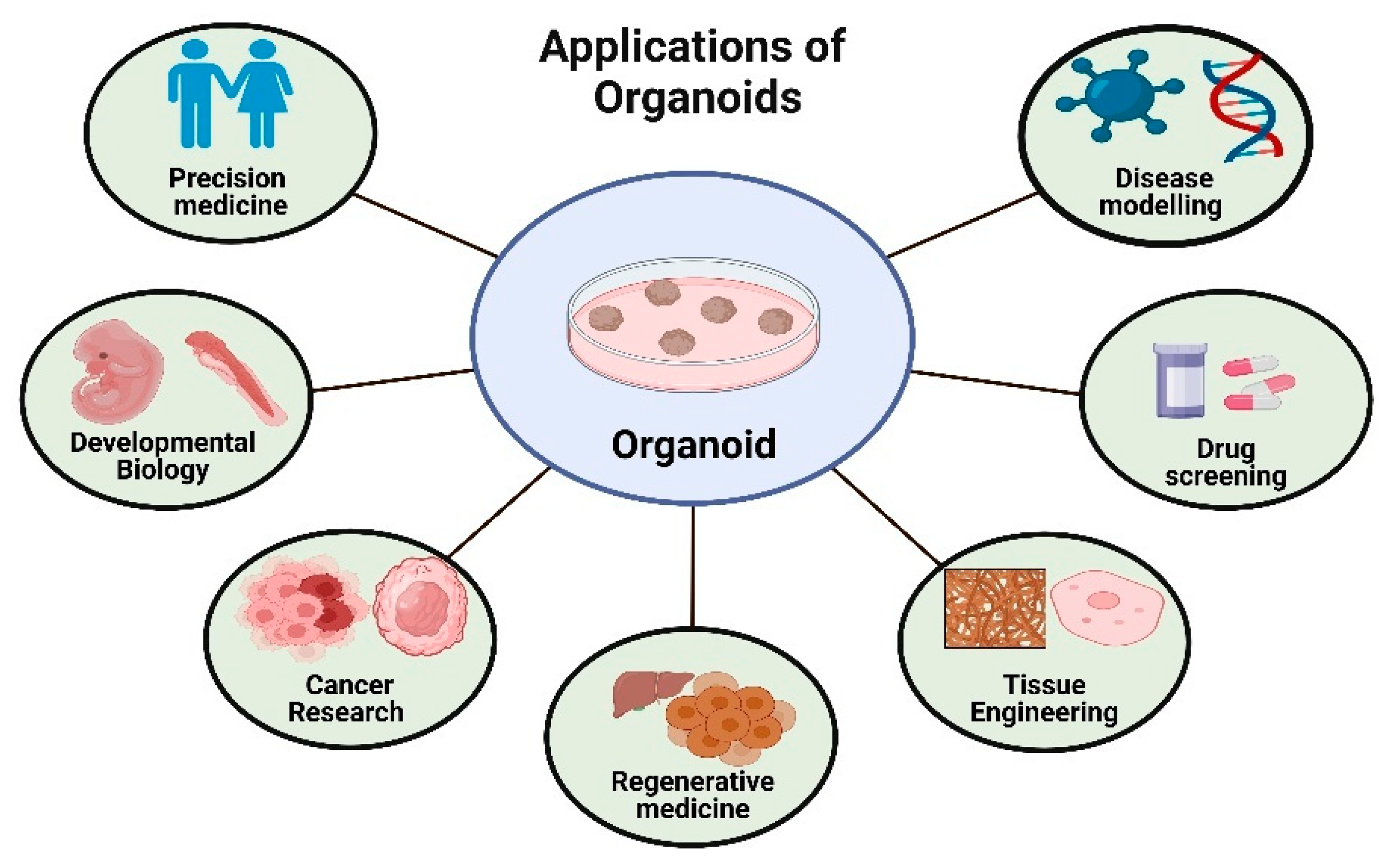
5. Application of Organoids

5.1. Cancer Research

Organoids have decisively advanced cancer research. They serve as true 3D in vitro models that mimic tumor architecture, genetic heterogeneity, and intercellular interactions. Such constructs form a bridge between conventional 2D cultures and in vivo studies [69]. They have triggered a new revolution in cancer research as organoids provide a more physiologically relevant framework for better disease modeling, drug discovery, and the application of precision medicine. Organoids serve as an important resource in translational research as they preserve important histological and molecular features, unlike conventional cancer cell lines, which often lose these complex features of primary tumors [70]. Another prominent use of organoids in oncology is personalized medicine. In this practice, PDOs are obtained by culturing tumor samples and retaining their genetic composition after propagation [71]. In contrast to trial-and-error experiments, these models allow the testing of different therapeutic agents against a patient’s tumor to determine the appropriate course of treatment [69]. Clinical studies have shown that the patterns of response to treatment of PDOs often reflect the reality of patients, reinforcing their prognostic importance in oncology [72].
Organs that withstand extensive drug treatment to clarify tumor evolution allow the elucidation of drug resistance mechanisms [73]. Major advances in the development of cancer therapies have been achieved through high-throughput drug screening in combination with organoid biobanks and personalized therapy [74]. In this case, in contrast to the preclinical models previously used, organoids enable the simultaneous evaluation of multiple drugs in many different tumor subtypes, accelerating the process of finding efficient treatment options [71,75]. Companies have used these organoid-based platforms to develop new combinations of drugs and therapies aimed at curing diseases [45,76]. Advanced neoplasms, such as colorectal and pancreatic malignancies, have been particularly helpful [71,77]. Organoid models have been used in immuno-oncology to study the efficacy of CAR T-cell immunotherapy, immune checkpoint inhibition, and other immunotherapeutic strategies [78,79]. By co-culturing tumor organoids with immune cells from patients, researchers can assess the response of each individual patient to immunotherapy, which improves the selection of patients for therapeutic trials and provides important information on resistance mechanisms [79] (Table 1).

5.2. Drug Development

In drug development, organoids have become a transformative tool, as they provide a physiologically relevant platform for screening drug response, toxicity, and therapeutic efficacy [84]. Organoids simulate the architecture and function of a natural tissue so closely that they offer an advantage over traditional 2D cell cultures for use in preclinical drug testing [85]. These models are essential for establishing a successful cure for many diseases, especially malignancies. They enable the study of tumor heterogeneity, differences in genetic composition, and patient response to drugs [86]. One of the most important uses of organoids in drug development is their ability to predict patient-specific responses to drugs [87]. PDOs, which reflect tumor variability and enable tailored drug screening, have been used extensively to mimic colorectal cancer (CRC) [88]. Several biobanks with PDOs have shown that drug response can be correlated with clinical and pathologic data [88], enabling more accurate therapy selection. In addition, organoids allow comparative studies of drug efficacy versus PDXs [89]. Studies have shown that while both models exhibit similar resistance to certain chemotherapeutic agents, organoids can show significant differences in drug sensitivity as they retain the tumor microenvironment, which is important for the field of personalized medicine [45]. Organoids offer high-throughput screening that enables the rapid identification of potential therapeutic candidates. Similarly, thousands of molecules can be evaluated in just a few days, increasing the overall effectiveness of treatment development.
The Cellular Tumor Organoid System (CTOS) has been particularly effective in developing xenografts and harvesting organoids for extensive drug screening with high tumor cell recovery rates for rapid, targeted therapy testing [85]. This approach has been widely applied in some cancers, including endometrial, where CTOS-based screening has identified potent inhibitors of tumor growth [87]. In addition, organoids facilitate the conduct of combination chemotherapy studies to investigate synergies among different drugs [87]. Another example is the study in which CRC organoids were used to test combinations of EGFR and MEK inhibitors, which were found to be more effective than either drug alone, a finding that is very important for the goal of optimizing cancer therapies. Outside of oncology, organoids derived from human pluripotent stem cells (hPSCs) have contributed significantly to the modeling of drug responses in other diseases [45]. For example, cardiac organoids have been used to study drug-induced cardiotoxicity. It became clear that drugs that are cardiotoxic in 2D can pass through the test screens in 3D, simply because there is a lack of tissue interaction [90]. Liver organoids and kidney organoids were produced to study hepatotoxicity and nephrotoxicity, respectively, and were more predictive assays than conventional cell lines or animal models [91]. The ability of organoids to recapitulate tissue-specific functions makes them a very powerful tool for the study of disease mechanisms and the development of safe drugs [47]. Organoids contribute to functional precision medicine by enabling the development of functional biomarkers that predict drug efficacy beyond their genetic markers [88]. In contrast to classical genomic approaches that rely solely on sequencing data, organoid-based screening evaluates tumor responses to therapies in real-time, leading to actionable insights for treatment decisions [45].
Biobanks of these organoids are therefore essential to preserve patient-derived samples for future research, enable longitudinal studies to track disease progression and mechanisms of treatment resistance, and explore other therapeutic strategies [92]. The coexistence of organoid technology and bioengineering with artificial intelligence has further expanded the application of organoids in drug development [93]. Microfluidic platforms enable real-time monitoring of organoid responses to drugs, increasing the physiological relevance of preclinical studies [94]. Since vascular organoids expressing viral entry receptors provide a platform for the evaluation of antiviral drugs, organoid research in the field of infectious diseases has also included studies on SARS-CoV-2 [95]. Despite its many advantages, organoid technology still has obstacles to overcome to maximize its use in drug discovery [6]. Current efforts are focused on improving organoid culture methods, improving the correlation between in vitro drug efficacy and clinical outcomes, and adapting organoids to the drug regulatory framework. With such ongoing developments, organoids are likely to serve as the backbone for drug discovery and thus stand at the interface between preclinical models and human trials, ultimately aiming to improve treatment strategies for numerous diseases (Table 2).

5.3. Precision Medicine

Human organoids are advancing cancer research by providing in vitro models for the study of tumors. In contrast to conventional methods using cancer cell lines, mouse models, or PDX, AdSC-derived organoid technology enables the cultivation of transformed cancer tissue, referred to as tumoroids or canceroids, from both diseased and normal tissue [11]. This technique is applicable to both diseased and normal tissue. Patient-derived cancer organoids have been successfully developed from colorectal, brain, prostate, pancreatic, liver, breast, bladder, stomach, esophageal, endometrial, and lung cancers [11]. Organoids developed from various pediatric and adult epithelial tumors are generated more efficiently than patient-derived 2D cancer cell cultures and better retain the histopathological, genetic, and drug response characteristics of the original tumor. This enables personalized therapies based on individual patient profiles rather than static genetic analyses of large cohorts [33].
In precision medicine, treatments are tailored using organoids to capture tumor diversity, while precision oncology traditionally relies on predictive mutational biomarkers for targeted therapies [101]. Tumoroid analysis not only serves as a guide for patient treatment but also helps in the discovery of biomarkers. For example, Burkhart’s team found that PDAC patients with better response to chemotherapy had fewer KRAS, TP53, and SMAD4 mutations, with PDO pharmacotypes serving as predictive biomarkers in an ongoing clinical trial [102,103]. However, these biomarkers are often not present. For example, in a mixed tumor cohort, whole-exome sequencing found FDA-approved treatment options for only 3 of 737 patients (0.4%) [101]. Tumoroids hold great potential for tumor biomarker discovery. Ukai and colleagues developed 5-FU-resistant GCOs, and microarray analysis identified KH domain-containing, RNA-binding, and signal transduction-associated 3 as an independent prognostic factor in gastric cancer patients, especially those treated with 5-FU [104].
Targeted therapy is a precision medicine approach that focuses on inhibiting specific molecular markers or signaling pathways in tumors or other diseases, ensuring a more precise and effective treatment approach. Organoid technology is revolutionizing cancer research and treatment by helping to produce immune cells, such as CAR T cells, that show strong anti-tumor activity against leukemia, while also creating transplantable cells for organs such as the liver, pancreas, retina, kidney, and intestine [105].
While the holistic approach preserves the diversity of the immune system, the reductionist approach allows controlled studies of the interaction between the immune system and the tumor but may limit the diversity of immune cells [106]. In contrast, the second approach isolates immune cells separately before culturing them with patient-derived cancer organoids. It provides better control over the system and allows long-term investigation of immune−tumor interactions, although it may limit the diversity of immune cells [107]. A novel co-culture strategy stimulates antigen-presenting dendritic cells with tumor antigens, enhances the lysis and proliferation of CD8+ T cells, and then cultures them with organoids from gastric cancer patients, effectively predicting the efficacy of precision medicine for improved patient prognosis [108]. Genetically engineered CAR T cells designed to recognize and bind cancer cell antigens have been co-cultured with patient-derived tumor organoids, such as glioblastoma and colorectal cancer, to evaluate their precision and efficacy in targeting tumors [109,110].
In addition to oncology, organoids have also delivered promising results in other areas of precision medicine. In cystic fibrosis, patient-derived intestinal organoids have been used to assess individual response to CFTR-modulating drugs to enable effective treatment selection [111]. Liver organoids derived from patients with genetic and metabolic disorders, such as alpha-1 antitrypsin deficiency and Wilson’s disease, have facilitated personalized drug testing and disease modeling [112]. In neurological and neurodegenerative diseases, brain organoids derived from the iPSCs of patients have been used for modeling diseases such as Rett syndrome, Alzheimer’s disease, and Parkinson’s disease, and have enabled mechanism research and screening of drugs tailored to the individual patient [113] (Table 3).

5.4. Developmental Biology

Models based on human cells are essential because many human-specific biological phenomena cannot be reproduced in animal models [18]. The need for human cell-based models is obvious, as human physiology differs greatly from that of mice in terms of developmental speed, drug metabolism (where ibuprofen and warfarin are safe for humans but toxic to rats), and genetic diversity [18]. iPSCs generated by reprogramming PSCs with transcription factors and ESCs derived from blastocyst cell masses both have multidirectional differentiation potential that allows the formation of PSC-derived organoids by directed differentiation [47]. The process begins with the formation of germ layers, such as endoderm, mesoderm, or ectoderm, followed by the addition of growth factors, signaling molecules, and cytokines to direct cell development and maturation. The result is PSC-derived organoids with a diverse composition of mesenchymal, epithelial, and endothelial cells [47]. With the advent of iPSC technology and advanced cell culture methods, scientists can now create laboratory models that are unique to each individual [18]. Reprogramming cells into iPSCs is commonplace, but creating disease models from these cell lines is still a challenge, as initial efforts have focused on differentiating iPSCs into a single cell type [18]. Organoids from hiPSCs are valuable for engineering hard-to-obtain tissues such as the brain and retina [118]. Organoids enable knockout studies of essential genes, overcoming embryonic lethality in vivo, while PSC-derived organoids serve as important models for developmental biology, revealing human-specific features of neurodevelopment by analyzing human and primate brain organoids at the single-cell level [118]. ASC-derived and PSC-derived organoids differ technically and in their applications. PSC-derived organoids serve as developmental models because they are derived from ESCs or iPSCs that can form all three germ layers [119]. Organoids at early to late fetal developmental stages provide insights into human development, but they have difficulty modeling adult tissues, making them more suitable for studying embryonic and fetal development and pregnancy-related diseases [120] (Table 4). One study reported on the use of iPSC-derived organoids from a patient with microcephaly, a genetic disorder caused by a gene mutation in a gene encoding CDK5-regulating downstream subunit-associated protein 2 (CDK5RAP2) that results in reduced brain size and is difficult to model in mice [121]. Organoid models of the human brain are essential for research into psychiatric disorders, as shown in a 2015 study in which iPSC-derived organoids from four patients with severe autism showed increased inhibitory GABAergic neurons due to upregulation of the FoxG1 gene when patient-derived organoids were subsequently grown [122].

5.5. Tissue Engineering and Regenerative Medicine

Regenerative medicine is a new and rapidly growing field of biomedical science that aims to build functional biological tissues in an in vitro system through the integration of stem cells, biocompatible scaffolds, and biochemical factors [133]. These bioengineered tissues closely resemble the structure and function of the body’s own tissues and offer potential solutions for the repair or replacement of injured organs [133]. Among the numerous strategies of regenerative medicine, organoids represent a promising innovation. In contrast to previous strategies such as cell transplantation or 2D cell cultures, organoids are 3D structures that self-assemble and maintain their functional and structural properties over extended periods of time [133,134]. Organoids are genetically stable, have robust expansion and differentiation potential, and possess the ability to self-organize into complex tissue-like architectures [134,135]. By creating an optimal chemical–physical and mechanical microenvironment, researchers are able to influence the functionality and self-organization of organoids to such an extent that they can closely mimic the physiological conditions of native tissue [136].
The potential of organoids has been demonstrated by a series of proof-of-concept experiments in animal models. For example, retinal sheets derived from mouse ESCs or iPSCs using customized optic-nerve-head organoid protocols were successfully transplanted into mouse models with retinal degeneration [137]. These transplanted tissues not only developed mature photoreceptors but also established synaptic contacts with the host cells, restoring light sensitivity in some cases. Similarly, intestinal organoids grown from dissociated mouse intestinal epithelia or single stem cells have been transplanted into mice and shown to be able to regenerate wounds in the intestinal mucosa to a certain degree [133,138]. Another study showed that enterospheres derived from fetal intestinal progenitor cells were able to integrate into an injured mouse intestine and differentiate in vivo, further confirming the regenerative capacity of organoids [139].
Despite advances in organ transplantation as a therapeutic intervention for a variety of diseases, its widespread use is hampered by major obstacles, most notably the lack of suitable donors and the need for long-term immunosuppression to prevent graft rejection [140]. Alternatives such as cell transplantation and artificial organs offer a short-term substitute but cannot fully replicate the complexity of structure and function of native tissue [134]. Organoids are now seen as a revolutionary and promising solution in this regard. Derived from tissue-specific stem cells, organoids provide a more physiologically relevant in vitro model than conventional cell lines and are therefore crucial for the study of both normal and pathological processes [11,53]. Their potential applications range from drug discovery and toxicity testing to personalized medicine, where they can be used to personalize treatments for individual patients [135].
Advances in molecular analysis have driven organoid technology to further elucidate molecular mechanisms in development, physiology, and disease. Organoid models have proven useful in the study of a number of organ-specific diseases, including neurological diseases such as ZIKV infection and psychiatric disorders [141]. Organoid technology is also widely used in cancer research. Organoid biobanks from patient tissue provide a platform for personalized drug screening and improve our understanding of the genetic and epigenetic landscapes of different cancer samples, advancing our mechanistic understanding of tumorigenesis [142].
In addition, the integration of organoid technology with other disciplines, such as tissue engineering, microfluidics, and human organoid biology, has enabled the development of sophisticated chip-based in vitro models [135,143] (Table 5). These models, commonly referred to as “organs-on-chips,” allow researchers to study the pathophysiological properties of human organs in a reproducible and controlled environment [143]. In the long term, the use of organoids in regenerative medicine could be complemented by their integration with in vitro genetic correction techniques [144]. Such a strategy holds great promise for autologous tissue repair of genetically diseased tissue, as it enables the generation of genetically modified, patient-specific organoids that can be transplanted without the risk of immune rejection [145]. Organoid technology is a major advance in the field of regenerative medicine and opens up new horizons for tissue repair, disease modelling, and personalized therapeutic approaches [22,28].

5.6. Emerging Applications of Organoids

In addition to their established role in cancer research, regenerative medicine, precision therapy, and tissue engineering, organoids are now also being researched for various emerging biomedical applications. For example, cerebral organoids have proven valuable in environmental toxicology, where exposure to neurotoxic substances, such as methylmercury and bisphenol A, resulted in the disruption of cortical layering and synaptogenesis, providing a model for the assessment of environmental risk factors for neurodevelopmental disorders [151]. In the context of infectious diseases, lung and gut organoids have been used to model host−pathogen interactions under physiologically relevant conditions. Lung organoids enabled the analysis of SARS-CoV-2 infection dynamics and response to antiviral drugs [151], while gut organoids facilitated the study of rotavirus, norovirus, and Helicobacter pylori pathogenesis [84]. Organoids have also proven to be powerful tools for vaccine development and immunological studies. For example, tonsil-derived organoids have recently been shown to mimic germinal center formation and support antigen-specific B-cell activation, allowing vaccine efficacy to be studied in vitro [152]. In gene therapy, organoid platforms are used to test gene correction strategies. For example, CRISPR-mediated editing of CFTR mutations in cystic fibrosis patient-derived intestinal organoids has successfully restored duct function, allowing for personalized therapeutic evaluation [153]. In addition, cross-species organoid models generated from human, non-human primate, and rodent stem cells have enabled comparative studies of organ development and species-specific gene regulatory pathways, thus contributing significantly to our understanding of human evolutionary biology [154] (Figure 3, Table 6).

6. Genetic Modification in Organoids

Genetic editing in organoids allows screening for effective drug responses, as demonstrated by Verissimo and colleagues, who used colorectal organoids with different RAS mutations to assess their effects on sensitivity to EGFR and MEK inhibitors [157]. The combination of organoid systems with CRISPR-Cas9 enables the modeling of monogenic diseases such as cystic fibrosis (CF), as demonstrated by the correction of the F508del mutation in human intestinal organoids, which restored cystic fibrosis transmembrane conductance regulator (CFTR) channel activity and showed potential for autologous organoid-based therapy [158]. Reduced CFTR function affects multiple organs by decreasing chloride transport, reducing water flow, and increasing mucus density [159]. In cystic fibrosis, reduced CFTR function leads to thick mucus accumulation, causing recurrent lung infections and fibrosis. However, researchers have found that rectal organoids can help assess CFTR activity. Healthy organoids swell when activated with forskolin, while CF organoids do not, although this can be restored with CF drugs or gene correction, making the test a reliable way to predict response to treatment [160].
In 2013, Schwank et al. demonstrated the use of a transient CRISPR/Cas9 system delivered by lipofection to enable genetic knockouts or mutation repair in organoids [153]. The authors first induced a frameshift mutation in the APC gene of mouse and human organoids, which resulted in WNT signaling-independent growth, as APC is a known tumor suppressor and negative regulator of WNT signaling [3]. After introducing the CRISPR/Cas9 tool, they repaired mutations in the CFTR gene in colon organoids derived from cystic fibrosis patients [153]. The restored swelling of target organoids in response to forskolin confirmed the successful correction of the CFTR locus mutation (disease-causing deletion mutation) and demonstrated the potential of CRISPR/Cas9 for organoid research and treatment of monogenic disorders [161]. Recently, Geurts et al. successfully restored CFTR function in human rectal and airway organoids by using CRISPR-based adenine base editors to correct the W1282X and R553X mutations by converting A-T to G-C [162]. In 2015, Hans Clevers’ team used CRISPR/Cas9 to create CRC organoids and establish tumor organoids from 20 CRC patients [46]. High-throughput sequencing confirmed that their genetic profiles matched large-scale CRC mutation data, making them valuable for drug screening and precision medicine [163]. Using CRISPR-Cas9, researchers introduced a TMPRSS2–ERG fusion into mouse prostate organoids by joining two DNA regions, resulting in AR-driven ERG overexpression that was prevented by androgen inhibitors, mirroring the results in vivo [164]. The “Inference of CRISPR Edits” (ICE) assay has become a valuable bioinformatics tool for assessing genetic alterations in organoids by deconvoluting Sanger sequencing data from mixed-cell populations. However, incomplete editing complicates the direct phenotypic analysis of organoid pools after Cas9 treatment [165]. CRISPR gRNA library screening analysis in human organoids has improved human genetic studies and, in combination with PSC and organoid technologies, enables genetic experiments in small human organ models that closely mimic human physiology [18,166]. Although genome-wide CRISPR/Cas9 screens on immortalized human and mouse cells are widely used in 2D cultures, technical challenges have limited their use in 3D organoids, which better mimic in vivo responses. The large size of the sgRNA library and background noise require a significant number of cells to ensure comprehensive coverage in pooled CRISPR/Cas9 screens [167].
Figure 4 illustrates the step-by-step process of genome editing in organoids using CRISPR-Cas9 technology. First, the organoids are split into individual cells to enable genetic modification at the individual cell level. The CRISPR-Cas9 system, delivered via vectors or electroporation, induces targeted DNA changes via two primary repair mechanisms: non-homologous end joining (NHEJ) for gene knockouts and homology-directed repair (HDR) for precise edits. Successfully edited cells undergo clonal expansion followed by selection of the genetically modified clones. Finally, selected organoids are expanded to provide genetically stable models for functional genomics, disease modeling, and the development of therapies.

7. Next-Generation Organoids

Next-generation organoids have revolutionized cancer research by supporting advanced 3D models that closely mimic the TME in vivo, providing insights into tumor biology, therapeutic response, and precision medicine [105]. The organoids retain the fundamental characteristics of native tumors, including cellular heterogeneity, genetic stability, and interactions with the TME, and they are powerful organoid models that overcome the limitations of traditional 2D cultures [70]. By mimicking the natural interactions between tumor and stroma, different cell types, such as cancer-associated fibroblasts (CAFs), endothelial cells, and immune cells, which increase their physiological relevance, are now being incorporated into next-generation organoids [168]. Effective replication of pancreatic ductal adenocarcinoma (PDAC) cellular heterogeneity by PDOs has enabled real-time assessment of treatment responses and resistance mechanisms [150]. In addition, co-culture models combining tumor organoids with tumor-infiltrating lymphocytes (TILs) enable an important new understanding of immune evasion and immunotherapy resistance [169]. Microfluidic platforms and organoid-on-a-chip technology have further advanced the field by allowing precise control of environmental factors, such as oxygen gradients, nutrient availability, and mechanical forces, which are essential for studying interactions between tumor and stroma and metastatic behavior [170].
Recent advances in 3D bioprinting have enabled the production of vascularized tumor organoids that closely resemble the morphological and functional characteristics of real tumors [171]. They have successfully generated bio-printed organoids of lung, breast, and liver tumors with permeable vascular networks, allowing researchers to analyze tumor invasion and metastases with remarkable accuracy [172]. Next-generation sequencing (NGS) and multi-omics techniques, such as proteomics and transcriptomics, have improved the use of organoid models by enabling complete genetic profiling of malignant tumors [173].
Next-generation organoids are of promising importance for immunotherapy research with regard to immune checkpoint inhibitors and chimeric antigen receptor (CAR) T-cell therapies [174]. This has opened up new perspectives on the molecular processes that control tumor spread and resistance to conventional treatments [175]. Despite these major advances, there are still some challenges in next-generation organoid research. These include the need for improved vascularization, prolonged culture stability, and the inclusion of neuronal and endocrine elements to more faithfully mimic interactions between the tumor and the nervous system [176,177].
Areas currently under discussion include ethical issues and the standardization of procedures for clinical transmission [178]. With a focus on the development of functional, patient-specific tumor organoids for tailored oncology, the future of organoid technology is anchored in the convergence of bioengineering, computational biology, and regenerative medicine [179]. Synthetic biology and organoid engineering are expected to converge to increase the scalability of these models, improving their accessibility for comprehensive drug screening and biomarker discovery [180]. Next-generation organoids represent a revolution in cancer research, providing powerful tools to elucidate tumor heterogeneity, predict treatment response, and create more effective patient-tailored treatment plans [72]. Advances in bioprinting, gene editing, microfluidics, and artificial intelligence are enabling organoid technology to transform oncology and precision medicine, enabling customized and more effective cancer treatments.

8. Challenges and Limitations in Organoid Research

Organoid technology has made considerable progress, providing 3D models that closely mimic human organs in structure and function and exhibit key properties, such as cell proliferation, differentiation, self-renewal, and genetic stability [19,46,51,120,166]. Despite these advances, several challenges and limitations persist, impacting reproducibility, scalability, and physiological relevance.
A major challenge is the variability and lack of standardization in organoid production protocols. Different laboratories use diverse methodologies, resulting in organoids with varying characteristics, even when derived from the same tissue type. For example, human intestinal organoids generated from adult stem cells (AdSCs) versus pluripotent stem cells (PSCs) show distinct differences, despite both being termed intestinal organoids [181,182]. This variability extends to patient-derived organoids (PDOs), where the degree of genetic drift, clonal selection, and tumor evolution over multiple passages remains poorly understood and tumor-dependent [75].
In addition to technical challenges, ethical considerations play a pivotal role in organoid research and its clinical translation [183]. Obtaining informed consent from donors of primary tissues or stem cells is essential and must comply with stringent ethical guidelines to protect donor autonomy, confidentiality, and privacy throughout the research process [56,183]. Given the personalized nature of organoid-based medicine, questions arise concerning the ownership of biological materials and data, the sharing of potentially sensitive genetic information, and the equitable distribution of resulting therapies, raising important ethical and social justice issues [183,184]. Moreover, as organoids become more complex and begin to recapitulate features of human organ function, especially in the case of brain organoids, emerging concerns about their moral status and the possibility of consciousness or sentience necessitate careful ethical scrutiny [185]. Addressing these concerns requires the development of robust regulatory frameworks and standardized ethical protocols to guide researchers and clinicians, ensuring responsible innovation while safeguarding human rights and public trust [56].
The culture environment and the composition of the matrix significantly influence the organoid consistency and function of the organoids. Most organoids are cultured in Matrigel, a mouse-derived extracellular matrix rich in proteins, but its undefined composition and batch-to-batch variability limit reproducibility and clinical translation [186]. Alternatives such as synthetic or decellularized tissue matrices tailored to specific organs are currently being explored to improve standardization and biocompatibility [187]. In addition, the exogenous and murine origin of Matrigel complicates regulatory approval for therapeutic applications [61].
Another intrinsic limitation is the lack of key physiological components in organoid cultures. Organoids generally lack vasculature, immune cells, and stromal elements, which are critical for accurately modeling tissue microenvironments and complex disease processes such as inflammation, fibrosis, and cancer metastasis [18,49,80,134]. Efforts to incorporate these components via co-culture systems, microfluidics, and organ-on-a-chip technologies are ongoing to better replicate in vivo conditions by providing dynamic nutrient flow, oxygenation, and intercellular interactions [25,41].
Despite advances, static culture conditions fail to replicate the dynamic in vivo environment where cells interact with constantly changing nutrient levels, growth factors, and mechanical cues delivered via blood vessels. This limitation restricts the physiological relevance of organoids and impacts drug testing and disease modeling [4,47]. Technologies like microfluidic devices that simulate blood flow and enable controlled microenvironments are promising solutions [188].
Organoids derived from single stem-cell lines tend to be intrinsically homogeneous, lacking the cellular heterogeneity found in native tissues composed of multiple interacting cell types [47,53]. This limits their ability to model organ-level complexity and function. Co-culture and multicellular organoid models are being developed to address this gap and better mimic the cellular diversity of organs [189].
The limited size and lifespan of organoids pose further constraints. Nutrient diffusion limits growth, often resulting in central hypoxia, apoptosis, or necrosis within the organoid core. Bioreactors and engineered culture systems that enhance nutrient delivery and waste removal enable the development of larger, more mature organoids suitable for regenerative medicine [190]. However, controlling self-organization to achieve physiologically relevant size, shape, and maturity remains challenging. Organoids derived from PSCs often show immature phenotypes, limiting their use as adult tissue models [5,53].
Phenotypic and genetic heterogeneity among organoids introduces variability that complicates reproducibility, especially in drug screening and translational research [4,28]. More robust and reproducible protocols are needed to minimize batch effects and increase the reliability of results. In addition, there is a lack of diverse organoid models representing metastatic cancers and other complex diseases. Combining organoid technology with patient-derived xenograft models may provide more representative systems [191].
Finally, scalability and cost are significant barriers to widespread application. Current differentiation and culture protocols often rely on expensive recombinant proteins and are not optimized for industrial-scale production, limiting their feasibility for large studies or clinical use [192]. Addressing these challenges is essential for translating organoid research into practical therapies and drug development.

9. Future Directions

Organoid technology is making progress in creating more physiologically relevant models by incorporating vascular networks that enhance nutrient and oxygen delivery for improved maturation and long-term culture stability [4,67]. The integration of immune and stromal cells into organoid cultures is a promising approach to better simulate the tumor microenvironment and immune responses for more accurate immunotherapy studies [54,83].
New bioengineering methods, such as microfluidic organ-on-a-chip systems and 3D bioprinting, facilitate dynamic control over the organoid microenvironment and increase reproducibility by bridging the gap between in vitro and in vivo conditions [193]. Genome editing technologies, particularly CRISPR/Cas9, help to generate disease-specific organoids, enabling functional genetic studies and applications in precision medicine [24,167].
The application of artificial intelligence to analyze large datasets from high-throughput organoid drug screening is expected to accelerate drug discovery and predictive modeling [194]. However, scaling up organoid production while ensuring quality and standardization remains a major challenge that requires robust protocols [180].
In the future, the combination of organoid platforms with regenerative medicine techniques may enable the development of transplantable tissues, although issues such as vascularization, immunocompatibility, and functional integration still need to be addressed [4,195]. Overall, continued technological innovation is likely to overcome current limitations and expand the clinical applications of organoids [196].

10. Conclusions

Organoids have gradually become the latest transformative tools in biomedical research, offering unparalleled opportunities for modeling human development and therapeutic interventions. In recent decades, organoid technologies have been advanced through developments in stem-cell biology, tissue engineering, and biomaterials. These 3D self-organizing cellular structures have proven to be highly effective models for reconstructing the architecture and function of native tissue, thereby mediating current understanding between conventional 2D cultures and in vivo models.
The major advantage of patient-derived organoids is that they have revolutionized precision medicine by enabling the individual study of disease profiles, drug screening methods, and personalized therapeutic strategies. Organoids have proven invaluable for the study of complex diseases, but they have been most spectacularly utilized in oncology, neurology, and gastrointestinal disorders. The advent of advanced technologies such as CRISPR-Cas9, single-cell RNA sequencing, and organ-on-a-chip systems has extended the reach of organoid research by enabling more accurate genetic modifications, high-throughput screening, and better physiological relevance.
However, this is an area that offers even more opportunities in the future, and there are important critical issues in organoid research that need to be addressed before widespread clinical implementation can occur. One of the major obstacles in developing organoids into reliable in vitro models is the fact that advances in organoid research have addressed both the limited vascularization and functional immaturity of organoids, as well as reproducibility. There are still complications with standardization of culture protocols and scalability on the one hand and ethical issues related to patient-derived organs on the other, which often affect uniformity and broad applicability in both research and medical applications.
Future advances in bioproduction methods will improve the complexity and functionality of organoids and enable them to better mimic in vivo models, for example, through 3D bioprinting and microfluidics. The integration of AI can further enhance organoid-based monitoring of drug responses and disease modeling through improved data analysis and predictive modeling. Last but not least, co-culture systems and immune-organoid models are expected to make further progress due to their potential applications to deepen our understanding of hostmicrobe interactions, immune responses, and tumor microenvironments.
Organoids thus hold great promise for the future of biomedical research, regenerative medicine, and drug discovery. Continued innovation and interdisciplinary collaboration will allow organoid technology to finally enter the realm of patient research and application, improving patient outcomes in the future of personalized medicine.

Author Contributions

K.Y.M., A.N., M.A. were written manuscript, K.Y.M. involved in the graphical work. G.M.-R. and A.C. were overlooking the manuscript. D.G. and K.P. edited the manuscript. All authors have read and agreed to the published version of the manuscript.

Funding

This study was supported by the Department of Science and Technology, Science Engineering Research Board (DST-SERB), Government of India under grant number EEQ/2017/000567 to KP. Industrial funding from BogaR Laboratories, Hyderabad to KP and DG.

Data Availability Statement

No new data were created or analyzed in this study. Data sharing is not applicable to this article.

Conflicts of Interest

The authors declare no conflict of interest.

References

  1. Wilson, H.V. A New Method by Which Sponges May Be Artificially Reared. Science 1907, 25, 912–915. [Google Scholar] [CrossRef] [PubMed]
  2. Heydari, Z.; Moeinvaziri, F.; Agarwal, T.; Pooyan, P.; Shpichka, A.; Maiti, T.K.; Timashev, P.; Baharvand, H.; Vosough, M. Organoids: A novel modality in disease modeling. Biodes Manuf. 2021, 4, 689–716. [Google Scholar] [CrossRef]
  3. Teriyapirom, I.; Batista-Rocha, A.S.; Koo, B.K. Genetic engineering in organoids. J. Mol. Med. 2021, 99, 555–568. [Google Scholar] [CrossRef]
  4. Zhao, Z.; Chen, X.; Dowbaj, A.M.; Sljukic, A.; Bratlie, K.; Lin, L.; Fong, E.L.S.; Balachander, G.M.; Chen, Z.; Soragni, A.; et al. Organoids. Nat. Rev. Methods Primers 2022, 2, 168. [Google Scholar] [CrossRef]
  5. Tang, X.Y.; Wu, S.; Wang, D.; Chu, C.; Hong, Y.; Tao, M.; Hu, H.; Xu, M.; Guo, X.; Liu, Y. Human organoids in basic research and clinical applications. Signal Transduct. Target. Ther. 2022, 7, 168. [Google Scholar] [CrossRef] [PubMed]
  6. Devarasetty, M.; Mazzocchi, A.R.; Skardal, A. Applications of Bioengineered 3D Tissue and Tumor Organoids in Drug Development and Precision Medicine: Current and Future. BioDrugs 2018, 32, 53–68. [Google Scholar] [CrossRef]
  7. Yamada, K.M.; Doyle, A.D.; Lu, J. Cell-3D matrix interactions: Recent advances and opportunities. Trends Cell Biol. 2022, 32, 883–895. [Google Scholar] [CrossRef]
  8. Cerneckis, J.; Cai, H.; Shi, Y. Induced pluripotent stem cells (iPSCs): Molecular mechanisms of induction and applications. Signal Transduct. Target. Ther. 2024, 9, 112. [Google Scholar] [CrossRef] [PubMed]
  9. Zhou, C.; Wu, Y.; Wang, Z.; Liu, Y.; Yu, J.; Wang, W.; Chen, S.; Wu, W.; Wang, J.; Qian, G.; et al. Standardization of organoid culture in cancer research. Cancer Med. 2023, 12, 14375–14386. [Google Scholar] [CrossRef]
  10. Bartfeld, S.; Clevers, H. Stem cell-derived organoids and their application for medical research and patient treatment. J. Mol. Med. 2017, 95, 729–738. [Google Scholar] [CrossRef]
  11. Huang, Y.; Huang, Z.; Tang, Z.; Chen, Y.; Huang, M.; Liu, H.; Huang, W.; Ye, Q.; Jia, B. Research Progress, Challenges, and Breakthroughs of Organoids as Disease Models. Front. Cell Dev. Biol. 2021, 9, 740574. [Google Scholar] [CrossRef]
  12. Roman, V.; Mihaila, M.; Radu, N.; Marineata, S.; Diaconu, C.C.; Bostan, M. Cell Culture Model Evolution and Its Impact on Improving Therapy Efficiency in Lung Cancer. Cancers 2023, 15, 4996. [Google Scholar] [CrossRef] [PubMed]
  13. Silva-Pedrosa, R.; Salgado, A.J.; Ferreira, P.E. Revolutionizing Disease Modeling: The Emergence of Organoids in Cellular Systems. Cells 2023, 12, 930. [Google Scholar] [CrossRef] [PubMed]
  14. Simian, M.; Bissell, M.J. Organoids: A historical perspective of thinking in three dimensions. J. Cell Biol. 2017, 216, 31–40. [Google Scholar] [CrossRef] [PubMed]
  15. Qu, B.; Mu, Q.; Bi, H.; Chen, Y.; Wang, Q.; Ma, X.; Lu, L. Interpretation of the past, present, and future of organoid technology: An updated bibliometric analysis from 2009 to 2024. Front. Cell Dev. Biol. 2024, 12, 1433111. [Google Scholar] [CrossRef]
  16. Corro, C.; Novellasdemunt, L.; Li, V.S.W. A brief history of organoids. Am. J. Physiol. Cell Physiol. 2020, 319, C151–C165. [Google Scholar] [CrossRef]
  17. Lancaster, M.A.; Knoblich, J.A. Generation of cerebral organoids from human pluripotent stem cells. Nat. Protoc. 2014, 9, 2329–2340. [Google Scholar] [CrossRef]
  18. Kim, J.; Koo, B.K.; Knoblich, J.A. Human organoids: Model systems for human biology and medicine. Nat. Rev. Mol. Cell Biol. 2020, 21, 571–584. [Google Scholar] [CrossRef]
  19. Cala, G.; Sina, B.; De Coppi, P.; Giobbe, G.G.; Gerli, M.F.M. Primary human organoids models: Current progress and key milestones. Front. Bioeng. Biotechnol. 2023, 11, 1058970. [Google Scholar] [CrossRef]
  20. Roshanravan, N.; Ghaffari, S.; Bastani, S.; Pahlavan, S.; Asghari, S.; Doustvandi, M.A.; Jalilzadeh-Razin, S.; Dastouri, M. Human cardiac organoids: A recent revolution in disease modeling and regenerative medicine. J. Cardiovasc. Thorac. Res. 2023, 15, 68–72. [Google Scholar] [CrossRef]
  21. Wensink, G.E.; Elias, S.G.; Mullenders, J.; Koopman, M.; Boj, S.F.; Kranenburg, O.W.; Roodhart, J.M.L. Patient-derived organoids as a predictive biomarker for treatment response in cancer patients. NPJ Precis. Oncol. 2021, 5, 30. [Google Scholar] [CrossRef]
  22. Wang, Q.; Guo, F.; Jin, Y.; Ma, Y. Applications of human organoids in the personalized treatment for digestive diseases. Signal Transduct. Target. Ther. 2022, 7, 336. [Google Scholar] [CrossRef] [PubMed]
  23. Mierke, C.T. Bioprinting of Cells, Organoids and Organs-on-a-Chip Together with Hydrogels Improves Structural and Mechanical Cues. Cells 2024, 13, 1638. [Google Scholar] [CrossRef]
  24. Li, T.; Yang, Y.; Qi, H.; Cui, W.; Zhang, L.; Fu, X.; He, X.; Liu, M.; Li, P.F.; Yu, T. CRISPR/Cas9 therapeutics: Progress and prospects. Signal Transduct. Target. Ther. 2023, 8, 36. [Google Scholar] [CrossRef] [PubMed]
  25. Leung, C.M.; de Haan, P.; Ronaldson-Bouchard, K.; Kim, G.-A.; Ko, J.; Rho, H.S.; Chen, Z.; Habibovic, P.; Jeon, N.L.; Takayama, S.; et al. A guide to the organ-on-a-chip. Nat. Rev. Methods Primers 2022, 2, 33. [Google Scholar] [CrossRef]
  26. Kimura, H.; Nishikawa, M.; Kutsuzawa, N.; Tokito, F.; Kobayashi, T.; Kurniawan, D.A.; Shioda, H.; Cao, W.; Shinha, K.; Nakamura, H.; et al. Advancements in Microphysiological systems: Exploring organoids and organ-on-a-chip technologies in drug development -focus on pharmacokinetics related organs. Drug Metab. Pharmacokinet. 2025, 60, 101046. [Google Scholar] [CrossRef]
  27. Li, P.; Huang, M.; Ma, Y.; Zhang, Y.; Shi, C. Novel research model for in vitro immunotherapy: Co-culturing tumor organoids with peripheral blood mononuclear cells. Cancer Cell Int. 2024, 24, 438. [Google Scholar] [CrossRef]
  28. Bose, S.; Clevers, H.; Shen, X. Promises and Challenges of Organoid-Guided Precision Medicine. Med 2021, 2, 1011–1026. [Google Scholar] [CrossRef]
  29. Licata, J.P.; Schwab, K.H.; Har-El, Y.E.; Gerstenhaber, J.A.; Lelkes, P.I. Bioreactor Technologies for Enhanced Organoid Culture. Int. J. Mol. Sci. 2023, 24, 11427. [Google Scholar] [CrossRef]
  30. Wasson, E.M.; He, W.; Ahlquist, J.; Hynes, W.F.; Triplett, M.G.; Hinckley, A.; Karelehto, E.; Gray-Sherr, D.R.; Friedman, C.F.; Robertson, C.; et al. A perfused multi-well bioreactor platform to assess tumor organoid response to a chemotherapeutic gradient. Front. Bioeng. Biotechnol. 2023, 11, 1193430. [Google Scholar] [CrossRef]
  31. Kim, M.B.; Hwangbo, S.; Jang, S.; Jo, Y.K. Bioengineered Co-culture of organoids to recapitulate host-microbe interactions. Mater. Today Bio 2022, 16, 100345. [Google Scholar] [CrossRef] [PubMed]
  32. Price, S.; Bhosle, S.; Goncalves, E.; Li, X.; McClurg, D.P.; Barthorpe, S.; Beck, A.; Hall, C.; Lightfoot, H.; Farrow, L.; et al. A suspension technique for efficient large-scale cancer organoid culturing and perturbation screens. Sci. Rep. 2022, 12, 5571. [Google Scholar] [CrossRef] [PubMed]
  33. Sun, J.; Ahmed, I.; Brown, J.; Khosrotehrani, K.; Shafiee, A. The empowering influence of air-liquid interface culture on skin organoid hair follicle development. Burns Trauma 2025, 13, tkae070. [Google Scholar] [CrossRef] [PubMed]
  34. Qin, M.; Deng, C.; Wen, L.; Luo, G.; Meng, Y. CRISPR-Cas and CRISPR-based screening system for precise gene editing and targeted cancer therapy. J. Transl. Med. 2024, 22, 516. [Google Scholar] [CrossRef]
  35. Hong, Z.X.; Zhu, S.T.; Li, H.; Luo, J.Z.; Yang, Y.; An, Y.; Wang, X.; Wang, K. Bioengineered skin organoids: From development to applications. Mil. Med. Res. 2023, 10, 40. [Google Scholar] [CrossRef]
  36. Jeong, S.; Na, Y.; Nam, H.M.; Sung, G.Y. Skin-on-a-chip strategies for human hair follicle regeneration. Exp. Dermatol. 2023, 32, 13–23. [Google Scholar] [CrossRef]
  37. Wang, X.; Luo, Y.; Ma, Y.; Wang, P.; Yao, R. Converging bioprinting and organoids to better recapitulate the tumor microenvironment. Trends Biotechnol. 2024, 42, 648–663. [Google Scholar] [CrossRef]
  38. Porter, R.J.; Murray, G.I.; McLean, M.H. Current concepts in tumour-derived organoids. Br. J. Cancer 2020, 123, 1209–1218. [Google Scholar] [CrossRef]
  39. Tibbitt, M.W.; Anseth, K.S. Hydrogels as extracellular matrix mimics for 3D cell culture. Biotechnol. Bioeng. 2009, 103, 655–663. [Google Scholar] [CrossRef]
  40. Grebenyuk, S.; Abdel Fattah, A.R.; Kumar, M.; Toprakhisar, B.; Rustandi, G.; Vananroye, A.; Salmon, I.; Verfaillie, C.; Grillo, M.; Ranga, A. Large-scale perfused tissues via synthetic 3D soft microfluidics. Nat. Commun. 2023, 14, 193. [Google Scholar] [CrossRef]
  41. Saorin, G.; Caligiuri, I.; Rizzolio, F. Microfluidic organoids-on-a-chip: The future of human models. Semin. Cell Dev. Biol. 2023, 144, 41–54. [Google Scholar] [CrossRef] [PubMed]
  42. Dellaquila, A.; Le Bao, C.; Letourneur, D.; Simon-Yarza, T. In Vitro Strategies to Vascularize 3D Physiologically Relevant Models. Adv. Sci. 2021, 8, e2100798. [Google Scholar] [CrossRef] [PubMed]
  43. Zhang, M.; Liu, Y.; Chen, Y.G. Generation of 3D human gastrointestinal organoids: Principle and applications. Cell Regen. 2020, 9, 6. [Google Scholar] [CrossRef] [PubMed]
  44. Zhu, Z.; Cheng, Y.; Liu, X.; Ding, W.; Liu, J.; Ling, Z.; Wu, L. Advances in the Development and Application of Human Organoids: Techniques, Applications, and Future Perspectives. Cell Transplant. 2025, 34, 9636897241303271. [Google Scholar] [CrossRef]
  45. Yao, Q.; Cheng, S.; Pan, Q.; Yu, J.; Cao, G.; Li, L.; Cao, H. Organoids: Development and applications in disease models, drug discovery, precision medicine, and regenerative medicine. MedComm 2024, 5, e735. [Google Scholar] [CrossRef]
  46. Clevers, H. Modeling Development and Disease with Organoids. Cell 2016, 165, 1586–1597. [Google Scholar] [CrossRef]
  47. Yang, S.; Hu, H.; Kung, H.; Zou, R.; Dai, Y.; Hu, Y.; Wang, T.; Lv, T.; Yu, J.; Li, F. Organoids: The current status and biomedical applications. MedComm 2023, 4, e274. [Google Scholar] [CrossRef]
  48. Sieben, C.J.; Harris, P.C. Experimental Models of Polycystic Kidney Disease: Applications and Therapeutic Testing. Kidney360 2023, 4, 1155–1173. [Google Scholar] [CrossRef]
  49. Lancaster, M.A.; Knoblich, J.A. Organogenesis in a dish: Modeling development and disease using organoid technologies. Science 2014, 345, 1247125. [Google Scholar] [CrossRef]
  50. Vlachogiannis, G.; Hedayat, S.; Vatsiou, A.; Jamin, Y.; Fernandez-Mateos, J.; Khan, K.; Lampis, A.; Eason, K.; Huntingford, I.; Burke, R.; et al. Patient-derived organoids model treatment response of metastatic gastrointestinal cancers. Science 2018, 359, 920–926. [Google Scholar] [CrossRef]
  51. Drost, J.; Clevers, H. Organoids in cancer research. Nat. Rev. Cancer 2018, 18, 407–418. [Google Scholar] [CrossRef] [PubMed]
  52. Rossi, G.; Manfrin, A.; Lutolf, M.P. Progress and potential in organoid research. Nat. Rev. Genet. 2018, 19, 671–687. [Google Scholar] [CrossRef]
  53. Heinzelmann, E.; Piraino, F.; Costa, M.; Roch, A.; Norkin, M.; Garnier, V.; Homicsko, K.; Brandenberg, N. iPSC-derived and Patient-Derived Organoids: Applications and challenges in scalability and reproducibility as pre-clinical models. Curr. Res. Toxicol. 2024, 7, 100197. [Google Scholar] [CrossRef] [PubMed]
  54. Wang, Q.; Yuan, F.; Zuo, X.; Li, M. Breakthroughs and challenges of organoid models for assessing cancer immunotherapy: A cutting-edge tool for advancing personalised treatments. Cell Death Discov. 2025, 11, 222. [Google Scholar] [CrossRef] [PubMed]
  55. Konoe, R.; Morizane, R. Strategies for Improving Vascularization in Kidney Organoids: A Review of Current Trends. Biology 2023, 12, 503. [Google Scholar] [CrossRef]
  56. Munsie, M.; Hyun, I.; Sugarman, J. Ethical issues in human organoid and gastruloid research. Development 2017, 144, 942–945. [Google Scholar] [CrossRef]
  57. Takebe, T.; Sekine, K.; Enomura, M.; Koike, H.; Kimura, M.; Ogaeri, T.; Zhang, R.R.; Ueno, Y.; Zheng, Y.W.; Koike, N.; et al. Vascularized and functional human liver from an iPSC-derived organ bud transplant. Nature 2013, 499, 481–484. [Google Scholar] [CrossRef]
  58. Suhito, I.R.; Kim, T.-H. Recent advances and challenges in organoid-on-a-chip technology. Organoid 2022, 2, e4. [Google Scholar] [CrossRef]
  59. Aazmi, A.; Zhang, D.; Mazzaglia, C.; Yu, M.; Wang, Z.; Yang, H.; Huang, Y.Y.S.; Ma, L. Biofabrication methods for reconstructing extracellular matrix mimetics. Bioact. Mater. 2024, 31, 475–496. [Google Scholar] [CrossRef]
  60. Gan, Z.; Qin, X.; Liu, H.; Liu, J.; Qin, J. Recent advances in defined hydrogels in organoid research. Bioact. Mater. 2023, 28, 386–401. [Google Scholar] [CrossRef]
  61. Passaniti, A.; Kleinman, H.K.; Martin, G.R. Matrigel: History/background, uses, and future applications. J. Cell Commun. Signal 2022, 16, 621–626. [Google Scholar] [CrossRef]
  62. Shi, J.; Yu, L.; Ding, J. PEG-based thermosensitive and biodegradable hydrogels. Acta Biomater. 2021, 128, 42–59. [Google Scholar] [CrossRef]
  63. Zhan, Y.; Jiang, W.; Liu, Z.; Wang, Z.; Guo, K.; Sun, J. Utilizing bioprinting to engineer spatially organized tissues from the bottom-up. Stem Cell Res. Ther. 2024, 15, 101. [Google Scholar] [CrossRef] [PubMed]
  64. Fang, Y.; Guo, Y.; Liu, T.; Xu, R.; Mao, S.; Mo, X.; Zhang, T.; Ouyang, L.; Xiong, Z.; Sun, W. Advances in 3D Bioprinting. Chin. J. Mech. Eng. Addit. Manuf. Front. 2022, 1, 100011. [Google Scholar] [CrossRef]
  65. Klotz, B.J.; Gawlitta, D.; Rosenberg, A.; Malda, J.; Melchels, F.P.W. Gelatin-Methacryloyl Hydrogels: Towards Biofabrication-Based Tissue Repair. Trends Biotechnol. 2016, 34, 394–407. [Google Scholar] [CrossRef]
  66. Li, S.; Dan, X.; Chen, H.; Li, T.; Liu, B.; Ju, Y.; Li, Y.; Lei, L.; Fan, X. Developing fibrin-based biomaterials/scaffolds in tissue engineering. Bioact. Mater. 2024, 40, 597–623. [Google Scholar] [CrossRef] [PubMed]
  67. Zhou, R.; Brislinger, D.; Fuchs, J.; Lyons, A.; Langthaler, S.; Hauser, C.A.E.; Baumgartner, C. Vascularised organoids: Recent advances and applications in cancer research. Clin. Transl. Med. 2025, 15, e70258. [Google Scholar] [CrossRef]
  68. Driehuis, E.; Clevers, H. CRISPR/Cas 9 genome editing and its applications in organoids. Am. J. Physiol. Gastrointest. Liver Physiol. 2017, 312, G257–G265. [Google Scholar] [CrossRef]
  69. Xu, H.; Lyu, X.; Yi, M.; Zhao, W.; Song, Y.; Wu, K. Organoid technology and applications in cancer research. J. Hematol. Oncol. 2018, 11, 116. [Google Scholar] [CrossRef]
  70. Liu, J.; Huang, X.; Huang, L.; Huang, J.; Liang, D.; Liao, L.; Deng, Y.; Zhang, L.; Zhang, B.; Tang, W. Organoid: Next-Generation Modeling of Cancer Research and Drug Development. Front. Oncol. 2021, 11, 826613. [Google Scholar] [CrossRef]
  71. Fang, Z.; Li, P.; Du, F.; Shang, L.; Li, L. The role of organoids in cancer research. Exp. Hematol. Oncol. 2023, 12, 69. [Google Scholar] [CrossRef] [PubMed]
  72. Love, J.R.; Karthaus, W.R. Next-Generation Modeling of Cancer Using Organoids. Cold Spring Harb. Perspect. Med. 2024, 14, a041380. [Google Scholar] [CrossRef] [PubMed]
  73. Mengistu, B.A.; Tsegaw, T.; Demessie, Y.; Getnet, K.; Bitew, A.B.; Kinde, M.Z.; Beirhun, A.M.; Mebratu, A.S.; Mekasha, Y.T.; Feleke, M.G.; et al. Comprehensive review of drug resistance in mammalian cancer stem cells: Implications for cancer therapy. Cancer Cell Int. 2024, 24, 406. [Google Scholar] [CrossRef]
  74. Zuo, J.; Fang, Y.; Wang, R.; Liang, S. High-throughput solutions in tumor organoids: From culture to drug screening. Stem Cells 2025, 43, sxae070. [Google Scholar] [CrossRef] [PubMed]
  75. Thorel, L.; Perreard, M.; Florent, R.; Divoux, J.; Coffy, S.; Vincent, A.; Gaggioli, C.; Guasch, G.; Gidrol, X.; Weiswald, L.B.; et al. Patient-derived tumor organoids: A new avenue for preclinical research and precision medicine in oncology. Exp. Mol. Med. 2024, 56, 1531–1551. [Google Scholar] [CrossRef] [PubMed]
  76. Soto-Gamez, A.; Gunawan, J.P.; Barazzuol, L.; Pringle, S.; Coppes, R.P. Organoid-based personalized medicine: From tumor outcome prediction to autologous transplantation. Stem Cells 2024, 42, 499–508. [Google Scholar] [CrossRef]
  77. Seppala, T.T.; Burkhart, R.A. Can Pancreatic Organoids Help in the Treatment of Pancreatic Cancer? Adv. Surg. 2021, 55, 215–229. [Google Scholar] [CrossRef]
  78. Yuki, K.; Cheng, N.; Nakano, M.; Kuo, C.J. Organoid Models of Tumor Immunology. Trends Immunol. 2020, 41, 652–664. [Google Scholar] [CrossRef]
  79. Sun, C.P.; Lan, H.R.; Fang, X.L.; Yang, X.Y.; Jin, K.T. Organoid Models for Precision Cancer Immunotherapy. Front. Immunol. 2022, 13, 770465. [Google Scholar] [CrossRef]
  80. Fontoura, J.C.; Viezzer, C.; Dos Santos, F.G.; Ligabue, R.A.; Weinlich, R.; Puga, R.D.; Antonow, D.; Severino, P.; Bonorino, C. Comparison of 2D and 3D cell culture models for cell growth, gene expression and drug resistance. Mater. Sci. Eng. C Mater. Biol. Appl. 2020, 107, 110264. [Google Scholar] [CrossRef]
  81. Peng, Z.; Lv, X.; Sun, H.; Zhao, L.; Huang, S. 3D tumor cultures for drug resistance and screening development in clinical applications. Mol. Cancer 2025, 24, 93. [Google Scholar] [CrossRef] [PubMed]
  82. Xie, X.; Li, X.; Song, W. Tumor organoid biobank-new platform for medical research. Sci. Rep. 2023, 13, 1819. [Google Scholar] [CrossRef] [PubMed]
  83. Wang, J.; Tao, X.; Zhu, J.; Dai, Z.; Du, Y.; Xie, Y.; Chu, X.; Fu, G.; Lei, Z. Tumor organoid-immune co-culture models: Exploring a new perspective of tumor immunity. Cell Death Discov. 2025, 11, 195. [Google Scholar] [CrossRef]
  84. Zhou, L.; Luo, D.; Lu, W.; Han, J.; Zhao, M.; Li, X.; Shen, T.; Jin, Z.; Zeng, J.; Wen, Y. Gastrointestinal tract organoids as novel tools in drug discovery. Front. Pharmacol. 2024, 15, 1463114. [Google Scholar] [CrossRef] [PubMed]
  85. Kondo, J.; Inoue, M. Application of Cancer Organoid Model for Drug Screening and Personalized Therapy. Cells 2019, 8, 470. [Google Scholar] [CrossRef]
  86. Zhao, B.; Hemann, M.T.; Lauffenburger, D.A. Intratumor heterogeneity alters most effective drugs in designed combinations. Proc. Natl. Acad. Sci. USA 2014, 111, 10773–10778. [Google Scholar] [CrossRef]
  87. Vandana, J.J.; Manrique, C.; Lacko, L.A.; Chen, S. Human pluripotent-stem-cell-derived organoids for drug discovery and evaluation. Cell Stem Cell 2023, 30, 571–591. [Google Scholar] [CrossRef] [PubMed]
  88. Takahashi, T. Organoids for Drug Discovery and Personalized Medicine. Annu. Rev. Pharmacol. Toxicol. 2019, 59, 447–462. [Google Scholar] [CrossRef]
  89. Tentler, J.J.; Tan, A.C.; Weekes, C.D.; Jimeno, A.; Leong, S.; Pitts, T.M.; Arcaroli, J.J.; Messersmith, W.A.; Eckhardt, S.G. Patient-derived tumour xenografts as models for oncology drug development. Nat. Rev. Clin. Oncol. 2012, 9, 338–350. [Google Scholar] [CrossRef]
  90. Muniyandi, P.; O’Hern, C.; Popa, M.A.; Aguirre, A. Biotechnological advances and applications of human pluripotent stem cell-derived heart models. Front. Bioeng. Biotechnol. 2023, 11, 1214431. [Google Scholar] [CrossRef]
  91. Afonso, M.B.; Marques, V.; van Mil, S.W.C.; Rodrigues, C.M.P. Human liver organoids: From generation to applications. Hepatology 2024, 79, 1432–1451. [Google Scholar] [CrossRef] [PubMed]
  92. Li, H.; Liu, H.; Chen, K. Living biobank-based cancer organoids: Prospects and challenges in cancer research. Cancer Biol. Med. 2022, 19, 965–982. [Google Scholar] [CrossRef] [PubMed]
  93. Shi, H.; Kowalczewski, A.; Vu, D.; Liu, X.; Salekin, A.; Yang, H.; Ma, Z. Organoid intelligence: Integration of organoid technology and artificial intelligence in the new era of in vitro models. Med. Nov. Technol. Devices 2024, 21, 5271. [Google Scholar] [CrossRef]
  94. Schuster, B.; Junkin, M.; Kashaf, S.S.; Romero-Calvo, I.; Kirby, K.; Matthews, J.; Weber, C.R.; Rzhetsky, A.; White, K.P.; Tay, S. Automated microfluidic platform for dynamic and combinatorial drug screening of tumor organoids. Nat. Commun. 2020, 11, 5271. [Google Scholar] [CrossRef] [PubMed]
  95. Han, Y.; Yang, L.; Lacko, L.A.; Chen, S. Human organoid models to study SARS-CoV-2 infection. Nat. Methods 2022, 19, 418–428. [Google Scholar] [CrossRef]
  96. Gorshkov, K.; Chen, C.Z.; Marshall, R.E.; Mihatov, N.; Choi, Y.; Nguyen, D.T.; Southall, N.; Chen, K.G.; Park, J.K.; Zheng, W. Advancing precision medicine with personalized drug screening. Drug Discov. Today 2019, 24, 272–278. [Google Scholar] [CrossRef]
  97. Takagi, K.; Takagi, M.; Hiyama, G.; Goda, K. A deep-learning model for characterizing tumor heterogeneity using patient-derived organoids. Sci. Rep. 2024, 14, 22769. [Google Scholar] [CrossRef]
  98. Duarte, D.; Vale, N. Evaluation of synergism in drug combinations and reference models for future orientations in oncology. Curr. Res. Pharmacol. Drug Discov. 2022, 3, 100110. [Google Scholar] [CrossRef]
  99. Davis, K.D.; Aghaeepour, N.; Ahn, A.H.; Angst, M.S.; Borsook, D.; Brenton, A.; Burczynski, M.E.; Crean, C.; Edwards, R.; Gaudilliere, B.; et al. Discovery and validation of biomarkers to aid the development of safe and effective pain therapeutics: Challenges and opportunities. Nat. Rev. Neurol. 2020, 16, 381–400. [Google Scholar] [CrossRef]
  100. Zuieva, A.; Can, S.; Boelke, F.; Reuter, S.; Schattscheider, S.; Topfer, E.; Westphal, A.; Mrowka, R.; Wolfl, S. Real-time monitoring of immediate drug response and adaptation upon repeated treatment in a microfluidic chip system. Arch. Toxicol. 2022, 96, 1483–1487. [Google Scholar] [CrossRef]
  101. Li, C.; Fleck, J.S.; Martins-Costa, C.; Burkard, T.R.; Themann, J.; Stuempflen, M.; Peer, A.M.; Vertesy, A.; Littleboy, J.B.; Esk, C.; et al. Single-cell brain organoid screening identifies developmental defects in autism. Nature 2023, 621, 373–380. [Google Scholar] [CrossRef] [PubMed]
  102. Seppala, T.T.; Zimmerman, J.W.; Suri, R.; Zlomke, H.; Ivey, G.D.; Szabolcs, A.; Shubert, C.R.; Cameron, J.L.; Burns, W.R.; Lafaro, K.J.; et al. Precision Medicine in Pancreatic Cancer: Patient-Derived Organoid Pharmacotyping Is a Predictive Biomarker of Clinical Treatment Response. Clin. Cancer Res. 2022, 28, 3296–3307. [Google Scholar] [CrossRef]
  103. Ono, M.; Ono, Y.; Nakamura, T.; Tsuchikawa, T.; Kuraya, T.; Kuwabara, S.; Nakanishi, Y.; Asano, T.; Matsui, A.; Tanaka, K.; et al. Predictors of Long-Term Survival in Pancreatic Ductal Adenocarcinoma after Pancreatectomy: TP53 and SMAD4 Mutation Scoring in Combination with CA19-9. Ann. Surg. Oncol. 2022, 29, 5007–5019. [Google Scholar] [CrossRef]
  104. Ukai, S.; Honma, R.; Sakamoto, N.; Yamamoto, Y.; Pham, Q.T.; Harada, K.; Takashima, T.; Taniyama, D.; Asai, R.; Fukada, K.; et al. Molecular biological analysis of 5-FU-resistant gastric cancer organoids; KHDRBS3 contributes to the attainment of features of cancer stem cell. Oncogene 2020, 39, 7265–7278. [Google Scholar] [CrossRef] [PubMed]
  105. Zeng, G.; Yu, Y.; Wang, M.; Liu, J.; He, G.; Yu, S.; Yan, H.; Yang, L.; Li, H.; Peng, X. Advancing cancer research through organoid technology. J. Transl. Med. 2024, 22, 1007. [Google Scholar] [CrossRef] [PubMed]
  106. Shelton, S.E.; Nguyen, H.T.; Barbie, D.A.; Kamm, R.D. Engineering approaches for studying immune-tumor cell interactions and immunotherapy. iScience 2021, 24, 101985. [Google Scholar] [CrossRef]
  107. Gronholm, M.; Feodoroff, M.; Antignani, G.; Martins, B.; Hamdan, F.; Cerullo, V. Patient-Derived Organoids for Precision Cancer Immunotherapy. Cancer Res. 2021, 81, 3149–3155. [Google Scholar] [CrossRef]
  108. Gu, Z.; Wu, Q.; Shang, B.; Zhang, K.; Zhang, W. Organoid co-culture models of the tumor microenvironment promote precision medicine. Cancer Innov. 2024, 3, e101. [Google Scholar] [CrossRef]
  109. Luksik, A.S.; Yazigi, E.; Shah, P.; Jackson, C.M. CAR T Cell Therapy in Glioblastoma: Overcoming Challenges Related to Antigen Expression. Cancers 2023, 15, 1414. [Google Scholar] [CrossRef]
  110. Shin, J.H.; Jeong, J.; Maher, S.E.; Lee, H.W.; Lim, J.; Bothwell, A.L.M. Colon cancer cells acquire immune regulatory molecules from tumor-infiltrating lymphocytes by trogocytosis. Proc. Natl. Acad. Sci. USA 2021, 118, e2110241118. [Google Scholar] [CrossRef]
  111. Beekman, J.M. Individualized medicine using intestinal responses to CFTR potentiators and correctors. Pediatr. Pulmonol. 2016, 51, S23–S34. [Google Scholar] [CrossRef] [PubMed]
  112. Sun, X.C.; Kong, D.F.; Zhao, J.; Faber, K.N.; Xia, Q.; He, K. Liver organoids: Established tools for disease modeling and drug development. Hepatol. Commun. 2023, 7, e0105. [Google Scholar] [CrossRef]
  113. Li, Y.; Zeng, P.M.; Wu, J.; Luo, Z.G. Advances and Applications of Brain Organoids. Neurosci. Bull. 2023, 39, 1703–1716. [Google Scholar] [CrossRef]
  114. Zahid, M.U.; Mohsin, N.; Mohamed, A.S.R.; Caudell, J.J.; Harrison, L.B.; Fuller, C.D.; Moros, E.G.; Enderling, H. Forecasting Individual Patient Response to Radiation Therapy in Head and Neck Cancer With a Dynamic Carrying Capacity Model. Int. J. Radiat. Oncol. Biol. Phys. 2021, 111, 693–704. [Google Scholar] [CrossRef] [PubMed]
  115. Kastner, C.; Hendricks, A.; Deinlein, H.; Hankir, M.; Germer, C.T.; Schmidt, S.; Wiegering, A. Organoid Models for Cancer Research-From Bed to Bench Side and Back. Cancers 2021, 13, 4812. [Google Scholar] [CrossRef] [PubMed]
  116. Jose, A.; Kulkarni, P.; Thilakan, J.; Munisamy, M.; Malhotra, A.G.; Singh, J.; Kumar, A.; Rangnekar, V.M.; Arya, N.; Rao, M. Integration of pan-omics technologies and three-dimensional in vitro tumor models: An approach toward drug discovery and precision medicine. Mol. Cancer 2024, 23, 50. [Google Scholar] [CrossRef]
  117. Liang, W.H.; Federico, S.M.; London, W.B.; Naranjo, A.; Irwin, M.S.; Volchenboum, S.L.; Cohn, S.L. Tailoring Therapy for Children With Neuroblastoma on the Basis of Risk Group Classification: Past, Present, and Future. JCO Clin. Cancer Inform. 2020, 4, 895–905. [Google Scholar] [CrossRef]
  118. Hofer, M.; Lutolf, M.P. Engineering organoids. Nat. Rev. Mater. 2021, 6, 402–420. [Google Scholar] [CrossRef]
  119. McCauley, H.A.; Wells, J.M. Pluripotent stem cell-derived organoids: Using principles of developmental biology to grow human tissues in a dish. Development 2017, 144, 958–962. [Google Scholar] [CrossRef]
  120. Corsini, N.S.; Knoblich, J.A. Human organoids: New strategies and methods for analyzing human development and disease. Cell 2022, 185, 2756–2769. [Google Scholar] [CrossRef]
  121. Nasser, H.; Vera, L.; Elmaleh-Berges, M.; Steindl, K.; Letard, P.; Teissier, N.; Ernault, A.; Guimiot, F.; Afenjar, A.; Moutard, M.L.; et al. CDK5RAP2 primary microcephaly is associated with hypothalamic, retinal and cochlear developmental defects. J. Med. Genet. 2020, 57, 389–399. [Google Scholar] [CrossRef] [PubMed]
  122. Mariani, J.; Coppola, G.; Zhang, P.; Abyzov, A.; Provini, L.; Tomasini, L.; Amenduni, M.; Szekely, A.; Palejev, D.; Wilson, M.; et al. FOXG1-Dependent Dysregulation of GABA/Glutamate Neuron Differentiation in Autism Spectrum Disorders. Cell 2015, 162, 375–390. [Google Scholar] [CrossRef] [PubMed]
  123. Barre-Sinoussi, F.; Montagutelli, X. Animal models are essential to biological research: Issues and perspectives. Future Sci. OA 2015, 1, FSO63. [Google Scholar] [CrossRef] [PubMed]
  124. Langhans, S.A. Three-Dimensional in Vitro Cell Culture Models in Drug Discovery and Drug Repositioning. Front. Pharmacol. 2018, 9, 6. [Google Scholar] [CrossRef]
  125. Paik, D.T.; Chandy, M.; Wu, J.C. Patient and Disease-Specific Induced Pluripotent Stem Cells for Discovery of Personalized Cardiovascular Drugs and Therapeutics. Pharmacol. Rev. 2020, 72, 320–342. [Google Scholar] [CrossRef]
  126. Marcos, L.F.; Wilson, S.L.; Roach, P. Tissue engineering of the retina: From organoids to microfluidic chips. J. Tissue Eng. 2021, 12, 20417314211059876. [Google Scholar] [CrossRef]
  127. Pollen, A.A.; Bhaduri, A.; Andrews, M.G.; Nowakowski, T.J.; Meyerson, O.S.; Mostajo-Radji, M.A.; Di Lullo, E.; Alvarado, B.; Bedolli, M.; Dougherty, M.L.; et al. Establishing Cerebral Organoids as Models of Human-Specific Brain Evolution. Cell 2019, 176, 743–756 e717. [Google Scholar] [CrossRef]
  128. Wu, Y.; Zhang, J.; Peng, B.; Tian, D.; Zhang, D.; Li, Y.; Feng, X.; Liu, J.; Li, J.; Zhang, T.; et al. Generating viable mice with heritable embryonically lethal mutations using the CRISPR-Cas9 system in two-cell embryos. Nat. Commun. 2019, 10, 2883. [Google Scholar] [CrossRef]
  129. Frum, T.; Spence, J.R. hPSC-derived organoids: Models of human development and disease. J. Mol. Med. 2021, 99, 463–473. [Google Scholar] [CrossRef]
  130. Gabriel, E.; Ramani, A.; Altinisik, N.; Gopalakrishnan, J. Human Brain Organoids to Decode Mechanisms of Microcephaly. Front. Cell. Neurosci. 2020, 14, 115. [Google Scholar] [CrossRef]
  131. Santos, J.L.S.; Araujo, C.A.; Rocha, C.A.G.; Costa-Ferro, Z.S.M.; Souza, B.S.F. Modeling Autism Spectrum Disorders with Induced Pluripotent Stem Cell-Derived Brain Organoids. Biomolecules 2023, 13, 260. [Google Scholar] [CrossRef] [PubMed]
  132. Jeon, S.; Park, J.; Moon, J.H.; Shin, D.; Li, L.; O’Shea, H.; Hwang, S.U.; Lee, H.J.; Brimble, E.; Lee, J.W.; et al. The patient-specific mouse model with Foxg1 frameshift mutation provides insights into the pathophysiology of FOXG1 syndrome. Nat. Commun. 2025, 16, 4760. [Google Scholar] [CrossRef]
  133. Arjmand, B.; Rabbani, Z.; Soveyzi, F.; Tayanloo-Beik, A.; Rezaei-Tavirani, M.; Biglar, M.; Adibi, H.; Larijani, B. Advancement of Organoid Technology in Regenerative Medicine. Regen. Eng. Transl. Med. 2023, 9, 83–96. [Google Scholar] [CrossRef] [PubMed]
  134. He, J.; Zhang, X.; Xia, X.; Han, M.; Li, F.; Li, C.; Li, Y.; Gao, D. Organoid technology for tissue engineering. J. Mol. Cell Biol. 2020, 12, 569–579. [Google Scholar] [CrossRef]
  135. Septiana, W.L.; Pawitan, J.A. Potential Use of Organoids in Regenerative Medicine. Tissue Eng. Regen. Med. 2024, 21, 1125–1139. [Google Scholar] [CrossRef]
  136. Sullivan, K.M.; Ko, E.; Kim, E.M.; Ballance, W.C.; Ito, J.D.; Chalifoux, M.; Kim, Y.J.; Bashir, R.; Kong, H. Extracellular Microenvironmental Control for Organoid Assembly. Tissue Eng. Part. B Rev. 2022, 28, 1209–1222. [Google Scholar] [CrossRef] [PubMed]
  137. Xue, Y.; Lin, B.; Chen, J.T.; Tang, W.C.; Browne, A.W.; Seiler, M.J. The Prospects for Retinal Organoids in Treatment of Retinal Diseases. Asia Pac. J. Ophthalmol. 2022, 11, 314–327. [Google Scholar] [CrossRef]
  138. Miura, S.; Suzuki, A. Generation of Mouse and Human Organoid-Forming Intestinal Progenitor Cells by Direct Lineage Reprogramming. Cell Stem Cell 2017, 21, 456–471 e5. [Google Scholar] [CrossRef]
  139. Wu, Y.; Ye, W.; Gao, Y.; Yi, Z.; Chen, Z.; Qu, C.; Huang, J.; Liu, F.; Liu, Z. Application of Organoids in Regenerative Medicine. Stem Cells 2023, 41, 1101–1112. [Google Scholar] [CrossRef]
  140. Wojciechowski, D.; Wiseman, A. Long-Term Immunosuppression Management: Opportunities and Uncertainties. Clin. J. Am. Soc. Nephrol. 2021, 16, 1264–1271. [Google Scholar] [CrossRef]
  141. Su, X.; Yue, P.; Kong, J.; Xu, X.; Zhang, Y.; Cao, W.; Fan, Y.; Liu, M.; Chen, J.; Liu, A.; et al. Human Brain Organoids as an In Vitro Model System of Viral Infectious Diseases. Front. Immunol. 2021, 12, 792316. [Google Scholar] [CrossRef] [PubMed]
  142. Qu, S.; Xu, R.; Yi, G.; Li, Z.; Zhang, H.; Qi, S.; Huang, G. Patient-derived organoids in human cancer: A platform for fundamental research and precision medicine. Mol. Biomed. 2024, 5, 6. [Google Scholar] [CrossRef]
  143. Ingber, D.E. Human organs-on-chips for disease modelling, drug development and personalized medicine. Nat. Rev. Genet. 2022, 23, 467–491. [Google Scholar] [CrossRef] [PubMed]
  144. Roper, J.; Yilmaz, O.H. Breakthrough Moments: Genome Editing and Organoids. Cell Stem Cell 2019, 24, 841–842. [Google Scholar] [CrossRef]
  145. Suhito, I.R.; Sunil, C.; Tay, A. Engineering human immune organoids for translational immunology. Bioact. Mater. 2025, 44, 164–183. [Google Scholar] [CrossRef]
  146. Yousefi, F.; Foster, L.A.; Selim, O.A.; Zhao, C. Integrating Physical and Biochemical Cues for Muscle Engineering: Scaffolds and Graft Durability. Bioengineering 2024, 11, 1245. [Google Scholar] [CrossRef]
  147. Lin, B.; McLelland, B.T.; Aramant, R.B.; Thomas, B.B.; Nistor, G.; Keirstead, H.S.; Seiler, M.J. Retina Organoid Transplants Develop Photoreceptors and Improve Visual Function in RCS Rats With RPE Dysfunction. Investig. Ophthalmol. Vis. Sci. 2020, 61, 34. [Google Scholar] [CrossRef] [PubMed]
  148. Sprangers, J.; Zaalberg, I.C.; Maurice, M.M. Organoid-based modeling of intestinal development, regeneration, and repair. Cell Death Differ. 2021, 28, 95–107. [Google Scholar] [CrossRef]
  149. Michalska, N.; Toton, E.; Kopczynski, P.; Jankowska-Wajda, M.; Rubis, B. Alternative Therapies in Transplantology as a Promising Perspective in Medicine. Ann. Transplant. 2024, 29, e943387. [Google Scholar] [CrossRef]
  150. Matsumoto, K.; Fujimori, N.; Ichihara, K.; Takeno, A.; Murakami, M.; Ohno, A.; Kakehashi, S.; Teramatsu, K.; Ueda, K.; Nakata, K.; et al. Patient-derived organoids of pancreatic ductal adenocarcinoma for subtype determination and clinical outcome prediction. J. Gastroenterol. 2024, 59, 629–640. [Google Scholar] [CrossRef]
  151. Caipa Garcia, A.L.; Arlt, V.M.; Phillips, D.H. Organoids for toxicology and genetic toxicology: Applications with drugs and prospects for environmental carcinogenesis. Mutagenesis 2022, 37, 143–154. [Google Scholar] [CrossRef] [PubMed]
  152. Wagar, L.E.; Salahudeen, A.; Constantz, C.M.; Wendel, B.S.; Lyons, M.M.; Mallajosyula, V.; Jatt, L.P.; Adamska, J.Z.; Blum, L.K.; Gupta, N.; et al. Modeling human adaptive immune responses with tonsil organoids. Nat. Med. 2021, 27, 125–135. [Google Scholar] [CrossRef] [PubMed]
  153. Schwank, G.; Koo, B.-K.; Sasselli, V.; Dekkers, J.F.; Heo, I.; Demircan, T.; Sasaki, N.; Boymans, S.; Cuppen, E.; van der Ent, C.K.; et al. Functional Repair of CFTR by CRISPR/Cas9 in Intestinal Stem Cell Organoids of Cystic Fibrosis Patients. Cell Stem Cell 2013, 13, 653–658. [Google Scholar] [CrossRef]
  154. Sun, Y.; Li, H.; Liu, Y.; Shin, S.; Mattson, M.P.; Rao, M.S.; Zhan, M. Cross-species transcriptional profiles establish a functional portrait of embryonic stem cells. Genomics 2007, 89, 22–35. [Google Scholar] [CrossRef] [PubMed]
  155. Dutta, D.; Clevers, H. Organoid culture systems to study host-pathogen interactions. Curr. Opin. Immunol. 2017, 48, 15–22. [Google Scholar] [CrossRef]
  156. Bonaiti, E.; Muraro, M.G.; Robert, P.A.; Jakscha, J.; Dirnhofer, S.; Martin, I.; Berger, C.T. Tonsil explants as a human in vitro model to study vaccine responses. Front. Immunol. 2024, 15, 1425455. [Google Scholar] [CrossRef]
  157. Verissimo, C.S.; Overmeer, R.M.; Ponsioen, B.; Drost, J.; Mertens, S.; Verlaan-Klink, I.; Gerwen, B.V.; van der Ven, M.; Wetering, M.V.; Egan, D.A.; et al. Targeting mutant RAS in patient-derived colorectal cancer organoids by combinatorial drug screening. Elife 2016, 5, e18489. [Google Scholar] [CrossRef]
  158. de Poel, E.; Lefferts, J.W.; Beekman, J.M. Intestinal organoids for Cystic Fibrosis research. J. Cyst. Fibros. 2020, 19 (Suppl. S1), S60–S64. [Google Scholar] [CrossRef]
  159. Saint-Criq, V.; Gray, M.A. Role of CFTR in epithelial physiology. Cell. Mol. Life Sci. 2017, 74, 93–115. [Google Scholar] [CrossRef]
  160. Lomunova, M.A.; Gershovich, P.M. Gene Therapy for Cystic Fibrosis: Recent Advances and Future Prospects. Acta Naturae 2023, 15, 20–31. [Google Scholar] [CrossRef]
  161. Aalbers, B.L.; Brunsveld, J.E.; van der Ent, C.K.; van den Eijnden, J.C.; Beekman, J.M.; Heijerman, H.G.M. Forskolin induced swelling (FIS) assay in intestinal organoids to guide eligibility for compassionate use treatment in a CF patient with a rare genotype. J. Cyst. Fibros. 2022, 21, 254–257. [Google Scholar] [CrossRef] [PubMed]
  162. Geurts, M.H.; de Poel, E.; Amatngalim, G.D.; Oka, R.; Meijers, F.M.; Kruisselbrink, E.; van Mourik, P.; Berkers, G.; de Winter-de Groot, K.M.; Michel, S.; et al. CRISPR-Based Adenine Editors Correct Nonsense Mutations in a Cystic Fibrosis Organoid Biobank. Cell Stem Cell 2020, 26, 503–510 e7. [Google Scholar] [CrossRef] [PubMed]
  163. Altunel, E.; Roghani, R.S.; Chen, K.Y.; Kim, S.Y.; McCall, S.; Ware, K.E.; Shen, X.; Somarelli, J.A.; Hsu, D.S. Development of a precision medicine pipeline to identify personalized treatments for colorectal cancer. BMC Cancer 2020, 20, 592. [Google Scholar] [CrossRef] [PubMed]
  164. St John, J.; Powell, K.; Conley-Lacomb, M.K.; Chinni, S.R. TMPRSS2-ERG Fusion Gene Expression in Prostate Tumor Cells and Its Clinical and Biological Significance in Prostate Cancer Progression. J. Cancer Sci. Ther. 2012, 4, 94–101. [Google Scholar] [CrossRef]
  165. Menche, C.; Farin, H.F. Strategies for genetic manipulation of adult stem cell-derived organoids. Exp. Mol. Med. 2021, 53, 1483–1494. [Google Scholar] [CrossRef]
  166. Andreatta, F.; Hendriks, D.; Artegiani, B. Human Organoids as an Emerging Tool for Genome Screenings. Annu. Rev. Biomed. Eng. 2025, 27, 157–183. [Google Scholar] [CrossRef]
  167. Bock, C.; Datlinger, P.; Chardon, F.; Coelho, M.A.; Dong, M.B.; Lawson, K.A.; Lu, T.; Maroc, L.; Norman, T.M.; Song, B.; et al. High-content CRISPR screening. Nat. Rev. Methods Primers 2022, 2, 8. [Google Scholar] [CrossRef]
  168. Chen, Y.; McAndrews, K.M.; Kalluri, R. Clinical and therapeutic relevance of cancer-associated fibroblasts. Nat. Rev. Clin. Oncol. 2021, 18, 792–804. [Google Scholar] [CrossRef]
  169. Paijens, S.T.; Vledder, A.; de Bruyn, M.; Nijman, H.W. Tumor-infiltrating lymphocytes in the immunotherapy era. Cell. Mol. Immunol. 2021, 18, 842–859. [Google Scholar] [CrossRef]
  170. Yu, F.; Hunziker, W.; Choudhury, D. Engineering Microfluidic Organoid-on-a-Chip Platforms. Micromachines 2019, 10, 165. [Google Scholar] [CrossRef]
  171. Augustine, R.; Kalva, S.N.; Ahmad, R.; Zahid, A.A.; Hasan, S.; Nayeem, A.; McClements, L.; Hasan, A. 3D Bioprinted cancer models: Revolutionizing personalized cancer therapy. Transl. Oncol. 2021, 14, 101015. [Google Scholar] [CrossRef] [PubMed]
  172. LeSavage, B.L.; Suhar, R.A.; Broguiere, N.; Lutolf, M.P.; Heilshorn, S.C. Next-generation cancer organoids. Nat. Mater. 2022, 21, 143–159. [Google Scholar] [CrossRef]
  173. Liu, Y.; Gan, Y.; AiErken, N.; Chen, W.; Zhang, S.; Ouyang, J.; Zeng, L.; Tang, D. Combining Organoid Models with Next-Generation Sequencing to Reveal Tumor Heterogeneity and Predict Therapeutic Response in Breast Cancer. J. Oncol. 2022, 2022, 9390912. [Google Scholar] [CrossRef] [PubMed]
  174. Guan, D.; Liu, X.; Shi, Q.; He, B.; Zheng, C.; Meng, X. Breast cancer organoids and their applications for precision cancer immunotherapy. World J. Surg. Oncol. 2023, 21, 343. [Google Scholar] [CrossRef]
  175. Garg, P.; Malhotra, J.; Kulkarni, P.; Horne, D.; Salgia, R.; Singhal, S.S. Emerging Therapeutic Strategies to Overcome Drug Resistance in Cancer Cells. Cancers 2024, 16, 2478. [Google Scholar] [CrossRef] [PubMed]
  176. Werschler, N.; Quintard, C.; Nguyen, S.; Penninger, J. Engineering next generation vascularized organoids. Atherosclerosis 2024, 398, 118529. [Google Scholar] [CrossRef]
  177. Chhibber, T.; Bagchi, S.; Lahooti, B.; Verma, A.; Al-Ahmad, A.; Paul, M.K.; Pendyala, G.; Jayant, R.D. CNS organoids: An innovative tool for neurological disease modeling and drug neurotoxicity screening. Drug Discov. Today 2020, 25, 456–465. [Google Scholar] [CrossRef]
  178. Hartung, T.; Morales Pantoja, I.E.; Smirnova, L. Brain organoids and organoid intelligence from ethical, legal, and social points of view. Front. Artif. Intell. 2023, 6, 1307613. [Google Scholar] [CrossRef]
  179. Taurin, S.; Alzahrani, R.; Aloraibi, S.; Ashi, L.; Alharmi, R.; Hassani, N. Patient-derived tumor organoids: A preclinical platform for personalized cancer therapy. Transl. Oncol. 2025, 51, 102226. [Google Scholar] [CrossRef]
  180. Ge, J.Y.; Wang, Y.; Li, Q.L.; Liu, F.K.; Lei, Q.K.; Zheng, Y.W. Trends and challenges in organoid modeling and expansion with pluripotent stem cells and somatic tissue. PeerJ 2024, 12, e18422. [Google Scholar] [CrossRef]
  181. Tsuruta, S.; Uchida, H.; Akutsu, H. Intestinal Organoids Generated from Human Pluripotent Stem Cells. JMA J. 2020, 3, 9–19. [Google Scholar] [PubMed]
  182. Funata, M.; Nio, Y.; Erion, D.M.; Thompson, W.L.; Takebe, T. The promise of human organoids in the digestive system. Cell Death Differ. 2021, 28, 84–94. [Google Scholar] [CrossRef] [PubMed]
  183. Mollaki, V. Ethical Challenges in Organoid Use. BioTech 2021, 10, 12. [Google Scholar] [CrossRef]
  184. de Jongh, D.; Massey, E.K.; VANGUARD Consortium; Bunnik, E.M. Organoids: A systematic review of ethical issues. Stem Cell Res. Ther. 2022, 13, 337. [Google Scholar] [CrossRef] [PubMed]
  185. Jeziorski, J.; Brandt, R.; Evans, J.H.; Campana, W.; Kalichman, M.; Thompson, E.; Goldstein, L.; Koch, C.; Muotri, A.R. Brain organoids, consciousness, ethics and moral status. Semin. Cell Dev. Biol. 2023, 144, 97–102. [Google Scholar] [CrossRef]
  186. Aisenbrey, E.A.; Murphy, W.L. Synthetic alternatives to Matrigel. Nat. Rev. Mater. 2020, 5, 539–551. [Google Scholar] [CrossRef]
  187. Golebiowska, A.A.; Intravaia, J.T.; Sathe, V.M.; Kumbar, S.G.; Nukavarapu, S.P. Decellularized extracellular matrix biomaterials for regenerative therapies: Advances, challenges and clinical prospects. Bioact. Mater. 2024, 32, 98–123. [Google Scholar] [CrossRef]
  188. Xiang, N.; Ni, Z. Innovations in Microfluidics to Enable Novel Biomedical Applications. Biosensors 2024, 14, 507. [Google Scholar] [CrossRef]
  189. Zahmatkesh, E.; Khoshdel-Rad, N.; Mirzaei, H.; Shpichka, A.; Timashev, P.; Mahmoudi, T.; Vosough, M. Evolution of organoid technology: Lessons learnt in Co-Culture systems from developmental biology. Dev. Biol. 2021, 475, 37–53. [Google Scholar] [CrossRef]
  190. Reina-Mahecha, A.; Beers, M.J.; van der Veen, H.C.; Zuhorn, I.S.; van Kooten, T.G.; Sharma, P.K. A Review of the Role of Bioreactors for iPSCs-Based Tissue-Engineered Articular Cartilage. Tissue Eng. Regen. Med. 2023, 20, 1041–1052. [Google Scholar] [CrossRef]
  191. Bleijs, M.; van de Wetering, M.; Clevers, H.; Drost, J. Xenograft and organoid model systems in cancer research. EMBO J. 2019, 38, e101654. [Google Scholar] [CrossRef] [PubMed]
  192. Tripathi, N.K.; Shrivastava, A. Recent Developments in Bioprocessing of Recombinant Proteins: Expression Hosts and Process Development. Front. Bioeng. Biotechnol. 2019, 7, 420. [Google Scholar] [CrossRef] [PubMed]
  193. De Spirito, M.; Palmieri, V.; Perini, G.; Papi, M. Bridging the Gap: Integrating 3D Bioprinting and Microfluidics for Advanced Multi-Organ Models in Biomedical Research. Bioengineering 2024, 11, 664. [Google Scholar] [CrossRef] [PubMed]
  194. Carreras-Puigvert, J.; Spjuth, O. Artificial intelligence for high content imaging in drug discovery. Curr. Opin. Struct. Biol. 2024, 87, 102842. [Google Scholar] [CrossRef]
  195. Jin, H.; Xue, Z.; Liu, J.; Ma, B.; Yang, J.; Lei, L. Advancing Organoid Engineering for Tissue Regeneration and Biofunctional Reconstruction. Biomater. Res. 2024, 28, 0016. [Google Scholar] [CrossRef]
  196. Liu, X.; Zhou, Z.; Zhang, Y.; Zhong, H.; Cai, X.; Guan, R. Recent progress on the organoids: Techniques, advantages and applications. Biomed. Pharmacother. 2025, 185, 117942. [Google Scholar] [CrossRef]
Figure 1. History of organoids from 1907–2024.
Organoids 04 00016 g001
Figure 2. Commonly used bioreactors for organoid culture. (A) Stirred tank bioreactor with impeller; arrows show the direction of fluid movement. (B) Microfluidic bioreactor with different chambers for different media. (C) Bioreactor with rotating wall; arrows show the direction of rotation.
Organoids 04 00016 g002
Figure 3. Various applications of organoids.
Organoids 04 00016 g003
Figure 4. Genome editing in organoids using CRISPR-Cas9 technology.
Organoids 04 00016 g004
Table 1. Applications of organoids in cancer research.
Application AreaDescriptionReferences
Pharmaceutical DevelopmentUsed by biotech companies to develop and optimize novel drug combinations and therapeutic strategies.[71]
Translational ResearchPreserve histological and molecular features of primary tumors, improving the relevance of preclinical studies.[72]
Personalized Medicine Allow drug testing on patient-specific tumor organoids to guide therapy decisions based on individual responses.[75]
Tumor ModelingRecapitulate 3D tumor architecture, genetic heterogeneity, and cell–cell interactions more accurately than 2D cultures.[80]
Drug Resistance StudiesEnable long-term drug exposure studies to understand mechanisms of acquired resistance.[81]
High-Throughput Drug ScreeningFacilitate rapid testing of multiple drugs across various tumor subtypes using organoid biobanks.[74,82]
Immuno-OncologyCo-culture with immune cells to evaluate immunotherapy responses, such as CAR T-cell therapy and immune checkpoint inhibitors.[83]
Table 2. Applications of organoids in drug development.
Application AreaDescriptionReferences
Drug ScreeningProvide physiologically relevant models for screening drug response, toxicity, and efficacy.[74]
Comparative Studies (PDX vs. Organoids)Assess drug sensitivity differences and complement PDX models in preclinical testing.[79]
Hepatotoxicity and NephrotoxicityLiver and kidney organoids outperform traditional cell lines and animal models in predicting drug toxicity.[81]
Infectious Disease Drug TestingEnable antiviral drug evaluation using vascular organoids, e.g., for SARS-CoV-2.[95]
Personalized Drug Testing (PDOs)Enable patient-specific drug response prediction and therapy selection.[96]
Tumor Heterogeneity AnalysisModel diverse genetic and phenotypic profiles of tumors to inform treatment strategies.[97]
Combination ChemotherapyFacilitate studies to assess synergistic effects between multiple drug treatments.[98]
Cardiotoxicity ModelingCardiac organoids help evaluate cardiotoxic effects of drugs with greater physiological relevance than 2D cultures.[98]
Functional Biomarker DiscoveryEnable discovery of biomarkers predicting drug efficacy beyond genetic markers.[99]
Microfluidics IntegrationMicrofluidic platforms improve real-time drug response monitoring.[100]
Table 3. Applications of organoids in precision medicine.
Application AreaDescriptionReferences
Longitudinal Disease MonitoringMonitor tumor evolution and resistance mechanisms through serial PDO generation and testing.[81]
Immunotherapy Prediction (PDO + Immune Cells)Co-culture organoids with autologous immune cells to evaluate immune response and predict efficacy of checkpoint inhibitors and CAR-T therapy.[83]
Functional Precision OncologyProvide functional validation of treatment strategies in real time, complementing genomic approaches.[85]
Patient-Derived Organoids (PDOs)Enable personalized treatment by testing therapies on patient-specific tumor organoids.[88,89]
Rapid Clinical Decision SupportReduce turnaround time from biopsy to treatment planning using organoid-based testing workflows.[89]
Companion Diagnostic DevelopmentAssist in developing biomarkers or tests to predict which patients will benefit from specific treatments.[102]
Therapy Response PredictionAccurately forecast patient response to chemotherapies, targeted agents, and radiation.[114]
Rare Cancer ModelsFacilitate treatment decision-making for patients with rare or atypical cancers using PDO-based drug response data.[115]
Molecular Profiling IntegrationCombine organoid testing with genomic, transcriptomic, and proteomic data to tailor therapy.[116]
Pediatric Oncology ApplicationsOffer a viable model for tailoring therapies in pediatric tumors with limited treatment options.[117]
Table 4. Applications of organoids in developmental biology.
Application AreaDescriptionReferences
PSC-Derived OrganoidsGenerated from ESCs or iPSCs, capable of forming all three germ layers to model early development stages.[107]
Human-Specific Developmental ModelingEnable modeling of human-specific biological processes not reproducible in animal models due to species differences in physiology and genetics.[123]
Directed DifferentiationUse of growth factors and cytokines to guide germ layer formation and cell maturation into complex tissues.[124]
Patient-Specific Disease ModelsiPSC-derived organoids allow the creation of individualized models to study genetic disorders and patient-specific pathologies.[53,125]
Hard-to-Obtain Tissue ModelingFacilitate the engineering of inaccessible tissues such as brain and retina.[126]
Neurodevelopmental ResearchReveal human-specific features by comparing brain organoids from humans and primates at the single-cell level.[127]
Overcoming Embryonic LethalityEnable knockout studies of essential genes that would be lethal in animal embryos.[128]
Embryonic and Fetal Development InsightsPSC-derived organoids model early- to mid-gestation stages, aiding the study of human development and pregnancy-related diseases.[129]
Neuropsychiatric Disease ModelingAllow investigation of disorders such as microcephaly and autism via patient-derived brain organoids.[130,131]
Genetic Pathway AnalysisEnable identification of gene dysregulation (e.g., FoxG1 upregulation in autism), providing insights into developmental gene networks.[132]
Table 5. Applications for organoids in tissue engineering and regenerative medicine.
Application AreaDescriptionReferences
Organ-on-Chip SystemsIntegration with microfluidics allows the creation of organoids-on-chips to study organ function in dynamic and controlled conditions.[25,36]
Genetic Correction and Autologous RepairCombine with genetic editing to enable patient-specific therapies with reduced risk of immune rejection.[44]
Disease ModelingUsed to study organ-specific diseases, including neurological and psychiatric conditions.[70]
Functional 3D Tissue ModelsSelf-assemble into complex, stable, and functional tissue-like architectures, unlike traditional 2D cell cultures.[4,118]
Regenerative MedicineMimic the structure and function of native tissues, offering potential for repair or replacement of damaged organs.[133]
Tissue Engineering IntegrationCombine stem cells, scaffolds, and biochemical cues to create bioengineered tissues that replicate physiological conditions.[146]
Transplantation PotentialDemonstrate regenerative ability in animal models (e.g., retinal sheets and intestinal organoids restoring tissue function).[147,148]
Overcoming Transplantation BarriersOffer an alternative to donor-dependent organ transplantation, eliminating issues like donor shortage and immune rejection.[149]
Personalized TherapeuticsEnable individualized treatment development through patient-specific organoids for drug screening and toxicity assessment.[18,150]
Table 6. Emerging applications of organoids.
Application AreaDescriptionReferences
Environmental ToxicologyCerebral organoids model neurotoxicant effects (e.g., methyl-mercury, bisphenol A), disrupting cortical development and synaptogenesis.[17]
Infectious Disease ModelingLung and intestinal organoids simulate host-pathogen interactions, e.g., SARS-CoV-2, rotavirus, norovirus, Helicobacter pylori.[155]
Vaccine Development and ImmunologyTonsil-derived organoids mimic germinal center formation and antigen-specific B cell activation to evaluate vaccine efficacy in vitro.[156]
Gene-Therapy TestingOrganoids serve as platforms for gene-editing validation, such as CRISPR correction of CFTR mutations in cystic fibrosis organoids.[153]
Comparative Evolutionary BiologyCross-species organoids from human, primate, and rodent stem cells allow the study of species-specific development and gene regulation.[154]
Disclaimer/Publisher’s Note: The statements, opinions and data contained in all publications are solely those of the individual author(s) and contributor(s) and not of MDPI and/or the editor(s). MDPI and/or the editor(s) disclaim responsibility for any injury to people or property resulting from any ideas, methods, instructions or products referred to in the content.

