Skip Content
You are currently on the new version of our website. Access the old version .

Journal of Clinical Medicine

Journal of Clinical Medicine is an international, peer-reviewed, open access journal of clinical medicine, published semimonthly online by MDPI.
Indexed in PubMed | Quartile Ranking JCR - Q1 (Medicine, General and Internal)

All Articles (46,872)

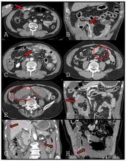
Background: Delayed bowel ischemia is a major cause of failure of nonoperative management in patients with blunt mesenteric injury. Although decreased bowel wall enhancement on computed tomography (CT) is a definitive sign of bowel ischemia, it is uncommon and may be absent on early imaging. This study aimed to identify specific CT findings that predict bowel ischemia to distinguish patients requiring surgery from those suitable for conservative management. Methods: We retrospectively reviewed 174 patients with blunt mesenteric injury treated at a Level 1 trauma center between January 2013 and December 2024. Initial CT findings were classified as mesenteric contrast extravasation freely extending into the peritoneal cavity (extravasation type 1), contrast extravasation tracking along the bowel contour (extravasation type 2), pseudoaneurysm, mesenteric haziness, mesenteric hematoma, interloop fluid, dependent portion fluid, and decreased bowel wall enhancement. Predictors of bowel ischemia were evaluated using univariate analysis and ridge-penalized multivariable logistic regression. Results: Bowel ischemia occurred in 30 patients (17.2%). Decreased bowel wall enhancement was rare (4.6%) but demonstrated perfect specificity and positive predictive value (both 100%), with low sensitivity (26.7%). Extravasation type 2 showed high specificity (97.2%) and remained an independent predictor of bowel ischemia. Dependent portion fluid showed relatively high sensitivity, whereas mesenteric haziness and mesenteric hematoma were inversely associated with ischemia. Conclusions: Contrast extravasation tracking along the bowel contour and decreased bowel wall enhancement on early CT are strong predictors of bowel ischemia in patients with blunt mesenteric injury. These findings should prompt consideration of early surgical exploration, even in patients who initially appear hemodynamically stable.

7 February 2026

Representative CT findings in mesenteric injury after blunt trauma. (A). Extravasation type 1 (arrow). (B). Extravasation type 2 (arrow). (C). Pseudoaneurysm (arrow). (D). Mesenteric haziness (circle). (E). Mesenteric hematoma (circle). (F). Interloop fluid (arrow). (G). Dependent portion fluid (arrow). (H). Decreased bowel wall enhancement (arrow).

Optical Coherence Tomography Angiography-Derived Peripapillary Vessel Density Findings in Multiple Sclerosis

  • Angeliki G. Filippatou,
  • Vasilios S. Liarakos and
  • Georgios Tsivgoulis
  • + 10 authors

Background/Objectives: Multiple sclerosis (MS)-related optic neuritis (ON) results in thinning of the peripapillary nerve fiber layer (pRNFL) which tends to be temporal quadrant-predominant. Optical coherence tomography angiography (OCTA) enables visualization of the retinal vasculature. Prior studies have shown reduced peripapillary vessel density (VD) in MS but data on the quadrantic pattern of peripapillary VD loss are limited. Our objective was to investigate the pattern of OCTA-derived peripapillary VD reduction in MS. Methods: People with MS (PwMS) and healthy controls (HC) underwent optic disc OCTA scans (Solix, Optovue) and VD was derived for the peripapillary region and quadrants. Eyes with ON within six months were excluded. Analyses were performed with generalized estimating equations models and standardized coefficients are presented. Results: We included 50 eyes from 29 PwMS (12 ON, 38 non-ON) and 12 eyes from 6 HC. VD in the peripapillary region was lower in MS ON eyes compared to HC with the largest effect size observed in the temporal quadrant (average: −1.47, p < 0.001; superior: −1.08, p = 0.006; inferior: −0.94; p = 0.017; temporal: −1.55; p < 0.001; nasal: −1.06, p = 0.007). In MS non-ON eyes, only temporal VD was significantly lower compared to HC eyes (temporal: −0.77, p = 0.004). Moderate to strong correlations were observed between OCT and corresponding OCTA metrics from the same regions. Conclusions: Our findings suggest that vascular alterations in the peripapillary region may exhibit a temporal quadrant predominant pattern. Larger studies are needed to further characterize the patterns and temporal evolution of retinal peripapillary vascular injury in MS.

7 February 2026

Example of peripapillary OCT and OCTA scan. Panel (A): En-face visualization of peripapillary vascular network with color-coded vessel density; Panel (B): Peripapillary OCT B-scan example; Panel (C): OCTA-derived peripapillary vessel density for superior (S), inferior (I), temporal (T) and nasal (N) quadrants (in %); Panel (D): OCT-derived peripapillary RNFL thickness for superior (S), inferior (I), temporal (T) and nasal (N) quadrants (in μm). OCT: optical coherence tomography; OCTA: OCT angiography; RNFL: retinal nerve fiber layer.

Macular Telangiectasia Type 2: The Role of Optical Coherence Tomography and Management Options

  • David-Ionuț Beuran,
  • Ioana Ruxandra Boca and
  • Cosmin Adrian Teodoru
  • + 4 authors

Background/Objectives: Macular Telangiectasia Type 2 (MacTel type 2) is a rare, progressive retinal disease that can lead to central vision loss. Optical coherence tomography (OCT) plays a crucial role in the early diagnosis, monitoring, and prognostic assessment of this condition. This narrative review aims to summarize the clinical features, OCT findings, and current management strategies for MacTel type 2. Methods: A literature search of PubMed, MEDLINE, and Google Scholar was performed for articles published from October 1993 to September 2025 using keywords related to MacTel type 2, OCT, clinical features, and treatment. All relevant clinical studies, including observational studies, clinical trials, and case series, were considered. The literature was screened independently by two authors, and a total of 69 articles were included. Results: Characteristic OCT findings include foveal cavitation, hyperreflective middle retinal layers, inner and outer retinal cavities, ellipsoid zone disruption, and retinal pigment clumps. Central macular thickness is consistently reduced, and structural biomarkers identified on OCT correlate with visual acuity decline. Treatment strategies vary by disease stage: non-proliferative MacTel type 2 currently has no universally effective therapy, although neuroprotective interventions such as ciliary neurotrophic factor (CNTF) show promising results. Proliferative MacTel type 2 is primarily managed with anti-vascular endothelial growth factor therapy (anti-VEGF), demonstrating functional and anatomical improvements. Conclusions: OCT provides essential structural evaluation for monitoring MacTel type 2, while treatment approaches remain stage-dependent. Emerging therapies, including CNTF implants and novel anti-VEGF agents, hold potential for improving outcomes.

7 February 2026

Fundus photo of a patient with early MacTel Type 2 changes, showing a subtle grayish area temporal to the fovea in the right eye. Cup-to-disc ratio asymmetry is noted. OCT demonstrated bilateral features. Images correspond with scans A and B from Figure 2.

Background: Silicone chin implants have been widely used is plastic and esthetic surgery of the face being considered as safe and efficient way for chin augmentation. However, complications such as bone resorption, displacement or ectopic bone formation may occur. Methods: The objective of this study was to evaluate complications associated with silicone chin implants and revision surgery protocols. Results: Among 98 patients who received silicone chin implants, 24 (11 males, 13 females) exhibited complications. The most commonly diagnosed issues were displacement (n = 3), bone resorption (n = 9), both conditions (n = 3), and patient dissatisfaction (n = 7). All patients were qualified for revision surgery, which included silicone implant removal followed by sliding genioplasty (n = 7), orthognathic surgery (n = 4), custom-made chin implant placement (n = 7), and repositioning and fixation (n = 1). After revision surgery, no complications occurred. Conclusions: Observations from this revision cohort suggest that careful patient selection and consideration of orthognathic or customized implant-based approaches may reduce the risk of dissatisfaction and revision surgery in patients with dentofacial deformities, or those seeking gender confirmation surgeries, compared to stock silicone implants.

7 February 2026

A 42-year-old Caucasian woman presented for consultation because of a mispositioned chin implant placed elsewhere four years earlier (A). She complained of asymmetry, palpable implant mobility, tissue irritation, and mild pain. CT showed implant malposition due to lack of fixation (A,C), with bone resorption and ectopic bone formation (B,D). The patient was scheduled for implant removal with immediate osteoplasty and placement of a custom-made PEEK chin implant.

News & Conferences

Issues

Open for Submission

Editor's Choice

Get Alerted

Add your email address to receive forthcoming issues of this journal.

XFacebookLinkedIn
J. Clin. Med. - ISSN 2077-0383