ijms-logo

Journal Browser

Journal Browser

State-of-the-Art Macromolecules in Japan

A topical collection in International Journal of Molecular Sciences (ISSN 1422-0067). This collection belongs to the section "Macromolecules".

Viewed by 18555

Editors


E-Mail Website
Collection Editor
Faculty of Textile Science and Technology, Department of Applied Biology, Shinshu University, Nagano, Japan
Interests: protein engineering; structural biology; biotechnology; synthetic biology

E-Mail Website
Collection Editor
Biomedical Research Institute, National Institute of Advanced Industrial Science and Technology (AIST), Tsukuba 305-8560, Japan
Interests: biomacromolecules; biosensors; liquid–liquid phase separation
Special Issues, Collections and Topics in MDPI journals

E-Mail Website
Collection Editor
Department of Chemistry, Ehime University, Ehime, Japan
Interests: nanoparticle; sensitive detection; amyloid

E-Mail Website
Collection Editor
Department of Biotechnology and Life Science, Tokyo University of Agriculture and Technology, Tokyo 184-8588, Japan
Interests: chaperone; olfaction; structural biology

E-Mail Website
Collection Editor
Department of Life Science, Akita University, Akita, Japan
Interests: enzyme; structural biology; computational biology

Topical Collection Information

Dear Colleagues, 

Molecules are the smallest units of materials. Among them, macromolecules are huge molecules, usually diameter ranging from about 10 to 1,000 nm. Our lives depend on various macromolecules, plastics, resins, and fibers. Our bodies are also made of macromolecules, such as proteins, nucleic acids, sugar chains. Most macromolecules are polymers. Proteins are polymers of amino acids. Nylon is a polyamide polymer. The development of novel materials and the advancement of life science depend on the studies of macromolecules.

Macromolecular science pursues a deeper understanding of the structure, property, and reactivity of macromolecules.

Macromolecular science and technology is multi-disciplinary research that combines physics, chemistry, biology, and engineering. A large number of research teams in Japan from different institutions and universities are working together and devoting considerable efforts to develop and study various macromolecules. This Topic Collection is committed to providing an overview of the macromolecular sciences and technologies in Japan.

Prof. Dr. Ryoichi Arai
Dr. Shunsuke Tomita
Prof. Dr. Tamotsu Zako
Prof. Dr. Masafumi Yohda
Prof. Dr. Masafumi Odaka
Collection Editors

Manuscript Submission Information

Manuscripts should be submitted online at www.mdpi.com by registering and logging in to this website. Once you are registered, click here to go to the submission form. Manuscripts can be submitted until the deadline. All submissions that pass pre-check are peer-reviewed. Accepted papers will be published continuously in the journal (as soon as accepted) and will be listed together on the collection website. Research articles, review articles as well as short communications are invited. For planned papers, a title and short abstract (about 100 words) can be sent to the Editorial Office for announcement on this website.

Submitted manuscripts should not have been published previously, nor be under consideration for publication elsewhere (except conference proceedings papers). All manuscripts are thoroughly refereed through a single-blind peer-review process. A guide for authors and other relevant information for submission of manuscripts is available on the Instructions for Authors page. International Journal of Molecular Sciences is an international peer-reviewed open access semimonthly journal published by MDPI.

Please visit the Instructions for Authors page before submitting a manuscript. There is an Article Processing Charge (APC) for publication in this open access journal. For details about the APC please see here. Submitted papers should be well formatted and use good English. Authors may use MDPI's English editing service prior to publication or during author revisions.

Keywords

  • protein
  • supramolecule
  • biomaterials
  • enzyme
  • polymers
  • nanoparticle

Published Papers (9 papers)

2024

Jump to: 2023, 2022

12 pages, 1924 KiB  
Article
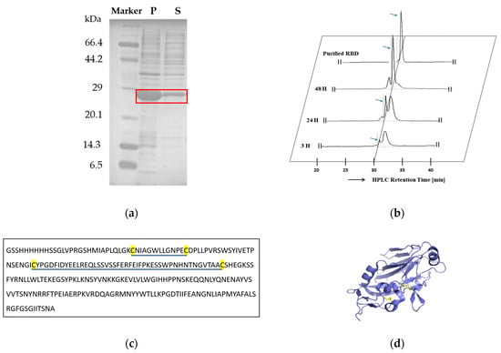
Design and Escherichia coli Expression of a Natively Folded Multi-Disulfide Bonded Influenza H1N1-PR8 Receptor-Binding Domain (RBD)
by Thao Tu, Tharangani Rathnayaka, Toshiyo Kato, Kenji Mizutani, Tomonori Saotome, Keiichi Noguchi, Shun-ichi Kidokoro and Yutaka Kuroda
Int. J. Mol. Sci. 2024, 25(7), 3943; https://doi.org/10.3390/ijms25073943 - 1 Apr 2024
Viewed by 1118
Abstract
Refolding multi-disulfide bonded proteins expressed in E. coli into their native structure is challenging. Nevertheless, because of its cost-effectiveness, handiness, and versatility, the E. coli expression of viral envelope proteins, such as the RBD (Receptor-Binding Domain) of the influenza Hemagglutinin protein, could significantly [...] Read more.
Refolding multi-disulfide bonded proteins expressed in E. coli into their native structure is challenging. Nevertheless, because of its cost-effectiveness, handiness, and versatility, the E. coli expression of viral envelope proteins, such as the RBD (Receptor-Binding Domain) of the influenza Hemagglutinin protein, could significantly advance research on viral infections. Here, we show that H1N1-PR8-RBD (27 kDa, containing four cysteines forming two disulfide bonds) expressed in E. coli and was purified with nickel affinity chromatography, and reversed-phase HPLC was successfully refolded into its native structure, as assessed with several biophysical and biochemical techniques. Analytical ultracentrifugation indicated that H1N1-PR8-RBD was monomeric with a hydrodynamic radius of 2.5 nm. Thermal denaturation, monitored with DSC and CD at a wavelength of 222 nm, was cooperative with a midpoint temperature around 55 °C, strongly indicating a natively folded protein. In addition, the 15N-HSQC NMR spectrum exhibited several 1H-15N resonances indicative of a beta-sheeted protein. Our results indicate that a significant amount (40 mg/L) of pure and native H1N1-PR8-RBD can be produced using an E. coli expression system with our refolding procedure, offering potential insights into the molecular characterization of influenza virus infection. Full article
Show Figures

Figure 1

17 pages, 3396 KiB  
Article
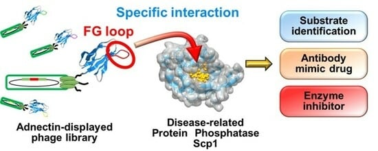
Identification of Inhibitors of the Disease-Associated Protein Phosphatase Scp1 Using Antibody Mimetic Molecules
by Tamaki Kobayashi, Kazuki Yamazaki, Junki Shinada, Masataka Mizunuma, Kazuhiro Furukawa and Yoshiro Chuman
Int. J. Mol. Sci. 2024, 25(7), 3737; https://doi.org/10.3390/ijms25073737 - 27 Mar 2024
Viewed by 1136
Abstract
Protein phosphorylation is a prevalent translational modification, and its dysregulation has been implicated in various diseases, including cancer. Despite its significance, there is a lack of specific inhibitors of the FCP/SCP-type Ser/Thr protein phosphatase Scp1, characterized by high specificity and affinity. In this [...] Read more.
Protein phosphorylation is a prevalent translational modification, and its dysregulation has been implicated in various diseases, including cancer. Despite its significance, there is a lack of specific inhibitors of the FCP/SCP-type Ser/Thr protein phosphatase Scp1, characterized by high specificity and affinity. In this study, we focused on adnectin, an antibody-mimetic protein, aiming to identify Scp1-specific binding molecules with a broad binding surface that target the substrate-recognition site of Scp1. Biopanning of Scp1 was performed using an adnectin-presenting phage library with a randomized FG loop. We succeeded in identifying FG-1Adn, which showed high affinity and specificity for Scp1. Ala scanning analysis of the Scp1-binding sequence in relation to the FG-1 peptide revealed that hydrophobic residues, including aromatic amino acids, play important roles in Scp1 recognition. Furthermore, FG-1Adn was found to co-localize with Scp1 in cells, especially on the plasma membrane. In addition, Western blotting analysis showed that FG-1Adn increased the phosphorylation level of the target protein of Scp1 in cells, indicating that FG-1Adn can inhibit the function of Scp1. These results suggest that FG-1Adn can be used as a specific inhibitor of Scp1. Full article
Show Figures

Graphical abstract

13 pages, 15032 KiB  
Article
Design of Protease-Responsive Antifungal Liposomal Formulation Decorated with a Lipid-Modified Chitin-Binding Domain
by Hendra Saputra, Muhammad Safaat, Pugoh Santoso, Rie Wakabayashi, Masahiro Goto, Toki Taira and Noriho Kamiya
Int. J. Mol. Sci. 2024, 25(7), 3567; https://doi.org/10.3390/ijms25073567 - 22 Mar 2024
Viewed by 1032
Abstract
Candida albicans is a prevalent fungal pathogen that displays antibiotic resistance. The polyene antifungal amphotericin B (AmB) has been the gold standard because of its broad antifungal spectra, and its liposomal formulation, AmBisome, has been used widely and clinically in treating fungal infections. [...] Read more.
Candida albicans is a prevalent fungal pathogen that displays antibiotic resistance. The polyene antifungal amphotericin B (AmB) has been the gold standard because of its broad antifungal spectra, and its liposomal formulation, AmBisome, has been used widely and clinically in treating fungal infections. Herein, we explored enhancing the antifungal activity of AmBisome by integrating a small chitin-binding domain (LysM) of chitinase A derived from Pteris ryukyuensis. LysM conjugated with a lipid (LysM–lipid) was initially prepared through microbial transglutaminase (MTG)-mediated peptide tag-specific conjugation of LysM with a lipid–peptide substrate. The AmBisome formulation modified with LysM–lipid conjugates had a size distribution that was comparable to the native liposomes but an increased zeta potential, indicating that LysM–lipid conjugates were anchored to AmBisome. LysM–lipid-modified AmBisome exhibited long-term stability at 4 °C while retaining the capacity to bind chitin. Nevertheless, the antifungal efficacy of LysM–lipid-modified AmBisome against C. albicans was modest. We then redesigned a new LysM–lipid conjugate by introducing a peptide linker containing a thrombin digestion (TD) site at the C-terminus of LysM (LysM–TD linker–lipid), thereby facilitating the liberation of the LysM domain from AmBisome upon the addition of thrombin. This new AmBisome formulation anchored with LysM–TD linker–lipid exhibited superior performance in suppressing C. albicans growth in the presence of thrombin compared with the LysM–lipid formulation. These results provide a platform to design stimuli-responsive AmBisome formulations that respond to external environments and thus advance the treatment of pathogenic fungi infections. Full article
Show Figures

Figure 1

13 pages, 2464 KiB  
Article
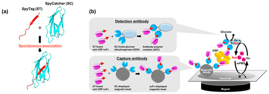
Rapid and Convenient Single-Chain Variable Fragment-Employed Electrochemical C-Reactive Protein Detection System
by Daimei Miura, Saki Motohashi, Ayaka Goto, Hayato Kimura, Wakako Tsugawa, Koji Sode, Kazunori Ikebukuro and Ryutaro Asano
Int. J. Mol. Sci. 2024, 25(5), 2859; https://doi.org/10.3390/ijms25052859 - 1 Mar 2024
Cited by 1 | Viewed by 1049
Abstract
Although IgG-free immunosensors are in high demand owing to ethical concerns, the development of convenient immunosensors that alternatively integrate recombinantly produced antibody fragments, such as single-chain variable fragments (scFvs), remains challenging. The low affinity of antibody fragments, unlike IgG, caused by monovalent binding [...] Read more.
Although IgG-free immunosensors are in high demand owing to ethical concerns, the development of convenient immunosensors that alternatively integrate recombinantly produced antibody fragments, such as single-chain variable fragments (scFvs), remains challenging. The low affinity of antibody fragments, unlike IgG, caused by monovalent binding to targets often leads to decreased sensitivity. We improved the affinity owing to the bivalent effect by fabricating a bivalent antibody–enzyme complex (AEC) composed of two scFvs and a single glucose dehydrogenase, and developed a rapid and convenient scFv-employed electrochemical detection system for the C-reactive protein (CRP), which is a homopentameric protein biomarker of systemic inflammation. The development of a point-of-care testing (POCT) system is highly desirable; however, no scFv-based CRP-POCT immunosensors have been developed. As expected, the bivalent AEC showed higher affinity than the single scFv and contributed to the high sensitivity of CRP detection. The electrochemical CRP detection using scFv-immobilized magnetic beads and the bivalent AEC as capture and detection antibodies, respectively, was achieved in 20 min without washing steps in human serum and the linear range was 1–10 nM with the limit of detection of 2.9 nM, which has potential to meet the criteria required for POCT application in rapidity, convenience, and hand-held detection devices without employing IgGs. Full article
Show Figures

Figure 1

2023

Jump to: 2024, 2022

19 pages, 4154 KiB  
Article
Development of Mn2+-Specific Biosensor Using G-Quadruplex-Based DNA
by Masataka Mizunuma, Mirai Suzuki, Tamaki Kobayashi, Yuki Hara, Atsushi Kaneko, Kazuhiro Furukawa and Yoshiro Chuman
Int. J. Mol. Sci. 2023, 24(14), 11556; https://doi.org/10.3390/ijms241411556 - 17 Jul 2023
Cited by 1 | Viewed by 1566
Abstract
Metal ions are used in various situations in living organisms and as a part of functional materials. Since the excessive intake of metal ions can cause health hazards and environmental pollution, the development of new molecules that can monitor metal ion concentrations with [...] Read more.
Metal ions are used in various situations in living organisms and as a part of functional materials. Since the excessive intake of metal ions can cause health hazards and environmental pollution, the development of new molecules that can monitor metal ion concentrations with high sensitivity and selectivity is strongly desired. DNA can form various structures, and these structures and their properties have been used in a wide range of fields, including materials, sensors, and drugs. Guanine-rich sequences respond to metal ions and form G-quadruplex structures and G-wires, which are the self-assembling macromolecules of G-quadruplex structures. Therefore, guanine-rich DNA can be applied to a metal ion-detection sensor and functional materials. In this study, the IRDAptamer library originally designed based on G-quadruplex structures was used to screen for Mn2+, which is known to induce neurodegenerative diseases. Circular dichroism and fluorescence analysis using Thioflavin T showed that the identified IRDAptamer sequence designated MnG4C1 forms a non-canonical G-quadruplex structure in response to low concentrations of Mn2+. A serum resistance and thermostability analysis revealed that MnG4C1 acquired stability in a Mn2+-dependent manner. A Förster resonance energy transfer (FRET) system using fluorescent molecules attached to the termini of MnG4C1 showed that FRET was effectively induced based on Mn2+-dependent conformational changes, and the limit of detection (LOD) was 0.76 µM for Mn2+. These results suggested that MnG4C1 can be used as a novel DNA-based Mn2+-detecting molecule. Full article
Show Figures

Figure 1

2022

Jump to: 2024, 2023

9 pages, 2043 KiB  
Communication
Relative Nuclease Resistance of a DNA Aptamer Covalently Conjugated to a Target Protein
by Yudai Tabuchi, Jay Yang and Masumi Taki
Int. J. Mol. Sci. 2022, 23(14), 7778; https://doi.org/10.3390/ijms23147778 - 14 Jul 2022
Cited by 6 | Viewed by 2332
Abstract
A major obstacle to the therapeutic application of an aptamer is its susceptibility to nuclease digestion. Here, we confirmed the acquisition of relative nuclease resistance of a DNA-type thrombin binding aptamer with a warhead (TBA3) by covalent binding to a target [...] Read more.
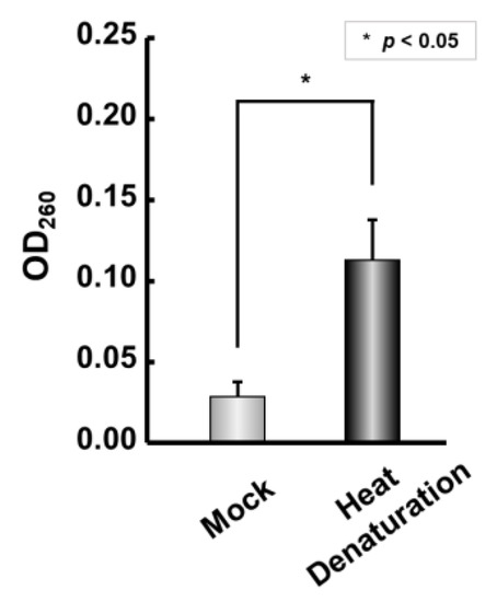
A major obstacle to the therapeutic application of an aptamer is its susceptibility to nuclease digestion. Here, we confirmed the acquisition of relative nuclease resistance of a DNA-type thrombin binding aptamer with a warhead (TBA3) by covalent binding to a target protein in the presence of serum/various nucleases. When the thrombin-inhibitory activity of TBA3 on thrombin was reversed by the addition of the complementary strand, the aptamer was instantly degraded by the nucleases, showing that the properly folded/bound aptamer conferred the resistance. Covalently binding aptamers possessing both a prolonged drug effect and relative nuclease resistance would be beneficial for in vivo translational applications. Full article
Show Figures

Graphical abstract

13 pages, 2678 KiB  
Article
Long-Term Fluorescent Tissue Marking Using Tissue-Adhesive Porphyrin with Polycations Consisting of Quaternary Ammonium Salt Groups
by Yoshiki Komatsu, Toru Yoshitomi, Kinji Furuya, Takafumi Ikeda, Azusa Terasaki, Aoi Hoshi, Naoki Kawazoe, Guoping Chen and Hirofumi Matsui
Int. J. Mol. Sci. 2022, 23(8), 4218; https://doi.org/10.3390/ijms23084218 - 11 Apr 2022
Cited by 4 | Viewed by 2314
Abstract
Localization of tumors during laparoscopic surgery is generally performed by locally injecting India ink into the submucosal layer of the gastrointestinal tract using endoscopy. However, the location of the tumor is obscured because of the black-stained surgical field and the blurring caused by [...] Read more.
Localization of tumors during laparoscopic surgery is generally performed by locally injecting India ink into the submucosal layer of the gastrointestinal tract using endoscopy. However, the location of the tumor is obscured because of the black-stained surgical field and the blurring caused by India ink. To solve this problem, in this study, we developed a tissue-adhesive porphyrin with polycations consisting of quaternary ammonium salt groups. To evaluate the ability of tissue-adhesive porphyrin in vivo, low-molecular-weight hematoporphyrin and tissue-adhesive porphyrin were injected into the anterior wall of the exposed stomach in rats. Local injection of low-molecular-weight hematoporphyrin into the anterior wall of the stomach was not visible even after 1 day because of its rapid diffusion. In contrast, the red fluorescence of the tissue-adhesive porphyrin was visible even after 7 days due to the electrostatic interactions between the positively-charged moieties of the polycation in the tissue-adhesive porphyrin and the negatively-charged molecules in the tissue. In addition, intraperitoneal injection of tissue-adhesive porphyrin in rats did not cause adverse effects such as weight loss, hepatic or renal dysfunction, or organ adhesion in the abdominal cavity. These results indicate that tissue-adhesive porphyrin is a promising fluorescent tissue-marking agent. Full article
Show Figures

Figure 1

12 pages, 1789 KiB  
Article
A Multi-Disulfide Receptor-Binding Domain (RBD) of the SARS-CoV-2 Spike Protein Expressed in E. coli Using a SEP-Tag Produces Antisera Interacting with the Mammalian Cell Expressed Spike (S1) Protein
by Subbaian Brindha and Yutaka Kuroda
Int. J. Mol. Sci. 2022, 23(3), 1703; https://doi.org/10.3390/ijms23031703 - 1 Feb 2022
Cited by 16 | Viewed by 3413
Abstract
An Escherichia coli (E. coli) production of the receptor-binding domain (RBD) of the SARS-CoV-2 (isolate Wuhan-Hu-1) spike protein would significantly accelerate the search for anti-COVID-19 therapeutics because of its versatility and low cost. However, RBD contains four disulfide bonds and its [...] Read more.
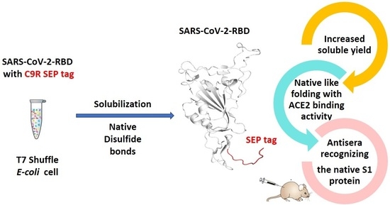
An Escherichia coli (E. coli) production of the receptor-binding domain (RBD) of the SARS-CoV-2 (isolate Wuhan-Hu-1) spike protein would significantly accelerate the search for anti-COVID-19 therapeutics because of its versatility and low cost. However, RBD contains four disulfide bonds and its expression in E. coli is limited by the formation of aberrant disulfide bonds resulting in inclusion bodies. Here, we show that a solubility-enhancing peptide (SEP) tag containing nine arginine residues (RBD-C9R) attached at the C-terminus can overcome this problem. The SEP-tag increased the expression in the soluble fraction and the final yield by five times (2 mg/L). The folding properties of the E. coli expressed RBD-C9R were demonstrated with biophysical characterization using RP-HPLC, circular dichroism, thermal denaturation, fluorescence, and light scattering. A quartz crystal microbalance (QCM) analysis confirmed the binding activity of RBD-C9R with ACE2, the host cell’s receptor. In addition, RBD-C9R elicited a Th-2 immune response with a high IgG titer in Jcl: ICR mice. The RBD-C9R antisera interacted with both itself and the mammalian-cell expressed spike protein (S1), as demonstrated by ELISA, indicating that the E. coli expressed RBD-C9R harbors native-like epitopes. Overall, these results emphasize the potential of our SEP-tag for the E. coli production of active multi-disulfide-bonded RBD. Full article
Show Figures

Graphical abstract

19 pages, 3483 KiB  
Article
Self-Assembling Lectin Nano-Block Oligomers Enhance Binding Avidity to Glycans
by Shin Irumagawa, Keiko Hiemori, Sayoko Saito, Hiroaki Tateno and Ryoichi Arai
Int. J. Mol. Sci. 2022, 23(2), 676; https://doi.org/10.3390/ijms23020676 - 8 Jan 2022
Cited by 7 | Viewed by 2824
Abstract
Lectins, carbohydrate-binding proteins, are attractive biomolecules for medical and biotechnological applications. Many lectins have multiple carbohydrate recognition domains (CRDs) and strongly bind to specific glycans through multivalent binding effect. In our previous study, protein nano-building blocks (PN-blocks) were developed to construct self-assembling supramolecular [...] Read more.
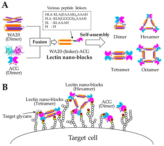
Lectins, carbohydrate-binding proteins, are attractive biomolecules for medical and biotechnological applications. Many lectins have multiple carbohydrate recognition domains (CRDs) and strongly bind to specific glycans through multivalent binding effect. In our previous study, protein nano-building blocks (PN-blocks) were developed to construct self-assembling supramolecular nanostructures by linking two oligomeric proteins. A PN-block, WA20-foldon, constructed by fusing a dimeric four-helix bundle de novo protein WA20 to a trimeric foldon domain of T4 phage fibritin, self-assembled into several types of polyhedral nanoarchitectures in multiples of 6-mer. Another PN-block, the extender PN-block (ePN-block), constructed by tandemly joining two copies of WA20, self-assembled into cyclized and extended chain-type nanostructures. This study developed novel functional protein nano-building blocks (lectin nano-blocks) by fusing WA20 to a dimeric lectin, Agrocybe cylindracea galectin (ACG). The lectin nano-blocks self-assembled into various oligomers in multiples of 2-mer (dimer, tetramer, hexamer, octamer, etc.). The mass fractions of each oligomer were changed by the length of the linkers between WA20 and ACG. The binding avidity of the lectin nano-block oligomers to glycans was significantly increased through multivalent effects compared with that of the original ACG dimer. Lectin nano-blocks with high avidity will be useful for various applications, such as specific cell labeling. Full article
Show Figures

Figure 1

Back to TopTop