


Int. J. Mol. Sci. 2023, 24(8), 7237; https://doi.org/10.3390/ijms24087237 - 14 Apr 2023
Cited by 10 | Viewed by 3072
Abstract
►
Show Figures
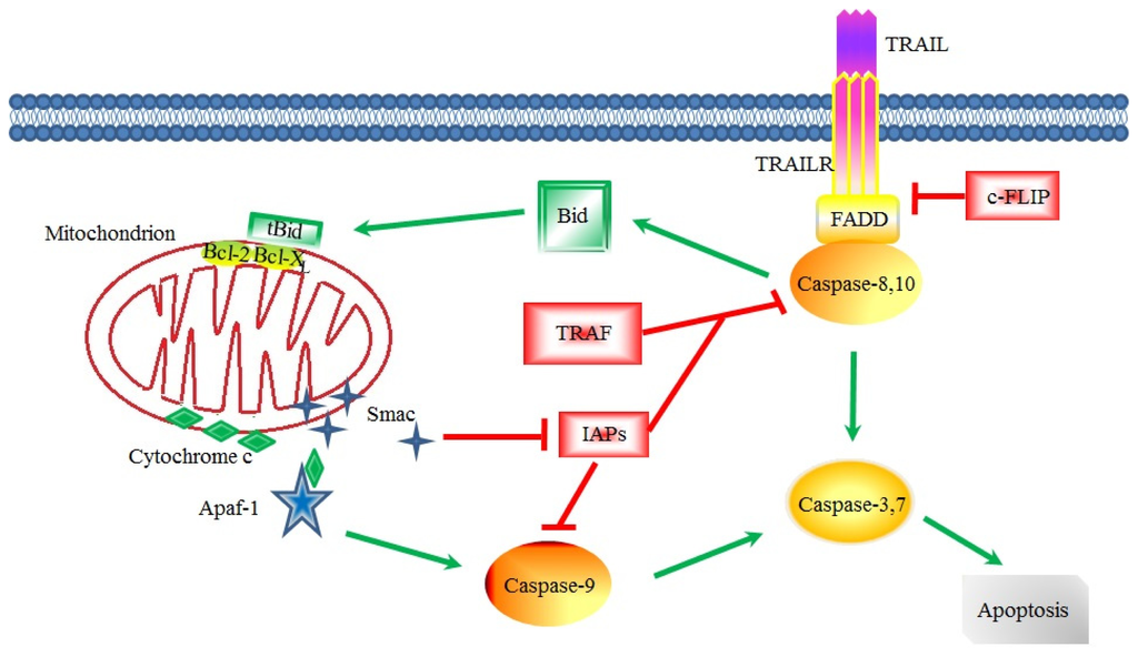
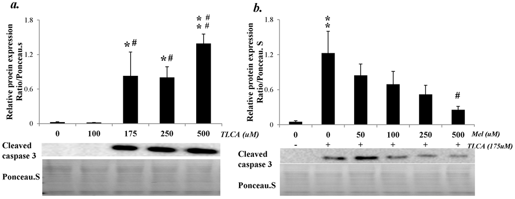
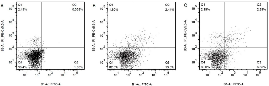
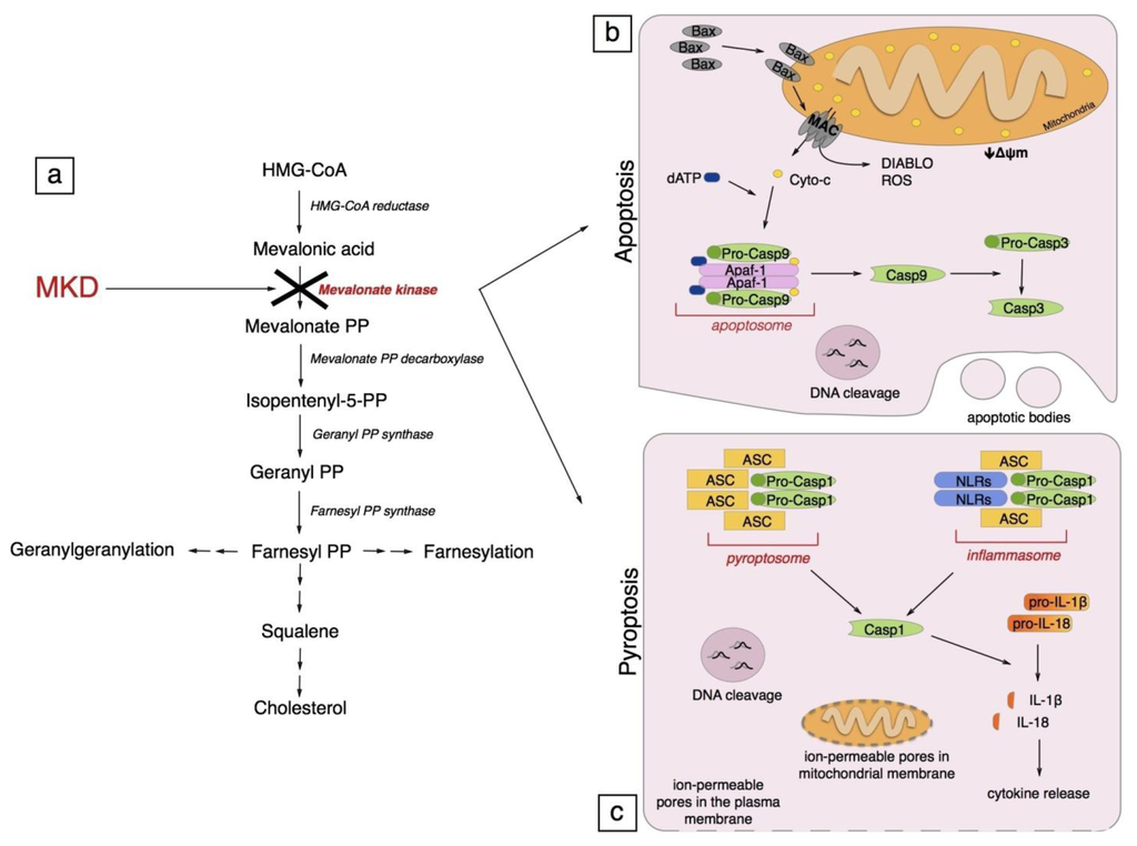
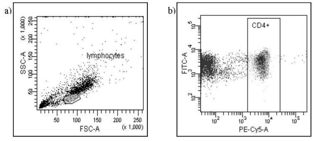
Among the extracellular vesicles, apoptotic bodies (ABs) are only formed during the apoptosis and perform a relevant role in the pathogenesis of different diseases. Recently, it has been demonstrated that ABs from human renal proximal tubular HK-2 cells, either induced by cisplatin or
[...] Read more.
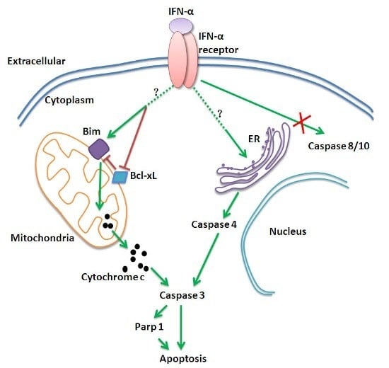
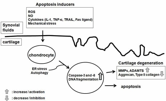
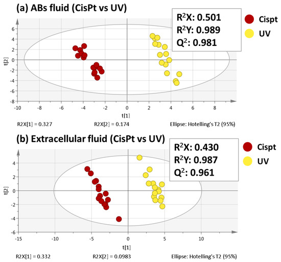
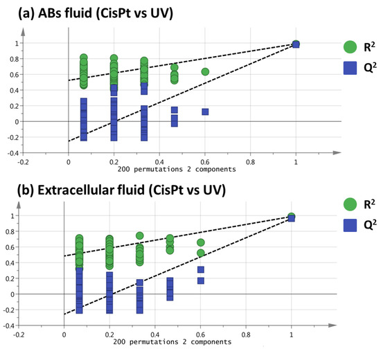
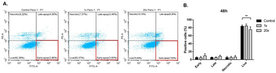
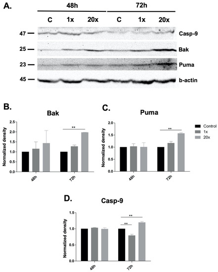
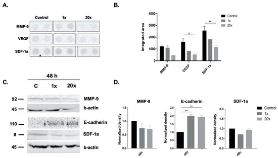
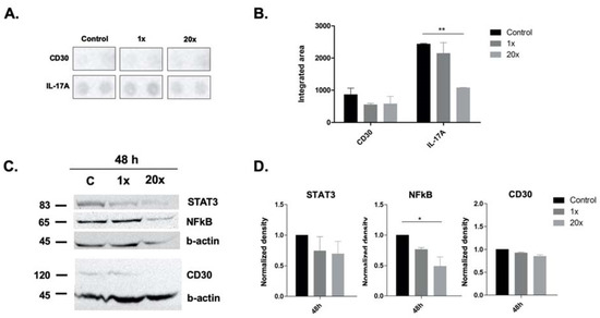
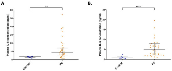
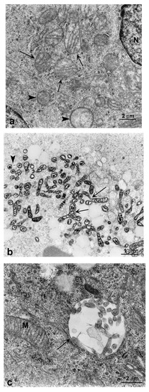
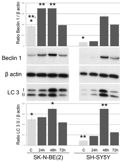
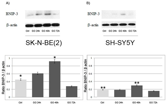
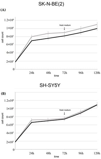
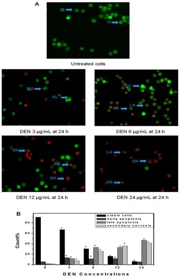
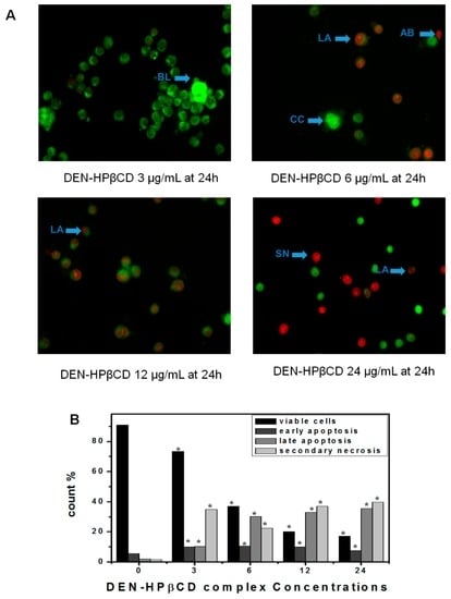
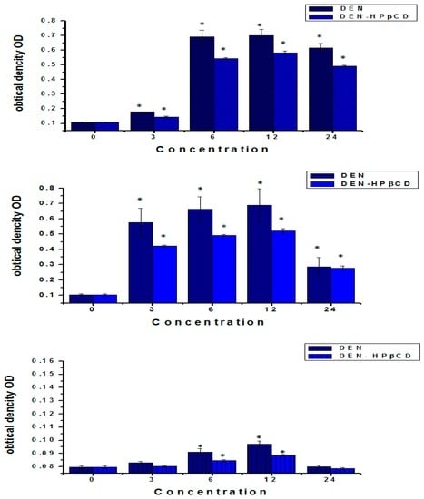
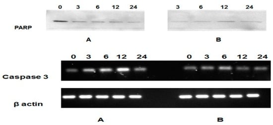
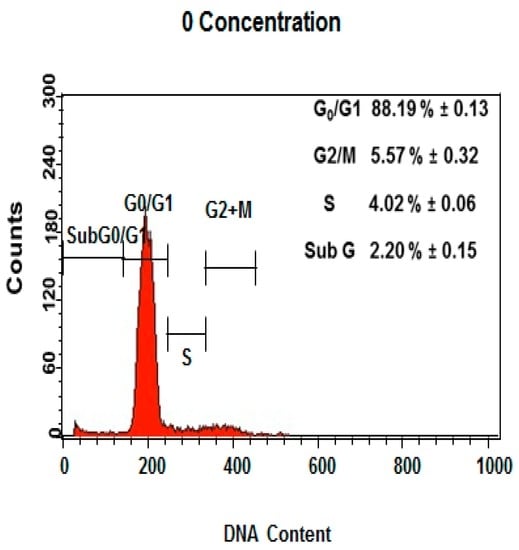
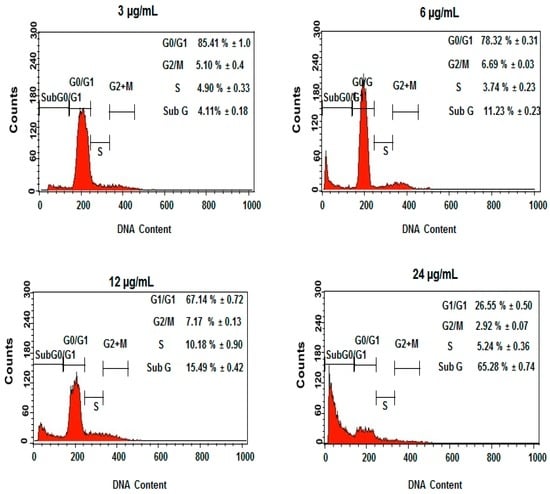
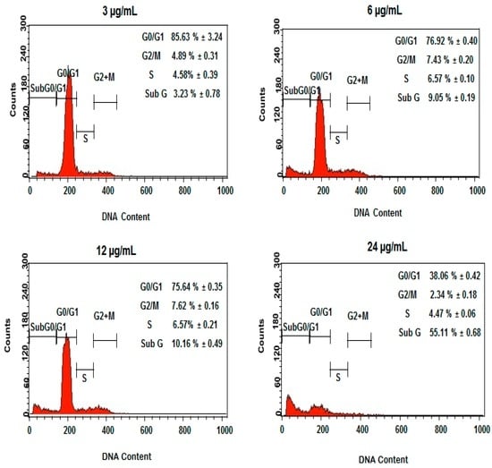
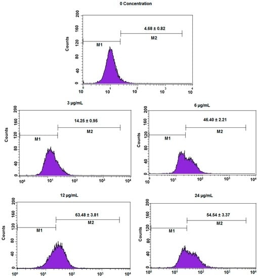
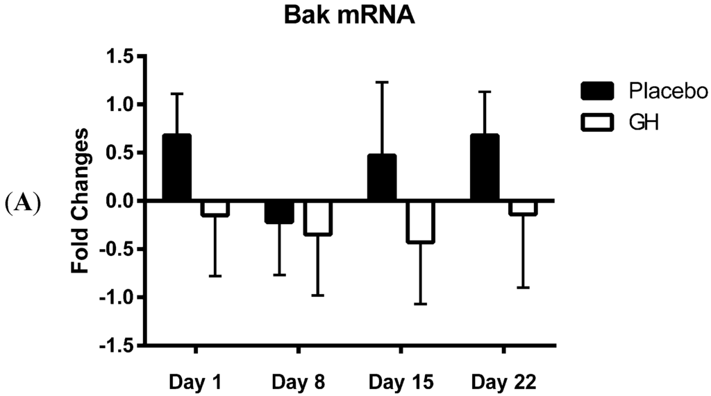
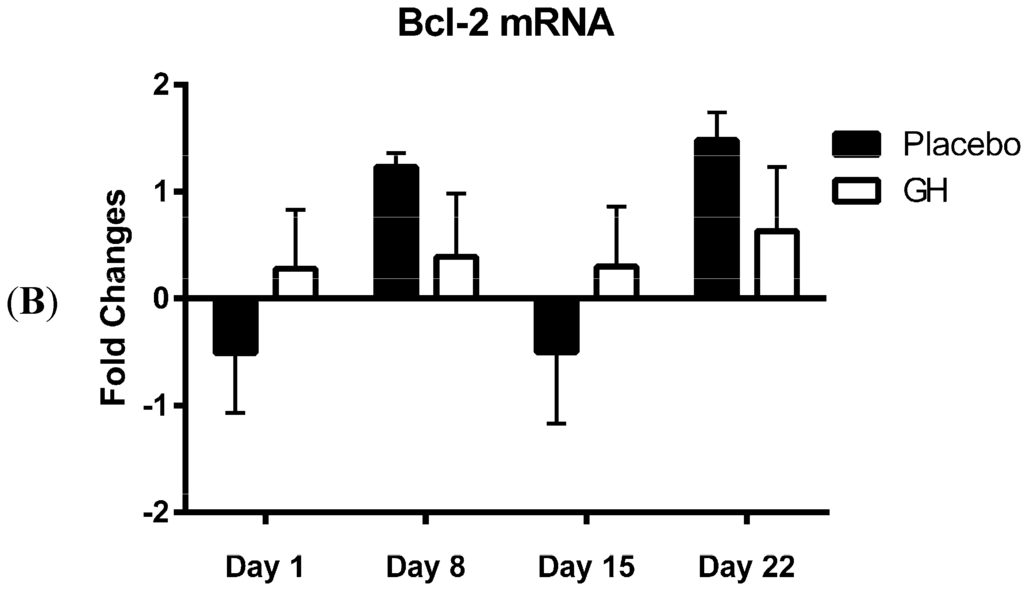
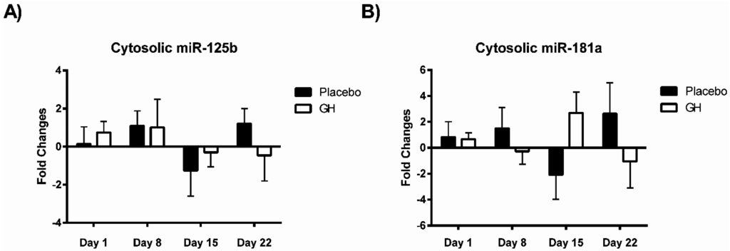
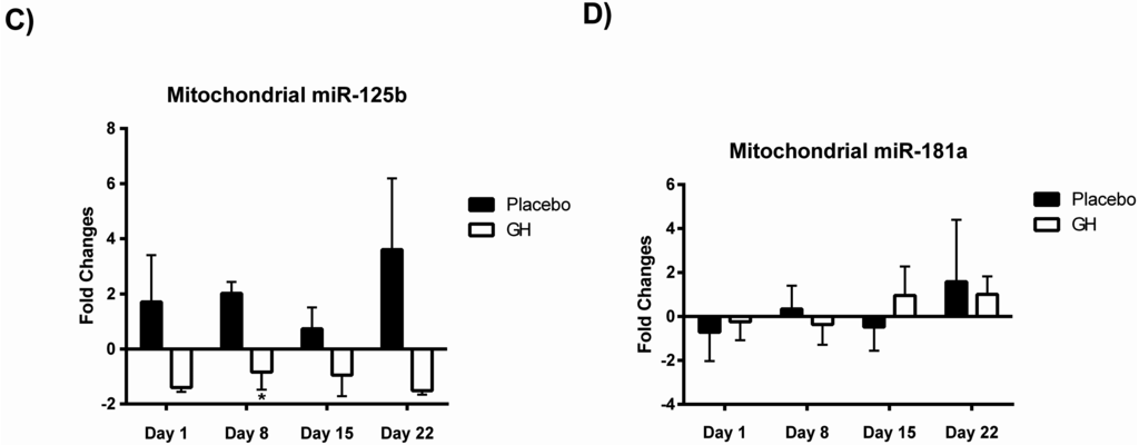
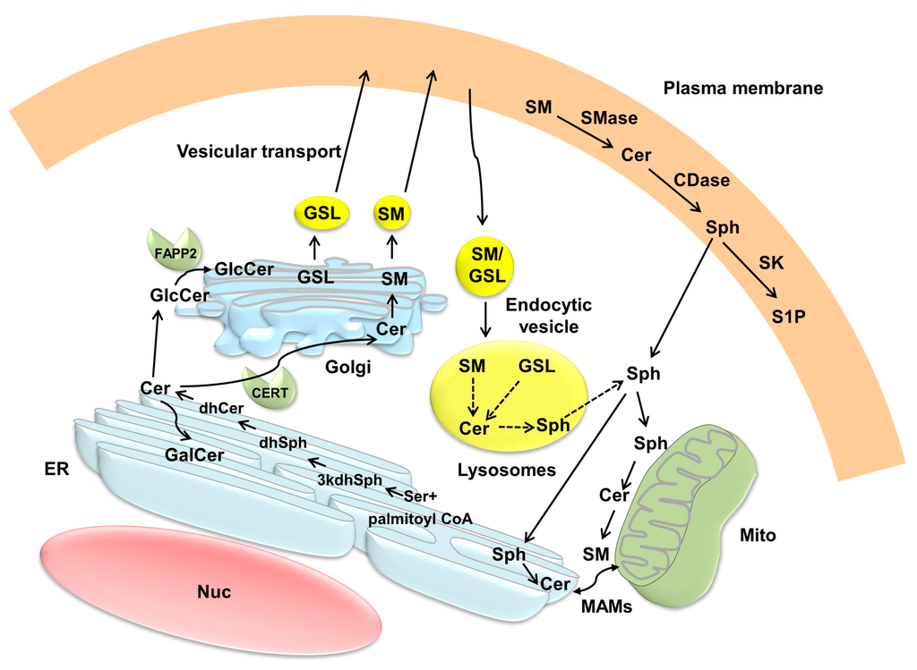
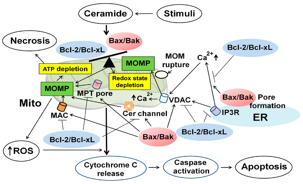
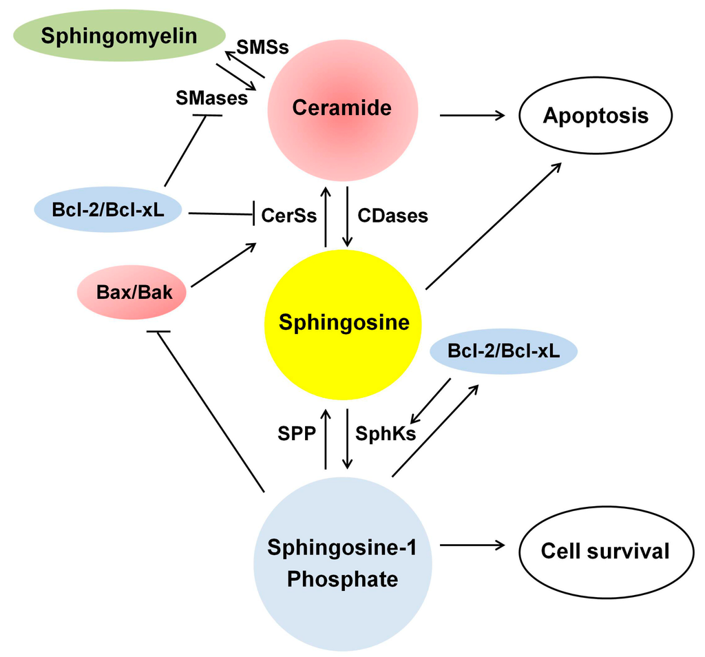
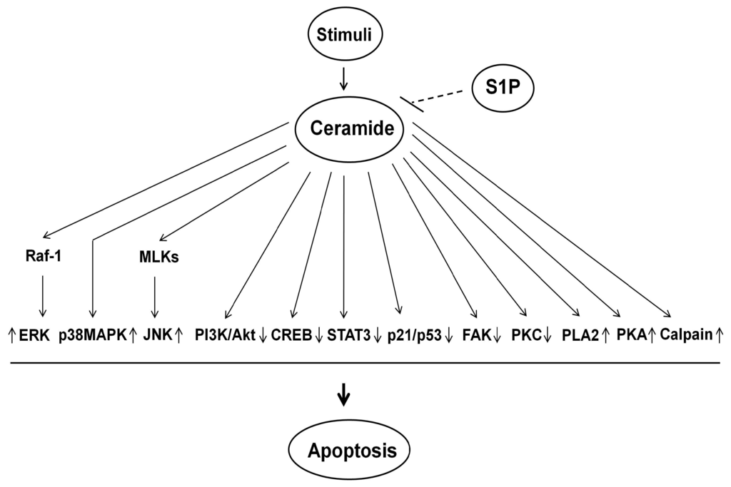
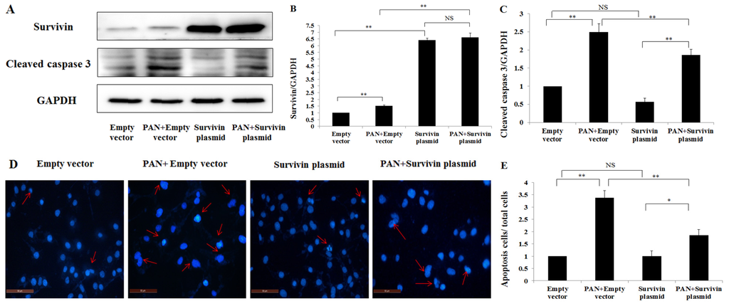
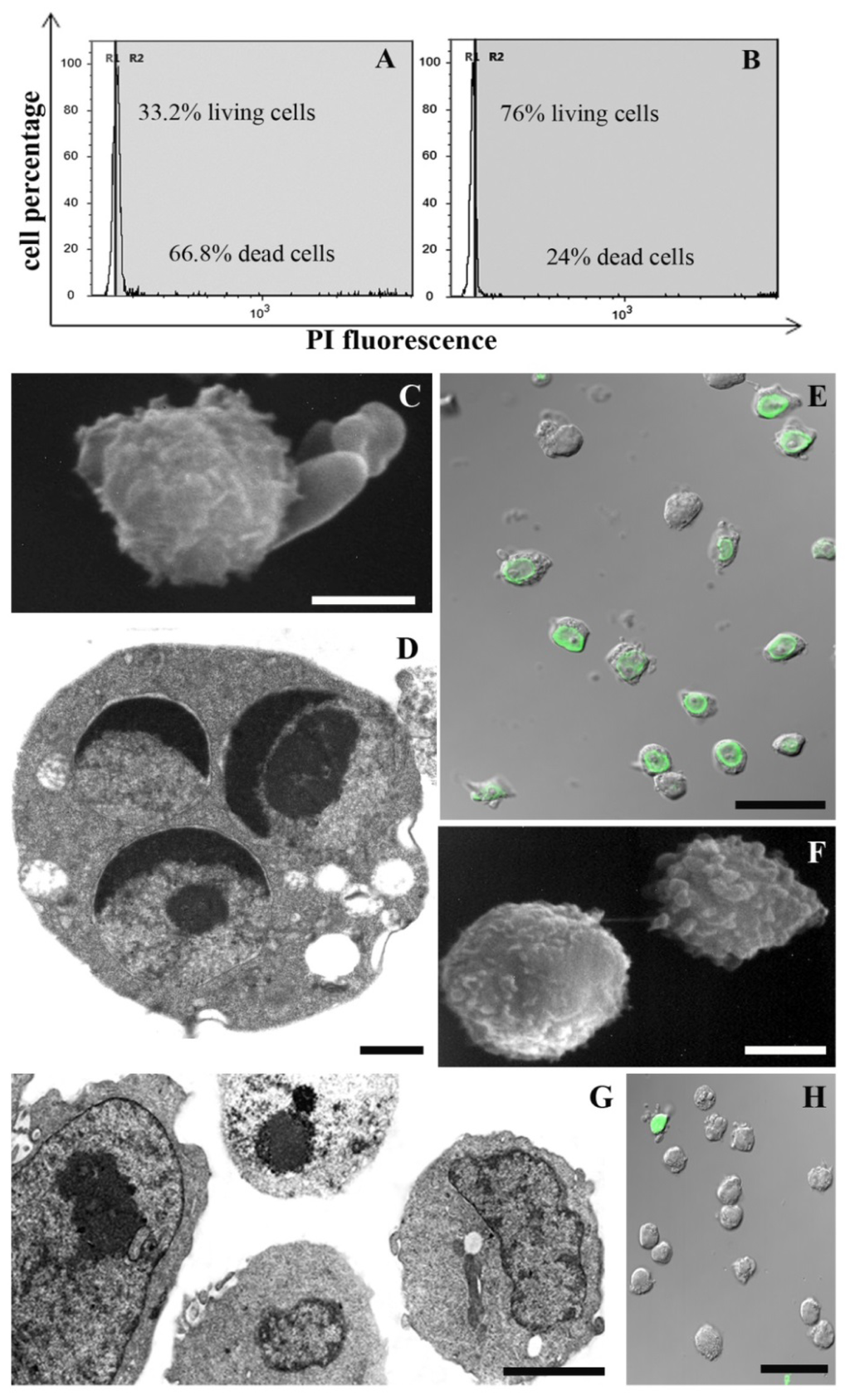
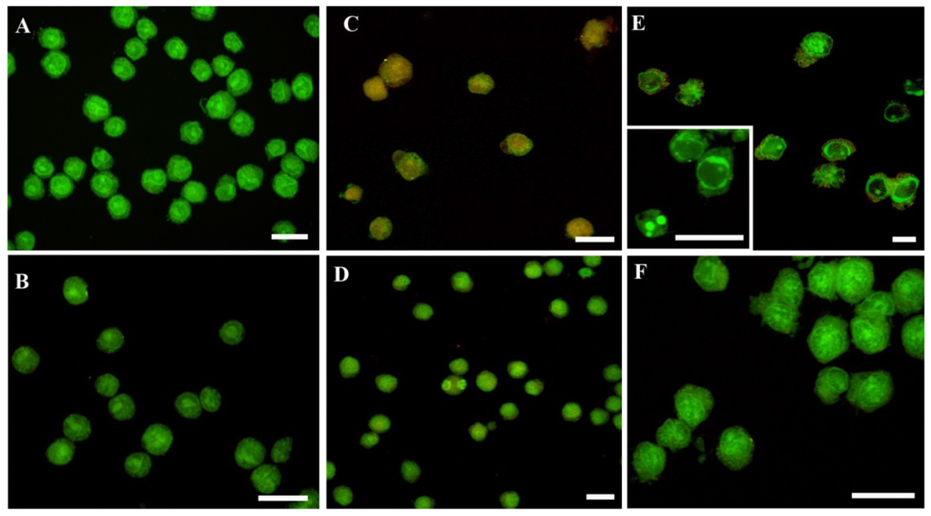
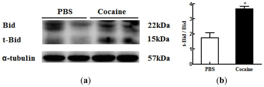
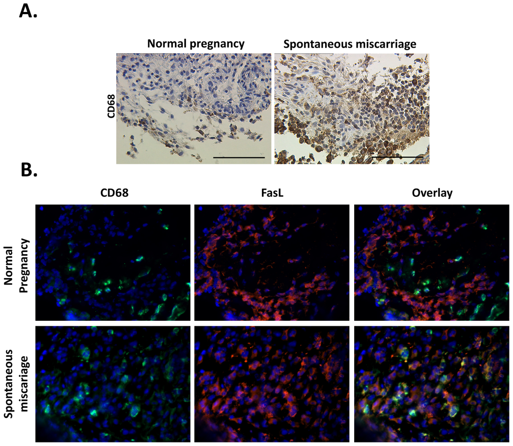
Among the extracellular vesicles, apoptotic bodies (ABs) are only formed during the apoptosis and perform a relevant role in the pathogenesis of different diseases. Recently, it has been demonstrated that ABs from human renal proximal tubular HK-2 cells, either induced by cisplatin or by UV light, can lead to further apoptotic death in naïve HK-2 cells. Thus, the aim of this work was to carry out a non-targeted metabolomic approach to study if the apoptotic stimulus (cisplatin or UV light) affects in a different way the metabolites involved in the propagation of apoptosis. Both ABs and their extracellular fluid were analyzed using a reverse-phase liquid chromatography-mass spectrometry setup. Principal components analysis showed a tight clustering of each experimental group and partial least square discriminant analysis was used to assess the metabolic differences existing between these groups. Considering the variable importance in the projection values, molecular features were selected and some of them could be identified either unequivocally or tentatively. The resulting pathways indicated that there are significant, stimulus-specific differences in metabolites abundancies that may propagate apoptosis to healthy proximal tubular cells; thus, we hypothesize that the share in apoptosis of these metabolites might vary depending on the apoptotic stimulus.
Full article
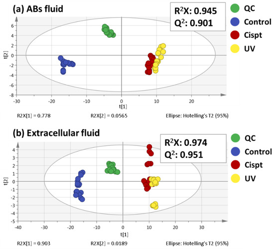
Figure 1


