


Int. J. Mol. Sci. 2024, 25(7), 3974; https://doi.org/10.3390/ijms25073974 - 3 Apr 2024
Cited by 6 | Viewed by 3636
Abstract
►
Show Figures
Trueperella pyogenes can cause various infections in the organs and tissues of different livestock (including pigs, cows, goats, and sheep), including mastitis, endometritis, pneumonia, or abscesses. Moreover, diseases induced by T. pyogenes cause significant economic losses in animal husbandry. In recent large-scale investigations,
[...] Read more.
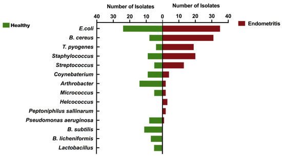
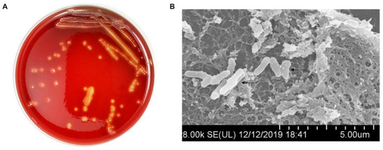
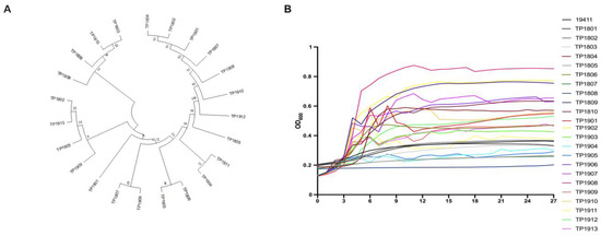
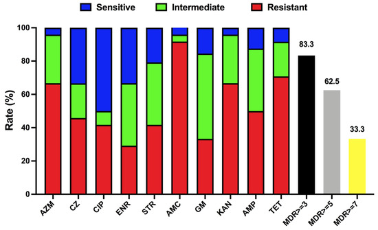
Trueperella pyogenes can cause various infections in the organs and tissues of different livestock (including pigs, cows, goats, and sheep), including mastitis, endometritis, pneumonia, or abscesses. Moreover, diseases induced by T. pyogenes cause significant economic losses in animal husbandry. In recent large-scale investigations, T. pyogenes has been identified as one of the main pathogens causing endometritis in lactating cows. However, the main treatment for the above-mentioned diseases is still currently antibiotic therapy. Understanding the impact of endometritis associated with T. pyogenes on the fertility of cows can help optimize antibiotic treatment for uterine diseases, thereby strategically concentrating the use of antimicrobials on the most severe cases. Therefore, it is particularly important to continuously monitor the prevalence of T. pyogenes and test its drug resistance. This study compared the uterine microbiota of healthy cows and endometritis cows in different cattle farms, investigated the prevalence of T. pyogenes, evaluated the genetic characteristics and population structure of isolated strains, and determined the virulence genes and drug resistance characteristics of T. pyogenes. An amount of 186 dairy cows were involved in this study and 23 T. pyogenes strains were isolated and identified from the uterine lavage fluid of dairy cows with or without endometritis.
Full article

Figure 1

