Article

Peroxisome Proliferator FpPEX11 Is Involved in the Development and Pathogenicity in Fusarium pseudograminearum

Key Laboratory of Agricultural Microbiology, College of Plant Protection, Shandong Agricultural University, Tai’an 271018, China
*
Authors to whom correspondence should be addressed.
Int. J. Mol. Sci. 2022, 23(20), 12184; https://doi.org/10.3390/ijms232012184
Submission received: 8 September 2022 / Revised: 1 October 2022 / Accepted: 6 October 2022 / Published: 12 October 2022
(This article belongs to the Collection State-of-the-Art Molecular Microbiology in China)

Abstract

:
Fusarium crown rot (FCR) of wheat, an important soil-borne disease, presents a worsening trend year by year, posing a significant threat to wheat production. Fusarium pseudograminearum cv. b was reported to be the dominant pathogen of FCR in China. Peroxisomes are single-membrane organelles in eukaryotes that are involved in many important biochemical metabolic processes, including fatty acid β-oxidation. PEX11 is important proteins in peroxisome proliferation, while less is known in the fungus F. pseudograminearum. The functions of FpPEX11a, FpPEX11b, and FpPEX11c in F. pseudograminearum were studied using reverse genetics, and the results showed that FpPEX11a and FpPEX11b are involved in the regulation of vegetative growth and asexual reproduction. After deleting FpPEX11a and FpPEX11b, cell wall integrity was impaired, cellular metabolism processes including active oxygen metabolism and fatty acid β-oxidation were significantly blocked, and the production ability of deoxynivalenol (DON) decreased. In addition, the deletion of genes of FpPEX11a and FpPEX11b revealed a strongly decreased expression level of peroxisome-proliferation-associated genes and DON-synthesis-related genes. However, deletion of FpPEX11c did not significantly affect these metabolic processes. Deletion of the three protein-coding genes resulted in reduced pathogenicity of F. pseudograminearum. In summary, FpPEX11a and FpPEX11b play crucial roles in the growth and development, asexual reproduction, pathogenicity, active oxygen accumulation, and fatty acid utilization in F. pseudograminearum.

1. Introduction

Fusarium crown rot (FCR) is a common wheat disease. In Australia, where it was first reported, the direct economic loss due to FCR was as high as AUD 56.3 million per year, with a potential loss of up to AUD 160 million. In the United States, FCR causes wheat yields to decrease by 10 million tons [1,2]. In recent years, the damage caused by FCR has gradually worsened in the Huang Huai wheat region of China; for example, the yield loss caused by FCR is up to 30–50% in many wheat-producing areas in the Henan Province. These data indicate that FCR is a serious threat to wheat grain security and should be controlled immediately. FCR is a typical soil-borne disease. It overwinters mainly in the form of mycelium in soil and diseased plant residues, which can be preserved for more than 2 years in general and longer in arid or semi-arid climates. It is mainly transmitted during cultivation, and its hosts mainly include weeds and various gramineous crops, such as wheat, barley, and corn. It generally does not infect dicotyledonous crops [3,4]. F. pseudograminearum shows heterothallism, and the sexual state of Gibberella coronicola is not easily inducible indoors and is difficult to find in the field [3]. Pathogens generally invade the roots and stem bases of the plants, and the specific infection site depends on the distribution of the pathogen source in the soil. Fusarium head blight could also be induced at the flowering stage in warm and humid climates [5]. The environment in the field is the main factor controlling the development of FCR. It has been reported that early sowing can aggravate the disease, and appropriate late sowing can reduce the degree of the disease [6]. Cohesive, low-lying, poorly drained, or highly humid soil can promote disease development. Excessive application of nitrogenous fertilizer in the field also increases the occurrence of FCR [7,8]. However, an appropriate increase in zinc fertilizer can effectively reduce the occurrence of the disease [9], but the molecular mechanism of its pathogenesis has not yet been revealed.
Peroxisomes, also known as microbodies, are dynamic, small, single-membraned organelles [10], and their quantity and activity can be adjusted according to the state of the tissue and organ, as well as nutrition [11]. When needed, the endoplasmic reticulum (ER) can synthesize new peroxisomes, or they can rapidly divide and proliferate from pre-existing peroxisomes. When the external or cellular environment changes and the peroxisomes are no longer needed to perform their function, they respond to environmental changes through pexophagy. In S. cerevisiae, peroxisome division involves several processes. First, mature peroxisomes elongate under the action of Pex11. Matrix proteins and proteins involved in cleavage are delivered to the elongated peroxisomes. Then, Dnm1 localizes to the peroxisomal constriction site to initiate the membrane constriction process by hydrolyzing GTP. Finally, cooperating with Fis1, Dnm1, Mdv1, or Caf4, a daughter peroxisome is produced [12,13,14]. Increasing attention has been paid to the pathogenicity of peroxisomes and plant-pathogenic fungi. First, pathogenic fungi require a large amount of energy when infecting hosts, and most of the energy comes from fatty acid β-oxidation metabolism in the peroxisomes. Second, plants infected by pathogenic fungi produce large amounts of reactive oxygen species (ROS). Phytopathogens can infect plants when they respond to ROS [14]. The peroxisomes contain more than 50 enzymes—mainly catalases and oxidases that are beneficial for cell detoxification—and genes related to peroxisome synthesis are closely related to the pathogenicity of plant pathogens. For example, the deletion of the PEX5 from Penicillium chrysogenum affects asexual reproduction and vegetative growth [15], as well as the proliferation of peroxisomes which accompanies the infection [16]. Most eukaryotic cells contain peroxisomes, which play an integral role in a variety of biochemical pathways, including ether phospholipid biosynthesis, fatty acid α-and β-oxidation, bile acid and docosahexaenoic acid synthesis, glyoxylic acid metabolism, amino acid catabolism, polyamine oxidation, and reactive oxygen and nitrogen metabolism (ROS and RNS) [17]. Additionally, mammalian peroxisomes are not only metabolic organelles but signaling platforms for the regulation of various physiological and pathological processes, including inflammation and innate immunity. For instance, Zellweger syndrome (ZS) is an autosomal recessive peroxisome biogenesis disease, and some reports have pointed out that peroxisome abnormalities can directly or indirectly cause age-related diabetes, neurodegenerative diseases, and cancer [18,19,20]. In plants, peroxisomes are involved in embryonic development, photorespiration, host resistance, metabolism of nitrogen and sulfur compounds, synthesis of plant hormones (auxin, jasmonic acid, etc.), and the glyoxylic acid cycle [21,22,23,24,25]. In [1] seeds, peroxisomes participate in the glyoxylic acid cycle, mobilizing the lipids stored in oleaginous seeds to degrade them into energy for germination, and are therefore also known as a glyoxysomes [26].
PEX11 is a peroxisomal membrane protein and the first protein identified to be involved in peroxisome proliferation and membrane extension [27] and is necessary for the polarization and division of peroxisomal membranes. The PEX11 protein belongs to the PEX gene family, with the most members at present. The number of members in this family varies significantly among different species [28]. Five members of the PEX11 protein family were detected in Arabidopsis thaliana—PEX11a, PEX11b, PEX11c, PEX11d, and PEX11e. These are divided into three subfamilies, all of which promote peroxisomal proliferation. Each family member plays a specific role in different environmental conditions, and perhaps in different steps of peroxisome proliferation [28,29]. In mammals, like humans and mice, three PEX11-related proteins exist: PEX11α, PEX11β, and PEX11γ [30]. In fungi, the composition of the PEX11 protein family is relatively complex, containing two to three members in most fungi, and up to five members in ascomycetes. For example, Magnaporthe oryzae contains three PEX11 family members, and deletion of the MoPEX11A gene has the greatest effect on its pathogenicity [31]. In S. cerevisiae, the C-terminal ends of PEX11 are similar to those of the homologous proteins, PEX25p (YPL112c) and PEX27 (YOR193w). PEX11 localizes to the peroxisomal membrane and may form homo-oligomers. These proteins form the S. cerevisiae PEX11 protein family, whose members are necessary for peroxisome biosynthesis, and play a role in the regulation of their size and number [32,33]. However, PEX25 is necessary for the de novo biosynthesis of peroxisomes, and PEX27 competes with PEX25 to inhibit peroxisomal proliferation [14]. Thus far, most reports are from studies on yeast and mammals, while the functions of PEX11 in F. pseudograminearum are largely unclear.
In this study, reverse genetics was used to investigate the functions of FpPEX11a, FpPEX11b, and FpPEX11c proteins. Our results indicate that FpPEX11a and FpPEX11b play important roles in the growth, asexual reproduction, active oxygen metabolism, and fatty acid utilization of F. pseudograminearum. However, the function of FpPEX11c could be confirmed by further study in the future. Most importantly, the absence of any FpPEX11 protein could reduce the pathogenicity of F. pseudograminearum.

2. Results

2.1. Identification and Knockout of FpPEX11 in F. pseudograminearum

Local BLASTp sequence alignment was performed using three members of the PEX11 protein family of M. oryzae (MGG_08896, MGG_00648, and MGG_05271), and the PEX11 protein (NP_014494) from S. cerevisiae against the F. pseudograminearum genome database available in NCBI aided identification of the orthologous genes FPSE-09675, FPSE-09643, and FPSE-04578, which we named FpPEX11a, FpPEX11b, and FpPEX11c, respectively. Further analysis showed that this group of genes had the typical domain of the PEX11 protein in SMART, which was similar to the protein structure of homologous genes in S. cerevisiae, M. oryzae, and F. graminearum (Figure 1). According to MEME, evolutionarily conserved motifs were found at the ends of FPSE-09675, FPSE-09643, and FPSE-04578 (Figure S1A). Phylogenetic tree analysis was carried out using undetermined PEX11 proteins of Neurospora crassa, Ogataea angusta, Penicillium chrysogenum, S. cerevisiae, M. oryzae, Aspergillus oryzae, Aspergillus fumigatus, Homo sapiens, Candida albicans, and F. pseudograminearum (Figure 1). When compared to yeast and other eukaryotes, FpPEX11 appeared to be closely related to F. graminearum and M. oryzae. Therefore, we designated FpPEX11a, FpPEX11b, and FpPEX11c as PEX11 protein family members.
To determine the function of FpPEX11 in F. pseudograminearum based on the principle of homologous recombination, several hygromycin-resistant transformants were obtained by PEG-mediated protoplast transformation (Figure S1). Taking the FpPEX11a gene as an example, the transformant was first preliminarily determined using the detection primer (Table S1), and then the clean knockout mutant strain ∆FpPEX11a was identified by Southern blotting (Table S1). The complementary gene FpPEX11a was integrated into the plasmid pFL2 to obtain the complementary strain ∆FpPEX11a-C; ∆FpPEX11b, ∆FpPEX11c, ∆FpPEX11b-C, and ∆FpPEX11c-C were obtained in the same manner.

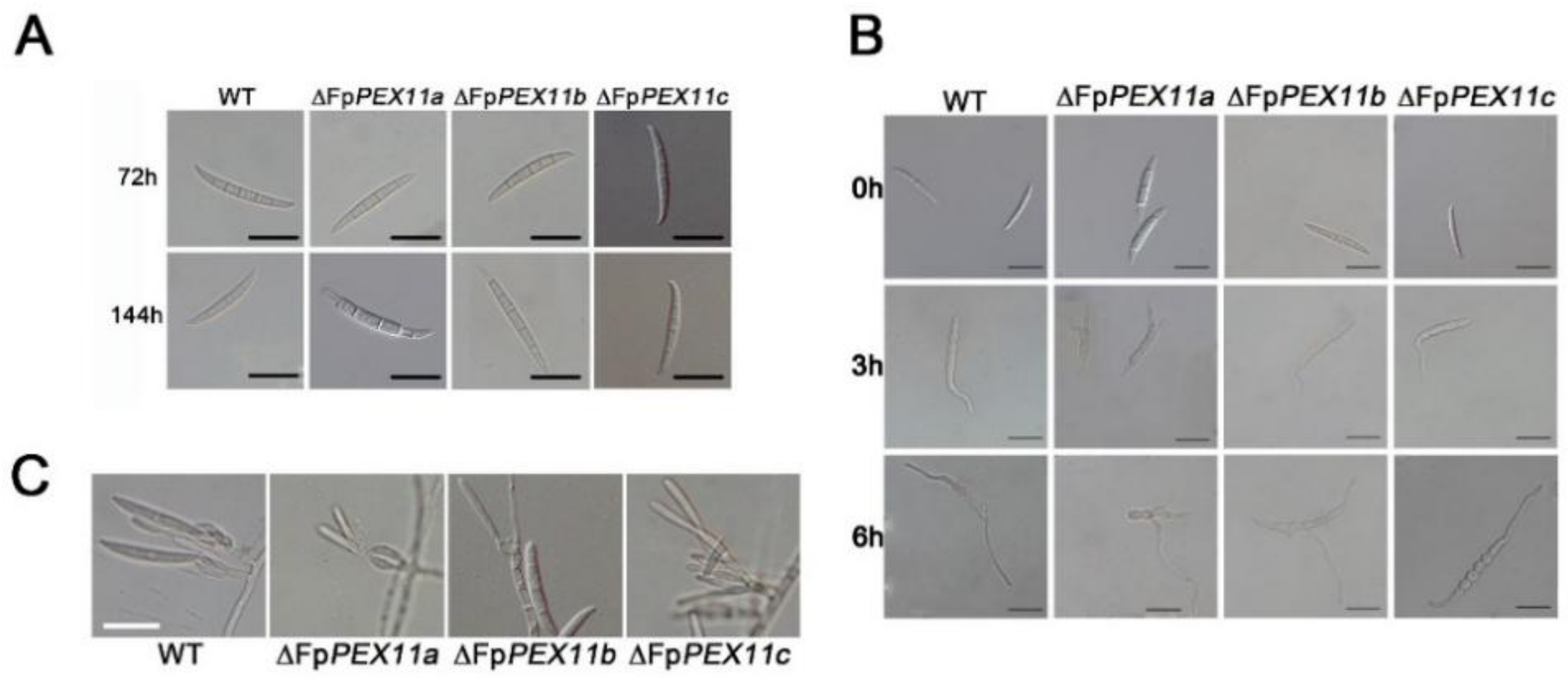
2.2. FpPEX11 Is Involved in Vegetative Growth and Asexual Reproduction

We compared the vegetative growth of the ∆FpPEX11a, ∆FpPEX11b, ∆FpPEX11c, WT, and complementation strains on PDA and CM media. When compared to WT, the colony diameter of ∆FpPEX11a on PDA medium was significantly lower, the color of the colony was darker, the aerial hyphae were sparser and more curved, and the edge of the ∆FpPEX11a aerial hyphae collapsed on CM medium, resulting in premature aging. There was no significant difference between ∆FpPEX11b colony diameter and that of WT, but the colony color was darker, the aerial hyphae were significantly reduced, the edge of ∆FpPEX11b aerial hyphae collapsed on CM, and the hyphae aged prematurely. No significant variation in vegetative growth of ∆FpPEX11c and WT was detected (Figure 2). The results indicate that FpPEX11a and FpPEX11b regulate colony morphology and growth rate, which significantly affect the vegetative growth of F. pseudograminearum; however, FpPEX11c had little effect on vegetative growth.
After 3 days of incubation, spore production of ∆FpPEX11a and ∆FpPEX11b growing in CMC media was reduced by 53.28% and 71.04%, respectively, when compared to that of the WT (Table 1). The clustered conidiophore of ∆FpPEX11a and ∆FpPEX11b were less than those of ∆FpPEX11c. In ∆FpPEX11a and ∆FpPEX11b, abnormal spore morphology with constriction at the tip was noticed after 6 d incubation. There was no significant difference in spore production or morphology of ∆FpPEX11c compared to that of WT. When incubated in YEPD medium, the conidium germination of ∆FpPEX11a was reduced by 50% at 3 h post-incubation when compared to that of WT, and the germ tubes were shorter than those of WT. Moreover, ∆FpPEX11b and ∆FpPEX11c were not significantly different from the WT. From these data, it can be seen that the significant decrease in the number of spores of ∆FpPEX11a and ∆FpPEX11b was probably due to abnormal sporulation structure, and spore deformity further affected their germination efficiency; ∆FpPEX11c showed no significant change in asexual reproduction ability when compared to WT (Figure 3). Summarily, FpPEX11a, FpPEX11b, and FpPEX11c played different roles in asexual reproduction of F. pseudograminearum. FpPEX11a was involved in the production, morphology, and germination of F. pseudograminearum spores to regulate asexual reproduction, while FpPEX11b was not involved in the germination of F. pseudograminearum spores and FpPEX11c had little effect on asexual reproduction.

2.3. FpPEX11 Regulates ROS Metabolism

To investigate whether FpPEX11 is involved in the response to oxidative stress, we analyzed the growth of ∆FpPEX11a and ∆FpPEX11b on CM medium containing 20 mM H2O2. Under these growth conditions, we observed obvious defects in the mutants when compared to WT, while ∆FpPEX11c was as resistant to oxidative stress as WT. The production of cellular ROS in each strain was qualitatively analyzed by NBT staining. As shown in Figure 4C, the edge color of the ∆FpPEX11a and ∆FpPEX11b colonies was darker than that of the WT, whereas the color change of ∆FpPEX11c was not significant. To verify this result, generation of ROS was visualized by using DHE. Compared to the wild type, the fluorescence of ∆FpPEX11b was the strongest, followed by ∆FpPEX11a and ∆FpPEX11c as the weakest, similar to the WT strain (Figure S2). These results fall in line with NBT findings. This indicates that ROS accumulation and metabolism were inhibited in ∆FpPEX11a and ∆FpPEX11b, but not in ∆FpPEX11c. The above results indicate that the absence of FpPEX11a and FpPEX11b reduced the resistance of the strain to oxidative stress, affected the metabolism of ROS, and thereby caused ROS accumulation, ultimately damaging the detoxification function of peroxisomes. Deletion of FpPEX11c had little effect on the response of the strain to oxidative stress.

2.4. FpPEX11 Is Involved in Lipid Metabolism

Unlike short-chain fatty acids, which act as substrates for mitochondrial β-oxidation, the main substrate for peroxisome oxidation is medium-long-chain fatty acids [34]. The strain was cultured in MM containing C14H28O2, C16H32O2, C18H36O2, and C22H44O2 to explore the effect of FpPEX11 on the β-oxidation function of peroxisomes.
The number of aerial hyphae of mutants was obviously less than that of the WT, and the longer the carbon chain length, the fewer aerial hyphae. When compared to the WT, the relative growth of ∆FpPEX11a and ∆FpPEX11b were significantly reduced, and only extremely sparse hyphae were observed in ∆FpPEX11b (Figure 5A). No significant difference was noticed in the colony diameter extension of ∆FpPEX11b; ∆FpPEX11c was not significantly different from the WT strain (Figure 5B). These results indicate that FpPEX11a and FpPEX11b can regulate the utilization of medium-, long-, and very-long-chain fatty acids, while FpPEX11c had little effect on the utilization of fatty acids.

2.5. FpPEX11 Is Involved in Responding to Cell Membrane and Cell Wall Stresses

Congo red (CR), a cell wall inhibitor, can hinder the normal assembly of cell walls to produce cell wall stress and inhibit fungal growth [35]. SDS destroys the stability of proteins and fungal cell walls. On CM medium supplemented with 0.01% SDS and 0.2% CR, the colony growth of the mutants was stunted, and the inhibition of ∆FpPEX11a and ∆FpPEX11b was significantly higher than that of the WT. The growth of aerial hyphae was abnormal, and the sensitivity to cell wall inhibitors increased; ΔFpPEX11c growth had no obvious defects when compared to WT and the complemented strain ΔFpPEX11c-C (Figure 6). These results indicate that knockdown of FpPEX11a and FpPEX11b reduces the resistance of the cell membrane and cell wall to the external stress.

2.6. The ∆FpPEX11 Exhibits Abnormal Peroxisome Number

To investigate the effect of FpPEX11 deletion on peroxisomes, the peroxisomal membrane protein PMP70 was labeled with GFP. A green punctate distribution was displayed in the WT and all mutants, but fluorescence in ∆FpPEX11a and ∆FpPEX11b was significantly lower, which indicated that the deletion of FpPEX11a and FpPEX11b led to a decrease in the number of peroxisomes. The fluorescence intensity in ∆FpPEX11c increased, and its volume decreased slightly (Figure 7). This also indicates abnormal biochemical processes, such as active oxygen degradation and fatty acid metabolism, in ∆FpPEX11a and ∆FpPEX11b. In ∆FpPEX11c, the volume of peroxisomes decreased, but their number increased, leading to peroxisomes being supplemented with some functions.

2.7. FpPEX11 Regulates Expression of Peroxisome-Proliferator-Associated Gene

While further studying the effect of FpPEX11 on the pathogenicity and functional integrity of F. pseudograminearum at the expression level, we detected the effects of the deletion of FpPEX11a, FpPEX11b, or FpPEX11c on each other’s expression levels, peroxisomal proliferation, and β-oxidation-related gene expression levels using quantitative PCR and analyzed their association with the phenotype of each strain.
Through analyzing the FpPEX11 gene expression difference between ∆FpPEX11 and WT, we found that the expression of PEX11b was downregulated and that of PEX11c was upregulated by 2.9 times in ∆FpPEX11a. The expression levels of PEX11a and PEX11c were both significantly upregulated in ∆FpPEX11b. The expression of both PEX11a and PEX11b was significantly downregulated in ∆FpPEX11c (Figure 8).
The number of peroxisomes increases through growth and division of pre-existing peroxisomes [36] or, when required, peroxisome biogenesis is initiated de novo from the ER [37]. PEX3 and PEX19 are key genes involved in the de novo biosynthesis of peroxisomes. PEX3 is responsible for the secretion of peroxisomal membrane proteins from the ER and acts as a docking protein for PEX19 [38,39]. As a membrane protein receptor, PEX19 recognizes and localizes to peroxisomes [40]. The expression of PEX3 and PEX19 was upregulated by 3- to 3.9-fold and 1.1- to 4.3-fold in each mutant when compared to the WT (Figure 8C), but the number of peroxisomes in ∆FpPEX11a and ∆FpPEX11b was not enhanced (Figure 7). The other pathway for peroxisomal proliferation is the division and proliferation of existing peroxisomes, which occurs through polarized extension and membrane constriction and division. FIS1 is a division factor recruited during division [41], and the expression levels of FIS1 in the mutants were significantly lower than those in the WT (Figure 8D). DNM1 is a dynamin-related protein involved in membrane constriction that promotes membrane division [42]. The expression of DNM1 was upregulated in each mutant when compared to that in the WT (Figure 8D). The number of peroxisomes was significantly increased, and the volume was decreased in ∆FpPEX11c (Figure 7).
Carnitine acetyltransferase plays an important role in fatty acid degradation. PTH2 is a peroxisome carnitine acetyltransferase that catalyzes the generation of acetyl-CoA from acetyl-CoA and facilitates the transport of acetyl-CoA between organelles. The expression of PTH2 was upregulated in each mutant compared to that in the wild type in each mutant (Figure 8B). PIP2 is involved in fatty acid degradation. Induced by the fatty acid metabolism signal from the body, PIP2 binds to Adr1 to initiate expression and promote fatty acid degradation [43]. The expression of PIP2 in each mutant was upregulated when compared to that in the wild type (Figure 8B).

2.8. FpPEX11 Is Essential for the Pathogenicity of F. pseudograminearum

To explore the effect of FpPEX11 knockdown on the pathogenicity of F. pseudograminearum in wheat, wheat coleoptiles were inoculated with spore suspensions of each strain. As shown in Figure 9, when compared to WT, the coleoptile infected length for ∆FpPEX11a and ∆FpPEX11b was reduced by 70% and 83.4%, respectively, while ∆FpPEX11c showed no significant difference when compared to the WT. Further inoculation at the blooming stage of wheat in the field was conducted, and spore suspensions of each strain were prepared in advance. After 10 days of investigation, the disease indices of ∆FpPEX11a, ∆FpPEX11b, and ∆FpPEX11c were significantly reduced when compared to those of the WT. These results indicate that FpPEX11a, FpPEX11b, and FpPEX11c are necessary for the pathogenicity of F. pseudograminearum (Figure 9).
DON is an important factor in Fusarium pathogenicity. In recent years, F. pseudograminearum has been found to produce DON. We explored whether the altered pathogenicity change of ∆FpPEX11 is related to the abnormal DON synthesis. We tested DON yield, and the results are shown in Table 2. When compared to the WT, the yields of ∆FpPEX11a and ∆FpPEX11b are significantly reduced, and the yield of ∆FpPEX11c is even higher than that of the WT. The yield of DON in the culture medium was quantitatively determined using a beacon DON detection kit. The yields of ∆FpPEX11a and ∆FpPEX11b were 0. The yield of ∆FpPEX11c was 121.43 µg/mg, which was not significantly different from that of the WT (133.78 µg/mg), which is consistent with the trend determined by LC-MS/MS.
To further verify this result, we analyzed the expression levels of TRI5, TRI6, and TRI10, which are related to DON synthesis. The results of fluorescence quantitative analysis showed that the expression levels of TRI5, TRI6, and TRI10 in ∆FpPEX11a and ∆FpPEX11b were all downregulated to different degrees, while the expression levels of TRI5 and TRI6 in ∆FpPEX11c were upregulated to different degrees. There was no significant difference between the expression levels of TRI10 and WT (Table 2). In summary, these results indicate that FpPEX11a and FpPEX11b are indispensable for the pathogenicity of F. pseudograminearum and affect the expression and biosynthesis of genes related to DON synthesis. The role of FpPEX11c in DON synthesis requires further investigation.

3. Discussion

In S. cerevisiae, the PEX11 protein family, consisting of PEX11, PEX25, and PEX27, has been shown to play a major role in peroxisome proliferation [44], and Huber demonstrated that PEX27 plays an inhibitory role in peroxisome proliferation in S. cerevisiae [14]. Deletion of PEX11 results in a smaller number of larger peroxisomes, whereas overexpression of PEX11 results in larger number of smaller peroxisomes. In M. oryzae, one of three PEX11 are required for peroxisomal proliferation [31]. To verify the presence or absence of peroxisome structures in the mutants, we determined the localization in the mycelia of a known peroxisomal membrane protein, PMP70, and the number of peroxisomes was found to be significantly reduced in ∆FpPEX11a and ∆FpPEX11b when compared with the WT (Figure 7). To compensate for the number of peroxisomes, the expression of the PEX3 and PEX19 genes associated with the de novo biosynthesis of peroxisomes was significantly upregulated, and the number and function of peroxisomes in ∆FpPEX11 did not recover well.
Downregulation of expression of the mitogen FIS1 involved in peroxisome proliferation and upregulation of DNM1 expression with PEX11 as the activator protein might be due to the feedback mechanism of the PEX11 protein (Figure 8). These abnormalities in expression levels of PEX11-related genes also reflect changes in the number of peroxisomes in ∆FpPEX11. In ∆FpPEX11c, the number of peroxisomes increased, and the volume decreased. Expression analysis of FpPEX11 revealed a decreased number of peroxisomes when the expression of PEX11c was higher. It has been speculated that FpPEX11c and PEX27 [14] of yeast also play an inhibitory role in the peroxisome proliferation. FpPEX11a and FpPEX11b are also involved in the regulation of peroxisome proliferation. Therefore, the three PEX11 family members in F. pseudograminearum are involved in the regulation of peroxisome proliferation.
A variety of metabolic reactions occur in peroxisomes; these include fatty acid β-oxidation, the glyoxylic acid cycle, melanin biosynthesis, and glycerol accumulation, which are related to the growth and development of pathogens. Currently, it has been shown that the pathogenicity of many plant pathogens, including M. oryzae, Colletotrichum gloeosporioides, F. graminearum, Ustilaginoidea maydis, and Phaeosphaeria nodorum, has inevitable connection with peroxisomes [45]. In this study, the peroxisome metabolism of the PEX11 knockout was diminished, and the fatty acid β-oxidative metabolism of ∆FpPEX11a and ∆FpPEX11b was distinctly affected. In media containing medium- or long-chain fatty acids, hyphal growth was inhibited (Figure 5), fatty acids could not be normally used, and supply of the metabolite acetyl-CoA was decreased. Fungal cell walls are mainly composed of chitin, β-1,3-glucan, β-1,6-glucan, and mannoproteins. Some studies have suggested that chitin and glucan in fungal cell walls are derived from acetyl-CoA [46]. Deletion of PEX5, PEX6, and PEX19 were all sensitive to Congo red and calcofluor-white in M. oryzae [47]. In this study, ∆FpPEX11a and ∆FpPEX11b were sensitive to cell wall inhibitor stress and showed impaired cell wall integrity (Figure 6), which reflects the impaired peroxisome function observed in ∆FpPEX11a and ∆FpPEX11b. The number of peroxisomes was significantly increased, and their volumes reduced in ∆FpPEX11c (Figure 7), hence, no significant change in the β-oxidation function of peroxisomes was observed. In S. cerevisiae, acetyl-CoA, a product of β-oxidation, can be transferred to the cytoplasm by carnitine acetyltransferase (CrAT2), or to the mitochondria by CrAT1, where it enters the tricarboxylic acid cycle [48]. The M. oryzae gene PTH2 (encoding CrAT1) plays a major role in acetyl-CoA metastasis. In the PTH2 deletion mutant, melanin was reduced, invasion was abnormal, and pathogenicity was lost [49]. In ∆FpPEX11a and ∆FpPEX11b, PTH2 satisfies the need of peroxisome enzyme metabolism by increasing its expression level (Figure 8). Being stimulated by fatty acid signals, PIP2 was upregulated. Fatty acid degradation was still severely impaired in ∆FpPEX11a and ∆FpPEX11b, especially the degradation of long-chain fatty acids. Therefore, FpPEX11a and FpPEX11b may play a significant role in regulating β-oxidation.
The early defense response of plants to pathogen infection involves the production of ROS to resist pathogen infection, and the ability of the pathogen to remove ROS is essential for successful invasion and colonization despite the host defense. The abundant enzymes in peroxisomes play an important role in regulating active oxygen balance. In yeast, peroxisome defects lead to ROS accumulation [50]. ROS are also produced during the interaction between Alternaria alternata and the host, and the destruction of the host ROS scavenging system leads to the reduction or loss of pathogenicity of A. alternata. [51]. In this study, it was found that the susceptibility of ∆FpPEX11a and ∆FpPEX11b to the oxidant H2O2 was increased, the accumulation of ROS was increased in hyphae, and the removal of ROS was blocked (Figure 4). On the other hand, the abnormal response of F. pseudograminearum to ROS may also be associated with its reduced pathogenicity; ∆FpPEX11c showed normal ability to respond to oxidative stress (Figure 4). Indoor coleoptile and field wheat inoculation showed that the pathogenicity of ∆FpPEX11a and ∆FpPEX11b significantly decreased (Figure 9), and the yield of DON decreased (Table 2). Previous studies have shown that fungi can recognize the host environment and induce gene expression for DON biosynthesis during infection, leading to the production of DON. DON has also been detected in tissues inoculated with F. graminearum or F. pseudograminearum in the stalk portion and not inoculated at the apex [52]. Thus, the effects of the deletion of FpPEX11a and FpPEX11b on DON contamination in grains and stalks can be continued. In the laboratory conditions, ∆FpPEX11c had no significant effect on the pathogenicity when compared with the WT; however, in the field, the pathogenicity of ∆FpPEX11c was significantly decreased (Figure 9), which might be due to differences in the environmental factors of the host. The expression levels of TRI5, TRI6, and TRI10, which are related to DON biosynthesis, were significantly downregulated in ∆FpPEX11a and ∆FpPEX11b, while the expression of TRI5 was significantly upregulated in the ∆FpPEX11c, and the yield of DON was not significantly different from that in WT. Therefore, comprehensive knowledge about the function of FpPEX11c requires further study in the future.
In conclusion, our results indicate that FpPEX11a and FpPEX11b are involved in the regulation of vegetative growth and asexual reproduction of F. pseudograminearum, and act through regulation in the number and functions of peroxisomes, such as via fatty acid β-oxidation and active oxygen metabolism. After deletion of FpPEX11a and FpPEX11b, the pathogenicity and yield of DON in F. pseudograminearum was significantly weakened, and disease progress was hindered (Figure 9). After deletion of FpPEX11c, the number of peroxisomes increased, and their volume decreased (Figure 7). Therefore, the three members of the FpPEX11 family, FpPEX11a, FpPEX11b and FpPEX11c, perform their respective roles, and their mutual balance regulates the number of peroxisomes and metabolic activities. Furthermore, we characterized part of the function of the FpPEX11 family in F. pseudograminearum for the first time, which can aid in the understanding of mechanisms underlying pathogenicity of F. pseudograminearum and other filamentous phytopathogens.

4. Materials and Methods

4.1. Fungal Strain and Culture Medium

The F. pseudograminearum wild-type (WT) strain used in this study was the local isolate Fp3-3, which was reserved at Shandong Agricultural University. Solid potato dextrose agar (PDA; 200 g peeled potato, 20 g dextrose, 15 g agar, and 1000 mL water), complete medium (CM), yeast extract peptone dextrose (YEPD) medium (yeast extract, 10.0 g; peptone, 20.0 g; glucose, 20.0 g; and 1000 mL distilled water), and sporulation media consisting of carboxymethyl cellulose liquid media (CMC) preparations, as well as the culture conditions, were adopted from a previous report [53,54].

4.2. Sequence Analysis

FpPEX11a (FPSE-09675) was identified using the BlastP and tBlastN algorithms from S. cerevisiae PEX11 (NC_001147.6) of NCBI (https://www.ncbi.nlm.nih.gov/) (accessed on 15 January 2020), and FpPEX11b (FPSE-09643) and FpPEX11c (FPSE_04578) were identified using the BlastP and tBlastN algorithms from M. oryzae PEX11 (MGG-00648, MGG-05271). SMART (http://smart.embl-heidelberg.de/) (accessed on 16 January 2020) predicted the domains. Motif prediction was carried out using MEME (https://meme-suite.org/meme/tools/meme) (accessed on 16 January 2020), phylogenetic analysis of the predicted PEX11 protein of F. pseudograminearum with those that have been reported in other species was performed using MEGA 7, and the evolutionary tree was constructed by multiple sequence alignment using Clustal W with 1000 neighbor-joining definite tests. FpPEX11 was used to construct a phylogenetic tree with homologous proteins from different species.

4.3. Strain Construction

At least 1 kb upstream and downstream fragments for each gene were amplified from genomic DNA, and the hygromycin-phosphotransferase (hph) gene was amplified from the pCB1003 plasmid using the primer pairs zH1-F/R and zH2-F/R and inserted into pCE-Zero to generate vectors pCE-aAH1, pCE-aH2B, pCE-bAH1, pCE-bH2B, pCE-cAH1, and pCE-cH2B. The fusion fragments were transformed into the WT strain to yield the transformants ∆FpPEX11a, ∆FpPEX11b, and ∆FpPEX11c by PEG-mediated protoplast transformation. Potential gene deletion mutants were further confirmed by Southern blot analysis [31].
For the complementation strains, genomic fragments containing full lengths of ORFs and 1.5 kb upstream of FpPEX11a, FpPEX11b, and FpPEX11c were amplified and inserted into pFL2-GRP to generate complementary vectors pFL2-GRP-aDW, pFL2-GRP-bDW, and pFL2-GRP-cDW, which were integrated into mutants. Candidate complementary strains were further determined by PCR.

4.4. Vegetative Growth Detection

In order to determine mycelial growth, each strain was cultured on PDA and CM at 25 °C for 3 days, and the colony diameter was measured by the perpendicular cross measurement method [55]. To determine cell wall sensitivity, each strain was cultured on CM containing 0.01% sodium dodecyl sulfate (SDS) and CM containing 0.2% Congo red for 3 days and then measured using the same method.
Utilization of carbon sources was measured in minimal medium (MM) supplemented with various carbon sources instead of sucrose at the following concentrations: 2.5 mM myristic acid (C14), 2.5 mM palmitic acid (C16), 2.5 mM oleic acid (C18), and 2.5 mM erucic acid (C22). Emulsifier NP40 was added to the MM. Colony diameters were measured after three days of incubation at 25 °C [56].
Three 5 mm bacterial cakes were cultured in CMC at a controlled environment of 25 °C and 150 rpm for 3 days to determine the conidia yield and observe the sporulation structure of each strain. Conidia were cultured in YEPD for 3 h, and the germination rate was calculated after microscopic observation.

4.5. ROS Detection

The strains were cultured in CM at 25 °C for 4 days to detect ROS by nitro tetrazolium blue chloride (NBT) staining. Each plate was stained with 15 mL of 0.2% (w/v) NBT solution and incubated in the dark at 25 °C for 45 min. The solution was drained, and the plates were washed with anhydrous ethanol. The plates were incubated for 30 min in the dark at 25 °C before imaging [16]. Generation of ROS was visualized by using DHE: mycelia harvested from YEPD cultures and were washed with phosphate-buffered saline (PBS). The samples were stained with DHE solution at 2.5 μg/mL in PBS and incubated in the dark for 5 min. The DHE stained cells were then examined under the same fluorescence filter set used for RFP.

4.6. Fluorescence Microscopy

Plasmids containing GFP-FpPMP70 were transformed into WT and mutants by PEG-mediated protoplast transformation to obtain fluorescently labeled strains. The fluorescence intensity of tagged proteins was observed using a Zeiss LSM880 confocal microscope (Gottingen, Niedersachsen, Germany). Image analysis was performed using ImageJ (http://rsb.info.nih.gov/nih-image/) (accessed on 20 February 2022) and Adobe Photoshop software. Peroxisome quantification was performed on 150 μm hyphae, and statistical data analysis was performed using Statistica 8.0.

4.7. Relative Quantitative Real-Time PCR

Total RNA was isolated from the hyphae of the WT, mutant, and complemented strains using TransZol Up (TransGen Biotech, Beijing, China). Relative quantification relates the PCR signal of the mutant transcript in a treatment group to that of another sample, such as the WT. For each sample, FP-β-tubulin was used as an internal control [57]. Quantitative results were calculated and analyzed using the 2−ΔΔCt method [58]. The primers used are listed in the Supplementary Material.

4.8. Pathogenicity Test

The inoculum was prepared containing 1 × 10⁷ conidia in 1 mL of sterile water, to which Triton 60 was added at a final concentration of 0.01% for better adherence of the inoculum to susceptible wheat Jimai 22 [59]. After inoculation, humidity was maintained with a sealing film for 48 h, and wheat heads were photographed and assayed [56] 10 days post-inoculation (dpi). Wheat coleoptiles were inoculated with inoculum and examined at 5 dpi and 25 °C [60].

Supplementary Materials

The following supporting information can be downloaded at: https://www.mdpi.com/article/10.3390/ijms232012184/s1.

Author Contributions

L.Z. and J.Y. designed the experiments, supervised the work, and edited the manuscript; M.W. performed the majority of the experiments, analyzed the data, and wrote the draft manuscript; H.X. and C.L. participated in the article modification; Y.T. and X.W. performed data curation; Y.L. provided guidance and revised the manuscript. All authors have read and agreed to the published version of the manuscript.

Funding

This work was supported by the Wheat Innovation Team of Shandong Province Modern Agricultural Industry Technology System (SDAIT-01-09), Natural science gene of Shandong Province (ZR2021QC141), and Funds of Shandong “Double Tops” Program (SYL2017XTTD11).

Conflicts of Interest

The authors declare no conflict of interest.

Abbreviations

DONDeoxynivalenol
FCRFusarium crown rot
EREndoplasmic reticulum
Reactive oxygen species
ZSZellweger syndrome
WTWild type
PDAPotato dextrose agar
CMComplete medium
YEPDYeast extract peptone dextrose
SDSSodium dodecyl sulfate

References

  1. Hogg, A.C.; Johnston, R.H.; Johnston, J.A.; Klouser, L.; Kephart, K.D.; Dyer, A.T. Monitoring fusarium crown rot populations in spring wheat residues using quantitative real-time polymerase chain reaction. Phytopathology 2010, 100, 149–157. [Google Scholar] [CrossRef] [Green Version]
  2. Smiley, R.W.; Gourlie, J.A.; Easley, S.A.; Patterson, L.-M.; Whittaker, R.G. Crop Damage Estimates for Crown Rot of Wheat and Barley in the Pacific Northwest. Plant Dis. 2005, 89, 595–604. [Google Scholar] [CrossRef] [Green Version]
  3. Burgess, L.W.; Backhouse, D.; Summerell, B.A.; Swan, L.J. Crown Rot of Wheat. In Fusarium: Paul E. Nelson Memorial Symposium; The American Phytopathological Society: St Paul, MN, USA, 2001. [Google Scholar]
  4. Knight, N.L.; Sutherland, M.W. Histopathological Assessment of Fusarium pseudograminearum Colonization of Cereal Culms During Crown Rot Infections. Plant Dis. 2016, 100, 252–259. [Google Scholar] [CrossRef] [PubMed] [Green Version]
  5. Obanor, F.; Neate, S.; Simpfendorfer, S.; Sabburg, R.; Wilson, P.; Chakraborty, S. Fusarium graminearumandFusarium pseudograminearumcaused the 2010 head blight epidemics in Australia. Plant Pathol. 2013, 62, 79–91. [Google Scholar] [CrossRef] [Green Version]
  6. Chen, C.; Payne, W.A.; Smiley, R.W.; Stoltz, M.A. Yield and water-use efficiency of eight wheat cultivars planted on seven dates in Northeastern Oregon. Agron. J. 2003, 95, 836. [Google Scholar] [CrossRef]
  7. Papendick, R.I.; Cook, R.J. Plant water stress and development of Fusarium foot rot in wheat subjected to different cultural practices. Phytopathology 1974, 64, 358–363. [Google Scholar] [CrossRef]
  8. Paulitz, T.C. Low Input No-till Cereal Production in the Pacific Northwest of the U.S.: The Challenges of Root Diseases. Eur. J. Plant Pathol. 2006, 115, 271–281. [Google Scholar] [CrossRef]
  9. Al-Fahdawi, M.S.; Able, J.A.; Evans, M.; Able, A.J. Response of durum wheat to different levels of zinc and Fusarium pseudograminearum. Crop. Pasture Sci. 2014, 65, 61–73. [Google Scholar] [CrossRef]
  10. Veenhuis, M.; Salomons, F.A.; Klei, I.J.V.D. Peroxisome biogenesis and degradation in yeast a structurefunction analysis. Microsc. Res. Tech. 2000, 15, 584–600. [Google Scholar] [CrossRef]
  11. Fransen, M. Peroxisome Dynamics: Molecular Players, Mechanisms, and (Dys)functions. ISRN Cell. Biol. 2012, 2012, 1–24. [Google Scholar] [CrossRef]
  12. Fagarasanu, A.; Mast, F.D.; Knoblach, B.; Rachubinski, R.A. Molecular mechanisms of organelle inheritance: Lessons from peroxisomes in yeast. Nat. Rev. Mol. Cell. Biol. 2010, 11, 644–654. [Google Scholar] [CrossRef] [PubMed]
  13. Nagotu, S.; Veenhuis, M.; van der Klei, I.J. Divide et impera: The dictum of peroxisomes. Traffic 2010, 11, 175–184. [Google Scholar] [CrossRef] [PubMed]
  14. Huber, A.; Koch, J.; Kragler, F.; Brocard, C.; Hartig, A. A subtle interplay between three Pex11 proteins shapes de novo formation and fission of peroxisomes. Traffic 2012, 13, 157–167. [Google Scholar] [CrossRef] [Green Version]
  15. Kiel, J.A.; van den Berg, M.; Bovenberg, R.A.; van der Klei, I.J.; Veenhuis, M. Penicillium chrysogenum Pex5p mediates differential sorting of PTS1 proteins to microbodies of the methylotrophic yeast Hansenula polymorpha. Fungal. Genet. Biol. 2004, 41, 708–720. [Google Scholar] [CrossRef] [PubMed] [Green Version]
  16. Chen, Y.; Zheng, S.; Ju, Z.; Zhang, C.; Tang, G.; Wang, J.; Wen, Z.; Chen, W.; Ma, Z. Contribution of peroxisomal docking machinery to mycotoxin biosynthesis, pathogenicity and pexophagy in the plant pathogenic fungus Fusarium graminearum. Environ. Microbiol. 2018, 20, 3224–3245. [Google Scholar] [CrossRef]
  17. Fransen, M.; Nordgren, M.; Wang, B.; Apanasets, O. Role of peroxisomes in ROS/RNS-metabolism: Implications for human disease. Biochim. Biophys. Acta 2012, 1822, 1363–1373. [Google Scholar] [CrossRef] [Green Version]
  18. Van Veldhoven, P.P. Biochemistry and genetics of inherited disorders of peroxisomal fatty acid metabolism. J. Lipid. Res. 2010, 51, 2863–2895. [Google Scholar] [CrossRef] [Green Version]
  19. Waterham, H.R.; Ferdinandusse, S.; Wanders, R.J. Human disorders of peroxisome metabolism and biogenesis. Biochim. Biophys. Acta 2016, 1863, 922–933. [Google Scholar] [CrossRef]
  20. Cipolla, C.M.; Lodhi, I.J. Peroxisomal Dysfunction in Age-Related Diseases. Trends Endocrinol. Metab. 2017, 28, 297–308. [Google Scholar] [CrossRef] [Green Version]
  21. Hu, J.; Aguirre, M.; Peto, C.; Alonso, J.; Ecker, J.; Chory, J. A Role for Peroxisomes in Photomorphogenesis and Development of Arabidopsis. Science 2002, 297, 405–409. [Google Scholar] [CrossRef]
  22. Hu, J.; Baker, A.; Bartel, B.; Linka, N.; Mullen, R.T.; Reumann, S.; Zolman, B.K. Plant peroxisomes: Biogenesis and function. Plant Cell. 2012, 24, 2279–2303. [Google Scholar] [CrossRef] [PubMed] [Green Version]
  23. Del Rio, L.A. Peroxisomes as a cellular source of reactive nitrogen species signal molecules. Arch. Biochem. Biophys. 2011, 506, 1–11. [Google Scholar] [CrossRef] [PubMed]
  24. Sparkes, I.A.; Brandizzi, F.; Slocombe, S.P.; El-Shami, M.; Hawes, C.; Baker, A. An Arabidopsis pex10 null mutant is embryo lethal, implicating peroxisomes in an essential role during plant embryogenesis. Plant Physiol. 2003, 133, 1809–1819. [Google Scholar] [CrossRef] [Green Version]
  25. Shabab, M. Role of plant peroxisomes in protection against herbivores. Subcell Biochem. 2013, 69, 315–328. [Google Scholar] [CrossRef] [PubMed]
  26. Kresge, N.; Simoni, R.D.; Hill, R.L. The discovery of glyoxysomes: The work of Harry Beevers, 1961. J. Biol. Chem. 2010, 285, e6–e7. [Google Scholar] [CrossRef] [PubMed] [Green Version]
  27. Marshall, P.A.; Krimkevich, Y.I.; Lark, R.H.; Dyer, J.M.; Veenhuis, M.; Goodman, J.M. Pmp27 promotes peroxisomal proliferation. J. Cell Biol. 1995, 129, 345–355. [Google Scholar] [CrossRef] [Green Version]
  28. Orth, T.; Reumann, S.; Zhang, X.; Fan, J.; Wenzel, D.; Quan, S.; Hu, J. The PEROXIN11 protein family controls peroxisome proliferation in Arabidopsis. Plant Cell 2007, 19, 333–350. [Google Scholar] [CrossRef] [PubMed] [Green Version]
  29. Koch, J.; Pranjic, K.; Huber, A.; Ellinger, A.; Hartig, A.; Kragler, F.; Brocard, C. PEX11 family members are membrane elongation factors that coordinate peroxisome proliferation and maintenance. J. Cell Sci. 2010, 123, 3389–3400. [Google Scholar] [CrossRef] [Green Version]
  30. Weng, H.; Ji, X.; Naito, Y.; Endo, K.; Ma, X.; Takahashi, R.; Shen, C.; Hirokawa, G.; Fukushima, Y.; Iwai, N. Pex11alpha deficiency impairs peroxisome elongation and division and contributes to nonalcoholic fatty liver in mice. Am. J. Physiol. Endocrinol. Metab. 2013, 304, E187–E196. [Google Scholar] [CrossRef] [Green Version]
  31. Wang, J.; Li, L.; Zhang, Z.; Qiu, H.; Li, D.; Fang, Y.; Jiang, H.; Chai, R.Y.; Mao, X.; Wang, Y.; et al. One of Three Pex11 Family Members Is Required for Peroxisomal Proliferation and Full Virulence of the Rice Blast Fungus Magnaporthe oryzae. PLoS ONE 2015, 10, e0134249. [Google Scholar] [CrossRef]
  32. Tam, Y.Y.; Torres-Guzman, J.C.; Vizeacoumar, F.J.; Smith, J.J.; Marelli, M.; Aitchison, J.D.; Rachubinski, R.A. Pex11-related proteins in peroxisome dynamics: A role for the novel peroxin Pex27p in controlling peroxisome size and number in Saccharomyces cerevisiae. Mol. Biol. Cell 2003, 14, 4089–4102. [Google Scholar] [CrossRef] [PubMed] [Green Version]
  33. Rottensteiner, H.; Stein, K.; Sonnenhol, E.; Erdmann, R. Conserved function of pex11p and the novel pex25p and pex27p in peroxisome biogenesis. Mol. Biol. Cell 2003, 14, 4316–4328. [Google Scholar] [CrossRef] [PubMed] [Green Version]
  34. Latruffe, N.; Vamecq, J.; Cherkaoui Malki, M. Genetic-dependency of peroxisomal cell functions-emerging aspects. J. Cell Mol. Med. 2003, 7, 238–248. [Google Scholar] [CrossRef] [PubMed]
  35. Ram, A.F.; Klis, F.M. Identification of fungal cell wall mutants using susceptibility assays based on Calcofluor white and Congo red. Nat. Protoc. 2006, 1, 2253–2256. [Google Scholar] [CrossRef]
  36. Lazarow, P.B.; Fujiki, Y. Biogenesis of peroxisomes. Annu. Rev. Cell Biol. 1985, 1, 489–530. [Google Scholar] [CrossRef]
  37. Hettema, E.H.; Motley, A.M. How peroxisomes multiply. J. Cell Sci. 2009, 122, 2331–2336. [Google Scholar] [CrossRef] [Green Version]
  38. Snyder, W.B.; Koller, A.; Choy, A.J.; Subramani, S. The peroxin Pex19p interacts with multiple, integral membrane proteins at the peroxisomal membrane. J. Cell Biol. 2000, 149, 1171–1178. [Google Scholar] [CrossRef] [Green Version]
  39. William, B.S.; Faber, K.N.; Thibaut, J.W.; Koller, A.; Luers, G.H.; Rangell, L.; Keller, G.A.; Subramani, S. Pex19p interacts with Pex3p and Pex10p and is essential for peroxisome biogenesis in Pichia pastoris. Mol. Biol. Cell 1999, 10, 1745–1761. [Google Scholar] [CrossRef] [Green Version]
  40. Cichocki, B.A.; Krumpe, K.; Vitali, D.G.; Rapaport, D. Pex19 is involved in importing dually targeted tail-anchored proteins to both mitochondria and peroxisomes. Traffic 2018, 19, 770–785. [Google Scholar] [CrossRef] [Green Version]
  41. Koch, J.; Brocard, C. PEX11 proteins attract Mff and human Fis1 to coordinate peroxisomal fission. J. Cell Sci. 2012, 125, 3813–3826. [Google Scholar] [CrossRef]
  42. Williams, C.; Opalinski, L.; Landgraf, C.; Costello, J.; Schrader, M.; Krikken, A.M.; Knoops, K.; Kram, A.M.; Volkmer, R.; van der Klei, I.J. The membrane remodeling protein Pex11p activates the GTPase Dnm1p during peroxisomal fission. Proc. Natl. Acad. Sci. USA 2015, 112, 6377–6382. [Google Scholar] [CrossRef] [PubMed] [Green Version]
  43. Rottensteiner, H.; Kal, A.J.; Filipits, M.; Binder, M.; Hamilton, B.; Tabak, H.F.; Ruis, H. Pip2p_ a transcriptional regulator of peroxisome proliferation in the yeast Saccharomyces cerevisiae. EMBO J. 1996, 15, 2924–2934. [Google Scholar] [CrossRef] [PubMed]
  44. Tower, R.J.; Fagarasanu, A.; Aitchison, J.D.; Rachubinski, R.A. The peroxin Pex34p functions with the Pex11 family of peroxisomal divisional proteins to regulate the peroxisome population in yeast. Mol. Biol. Cell 2011, 22, 1727–1738. [Google Scholar] [CrossRef] [PubMed]
  45. Kimura, A.; Takano, Y.; Furusawa, I.; Okuno, T.; Notes, A. Peroxisomal Metabolic Function Is Required for Appressorium-Mediated Plant Infection by Colletotrichum lagenarium. Plant Cell 2001, 13, 1945–1957. [Google Scholar] [CrossRef] [Green Version]
  46. Bowman, S.M.; Free, S.J. The structure and synthesis of the fungal cell wall. Bioessays 2006, 28, 799–808. [Google Scholar] [CrossRef]
  47. Li, L.; Wang, J.; Zhang, Z.; Wang, Y.; Liu, M.; Jiang, H.; Chai, R.; Mao, X.; Qiu, H.; Liu, F.; et al. MoPex19, which is essential for maintenance of peroxisomal structure and woronin bodies, is required for metabolism and development in the rice blast fungus. PLoS ONE 2014, 9, e85252. [Google Scholar] [CrossRef] [Green Version]
  48. Wanders, R.J.; Waterham, H.R. Biochemistry of mammalian peroxisomes revisited. Annu. Rev. Biochem. 2006, 75, 295–332. [Google Scholar] [CrossRef]
  49. Zhang, Z.; Wang, J.; Zhang, Z.; Wang, Y.; Li, L.; Chai, R.; Mao, X.; Jiang, H.; Qiu, H.; Du, X.; et al. PTS1 Peroxisomal Import Pathway Plays Shared and Distinct Roles to PTS2 Pathway in Development and Pathogenicity of Magnaporthe oryzae. PLoS ONE 2013, 8, e55554. [Google Scholar] [CrossRef] [Green Version]
  50. Bonekamp, N.A.; Volkl, A.; Fahimi, H.D.; Schrader, M. Reactive oxygen species and peroxisomes: Struggling for balance. Biofactors 2009, 35, 346–355. [Google Scholar] [CrossRef]
  51. Ma, H.; Wang, M.; Gai, Y.; Fu, H.; Zhang, B.; Ruan, R.; Chung, K.-R.; Li, H. Thioredoxin and Glutaredoxin Systems Required for Oxidative Stress Resistance, Fungicide Sensitivity, and Virulence of Alternaria alternata. Appl. Environ. Microbiol. 2018, 84, e00086-18. [Google Scholar] [CrossRef]
  52. Mudge, A.M.; Dill-Macky, R.; Dong, Y.; Gardiner, D.M.; White, R.G.; Manners, J.M. A role for the mycotoxin deoxynivalenol in stem colonisation during crown rot disease of wheat caused by Fusarium graminearum and Fusarium pseudograminearum. Physiol. Mol. Plant Pathol. 2006, 69, 73–85. [Google Scholar] [CrossRef]
  53. Wang, L.-m.; Zhang, Y.-f.; Du, Z.-l.; Kang, R.-j.; Chen, L.-l.; Xing, X.-p.; Yuan, H.; Ding, S.-l.; Li, H.-l. FpPDE1 function of Fsarium pseudograminearum on pathogenesis in wheat. J. Integr. Agric. 2017, 16, 2504–2512. [Google Scholar] [CrossRef]
  54. Zhang, L.; Liu, C.; Wang, L.; Sun, S.; Liu, A.; Liang, Y.; Yu, J.; Dong, H. FgPEX1 and FgPEX10 are required for the maintenance of Woronin bodies and full virulence of Fusarium graminearum. Curr. Genet 2019, 65, 1383–1396. [Google Scholar] [CrossRef] [PubMed]
  55. Chen, L.; Geng, X.; Ma, Y.; Zhao, J.; Li, T.; Ding, S.; Shi, Y.; Li, H. Identification of basic helix-loop-helix transcription factors reveals candidate genes involved in pathogenicity of Fusarium pseudograminearum. Can. J. Plant Pathol. 2019, 41, 200–208. [Google Scholar] [CrossRef]
  56. Zhang, L.; Wang, L.; Liang, Y.; Yu, J. FgPEX4 is involved in development, pathogenicity, and cell wall integrity in Fusarium graminearum. Curr. Genet. 2019, 65, 747–758. [Google Scholar] [CrossRef]
  57. Zhang, Y.; Wang, L.; Liang, S.; Zhang, P.; Kang, R.; Zhang, M.; Wang, M.; Chen, L.; Yuan, H.; Ding, S.; et al. FpDep1, a component of Rpd3L histone deacetylase complex, is important for vegetative development, ROS accumulation, and pathogenesis in Fusarium pseudograminearum. Fungal. Genet. Biol. 2020, 135, 103299. [Google Scholar] [CrossRef]
  58. Livak, K.J.; Schmittgen, T.D. Analysis of relative gene expression data using real-time quantitative PCR and the 2(-Delta Delta C(T)) Method. Methods 2001, 25, 402–408. [Google Scholar] [CrossRef] [PubMed]
  59. Rubella, S.; Goswami, H.C.K. Pathogenicity and In Planta Mycotoxin Accumulation Among Members of the Fusarium graminearum Species Complex on Wheat and Rice. Phytopathology 2005, 95, 1397–1404. [Google Scholar] [CrossRef] [Green Version]
  60. Tang, L.; Yu, X.; Zhang, L.; Zhang, L.; Chen, L.; Zou, S.; Liang, Y.; Yu, J.; Dong, H. Mitochondrial FgEch1 is responsible for conidiation and full virulence in Fusarium graminearum. Curr. Genet. 2020, 66, 361–371. [Google Scholar] [CrossRef]
Figure 1. Bioinformatics analysis of PEX11 in F. pseudograminearum. (A) MEME analysis. (B) Domain analysis. (C) Phylogenetic tree. (A) FpPex11 contains conserved C-terminal motif as revealed by MEME analysis. Sc- Saccharomyces cerevisiae; Fg—Fusarium graminearum; Mo—Magnaporthe oryzae; Nc—Neurospora crassa; Pc—Penicillium chrysogenum; Ao—Aspergillus oryzae; Af—Aspergillus fumigatus; Ca—Candida albicans; Oa—Ogataea angusta; Hs—Homo sapiens. (B) Prediction domain of PEX11 by SMART analysis. (C) Phylogenetic relationship of PEX11 homologs calculated with neighbor-joining method using the MEGA 7.0.
Ijms 23 12184 g001
Figure 2. Phenotypic test assay for WT, ΔFpPEX11, and ΔFpPEX11-C strains in F. pseudograminearum. (A) Mycelial growth of all strains on PDA and CM medium for 3 days. (B) Strain colony diameter statistics. Different letter on the bars for each treatment indicate significant difference at p < 0.05 by Duncan’s multiple range test.
Ijms 23 12184 g002
Figure 3. Asexual reproduction assay in WT and ΔFpPEX11 strains in F. pseudograminearum. (A) Conidia of WT and the mutants incubated in liquid CMC for 3 days. (B) Strains were incubated in YEPD culture medium for 3 h or 6 h. Scale bar = 20 μm. (C) Conidiogenous structure.
Ijms 23 12184 g003
Figure 4. Sensitivity to H2O2 and ROS accumulation. (A) Strains were grown on complete medium (CM) supplemented with H2O2. (B) Mycelial growth inhibition. (C) Nitroblue tetrazolium (NBT) staining for ROS production in mycelia of strains. Different letter on the bars for each treatment indicate significant difference at p < 0.05 by Duncan’s multiple range test.
Ijms 23 12184 g004
Figure 5. Deletion of strain affected fatty acid β-oxidation. (A) Strains were cultured at 25 °C for 3 d on minimal medium (MM) supplemented with different carbon chain length of fatty acids as the sole carbon source. (B) Relative mycelial growth of WT and the mutants on various media. Different letter on the bars for each treatment indicate significant difference at p < 0.05 by Duncan’s multiple range test.
Ijms 23 12184 g005
Figure 6. Cell wall integrity of the strains was defective. (A) Strains grown on CM supplemented with CR and SDS. Photographs were taken after incubation for 3 days at 25 °C. (B) Percent inhibition of strains (%) Inhibition = (diameter of untreated strain-diameter of treated strain)/(diameter of untreated strain) × 100%. Different letters on the bars for each treatment indicate significant difference at p < 0.05 by Duncan’s multiple range test.
Ijms 23 12184 g006
Figure 7. The expression and localization of FpPMP70-GFP in WT and ΔFpPEX11 mutants using its native promoter by fluorescence microscopy. DIC: differential interference contrast. Scale bar = 5 μm.
Ijms 23 12184 g007
Figure 8. FpPEX11−related gene expression assay results. (A) Relative expression levels of mutual PEX11 in the ∆FpPEX11 mutants. (B) Relative expression levels of PTH2 and PIP2 in ∆FpPEX11 mutants. (C) Relative expression levels of PEX3 and PEX19 in ∆FpPEX11 mutants. (D) Relative expression levels of FIS1 and DNM1 in ∆FpPEX11 mutants.
Ijms 23 12184 g008
Figure 9. Effect of FpPEX11 knockout on the pathogenicity of F. pseudograminearum. (A) Infestation of coleoptile 10 d after inoculation. (B) Determination of the length of infestation of coleoptile 10 d after inoculation. (C) Infestation of wheat ears 10 d after inoculation. (D) Disease index statistics of wheat ears 10 d after inoculation. Different letter on the bars for each treatment indicate significant difference at p < 0.05 by Duncan’s multiple range test.
Ijms 23 12184 g009
Table 1. Conidiation and conidial germination for 3 h or 6 h in the WT and ΔFpPEX11 strains.
StrainConidiation
(106 Conidia/mL)
Germination (%)
3 h6 h
WT2.59 ± 0.15 a51.6 ± 0.75 a100.00 ± 0.00 a
FpPEX11a1.21 ± 0.10 b27.2 ± 3.32 b97.00 ± 3.00 a
FpPEX11b0.75 ± 0.09 b49.2 ± 1.16 a95.00 ± 1.00 a
FpPEX11c2.37 ± 0.35 a52.8 ± 1.59 a99.00 ± 1.00 a
Different letters for each treatment indicate significant difference at p < 0.05 by Duncan’s multiple rang.
Table 2. DON production in the WT and ∆FpPEX11 strains.
DON ProductionRelative Expression Levels of TRI
ELISA (μg/mg)HPLC (μg/g) *TRI5TRI6TRI10
WT133.78 ± 18.36 a14.59 ± 3.47 b1.00 ± 0.09 1.00 ± 0.221.00 ± 0.22
FpPEX11a00.16 ± 0.01 c0.11 ± 0.04 0.16 ± 0.060.19 ± 0.02
FpPEX11b00.00763 ± 0 c0.13 ± 0.02 0.10 ± 0.020.15 ± 0.08
FpPEX11c121.43 ± 21.65 a32.05 ± 6.00 a7.198 ± 0.09 1.82 ± 0.240.90 ± 0.21
DON production was analyzed by HPLC–MS/MS (HPLC) and Deoxynivalenol (DON) Rapid Test Kit (ELISA). * Logarithmic transformation of DON production by HPLC and the different letter on the bars for each treatment indicate significant difference at p < 0.05 by Duncan’s multiple range test.
Publisher’s Note: MDPI stays neutral with regard to jurisdictional claims in published maps and institutional affiliations.

