Article

Whole-Genome Analysis of Mycobacterium neoaurum DSM 1381 and the Validation of Two Key Enzymes Affecting C22 Steroid Intermediates in Sterol Metabolism

1
Lab of Biorefinery, Shanghai Advanced Research Institute, Chinese Academy of Sciences, No. 99 Haike Road, Pudong, Shanghai 201210, China
2
University of Chinese Academy of Sciences, Beijing 100049, China
3
BioTechnology Institute, University of Minnesota, 140 Gortner Lab, 1479 Gortner Avenue Saint Paul, Minneapolis, MN 55108, USA
4
Protein Engineering and Biopharmaceutical Sciences Laboratory, Hubei University of Technology, Wuhan 430068, China
*
Author to whom correspondence should be addressed.
These authors contributed equally to this work.
Int. J. Mol. Sci. 2023, 24(7), 6148; https://doi.org/10.3390/ijms24076148
Submission received: 10 January 2023 / Revised: 1 February 2023 / Accepted: 6 February 2023 / Published: 24 March 2023
(This article belongs to the Collection State-of-the-Art Molecular Microbiology in China)

Abstract

:
Mycobacterium neoaurum DSM 1381 originated from Mycobacterium neoaurum ATCC 25790 by mutagenesis screening is a strain of degrading phytosterols and accumulating important C22 steroid intermediates, including 22-hydroxy-23, 24-bisnorchola-4-en-3-one (4-HP) and 22-hydroxy-23, 24-bisnorchola-1,4-dien-3-one (HPD). However, the metabolic mechanism of these C22 products in M. neoaurum DSM 1381 remains unknown. Therefore, the whole-genome sequencing and comparative genomics analysis of M. neoaurum DSM 1381 and its parent strain M. neoaurum ATCC 25790 were performed to figure out the mechanism. As a result, 28 nonsynonymous single nucleotide variants (SNVs), 17 coding region Indels, and eight non-coding region Indels were found between the genomes of the two strains. When the wild-type 3-ketosteroid-9α-hydroxylase subunit A1 (KshA1) and β-hydroxyacyl-CoA dehydrogenase (Hsd4A) were overexpressed in M. neoaurum DSM 1381, the steroids were transformed into the 4-androstene-3, 17- dione (AD) and 1,4-androstadiene-3,17-dione (ADD) instead of C22 intermediates. This result indicated that 173N of KshA1 and 171K of Hsd4A are indispensable to maintaining their activity, respectively. Amino acid sequence alignment analysis show that both N173D in KshA1 and K171E in Hsd4A are conservative sites. The 3D models of these two enzymes were predicted by SWISS-MODEL and AlphaFold2 to understand the inactivation of the two key enzymes. These results indicate that K171E in Hsd4A may destroy the inaction between the NAD+ with the NH3+ and N173D in KshA1 and may disrupt the binding of the catalytic domain to the substrate. A C22 steroid intermediates–accumulating mechanism in M. neoaurum DSM 1381 is proposed, in which the K171E in Hsd4A leads to the enzyme’s inactivation, which intercepts the C19 sub-pathways and accelerates the C22 sub-pathways, and the N173D in KshA1 leads to the enzyme’s inactivation, which blocks the degradation of C22 intermediates. In conclusion, this study explained the reasons for the accumulation of C22 intermediates in M. neoaurum DSM 1381 by exploring the inactivation mechanism of the two key enzymes.

1. Introduction

Steroid drugs have a variety of pharmacological and physiological activities, are widely used in anti-inflammatory and anti-tumor treatments and to regulate sexual ability and birth control [1,2]. Steroid drugs have become the second-largest marketed pharmaceutical after antibiotics, and the market demand for steroid drugs is continuously increasing [2]. C22 intermediates are highly valuable precursors in steroid drugs, for example in synthesizing progestational and adrenocortical hormones, compared with other steroid intermediates [3]. At present, most of the steroid intermediates used in industrial production are chemical modifications from a natural steroid [4]. Compared with traditional chemical synthesis methods, the biotransformation method has the advantages of mild transformation conditions, fewer production steps, higher transformation efficiency, stereo-selectivity and less environmental pollution. Therefore, biotransformation methods are increasingly used in the production of steroid drugs and intermediates and greatly promote the industrial production of steroid intermediates [5]. Most of the strains used in the industrial production of steroid drug intermediates are from mycobacteria. The development of ideal industry strains for diverse steroid intermediates will significantly promote the application of biotransformation in steroid drug production [5,6,7]. Several efficient steroid intermediate producers have been reported in the last decades [2,6,8]. However, the products are limited to a few intermediates, such as C19 intermediates (AD, ADD, and 9 α-OH-AD) and C22 intermediates (4-HP, HPD, and 9 α-OH-HP) [9].
At present, the steroids that can be obtained by one-step fermentation by microbial transformation are mainly C19 intermediates. The accumulation of C22 intermediates is less studied and developed [3,10]. C22 intermediates are mostly derived from the transformation of steroids by natural or mutagenic strains. The accumulation of C22 intermediates is due to the existence of two sub-pathways in the steroid side-chain degradation pathway by microorganisms: one is the complete degradation of the side chain, of which the final product is a C19 steroid intermediate; the other is the incomplete chain degradation, of which the final product is a C22 steroid intermediate. The steroid side chain metabolic pathway branches at the intermediate 22-Hydroxy-3-oxo-cholest-4-ene-24-carboxyl-CoA. Bifunctional enzyme hydroxyacyl-CoA dehydrogenase/17β-hydroxysteroid dehydrogenase Hsd4A catalysis is reported to account for the branches [3]. A common strategy to obtain intermediates including the steroids mentioned above is to retain the steroid nucleus by knocking out 3-ketosteroid-Δ1-dehydrogenase (KstD) or 3-ketosteroid-9α-hydroxylase (KSH) to prevent the ring-opening reaction of the B ring [10].
Some efficient steroid drug intermediate producers have been obtained by traditional mutagenesis [11]. However, it is hard to further reconstruct these strains to achieve higher yield and fewer by-products due to the lack of knowledge of steroid degradation pathways [12]. Genome sequencing is one of the important methods to reveal the steroid degradation mechanism. Until now, several M. neoaurum strains have been sequenced, including type strains M. neoaurum ATCC 25795 and M. neoaurum NRRLB 3805, and industrial steroid-producing strains Mycobacterium sp. VKM Ac-1815D and Mycobacterium sp. VKM Ac-1816D [13,14]. However, the strains that can accumulate C22 intermediates were not sequenced and studied. Among the various mutants, M. neoaurum DSM 1381 originated from M. neoaurum ATCC 25790 by mutagenesis screening was reported to be able to accumulate HPD as the main product with a small portion of 4-HP [15]. Our previous research has led to the construction of an ideal 4-HP-producing strain (ΔkstD1), which is obtained by deleting kstD1 in M. neoaurum DSM 1381. This ΔkstD1 mutant could produce 14.18 g/L 4-HP from 20 g/L phytosterols in 168h [16]. However, the mechanism to accumulate 4-HP and HPD is rarely studied. In this study, the genome sequencings of M. neoaurum DSM 1381 and its parent strain M. neoaurum ATCC 25790, which have the ability to completely degrade sterols without accumulating any product, were carried out [15], and comparative genomic analysis was performed to identify the mutant site responsible for the accumulation of the C22 intermediate. With the comparative genomic analysis, the function and role of related genes were verified by the complement gene expression, and the effects of mutation sites on enzymes were discussed. These findings demonstrated that M. neoaurum DSM 1381 is an excellent target strain to discover the key enzymes of the steroid degradation pathway and provided some new insights into the accumulation of C22 steroids.

2. Results and Discussion

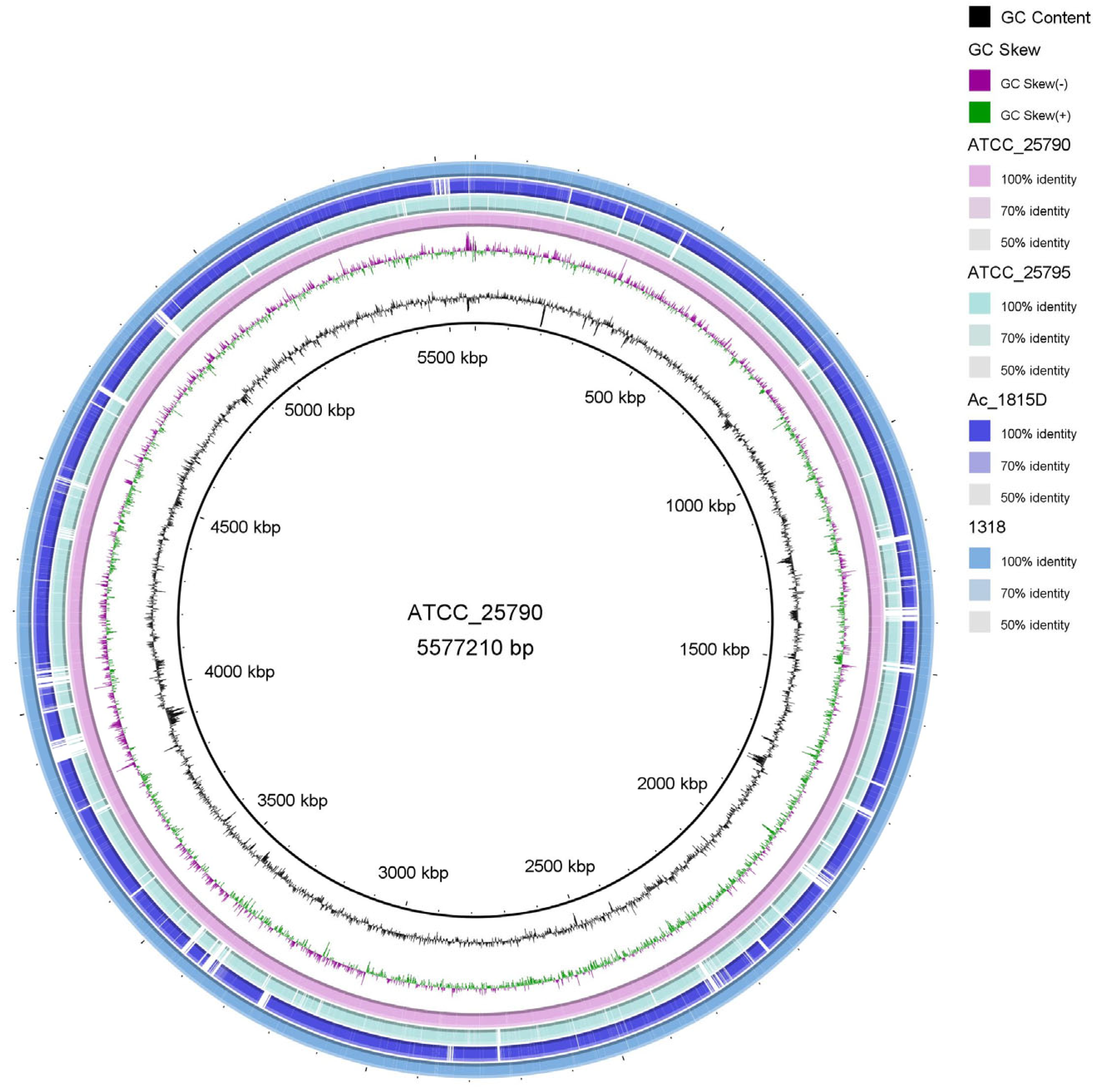
2.1. Sequencing and Gene Annotation of the Whole Genome of M. neoaurum DSM 1381 and M. neoaurum ATCC 25790

To analyze the presence of the steroid-related genes in M. neoaurum DSM 1381 and M. neoaurum ATCC 25790, the genomes were sequenced and annotated. As shown in Table 1 and Figure 1, their genome sizes are almost the same, with a total length of 5.58 Mb, which are similar to the genome sizes of M. neoaurum strains reported previously (M. neoaurum MN2 5.38 Mb, M. neoaurum NRRLB-3805 5.42 Mb, M. neoaurum ATCC 25795 5.47 Mb) [13,17]. There are 5232 and 5348 possible genes predicted in the genome of M. neoaurum DSM 1381 and M. neoaurum ATCC 25790, respectively. Moreover, the GC content in the gene region and intergenetic region is as high as 67% and 62.2%, respectively, which agrees well with previous reports [17,18]. Then the model strains Mycobacterium sp. VKM Ac 1815D and M. neoaurum ATCC 25795, whose whole-genome sequencings were completed previously, were selected for genome sequence alignment analysis and visualized [14] (Figure 1). The results show that, although they belong to the same genus, there is a huge number of differences among these genomes, among which M. neoaurum DSM 1381 is most similar to M. neoaurum ATCC 25790 while showing notable numbers of SNP, InDel, and deletions compared with the genomes of the Mycobacterium sp. VKM Ac 1815D and M. neoaurum ATCC 25795.

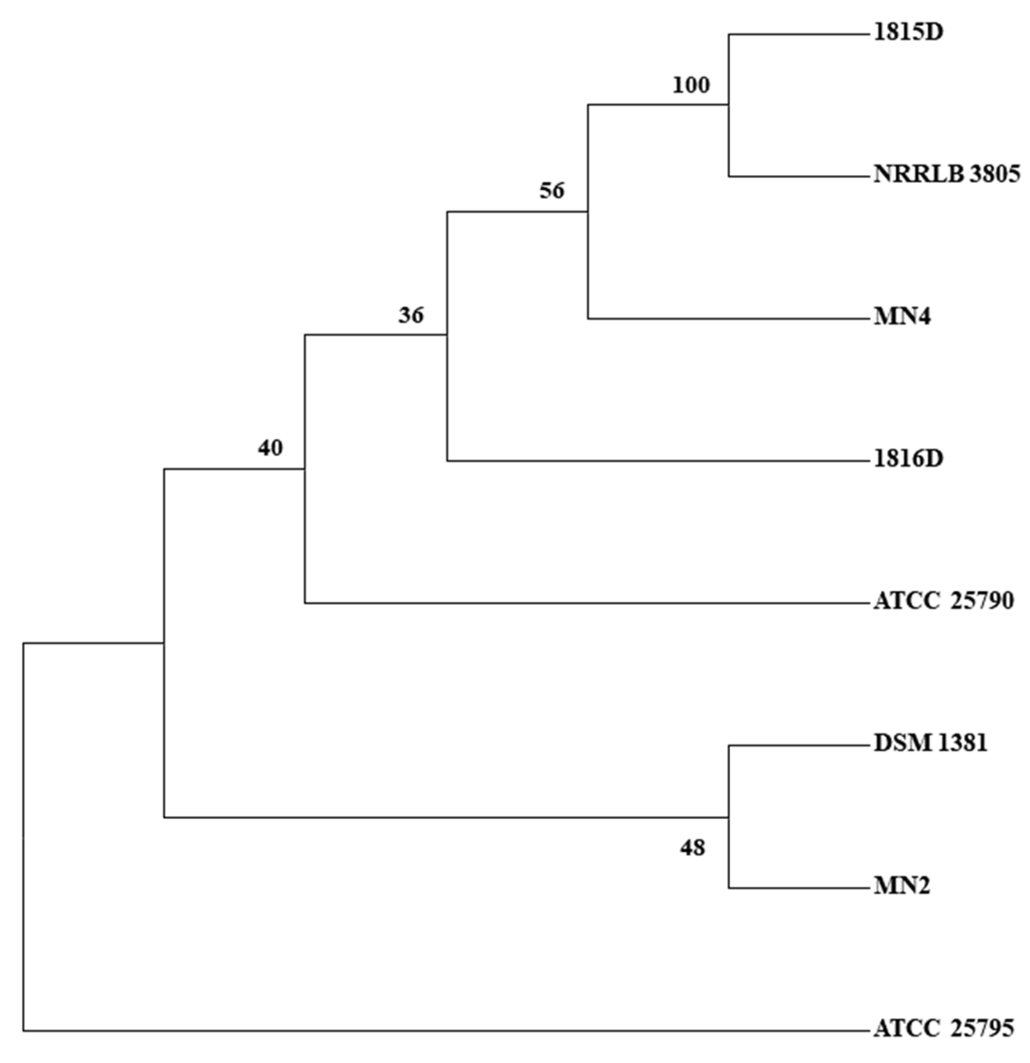
2.2. SNP Discovery Tree Analysis

Except for the two M. neoaurum strains mentioned above, four more strains were previously sequenced and deposited into the NCBI database, including M. neoaurum MN2, M. neoaurum MN4, Mycobacterium sp. VKM Ac 1816D, and M. neoaurum NRRL LB 3805. The characteristics of these strains were described briefly in the materials and methods section. M. neoaurum MN2 is an AD-producing strain, and M. neoaurum MN4 is mutated from M. neoaurum MN2 and has a higher AD yield [17]. To further confirm the genetic relationship between these strains, we conducted SNP discovery tree analysis. As shown in Figure 2, we found M. neoaurum ATCC 25790 and Mycobacterium sp. VKM Ac 1816D were clustered together as one branch that then clustered with M. neoaurum MN4, Mycobacterium sp. VKM Ac 1815D, and M. neoaurum NRRLB 3805. M. neoaurum DSM 1381 had the closest relationship with M. neoaurum MN2, followed by its origin strain M. neoaurum ATCC 25790, while the wild-type strain M. neoaurum ATCC 25795 had a farther relationship with other strains.

2.3. Prediction of Genes Related to Sterol Metabolism

In good agreement with previous reports [6,19,20], there is a gene cluster responsible for sterol metabolism in M. neoaurum DSM 1381 and M. neoaurum ATCC 25790. The well-studied strains, M. neoaurum ATCC 25795 and Mycobacterium sp. VKM Ac 1815D, were selected to annotate and analyze the homologs from the two newly sequenced strains, and the results were illustrated in Figure 3 [3]. The results were similar to those shown in the global comparison above. Gene deletions or insertions also occurred within the gene cluster within the same genus. Most of the identified genes, including KshA1, KshB1, KstD1, Hsd4A, KstR, KstR2, and mce4 operon, are present in the four strains’ gene clusters. Meanwhile, there are notable differences among the four clusters. For example, compared to M. neoaurum ATCC 25795, 10 genes were found inserted into the mec4 operons of the other three strains. In the downstream region of Hsd4B from Mycobacterium sp. VKM Ac 1815D, a large fragment as long as 51.5 kb was unique relative to the other three genomes. In addition, there are three KstDs in the genomes of M. neoaurum ATCC 25795, M. neoaurum DSM 1381, and M. neoaurum ATCC 25790, but only one in Mycobacterium sp. VKM Ac 1815D. To sum up, these strains share a high identity as well as amounts of differences in sterol metabolic genes.

2.4. Mutation Analysis of Sterol Metabolism-Related Genes

To figure out the reasons for the phenotype differences between the two strains, the SNP and InDel were analyzed between the M. neoaurum DSM 1381 and its parent strain M. neoaurum ATCC 25790. Thirty-eight SNPs, including 28 nonsynonymous SNV and 10 synonymous SNV, and 25 InDels, in which 17 were located in the coding region and eight in the non-coding region, were found; the specific information of the mutants is shown in Tables S1 and S2. Two of the mutations appeared in the above-mentioned gene cluster and are located in the kshA1, which codes for 3-ketosteroid 9α-hydroxylase subunit A, and hsd4A, whose encoding protein has double-function of 17-hydroxytryptamine dehydrogenase and β-hydroxyacyl-CoA dehydrogenase, respectively [3,21,22,23]. The former is involved in the open-loop reaction of the B-ring and the latter is involved in the degradation of the C17 side chain.
As reported previously [24,25,26,27], the C17 side chain degradation process is similar to the β-oxidation cycle involved in lipid catabolism. The enzymes involved in this process are mainly oxidoreductases. All mutated genes, after excluding identified genes, are involved in other physiological processes and genes encoding hypothetical proteins; the remaining six genes, including orf2435, orf2617-orf2616, orf187, orf1328, orf2235, orf1151, were predicted to function as oxidoreductase and to be involved in lipid catabolism or sterol degradation. Moreover, orf2188 was predicted to be a possible regulatory factor and had not yet been identified. The six mutated genes were selected to identify if they participate in sterol metabolism. To sum up, based on the gene annotation results, we selected eight mutated genes for further experiment to test whether they were involved in sterol side-chain degradation (Table 2).

2.5. The Functional Complement of the Selected Mutated Genes

The eight selected genes mentioned above, from M. neoaurum ATCC 25790, were cloned into pMV261. The resulting plasmids were transformed and expressed in M. neoaurum DSM 1381. The products from the sterol of the complemented strain were identified to test the complement results.
3-ketosteroid-9α-hydroxylase (KSH) is involved in the ring-opening oxidation of the steroid nucleus, which catalyzes the formation of 9α-hydroxyl-1,4-dienosteroid from 1,4-dien-3-one steroid [28], 9α-hydroxyl-1,4-dienosteroid are structurally unstable, leading to cleavage at the 9 and 10 positions of the B ring, disrupting the core structure of the steroid (Figure 3b). As shown in Figure 4, after the expression of the kshA1 gene from M. neoaurum ATCC 25790 in M. neoaurum DSM 1381, the products 4HP and HPD were degraded without new 3-sterone compounds appearing, indicating that the N173D substitution destroyed the 3-ketosteroid-9α-hydroxylase activity of KshA1 which is responsible for steroid nucleus degradation. The expression of wild-type hsd4A leads to the transformation of HPD/4-HP to ADD/AD, which agrees well with the previously reported function [3], which is responsible for removing the last molecule propionyl CoA during the side chain degradation. In a word, the single base mutation in these two kshA1 and hsd4A leads to the accumulation of 4-HP and HPD in M. neoaurum DSM 1381. In addition, there was no phenotypic difference after single-gene complementation of the other six mutated genes or co-expression with hsd4A, indicating they probably do not participate in sterol side-chain degradation.

2.6. The Structure and Sequence Analysis of Hsd4A

Hsd4A from ATCC 25795 was identified as a dual-function protein of 17-hydroxytryptamine dehydrogenase and β-hydroxyacyl-CoA dehydrogenase [3,21,22]. FabGs (β-oxoacyl reductases) are ubiquitous enzymes involved in fatty acid synthesis. The reaction entails NADPH/NADH-mediated conversion of β-oxoacyl-ACP (acyl-carrier-protein) into β-hydroxyacyl-ACP. The Hsd4A was rarely studied; however, in this study, we obtained the clue that the K171E mutation significantly damages the 17-hydroxytryptamine dehydrogenase activity of Hsd4A. To further figure out the possible reason, the homolog structure was built using SWISS-MODEL server [29] and AlphaFold2 [30,31].
Various Hsd4A homolog structures including a putative short-chain dehydrogenase from Mycobacterium smegmatis (4KZP) [32], a short-chain dehydrogenase from Mycobacterium avium (3QLJ), Human 17-beta-hydroxysteroid dehydrogenase Type 4 (1ZBQ), (3R)-Hydroxyacyl-CoA Dehydrogenase Domain of Candida tropicalis Peroxisomal Multifunctional Enzyme Type 2 (2ET6), (3r)-Hydroxyacyl-Coa Dehydrogenase Fragment Of Rat Peroxisomal Multifunctional Enzyme Type 2 (1GZ6) were resolved previously, and the identity is 79.5%/98%, 45.61%/94%, 41.67%/81%, 40.23%/80%, and 39.68%/81%, respectively. In this study, the amino acid sequences of these homologous fragments were compared with DSM 1381_Hsd4A (Figure S1). The 4KZP, 3QLJ, and 1ZBQ share a higher identity with target DSM 44074_Hsd4A. However, their functions were not identified. To obtain the homology model of DSM 1381_Hsd4A, the protein structure prediction algorithm AlphaFold2 was used to generate the model of the overall domain organization of DSM 1381_Hsd4A and the structural arrangements of the cofactor NAD+ bound in the cleft. Figure 5 shows the comparison of the simulated structures of the two Hsd4A proteins and the binding of the coenzyme NAD+ with them. The difference between the two proteins can be clearly observed; the distance between 171E residue of DSM 1381_ Hsd4A and NAD+ is 4.7 angstroms, which is obviously longer than the one from K171 residue of ATCC 25790_Hsd4A to NAD+, which is 3.8 angstroms. At the same time, Figure 5 shows the impact of K171E mutation on the overall configuration of Hsd4A protein.
Indicated by the structure analysis, K171 plays a crucial role in binding cofactor NAD+. Mutating the basic residue lysine to acidic residue glutamic acid destroys the inaction between the NAD+ with the NH3+. As a result, the function of Hsd4A was destroyed. In conclusion, the model we established has high reliability and can predict the structure of the Hsd4A well.

2.7. The Structure and Sequence Analysis of KshA1

KSH plays an important role during the degradation of steroids and is known to be a two-component Rieske oxygenase (Ro) system, consisting of a terminal oxygenase (KshA) and a ferredoxin reductase (KshB) [28,33]. The structure of KshA from M. tuberculosis and KshA1 as well as KshA5 from Rhodococcus rhodochrous DSM 43269 were solved previously; we used 2ZYL (3-ketosteroid-9α-hydroxylase from M. tuberculosis) as a template for homology modeling. Accordingly, the KshA monomer is arranged as a typical head-to-tail trimer [34]. KshA was identified as a flavoprotein reductase and two iron-sulfur proteins. KshA homologs have the typical Rieske Fe2S2 binding domain (C-X-H-X16,17-C-X2-H) and the nonheme Fe2+ motif (D-X3-D-X2-H-X4-H) [23,35,36]. As shown in Figure 6 and Figure S2, the Fe2+ motif region of KshA1_DSM 44074 appeared as D172-(NVT)-D-(MA)-H-(FFYV)-H184.
Combined with our experimental results, one of these SNPs was in a putative steroid-catabolism gene kshA1: an A517 nucleotide of M. neoaurum ATCC 25790 was substituted with a G nucleotide in M. neoaurum DSM 1381, thus resulting in the replacement of Asn173 with Asp. To explore the specific effect of the mutation site N173D on KshA, we used 2ZYL for homology modeling [34], and the amino acid sequence alignment is shown in Figure S2. According to previous reports, the mutation site N173D is located in the catalytic domain (amino acid residues 154–374) of KshA [34,36], which is consist with our predicted model. The mononuclear iron in M. neoaurum ATCC 25790is coordinated by His-179, His-184, Asp-302, and Asn-173, while the bidentate ligand Asn-173 in M. neoaurum DSM 1381 is replaced by Asp-173, which may disrupt the binding of the catalytic domain to the Fe, resulting in the inactivation of KshA1 and indicating the significance of N173 for the 3-ketosteroid 9α-hydroxylase function, which had not previously been mentioned and identified.

3. Materials and Methods

3.1. Chemicals, Strains, and Media

Prime STAR HS high-fidelity polymerase, Taq DNA polymerase, Kpn1, Sal1, EcoR1, and other restriction enzymes were purchased from TaKaRa Company; ClonExpress II One-Step Cloning Kit was purchased from Nanjing Vazyme Biotechnology Company; Genome Extraction Kit, Plasmid Miniprep kit, DNA fragment agarose gel recovery kit were purchased from Axygen; Kanamycin, Hygromycin were purchased from Shanghai McLean Biochemical Technology Co., Ltd. (Shanghai, China); ADD and AD standard were purchased from Yunnan Biological Products. Phytosterols (β-sitosterol 45%, campesterol 37%, stigmasterol 18%) was obtained from Jiangsu Yuehong Feed Co., Ltd. (Taizhou, Jiangsu, China); agar, yeast extracts, glycerol, and tryptone were purchased from Shanghai Macklin Biochemical Co., Ltd. (Shanghai, China) Glucose, Tween-80, ammonium salt, phosphate, methanol, ethyl acetate, n-hexane, etc. were purchased from Sinopharm Chemical Reagent Co., Ltd. (Shanghai, China)
E. coli was cultured with Luria–Bertani medium (LB medium) at 200 rpm, 37 °C. Mycobacterial competent cells were prepared and cultured in the medium including glycerol 20 g/L, (NH4)2PO4 8 g/L, K2HPO4 0.5 g/L, citric acid 3 g/L, ferric ammonium citrate 0.05 g/L, MgSO4 7H2O 0.5 g/L, Tween-80 2 g/L. The medium used to culture mycobacterial cells was yeast extract 15 g/L, glucose 6 g/L, MgSO4 7H2O 2 g/L, K2HPO4 1.0 g/L, KNO3 2.0 g/L, Tween-80 2 g/L, at pH 7.5–8.0. To perform steroid transformation the phytosterol 5 g/L and Tween-80 2 g/L were supplied, and 50 μg/mL kanamycin and 100 μg/mL hygromycin were added when needed.

3.2. Characteristics of the Used M. neoaurum Strains

M. neoaurum DSM 1381 is a mutant strain that was obtained by ultraviolet mutagenesis from wild-type strain M. neoaurum ATCC 25790 and is reported as a 4HP and HPD producer. M. neoaurum ATCC 25795 is a wild-type strain that is well studied. Mycobacterium sp. VKM Ac-1815D is the well-known producer for AD with a molar yield of 68–72% [37], but is also able to accumulate ADD (6–10%), 20-hydroxymethyl pregn-4-ene-3-one (HMP) (14–16%), and a small amount of 20-hydroxymethyl pregna-1,4-diene-3-one (HMPD). The main product of Mycobacterium sp. VKM Ac-1816D is ADD (70–72%), and AD (2–4%), HMPD (14–16%), and a small amount of HMP are by-products [14]. M. neoaurum NRRLB 3805 is another AD producer with less by-product ADD compared with Mycobacterium sp. VKM Ac-1815D. Mycobacterium neoaurum MN2 is a well-characterized AD producer which was obtained from soil organisms. M. neoaurum MN4 is a mutated strain from M. neoaurum MN2 with a higher AD yield and an increased tolerance to phytosterols (Table 3).

3.3. Genome Sequencing and Annotation

The genomes of M. neoaurum DSM 1381 and M. neoaurum ATCC 25790 were isolated using the DNA extraction kit (Axygen) according to modified manufacturers’ protocols, which are described briefly as follows: after the collection with centrifuges, the M. neoaurum pellets were resuspended with 150 µL Buffer S (RNase A added) and treated with 20 µL 5 mg/mL lysozyme at 37 °C for 30 min. After adding 30 µL 0.25 M EDTA, the reaction was ice-incubated for 5 min. Then 450 µL Buffer GA was mixed and incubated at 65 °C for 30 min. The subsequent procedures were processed strictly according to the manufacturers’ protocols. After certifying the DNA qualification with 1% agarose gel electrophoresis and nanodrop (Thermo Scientific), the genome was sequenced by Majorbio (Shanghai). The whole-genome sequencing was performed with Illumina Hiseq. The raw data was first quality-trimmed, and then assembled with SOAPdenovo v2.04 (http://soap.genomics.org.cn/, accessed on 20 November 2020) and adjusted with GapCloser v1.12. Then the assembled genome sequences were deposited in the National Center for Biotechnology Information (NCBI) database under accession number CP115600 and CP115466, respectively.
Genes of the assembled genomes were predicted with the software Glimmer 3.02 (http://www.cbcb.umd.edu/software/glimmer/, accessed on 25 November 2020) [39,40]. The predicted protein sequences were blasted using BLAST 2.2.28+ against the databases including Nr, genes, string, and GO.

3.4. Whole Gene Alignment Analysis and Visual Display

The annotation results were visualized by CGV software. There are four M. neoaurum strain genomes selected for sequence alignment: the genome sequencing results of M. neoaurum DSM 1381, Mycobacterium sp. VKM Ac 1815D, M. neoaurum ATCC 25790 and M. neoaurum ATCC 25795. The graphical view of the alignments was rendered using BLAST Ring Image Generator (BRIG).

3.5. SNP and Indel Analysis

To detect the single-nucleotide polymorphisms (SNPs), the reliable raw short read sequences of M. neoaurum ATCC 25790 obtained were compared to assembled genome sequences of M. neoaurum DSM 1381 with the BWA package. The results were analyzed with Samtools for significance values of each genome nucleotide. The calling results were checked out manually by aligning the two assembled genomes with the NCBI BLASTN SUITE. In the end, Annovar was employed to annotate the found SNPs and Indels [41].

3.6. Consensus Parsimonious Tree

kSNP 3.0 was used to scan SNPs across 18 genomes and to build a consensus parsimonious tree. One hundred equivalent parsimonious trees were constructed, and support values and consensus trees were derived from them.

3.7. Comparative Analysis of Steroid Degradation-Related Gene Clusters

Blast alignment was performed on the two groups of gene sequences, and the alignment results with high similarity (Similarity > 90%) were selected. The relationship between the alignment results, gene names, and annotation information was sorted out, and the graph was drawn by script.

3.8. Functional Complementation of Differential Genes

The complete coding sequences of hsd4A, kshA1, orf624, orf1123, orf1296, orf2188, and orf2389, amplified from the M. neoaurum ATCC 25790 genome, were inserted in the EcoRI site of pMV261. To perform functional complementation, the resulting plasmids were transformed into the mutant strain M. neoaurum DSM 1381 according to the procedure described previously [42]. The plasmid pMV261hyg-hsd4A was constructed based on pMV261-hsd4A to get co-expression of hsd4A with orf624, orf1123, orf1296, orf2188, and orf2389. The Hyg expression cassette was obtained from pSBY3_ligD-HygR and inserted in the SspI site on the Kan coding sequences. Then, pMV261hyg-hsd4A was introduced into DSM1381_orf624, DSM1381_orf1123, DSM1381_orf1296, DSM1381_orf2188, and DSM1381_orf2389 and selected with Hyg and Kan.

3.9. Analysis of Sterol Fermentation Products

3.9.1. Sample Processing

When conducting sterol fermentation experiments with M. neoaurum, samples were taken every 24 h. The fermentation samples were repeatedly extracted three times with an equal amount of ethyl acetate. The three extracts were mixed, filtered through a 0.22 μm membrane, and 75 μL of the filtrate was mixed with an equal volume of squalene (Sigma, Waltham, MA, USA) internal reference compound solution for gas chromatography detection. The procedure was to take 50 μL of the filtrate to evaporate the solvent, dissolve it with an appropriate amount of methanol ultrasonically, and carry out liquid chromatography detection.

3.9.2. TLC Detection Method for Steroids

TLC can be used as a qualitative detection method with ethyl acetate/n-hexane (6:4) as the developing solvent. Among them, 3-sterone compounds can be observed under ultraviolet irradiation, while the color development of sterols needs to be observed by spraying with 20% dilute sulfuric acid and then treating at 115 °C for 20 min.

3.9.3. The Detection Method of 3-Sterone Compounds

The 3-sterone compound has an absorption peak at 254 nm, so it was quantitatively detected at 254 nm by high-performance liquid chromatography. Chromatographic analysis conditions: C18 reversed-phase chromatography column (Agilent Extend-C18 column, 4.6 × 250 mm, 5 μm (Agilent, Santa Clara, CA, USA)); mobile phase: methanol/water (80:20, v/v); flow rate 0.8 mL/min; column temperature 40 °C; injection volume was 20 μL. The procedure was to take 50 μL of the treated product extract in a ventilated place to evaporate cleanly, and then to dissolve it with an appropriate amount of anhydrous methanol ultrasonically.

3.9.4. The Quantitative Detection Method of Sterols

Gas chromatography methods were used for the quantitative analysis of phytosterols. Gas chromatography analysis method: gas chromatography column (Agilent Rtx-5, 30 m × 0.53 mm × 5.0 µm), inlet temperature 320 °C, column temperature 300 °C; detector temperature 320 °C. Squalene (Sigma) was used as an internal reference compound.

3.10. Prediction of the Three-Dimensional Structures of the Hsd4A and KshA1 Enzymes

The structural models of the Hsd4A and KshA1 enzymes were predicted with AlphaFold2 using online resources and the protocol described by Jumper et al. [31]. To conduct the AlphaFold2 prediction, the Hsd4A and KshA1 enzyme sequences were input into the jupyter notebook for ColabFold [43]; then, the AlphaFold2 models of Hsd4A and KshA1 were obtained using as input the amino acid sequence and a HMMer [30] multiple sequence alignment (default method from Deepmind) [31]. The AlphaFold2 built an end-to-end network to optimize the final model [44]; furthermore, AlphaFold2 used attention modules to derive distance constraints and built structural models from them with 3D equivariant transformer neural networks [45,46], which operate directly on atoms in three-dimensional space. The prediction also contained side-chain information.

4. Conclusions

C22 steroid intermediates, including 4-HP, HPD, 1,4-HP, 9-OH-4-HP, are products of an incomplete degradation of steroid side chains. Compared with C19 steroid intermediates, C22 intermediates are more suitable for the synthesis of progesterone and adrenocortical hormones [3]. For example, the C22 intermediate 9-hydroxy-3-oxo-Pregna-4,17 (20)-diene-20-carboxylic acid was reported to be a precursor to generate 9-hydroxy-4, 16-pergnadiene-3, 20-dione under the catalysis of H2O2 in the presence of MoO42-, and the latter can be used to synthesize corticosteroid hormones [47]. Development of C22 steroid-producing strains continues to interest researchers. For instance, Xu found that the formation of C22 intermediate 4-HP is related to Hsd4A [3], which started the molecular research of the C22 intermediate production strain [27,48,49]. However, due to the complex pathway of steroid-degradation and the unclear accumulation mechanism of C22 intermediates, only a few industrial C22 steroid-producing strains have been developed, and the current strains retain some drawbacks, including low product purity and low molar yield. The key genes and enzymes involved in the steroid-degrading pathway in M. neoaurum have drawn more attention and have been selected as the origin strain to construct the admirable producers for important steroid intermediates. To research on the steroid degradation mechanism of M. neoaurum, we carried out the genome sequencing, gene annotation, and comparative genomic analysis of HPD/4-HP accumulation mutant M. neoaurum DSM 1381 and its parent strain M. neoaurum ATCC 25790. The sterol degradation-related genes were predicted and studied, and the key genes hsd4A and kshA1 responsible for the phenotypic difference of M. neoaurum DSM 1381 were identified by functional complementation.
According to the genome sequencing results, the GC content of the two genomes is more than 65% and the genomes’ size is around 5 Mb, which is consistent with the reported data. As shown in Figure 1, the two genomes share a high identity with the genome of wild type M. neoaurum ATCC 25795 and the AD-producer Mycobacterium sp. VKM Ac 1815D in most genomic regions. However, there are also plenty of low non-identical regions, deletions, and inserts found both inside and outside the sterol metabolism-related gene clusters. For example, the long insert observed in the gene cluster for steroid degradation is as long as 51.5 kb. Consistently, the M. neoaurum DSM 1381 shares a far genetic distance with M. neoaurum ATCC 25795, as shown in the SNP evolutionary tree (Figure 2). The numbers of genetic differences make it hard to locate the mutant sites responsible for the phenotype differences between these strains. Thus, SNP/InDel analysis was only performed between M. neoaurum DSM 1381 and its parent strain, which is more feasible to discover the key genes. Considering gene annotation and SNP/InDel results, eight mutated genes were selected as possible sites that lead to the accumulation of 4-HP and HPD in M. neoaurum DSM 1381.
Then the gene complementation experiment was processed to confirm the key mutation sites. The complementation of hsd4A led to the generation of AD and ADD, and the productions of 4-HP and HPD were almost undetectable, proving that hsd4A was involved in the removal of the last molecule of propionyl-CoA in the process of side-chain degradation and that 171Lys of Hsd4A is a key residue for enzyme activity. Loss of protease activity is the reason that M. neoaurum DSM 1381 accumulated HPD/4-HP rather than ADD/AD. In order to gain a deeper understanding of the effect of this mutation, we also performed homology modeling on Hsd4A. We proposed that the replacement of basic amino acids with acidic amino acids affects the previous inaction between NH3+ and NAD, thus destroying the enzymatic activity. This study explored whether the inactivation of DSM 1381_hsd4a led to the inhibition of the accumulation of C19 intermediates during the transformation of phytosterol in M. neoaurum DSM 1381, leading to the incomplete degradation pathway, which explained why the main accumulation product HPD and the secondary accumulation product 4HP of M. neoaurum DSM 1381 were both C22 intermediates.
Theoretically, when the target genes are supplied, the 4HP and HPD will be totally degraded. Thus, there must be another gene responsible for AD/ADD degradation that was inactive. Indeed, after expressing kshA1 from M. neoaurum ATCC 25790 in M. neoaurum DSM 1381, the 4-HP and HPD were no longer observed, and no new 3-sterone compounds were generated, indicating that the 3-ketosteroid-9α-hydroxylase, which is responsible for the ring-opening reaction, is inactivated in M. neoaurum DSM 1381. The amino acid substitution caused by the point mutation A517G in KshA1 does lead to the loss of KshA1 activity, which is a prerequisite for the accumulation of steroid-containing intermediates and the production of HPD/4HP. Similarly, we performed homology modeling on KshA1 and found that the N173D may lead to changes in the coordination of key amino acids in the catalytic domain with the mononuclear iron. This study explored whether the inactivation of DSM 1381_KshA1 causes no production of 9 α- hydroxylation products, which retain the integrity of the steroid nucleus and provide a prerequisite for the accumulation of HPD and 4HP in M. neoaurum DSM 1381.
In summary, we sequenced the genomes of two M. neoaurum strains. The resultant information and analysis lead to the discovery of two crucial residues related to C22 steroid accumulation. We demonstrated this is a promising way to discover the key enzymes on the steroid degradation pathway. Especially, there are plenty of steroid intermediate-producing strains developed using mutation and screen methods previously, and the genome sequencing cost is decreasing dramatically. The genome information obtained in this study is also useful as a reference for the strain development to reduce the side products and increase productivity and product purity.

Supplementary Materials

The supporting information can be downloaded at: https://www.mdpi.com/article/10.3390/ijms24076148/s1.

Author Contributions

Conceptualization, J.Z. and R.Z.; Data curation, J.Z. and R.Z.; formal analysis, J.Z. and R.Z.; investigation, J.Z. and R.Z.; writing—original draft, J.Z. and R.Z.; funding acquisition, B.Z.; methodology, S.S. and Z.S.; resources, H.C.; supervision, H.C.; visualization, J.S.; writing—review and editing, J.Z. and R.Z. All authors have read and agreed to the published version of the manuscript.

Funding

This study was supported by National Key’ R&D Program of China (No. 2017YFE0112700) and 2022 unveiling science and technology projects in Hubei Province.

Data Availability Statement

The data supporting these findings can be found in the Supplementary Materials.

Conflicts of Interest

The authors declare no conflict of interest. No role for the funder in the design and conduct of the study.

References

  1. Thakur, M.K.; Paramanik, V. Role of steroid hormone coregulators in health and disease. Horm. Res. 2009, 71, 194–200. [Google Scholar] [CrossRef] [PubMed]
  2. Fernández-Cabezón, L.; Galán, B.; García, J.L. New Insights on Steroid Biotechnology. Front. Microbiol. 2018, 9, 958. [Google Scholar] [CrossRef] [PubMed]
  3. Xu, L.Q.; Liu, Y.J.; Yao, K.; Liu, H.H.; Tao, X.Y.; Wang, F.Q.; Wei, D.Z. Unraveling and engineering the production of 23,24-bisnorcholenic steroids in sterol metabolism. Sci. Rep. 2016, 6, 21928. [Google Scholar] [CrossRef] [PubMed] [Green Version]
  4. Hanson, J.R. Steroids: Reactions and partial synthesis. Nat. Prod. Rep. 2004, 21, 386–394. [Google Scholar] [CrossRef] [PubMed]
  5. Donova, M.V. Steroid Bioconversions. Methods Mol. Biol. 2017, 1645, 1–13. [Google Scholar] [CrossRef] [PubMed]
  6. Donova, M.V.; Egorova, O.V. Microbial steroid transformations: Current state and prospects. Appl. Microbiol. Biotechnol. 2012, 94, 1423–1447. [Google Scholar] [CrossRef]
  7. Rugutt, J.K.; Rugutt, K.J. Antimycobacterial activity of steroids, long-chain alcohols and lytic peptides. Nat. Prod. Res. 2012, 26, 1004–1011. [Google Scholar] [CrossRef]
  8. Xie, R.; Shen, Y.; Qin, N.; Wang, Y.; Su, L.; Wang, M. Genetic differences in ksdD influence on the ADD/AD ratio of Mycobacterium neoaurum. J. Ind. Microbiol. Biotechnol. 2015, 42, 507–513. [Google Scholar] [CrossRef]
  9. Yuan, C.Y.; Ma, Z.G.; Zhang, J.X.; Liu, X.C.; Du, G.L.; Sun, J.S.; Shi, J.P.; Zhang, B.G. Production of 9,21-dihydroxy-20-methyl-pregna-4-en-3-one from phytosterols in Mycobacterium neoaurum by modifying multiple genes and improving the intracellular environment. Microb. Cell Fact. 2021, 20, 229. [Google Scholar] [CrossRef]
  10. Yao, K.; Xu, L.Q.; Wang, F.Q.; Wei, D.Z. Characterization and engineering of 3-ketosteroid-Δ1-dehydrogenase and 3-ketosteroid-9α-hydroxylase in Mycobacterium neoaurum ATCC 25795 to produce 9α-hydroxy-4-androstene-3,17-dione through the catabolism of sterols. Metab. Eng. 2014, 24, 181–191. [Google Scholar] [CrossRef]
  11. Egorova, O.V.; Gulevskaya, S.A.; Puntus, I.F.; Filonov, A.E.; Donova, M.V. Production of androstenedione using mutants of Mycobacterium sp. J. Chem. Technol. Biotechnol. Int. Res. Process Environ. Clean Technol. 2002, 77, 141–147. [Google Scholar]
  12. Wei, W.; Fan, S.Y.; Wang, F.Q.; Wei, D.Z. Accumulation of androstadiene-dione by overexpression of heterologous 3-ketosteroid Δ1-dehydrogenase in Mycobacterium neoaurum NwIB-01. World J. Microbiol. Biotechnol. 2014, 30, 1947–1954. [Google Scholar] [CrossRef] [PubMed]
  13. Rodríguez-García, A.; Fernández-Alegre, E.; Morales, A.; Sola-Landa, A.; Lorraine, J.; Macdonald, S.; Dovbnya, D.; Smith, M.C.; Donova, M.; Barreiro, C. Complete genome sequence of ‘Mycobacterium neoaurum’ NRRL B-3805, an androstenedione (AD) producer for industrial biotransformation of sterols. J. Biotechnol. 2016, 224, 64–65. [Google Scholar] [CrossRef]
  14. Bragin, E.Y.; Shtratnikova, V.Y.; Dovbnya, D.V.; Schelkunov, M.I.; Pekov, Y.A.; Malakho, S.G.; Egorova, O.V.; Ivashina, T.V.; Sokolov, S.L.; Ashapkin, V.V.; et al. Comparative analysis of genes encoding key steroid core oxidation enzymes in fast-growing Mycobacterium spp. strains. J. Steroid Biochem. Mol. Biol. 2013, 138, 41–53. [Google Scholar] [CrossRef] [PubMed]
  15. Imada, Y.; Takahashi, K. Process for Producing Steroidal Alcohols. United States patent US 4,223,091, 16 September 1980. [Google Scholar]
  16. Zhang, R.; Liu, X.; Wang, Y.; Han, Y.; Sun, J.; Shi, J.; Zhang, B. Identification, function, and application of 3-ketosteroid Δ1-dehydrogenase isozymes in Mycobacterium neoaurum DSM 1381 for the production of steroidic synthons. Microb. Cell Fact. 2018, 17, 77. [Google Scholar] [CrossRef] [Green Version]
  17. Xu, L.X.; Yang, H.L.; Kuang, M.A.; Tu, Z.C.; Wang, X.L. Comparative genomic analysis of Mycobacterium neoaurum MN2 and MN4 substrate and product tolerance. 3 Biotech. 2017, 7, 181. [Google Scholar] [CrossRef] [PubMed]
  18. Wang, H.; Song, S.; Peng, F.; Yang, F.; Chen, T.; Li, X.; Cheng, X.; He, Y.; Huang, Y.; Su, Z. Whole-genome and enzymatic analyses of an androstenedione-producing Mycobacterium strain with residual phytosterol-degrading pathways. Microb. Cell Fact. 2020, 19, 187. [Google Scholar] [CrossRef]
  19. Van der Geize, R.; Yam, K.; Heuser, T.; Wilbrink, M.H.; Hara, H.; Anderton, M.C.; Sim, E.; Dijkhuizen, L.; Davies, J.E.; Mohn, W.W.; et al. A gene cluster encoding cholesterol catabolism in a soil actinomycete provides insight into Mycobacterium tuberculosis survival in macrophages. Proc. Natl. Acad. Sci. USA 2007, 104, 1947–1952. [Google Scholar] [CrossRef] [Green Version]
  20. Yao, K.; Wang, F.Q.; Zhang, H.C.; Wei, D.Z. Identification and engineering of cholesterol oxidases involved in the initial step of sterols catabolism in Mycobacterium neoaurum. Metab. Eng. 2013, 15, 75–87. [Google Scholar] [CrossRef]
  21. Mindnich, R.; Möller, G.; Adamski, J. The role of 17 beta-hydroxysteroid dehydrogenases. Mol. Cell. Endocrinol. 2004, 218, 7–20. [Google Scholar] [CrossRef]
  22. Uhía, I.; Galán, B.; Kendall, S.L.; Stoker, N.G.; García, J.L. Cholesterol metabolism in Mycobacterium smegmatis. Environ. Microbiol. Rep. 2012, 4, 168–182. [Google Scholar] [CrossRef]
  23. Kreit, J. Aerobic catabolism of sterols by microorganisms: Key enzymes that open the 3-ketosteroid nucleus. FEMS Microbiol. Lett. 2019, 366, fnz173. [Google Scholar] [CrossRef] [PubMed]
  24. Capyk, J.K.; Kalscheuer, R.; Stewart, G.R.; Liu, J.; Kwon, H.; Zhao, R.; Okamoto, S.; Jacobs, W.R., Jr.; Eltis, L.D.; Mohn, W.W. Mycobacterial cytochrome p450 125 (cyp125) catalyzes the terminal hydroxylation of c27 steroids. J. Biol. Chem. 2009, 284, 35534–35542. [Google Scholar] [CrossRef] [Green Version]
  25. Fernandes, P.; Cruz, A.; Angelova, B.; Pinheiro, H.M.; Cabral, J.M.S. Microbial conversion of steroid compounds: Recent developments. Enzym. Microb. Technol. 2003, 32, 688–705. [Google Scholar] [CrossRef]
  26. García, J.L.; Uhía, I.; Galán, B. Catabolism and biotechnological applications of cholesterol degrading bacteria. Microb. Biotechnol. 2012, 5, 679–699. [Google Scholar] [CrossRef]
  27. Xiong, L.B.; Liu, H.H.; Xu, L.Q.; Sun, W.J.; Wang, F.Q.; Wei, D.Z. Improving the production of 22-hydroxy-23,24-bisnorchol-4-ene-3-one from sterols in Mycobacterium neoaurum by increasing cell permeability and modifying multiple genes. Microb. Cell Fact. 2017, 16, 89. [Google Scholar] [CrossRef] [PubMed] [Green Version]
  28. van der Geize, R.; Hessels, G.I.; van Gerwen, R.; van der Meijden, P.; Dijkhuizen, L. Molecular and functional characterization of kshA and kshB, encoding two components of 3-ketosteroid 9alpha-hydroxylase, a class IA monooxygenase, in Rhodococcus erythropolis strain SQ1. Mol. Microbiol. 2002, 45, 1007–1018. [Google Scholar] [CrossRef] [PubMed]
  29. Waterhouse, A.; Bertoni, M.; Bienert, S.; Studer, G.; Tauriello, G.; Gumienny, R.; Heer, F.T.; de Beer, T.A.P.; Rempfer, C.; Bordoli, L.; et al. SWISS-MODEL: Homology modelling of protein structures and complexes. Nucleic Acids Res. 2018, 46, W296–W303. [Google Scholar] [CrossRef] [Green Version]
  30. Eddy, S.R. Accelerated Profile HMM Searches. PLoS Comput. Biol. 2011, 7, e1002195. [Google Scholar] [CrossRef] [Green Version]
  31. Jumper, J.; Evans, R.; Pritzel, A.; Green, T.; Figurnov, M.; Ronneberger, O.; Tunyasuvunakool, K.; Bates, R.; Žídek, A.; Potapenko, A.; et al. Highly accurate protein structure prediction with AlphaFold. Nature 2021, 596, 583–589. [Google Scholar] [CrossRef]
  32. Dranow, D.M.; Fairman, J.W.; Edwards, T.E.; Lorimer, D. Crystal Structure of a Putative Short Chain Dehydrogenase from Mycobacterium smegmatis. Acta Crystallogr. Sect. F Struct. Biol. Commun. 2022, 78, 25–30. [Google Scholar]
  33. Petrusma, M.; Dijkhuizen, L.; van der Geize, R. Rhodococcus rhodochrous DSM 43269 3-ketosteroid 9alpha-hydroxylase, a two-component iron-sulfur-containing monooxygenase with subtle steroid substrate specificity. Appl. Environ. Microbiol. 2009, 75, 5300–5307. [Google Scholar] [CrossRef] [PubMed] [Green Version]
  34. Capyk, J.K.; D’Angelo, I.; Strynadka, N.C.; Eltis, L.D. Characterization of 3-ketosteroid 9{alpha}-hydroxylase, a Rieske oxygenase in the cholesterol degradation pathway of Mycobacterium tuberculosis. J. Biol. Chem. 2009, 284, 9937–9946. [Google Scholar] [CrossRef] [PubMed] [Green Version]
  35. Capyk, J.K.; Casabon, I.; Gruninger, R.; Strynadka, N.C.; Eltis, L.D. Activity of 3-ketosteroid 9α-hydroxylase (KshAB) indicates cholesterol side chain and ring degradation occur simultaneously in Mycobacterium tuberculosis. J. Biol. Chem. 2011, 286, 40717–40724. [Google Scholar] [CrossRef] [Green Version]
  36. Penfield, J.S.; Worrall, L.J.; Strynadka, N.C.; Eltis, L.D. Substrate specificities and conformational flexibility of 3-ketosteroid 9α-hydroxylases. J. Biol. Chem. 2014, 289, 25523–25536. [Google Scholar] [CrossRef] [Green Version]
  37. Donova, M.V.; Gulevskaya, S.A.; Dovbnya, D.V.; Puntus, I.F. Mycobacterium sp. mutant strain producing 9alpha-hydroxyandrostenedione from sitosterol. Appl. Microbiol. Biotechnol. 2005, 67, 671–678. [Google Scholar] [CrossRef]
  38. Marsheck, W.J.; Kraychy, S.; Muir, R.D. Microbial degradation of sterols. Appl. Microbiol. 1972, 23, 72–77. [Google Scholar] [CrossRef]
  39. Delcher, A.L.; Bratke, K.A.; Powers, E.C.; Salzberg, S.L. Identifying bacterial genes and endosymbiont DNA with Glimmer. Bioinformatics 2007, 23, 673–679. [Google Scholar] [CrossRef] [Green Version]
  40. Delcher, A.L.; Harmon, D.; Kasif, S.; White, O.; Salzberg, S.L. Improved microbial gene identification with GLIMMER. Nucleic Acids Res. 1999, 27, 4636–4641. [Google Scholar] [CrossRef]
  41. Wang, K.; Li, M.; Hakonarson, H. ANNOVAR: Functional annotation of genetic variants from high-throughput sequencing data. Nucleic Acids Res. 2010, 38, e164. [Google Scholar] [CrossRef]
  42. Stover, C.K.; de la Cruz, V.F.; Fuerst, T.R.; Burlein, J.E.; Benson, L.A.; Bennett, L.T.; Bansal, G.P.; Young, J.F.; Lee, M.H.; Hatfull, G.F.; et al. New use of BCG for recombinant vaccines. Nature 1991, 351, 456–460. [Google Scholar] [CrossRef] [PubMed]
  43. Mirdita, M.; Schütze, K.; Moriwaki, Y.; Heo, L.; Ovchinnikov, S.; Steinegger, M. ColabFold: Making protein folding accessible to all. Nat. Methods 2022, 19, 679–682. [Google Scholar] [CrossRef] [PubMed]
  44. AlQuraishi, M. End-to-End Differentiable Learning of Protein Structure. Cell Syst. 2019, 8, 292–301.e3. [Google Scholar] [CrossRef] [PubMed]
  45. Fuchs, F.B.; Wagstaff, E.; Dauparas, J.; Posner, I. Iterative SE(3)-Transformers. In Proceedings of the 5th International Conference on Geometric Science of Information (GSI), Sorbonne University, Paris, France, 21–23 July 2021; pp. 585–595. [Google Scholar]
  46. Vaswani, A.; Shazeer, N.; Parmar, N.; Uszkoreit, J.; Jones, L.; Gomez, A.N.; Kaiser, L.; Polosukhin, I. Attention Is All You Need. In Proceedings of the 31st Annual Conference on Neural Information Processing Systems (NIPS), Long Beach, CA, USA, 4–9 December 2017. [Google Scholar]
  47. Toro, A.; Ambrus, G. Oxidative decarboxylation of 17(20)-dehydro-23,24-dinorcholanoic acids. Tetrahedron Lett. 1990, 31, 3475–3476. [Google Scholar] [CrossRef]
  48. Xiong, L.B.; Liu, H.H.; Zhao, M.; Liu, Y.J.; Song, L.; Xie, Z.Y.; Xu, Y.X.; Wang, F.Q.; Wei, D.Z. Enhancing the bioconversion of phytosterols to steroidal intermediates by the deficiency of kasB in the cell wall synthesis of Mycobacterium neoaurum. Microb. Cell Fact. 2020, 19, 80. [Google Scholar] [CrossRef]
  49. Sun, W.J.; Wang, L.; Liu, H.H.; Liu, Y.J.; Ren, Y.H.; Wang, F.Q.; Wei, D.Z. Characterization and engineering control of the effects of reactive oxygen species on the conversion of sterols to steroid synthons in Mycobacterium neoaurum. Metab. Eng. 2019, 56, 97–110. [Google Scholar] [CrossRef]
Figure 1. Global comparison of M. neoaurum DSM 1381. M. neoaurum ATCC 25790genomes, M. neoaurum ATCC 25795, and Mycobacterium sp. VKM Ac 1815D sequence.
Ijms 24 06148 g001
Figure 2. Consensus parsimonious tree derived from SNPs detected in eight strains of M. neoaurum. DSM 1381, M. neoaurum DSM 1381; ATCC 25790, M. neoaurum ATCC 25790; ATCC 25795, M. neoaurum ATCC 25795; VKM Ac 1815D, Mycobacterium sp. VKM Ac 1815D; VKM Ac 1816D, Mycobacterium sp. VKM Ac 1816D; MN2, M. neoaurum MN2; MN4, M. neoaurum MN4; NRRLB 3805, M. neoaurum NRRLB 3805.
Ijms 24 06148 g002
Figure 3. Partial gene cluster encoding the catabolism of sterols in M. neoaurum DSM 1381and putative phytosterol metabolic pathway by M. neoaurum DSM 1381. (a) Genes in the map are color-coded according to assigned or proposed function: Orange, mce cluster genes for steroids transportation; Green, side-chain degradation genes; Blue, nucleus cleavage genes; Light green, the genes under the control of KstR2; Yellow, the gene encoding the transcriptional repressor KstRs; (b) The red arrow indicates the step blocked in M. neoaurum DSM 1381.
Ijms 24 06148 g003
Figure 4. HPLC chromatogram comparison of the products from the transformation of phytosterols by strains DSM 1381_hsd4A (red), DSM 1381_kshA1 (blue), and M. neoaurum DSM 1381 (black).
Ijms 24 06148 g004
Figure 5. Structural model of Hsd4A. (a) DSM 1381_Hsd4A, aquamarine; ATCC 25790_Hsd4A, light magenta; (b) DSM 1381_Hsd4A, gray, 171E and NAD+ shown in sticks; (c) DSM 1381_Hsd4A, gray, K171 and NAD+ shown in sticks.
Ijms 24 06148 g005
Figure 6. Structure model of KshA1. DSM 1381_ KshA1, pink; DSM 44074_KshA1, blue; structural template, gray; substrate AD, green; the non-heme Fe2+, gold (a,b). The residues within 4 Å of Fe are shown in sticks (c,d).
Ijms 24 06148 g006
Table 1. Results of genome sequencing and gene annotation.
StrainsDSM 1381ATCC 25790
Gene num52325348
Gene total length5,577,916 bp5,577,210 bp
Gene average length971 bp951 bp
Gene density0.937 genes per kb0.958 genes per kb
GC content in gene region (%)67.267.2
Gene/Genome (%)91.191.3
Intergenetic region length494,836 bp487,875 bp
GC content in the intergenetic region (%)62.262.2
Intergenetic length/Genome (%)8.978.74
Table 2. The mutated genes in M. neoaurum DSM 1381 predicted to be responsible for steroid degradation.
Description of the Gene ProductGene IDSNVaa Mutation
Nonsynonymous SNVATCC 25790DSM 1381ATCC 25790DSM 1381ATCC 25790DSM 1381
KshA180714517A517G173Asn173Asp
Hsd4A77546511A511G171Lys171Glu
NAD(P)/FAD-dependent oxidoreductase132812961025C1025G342Ala342Gly
FadR family transcriptional regulator22352188446T446A149Leu149Gln
SDR family NAD(P)-dependent oxidoreductase11511123upstream 34Gupstream 34T
IndelATCC 25790DSM 1381Indel in DSM 1381Mutation start position
2,4-dienoyl-CoA reductase24352389insert, 4 bp660
acyl-CoA synthetase2617–26162573gap, 34 bp807
acyltransferase187624gap, 1 bp461
Table 3. Information on the strains used for comparative analysis.
StrainAbbreviationMain ProductSide ProductsReference
M. neoaurum DSM 1381DSM 1381HPD4HP[15]
M. neoaurum ATCC 25790 (M. neoaurum ATCC 25790)ATCC 25790NANA[15]
M. neoaurum ATCC 25795ATCC 25795NANA
Mycobacterium sp. VKM Ac-1815DAc 1815DADADD, HP, HPD[37]
Mycobacterium sp. VKM Ac-1816DAc 1816DADDAD, HP, HPD[14]
M. neoaurum MN2MN2ADADD[17]
M. neoaurum MN4MN4ADADD[17]
M. neoaurum NRRLB 3805NRRLB 3805NANA[38]
Disclaimer/Publisher’s Note: The statements, opinions and data contained in all publications are solely those of the individual author(s) and contributor(s) and not of MDPI and/or the editor(s). MDPI and/or the editor(s) disclaim responsibility for any injury to people or property resulting from any ideas, methods, instructions or products referred to in the content.

