Journal Description
Organoids
Organoids
is an international, peer-reviewed, open access journal on all aspects of organoids published quarterly online by MDPI.
- Open Access— free for readers, with article processing charges (APC) paid by authors or their institutions.
- High Visibility: indexed within ESCI (Web of Science), Scopus, and many other databases.
- Journal Rank: CiteScore - Q2 (Biochemistry, Genetics and Molecular Biology (miscellaneous))
- Rapid Publication: manuscripts are peer-reviewed and a first decision is provided to authors approximately 26.3 days after submission; acceptance to publication is undertaken in 5.6 days (median values for papers published in this journal in the first half of 2026).
- Recognition of Reviewers: Reviewers whose reports are timely and of high quality receive an APC discount voucher for a future publication in an MDPI journal. Become a reviewer.
- Organoids is a companion journal of Cells.
Impact Factor:
2.8 (2025);
5-Year Impact Factor:
3.3 (2025)
Latest Articles
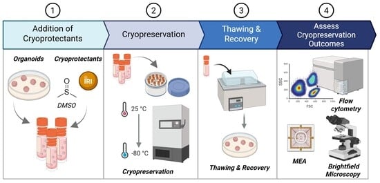
Efficient Cryopreservation of Human Midbrain Organoids Using Conventional DMSO Protocols with Ice Recrystallization Inhibitors
Organoids 2026, 5(3), 32; https://doi.org/10.3390/organoids5030032 - 19 Sep 2026
Abstract
►
Show Figures
Human midbrain organoids (hMOs) are a powerful stem cell-derived model for studying human neurodevelopment and neurological disease in vitro; however, their broad adoption is limited by long culture times, high cost, and inter-batch variability. Cryopreservation offers a strategy to address these limitations by
[...] Read more.
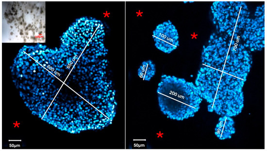
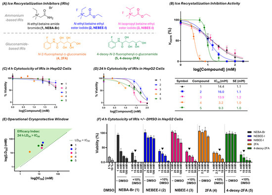
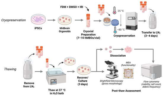
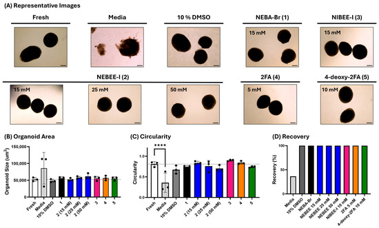
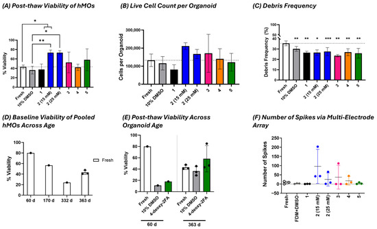
Human midbrain organoids (hMOs) are a powerful stem cell-derived model for studying human neurodevelopment and neurological disease in vitro; however, their broad adoption is limited by long culture times, high cost, and inter-batch variability. Cryopreservation offers a strategy to address these limitations by enabling long-term storage and reducing experimental heterogeneity, but its effectiveness depends on robust cryoprotective strategies. Here, we evaluated hMO cryopreservation using a standard 10% dimethyl sulfoxide (DMSO) protocol, alone and in combination with a panel of five ice recrystallization inhibitors (IRIs), including betaine-derived ammonium salts and N-aryl-d-gluconamides. Across all conditions, hMOs were successfully recovered following freeze–thawing, with preservation of overall structural integrity, including maintained morphology and reduced cellular debris. Importantly, post-thaw viability and cell recovery in the 10% DMSO condition were comparable to those of fresh controls, demonstrating that conventional DMSO-based cryopreservation is sufficient to maintain hMO survival and gross structural preservation. While supplementation with select IRIs, particularly N-ethyl betaine ethyl ester iodide (NEBEE-I), yielded modest improvements in viability relative to DMSO alone and a non-significant trend toward enhanced electrophysiological activity in multielectrode array recordings, these effects were secondary to the robust performance of the baseline DMSO protocol. Collectively, these findings establish standard 10% DMSO cryopreservation as an effective and broadly applicable method for hMO preservation, while suggesting that small-molecule IRIs may provide incremental benefits in selected outcome measures.
Full article
Open AccessArticle
Organoid Culture Using Single-Layer Matrigel Method Recapitulates Cervical Cancer Subtypes In Vitro—A Tool for Precision Medicine Applications
by
Surbhi Singla, Rashmi Bagga, Radhika Srinivasan, Prateek Bhatia and Shalmoli Bhattacharyya
Organoids 2026, 5(3), 31; https://doi.org/10.3390/organoids5030031 - 15 Sep 2026
Abstract
►▼
Show Figures
Advanced cervical cancer remains a major cause of mortality in women worldwide as it has limited treatment options and recurrence is very common. This highlights the necessity to develop patient-derived organoids (PDOs) as preclinical models that can recapitulate the clinical heterogeneity of the
[...] Read more.
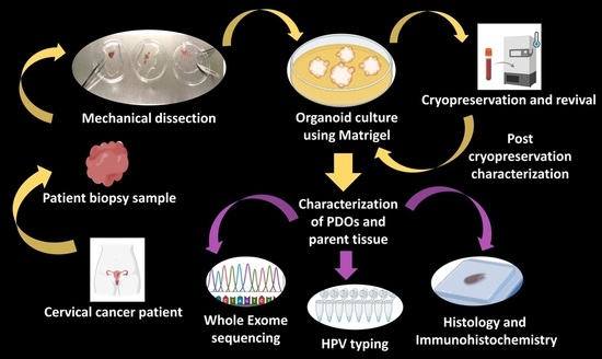
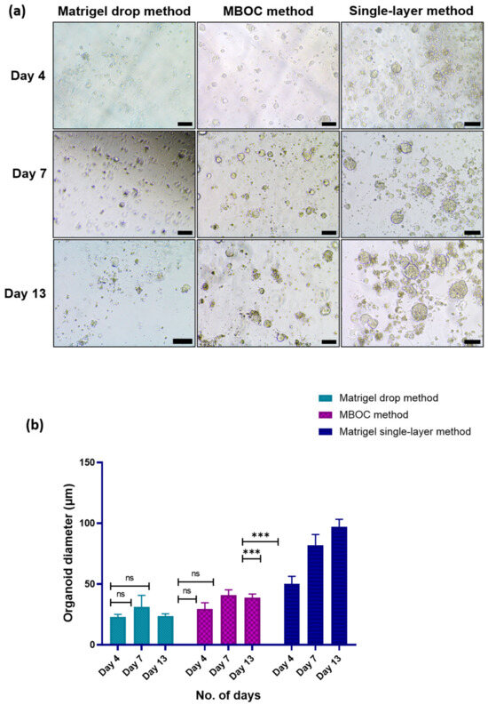
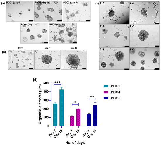
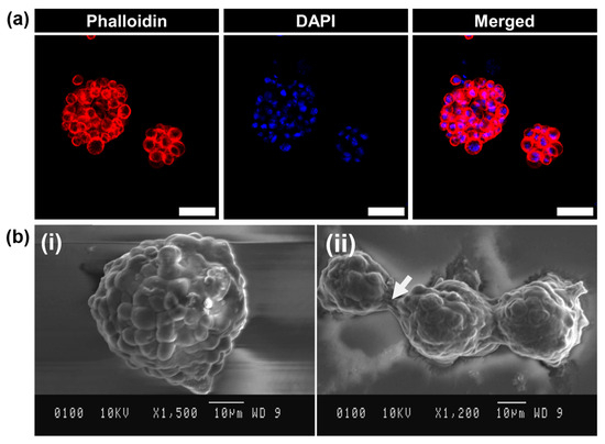
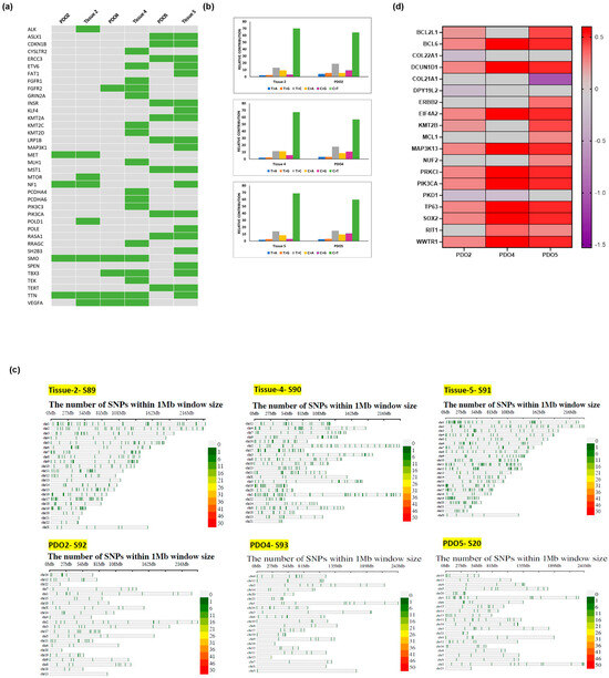
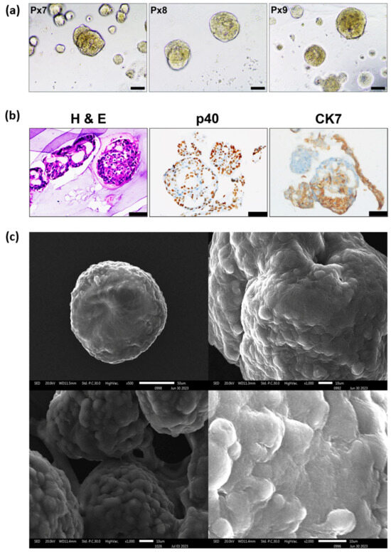
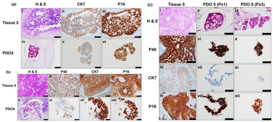
Advanced cervical cancer remains a major cause of mortality in women worldwide as it has limited treatment options and recurrence is very common. This highlights the necessity to develop patient-derived organoids (PDOs) as preclinical models that can recapitulate the clinical heterogeneity of the cancer in terms of molecular features and genetic background. The PDOs have potential for guiding personalized treatment in clinical practice. In this study, we have established patient-derived cervical cancer organoids from biopsy samples of five patients with two different histological subtypes (squamous cell carcinoma and adenocarcinoma) using a modified protocol. The organoids were characterized to assess their genetic and phenotypic similarity to the parental tumor tissue. The organoids developed in vitro preserved several characteristics of the parental tumors, including histological features, HPV status and a subset of genomic alterations. The expression of cervical cancer-related genes, including PIK3CA, MET, and LRP1B, was found to be comparable between the organoids and the parental tumor tissue. Moreover, characterization of the PDOs after cryopreservation showed the histopathological features of the parental tumor tissue. This study demonstrates that the CERvical Cancer OrganoidS (CERCOS) established using the current protocol hold potential to serve as a platform for personalized medicine.
Full article

Graphical abstract
Open AccessReview
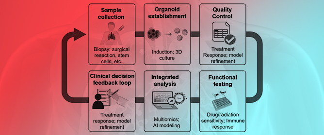
Organoid-on-Chip Technologies in Precision Oncology: Bridging Patient-Specific Tumor Biology and Physiologically Relevant Drug Screening
by
Muhammet Volkan Bulbul and Turan Demircan
Organoids 2026, 5(3), 30; https://doi.org/10.3390/organoids5030030 - 11 Sep 2026
Abstract
The inadequacy of traditional preclinical oncology models, specifically two-dimensional (2D) monolayer cultures and murine in vivo systems, in predicting human drug responses has led to the development of patient-derived organoids (PDOs) and microfluidic organ-on-chip (OoC) technologies. These innovations represent significant recent methodological advancements
[...] Read more.
The inadequacy of traditional preclinical oncology models, specifically two-dimensional (2D) monolayer cultures and murine in vivo systems, in predicting human drug responses has led to the development of patient-derived organoids (PDOs) and microfluidic organ-on-chip (OoC) technologies. These innovations represent significant recent methodological advancements in the field of cancer research. This review synthesizes the biological rationale, technical principles, and translational applications of PDO–OoC integration, with an emphasis on recent clinical validation studies, AI integration, and post-FDA Modernization Act 2.0 regulatory evolution—areas that have not been comprehensively addressed in prior reviews. We examined the predictive limitations of 2D models, organoid generation, and ToC engineering principles. The synergistic integration of organoids into chip-based systems, extended into multi-organ “Body-on-a-Chip” architectures, is presented as a unifying framework that combines patient-specific biological fidelity with dynamic microenvironmental control. We further reviewed the research applications and early clinical validation studies of high-throughput drug screening, immuno-oncology modeling, and patient-specific drug response prediction across multiple tumor types. Clinical validation studies have reported moderate correlations (r ~ 0.4–0.6) between organoid responses and outcomes, indicating partial predictive capacity. Despite this progress, clinical translation remains constrained by standardization and reproducibility deficits, biomaterial limitations (e.g., PDMS drug absorption and Matrigel batch variability), and regulatory ambiguities within the evolving FDA Modernization Act 2.0. Finally, we discuss the emerging integration of artificial intelligence, including transfer learning-based drug response prediction and real-time organoid avatar systems in clinical trials, as a pathway toward closed-loop individualized functional precision oncology. Organoid and tumor-on-chip platforms have advanced toward clinical utility, although barriers remain.
Full article
(This article belongs to the Special Issue Organs-on-Chips and Organoids: From Disease Modeling to Advanced Therapeutics)
►▼
Show Figures

Graphical abstract
Open AccessEditorial
Advances in Organoid Technology: Bridging the Gap Between Research and Therapy
by
Elizabeth Vincan, Somponnat Sampattavanich, Joao Ferreira and Ramanuj DasGupta
Organoids 2026, 5(3), 29; https://doi.org/10.3390/organoids5030029 - 9 Sep 2026
Abstract
Organoids Are Us is a symposium series that provides a snapshot in time of the latest and emerging developments in organoid technology [...]
Full article
(This article belongs to the Special Issue Advances in Organoid Technology: Bridging the Gap between Research and Therapy)
►▼
Show Figures

Graphical abstract
Open AccessTechnical Note
3D Imaging Without Light-Sheet: An Accessible Tissue-Clearing and Confocal Workflow for Human Cortical and Retinal Organoids
by
Erica Debbi, Lorenza Mautone, Chiara D’Antoni, Caterina Sanchini, Federica Cordella, Cristina Bertollini, Silvia Ghirga, Chloe Goemans, Yao Du, Yuliia Mykhailovska, Carlo Brighi, Laura Ferrucci, Francesco Bacchi, Valeria de Turris, Nicolas Baeyens and Silvia Di Angelantonio
Organoids 2026, 5(3), 28; https://doi.org/10.3390/organoids5030028 - 7 Sep 2026
Abstract
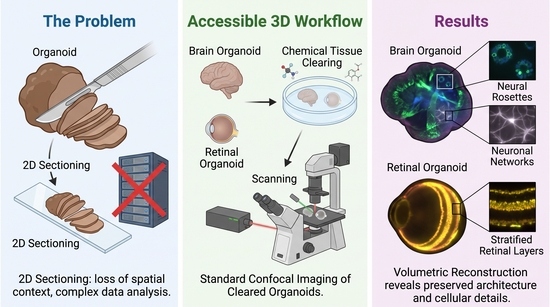
Human induced pluripotent stem cell (iPSC)-derived neural organoids have emerged as valuable models for investigating human neurodevelopment and neurological disorders. However, their complex three-dimensional architecture poses significant challenges for conventional histological approaches, which rely on physical sectioning and inevitably disrupt spatial relationships within
[...] Read more.
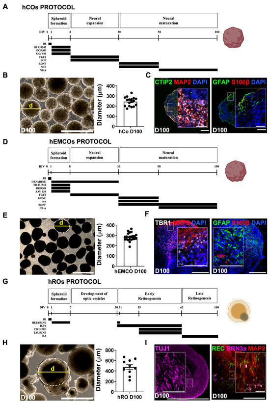
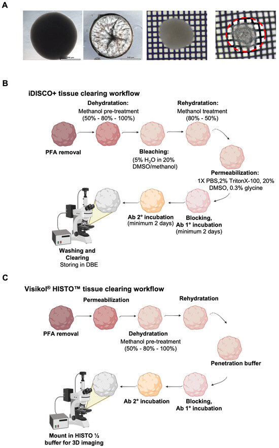
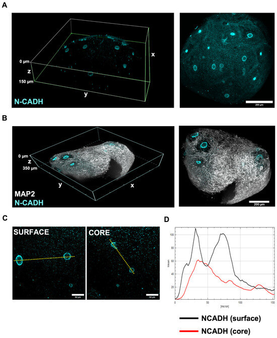
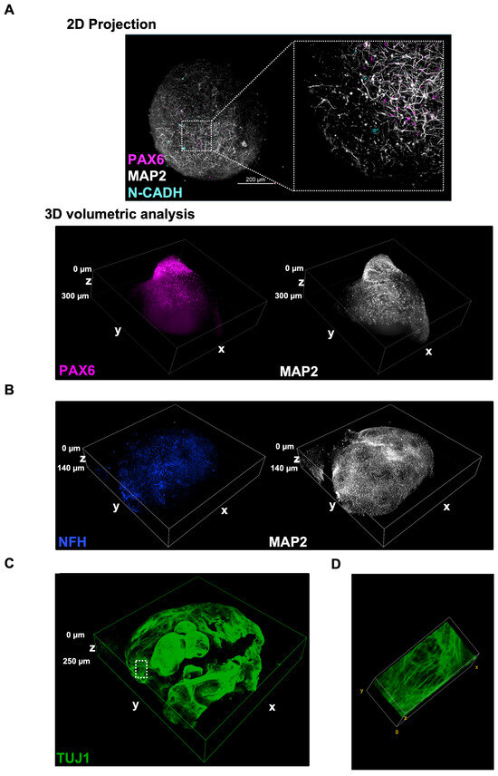
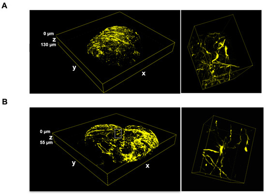
Human induced pluripotent stem cell (iPSC)-derived neural organoids have emerged as valuable models for investigating human neurodevelopment and neurological disorders. However, their complex three-dimensional architecture poses significant challenges for conventional histological approaches, which rely on physical sectioning and inevitably disrupt spatial relationships within the tissue. Volumetric imaging of intact organoids typically requires light-sheet fluorescence microscopy, a technology not widely accessible to standard cell biology laboratories. Here, we show that solvent-based tissue clearing, using the iDISCO+ and Visikol® HISTO protocols, combined with conventional laser-scanning and spinning-disk confocal microscopy, platforms already available in most imaging facilities, is sufficient to resolve neuroepithelial rosette-like structures, neuronal networks, and astroglial components within intact human cortical and retinal organoids, while preserving immunofluorescent labeling and tissue architecture. The workflow was also compatible with commonly used immunofluorescence markers. Although light-sheet fluorescence microscopy remains advantageous for large-scale whole-sample imaging, our results show that cleared human organoids within the size range analyzed here can be effectively visualized using accessible confocal systems. This study provides a practical strategy for three-dimensional imaging of intact human neural organoids, facilitating spatial analysis of developmental organization and disease-relevant phenotypes in laboratories without dedicated light-sheet microscopy infrastructure.
Full article
(This article belongs to the Special Issue From Development to Degeneration: Advances in Stem Cell and Organoid Models of the Nervous System)
►▼
Show Figures

Graphical abstract
Open AccessReview
Building Fit-for-Purpose, Multi-Lineage Immune–Organoid Models for Urologic Cancers: A Critical Narrative Review of Prostate, Urothelial and Renal Cell Carcinoma Models
by
Emmanuel O. Oisakede, Okhibhamen Ehizokhale, Olawunmi O. Oyedeji and David B. Olawade
Organoids 2026, 5(3), 27; https://doi.org/10.3390/organoids5030027 - 3 Sep 2026
Abstract
Immune–organoid co-culture has been proposed as a bridge between reductionist assays and clinical immuno-oncology, but the urologic evidence remains substantially thinner than this description suggests. This critical narrative review evaluates peer-reviewed work in prostate cancer, urothelial carcinoma and renal cell carcinoma from January
[...] Read more.
Immune–organoid co-culture has been proposed as a bridge between reductionist assays and clinical immuno-oncology, but the urologic evidence remains substantially thinner than this description suggests. This critical narrative review evaluates peer-reviewed work in prostate cancer, urothelial carcinoma and renal cell carcinoma from January 2014 through to 31 July 2026, separating patient-derived immune-preserving models from reconstituted cytotoxicity assays and adjacent engineering studies. Native air–liquid interface cultures have retained endogenous lymphoid, myeloid and stromal compartments in renal cell carcinoma for short-term checkpoint-inhibitor experiments. Reconstituted bladder and kidney organoids have supported mechanistic testing of chimeric antigen receptor T cells, and a bladder study has examined treatment-induced Jurkat-cell migration. These studies establish technical feasibility, but most use small cohorts, short endpoints and incomplete immune composition, and none has prospectively shown that a multi-lineage urologic organoid assay improves treatment selection. Published prostate systems remain largely epithelial or stromal, leaving a conspicuous immune-modelling gap. We therefore argue against equating greater cellular complexity with greater validity. The appropriate model is the least complex system that preserves the mechanism, spatial constraint and temporal window required by the question. A tiered framework is proposed that progresses from analytical quality control, through defined effector and suppressor modules, to perfused or spatially organised cultures only when these features are necessary. Minimum reporting standards, disease-specific immune modules, clinically meaningful endpoints and a four-stage validation ladder are specified. Multi-lineage systems can clarify resistance mechanisms and screen combinations, but predictive or clinical claims require blinded patient concordance and prospective utility studies rather than architectural sophistication alone.
Full article
(This article belongs to the Special Issue Organs-on-Chips and Organoids: From Disease Modeling to Advanced Therapeutics)
►▼
Show Figures

Figure 1
Open AccessReview
Harnessing Bioactive Ceramic for Organoid Engineering: Mechanisms, Applications, and Prospects in Regenerative Medicine
by
Shihan Sun, Sixuan Chen, Wenping Ma, Jun Xu, Mingxia Lu and Hongxu Lu
Organoids 2026, 5(3), 26; https://doi.org/10.3390/organoids5030026 - 17 Aug 2026
Abstract
►▼
Show Figures
Organoids are three-dimensional, stem-cell-derived tissue constructs that recapitulate the architecture and function of native organs, and they have rapidly emerged as transformative tools in regenerative medicine. Their translation from laboratory models to clinical therapies remains constrained, however, by the limitations of conventional culture
[...] Read more.
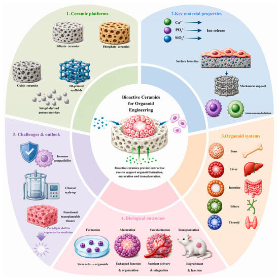
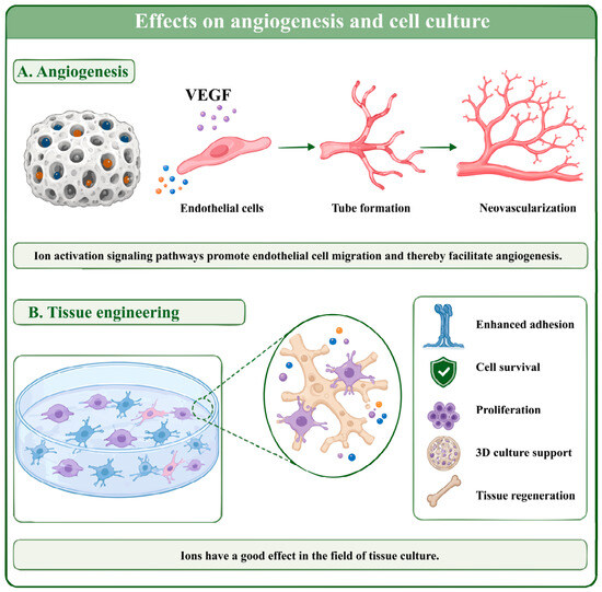
Organoids are three-dimensional, stem-cell-derived tissue constructs that recapitulate the architecture and function of native organs, and they have rapidly emerged as transformative tools in regenerative medicine. Their translation from laboratory models to clinical therapies remains constrained, however, by the limitations of conventional culture matrices. Matrigel is the most widely used matrix. It suffers from batch-to-batch variability, an undefined composition, poor mechanical tunability, and a lack of instructive bioactivity. Bioactive ceramics offer a compelling alternative. Encompassing silicate-, phosphate-, and oxide-based formulations, these materials provide controllable ion-release profiles and structural versatility. They also possess a proven capacity to modulate cell behavior through biochemical and biophysical cues. This review systematically examines how the defining properties of bioactive ceramics intersect with the requirements of organoid formation, maturation, and transplantation. We focus on four key properties: ion release, surface bioactivity, mechanical support, and immunomodulation. We survey established and emerging combinations across bone, liver, intestine, biliary, and thyroid organoid systems. Fabrication strategies, including 3D-printed and sol–gel-derived ceramic scaffolds, are also discussed. Finally, we critically assess remaining challenges in vascularization, immune compatibility, and clinical scale-up, and we propose that the deliberate co-design of bioactive ceramics and organoid biology represents a paradigm shift in regenerative medicine. This approach offers a path toward functional, transplantable tissue constructs with genuine therapeutic potential.
Full article

Figure 1
Open AccessOpinion
Physiological Relevance of Engineered Brain Models
by
Bram Servais, David J. Collins and David R. Nisbet
Organoids 2026, 5(3), 25; https://doi.org/10.3390/organoids5030025 - 5 Aug 2026
Abstract
►▼
Show Figures
Engineered brain models, including brain organoids and brain-on-a-chip systems, are generally assessed in terms of their physiological relevance. Although this language is useful for emphasizing the need to better approximate human biology, it can also obscure important differences among context of use, required
[...] Read more.
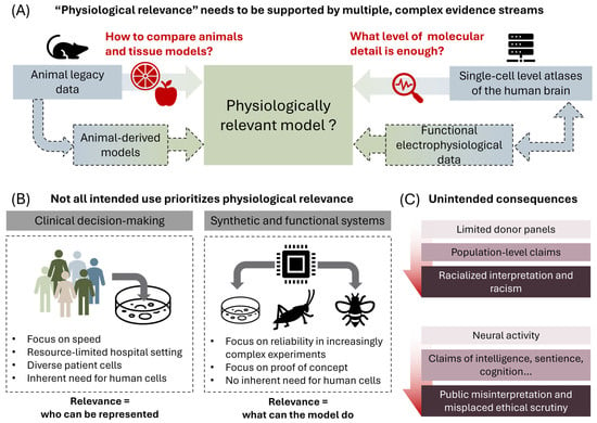
Engineered brain models, including brain organoids and brain-on-a-chip systems, are generally assessed in terms of their physiological relevance. Although this language is useful for emphasizing the need to better approximate human biology, it can also obscure important differences among context of use, required validation strategy and ethical considerations. In this Opinion, we argue that physiological relevance should not be treated as a universal measure of model quality. Instead, its meaning should be defined relative to its application domain, including animal-model comparison, interpretation of single-cell atlases, clinical translation, donor representation, and emerging functional applications such as synthetic biological intelligence. For some applications, particularly patient-specific disease modeling and therapeutic screening, greater human physiological relevance may be required. For others, including biohybrid computing, controllable neural interfaces, interpretability, and ethical considerations may be more important. Moving beyond simplistic terminology will help improve scientific interpretation, prevent overstating findings, and support more responsible development of engineered brain models.
Full article

Figure 1
Open AccessReview
From Cells to Microphysiological Systems: 3D Cell Cultures and Organ-on-Chip Systems for Studying cAMP and cGMP Signaling
by
Maria Rita Assenza, Nicole Bertani, Martina Pinna and Federica Campolo
Organoids 2026, 5(3), 24; https://doi.org/10.3390/organoids5030024 - 4 Aug 2026
Cited by 1
Abstract
Cyclic adenosine monophosphate and cyclic guanosine monophosphate are key regulators of cellular physiology and tissue homeostasis. Conventional experimental models have provided fundamental insights into cyclic nucleotide pathways; however, they often fail to fully recapitulate essential features of in vivo systems. Two-dimensional cell cultures
[...] Read more.
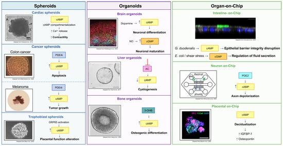
Cyclic adenosine monophosphate and cyclic guanosine monophosphate are key regulators of cellular physiology and tissue homeostasis. Conventional experimental models have provided fundamental insights into cyclic nucleotide pathways; however, they often fail to fully recapitulate essential features of in vivo systems. Two-dimensional cell cultures lack spatial organization, while animal models incompletely capture cell–cell interactions and the dynamic microenvironment shaping signaling processes. In recent years, advanced three-dimensional and microengineered systems have emerged as tools to bridge this gap. In this review, we discuss how three-dimensional and organ-on-chip systems are transforming the study of cyclic nucleotide signaling by enabling reconstruction of tissue architecture and signaling niches. Spheroids and organoids provide robust models to investigate compartmentalized signaling and intercellular communication. Complementarily, microfluidic organ-on-chip devices introduce controlled mechanical cues, perfusion, and tissue interfaces, enabling real-time monitoring of signaling dynamics. We highlight recent advances in microphysiological systems for investigating the spatial and temporal dynamics of cyclic adenosine monophosphate and cyclic guanosine monophosphate signaling, including biosensors, live-cell imaging, and genome editing. We further discuss applications in physiological and pathological contexts, including metabolic, cardiovascular and cancer diseases, and outline current challenges and future perspectives for integrating three-dimensional and organ-on-chip technologies.
Full article
(This article belongs to the Special Issue Organs-on-Chips and Organoids: From Disease Modeling to Advanced Therapeutics)
►▼
Show Figures

Figure 1
Open AccessArticle
Neuroimmune Organoid Models Early Glioblastoma Establishment and the Invasive Niche
by
Nina Y. Yuan, William D. Richards, Kailyn T. Parham, Kaylie Greuel, Joshua A. Zimmermann, Jack Shireman, Lei Zhao, Mahua Dey and Connie S. Lebakken
Organoids 2026, 5(3), 23; https://doi.org/10.3390/organoids5030023 - 2 Aug 2026
Abstract
Glioblastoma (GBM) is a highly aggressive malignant brain tumor accounting for 15% of all brain tumors and 50% of all gliomas. The exact cause of GBM is not fully understood but risk factors include age, genetic mutations, exposure to ionizing radiation, and certain
[...] Read more.
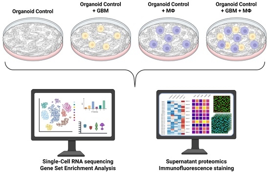
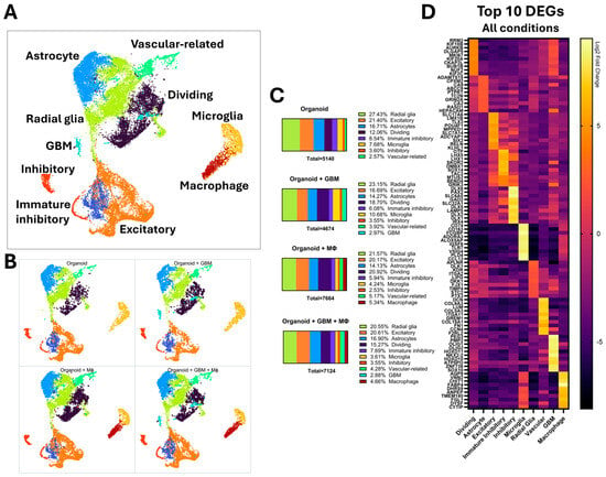
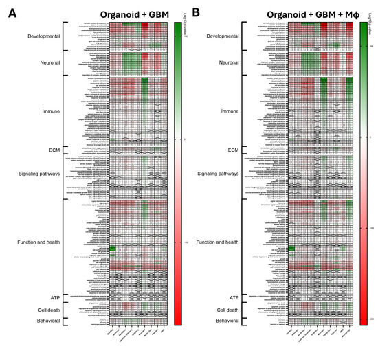
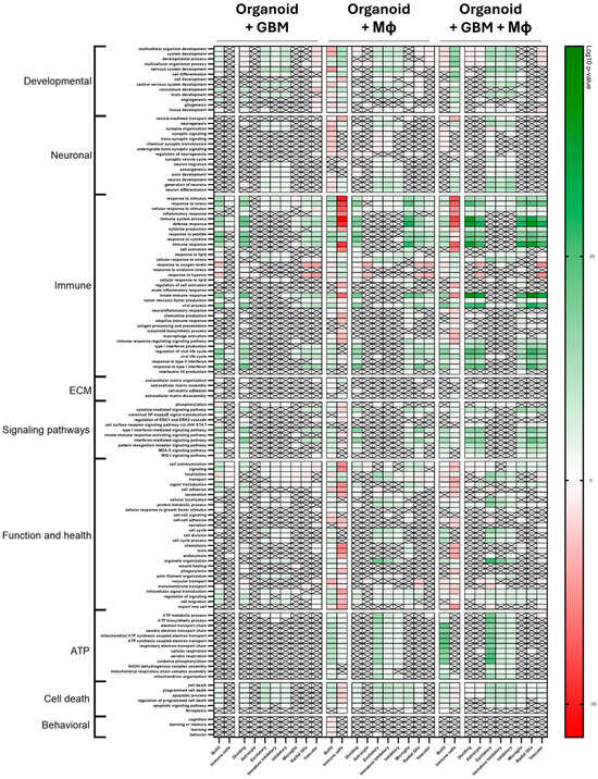
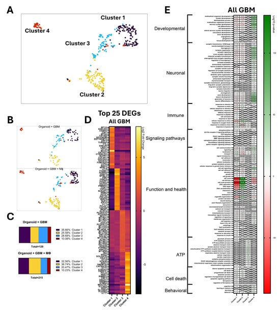
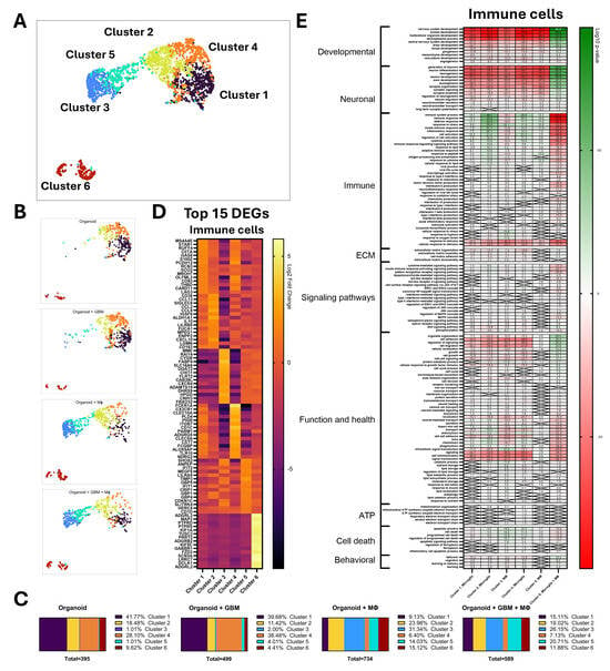
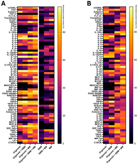
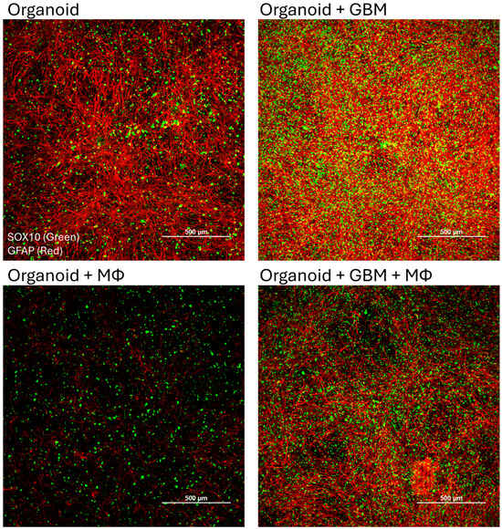
Glioblastoma (GBM) is a highly aggressive malignant brain tumor accounting for 15% of all brain tumors and 50% of all gliomas. The exact cause of GBM is not fully understood but risk factors include age, genetic mutations, exposure to ionizing radiation, and certain genetic disorders. Symptoms of GBM include headaches, seizures, cognitive impairment, and weaknesses on one side of the body. Myeloid cells account for 30–50% of the tumor mass and are instrumental in shaping the complex tumor microenvironment (TME). Inflammation in the TME is an important driver of tumor growth and invasion; however, as the environment evolves, the immunosuppressive TME poses a significant hurdle as it hinders the immune-mediated killing of tumor cells. Our work utilizes neuroimmune organoids containing neurons, astrocytes, microglia, and vascular-like cells, to which we add patient-derived GBM cells and/or iPSC-derived macrophages to model the GBM TME. Model characterization was performed using single-cell RNA sequencing and supernatant proteomics to determine cell-specific changes during coculturing. Our findings are consistent with this 7-day coculture model recapitulating key aspects of GBM early tumor establishment and immune activation, with transcriptomic and secretome signatures suggestive of an emerging immune evasion phenotype.
Full article
(This article belongs to the Special Issue From Development to Degeneration: Advances in Stem Cell and Organoid Models of the Nervous System)
►▼
Show Figures

Graphical abstract
Open AccessReview
Modeling Context-Dependent Tumor Metabolism in 3D Systems: Implications for Functional Precision Oncology
by
Maria Virginia Giolito, Olivier Feron and Cyril Corbet
Organoids 2026, 5(3), 22; https://doi.org/10.3390/organoids5030022 - 27 Jul 2026
Cited by 1
Abstract
►▼
Show Figures
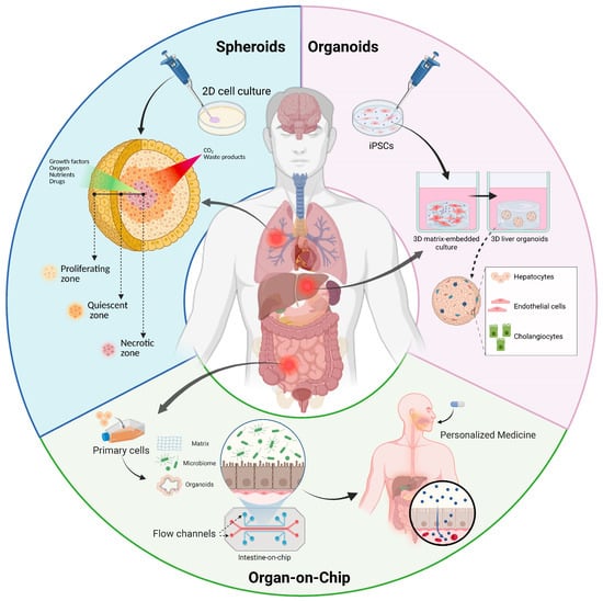
Cancer metabolism is a dynamic and context-dependent process shaped by both tumor-intrinsic programs and microenvironmental cues. Capturing this complexity remains a major challenge, which limits the translation of metabolic insights into clinically actionable strategies. Patient-derived tumor organoids, together with emerging engineered platforms such
[...] Read more.
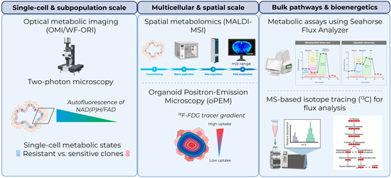
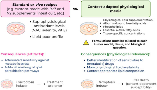
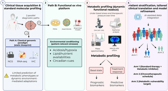
Cancer metabolism is a dynamic and context-dependent process shaped by both tumor-intrinsic programs and microenvironmental cues. Capturing this complexity remains a major challenge, which limits the translation of metabolic insights into clinically actionable strategies. Patient-derived tumor organoids, together with emerging engineered platforms such as organ-on-chip systems, vascularized assembloids, and bioprinted tumor models, have opened new avenues for investigating tumor metabolism in physiologically relevant settings. These models enable the study of metabolic heterogeneity across tumor types, disease stages, and treatment conditions while preserving clinically relevant tumor features. Importantly, they provide functional platforms for ex vivo metabolic profiling, identification of metabolic vulnerabilities, and prediction of therapeutic responses. In this review, we discuss recent advances in the use of patient-derived and engineered 3D tumor models to characterize context-dependent metabolic states and treatment-induced metabolic rewiring. We first review technologies currently available to interrogate metabolism in these systems, including optical metabolic imaging, spatial metabolomics, isotope tracing, and bioenergetic profiling. We then discuss how 3D tumor models are used to investigate metabolic interactions within the tumor microenvironment (TME), including stromal and immune crosstalk, acidosis, nutrient availability, and circadian regulation. Finally, we critically examine current limitations, particularly the insufficient physiological relevance of standard organoid culture conditions for metabolic studies, and discuss how advanced engineering approaches and computational modeling may contribute to metabolism-driven functional precision oncology.
Full article

Figure 1
Open AccessArticle
Spatial Analysis of Proteins in 3D Cell Culture Models: A Systematic Troubleshooting Guide for Whole-Mount Immunofluorescence
by
Olgu Enis Tok, Gamze Demirel, Ozgecan Kayalar, Nur Konyalilar, Hasan Bayram and Ranan Gulhan Aktas
Organoids 2026, 5(3), 21; https://doi.org/10.3390/organoids5030021 - 8 Jul 2026
Abstract
►▼
Show Figures
The rise of 3D cell culture systems—including organoids, spheroids, and organ-on-a-chip models—has transformed our understanding of tumor biology, disease pathology, and tissue development. However, accurately analyzing spatial phenotypic content within these complex architectures remains a formidable challenge. While contemporary protocols strive for precise
[...] Read more.
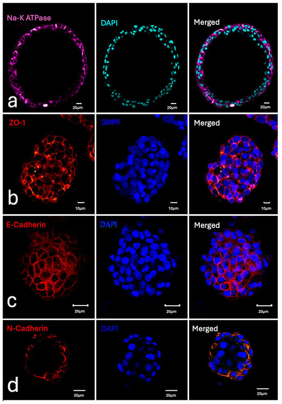
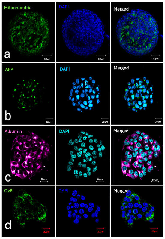
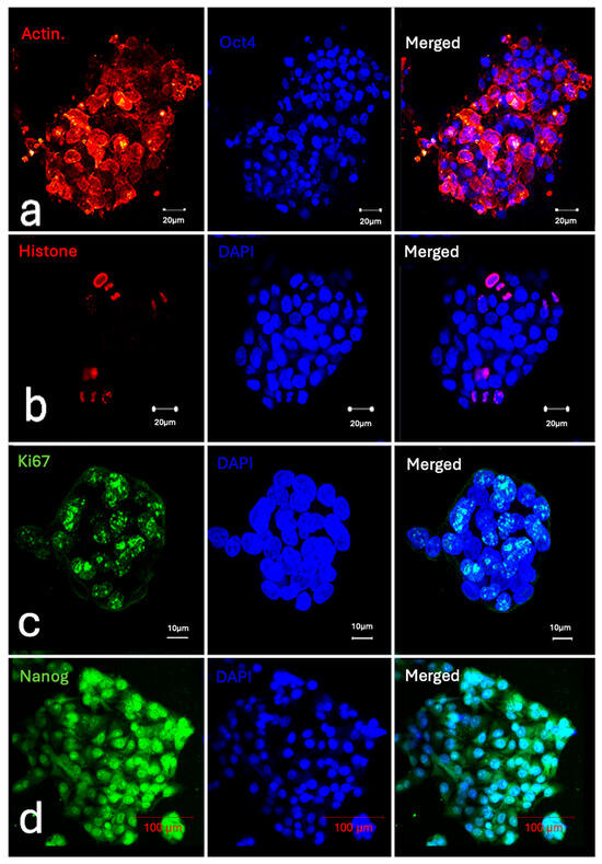
The rise of 3D cell culture systems—including organoids, spheroids, and organ-on-a-chip models—has transformed our understanding of tumor biology, disease pathology, and tissue development. However, accurately analyzing spatial phenotypic content within these complex architectures remains a formidable challenge. While contemporary protocols strive for precise protein localization, their reliability is frequently undermined by technical artifacts and the structural degradation of the 3D matrices. These distortions are often induced by invasive harvesting, harsh clearing agents, and frequent sample transfers. To bridge the gap between complex 3D tissue architectures and reliable assay readouts, this study establishes a systematic troubleshooting framework for whole-mount 3D immunofluorescence staining. Utilizing a diverse panel of 24 distinct antibodies targeting membrane, cytoplasmic, and nuclear proteins across human airway organoids and liver cancer spheroids, we executed comprehensive mono-, double-, and triple-labeling configurations. To evaluate workflow boundaries, we conducted a series of controlled whole-mount experiments where specific, common technical mistakes were deliberately introduced. By documenting the exact imaging artifacts, structural distortions, and aberrant signal profiles generated by these intentional procedural errors, this study provides a unique visual “atlas of failure” paired directly with validated methodological solutions. The study offers a practical, high-throughput diagnostic resource to eliminate technical error and experimental noise for whole-mount immunofluorescence labeling experiments, thereby facilitating high-quality imaging and consistent phenotypic validation.
Full article

Figure 1
Open AccessReview
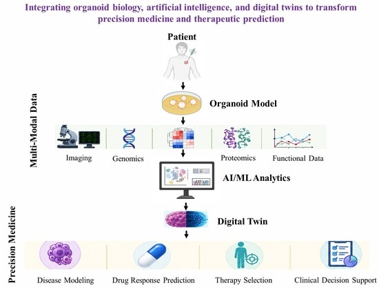
Artificial Intelligence–Enabled Organoid Platforms for Precision Medicine: Integrating Multi-Omics, Digital Twins, and Microphysiological Systems
by
Ramandeep Saini, Bishakha Thakur, Bikram Kumar Basaba and Mantosh Kumar Satapathy
Organoids 2026, 5(3), 20; https://doi.org/10.3390/organoids5030020 - 2 Jul 2026
Abstract
►▼
Show Figures
The convergence of artificial intelligence (AI) and organoid technology represents a transformative advance toward precision and predictive medicine. Organoids derived from pluripotent stem cells or patient tissues provide physiologically relevant three-dimensional models that recapitulate key aspects of native organ architecture and function. However,
[...] Read more.
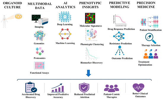
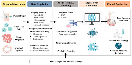
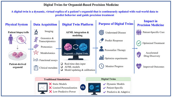
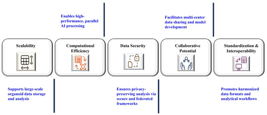
The convergence of artificial intelligence (AI) and organoid technology represents a transformative advance toward precision and predictive medicine. Organoids derived from pluripotent stem cells or patient tissues provide physiologically relevant three-dimensional models that recapitulate key aspects of native organ architecture and function. However, intrinsic biological heterogeneity, high-content imaging outputs, and dynamic spatiotemporal processes pose significant analytical challenges that exceed the capacity of conventional approaches. Recent advances in AI and machine learning enable automated image segmentation, quantitative morphometric profiling, and predictive modeling of organoid growth, differentiation, and therapeutic response, thereby enhancing reproducibility and translational relevance. The integration of multimodal datasets, including imaging, genomics, transcriptomics, epigenomics, proteomics, and metabolomics, has further enabled the development of organoid-based digital twins and in silico disease simulations to optimize personalized therapy. AI-enabled organoid-on-a-chip platforms, cloud-based analytics, and federated learning frameworks are accelerating the emergence of scalable, privacy-preserving, and data-driven biomedical ecosystems. Despite these advances, critical challenges persist, including data standardization, model interpretability, ethical governance, and clinical validation. In contrast to existing reviews that emphasize isolated AI applications, this study proposes a unified translational framework integrating AI-driven image analytics, multi-omics integration, digital twins, and organoid-on-a-chip systems within a precision medicine paradigm. By synthesizing current developments, methodological advances, and emerging trends, this study highlights how AI-powered organoid platforms can bridge experimental biology and clinical decision-making, with broad implications for drug discovery, disease modeling, and regenerative medicine. This review aims to provide a comprehensive overview of artificial intelligence–enabled organoid platforms by integrating advances in image analytics, multi-omics data integration, digital twins, and microphysiological systems, while highlighting their potential applications and future directions in precision medicine, drug discovery, and regenerative healthcare.
Full article

Graphical abstract
Open AccessPerspective
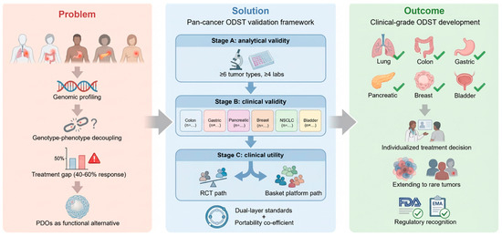
A Pan-Cancer Preclinical Validation Framework for Organoid-Based Drug Sensitivity Testing
by
Jia Shang, Caixia Xia, Zilin Xu, Sheng Tu, Gang Li, Fangjin Chen, Lingao Ju, Gang Wang, Yu Xiao and Kaiyu Qian
Organoids 2026, 5(2), 19; https://doi.org/10.3390/organoids5020019 - 12 Jun 2026
Cited by 1
Abstract
Patient-derived organoids (PDOs) provide ex vivo functional models that capture tumor drug-response patterns across multiple cancer types. Organoid drug sensitivity testing (ODST) has accumulated supportive evidence in single-tumor studies, yet it lacks a pan-cancer biostatistical framework that can support multi-cancer clinical decision-making. This
[...] Read more.
Patient-derived organoids (PDOs) provide ex vivo functional models that capture tumor drug-response patterns across multiple cancer types. Organoid drug sensitivity testing (ODST) has accumulated supportive evidence in single-tumor studies, yet it lacks a pan-cancer biostatistical framework that can support multi-cancer clinical decision-making. This article presents a pan-cancer ODST validation framework that integrates evidence synthesis, regulatory mapping, and adaptive trial design. The framework specifies analytical-performance standards, a three-stage validation architecture, and an explicit cross-tumor portability coefficient that quantifies the transferability of validated evidence among cancer types. Implementation barriers, including heterogeneous tissue-collection standards, variable establishment success, immunotherapy modeling limitations, and regulatory misalignment, are identified, and corresponding mitigation strategies are described. The framework supports a structured pathway from analytical validity to clinical utility for ODST across solid-tumor indications.
Full article
(This article belongs to the Special Issue Advances in Organoid Technology: Bridging the Gap between Research and Therapy)
►▼
Show Figures

Figure 1
Open AccessArticle
Engineering Chimeric Cardio-Vascular Assembloids Using Human iPSC-Derived Cardiomyocytes and Vascular Rings
by
Hannah Klör, Kornelia Kenst, Berin Upcin, Süleyman Ergün and Philipp Wörsdörfer
Organoids 2026, 5(2), 18; https://doi.org/10.3390/organoids5020018 - 10 Jun 2026
Abstract
►▼
Show Figures
The myocardium possesses one of the highest vascular densities in the body. The outermost wall layer of large and medium-sized vessels, the adventitia, forms a critical interface between the vasculature and the myocardium and serves as a reservoir for stem and progenitor cells
[...] Read more.
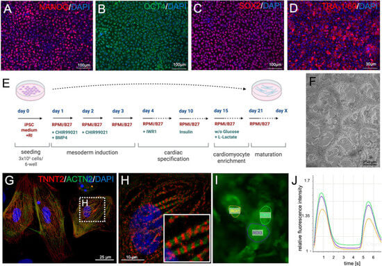
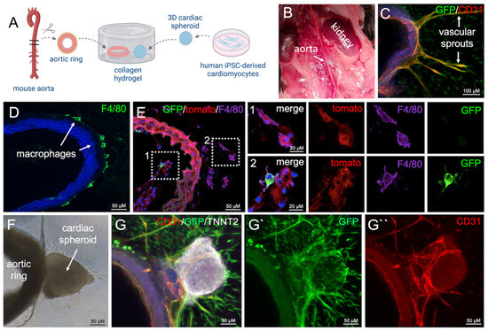
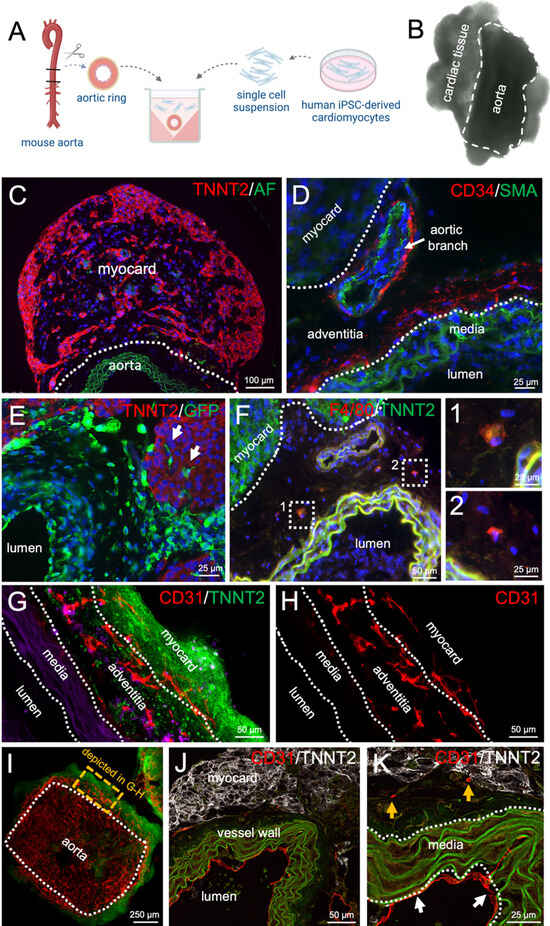
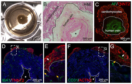
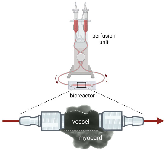
The myocardium possesses one of the highest vascular densities in the body. The outermost wall layer of large and medium-sized vessels, the adventitia, forms a critical interface between the vasculature and the myocardium and serves as a reservoir for stem and progenitor cells capable of differentiating into all vascular wall lineages as well as innate immune cells, including macrophages. Current cardiac organoid models intrinsically develop networks of endothelial cords and small capillary-like structures that resemble cardiac microvessels. However, these microvessels mostly lack an adventitial compartment in vivo. Here, we present a potential alternative assembloid strategy that combines vascular segments from mouse and human origin with either cardiomyocytes or cardiac spheroids derived from human induced pluripotent stem cells, thereby incorporating large diameter vessels and the vascular adventitia into a cardiac tissue model. Within the assembloids, the myocardial component remained contractile and connected to the vascular adventitia, which displayed cellular sprouting toward the hiPSC-derived cardiac tissue. Immunostaining for vascular and immune markers revealed that the adventitia gave rise to endothelial sprouts and macrophage-like cells which integrated into the myocardial tissue. In summary, we present proof of concept for complex assembloids composed of vessel segments and human iPSC-derived cardiomyocytes which contain and maintain an in vivo-like adventitial compartment. We suggest this model may serve as a platform for investigating myocardial–stromal interactions, cardiac tissue repair, and functional remodeling under both physiological and pathological conditions. Furthermore, the incorporation of large-lumen vessel segments may enable future experimental perfusion, rendering the model particularly suitable for drug testing via intravascular delivery.
Full article

Figure 1
Open AccessArticle
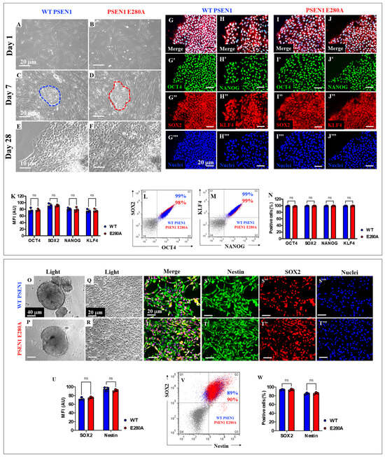
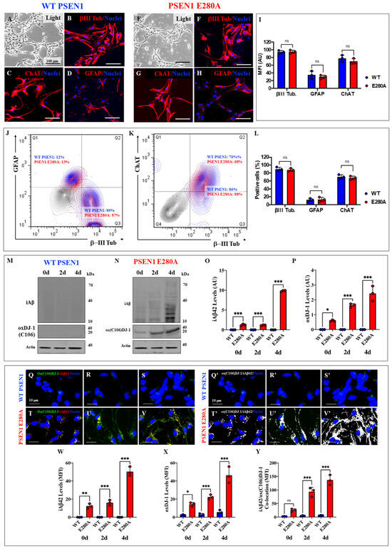
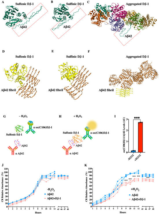
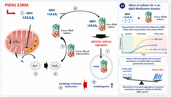
Sulfonic DJ-1 (Cys106-SO3H) Binds to and Colocalizes with the Intracellular Accumulation of Amyloid-Beta 42 (Aβ42) in Familial Alzheimer’s Disease PSEN1 E280A Cerebral Organoids Derived from Induced Pluripotent Stem Cells
by
Viviana Soto-Mercado, Miguel Mendivil-Perez, Carlos Velez-Pardo and Marlene Jimenez-Del-Rio
Organoids 2026, 5(2), 17; https://doi.org/10.3390/organoids5020017 - 3 Jun 2026
Cited by 1
Abstract
The intracellular accumulation of amyloid beta 42 (iAβ42) has been proposed as an early pathological indicator of familial Alzheimer’s disease (FAD). DJ-1 is a multifunctional protein sensitive to oxidative stress (OS) that has been associated with neurodegeneration; however, its role in iAβ42 pathology
[...] Read more.
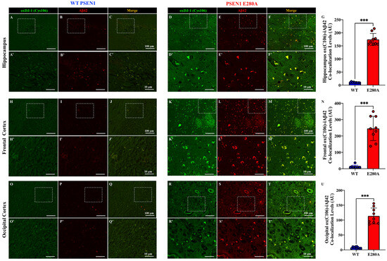
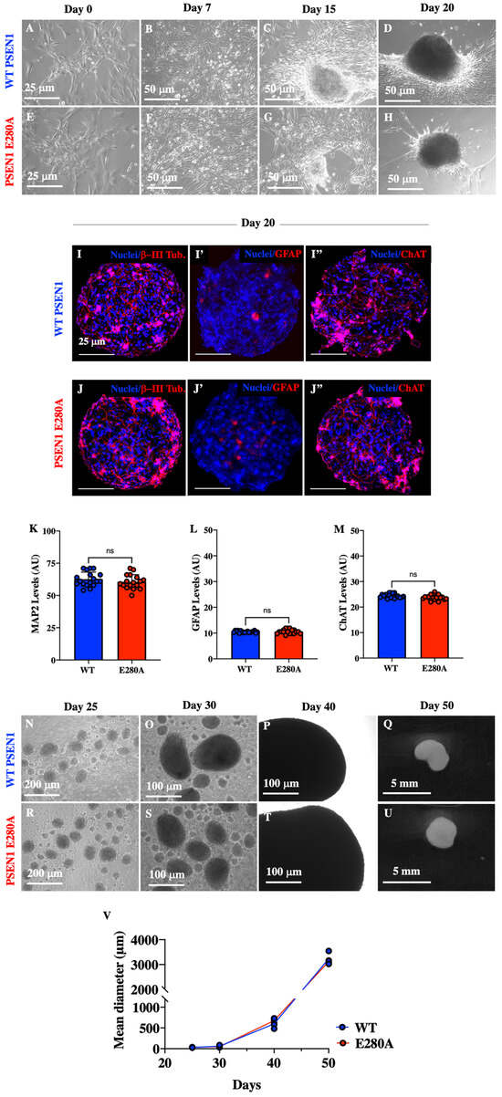
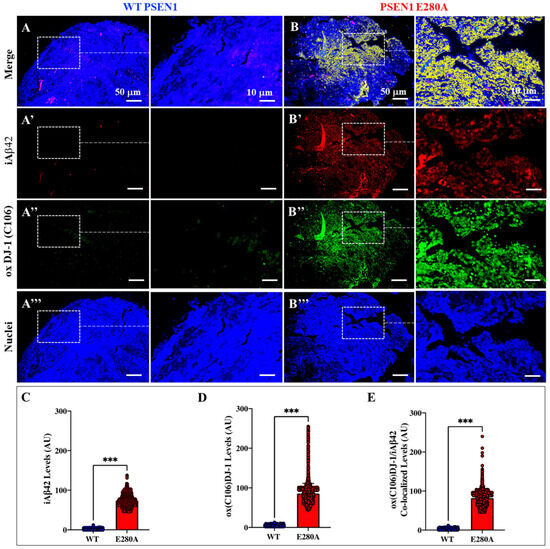
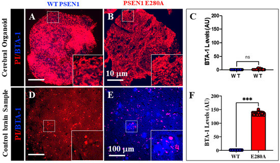
The intracellular accumulation of amyloid beta 42 (iAβ42) has been proposed as an early pathological indicator of familial Alzheimer’s disease (FAD). DJ-1 is a multifunctional protein sensitive to oxidative stress (OS) that has been associated with neurodegeneration; however, its role in iAβ42 pathology is unclear. In this study, we examined whether oxidized (sulfonic) DJ-1 (Cys106-SO3H) drives iAβ42 accumulation using postmortem brain samples and in vitro 3D iPSC-derived cerebral organoids (COs) or 2D induced pluripotent stem cells (iPSC)-derived ChLNs (cholinergic-like neurons) models from a PSEN1 E280A patient and a healthy volunteer (as a control sample). Post-mortem analyses of the temporal and frontal cortices and hippocampus from FAD PSEN1 E280A patients revealed strong intracellular co-localization of sulfonic DJ-1 and iAβ42, which was absent in control samples. To validate these findings, we generated COs from an iPSC PSEN1 E280A FAD patient and a healthy donor. In these organoids, we observed the co-localization of oxidized DJ-1 and Aβ42 in the absence of extracellular fibrils or plaques, as confirmed by BTA-1 staining. To further support these observations, 2D iPSC PSEN1 E280A-derived ChLNs cultures showed that intracellular Aβ42 accumulates progressively in direct correlation with increasing DJ-1 oxidation, as demonstrated by immunofluorescence microscopy and Western blotting analysis. These results indicate that DJ-1 oxidation accompanies the earliest intracellular stages of Aβ42 pathology. Furthermore, complementary in silico molecular docking analysis revealed a higher affinity between Aβ42 and oxidized sulfonic DJ-1 (DJ-1 Cys106-SO3H) compared to sulfenic (DJ-1 Cys106-SOH) or sulfinic acid (DJ-1 Cys106-SO2H) forms. Likewise, ELISA tests and seeding assays confirmed that oxidized DJ-1 binds to and decelerates Aβ42 aggregation kinetics. Together, our results identify DJ-1 oxidation as a critical molecular event in the accumulation of iAβ42 in FAD. These findings suggest that oxidized DJ-1 represents not only a potential early biomarker of intracellular pathology but also a pharmacological target. Preventing the oxidation of DJ-1 or its pathological aggregation could provide new biomarkers and therapeutic strategies for reducing the intracellular accumulation of Aβ42 and neurodegeneration in FAD.
Full article
(This article belongs to the Special Issue The Current Applications and Potential of Stem Cell-Derived Organoids)
►▼
Show Figures

Figure 1
Open AccessReview
Precision Oncology at a Crossroads: How Organoid Platforms Are Reshaping the Field
by
Seulbee Lee, Alyssa Kim, Rachel Hyunkyung Kim, Seo-Hee You, Hyun Soo Kim, Seok Chung, Sang-Haak Lee, Seung-Ah Yahng, In Kyoung Kim and Hye Joung Kim
Organoids 2026, 5(2), 16; https://doi.org/10.3390/organoids5020016 - 29 May 2026
Abstract
►▼
Show Figures
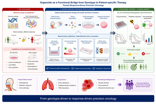
Tumor heterogeneity and microenvironmental complexity remain fundamental barriers to genomics-centered precision oncology, frequently causing discordance between molecular alterations and real-world therapeutic responses. Here, we reviewed patient-derived organoid (PDO) technologies as functional platforms that complement molecular profiling by directly investigating patient-specific sensitivity, resistance, and
[...] Read more.
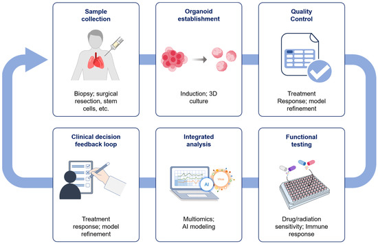
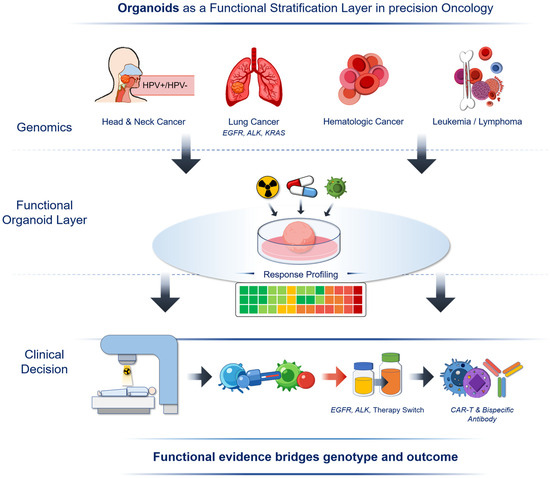
Tumor heterogeneity and microenvironmental complexity remain fundamental barriers to genomics-centered precision oncology, frequently causing discordance between molecular alterations and real-world therapeutic responses. Here, we reviewed patient-derived organoid (PDO) technologies as functional platforms that complement molecular profiling by directly investigating patient-specific sensitivity, resistance, and microenvironment dependent vulnerability. We first summarize why conventional preclinical systems, two-dimensional cell lines and patient-derived xenografts, are limited by reduced biological fidelity, impractical turnaround time, and scalability for clinical decision support. We then synthesized organoid-based evidence across three representative disease malignancies with distinct precision-medicine bottlenecks. Across these settings, we highlight advances that extend the PDO capability beyond the tumor epithelium alone, including air–liquid interface cultures, immune and stromal co-cultures, and microfluidic organoid-on-chip systems, as well as integration with multi-omics and artificial intelligence for scalable analytics. Finally, we discuss the key translational requirements, standardization of culture matrices and assay readouts, quality control, automation to reduce turnaround time, and regulatory/ethical frameworks, required to transition organoid-guided testing from proof-of-concept to routine implementation. Collectively, this review reframes organoids as functional stratification platforms supporting the integration of functional response profiling alongside genomics-guided precision oncology approaches.
Full article

Graphical abstract
Open AccessReview
Patient-Derived Organoids in Clinical Medicine: Proven Impact and Future Directions
by
Magdalena Skowronska, Ece Yildiz, Jens Grosch and Mairene Coto-Llerena
Organoids 2026, 5(2), 15; https://doi.org/10.3390/organoids5020015 - 21 May 2026
Cited by 1
Abstract
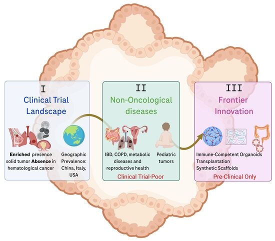
Patient-derived organoids (PDOs) have rapidly transitioned from research tools into promising platforms for clinical translation. In this review, we analyze 139 PDO-related clinical trials registered between 2023 and 2025 and contrast them with recent advances in disease modelling. Our analysis revealed a predominance
[...] Read more.
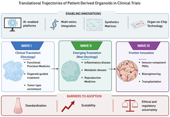
Patient-derived organoids (PDOs) have rapidly transitioned from research tools into promising platforms for clinical translation. In this review, we analyze 139 PDO-related clinical trials registered between 2023 and 2025 and contrast them with recent advances in disease modelling. Our analysis revealed a predominance of oncology-focused studies, with translational maturity spanning from foundational research to studies in which PDOs directly informed clinical decision-making. In contrast, non-oncology areas show extensive preclinical progress but remain trial-poor. We found that trial registration is geographically concentrated in a small number of countries, reflecting uneven global adoption. We then explored advances in disease modeling, mainly confined to preclinical studies, including immune-competent PDOs, complex organ-on-a-chip systems, synthetic matrices, AI-enabled platforms, and therapeutic transplantation. Based on these findings, we propose a conceptual framework outlining the trajectory of PDO adoption in clinical trials. This trajectory can be understood as three overlapping waves of translation: the first wave, focusing on oncology, has already demonstrated impacts on patient care; the second, targeting non-oncology diseases, is scientifically advanced but has not achieved widespread clinical application; and the third, involving frontier technologies, remains in the preclinical stage. Understanding these trajectories underscores the promise and challenges of PDOs that must be addressed for broader clinical adoption.
Full article
(This article belongs to the Special Issue Organs-on-Chips and Organoids: From Disease Modeling to Advanced Therapeutics)
►▼
Show Figures

Graphical abstract

Open AccessArticle
Functional Precision Oncology in Rectal Cancer Liver Metastasis: Integrated Genomic and Organoid-Based Drug Sensitivity Profiling
by
Ebrar Tutar-Torun, Begüm Kurt, Dila Sener-Akcora, Ayse Mine Yilmaz, Ali Sahin, Kazım Yalcin Arga, Muharrem Okan Cakir, Taha Bahsi, Mustafa Ozdogan and Betul Karademir-Yilmaz
Organoids 2026, 5(2), 14; https://doi.org/10.3390/organoids5020014 - 21 May 2026
Cited by 1
Abstract
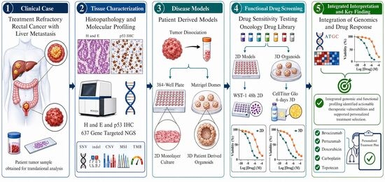
Treatment-refractory rectal cancer liver metastasis represents a major therapeutic challenge, particularly in the absence of actionable genomic biomarkers. Functional precision oncology approaches integrating genomic profiling with patient-derived organoid (PDO) drug testing may provide biologically informed therapeutic prioritization. A 50-year-old female patient with KRAS/TP53-mutant,
[...] Read more.
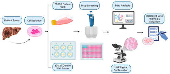
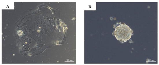
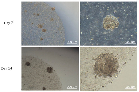
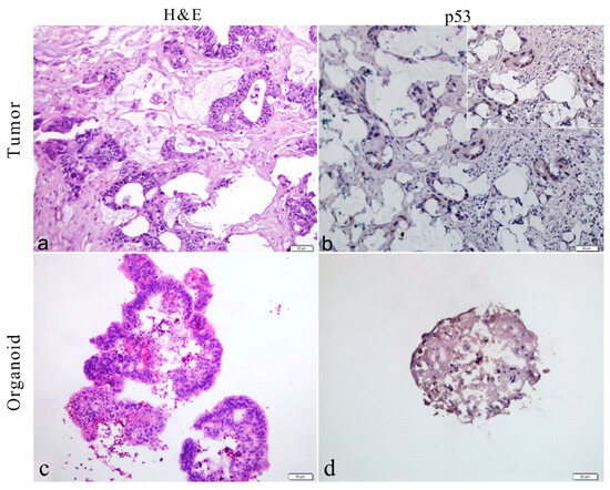
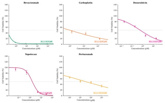
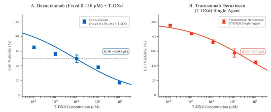
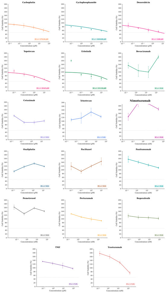
Treatment-refractory rectal cancer liver metastasis represents a major therapeutic challenge, particularly in the absence of actionable genomic biomarkers. Functional precision oncology approaches integrating genomic profiling with patient-derived organoid (PDO) drug testing may provide biologically informed therapeutic prioritization. A 50-year-old female patient with KRAS/TP53-mutant, microsatellite-stable (MSS) rectal adenocarcinoma refractory to FOLFIRINOX was enrolled. A liver metastasis from a treatment-refractory rectal cancer patient was processed to establish three-dimensional patient-derived organoids. Histopathological concordance was assessed using H&E and p53 immunohistochemistry. Comprehensive genomic profiling was performed using a 637-gene targeted next-generation sequencing panel, enabling detection of single-nucleotide variants, indels, copy number variations, microsatellite instability, and tumor mutational burden. Functional drug sensitivity profiling was conducted in parallel 2D and 3D platforms using a customized 17-agent panel, followed by exploratory combinatorial validation. The organoids demonstrated high phenotypic and genomic concordance with the parental tumor, preserving key driver alterations (KRAS^A146T, TP53^R175H, APC frameshifts, CCNE1 amplification), microsatellite stability, and low tumor mutational burden (TMB: 6.37 mut/Mb). Functional screening identified selective sensitivity to bevacizumab (IC50: 0.130 μM), doxorubicin (IC50: 0.570 μM), carboplatin (IC50: 0.950 μM), and topotecan (IC50: 1.600 μM) in the 3D organoid model, with consistent cross-platform validation. An exploratory combination assay further supported enhanced viability suppression under bevacizumab-based regimens. Critically, at the time of manuscript preparation, the patient demonstrated radiological disease stabilization under bevacizumab plus trastuzumab deruxtecan, consistent with the organoid-derived response profile. These findings highlight the capacity of integrated genomic and organoid-based profiling to uncover therapeutic vulnerabilities beyond standard biomarker assessment. This proof-of-concept case report study demonstrates the feasibility and translational relevance of an established organoid-based functional precision oncology platform for therapeutic prioritization in metastatic rectal cancer.
Full article
(This article belongs to the Special Issue Advances in Organoid Technology: Bridging the Gap between Research and Therapy)
►▼
Show Figures

Graphical abstract
Open AccessArticle
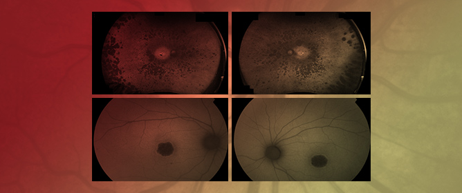
Dissecting PDE6-Associated Inherited Retinal Dystrophies Using Patient-Derived Retinal Models
by
Paula Gaudó, Anniken Burés-Jelstrup, Laura Siles, Rafael Navarro and Esther Pomares
Organoids 2026, 5(2), 13; https://doi.org/10.3390/organoids5020013 - 7 May 2026
Cited by 1
Abstract
Inherited retinal dystrophies (IRDs) comprise a diverse group of genetic disorders that frequently result in irreversible vision loss due to photoreceptor dysfunction or degeneration. Among them, retinitis pigmentosa (RP) and achromatopsia (ACHM) are, in some cases, associated with pathogenic variants in PDE6A and
[...] Read more.
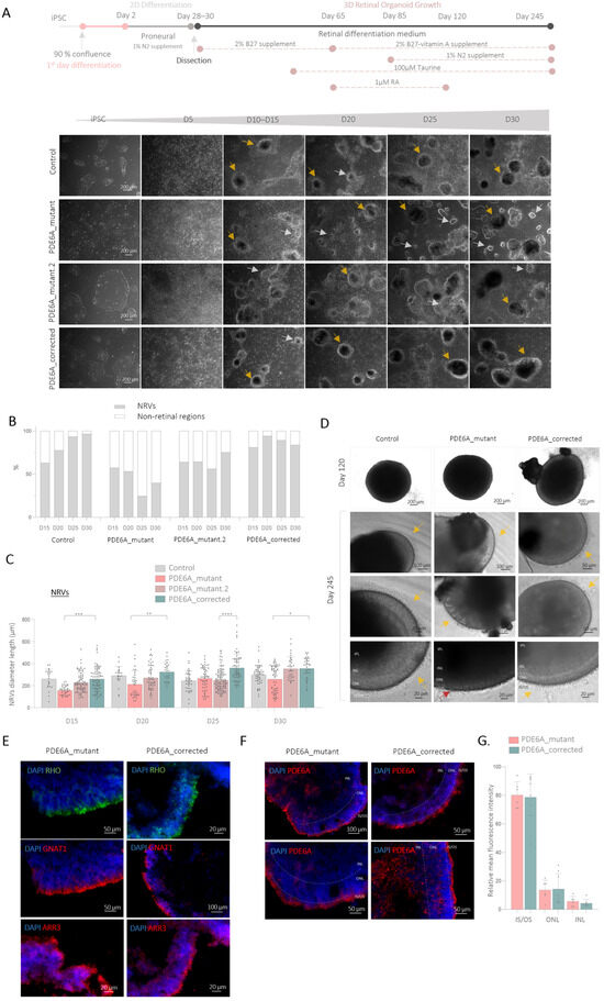
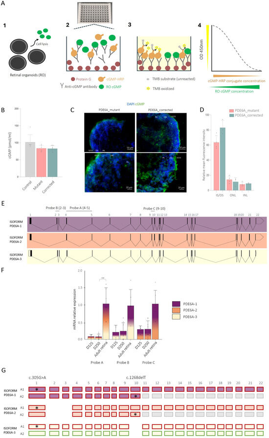
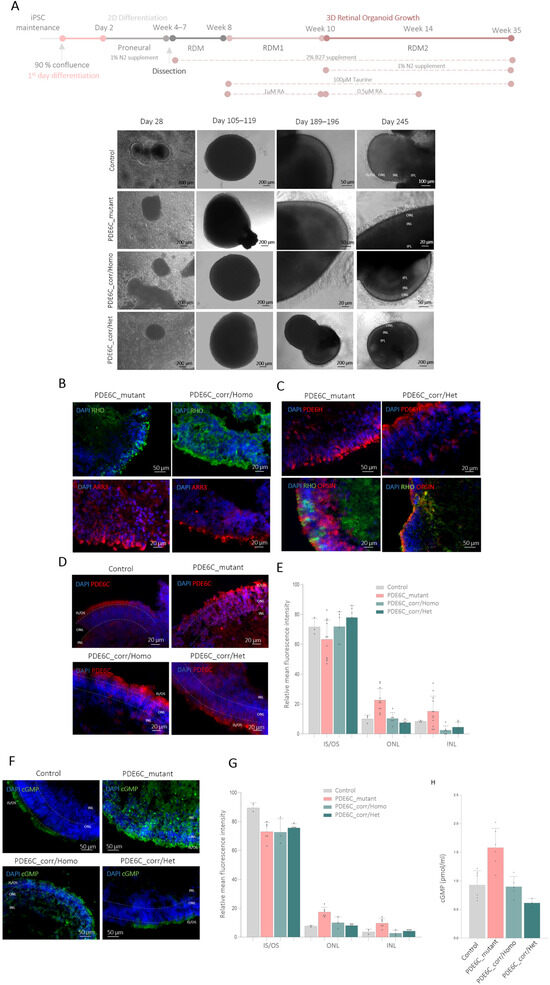
Inherited retinal dystrophies (IRDs) comprise a diverse group of genetic disorders that frequently result in irreversible vision loss due to photoreceptor dysfunction or degeneration. Among them, retinitis pigmentosa (RP) and achromatopsia (ACHM) are, in some cases, associated with pathogenic variants in PDE6A and PDE6C, respectively, which are key components of the phototransduction cascade. As most of IRDs still lack effective therapies, retinal organoids (ROs) provide a valuable in vitro model for the investigation of disease-associated mechanisms. Here, we generated induced pluripotent stem cell (iPSC)-derived ROs from an RP patient carrying compound heterozygous PDE6A mutations and from a patient with ACHM harboring a homozygous PDE6C mutation, along with their corresponding CRISPR/Cas9-corrected isogenic controls, which, to our knowledge, represent the first patient-derived RO models reported for the PDE6A and PDE6C genes. The mutant PDE6A line exhibited impaired neuroretinal vesicle formation and RO differentiation; however, a subset of RP-derived ROs matured appropriately and retained photoreceptor features. Moreover, the specific isoform expression pattern detected in retinal tissues reflected differences across developmental maturation stages that could influence disease severity. In contrast, the PDE6C_mutant ROs displayed normal structure and maturation, although cGMP hydrolysis within photoreceptors was likely compromised. In both models, CRISPR/Cas9-mediated correction restored the disease-associated phenotype resembling wild-type ROs. Collectively, these findings provide new insights into PDE6-associated pathogenesis, underscore the utility of patient-specific and gene-corrected ROs for elucidating IRD mechanisms, and support gene editing as a promising therapeutic strategy.
Full article
(This article belongs to the Special Issue Advances in Organoid Technology: Bridging the Gap between Research and Therapy)
►▼
Show Figures

Figure 1
Highly Accessed Articles
Latest Books
E-Mail Alert
News
Topics
Topic in
CIMB, IJMS, JCDD, Organoids, Biomedicines
Molecular and Cellular Mechanisms of Heart Disease
Topic Editors: Pasi Tavi, Ebru Arioglu-InanDeadline: 31 December 2026
Topic in
Cells, JCM, Organoids, JMP
Novel Discoveries in Oncology 2nd Edition
Topic Editors: Michela Campolo, Giovanna Casili, Alessia Filippone, Marika LanzaDeadline: 31 January 2027
Special Issues
Special Issue in
Organoids
Organs-on-Chips and Organoids: From Disease Modeling to Advanced Therapeutics
Guest Editor: Gautam MahajanDeadline: 30 November 2026
Special Issue in
Organoids
From Development to Degeneration: Advances in Stem Cell and Organoid Models of the Nervous System
Guest Editor: Elena CocciaDeadline: 30 November 2026
Special Issue in
Organoids
Modeling Lung Regeneration and Injury Responses Using Organoids and Engineered Niches
Guest Editor: Tristan FrumDeadline: 31 December 2026