
Review

From Cells to Microphysiological Systems: 3D Cell Cultures and Organ-on-Chip Systems for Studying cAMP and cGMP Signaling

1
Innate Lymphoid Cells Unit, Bambino Gesù Children’s Hospital, IRCCS, 00146 Rome, Italy
2
Department of Experimental Medicine, Sapienza University of Rome, 00161 Rome, Italy
3
Departmental Faculty of Medicine, Saint Camillus International University of Health Sciences, 00133 Rome, Italy
*
Authors to whom correspondence should be addressed.
Organoids 2026, 5(3), 24; https://doi.org/10.3390/organoids5030024
Submission received: 19 June 2026 / Revised: 29 July 2026 / Accepted: 30 July 2026 / Published: 4 August 2026

Abstract

Cyclic adenosine monophosphate and cyclic guanosine monophosphate are key regulators of cellular physiology and tissue homeostasis. Conventional experimental models have provided fundamental insights into cyclic nucleotide pathways; however, they often fail to fully recapitulate essential features of in vivo systems. Two-dimensional cell cultures lack spatial organization, while animal models incompletely capture cell–cell interactions and the dynamic microenvironment shaping signaling processes. In recent years, advanced three-dimensional and microengineered systems have emerged as tools to bridge this gap. In this review, we discuss how three-dimensional and organ-on-chip systems are transforming the study of cyclic nucleotide signaling by enabling reconstruction of tissue architecture and signaling niches. Spheroids and organoids provide robust models to investigate compartmentalized signaling and intercellular communication. Complementarily, microfluidic organ-on-chip devices introduce controlled mechanical cues, perfusion, and tissue interfaces, enabling real-time monitoring of signaling dynamics. We highlight recent advances in microphysiological systems for investigating the spatial and temporal dynamics of cyclic adenosine monophosphate and cyclic guanosine monophosphate signaling, including biosensors, live-cell imaging, and genome editing. We further discuss applications in physiological and pathological contexts, including metabolic, cardiovascular and cancer diseases, and outline current challenges and future perspectives for integrating three-dimensional and organ-on-chip technologies.

1. Introduction

The development of three-dimensional (3D) cell models has significantly impacted biomedical and drug-discovery research, offering a more physiologically relevant alternative to traditional two-dimensional (2D) cell culture systems. Too often, compounds that demonstrate high efficacy in 2D in vitro systems fail to show comparable effectiveness in clinical trials [1]. This discrepancy reflects the inability of 2D cultures to fully reproduce the structural and functional complexity of human tissues or to accurately predict therapeutic responses. At the preclinical stage, in vivo studies involving animal models still remain the gold standard and represent a crucial step in the drug development process. These models faithfully recapitulate the complexity of whole organisms and allow the assessment of pharmacological and toxicological properties of lead compounds. However, their predictive value is limited by phylogenetic differences between experimental models and humans, frequently resulting in poor translation to clinical outcomes. Furthermore, high costs, extended study durations, and ethical concerns continue to restrict the widespread use of animal models in biological and pharmaceutical research [2].
In light of the above-mentioned limitations, researchers are still developing in vitro cellular models that more accurately recapitulate human organ physiology than conventional preclinical approaches. These models span a continuum from spheroids to organoids and, more recently, to organ-on-chip (OoC) systems, collectively offering different advantages and applications for replicating the complexity of human beings.
Spheroids are 3D cellular aggregates composed of various cell types, including stem cells, primary cells, and cancer cells, and represent a well-defined model that more closely mimics physiological conditions for investigating cellular behavior and pharmacological responses [3,4,5].
Moreover, organoids are advanced 3D cell-culture systems in which stem or progenitor cells self-organize into small organ-like structures. These models have revolutionized developmental biology research and regenerative medicine. Organoids are able to recapitulate key aspects of the composition, architecture, and function of the native tissues, thereby providing a more realistic platform for studying organ physiology and pathology [6].
More recently, OoC technology has further enhanced the capabilities of 3D cell culture systems and has emerged as a powerful tool in organogenesis and tissue engineering research. These microfluidic devices incorporate living cells within a controlled microenvironment that simulates the microarchitecture and dynamic physiological processes of the human organ. In addition, their integration with sensors and imaging systems allows real-time monitoring of cell responses, offering more accurate details about cell behavior, drug effects and disease progression [7]. Three-dimensional models better recapitulate cell–extracellular matrix (ECM) interactions and nutrient gradients. These systems are valuable tools for studying second messenger-governed networks, as they enable the investigation of spatiotemporal signaling dynamics and mechano-transductive thresholds that cannot be faithfully reproduced in conventional 2D monolayer cultures.
Among the second messengers, cyclic adenosine monophosphate (cAMP) and cyclic guanosine monophosphate (cGMP) are two of the most important intracellular signal molecules, coupling extracellular stimuli, such as a hormones, neurotransmitters, and cytokines, to specific cellular responses [8]. The functional outcome of these signaling pathways is highly context-dependent, varying with cell type, extracellular environment, nature of the activating stimulus, subcellular localization, and the specific cyclic nucleotide involved. Once generated, cyclic nucleotides regulate a range of downstream effectors, including protein kinases, ion channels, transcription factors, and scaffolding proteins, thereby modulating different cellular processes [9].
Understanding how these signaling networks are dynamically coordinated within complex tissue microenvironments remains a major challenge in biomedical research. In this context, 3D cell culture and OoC systems provide a unique platform to dissect the molecular communication between cells and, ultimately, the regulation of second messenger signaling in physiological and pathological conditions.

2. cAMP and cGMP Signaling

cAMP and cGMP are ubiquitous intracellular second messengers that transduce extracellular stimuli into precise biochemical responses, orchestrating a remarkably broad spectrum of cellular processes ranging from proliferation to differentiation, and apoptosis [10]. Both cAMP and cGMP are involved in numerous cellular functions, including metabolism, hormonal regulation, and cancer development [11,12,13,14,15]. Their intracellular concentrations are tightly regulated through the balance between biosynthesis, mediated by adenylyl cyclase (AC) and guanylyl cyclase (GC) enzymes, and degradation, carried out by a large and functional superfamily of phosphodiesterases (PDEs) [16]. cAMP is synthesized from adenosine triphosphate (ATP) by the AC, whose activity is principally stimulated by the stimulatory G-protein subunit Gαs downstream of G-protein-coupled receptor (GPCR) activation and is suppressed by the inhibitory subunit Gαi [17]. In parallel, cGMP is synthesized from guanosine triphosphate (GTP) by GC, which is encoded by two distinct gene families: soluble GCs (sGC), cytosolic heterodimers activated by nitric oxide (NO), and particulate GCs (pGC), transmembrane receptors activated by natriuretic peptides such as atrial natriuretic peptide (ANP) and B-type natriuretic peptide (BNP) [18,19,20].
The intracellular amplitude, duration, and spatial extent of cAMP and cGMP signals are governed primarily by PDEs, a family of enzymes that hydrolyze the 3′-phosphodiester bond of cyclic nucleotides, converting them to the inactive 5′-monophosphates AMP and GMP, respectively. The human PDE superfamily encompasses 11 gene families (PDE1–PDE11), each comprising multiple splice variants and isoforms, yielding over 100 distinct molecular species with different substrate specificities, kinetic properties, regulatory mechanisms, and tissue distributions [20]. PDE families can be broadly classified according to substrate selectivity: PDE4, PDE7, and PDE8 are cAMP-specific; PDE5, PDE6, and PDE9 are cGMP-specific; whereas PDE1, PDE2, PDE3, PDE10, and PDE11 hydrolyze both cyclic nucleotides, albeit with different affinities [21]. This isoform diversity is not merely biochemical redundancy; rather, it constitutes the mechanistic basis for the compartmentalization of cyclic nucleotide signalling. The strategic intracellular localization of specific PDE isoforms creates discrete signalling microdomains in which local cAMP or cGMP concentrations can differ dramatically from bulk cytosolic levels, thereby allowing the cell to generate spatially restricted and functionally specific responses to a given stimulus.
The central principle governing cAMP and cGMP biology is that these two messengers do not function as independent pathways but instead engage in extensive bidirectional cross-talk, primarily mediated through PDEs with dual substrate specificity. PDE2A, for instance, is allosterically activated by cGMP and preferentially degrades cAMP when cGMP levels are elevated, thereby enabling cGMP to attenuate cAMP-dependent responses [22]. Conversely, PDE3 is competitively inhibited by cGMP, such that elevated cGMP indirectly increases cAMP by preventing its hydrolysis [23]. These interactions generate complex, non-linear signalling dynamics that are critically dependent on the local concentrations of both messengers, the isoform complement of PDEs expressed in a given cell type, and the subcellular localization of the relevant enzymes [24]. The concept of signalling compartmentalization, in which functionally distinct pools of cAMP or cGMP coexist within the same cell, has transformed the mechanistic understanding of cyclic nucleotide biology.
The existence of such a compartmentalized signalling architecture has profound implications for the design and interpretation of experimental models: only systems that faithfully replicate the 3D organization of cells, the paracrine and autocrine signalling environment, and the tissue-specific expression of receptors, cyclases, and PDEs can be expected to recapitulate the full complexity of cyclic nucleotide signalling as it operates in vivo.

3. Microphysiological Systems

The transition from conventional 2D cell cultures to microphysiological systems (MPSs) has substantially reshaped the investigation of cyclic nucleotide signaling, offering new insights into the long-standing “spatial paradox” associated with second messengers [25]. The “spatial paradox” of second messengers describes the apparent contradiction between the high diffusibility of cAMP and cGMP and their ability to generate highly localized and specific cellular responses.
Rather than diffusing freely throughout the cytosol, cAMP and cGMP are spatially confined to discrete intracellular microdomains in vivo. Within these signaling compartments, the coordinated balance between local cyclic nucleotide synthesis and degradation generates highly localized concentration gradients that ensure the specificity, fidelity, and temporal control of downstream signaling pathways [26,27]. By contrast, the loss of spatial organization in 2D systems leads to an artificial homogenization of these signals, obscuring the compartmentalized nature of cyclic nucleotide signaling and potentially misrepresenting downstream cellular responses [28]. The paradox is resolved by the existence of signaling microdomains shaped by cyclases, PDEs, scaffolding proteins, and downstream effectors, which spatially compartmentalize cyclic nucleotide signaling within cells [25]. The formation and maintenance of these signaling microdomains rely on the precise spatial arrangement of such molecular components, including A-kinase anchoring proteins (AKAPs) [28]. This intricate organization is inherently dependent on cell polarity, cytoskeletal architecture, and cell–matrix interactions, features that are largely absent or significantly altered in 2D cultures but are more faithfully reproduced in MPSs [29].
By restoring aspects of tissue geometry and microenvironmental cues, MPSs enable the preservation of spatiotemporal gradients and localized signaling events, thereby providing a more physiologically relevant framework to study cyclic nucleotide dynamics [30]. Beyond their impact on mechanistic understanding, MPSs also address critical translational and ethical challenges in biomedical research. Their ability to generate high-fidelity, human-relevant data makes them a valuable alternative to traditional animal models, which often fail to capture species-specific differences in signaling pathways and their spatial regulation [31]. Importantly, the adoption of MPSs align closely with the ethical principle of 3Rs (Replacement, Reduction, and Refinement), supporting efforts to minimize animal use while improving the predictive power of preclinical studies [32].
Three-dimensional microfluidic platforms represent a significant advancement in the study of cellular signaling. By bridging the gap between simplified in vitro systems and the complexity of human physiology, MPSs provide a powerful and ethically sustainable approach to investigate the molecular mechanisms underlying cyclic nucleotide signaling in both health and disease.

3.1. Spheroids

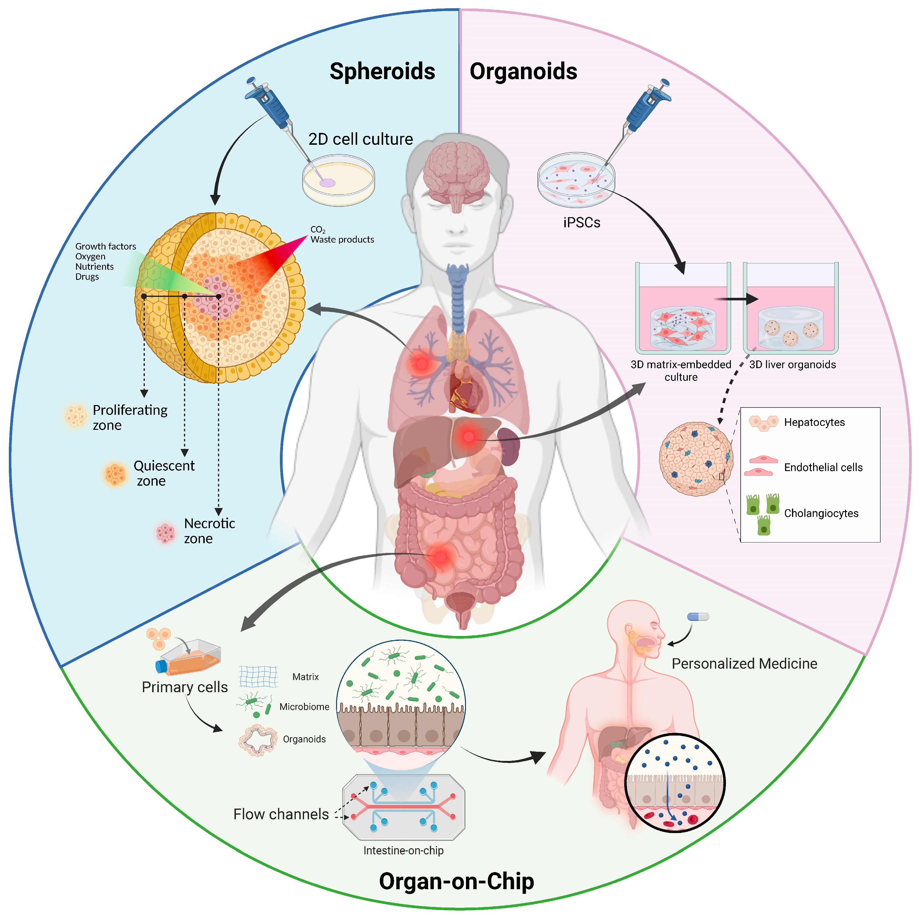
Spheroids represent one of the earliest and most widely adopted 3D culture models, providing an intermediate level of complexity between conventional 2D monolayers and more advanced MPSs [33]. By allowing cells to self-assemble into multicellular aggregates, they partially restore essential features of native tissues, including extensive cell–cell and cell–matrix interactions, ECM deposition, and the formation of physiological gradients of oxygen, nutrients, and metabolites (Figure 1). These characteristics create a microenvironment that more closely resembles in vivo tissue organization and profoundly influences intracellular signaling pathways [34].
Spheroid formation is mediated by membrane proteins, particularly integrins, together with ECM components. This occurs through three critical steps: (i) initially single cells form aggregates by interactions between cell-surface integrins and long-chain ECM fibers enriched in multiple RGD (Arginyl-Glycyl-Aspartic acid) motifs, leading to upregulation of cadherin expression; (ii) cadherins are accumulated at the membrane surface; and (iii) cells undergo compaction into solid aggregates to form spheroids due to the homophilic cadherin–cadherin binding [33]. Within this context, spheroids have emerged as valuable tools for investigating the spatial regulation of cyclic nucleotide signaling. Unlike 2D cultures, where signaling pathways are often artificially homogenized, spheroids introduce spatial heterogeneity that enables a more physiologically relevant organization of cAMP and cGMP signaling. Gradients in oxygenation and nutrient diffusion can directly affect the activity of AC, GC, and PDEs, thereby shaping localized cyclic nucleotide concentrations and downstream signaling responses [35]. Importantly, the 3D architecture of spheroids promotes the partial re-establishment of signaling microdomains [36].
Although spheroids do not fully reproduce the complexity of intact tissues, their dense multicellular organization supports a degree of compartmentalized signaling that is absent in 2D cultures, making them particularly useful for studying spatial aspects of second messenger regulation [27,28,29].
Despite multiple advantages, spheroids still present several limitations. Their architecture is frequently heterogeneous, leading to variability in size, cellular composition, and diffusion gradients [37]. In addition, the absence of vascularization and mechanical cues limits their ability to fully reproduce the dynamic spatiotemporal regulation observed in vivo [38]. Nevertheless, spheroids remain a highly accessible and biologically relevant platform that bridges the gap between oversimplified 2D systems and more sophisticated organoid or OoC technologies.

3.2. Organoids

Organoids represent a major advancement over simple 3D culture systems, offering a higher degree of biological fidelity through their ability to self-organize into tissue-like structures from stem or progenitor cells. This intrinsic capacity for spatial organization and lineage specification makes organoids particularly valuable for investigating complex signaling pathways, including GPCR-mediated cascades and cAMP-dependent networks [39].
Organoids are miniaturized organ-like structures generated from adult stem cells (ASCs) or pluripotent stem cells (PSCs), including embryonic stem cells (ESCs) and induced pluripotent stem cells (iPSCs) in a specific 3D culture system, which mimic key structural and functional features of native tissues (Figure 1). Their formation relies on tightly coordinated developmental and differentiation processes and requires ECM components that provide a basal lamina-like scaffold to support tissue organization and morphogenesis [40].
A central advantage of organoids lies in their ability to recapitulate developmental processes, allowing the study of how cAMP and cGMP signaling contribute to tissue maturation, differentiation, and gene transcription in a spatially and temporally controlled manner [41]. In organoid systems, this signaling cascade can be studied within a structurally organized context that preserves compartmentalization and microenvironmental cues, which are essential for generating specific biological outcomes. Despite their considerable advantages, organoids still face challenges related to variability, incomplete vascularization, and limited integration of systemic physiological inputs. Nevertheless, ongoing advances in bioengineering and microfluidic technologies are progressively enhancing their reproducibility and functional complexity [27,42].
Overall, organoids constitute a powerful and versatile platform for studying second messenger signaling in both physiological and pathological contexts. By preserving tissue architecture, cellular diversity, and signaling compartmentalization, they enable a more accurate and dynamic understanding of how cyclic nucleotides regulate human development, homeostasis, and disease.

3.3. Organ-on-Chip

OoC technologies represent a further evolution of 3D culture systems by integrating microfluidic engineering with tissue-specific cellular architectures [43]. While spheroids and organoids partially restore the spatial organization of native tissues, OoC platforms additionally reproduce the dynamic physical and chemical microenvironment that governs cellular behavior in vivo [44]. Through the controlled application of fluid flow, shear stress, mechanical deformation, and spatiotemporal gradients of soluble factors, these systems enable the investigation of signaling processes under physiologically relevant dynamic conditions [45].
Structurally, OoC devices consist of microfabricated platforms containing networks of micrometer-scale channels that mimic vascular and interstitial compartments (Figure 1).
Cells are cultured within these channels or in interconnected chambers, frequently separated by porous membranes or embedded within ECM-like hydrogels. Continuous perfusion generated by external pumps or pressure-driven systems establishes highly controlled laminar flow conditions and reproducible concentration gradients. This configuration enables the precise modulation of nutrient delivery, metabolite exchange, and biomechanical stimulation, while also allowing temporal control over signaling inputs and environmental perturbations [43]. A major advantage of OoC systems lies in their ability to recreate tissue–tissue interfaces and multicellular communication in a dynamically perfused environment. In vascularized platforms, endothelial channels exposed to physiological shear stress can be coupled to adjacent parenchymal compartments containing tissue-specific cells within 3D matrices [46]. This organization supports bidirectional signaling, barrier formation, and immune cell trafficking while preserving transport processes that are difficult to reproduce in static culture systems. More advanced devices further incorporate cyclic mechanical strain or compression to emulate organ-specific motions such as breathing, vascular pulsatility, or peristalsis [47]. Within this dynamic context, mechanical forces play a central role in the regulation of cyclic nucleotide signaling. Despite current limitations related to technical complexity, standardization, and scalability, OoC technologies are rapidly advancing and are increasingly recognized as essential components of the broader MPSs framework.

4. 3D Cell Culture and OoC to Study cAMP Signaling

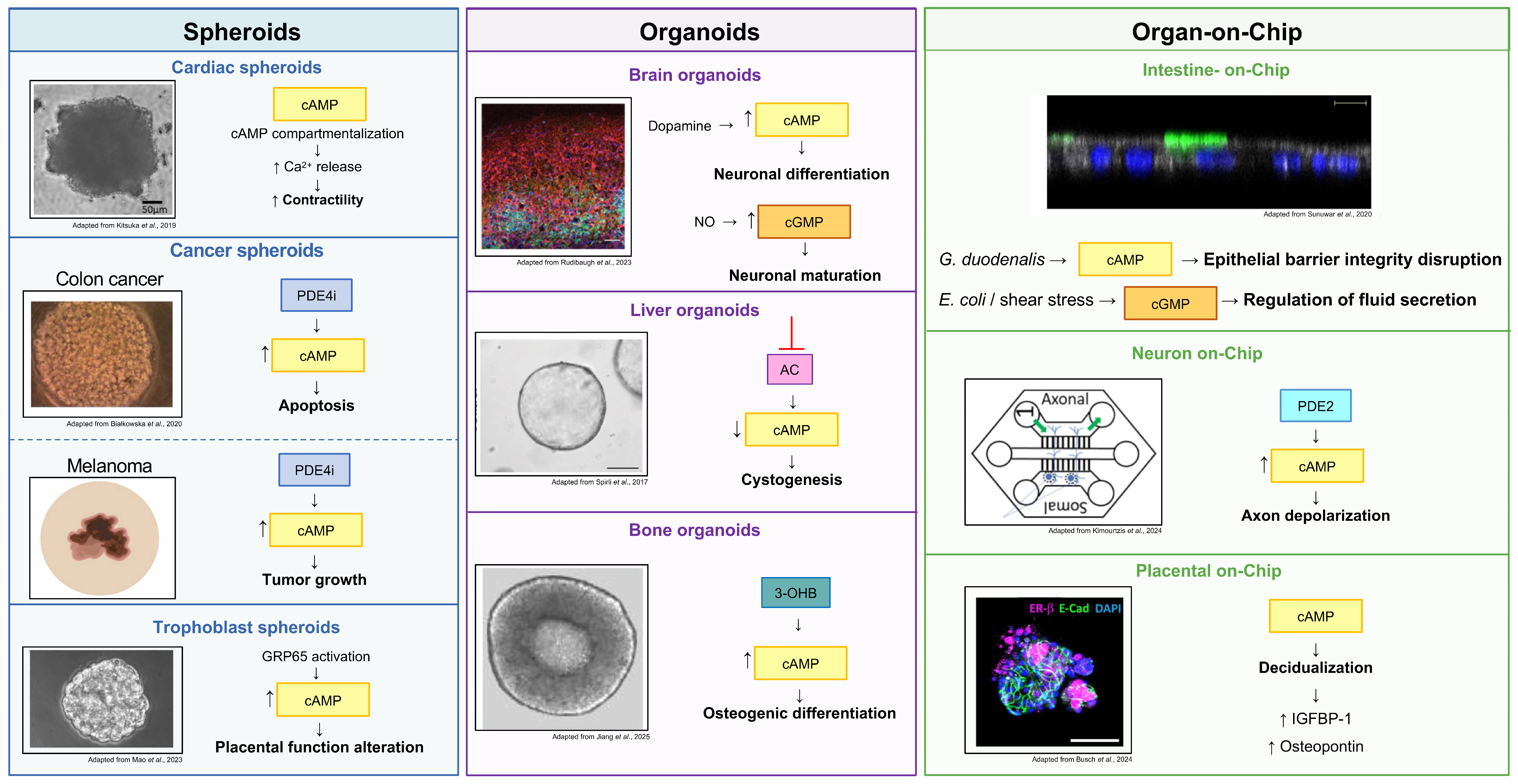
Three-dimensional culture systems and OoC technologies have significantly advanced the study of cAMP and cGMP signaling, overcoming several limitations associated with conventional 2D cultures (Figure 2).
In cardiac research, spheroids and organoids recapitulate key structural and functional properties of the myocardium, including pulsatility, excitation–contraction coupling, and contractile force generation. Protein kinase A (PKA) signaling is a major regulator of cardiac rhythm and calcium handling, and its dysregulation has been associated with arrhythmogenesis [48]. Human iPSC-derived cardiac organoids revealed that PKA activity is spatially heterogeneous rather than uniformly distributed throughout the tissue, supporting the existence of localized cAMP signaling microdomains. Pharmacological activation of the AC–cAMP–PKA axis further increased this spatial heterogeneity, suggesting the formation of discrete signaling domains within cardiac tissue [44].
Similarly, treatment of multicellular cardiac organoids with 2-chloro-carbocyclic oxetanocin A (COA-Cl), a synthetic adenosine analogue, partially inhibited PDE activity, leading to elevated intracellular cAMP levels, enhanced PKA signaling, increased calcium (Ca2+) release from the endoplasmic reticulum, and augmented contractile force [48].
In neurobiology, cerebral organoids have emerged as valuable tools for studying neuronal maturation, electrophysiological activity, and neurodevelopmental signaling pathways. According to the consensus nomenclature proposed by Pașca et al., neural organoids are designated according to the anatomical region they model, giving rise to region-specific systems such as cerebral, ventral forebrain, and medial ganglionic eminence organoids [49].
Comparative transcriptomic analyses demonstrated that cerebral organoids resemble fetal brain tissue, although pathways associated with synaptogenesis, cAMP response element-binding protein (CREB) signaling, glutamate receptor signaling, and Ca2+ signaling remain relatively immature compared with fetal brain samples [50].
Dopamine signaling in ventral forebrain organoids depends on cAMP-mediated pathways regulating neuronal differentiation, migration, memory, and stress responses. In human medial ganglionic eminence organoids (hMGEOs), dopamine stimulation increased intracellular cAMP levels, whereas chronic stimulation attenuated this response, suggesting receptor desensitization mechanisms associated with altered calcium activity and spontaneous Ca2+ transients [51]. Moreover, human iPSC-derived brain organoids demonstrated that pharmacological modulation of cAMP signaling regulates the GLI3 transcription factor and Sonic Hedgehog-dependent neural patterning through primary cilia-mediated mechanisms [52]. Altered cAMP signaling was also associated with impaired neuronal maturation and disrupted electrophysiological activity in human forebrain organoids [53]. In parallel, microfluidic neuronal culture systems highlighted the role of prostaglandin E2-dependent cAMP signaling in sustained depolarization of sensory axons mediated by calcium-activated chloride channels [54]. Recent advances in cerebral organoid technology have further demonstrated the presence of substantial cellular heterogeneity, including neuronal, glial, and vascular-associated populations, together with progressive maturation of electrophysiological properties over time. Importantly, cerebral organoids exhibit functional neuronal activity, ion channel responsiveness, and pharmacological sensitivity to anesthetic compounds such as propofol, indicating the establishment of active neuronal networks [51].
The role of cyclic nucleotide signaling has also been extensively investigated in gastrointestinal and epithelial organoids. In human iPSC-derived gut organoids, cAMP signaling regulated epithelial patterning and lineage allocation. cAMP deprivation promoted expansion of enteroendocrine populations and absorptive enterocyte-like cells, whereas cAMP supplementation favored epithelial progenitor maintenance through differential activation of NOTCH, MAPK, and WNT signaling pathways [55]. OoC-derived intestinal epithelium models incorporating fluid flow and mechanical stimulation further demonstrated that Giardia duodenalis infection disrupts epithelial barrier integrity through impairment of cAMP-dependent tight junction signaling pathways [56,57].
Aberrant cyclic nucleotide signaling has also been implicated in tumor progression and therapeutic resistance. In patient-derived gastric cancer organoids, Fibulin-5 released by cancer-associated fibroblasts promoted epithelial–mesenchymal transition (EMT) and organoid growth through activation of the CREB pathway [58]. In HCT116 colon cancer spheroids, resveratrol induced apoptosis and luminal cavity formation, likely through inhibition of PDE4 activity, similarly to the PDE4 inhibitor rolipram [59]. Likewise, combined treatment with the antineoplastic drug vemurafenib and the PDE4 inhibitor roflumilast reduced melanoma spheroid growth generated from resistant cell lines [60].
Three-dimensional reproductive models have provided additional insights into implantation and endocrine signaling. Trophoblast spheroids exposed to acidic microenvironmental conditions showed activation of the GPR65 receptor, leading to increased cAMP production, ERK phosphorylation, altered MYLK and fibronectin expression, and molecular alterations associated with early pregnancy loss [61]. Endometrial organoids further demonstrated that cAMP cooperates with ovarian steroid hormones to induce complex decidualization-associated transcriptomic programs that cannot be fully reproduced in 2D systems [62].
Additional applications include hepatic, renal, bone, and skin organoids. cAMP signaling was associated with maturation of human PSC-derived hepatoblasts into hepatocyte-like cells exhibiting metabolic and transcriptional profiles comparable to primary hepatocytes, a process dependent on activin/nodal signaling during early differentiation [63]. In biliary organoids, inhibition of AC reduced cyst growth, linking cAMP signaling to cystogenesis [64]. In developmental polycystic kidney disease (PKD), kidney organoids identified a pathogenic PTH1R–cAMP–PKA–CREB signaling loop involved in cyst formation [65].
Bone organoids further revealed that the ketone body 3-hydroxybutyrate (3-OHB) promotes osteogenic differentiation through activation of the HCAR2–cAMP/PKA/CREB pathway and RUNX2 upregulation [66].
In 3D dermal fibroblast spheroids, α-MSH–MC1R–cAMP signaling was investigated in the context of UVA-induced oxidative stress and photoaging responses, although canonical cAMP signaling appeared not to mediate the photoprotective effects observed in dermal fibroblasts [67].
Similarly, microfluidic approaches have enabled high-throughput single-cell analyses of cAMP-dependent chemotactic responses and collective cell migration under controlled gradient conditions [68].
The most significant studies discussed in this section are summarized in Table 1.

5. 3D Cell Culture and OoC to Study cGMP Signaling

Three-dimensional models naturally establish biochemical and oxygen gradients that strictly regulate cGMP generation and degradation. The relevance of spheroids for cyclic nucleotide research has been especially evident in neurological studies [71,72].
As self-organized neural aggregates, whole-brain spheroids have been shown to recapitulate important developmental aspects of NO-dependent signaling pathways that are difficult to reproduce in 2D systems (Figure 2). Teunissen and colleagues showed that differentiated brain spheroids preserved functional NOS and both soluble and particulate GC activities, supporting physiologically relevant cGMP production in both neurons and glial cells [69]. These findings indicate that the spatial organization and multicellular complexity of brain spheroids promote the maturation of NO-cGMP signaling during neurodevelopment. The translational potential of these models has also emerged from patient-derived iPSC-brain organoids. Using organoids generated from patients with Leigh syndrome, Zink and colleagues identified compounds capable of modulating signaling pathways associated with mitochondrial dysfunction [73].
Beyond neurobiology, advanced 3D intestinal models have demonstrated that cGMP signaling is highly responsive to the physical microenvironment. Gut-on-chip devices revealed that luminal and/or basolateral fluid flow regulate intracellular cGMP levels and influence the epithelial response to the Escherichia coli heat-stable enterotoxin [74]. Similarly, multicellular enteroids, comprising enterocytes, goblet cells, Paneth cells and enteroendocrine cells, have shown that cGMP signaling orchestrates intestinal fluid homeostasis by regulating luminal volume expansion [75]. These observations emphasize that mechanical forces represent an important regulatory component of cyclic nucleotide signaling, a feature that is essentially absent in static culture systems.
In the context of oncological applications, 3D models have also been used to study complex biological properties such as EMT, stem cell properties and drug resistance. Tumor spheroids more faithfully reproduce gradients of oxygen, nutrients, and drug penetration, thereby capturing cellular heterogeneity and plasticity. To this end, Li and colleagues showed that non-small cell lung cancer spheroids acquire enhanced EMT and stemness characteristics compared with 2D cultures, while treatment with the PDE5 inhibitor, tadalafil, altered both cGMP- and cAMP-mediated pathways, leading to a reprogramming of intracellular signaling and consequent alterations in cell survival [70]. This approach has therefore demonstrated that 3D models allow for a more physiologically relevant assessment of the interaction between cellular plasticity and response to pharmacological treatments. Likewise, Muir and colleagues demonstrated that NO donors and cGMP analogues increased the sensitivity of breast cancer spheroids to doxorubicin, supporting the concept that activation of the NO–cGMP axis may enhance the efficacy of conventional chemotherapy [76]. Collectively, these studies highlight the value of 3D tumor models not only for investigating cancer biology but also for evaluating therapeutic strategies targeting cyclic nucleotide signaling.
The versatility of advanced 3D systems further extends to tissue engineering and regenerative medicine Recently, Busch and colleagues developed an OoC model of the uterine wall, that reproduce hormone-dependent stromal decidualization, providing a physiologically relevant platform to investigate cyclic nucleotide-regulated reproductive processes [77].
Similarly, human iPSC-derived cartilaginous organoids have revealed that α2-adrenergic receptor (α-AR) signaling promotes cartilage hypertrophy through a cGMP-dependent secretory leukocyte protease inhibitor pathway, whereas pharmacological inhibition of this axis favors chondrogenic differentiation [78].
Collectively, these studies demonstrate that the principal strength of 3D culture systems lies not simply in their ability to reproduce tissue morphology, but in their capacity to reveal context-dependent regulation of cyclic nucleotide signaling that emerges from the interplay between cellular architecture, mechanical stimuli, and tissue-specific microenvironments. As these technologies continue to evolve, they are expected to play an increasingly important role in elucidating cAMP- and cGMP-mediated signaling in physiology and disease, while accelerating translational applications in drug discovery and precision medicine.
The most significant studies discussed in this section are summarized in Table 1.

6. Current Challenges and Future Perspectives

Although 3D culture systems and OoC technologies have substantially advanced the study of cyclic nucleotide signaling by better reproducing tissue architecture and microenvironmental complexity than conventional 2D cultures, several challenges still limit their translational potential.
Throughout this review, we highlighted how spheroids, organoids, and MPSs enable the investigation of compartmentalized cAMP and cGMP signaling in physiologically relevant contexts. However, further technological advances are needed to fully recapitulate human tissue complexity. A major limitation remains the lack of standardization and reproducibility. Stem cell-derived organoids often display variability in morphology, cellular composition, maturation, and function, while spheroids and OoC platforms are strongly influenced by culture conditions, ECM composition, and device design. Standardized manufacturing protocols and validation criteria will therefore be essential to improve reproducibility across laboratories.
Current models also incompletely reproduce physiological complexity, as they generally lack vascularization, innervation, immune components, and systemic endocrine and metabolic interactions. Since cyclic nucleotide signaling is tightly regulated by multicellular communication, integrating vascular, immune, stromal, and neuronal compartments will be crucial to better model physiological and pathological processes.
Another key challenge is the investigation of highly compartmentalized cAMP and cGMP signaling microdomains. Although 3D models preserve tissue organization, direct visualization of signaling nanodomains remains technically demanding. Combining organoids and OoC systems with genetically encoded biosensors, high-resolution live imaging, optogenetics, and CRISPR/Cas technologies will enable real-time monitoring of cyclic nucleotide dynamics with subcellular resolution.
Future developments will likely focus on multi-OoC platforms to investigate inter-organ communication, pharmacokinetics, and tissue-specific responses to PDEs inhibitors and other modulators of cAMP/cGMP signaling. In parallel, integration of imaging, multi-omics, and biosensor datasets with artificial intelligence will facilitate automated phenotyping, identification of therapeutic targets, and patient-specific prediction of drug responses.
Overall, the convergence of stem cell biology, bioengineering, advanced imaging, genome editing, biosensors, and computational approaches is rapidly transforming MPSs into powerful experimental platforms. As these limitations are progressively overcome, 3D culture systems and OoC technologies are expected to become indispensable tools for elucidating the spatial and temporal regulation of cAMP and cGMP signaling and for accelerating precision medicine and drug discovery.

7. Conclusions

Overall, 3D culture systems and OoC technologies have considerably expanded the study of cAMP- and cGMP-dependent signaling by enabling the investigation of cyclic nucleotide pathways within physiologically relevant and structurally complex microenvironments. By recapitulating tissue architecture, multicellular interactions, and ECM dynamics, these models provide unique opportunities to study the spatial compartmentalization and functional regulation of cyclic nucleotide signaling under both physiological and pathological conditions. Importantly, spheroids and organoids have revealed previously inaccessible aspects of cyclic nucleotide biology, including the regulation of contractility, epithelial organization, paracrine communication, and microenvironment-dependent responses such as hypoxia and pH sensing.
Furthermore, these platforms represent promising tools for drug screening and precision medicine approaches targeting cyclic nucleotide pathways, particularly for evaluating PDE modulation in systems that more faithfully reproduce human tissue physiology.

Author Contributions

Conceptualization, M.R.A. and F.C.; writing—original draft preparation, M.R.A., N.B., M.P. and F.C.; writing—review and editing, M.R.A. and F.C.; visualization, N.B. and M.P.; All authors have read and agreed to the published version of the manuscript.

Funding

This research received no external funding.

Institutional Review Board Statement

Not applicable.

Informed Consent Statement

Not applicable.

Data Availability Statement

No new data were created or analyzed in this study. Data sharing is not applicable to this article.

Acknowledgments

During the preparation of this manuscript, the authors used OpenAI Chat-GPT (GPT-5.5) exclusively to improve the English language. The authors have reviewed and edited the output and take full responsibility for the content of this publication. MRA would like to acknowledge the unwavering patience and encouragement of her beloved father Pietro Assenza, whose memory continues to walk beside her.

Conflicts of Interest

The authors declare no conflicts of interest.

Abbreviations

The following abbreviations are used in this manuscript:
3Dthree-dimensional
2Dtwo-dimensional
OoCOrgan-on-chip
ECMextracellular matrix
cAMPcyclic adenosine monophosphate
cGMPcyclic guanosine monophosphate
ACadenylyl cyclase
GCguanylyl cyclase
PDEsphosphodiesterases
ATPadenosine triphosphate
GTPguanosine triphosphate
GPCRG-protein coupled receptor
sGCsoluble guanylyl cyclase
pGCparticulate guanylyl cyclase
NOnitric oxide
ANPatrial natriuretic peptide
BNPB-type natriuretic peptide
MPSsmicrophysiological systems
AKAPsA-kinase anchoring proteins
RGDArginyl-Glycyl-Aspartic acid
iPSCsinduced pluripotent stem cells
CO2carbon dioxide
ASCsadult stem cells
PSCspluripotent stem cells
ESCsembryonic stem cells
PKAprotein kinase A
PDE4iphosphodiesterase 4 inhibitor
GPR65G-protein coupled receptor 65
3-OHB3-hydroxybutyrate;
PGE2prostaglandin E2
COA-Cl2-chloro-carbocyclic oxetanocin A
Ca2+calcium
hMGEOshuman medial ganglionic eminence organoids
EMTepithelial–mesenchymal transition
PKDpolycystic kidney disease
CREBcAMP responsive element binding
NOSnitric oxide synthase
DETAdiethylenetriamine
IGFBP-1insulin-like growth factor binding protein 1
α-ARα-adrenergic receptor

References

  1. Dave, R.; Pandey, K.; Patel, R.; Gour, N.; Bhatia, D. Leveraging 3D cell culture and AI technologies for next-generation drug discovery. Cell Biomater. 2025, 1, 100050. [Google Scholar] [CrossRef] [Scilit]
  2. Żuchowska, A.; Baranowska, P.; Flont, M.; Brzózka, Z.; Jastrzębska, E. Review: 3D cell models for organ-on-a-chip applications. Anal. Chim. Acta 2024, 1301, 342413. [Google Scholar] [CrossRef] [Scilit] [PubMed]
  3. Fan, T.W.-M.; El-Amouri, S.S.; Macedo, J.K.A.; Wang, Q.J.; Song, H.; Cassel, T.; Lane, A.N. Stable isotope-resolved metabolomics shows metabolic resistance to anti-cancer selenite in 3D spheroids versus 2D cell cultures. Metabolites 2018, 8, 40. [Google Scholar] [CrossRef] [Scilit] [PubMed]
  4. Assenza, M.R.; Barbagallo, F.; Moretti, F. A workflow for quantifying cell quiescence in 3D spheroids. Biochem. Biophys. Rep. 2026, 45, 102467. [Google Scholar] [CrossRef] [Scilit] [PubMed]
  5. Valentini, S.; Mele, G.; Attili, M.; Assenza, M.R.; Saccoccia, F.; Sardina, F.; Rinaldo, C.; Massari, R.; Tirelli, N.; Pontecorvi, A.; et al. Targeting the MDM2-MDM4 interaction interface reveals an otherwise therapeutically active wild-type p53 in colorectal cancer. Mol. Oncol. 2025, 19, 2412–2430. [Google Scholar] [CrossRef] [Scilit] [PubMed]
  6. Liu, X.; Zhou, Z.; Zhang, Y.; Zhong, H.; Cai, X.; Guan, R. Recent progress on the organoids: Techniques, advantages and applications. Biomed. Pharmacother. 2025, 185, 117942. [Google Scholar] [CrossRef] [Scilit] [PubMed]
  7. Leung, C.M.; de Haan, P.; Ronaldson-Bouchard, K.; Kim, G.-A.; Ko, J.; Rho, H.S.; Chen, Z.; Habibovic, P.; Jeon, N.L.; Takayama, S.; et al. A guide to the organ-on-a-chip. Nat. Rev. Methods Prim. 2022, 2, 33. [Google Scholar] [CrossRef] [Scilit]
  8. Newton, A.C.; Bootman, M.D.; Scott, J.D. Second messengers. Cold Spring Harb. Perspect. Biol. 2016, 8, a005926. [Google Scholar] [CrossRef] [Scilit] [PubMed]
  9. Fajardo, A.M.; Piazza, G.A.; Tinsley, H.N. The role of cyclic nucleotide signaling pathways in cancer: Targets for prevention and treatment. Cancers 2014, 6, 436–458. [Google Scholar] [CrossRef] [Scilit] [PubMed]
  10. Campolo, F.; Samidurai, A. Editorial: Cyclic nucleotide phosphodiesterases (PDEs) signaling in the endocrine system. Front. Endocrinol. 2025, 16, 1548972. [Google Scholar] [CrossRef] [Scilit] [PubMed]
  11. Bertani, N.; Assenza, M.R.; Sciarra, F.; D’aDdato, G.; Klinger, F.G.; Venneri, M.A.; Isidori, A.M.; Campolo, F. Beyond Cyclic Nucleotides: Emerging Roles of Phosphodiesterases in Metabolic Disorders. Front. Biosci. 2025, 30, 46323. [Google Scholar] [CrossRef] [Scilit] [PubMed]
  12. Campolo, F.; Giampaoli, O.; Barbagallo, F.; Palmisano, B.; Di Maio, A.; Sciarra, F.; Rizzo, F.; Monti, S.; Albanese, S.; Cardarelli, S.; et al. Pde5a deficiency prevents diet-induced obesity via adipose cAMP-PKA activation enhancing fat browning. Mol. Metab. 2025, 101, 102243. [Google Scholar] [CrossRef] [Scilit] [PubMed]
  13. Campolo, F.; Assenza, M.R.; Venneri, M.A.; Barbagallo, F. Once upon a Testis: The Tale of Cyclic Nucleotide Phosphodiesterase in Testicular Cancers. Int. J. Mol. Sci. 2023, 24, 7617. [Google Scholar] [CrossRef] [Scilit] [PubMed]
  14. Cardarelli, S.; Miele, A.E.; Campolo, F.; Massimi, M.; Mancini, P.; Biagioni, S.; Naro, F.; Giorgi, M.; Saliola, M. Cellular Redox Metabolism Is Modulated by the Distinct Localization of Cyclic Nucleotide Phosphodiesterase 5A Isoforms. Int. J. Mol. Sci. 2022, 23, 8587. [Google Scholar] [CrossRef] [Scilit] [PubMed]
  15. Campolo, F.; Pofi, R.; Venneri, M.A.; Isidori, A.M. Priming metabolism with the type 5 phosphodiesterase: The role of cGMP-hydrolyzing enzymes. Curr. Opin. Pharmacol. 2021, 60, 298–305. [Google Scholar] [CrossRef] [Scilit] [PubMed]
  16. Conti, M.; Beavo, J. Biochemistry and physiology of cyclic nucleotide phosphodiesterases: Essential components in cyclic nucleotide signaling. Annu. Rev. Biochem. 2007, 76, 481–511. [Google Scholar] [CrossRef] [Scilit] [PubMed]
  17. Williams, J.A. Cyclic Nucleotides as Mediators of Acinar and Ductal Function; The Pancreapedia: Ann Arbor, MI, USA, 2016. [Google Scholar] [CrossRef] [Scilit]
  18. Airhart, N.; Yang, Y.-F.; Roberts, C.T.; Silberbach, M. Atrial natriuretic peptide induces natriuretic peptide receptor-cGMP-dependent protein kinase interaction. J. Biol. Chem. 2003, 278, 38693–38698. [Google Scholar] [CrossRef] [Scilit] [PubMed]
  19. Lorenz, R.; Bertinetti, D.; Herberg, F.W. cAMP-dependent protein kinase and cGMP-dependent protein kinase as cyclic nucleotide effectors. In Handbook of Experimental Pharmacology; Springer: New York, NY, USA, 2017; pp. 105–122. [Google Scholar] [CrossRef] [Scilit] [PubMed]
  20. Bender, A.T.; Beavo, J.A. Cyclic nucleotide phosphodiesterases: Molecular regulation to clinical use. Pharmacol. Rev. 2006, 58, 488–520. [Google Scholar] [CrossRef] [Scilit] [PubMed]
  21. Francis, S.H.; Blount, M.A.; Corbin, J.D. Mammalian cyclic nucleotide phosphodiesterases: Molecular mechanisms and physiological functions. Physiol. Rev. 2011, 91, 651–690. [Google Scholar] [CrossRef] [Scilit] [PubMed]
  22. Wu, A.Y.; Tang, X.-B.; Martinez, S.E.; Ikeda, K.; Beavo, J.A. Molecular determinants for cyclic nucleotide binding to the regulatory domains of phosphodiesterase 2A. J. Biol. Chem. 2004, 279, 37928–37938. [Google Scholar] [CrossRef] [Scilit] [PubMed]
  23. Yan, C.; Miller, C.L.; Abe, J.-I. Regulation of phosphodiesterase 3 and inducible cAMP early repressor in the heart. Circ. Res. 2007, 100, 489–501. [Google Scholar] [CrossRef] [Scilit] [PubMed]
  24. Brescia, M.; Zaccolo, M. Modulation of compartmentalised cyclic nucleotide signalling via local inhibition of phosphodiesterase activity. Int. J. Mol. Sci. 2016, 17, 1672. [Google Scholar] [CrossRef] [Scilit] [PubMed]
  25. Fontana, F.; Marzagalli, M.; Sommariva, M.; Gagliano, N.; Limonta, P. In vitro 3D cultures to model the tumor microenvironment. Cancers 2021, 13, 2970. [Google Scholar] [CrossRef] [Scilit] [PubMed]
  26. Zaccolo, M.; Pozzan, T. Discrete microdomains with high concentration of cAMP in stimulated rat neonatal cardiac myocytes. Science 2002, 295, 1711–1715. [Google Scholar] [CrossRef] [Scilit] [PubMed]
  27. Musheshe, N.; Schmidt, M.; Zaccolo, M. cAMP: From Long-Range Second Messenger to Nanodomain Signalling. Trends Pharmacol. Sci. 2018, 39, 209–222. [Google Scholar] [CrossRef] [Scilit] [PubMed]
  28. Nussinov, R. The spatial structure of cell signaling systems. Phys. Biol. 2013, 10, 045004. [Google Scholar] [CrossRef] [Scilit] [PubMed]
  29. Zaccolo, M.; Movsesian, M.A. cAMP and cGMP signaling cross-talk: Role of phosphodiesterases and implications for cardiac pathophysiology. Circ. Res. 2007, 100, 1569–1578. [Google Scholar] [CrossRef] [Scilit] [PubMed]
  30. Tharp, K.M.; Weaver, V.M. Modeling Tissue Polarity in Context. J. Mol. Biol. 2018, 430, 3613–3628. [Google Scholar] [CrossRef] [Scilit] [PubMed]
  31. Marx, U.; Akabane, T.; Andersson, T.B.; Baker, E.; Beilmann, M.; Beken, S.; Brendler-Schwaab, S.; Cirit, M.; David, R.; Dehne, E.-M.; et al. Biology-Inspired Microphysiological Systems to Advance Patient Benefit and Animal Welfare in Drug Development. Altex 2020, 37, 365–394. [Google Scholar] [CrossRef] [Scilit] [PubMed]
  32. Gorzalczany, S.B.; Basso, A.G.R. Strategies to apply 3Rs in preclinical testing. Pharmacol. Res. Perspect. 2021, 9, e00863. [Google Scholar] [CrossRef] [Scilit] [PubMed]
  33. Białkowska, K.; Komorowski, P.; Bryszewska, M.; Miłowska, K. Spheroids as a type of three-dimensional cell cultures—Examples of methods of preparation and the most important application. Int. J. Mol. Sci. 2020, 21, 6225. [Google Scholar] [CrossRef] [Scilit] [PubMed]
  34. Kapałczyńska, M.; Kolenda, T.; Przybyła, W.; Zajączkowska, M.; Teresiak, A.; Filas, V.; Ibbs, M.; Bliźniak, R.; Łuczewski, L.; Lamperska, K. 2D and 3D cell cultures—A comparison of different types of cancer cell cultures. Arch. Med. Sci. 2018, 14, 910–919. [Google Scholar] [CrossRef] [Scilit] [PubMed]
  35. Ahmad, F.; Murata, T.; Shimizu, K.; Degerman, E.; Maurice, D.; Manganiello, V. Cyclic nucleotide phosphodiesterases: Important signaling modulators and therapeutic targets. Oral Dis. 2015, 21, e25–e50. [Google Scholar] [CrossRef] [Scilit] [PubMed]
  36. Zhu, X.; Li, Y.; Long, H.; Wu, Y.; Sun, N.; Li, S.; Liu, Y.; Xie, H.; Bao, J. Micro Pattern-Based 3D Cell Culture Platform: An Overview of Technologies and Applications. Exploration 2026, 6, 20240469. [Google Scholar] [CrossRef] [Scilit] [PubMed]
  37. Han, S.J.; Kwon, S.; Kim, K.S. Challenges of applying multicellular tumor spheroids in preclinical phase. Cancer Cell Int. 2021, 21, 152. [Google Scholar] [CrossRef] [Scilit] [PubMed]
  38. LaMontagne, E.; Muotri, A.R.; Engler, A.J. Recent advancements and future requirements in vascularization of cortical organoids. Front. Bioeng. Biotechnol. 2022, 10, 1048731. [Google Scholar] [CrossRef] [Scilit] [PubMed]
  39. Gao, Q.; Wu, Y.; Mao, Y.; Chen, X.; Geng, Z.; Su, J. Organoids: A new frontier in precision medicine and engineering. Precis. Med. Eng. 2025, 2, 100049. [Google Scholar] [CrossRef] [Scilit]
  40. Gunti, S.; Hoke, A.T.K.; Vu, K.; London, N.R., Jr. Organoid and spheroid tumor models: Techniques and applications. Cancers 2021, 13, 874. [Google Scholar] [CrossRef] [Scilit] [PubMed]
  41. Ahmed, M.B.; Alghamdi, A.A.A.; Islam, S.U.; Lee, J.-S.; Lee, Y.-S. cAMP Signaling in Cancer: A PKA-CREB and EPAC-Centric Approach. Cells 2022, 11, 2020. [Google Scholar] [CrossRef] [Scilit] [PubMed]
  42. Zaccolo, M.; Zerio, A.; Lobo, M.J. Subcellular organization of the camp signaling pathway. Pharmacol. Rev. 2021, 73, 278–309. [Google Scholar] [CrossRef] [Scilit] [PubMed]
  43. Morais, A.S.; Mendes, M.; Cordeiro, M.A.; Sousa, J.J.; Pais, A.C.; Mihăilă, S.M.; Vitorino, C. Organ-on-a-Chip: Ubi sumus? Fundamentals and Design Aspects. Pharmaceutics 2024, 16, 615. [Google Scholar] [CrossRef] [Scilit] [PubMed]
  44. Kong, J.S.; Kim, J.; Jang, J.; Cho, D.-W. Advances and applications of organ-on-a-chip technology. Cell Rep. Methods 2026, 6, 101361. [Google Scholar] [CrossRef] [Scilit] [PubMed]
  45. Low, L.A.; Mummery, C.; Berridge, B.R.; Austin, C.P.; Tagle, D.A. Organs-on-chips: Into the next decade. Nat. Rev. Drug Discov. 2021, 20, 345–361. [Google Scholar] [CrossRef] [Scilit] [PubMed]
  46. Olaizola-Rodrigo, C.; Bayona, C.; Oliván, S.; Monge, R. A review of organ-on-chip fabrication methods: From early developments to overcoming inert barriers. iScience 2025, 28, 113992. [Google Scholar] [CrossRef] [Scilit] [PubMed]
  47. Tehranirokh, M.; Kouzani, A.Z.; Francis, P.S.; Kanwar, J.R. Microfluidic devices for cell cultivation and proliferation. Biomicrofluidics 2013, 7, 51502. [Google Scholar] [CrossRef] [Scilit] [PubMed]
  48. Kitsuka, T.; Itoh, M.; Amamoto, S.; Arai, K.-I.; Oyama, J.; Node, K.; Toda, S.; Morita, S.; Nishida, T.; Nakayama, K. 2-Cl-C.OXT-A stimulates contraction through the suppression of phosphodiesterase activity in human induced pluripotent stem cell-derived cardiac organoids. PLoS ONE 2019, 14, e0213114. [Google Scholar] [CrossRef] [Scilit] [PubMed]
  49. Pașca, S.P.; Arlotta, P.; Bateup, H.S.; Camp, J.G.; Cappello, S.; Gage, F.H.; Knoblich, J.A.; Kriegstein, A.R.; Lancaster, M.A.; Ming, G.-L.; et al. A nomenclature consensus for nervous system organoids and assembloids. Nature 2022, 609, 907–910. [Google Scholar] [CrossRef] [Scilit] [PubMed]
  50. Logan, S.; Arzua, T.; Yan, Y.; Jiang, C.; Liu, X.; Yu, L.-K.; Liu, Q.-S.; Bai, X. Dynamic characterization of structural, molecular, and electrophysiological phenotypes of human-induced pluripotent stem cell-derived cerebral organoids, and comparison with fetal and adult gene profiles. Cells 2020, 9, 1301. [Google Scholar] [CrossRef] [Scilit] [PubMed]
  51. Rudibaugh, T.T.; Stuppy, S.R.; Keung, A.J. Reactive Oxygen Species Mediate Transcriptional Responses to Dopamine and Cocaine in Human Cerebral Organoids. Int. J. Mol. Sci. 2023, 24, 16474. [Google Scholar] [CrossRef] [Scilit] [PubMed]
  52. Shimada, I.S.; Goto, A.; Hashimoto, Y.; Takase, H.; Itoh, M.; Kato, Y. Spatial ciliary signaling regulates the dorsal/ventral regionalization of human brain organoids. bioRxiv 2024. [Google Scholar] [CrossRef] [Scilit]
  53. Xu, Z.; Yan, H.; Wang, B.; Wang, H.; Xia, Y.; Wan, J.; Jiang, J.; Zeng, L.; Tang, B.; Meng, Q. The ADCY1-mediated cAMP signaling pathway mediates functional effects of montelukast treatment in brain organoids. Cell. Mol. Life Sci. 2025, 82, 224. [Google Scholar] [CrossRef] [Scilit] [PubMed]
  54. Kimourtzis, G.; Rangwani, N.; Jenkins, B.J.; Jani, S.; McNaughton, P.A.; Raouf, R. Prostaglandin E2 depolarises sensory axons in vitro in an ANO1 and Nav1.8 dependent manner. Sci. Rep. 2024, 14, 17360. [Google Scholar] [CrossRef] [Scilit] [PubMed]
  55. Flores, E.Y.; Mithal, A.; Bawa, P.; Wang, F.; D’aMico, A.; O’cOnnell, A.K.; Gertje, H.P.; Tseng, A.E.; Crossland, N.A.; Alekseyev, Y.; et al. Human-induced Pluripotent Stem Cell-derived Gut Organoids Recapitulate Regional Specific Genetic Programs and a Role for cAMP in Lineage Specification. Cell. Mol. Gastroenterol. Hepatol. 2025, 19, 101534. [Google Scholar] [CrossRef] [Scilit] [PubMed]
  56. Walocha, R.; Kim, M.; Wong-Ng, J.; Gobaa, S.; Sauvonnet, N. Organoids and organ-on-chip technology for investigating host-microorganism interactions. Microbes Infect. 2024, 26, 105319. [Google Scholar] [CrossRef] [Scilit] [PubMed]
  57. Holthaus, D.; Kraft, M.R.; Krug, S.M.; Wolf, S.; Müller, A.; Betancourt, E.D.; Schorr, M.; Holland, G.; Knauf, F.; Schulzke, J.-D.; et al. Dissection of Barrier Dysfunction in Organoid-Derived Human Intestinal Epithelia Induced by Giardia duodenalis. Gastroenterology 2022, 162, 844–858. [Google Scholar] [CrossRef] [Scilit] [PubMed]
  58. Choi, J.; Kwak, Y.; Park, M.; Jo, J.Y.; Kang, J.H.; Myeong-Cherl, K.; Kim, H.-R.; Kim, G.; Kong, S.-H.; Park, D.-J.; et al. Cancer-associated fibroblast-derived fibulin-5 promotes epithelial–mesenchymal transition in diffuse-type gastric cancer via cAMP response element-binding protein pathway, showing poor prognosis. Exp. Mol. Med. 2025, 57, 1106–1119. [Google Scholar] [CrossRef] [Scilit] [PubMed]
  59. Tsunoda, T.; Ishikura, S.; Doi, K.; Matsuzaki, H.; Iwaihara, Y.; Shirasawa, S. Resveratrol induces luminal apoptosis of human colorectal cancer HCT116 cells in three-dimensional culture. Anticancer Res. 2014, 34, 4551–4555. [Google Scholar] [PubMed]
  60. Delyon, J.; Becherirat, S.; Roger, A.; Bernard-Cacciarella, M.; De Moura, C.R.; Louveau, B.; Mourah, S.; Lebbé, C.; Dumaz, N. PDE4D drives rewiring of the MAPK pathway in BRAF-mutated melanoma resistant to MAPK inhibitors. Cell Commun. Signal. 2024, 22, 559. [Google Scholar] [CrossRef] [Scilit] [PubMed]
  61. Mao, J.; Feng, Y.; Zheng, Y.; Gao, Y.; Zhang, L.; Sun, X.; Wu, Y.; Zhu, X.; Ma, F. GPR65 inhibits human trophoblast cell adhesion through upregulation of MYLK and downregulation of fibronectin via cAMP-ERK signaling in a low pH environment. Cell Commun. Signal. 2023, 21, 238, Correction in Cell Commun Signal. 2024, 22, 414. https://doi.org/10.1186/s12964-024-01801-9. [Google Scholar] [CrossRef] [Scilit] [PubMed]
  62. Liu, S.; Zhang, J.; Zhan, T.; Zhang, Q.; Douglas, N.C.; Ye, X.; Xiao, S. Hormonal stimulation induces broader decidualization responses than cyclic adenosine monophosphate alone in 3D human endometrial organoids. Biol. Reprod. 2026, 115, 5–8. [Google Scholar] [CrossRef] [Scilit] [PubMed]
  63. Ogawa, S.; Surapisitchat, J.; Virtanen, C.; Ogawa, M.; Niapour, M.; Sugamori, K.S.; Wang, S.; Tamblyn, L.; Guillemette, C.; Hoffmann, E.; et al. Three-dimensional culture and cAMP signaling promote the maturation of human pluripotent stem cell-derived hepatocytes. Development 2013, 140, 3285–3296. [Google Scholar] [CrossRef] [Scilit] [PubMed]
  64. Spirli, C.; Mariotti, V.; Villani, A.; Fabris, L.; Fiorotto, R.; Strazzabosco, M. Adenylyl cyclase 5 links changes in calcium homeostasis to cAMP-dependent cyst growth in polycystic liver disease. J. Hepatol. 2017, 66, 571–580. [Google Scholar] [CrossRef] [Scilit] [PubMed]
  65. Afrin, H.; Robichaud, J.H.; Qamar, U.; Harris, P.C.; Gupta, N. Kidney organoids demonstrate that PTH1R drives a cystogenic cAMP-pPKA-pCREB axis in developmental polycystic kidney disease. Am. J. Physiol. Physiol. 2025, 329, F257–F268. [Google Scholar] [CrossRef] [Scilit] [PubMed]
  66. Jiang, S.; Zhang, X.; Chen, F.; Zhao, Z.; Zhang, C.; Sun, Y. Ketone body 3-hydroxybutyrate mitigates apoptosis and enhances osteogenesis in bone organoid construction via the cAMP/PKA/CREB signaling pathway. FASEB J. 2025, 39, e70510. [Google Scholar] [CrossRef] [Scilit] [PubMed]
  67. Böhm, M.; Stegemann, A.; Wolnicka-Głubisz, A.; Olajossy, B.; Schäfer, N.; Niland, S.; Eble, J.; Raker, V.; Steinbrink, K.; Grässel, S.; et al. Dissecting the Impact of α-MSH-MC1R-cAMP Signaling on UVA-Induced Stress in Fibroblasts-Implications for Regulation of Cutaneous Photoaging. Aging Dis. 2026, 18, 1–15. [Google Scholar] [CrossRef] [Scilit] [PubMed]
  68. Ohtsuka, D.; Ota, N.; Amaya, S.; Matsuoka, S.; Tanaka, Y.; Ueda, M. A sub-population of Dictyostelium discoideum cells shows extremely high sensitivity to cAMP for directional migration. Biochem. Biophys. Res. Commun. 2021, 554, 131–137. [Google Scholar] [CrossRef] [Scilit] [PubMed]
  69. Teunissen, C.E.; Steinbusch, H.W.; Ittersum, M.M.-V.; De Bruijn, C.; Axer, H.; De Vente, J. Whole brain spheroid cultures as a model to study the development of nitric oxide synthase-guanylate cyclase signal transduction. Dev. Brain Res. 2000, 125, 99–115. [Google Scholar] [CrossRef] [Scilit] [PubMed]
  70. Li, M.; Liu, Y.; Zhang, W.; Li, C.; Zhu, Y.; Wang, S. Tadalafil Reverses the Effect of Three-Dimensional Cell Culture System on Stem Cell Features in A549 and SK-MES-1. DNA Cell Biol. 2021, 40, 869–880, Correction in DNA Cell Biol. 2024, 43, 56. https://doi.org/10.1089/dna.2020.6467.correx. [Google Scholar] [CrossRef] [Scilit] [PubMed]
  71. Oktar, N.; Darling, J.L.; Thomas, D.G.T. An experimental trial of cyclic nucleotides on multicellular spheroids derived from human brain tumours. J. Neurooncol. 1987, 5, 83–89. [Google Scholar] [CrossRef] [Scilit] [PubMed]
  72. Ueda, H.R.; Ertürk, A.; Chung, K.; Gradinaru, V.; Chédotal, A.; Tomancak, P.; Keller, P.J. Tissue clearing and its applications in neuroscience. Nat. Rev. Neurosci. 2020, 21, 61–79, Correction in Nat. Rev. Neurosci. 2020, 21, 298. https://doi.org/10.1038/s41583-020-0291-5. [Google Scholar] [CrossRef] [Scilit] [PubMed]
  73. Zink, A.; Dai, D.-F.; Wittich, A.; Henke, M.-T.; Pedrotti, G.; Heiduschka, S.; Santamaria, G.; Pentimalli, T.M.; Brueser, C.; Notopoulou, S.; et al. Pluripotent stem-cell-based screening uncovers sildenafil as a mitochondrial disease therapy. Cell 2026, 189, 1656–1679.e42. [Google Scholar] [CrossRef] [Scilit] [PubMed]
  74. Sunuwar, L.; Yin, J.; Kasendra, M.; Karalis, K.; Kaper, J.; Fleckenstein, J.; Donowitz, M. Mechanical stimuli affect Escherichia coli heat-stable enterotoxin-cyclic GMP signaling in a human enteroid intestine-chip model. Infect. Immun. 2020, 88, e00866-19. [Google Scholar] [CrossRef] [Scilit] [PubMed]
  75. Pattison, A.M.; Blomain, E.S.; Merlino, D.J.; Wang, F.; Crissey, M.A.S.; Kraft, C.L.; Rappaport, J.A.; Snook, A.E.; Lynch, J.P.; Waldman, S.A. Intestinal enteroids model guanylate cyclase C-dependent secretion induced by heat-stable enterotoxins. Infect. Immun. 2016, 84, 3083–3091. [Google Scholar] [CrossRef] [Scilit] [PubMed]
  76. Muir, C.; Adams, M.; Graham, C. Nitric oxide attenuates resistance to doxorubicin in three-dimensional aggregates of human breast carcinoma cells. Breast Cancer Res. Treat. 2006, 96, 169–176. [Google Scholar] [CrossRef] [Scilit] [PubMed]
  77. Busch, C.; Hill, C.J.; Paterson, K.; Mellin, R.; Zagnoni, M.; Hapangama, D.K.; Sandison, M.E. Functional, patient-derived 3D tri-culture models of the uterine wall in a microfluidic array. Hum. Reprod. 2024, 39, 2537–2550. [Google Scholar] [CrossRef] [Scilit] [PubMed]
  78. Wei, X.; Qiu, J.; Lai, R.; Wei, T.; Lin, Z.; Huang, S.; Jiang, Y.; Kuang, Z.; Zeng, H.; Gong, Y.; et al. A human organoid drug screen identifies α2-adrenergic receptor signaling as a therapeutic target for cartilage regeneration in vitro and in situ. Cell Stem Cell 2024, 31, 1813–1830.e8. [Google Scholar] [CrossRef] [Scilit] [PubMed]
Figure 1. Evolution of 3D modeling strategies: from spheroids to organ-on-chip. Schematic representation of the progression of in vitro models from spheroids to increasingly complex and physiologically relevant 3D systems. Spheroids represent the earliest 3D culture approach, improving cell–cell interactions and tissue-like organization compared with conventional 2D cultures. Organoids, generated from primary cells or iPSCs within ECM-based cultures, reproduce key aspects of different organ architecture and cellular heterogeneity, for example liver structure, including hepatocytes, cholangiocytes and endothelial cells. OoC technologies further enhance physiological relevance by integrating microfluidic flow, multicellular organization, and tissue–tissue interactions, enabling the incorporation of microbiome components and the dynamic exchange of nutrients, gases, metabolites, and waste products, and hold promise for personalized medicine applications (Created in BioRender. Pinna, M. 2026; https://BioRender.com/3k5qny8; accessed on 20 May 2026).
Organoids 05 00024 g001
Figure 2. Schematic representation of cAMP and cGMP signaling pathways in spheroids, organoids and organ-on-chip models. Spheroids (left panel): In cardiac spheroids, cAMP compartmentalization regulates Ca2+ release and enhances contractility. In cancer spheroids, PDE modulation produces divergent effects: PDE4 inhibition increases cAMP levels and induces apoptosis in colon cancer spheroids, whereas it promotes tumor growth in melanoma spheroids. In trophoblast spheroids, GPR65 activation modulates cAMP signaling, leading to alterations in placental function. Organoids (middle panel): In brain organoids, dopamine-induced cAMP elevation promotes neuronal differentiation, while NO-mediated cGMP signaling supports neuronal maturation. In liver organoids, AC activation stimulates cAMP-dependent cystogenesis. In bone organoids, 3-OHB modulates cAMP signaling to promote osteogenic differentiation. Organ-on-Chip (right panel): In intestine-on-chip models, Giardia duodenalis disrupts epithelial barrier integrity through a cAMP-dependent mechanism, whereas Escherichia coli infection and fluid shear stress modulate cGMP signaling to regulate fluid secretion. In neuron-on-chip systems, PGE2 and PDE2 activity regulate cAMP levels, resulting in axonal depolarization. In placental-on-chip system, cAMP induces stromal decidualization and epithelial differentiation.
Organoids 05 00024 g002
Table 1. Representative original studies employing 3D experimental models to investigate cyclic nucleotide signaling. Studies are categorized according to the 3D models, tissue of origin, predominant cAMP/cGMP-related signaling pathway, and their principal biological contribution, highlighting the utility of advanced in vitro systems for investigating cyclic nucleotide signaling in physiologically relevant microenvironments.
SystemModelSignalingImpactRef
SpheroidsColon cancercAMPIdentified PDE4 inhibition as a driver of apoptosis in 3D colorectal spheroids.[59]
MelanomacAMPLinked PDE4D to MAPK pathway rewiring and drug resistance in melanoma.[60]
TrophoblastcAMPDemonstrated GPR65-mediated regulation of trophoblast adhesion via the cAMP–ERK pathway.[61]
BraincGMPIntroduced brain spheroids to study NO–cGMP signaling in neural development.[69]
Lung cancercGMPDemonstrated that PDE5 inhibition reverses stemness in lung cancer spheroids.[70]
OrganoidsBraincAMPEstablished cerebral organoids as models to investigate brain development and cAMP-regulated transcriptional responses.[50,51]
GutIdentified cAMP as a key regulator of intestinal lineage specification in gut organoids.[55]
EndometriumValidated endometrial organoids for studying hormone- and cAMP-dependent decidualization.[62]
LiverEstablished liver organoids to investigate cAMP-driven hepatocyte maturation.[63]
KidneyEstablished kidney organoids to investigate cAMP-dependent cystogenesis.[65]
BoneDemonstrated that cAMP/PKA/CREB signaling promotes osteogenesis in bone organoids.[66]
Organ-on-ChipIntestinecAMP/cGMPDemonstrated the application of intestine-on-chip models to study cAMP- and cGMP-dependent signaling in physiologically relevant microenvironments.[57,63]
Disclaimer/Publisher’s Note: The statements, opinions and data contained in all publications are solely those of the individual author(s) and contributor(s) and not of MDPI and/or the editor(s). MDPI and/or the editor(s) disclaim responsibility for any injury to people or property resulting from any ideas, methods, instructions or products referred to in the content.