Article

Engineering Chimeric Cardio-Vascular Assembloids Using Human iPSC-Derived Cardiomyocytes and Vascular Rings


1
Institute of Anatomy and Cell Biology, University of Würzburg, 97070 Würzburg, Germany
2
Atlas University Research Center (ARC), Istanbul Atlas University, Istanbul 34408, Turkey
*
Author to whom correspondence should be addressed.
These authors contributed equally to this work.
These authors also contributed equally to this work.
Organoids 2026, 5(2), 18; https://doi.org/10.3390/organoids5020018
Submission received: 26 March 2026 / Revised: 31 May 2026 / Accepted: 8 June 2026 / Published: 10 June 2026

Abstract

The myocardium possesses one of the highest vascular densities in the body. The outermost wall layer of large and medium-sized vessels, the adventitia, forms a critical interface between the vasculature and the myocardium and serves as a reservoir for stem and progenitor cells capable of differentiating into all vascular wall lineages as well as innate immune cells, including macrophages. Current cardiac organoid models intrinsically develop networks of endothelial cords and small capillary-like structures that resemble cardiac microvessels. However, these microvessels mostly lack an adventitial compartment in vivo. Here, we present a potential alternative assembloid strategy that combines vascular segments from mouse and human origin with either cardiomyocytes or cardiac spheroids derived from human induced pluripotent stem cells, thereby incorporating large diameter vessels and the vascular adventitia into a cardiac tissue model. Within the assembloids, the myocardial component remained contractile and connected to the vascular adventitia, which displayed cellular sprouting toward the hiPSC-derived cardiac tissue. Immunostaining for vascular and immune markers revealed that the adventitia gave rise to endothelial sprouts and macrophage-like cells which integrated into the myocardial tissue. In summary, we present proof of concept for complex assembloids composed of vessel segments and human iPSC-derived cardiomyocytes which contain and maintain an in vivo-like adventitial compartment. We suggest this model may serve as a platform for investigating myocardial–stromal interactions, cardiac tissue repair, and functional remodeling under both physiological and pathological conditions. Furthermore, the incorporation of large-lumen vessel segments may enable future experimental perfusion, rendering the model particularly suitable for drug testing via intravascular delivery.

1. Introduction

Over the past decades, numerous strategies have been developed to model the human heart in vitro. Owing to the technical challenges of culturing primary cardiomyocytes from donor tissue, most current approaches rely on hiPSCs [1]. Robust protocols have been established to induce cardiac differentiation through biphasic modulation of the WNT signaling pathway using small molecules [2,3]. Additionally, lactate-based metabolic selection has been developed to enrich cardiomyocyte populations [4].
Beyond their use in traditional 2D cultures, hiPSC-derived cardiomyocytes (hiPSC-CMs) are used to engineer 3D heart muscle-like constructs which serve as platforms for disease modeling, e.g., cryo-infarction studies [5]. By integrating additional cell types, including endothelial cells, fibroblasts, and macrophages, researchers have achieved enhanced tissue maturation and functionality [6,7,8]. Similar improvements in tissue complexity and development have been observed in multi-lineage organoids that support the co-development of gut and cardiac tissues [9].
In recent years, several studies have reported the generation of self-organizing, iPSC-derived cardiac organoids of increasing complexity, featuring internal ventricle-like chambers, vascular networks, and epicardial tissue [10,11,12,13]. Moreover, region-specific chambered organoids, such as ventricular or atrial types, have been combined to form larger, multi-chamber constructs known as assembloids [14]. Finally, successful incorporation of tissue-resident macrophages has been demonstrated, achieved either via intrinsic development or through their addition in an assembloid-based approach [15,16].
Here, we propose an alternative strategy for generating vascularized human cardiac tissue models by incorporating large vessels and an in vivo-like vascular adventitia. Our approach involves co-culturing human induced pluripotent stem cell-derived cardiomyocytes (hiPSC-CMs), which mostly display immature, fetal-like properties, with fragments of adult mouse or human blood vessels. In this setup, hiPSC-CMs are directly exposed to the complex microenvironment of vascular tissue. Of particular interest is the vascular adventitia, the outermost layer of the vascular wall, which is primarily composed of connective tissue rich in fibroblasts, immune cells, and various populations of stem and progenitor cells. These progenitors give rise to endothelial cells, smooth muscle cells, and pericytes, thereby contributing to neovascularization through both angiogenesis and vasculogenesis [17,18]. Additionally, they serve as a source of macrophages [17,19,20]. A CD34+/Flk1+/Sca1 subpopulation of adventitial progenitor cells has been further shown to differentiate spontaneously into beating cardiomyocytes, at least in vitro [21]. Because stromal cell types have been shown to enhance the functionality and maturation of engineered cardiac microtissues [6,8], and because the vascular adventitia represents an important stromal component of the heart in vivo, we propose that the presented method could help to bring iPSC-derived, fetal-like tissue models closer to the in vivo situation.

2. Materials and Methods

2.1. Cell Culture

Commercially available human dermal fibroblasts (juvenile NHDF, C-12300, PromoCell, Heidelberg, Germany) were reprogrammed into iPSCs using the hSTEMCCA lentiviral construct [22].
Human iPSCs were cultured on hESC-qualified Matrigel (Corning, Corning, NY, USA)-coated plates in StemMACS iPS Brew medium (Miltenyi Biotec, Bergisch Gladbach, Germany), with regular medium changes. Upon reaching 80–85% confluency, cells were dissociated using StemPro Accutase (Thermo Fisher Scientific (Gibco), Waltham, MA, USA) and reseeded in StemMACS iPS Brew medium supplemented with 10 µM ROCK inhibitor (RI) Y-27632 (Miltenyi Biotec, Bergisch Gladbach, Germany). All cell culture procedures were conducted at 37 °C in a humidified incubator with 5% CO2.

2.2. Cardiac Differentiation of hiPSCs

At 80–90% confluency, hiPSCs were dissociated using StemPro Accutase and seeded at a density of 3 × 10⁶ cells per 6-well in StemMACS iPS Brew medium supplemented with 10 µM ROCK inhibitor (RI) Y-27632. After 24 h, the medium was replaced by mesoderm induction medium (MIM), consisting of RPMI 1640 (Thermo Fisher Scientific (Gibco), Waltham, MA, USA), 2% B27 supplement (Thermo Fisher Scientific (Gibco), Waltham, MA, USA), 50 µg/mL vitamin C (Sigma-Aldrich, Burlington, MA, USA), and 0.2% β-mercaptoethanol (Thermo Fisher Scientific (Gibco), Waltham, MA, USA). The medium was supplemented with 25 ng/mL BMP4 (PeproTech, Cranbury, NJ, USA) and 5 µM CHIR99021 (Axon Medchem, Groningen, The Netherlands) to induce mesoderm formation.
After 24 h, the medium was changed to MIM supplemented with 5 µM CHIR99021 only. On day 3, the medium was switched to cardiac induction medium (CIM), composed of RPMI 1640, 2% B27 without insulin (Thermo Fisher Scientific (Gibco), Waltham, MA, USA), 50 µg/mL vitamin C, and 0.2% β-mercaptoethanol. From day 4 onward, CIM was supplemented with 10 µM WNT inhibitor IWR-1 (Sigma-Aldrich, Burlington, MA, USA).
The first spontaneous beating of cardiomyocytes was typically observed between days 8 and 10. At this point, the medium was switched back to MIM for 4–5 days. To enrich cardiomyocytes, cultures were kept in cardiac enrichment medium (CEM), consisting of glucose-free RPMI 1640 supplemented with 400 µM L-lactate (Sigma-Aldrich, Burlington, MA, USA), for 5 days with daily medium changes. After the enrichment phase, cells were maintained in MIM until further use [23].

2.3. Animal Models

“FlkSwitch” mice [24], wild type and UBC6-GFP [25] mice were bred in the specified pathogen-free animal facility of the Center for Experimental Molecular Medicine at the University of Würzburg under a 12:12 h light-dark cycle and had ad libitum access to standard chow and autoclaved drinking water. All procedures were performed in accordance with German federal guidelines and local regulations (Regierung von Unterfranken, Würzburg, Germany). Under these regulations, decapitation of mice and subsequent removal of organs (e.g., the aorta) from the sacrificed animal for in vitro experiments does not require an animal experiment permit or ethics approval. “FlkSwitch” mice were generated by crossing Flk2-Cre mice with ROSA mT/mG reporter mice (Gt(ROSA)26Sortm4(ACTB-tdTomato-EGFP), Jackson Laboratory, Ben Harbor, ME, USA). In the absence of Cre recombinase, ROSA mT/mG mice constitutively express tdTomato under the control of the ubiquitous ACTB promoter. Upon Cre-mediated recombination in Flk2/Flt3-expressing cells (e.g., bone marrow-derived macrophage progenitors), the tdTomato cassette is excised, enabling expression of enhanced green fluorescent protein (eGFP). This genetic system allows to distinguish between macrophages derived from bone marrow progenitors (eGFP+) and those originating from bone marrow-independent, vascular-resident progenitor cells (tdTomato+).

2.4. Mouse Aortic Ring Culture

Aortas were dissected and cut into 1 mm rings. Rings were washed in PBS and incubated overnight in Opti-MEM (Thermo Fisher Scientific) supplemented with 1% penicillin/streptomycin (Sigma-Aldrich).
The following day, individual rings were placed lumen-side up in 96-well plates and embedded in collagen gel (1 mg/mL rat tail collagen type I (Merck Millipore, Darmstadt, Germany) in DMEM, pH 7.0). Vessel rings were cultured in Opti-MEM supplemented with 2.5% FCS (heat-inactivated) (Biochrom, Waterbeach, Cambridge, UK), 1% penicillin/streptomycin and 30 ng/mL murine VEGF (PeproTech, Cranbury, NJ, USA). Endothelial sprouting into the surrounding matrix was typically observed 6–9 days post-embedding, after which the samples were fixed.

2.5. Co-Culture of Mouse Aortic Rings with hiPSC-Derived Cardiac Spheroids in a Collagen Matrix

Cardiac spheroids were generated from 15-day-old hiPSC-CMs, using the 2D cardiac differentiation protocol described above. hiPSC-CMs were dissociated using STEMdiff™ Cardiomyocyte Dissociation Medium (Stemcell Technologies, Vancouver, BC, Canada), and 6000 cells per well were seeded into 1% agarose-coated 96-well plates to promote 3D aggregation in suspension. Aggregation was initiated in STEMdiff™ Cardiomyocyte Support Medium (Stemcell Technologies, Vancouver, BC, Canada) supplemented with 10 µM ROCK inhibitor Y-27632. After 24 h, the medium was replaced by MIM.
Mouse aortic rings were prepared as described above for mouse aortic ring culture. For co-culture, individual aortic rings were placed lumen-side up in separate wells of a 96-well plate. Eight-day-old cardiac spheroids were positioned near the vessel rings without direct contact. Aortic rings and cardiac spheroids were embedded together in collagen gel and cultured in MIM supplemented with 2.5% FCS, 1% penicillin/streptomycin and 100 ng/mL human VEGF (PeproTech, Cranbury, NJ, USA). Endothelial sprouting into the surrounding matrix and towards the cardiac spheroid was detected after 6 to 9 days.
To distinguish cells originating from the vessel ring and the cardiac spheroid, aortas from UBC6-GFP mice were used.

2.6. Co-Culture of Mouse and Human Vessel Rings with hiPSC-Derived Cardiomyocytes in 3D Suspension Culture

Mouse aortas were dissected and cut into 2 mm rings, which were stored on ice in PBS supplemented with 1% penicillin/streptomycin until further use. Mouse aortic rings were placed individually into wells of a 1% agarose-coated 48-well plate. In parallel, 3-week-old hiPSC-CMs, generated via 2D cardiac differentiation, were dissociated into small cell clusters using Accutase. Approximately 500,000 hiPSC-CMs were seeded per well containing a vessel ring in MIM supplemented with 10 µM ROCK inhibitor Y-27632.
For experiments involving human vessel rings, a segment of the great saphenous vein was obtained post mortem from a donor who had given informed consent for body donation through the institutional anatomical program. The vein was cut into 3–4 mm rings, which were placed individually into wells of a 1% agarose-coated 24-well plate. Approximately 1.5 million hiPSC-CMs were seeded per well in MIM supplemented with 10 µM ROCK inhibitor Y-27632.
To promote attachment of the hiPSC-CMs to the vessel rings, cultures were placed on an orbital shaker for the first day. Co-cultures were maintained for up to 3 weeks. In total, more than 100 murine cardio-vascular assembloids were generated from over 10 mice. In the human experiment, three cardio-vascular assembloids were generated from a single donor vein. The exact number of replicates underlying the representative images is specified in the figure legends for each respective experiment.

2.7. Tissue Fixation

Samples were washed with PBS (Sigma-Aldrich, Burlington, MA, USA) and fixed overnight at 4 °C in 4% paraformaldehyde (Roti-Histofix, Roth, Karlsruhe, Germany) in PBS. Fixed samples were washed in PBS and embedded either in Tissue-Tek (Sakura Finetek, Torrance, CA, USA) for cryosectioning or in paraffin. Sections were cut at a thickness of 5–10 µm.

2.8. Immunofluorescence Staining

Paraffin sections were deparaffinized in xylene and rehydrated through a descending ethanol series. Hematoxylin and eosin staining was performed according to standard protocols. For immunofluorescence staining, antigen retrieval was carried out in 10 mM sodium citrate buffer (pH 6.0) at 95 °C for 30 min. To block nonspecific antibody binding, sections were incubated for 1 h at room temperature in blocking solution containing 4% BSA (AppliChem, Darmstadt, Germany) and 0.2% Triton X-100 (Sigma-Aldrich, Burlington, MA, USA) in PBS. Primary antibodies were diluted in blocking solution and applied overnight at 4 °C. Secondary antibodies conjugated to Cy2, Cy3, or Cy5 fluorophores (Dianova, Hamburg, Germany) were applied for 1 h at room temperature. Nuclei were counterstained with DAPI (1 µg/mL in PBS; Sigma-Aldrich, Burlington, MA, USA) for 10 min at room temperature.
Primary antibodies used:
CD34 (Abcam, Cambridge, UK, ab8158), CD31 (Abcam, Cambridge, UK, ab28364; Dako, Hamburg, Germany, M0823), F4/80 (Abcam, Cambridge, UK, ab16911), GFP (Abcam, Cambridge, UK, ab13970), IBA1 (WAKO/Fujifilm, Osaka, Japan, 019-19741), Nanog (Cell Signaling Technology, Danvers, MA, USA, D73G4), OCT3/4 (Santa Cruz Biotechnology, Dallas, TX, USA, sc-5279), ACTN2 (Abcam, Cambridge, UK, ab68167), SMA (Abcam, Cambridge, UK, ab5694), SOX2 (R&D Systems, Minneapolis, MN, USA, AF3369), TNNT2 (Invitrogen, Carlsbad, CA, USA, MA512960), TRA-1-60 (Abcam, Cambridge, UK, ab16288).
Images were acquired using an Axiovert 40 CFL microscope (Zeiss, Oberkochen, Germany), a Nikon Eclipse Ti confocal laser scanning microscope (Nikon, Tokyo, Japan) or a Keyence BZ-9000 microscope (Keyence, Osaka, Japan).

2.9. Whole-Mount Staining and Tissue Clearing

Tissues were fixed overnight in 4% paraformaldehyde (PFA) at 4 °C, washed in PBS, and dehydrated through an ascending methanol (MeOH) series: 50% MeOH in PBS (30 min), 80% MeOH in PBS (30 min), and 2 × 100% MeOH (30 min each at room temperature). Samples were stored overnight in 100% MeOH at 4 °C.
The following day, tissues were washed in 20% DMSO (Carl Roth) in MeOH and rehydrated through a descending MeOH series: 80% MeOH in PBS (30 min), 50% MeOH in PBS (30 min), and PBS (30 min). All steps were performed on a shaker at room temperature. Samples were permeabilized with 0.2% Triton X-100 in PBS (2 × 30 min), followed by overnight incubation in penetration buffer (4 µL Triton X-100, 400 µL 1.5 M glycine, 400 µL DMSO ad 2 mL PBS) at 37 °C.
Blocking was performed overnight at room temperature in blocking buffer (4 µL Triton X-100, 120 mg BSA, 200 µL DMSO ad 2 mL PBS). Samples were washed twice for 1 h in washing buffer (0.2% Tween-20 (Sigma-Aldrich) in PBS) at room temperature. Primary antibodies were diluted in antibody buffer (4 µL Tween-20, 60 mg BSA, 100 µL DMSO ad 2 mL PBS) and incubated with the samples for 24 h at 37 °C. Samples were then washed 10 × 30 min in washing buffer at room temperature.
Secondary antibodies and DAPI were applied in antibody buffer and incubated overnight at 37 °C. The following day, samples were washed 3 × 30 min in washing buffer and dehydrated through an ascending 1-propanol series (3 × 30%, 3 × 50%, 3 × 70%, 3 × 100% in PBS, pH 9.0–9.5, 30 min each). Samples were cleared and stored in ethyl cinnamate until imaging. Imaging was performed using a Nikon (Tokyo, Japan) A1 confocal microscope (ECLIPSE Ti) with Nikon Plan Apo 2/0.75 or 60×/1.40 objectives.
Mouse Aortic Ring Culture and Co-culture of Mouse Aortic Rings with hiPSC-derived Cardiac Spheroids in a Collagen Matrix were stained similarly, but with slight adjustments as follows. After fixation and washing samples were directly transferred into penetration buffer (1 h at room temperature), then incubated in blocking buffer (1 h at room temperature). Primary and secondary antibodies were applied as described above, DAPI was added after the incubation with the secondary antibodies for 30 min at room temperature. After further washing steps in washing buffer (8 × 10 min), samples were dehydrated in an ascending ethanol (EtOH) series (50%, 70%, 90% EtOH in PBS, 60 min each, 100% EtOH 3 × 60 min), then cleared and stored in ethyl cinnamate and imaged as described above.

2.10. Calcium Imaging

Calcium transients in hiPSC-CMs were visualized using the calcium-sensitive fluorescent dye Fluo-4 AM (Thermo Fisher Scientific, Waltham, MA, USA). Cells were incubated in MIM containing 5 µM Fluo-4 AM at 37 °C for 30 min prior to imaging. Imaging was performed using a DM 6000 CFS microscope (Leica, Wetzlar, Germany).

3. Results

3.1. Differentiation of hiPSCs into hiPSC-CMs

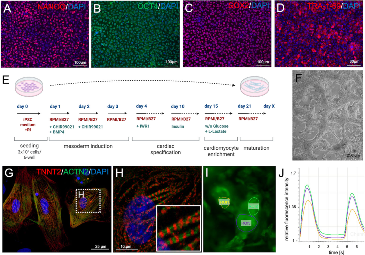
The hiPSCs were checked for the expression of pluripotency markers (Figure 1A–D) and subsequently differentiated into hiPSC-CMs using a protocol previously established in our laboratory [23]. The differentiation process involved an initial two-day activation of WNT and BMP4 signaling, followed by WNT inhibition and a final lactate enrichment step (Figure 1E).
The resulting hiPSC-CM cultures (Figure 1F) exhibited spontaneous contractile activity (Video S1). Immunostaining for TNNT2 and sarcomeric α-Actinin (ACTN2) on tissue sections revealed a regular, cross-striated, sarcomere-like expression pattern (Figure 1G,H). Calcium imaging of spontaneously beating cardiomyocytes using the Fluo-4 AM calcium indicator revealed regular, spontaneous calcium transients (Figure 1I,J).

3.2. Co-Culture of hiPSC-Derived Cardiac Spheroids with Mouse Aortic Rings in Collagen Hydrogel

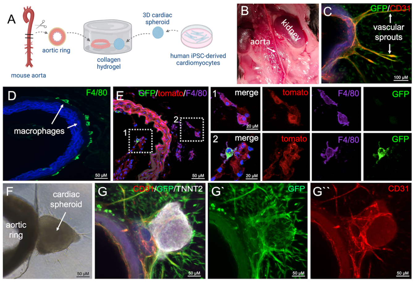
Mouse aortas were isolated from adult mice aged 8–12 weeks (Figure 2B) and cut into 1 mm rings. Individual rings were then embedded into a collagen hydrogel. In the co-culture setup, aortic rings were embedded together with a human cardiac spheroid (Figure 2A). Culturing the aortic rings in collagen induced angiogenic vascular sprouting (Figure 2C), as previously described for aorta or human arterial rings [17,26,27] and led to the generation of F4/80+ macrophages from adventitial stem and progenitor cells (Figure 2D), consistent with recently published findings from our lab [20].
To determine the origin of these macrophages, we embedded aortic rings from “FlkSwitch” mice, in which Cre recombinase expression is driven by the Flk2/Flt3 promoter [24]. Flk2/Flt3 is expressed in bone marrow-resident hematopoietic stem and progenitor cells during definitive hematopoiesis [28]. The “FlkSwitch” mice carry a reporter cassette that switches from red (tdTomato) to green (eGFP) fluorescence upon Cre-mediated recombination, which typically occurs in bone marrow-derived hematopoietic cells and in their derivatives circulating in peripheral blood. In contrast, tissue-resident cells of hematopoietic origin, such as microglia, which arise from yolk sac hematopoiesis during early embryonic development [29], do not undergo recombination and therefore remain tdTomato-positive, similar to all other somatic cells, including vascular wall cells.
In the presented experiments, most macrophages arising in the adventitial compartment exhibited red fluorescence, indicating that they did not originate from bone marrow–derived monocytes but instead from local adventitial stem and progenitor cells with hematopoietic potential. A small number of green-fluorescent, bone marrow-derived macrophages were also observed, primarily within the vascular lumen (Figure 2E). This finding is particularly relevant because tissue-resident macrophages, which arise during early hematopoiesis, play important roles in cardiac development, maintenance of cardiac function, support of electric conduction, and contribution to repair and regeneration, in contrast to monocyte-derived macrophages [8,30,31,32,33,34]. In the next step, we co-cultured hiPSC-derived cardiac spheroids with aortic rings from GFP+ mice and observed (Figure 2F,G) that CD31+/GFP+ mouse-derived endothelial sprouts were attracted to the cardiac spheroids, establishing close contact with the TNNT2+ human cardiomyocytes within the spheroids (Figure 2G–G″). Additionally, CD31/GFP+ non-endothelial mouse cells, likely fibroblasts, were also recruited to the spheroids (Figure 2G–G″). The cardiac spheroids maintained their spontaneous beating activity throughout the co-culture period (Video S2).

3.3. Co-Culture of hiPSC-Derived Cardiomyocytes with Mouse Aortic Rings in 3D Suspension Culture

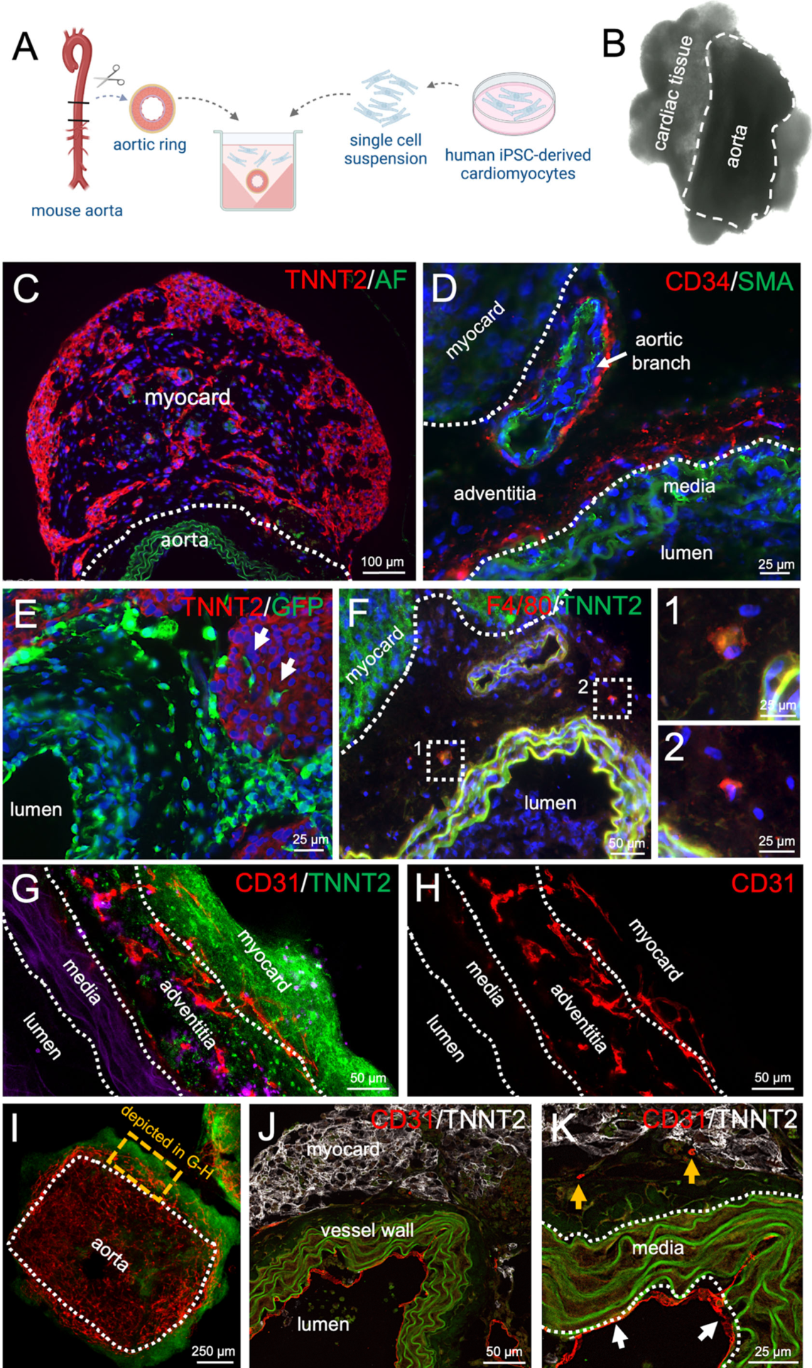
To generate free-floating 3D cardiac tissue constructs which are more suitable for high throughput production and do not require the use of expensive extracellular matrix-preparation, we modified the culture method. Using agarose coating, we prepared a non-adhesive 48-well plate with cone-shaped molds, as previously described [35]. In these molds, mouse aortic rings were combined with a suspension of hiPSC-CMs (Figure 3A). After 2–3 days, the hiPSC-CMs adhered to the vascular adventitia. The aortic rings and hiPSC-CMs formed a compact tissue construct (Figure 3B), which exhibited spontaneous beating activity. This beating was maintained for several weeks in culture (Figure 3B and Video S3).
Immunofluorescence analyses of tissue sections revealed TNNT2-positive human myocardial tissue directly adjacent to the murine vascular adventitia (Figure 3C). Moreover, the adventitial stem and progenitor cell niche, containing CD34+ cells with hematopoietic and endothelial differentiation capacity, remained stable throughout the culture period (Figure 3D). In addition to the aorta, the CD34+ cell niche was also observed around smaller connected aortic branches (Figure 3D). Using aortic segments derived from GFP-expressing mice, green fluorescent mouse cells were observed to migrate from the adventitia into the human cardiac tissue, indicating the contribution of mouse cells to the human cardiac tissue (Figure 3E). Furthermore, F4/80+ macrophages were detected within the adventitial niche adjacent to the myocardium-like tissue (Figure 3F). Finally, tissue clearing and whole-mount staining revealed an extensive CD31+ endothelial network within the vascular adventitia which extends endothelial sprouts into the adjacent TNNT2+ tissue composed of hiPSC-CMs (Figure 3G–I, Videos S5 and S6). These results from tissue clearing were confirmed in paraffin sections (Figure 3J,K) revealing an endothelial lining of the aortic lumen (Figure 3K, white arrows), endothelial cells within the adventitia as well as endothelial sprouts extending towards the human cardiac tissue (Figure 3K, yellow arrows).

3.4. Establishment of an Entirely Human Vessel–Cardiomyocyte Co-Culture Model

We initially began our experiments using human cardiomyocytes and mouse vessel rings. However, for applications such as disease modeling, drug screening, and toxicity testing, an entirely human tissue model represents the ideal setup. To assess whether our co-culture technique is also suitable for human vascular tissue, we co-cultured hiPSC-CMs with vessel rings derived from the human saphenous vein.
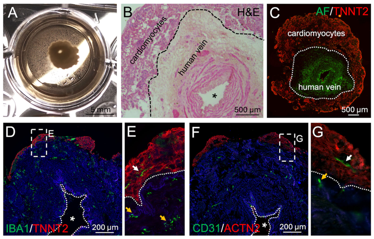
Our results demonstrate that hiPSC-CMs readily attach to the adventitial layer of the human vessel, forming a compact, cohesive tissue within 2–3 days (Figure 4A,B). Notably, spontaneous contractile activity of the cardiac tissue was observed and maintained for over three weeks (Video S4). Immunofluorescence analysis detecting TNNT2 confirmed the cardiac identity of the attached tissue (Figure 4C). Moreover, IBA1+ macrophage-like cells were detected throughout the vascular adventitia (Figure 4D) and also within the attached TNNT2+ human cardiac tissue (Figure 4D,E). IBA1+ cells were not detectable in control cultures of aggregated hiPSC-derived cells differentiated toward the cardiac lineage in the absence of vascular tissue (Figure S1A). Besides macrophages, CD31+ endothelial sprouts were detected throughout the adventitial compartment (Figure 4F) as well as within the attached ACTN2+ iPSC-derived cardiac tissue (Figure 4F,G). Notably, rare endothelial cells were also observed in control cultures lacking vascular segments, indicating that endothelial cells can arise as a byproduct of the differentiation protocol and partially survive lactate-based enrichment (Figure S1B). The observed endothelial network is therefore likely chimeric, consisting of both hiPSC-derived and vascular adventitia–derived components.

4. Discussion

In this study, we present a cardio-vascular assembloid model in which rings of primary vascular explants are co-cultured with hiPSC-derived cardiomyocytes (hiPSC-CM) that self-assemble into cardiac tissue at the outer, adventitial side of the vessel wall. We demonstrate that CD34+ progenitor cells, naturally residing in the mouse aortic adventitia, are maintained in the assembloid model, but also partially activated and mobilized into the cardiac compartment, where they form a network of endothelial cords. Moreover, macrophage-like cells appear within the aortic adventitia, at the interface between the cardiac spheroid and the vessel wall.
Our initially tested assembloid model was largely chimeric, as it involved the co-culture of hiPSC-CMs with vascular rings derived from the mouse aorta. Therefore, as a proof of concept, we also co-cultured hiPSC-CMs with human vascular rings from saphenous vein explants obtained postmortem from human cadavers donated to the Institute of Anatomy and Cell Biology of the University of Würzburg, Germany, demonstrating that the approach can also be adapted to an entirely human model.
Previous studies have already shown that co-culturing cardiomyocytes with fibroblasts, endothelial cells, and macrophages can enhance tissue maturation and function [6,7,8]. However, these approaches rely on mixtures of dissociated single cells, either individually derived from iPSCs or isolated directly from donor tissue, which are subsequently aggregated to form spheroids or engineered micro-tissues. In contrast, our method integrates hiPSC-CM with intact, millimeter-scale segments (vascular rings) of mature vascular tissue, resulting in a more in vivo-like structured cardio-vascular niche.
We observed that the adventitial stem cell niche, also referred to as the vasculogenic zone [18], remained structurally intact within the assembloids, while different adventitial cell populations got activated and migrated into the surrounding matrix or the attached human cardiac tissue. The adventitial niche harbors multiple predominantly CD34+ stem and progenitor cell types and is similarly found in human and murine vessel walls. A distinct subset of these progenitors has the capacity to continuously generate new macrophages [19]. Another subset exhibits vascular progenitor potential [17] and contributes to new vessel formation, thereby promoting tissue vascularization. We assume that the adventitial tissue, by providing fibroblasts, endothelial cells, and macrophages, all of which have been shown to enhance cardiac maturation in vitro [6,7,8], could support hiPSC-CM maturation and long-term functional maintenance, while also contributing new stromal components as the tissue grows, matures, and undergoes remodeling. However, this hypothesis needs to be carefully tested in future studies through in-depth histological and ultrastructural analyses (e.g., measurement of sarcomere length) as well as functional assessments (e.g., electrophysiological evaluations).
Looking ahead, our model could provide a valuable platform to investigate the contribution of the adventitial niche to cardiac tissue maintenance under both physiological and pathological conditions, as well as its potential significance as a site for therapeutic intervention, as previously suggested by our group [21]. In this context, the model could be used to study cardiac regeneration following experimental injury, such as cryoinfarction [5] or toxic damage [36], and to investigate how such injuries affect the vascular niche, particularly the adventitial compartment.
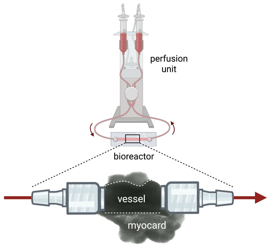
As a next step, we plan to scale up the model by using longer segments of human vasculature and larger numbers of seeded hiPSC-CM. The longer vessel segments will be connected to a bioreactor and a perfusion unit (Figure 5) [37]. Within the bioreactor, vascular sprouts from the explanted vessel could drive cardiac tissue vascularization and ultimately enable tissue perfusion, supporting extended culture durations and potentially further growth in terms of tissue size. This would promote enhanced tissue maturation. Furthermore, drug delivery via the perfusion system would allow for testing of novel therapeutics or investigation of drug-induced cardiotoxicity in a physiologically relevant setting [38].
In summary, the key strength of this novel approach, combining mature vessel segments with iPSC-derived cardiomyocytes, lies in its ability to recreate a realistic tissue microenvironment that can be maintained in long-term culture. This concept lies in the intermediate complexity between in vitro organoid technologies and the transplantation of organoids into living animals. It provides a complex, mature, in vivo-like environment to support organoid maturation without the need for costly and ethically sensitive animal experiments, while also offering improved scalability and potential for higher-throughput experimental approaches.
The needed vascular segments could be derived from mice or, if an all-human platform should be required, are readily available as byproducts from routine surgical procedures, such as bypass surgery, where remaining pieces of the human saphenous vein or internal thoracic artery are often discarded.
Here, we provide proof of concept for a previously unexplored cardiovascular assembloid approach, along with its initial characterization. This strategy could serve as a bridge between traditional 3D tissue culture systems and animal models. However, further studies are required to determine how closely the presented model recapitulates the native human heart and how it influences the functional maturation of fetal-like hiPSC-derived cardiomyocytes. Future work will focus on establishing perfusable constructs within a bioreactor environment and validating the applicability of this model for disease modeling and drug testing.

5. Conclusions

Taken together, we present proof-of-concept for a novel assembloid model, available in both chimeric and non-chimeric formats, that can be adapted to a wide range of tissue-specific spheroids and organoids beyond the heart, e.g., to neural tissue [39]. As such, with further refinement and standardization, this platform has strong potential for use in both basic and developmental research, as well as in translational applications, including drug testing and disease modeling.

Supplementary Materials

The following supporting information can be downloaded at: https://www.mdpi.com/article/10.3390/organoids5020018/s1, Figure S1: 60-day-old control cultures of aggregated hiPSC-derived cells differentiated toward the cardiac lineage in the absence of vascular tissue; Video S1: Spontaneously contracting human iPSC-derived cardiomyocytes in 2D culture; Video S2: Beating cardiac spheroid adjacent to a mouse aortic ring embedded in collagen I; Video S3: Spontaneously contracting hiPSC-derived cardiomyocytes attached to a mouse aortic ring in suspension culture; Video S4: Spontaneously contracting hiPSC-derived cardiomyocytes attached to a human saphenous vein ring in suspension culture; Video S5: 3D-reconstructed z-stack from whole-mount immunofluorescence of murine aortic adventitia and attached human cardiac tissue; Video S6: 3D-reconstructed z-stack highlighting the endothelial network.

Author Contributions

Conceptualization, P.W. and S.E.; Methodology, P.W., S.E., H.K., K.K. and B.U.; Validation, P.W., S.E., H.K., K.K. and B.U.; Formal Analysis, P.W. and S.E.; Investigation, H.K., K.K., B.U. and P.W.; Resources, P.W. and S.E.; Data Curation, H.K. and K.K.; Writing—Original Draft Preparation, P.W., H.K., K.K. and S.E.; Writing—Review & Editing, P.W., H.K., K.K. and S.E.; Visualization, P.W.; Supervision, P.W. and S.E.; Project Administration, P.W. and S.E.; Funding Acquisition, P.W. and S.E. All authors have read and agreed to the published version of the manuscript.

Funding

This research was funded by the IZKF Würzburg (grant number E-D-410) to P.W. and by the Deutsche Forschungsgemeinschaft (DFG) through the Collaborative Research Center TRR225 (project B04) to S.E.

Institutional Review Board Statement

Ethical review and approval were waived because mouse procedures only involved decapitation followed by postmortem tissue collection for in vitro experiments (e.g., aortic isolation), which, according to German federal guidelines and local regulations (Regierung von Unterfranken, Würzburg, Germany), does not require an animal experiment permit or specific ethical approval. Human saphenous veins were obtained post mortem from a body donor of the Institute of Anatomy and Cell Biology, Würzburg. The legally approved body donation agreement includes explicit consent for the use of tissues for scientific research. No identifiable personal data were used in this study.

Informed Consent Statement

Human saphenous veins were obtained postmortem from a body donor of the Institute of Anatomy and Cell Biology, Würzburg. The legally approved body donation agreement includes explicit consent for the use of tissues for scientific research. No identifiable personal data were used in this study.

Data Availability Statement

Data supporting the findings of this study are available from the corresponding author upon reasonable request.

Acknowledgments

We thank Doris Dettelbacher-Weber, Martina Gebhardt, Erna Kleinschroth, Ursula Roth, Elke Varin, and Lisa Wittstatt for their excellent technical assistance, as well as all members of the Stem Cell Lab for their support and valuable discussions. Figure 1E, Figure 2A, Figure 3A and Figure 5 were created using BioRender (BioRender.com).

Conflicts of Interest

The authors declare no conflicts of interest.

References

  1. Takahashi, K.; Tanabe, K.; Ohnuki, M.; Narita, M.; Ichisaka, T.; Tomoda, K.; Yamanaka, S. Induction of pluripotent stem cells from adult human fibroblasts by defined factors. Cell 2007, 131, 861–872. [Google Scholar] [CrossRef] [Scilit]
  2. Ren, Y.M.; Lee, M.Y.; Schliffke, S.; Paavola, J.; Amos, P.J.; Ge, X.; Ye, M.Y.; Zhu, S.J.; Senyei, G.; Lum, L.; et al. Small molecule Wnt inhibitors enhance the efficiency of BMP-4-directed cardiac differentiation of human pluripotent stem cells. J. Mol. Cell. Cardiol. 2011, 51, 280–287. [Google Scholar] [CrossRef] [Scilit]
  3. Burridge, P.W.; Keller, G.; Gold, J.D.; Wu, J.C. Production of de novo cardiomyocytes: Human pluripotent stem cell differentiation and direct reprogramming. Cell Stem Cell 2012, 10, 16–28. [Google Scholar] [CrossRef] [Scilit] [PubMed]
  4. Tohyama, S.; Hattori, F.; Sano, M.; Hishiki, T.; Nagahata, Y.; Matsuura, T.; Hashimoto, H.; Suzuki, T.; Yamashita, H.; Satoh, Y.; et al. Distinct metabolic flow enables large-scale purification of mouse and human pluripotent stem cell-derived cardiomyocytes. Cell Stem Cell 2013, 12, 127–137. [Google Scholar] [CrossRef] [Scilit]
  5. Voges, H.K.; Mills, R.J.; Elliott, D.A.; Parton, R.G.; Porrello, E.R.; Hudson, J.E. Development of a human cardiac organoid injury model reveals innate regenerative potential. Development 2017, 144, 1118–1127. [Google Scholar] [CrossRef] [Scilit]
  6. Giacomelli, E.; Meraviglia, V.; Campostrini, G.; Cochrane, A.; Cao, X.; van Helden, R.W.J.; Krotenberg Garcia, A.; Mircea, M.; Kostidis, S.; Davis, R.P.; et al. Human-iPSC-Derived Cardiac Stromal Cells Enhance Maturation in 3D Cardiac Microtissues and Reveal Non-cardiomyocyte Contributions to Heart Disease. Cell Stem Cell 2020, 26, 862–879.e11. [Google Scholar] [CrossRef] [Scilit] [PubMed]
  7. Voges, H.K.; Foster, S.R.; Reynolds, L.; Parker, B.L.; Devilee, L.; Quaife-Ryan, G.A.; Fortuna, P.R.J.; Mathieson, E.; Fitzsimmons, R.; Lor, M.; et al. Vascular cells improve functionality of human cardiac organoids. Cell Rep. 2023, 42, 112322. [Google Scholar] [CrossRef] [Scilit]
  8. Lock, R.I.; Graney, P.L.; Tavakol, D.N.; Nash, T.R.; Kim, Y.; Sanchez, E., Jr.; Morsink, M.; Ning, D.; Chen, C.; Fleischer, S.; et al. Macrophages enhance contractile force in iPSC-derived human engineered cardiac tissue. Cell Rep. 2024, 43, 114302. [Google Scholar] [CrossRef] [Scilit] [PubMed]
  9. Silva, A.C.; Matthys, O.B.; Joy, D.A.; Kauss, M.A.; Natarajan, V.; Lai, M.H.; Turaga, D.; Blair, A.P.; Alexanian, M.; Bruneau, B.G.; et al. Co-emergence of cardiac and gut tissues promotes cardiomyocyte maturation within human iPSC-derived organoids. Cell Stem Cell 2021, 28, 2137–2152.e6. [Google Scholar] [CrossRef] [Scilit]
  10. Hofbauer, P.; Jahnel, S.M.; Papai, N.; Giesshammer, M.; Deyett, A.; Schmidt, C.; Penc, M.; Tavernini, K.; Grdseloff, N.; Meledeth, C.; et al. Cardioids reveal self-organizing principles of human cardiogenesis. Cell 2021, 184, 3299–3317.e22. [Google Scholar] [CrossRef] [Scilit]
  11. Lewis-Israeli, Y.R.; Wasserman, A.H.; Gabalski, M.A.; Volmert, B.D.; Ming, Y.; Ball, K.A.; Yang, W.; Zou, J.; Ni, G.; Pajares, N.; et al. Self-assembling human heart organoids for the modeling of cardiac development and congenital heart disease. Nat. Commun. 2021, 12, 5142. [Google Scholar] [CrossRef] [Scilit]
  12. Meier, A.B.; Zawada, D.; De Angelis, M.T.; Martens, L.D.; Santamaria, G.; Zengerle, S.; Nowak-Imialek, M.; Kornherr, J.; Zhang, F.; Tian, Q.; et al. Epicardioid single-cell genomics uncovers principles of human epicardium biology in heart development and disease. Nat. Biotechnol. 2023, 41, 1787–1800. [Google Scholar] [CrossRef] [Scilit]
  13. Abilez, O.J.; Yang, H.; Guan, Y.; Shen, M.; Yildirim, Z.; Zhuge, Y.; Venkateshappa, R.; Zhao, S.R.; Gomez, A.H.; El-Mokahal, M.; et al. Gastruloids enable modeling of the earliest stages of human cardiac and hepatic vascularization. Science 2025, 388, eadu9375. [Google Scholar] [CrossRef] [Scilit] [PubMed]
  14. Schmidt, C.; Deyett, A.; Ilmer, T.; Haendeler, S.; Torres Caballero, A.; Novatchkova, M.; Netzer, M.A.; Ceci Ginistrelli, L.; Mancheno Juncosa, E.; Bhattacharya, T.; et al. Multi-chamber cardioids unravel human heart development and cardiac defects. Cell 2023, 186, 5587–5605.e27. [Google Scholar] [CrossRef] [Scilit] [PubMed]
  15. O’Hern, C.; Caywood, S.; Aminova, S.; Kiselev, A.; Volmert, B.; Cao, W.; Wang, F.; Dionise, M.; Sewavi, M.L.; Skoric, M.; et al. Human heart-macrophage assembloids mimic immune-cardiac interactions and enable arrhythmia disease modeling. Cell Stem Cell 2025, 32, 1671–1690.e13. [Google Scholar] [CrossRef] [Scilit] [PubMed]
  16. Rockel, A.F.; Brunnbauer, T.; Wagner, N.; Gerull, B.; Ergun, S.; Worsdorfer, P. Tissue-resident macrophages co-develop with myocardial tissue in human induced pluripotent stem cell-derived organoids. Front. Cell Dev. Biol. 2025, 13, 1629988. [Google Scholar] [CrossRef] [Scilit]
  17. Zengin, E.; Chalajour, F.; Gehling, U.M.; Ito, W.D.; Treede, H.; Lauke, H.; Weil, J.; Reichenspurner, H.; Kilic, N.; Ergun, S. Vascular wall resident progenitor cells: A source for postnatal vasculogenesis. Development 2006, 133, 1543–1551. [Google Scholar] [CrossRef] [Scilit]
  18. Worsdorfer, P.; Mekala, S.R.; Bauer, J.; Edenhofer, F.; Kuerten, S.; Ergun, S. The vascular adventitia: An endogenous, omnipresent source of stem cells in the body. Pharmacol. Ther. 2017, 171, 13–29. [Google Scholar] [CrossRef] [Scilit]
  19. Psaltis, P.J.; Puranik, A.S.; Spoon, D.B.; Chue, C.D.; Hoffman, S.J.; Witt, T.A.; Delacroix, S.; Kleppe, L.S.; Mueske, C.S.; Pan, S.; et al. Characterization of a resident population of adventitial macrophage progenitor cells in postnatal vasculature. Circ. Res. 2014, 115, 364–375. [Google Scholar] [CrossRef] [Scilit]
  20. Kleefeldt, F.; Upcin, B.; Bommel, H.; Schulz, C.; Eckner, G.; Allmanritter, J.; Bauer, J.; Braunger, B.; Rueckschloss, U.; Ergun, S. Bone marrow-independent adventitial macrophage progenitor cells contribute to angiogenesis. Cell Death Dis. 2022, 13, 220. [Google Scholar] [CrossRef] [Scilit]
  21. Mekala, S.R.; Worsdorfer, P.; Bauer, J.; Stoll, O.; Wagner, N.; Reeh, L.; Loew, K.; Eckner, G.; Kwok, C.K.; Wischmeyer, E.; et al. Generation of Cardiomyocytes From Vascular Adventitia-Resident Stem Cells. Circ. Res. 2018, 123, 686–699. [Google Scholar] [CrossRef] [Scilit]
  22. Sommer, C.A.; Stadtfeld, M.; Murphy, G.J.; Hochedlinger, K.; Kotton, D.N.; Mostoslavsky, G. Induced pluripotent stem cell generation using a single lentiviral stem cell cassette. Stem Cells 2009, 27, 543–549. [Google Scholar] [CrossRef] [Scilit]
  23. Kadari, A.; Mekala, S.; Wagner, N.; Malan, D.; Koth, J.; Doll, K.; Stappert, L.; Eckert, D.; Peitz, M.; Matthes, J.; et al. Robust Generation of Cardiomyocytes from Human iPS Cells Requires Precise Modulation of BMP and WNT Signaling. Stem Cell Rev. Rep. 2015, 11, 560–569. [Google Scholar] [CrossRef] [Scilit]
  24. Boyer, S.W.; Schroeder, A.V.; Smith-Berdan, S.; Forsberg, E.C. All hematopoietic cells develop from hematopoietic stem cells through Flk2/Flt3-positive progenitor cells. Cell Stem Cell 2011, 9, 64–73. [Google Scholar] [CrossRef] [Scilit] [PubMed]
  25. Okabe, M.; Ikawa, M.; Kominami, K.; Nakanishi, T.; Nishimune, Y. ‘Green mice’ as a source of ubiquitous green cells. FEBS Lett. 1997, 407, 313–319. [Google Scholar] [CrossRef] [Scilit] [PubMed]
  26. Nicosia, R.F.; Tchao, R.; Leighton, J. Histotypic angiogenesis in vitro: Light microscopic, ultrastructural, and radioautographic studies. In Vitro 1982, 18, 538–549. [Google Scholar] [CrossRef] [Scilit] [PubMed]
  27. Upcin, B.; Henke, E.; Kleefeldt, F.; Hoffmann, H.; Rosenwald, A.; Irmak-Sav, S.; Aktas, H.B.; Ruckschloss, U.; Ergun, S. Contribution of Adventitia-Derived Stem and Progenitor Cells to New Vessel Formation in Tumors. Cells 2021, 10, 1719. [Google Scholar] [CrossRef] [Scilit]
  28. Matthews, W.; Jordan, C.T.; Wiegand, G.W.; Pardoll, D.; Lemischka, I.R. A receptor tyrosine kinase specific to hematopoietic stem and progenitor cell-enriched populations. Cell 1991, 65, 1143–1152. [Google Scholar] [CrossRef] [Scilit]
  29. Gomez Perdiguero, E.; Schulz, C.; Geissmann, F. Development and homeostasis of “resident” myeloid cells: The case of the microglia. Glia 2013, 61, 112–120. [Google Scholar] [CrossRef] [Scilit]
  30. Dick, S.A.; Macklin, J.A.; Nejat, S.; Momen, A.; Clemente-Casares, X.; Althagafi, M.G.; Chen, J.; Kantores, C.; Hosseinzadeh, S.; Aronoff, L.; et al. Self-renewing resident cardiac macrophages limit adverse remodeling following myocardial infarction. Nat. Immunol. 2019, 20, 29–39. [Google Scholar] [CrossRef] [Scilit]
  31. Hamidzada, H.; Pascual-Gil, S.; Wu, Q.; Kent, G.M.; Masse, S.; Kantores, C.; Kuzmanov, U.; Gomez-Garcia, M.J.; Rafatian, N.; Gorman, R.A.; et al. Primitive macrophages induce sarcomeric maturation and functional enhancement of developing human cardiac microtissues via efferocytic pathways. Nat. Cardiovasc. Res. 2024, 3, 567–593. [Google Scholar] [CrossRef] [Scilit]
  32. Hulsmans, M.; Clauss, S.; Xiao, L.; Aguirre, A.D.; King, K.R.; Hanley, A.; Hucker, W.J.; Wulfers, E.M.; Seemann, G.; Courties, G.; et al. Macrophages Facilitate Electrical Conduction in the Heart. Cell 2017, 169, 510–522.e20. [Google Scholar] [CrossRef] [Scilit]
  33. Landau, S.; Zhao, Y.; Hamidzada, H.; Kent, G.M.; Okhovatian, S.; Lu, R.X.Z.; Liu, C.; Wagner, K.T.; Cheung, K.; Shawky, S.A.; et al. Primitive macrophages enable long-term vascularization of human heart-on-a-chip platforms. Cell Stem Cell 2024, 31, 1222–1238.e10. [Google Scholar] [CrossRef] [Scilit]
  34. Wong, N.R.; Mohan, J.; Kopecky, B.J.; Guo, S.; Du, L.; Leid, J.; Feng, G.; Lokshina, I.; Dmytrenko, O.; Luehmann, H.; et al. Resident cardiac macrophages mediate adaptive myocardial remodeling. Immunity 2021, 54, 2072–2088.e7. [Google Scholar] [CrossRef] [Scilit] [PubMed]
  35. Worsdorfer, P.; Rockel, A.; Alt, Y.; Kern, A.; Ergun, S. Generation of Vascularized Neural Organoids by Co-culturing with Mesodermal Progenitor Cells. STAR Protoc. 2020, 1, 100041. [Google Scholar] [CrossRef] [Scilit] [PubMed]
  36. Hyun, S.A.; Park, J.H.; Ko, M.Y.; Min, E.; Kim, M.; Kang, S.W.; Ka, M. Human cardiac organoids highlight cardiotoxicity of the tire rubber antioxidant 6PPD. Ecotoxicol. Environ. Saf. 2025, 308, 119496. [Google Scholar] [CrossRef] [Scilit]
  37. Lysyy, T.; Bracaglia, L.G.; Qin, L.; Albert, C.; Pober, J.S.; Tellides, G.; Saltzman, W.M.; Tietjen, G.T. Ex vivo isolated human vessel perfusion system for the design and assessment of nanomedicines targeted to the endothelium. Bioeng. Transl. Med. 2020, 5, e10154. [Google Scholar] [CrossRef] [Scilit] [PubMed]
  38. Mamoshina, P.; Rodriguez, B.; Bueno-Orovio, A. Toward a broader view of mechanisms of drug cardiotoxicity. Cell Rep. Med. 2021, 2, 100216. [Google Scholar] [CrossRef] [Scilit]
  39. Worsdorfer, P.; I, T.; Asahina, I.; Sumita, Y.; Ergun, S. Do not keep it simple: Recent advances in the generation of complex organoids. J. Neural Transm. 2020, 127, 1569–1577. [Google Scholar] [CrossRef] [Scilit]
Figure 1. Differentiation of hiPSCs into beating hiPSC-CMs. (AD) Immunofluorescence staining of undifferentiated hiPSCs cultured in 2D, showing expression of pluripotency markers: NANOG (A), OCT4 (B), SOX2 (C), and TRA-1-60 (D). Nuclei are counterstained with DAPI. (E) Schematic overview of the cardiac differentiation protocol, illustrating the timeline and culture media/small molecules used. Created in BioRender. Wörsdörfer, P. (2026) https://BioRender.com/8hq4vv5, accessed on 12 March 2026. (F) Phase contrast image of a typical hiPSC-derived 2D cardiomyocyte culture (see Video S1). (G) Immunofluorescence analyses of differentiated hiPSC-CMs using cardiac-specific markers TNNT2 and ACTN2. Nuclei are counterstained with DAPI. (H) High-resolution image highlighting the characteristic cross-striated staining pattern of TNNT2 and ACTN2 indicating sarcomere formation. (I) Calcium imaging using the calcium-sensitive fluorescent dye Fluo-4 AM. (J) Calcium transients in ROIs marked in (I) determined by Fluo-4 AM fluorescence recorded for 7 s.
Organoids 05 00018 g001
Figure 2. Co-culturing human iPSC-derived cardiac spheroids with aortic vessel rings in a collagen hydrogel. (A) Schematic overview of the co-culture setup combining mouse aortic rings and 3D cardiac spheroids embedded in a collagen I matrix. Created in BioRender. Wörsdörfer, P. (2026) https://BioRender.com/zd7clp0, accessed on 12 March 2026. (B) Dissection of the mouse aorta. (C) Immunofluorescence staining of aortic rings showing GFP+ aorta-derived CD31+ endothelial cells sprouting into the surrounding collagen. Some GFP+/CD31 non-endothelial cell types (e.g., fibroblasts) are also observed. (n = 3 aortic rings were analyzed and showed comparable results; a representative image is presented). (D) Immunofluorescence analysis of aortic rings reveals F4/80+ macrophages within the vascular adventitia. (n = 3 aortic rings were analyzed and showed comparable results; a representative image is presented). (E) Immunofluorescence staining of aortic rings derived from “FlkSwitch” mice exhibit F4/80+ macrophages in two distinct regions: (1) within the vessel lumen and (2) in the vascular adventitia. The macrophages located in the vessel lumen (1) express GFP, indicating their origin from circulating blood monocytes, whereas those in the adventitia (2) lack GFP expression, consistent with tissue-resident macrophages. (n = 3 aortic rings were analyzed and showed comparable results; a representative image is presented). (F) Brightfield image of the aortic ring with an adjacent cardiac spheroid (see Video S2). (n = 7 aortic rings + cardiac spheroids were generated and showed comparable results; a representative image and video are presented). (GG″) Immunofluorescence analyses reveal GFP+/CD31+ endothelial sprouts growing towards and into the TNNT2+ cardiac spheroid. Not all GFP+ cells sprouting from the aortic ring co-express CD31, suggesting cellular heterogeneity. Nuclei are counterstained with DAPI. (n = 3 aortic rings + cardiac spheroids were analyzed and showed comparable results; a representative image is presented).
Organoids 05 00018 g002
Figure 3. Co-culturing human iPSC-derived cardiomyocytes with aortic vessel rings in 3D suspension culture. (A) Schematic representation of the co-culture setup combining murine aortic rings with human iPSC-derived cardiomyocytes (hiPSC-CMs) in agarose molds. Created in BioRender. Wörsdörfer, P. (2026) https://BioRender.com/ixpu9o7, accessed on 12 March 2026. (B) Brightfield image of the 3D tissue generated by co-culture of hiPSC-CMs with a murine aortic ring at culture day 11 (see also Video S3). (C) Immunofluorescence analysis of tissue sections from 11-day-old co-cultures showing TNNT2+ hiPSC-CMs in close proximity to the aortic adventitia. Autofluorescence (AF) of elastic fibers within the tunica media highlights the localization of the vessel ring. The dotted line indicates the interface between the cardiac organoid and the adventitia. (n = 3 assembloids were analyzed and showed comparable results; a representative image is presented). (D) SMA+ smooth muscle cells are present in the tunica media, while CD34+ adventitial progenitor cells are detected within the adventitial compartment. The image shows the aorta and an aortic branch. (n = 4 assembloids were analyzed and showed comparable results; a representative image is presented). (E) GFP+ mouse cells are found around and within the TNNT2+ human myocard-like tissue attached to the vessel segment. (n = 3 assembloids were analyzed and showed comparable results; a representative image is presented). (F) F4/80+ macrophages are observed in the adventitia of the vessel rings at the interface with the TNNT2+ hiPSC-derived cardiac tissue. (n = 2 assembloids were analyzed and showed comparable results; a representative image is presented). (G,H) Maximum-intensity projection of a z-stack from whole-mount immunofluorescence analysis followed by tissue clearing of a 17-day-old cardio-vascular assembloid detecting CD31 (red), TNNT2 (green), and autofluorescence of elastic fibers (purple). Panel H shows only the CD31 (red) channel from panel G. For 3D reconstructions, see Videos S5 and S6. (n = 2 assembloids were analyzed and showed comparable results; a representative image and videos are presented). (I) Low-magnification whole-mount immunofluorescence image of a 17-day-old cardio-vascular assembloid detecting CD31 (red) and TNNT2 (green), showing the entire assembloid. Panels G–H depict the region indicated by the yellow box. (n = 2 assembloids were analyzed and showed comparable results; a representative image is presented). (J,K) Immunofluorescence analysis of paraffin sections from a cardio-vascular assembloid detecting TNNT2 (white), CD31 (red), and autofluorescence of elastic fibers (green). Panel (K) shows a higher magnification of panel (J). Endothelial cells lining the aortic lumen are indicated by white arrows, while endothelial sprouts in close proximity to cardiomyocytes are indicated by yellow arrows. (n = 8 assembloids were analyzed and showed comparable results; representative images are presented).
Organoids 05 00018 g003
Figure 4. Co-culturing human iPSC-derived cardiomyocytes with human saphenous vein–derived vessel rings in 3D suspension culture. (A) Co-culture of human saphenous vein rings with human iPSC-derived cardiomyocytes at day 21 (see also Video S4). (B) Haematoxylin and eosin staining of a paraffin section from day 14 of co-culture. The interface between the vein wall and the hiPSC-CM compartment is indicated by the dashed line. The black asterisk marks the vascular lumen. (C) Immunofluorescence analysis of paraffin sections from the all-human cardio-vascular assembloid showing TNNT2+ cardiomyocytes (red) and autofluorescence of elastic fibers (green). (D,E) Immunofluorescence analysis of paraffin sections detecting TNNT2 (red), IBA1 (green), and autofluorescence of elastic fibers (blue). (E) Higher magnification of the boxed area in (D). Yellow arrows indicate macrophage-like cells in the adventitia, and the white arrow indicates a macrophage-like cell within the attached cardiomyocyte compartment. The white asterisk marks vascular lumen. (F,G) Immunofluorescence analysis detecting ACTN2 (red), CD31 (green), and autofluorescence of elastic fibers (blue). (G) Higher magnification of the boxed area in (F). The yellow arrow indicates an endothelial sprout in the adventitia, and the white arrow indicates an endothelial sprout extending into the cardiomyocyte compartment. The white asterisk marks vascular lumen. (AG): n = 3 assembloids from 1 donor were analyzed and showed comparable results; a representative image is presented.
Organoids 05 00018 g004
Figure 5. Suggested setup for perfusion of the co-culture model in a bioreactor. This schematic illustrates a vessel–cardiomyocyte co-culture system in which the vessel is connected to a perfusion unit to enable continuous perfusion mimicking the in vivo situation. This configuration is intended to support the long-term growth and maintenance of the tissue model in future studies. Additionally, the perfusion circuit would potentially allow for controlled drug delivery. Created in BioRender. Wörsdörfer, P. (2026) https://BioRender.com/iqt5ouc, accessed on 12 March 2026.
Organoids 05 00018 g005
Disclaimer/Publisher’s Note: The statements, opinions and data contained in all publications are solely those of the individual author(s) and contributor(s) and not of MDPI and/or the editor(s). MDPI and/or the editor(s) disclaim responsibility for any injury to people or property resulting from any ideas, methods, instructions or products referred to in the content.