
Review

Modeling Context-Dependent Tumor Metabolism in 3D Systems: Implications for Functional Precision Oncology


by
Maria Virginia Giolito
1,*,
Olivier Feron
1,2 and
Cyril Corbet
1,2,*
1
Pole of Pharmacology and Therapeutics (FATH), Institut de Recherche Expérimentale et Clinique (IREC), UCLouvain, Avenue Hippocrate 57, 1200 Brussels, Belgium
2
WEL Research Institute, Avenue Pasteur 6, 1300 Wavre, Belgium
*
Authors to whom correspondence should be addressed.
Organoids 2026, 5(3), 22; https://doi.org/10.3390/organoids5030022
Submission received: 30 June 2026 / Revised: 22 July 2026 / Accepted: 23 July 2026 / Published: 27 July 2026

Abstract

Cancer metabolism is a dynamic and context-dependent process shaped by both tumor-intrinsic programs and microenvironmental cues. Capturing this complexity remains a major challenge, which limits the translation of metabolic insights into clinically actionable strategies. Patient-derived tumor organoids, together with emerging engineered platforms such as organ-on-chip systems, vascularized assembloids, and bioprinted tumor models, have opened new avenues for investigating tumor metabolism in physiologically relevant settings. These models enable the study of metabolic heterogeneity across tumor types, disease stages, and treatment conditions while preserving clinically relevant tumor features. Importantly, they provide functional platforms for ex vivo metabolic profiling, identification of metabolic vulnerabilities, and prediction of therapeutic responses. In this review, we discuss recent advances in the use of patient-derived and engineered 3D tumor models to characterize context-dependent metabolic states and treatment-induced metabolic rewiring. We first review technologies currently available to interrogate metabolism in these systems, including optical metabolic imaging, spatial metabolomics, isotope tracing, and bioenergetic profiling. We then discuss how 3D tumor models are used to investigate metabolic interactions within the tumor microenvironment (TME), including stromal and immune crosstalk, acidosis, nutrient availability, and circadian regulation. Finally, we critically examine current limitations, particularly the insufficient physiological relevance of standard organoid culture conditions for metabolic studies, and discuss how advanced engineering approaches and computational modeling may contribute to metabolism-driven functional precision oncology.

1. Introduction

Metabolic reprogramming is a hallmark of cancer, enabling tumor cells to sustain proliferation, survive under stress, and adapt to fluctuating environmental conditions [1]. However, tumor metabolism is not a fixed or cell-autonomous property. Rather, it is a highly dynamic and context-dependent process shaped by the interplay between tumor-intrinsic programs and extrinsic influences, including gradients of oxygen, pH, nutrients, and extracellular metabolites [2]. Cancer cells continuously adapt to these constraints by modulating their reliance on glycolysis, oxidative phosphorylation (OXPHOS), lipid metabolism, and alternative biosynthetic pathways [3]. In addition to spatial heterogeneity, tumor metabolism is influenced by temporal dynamics, including circadian regulation, and by therapeutic pressure, which can induce profound metabolic rewiring associated with resistance [4]. Furthermore, interactions with stromal, immune, neuronal, and vascular components of the TME critically shape metabolic states through nutrient competition, metabolite exchange, and signaling crosstalk [5].
Despite significant advances in cancer metabolism research, the integration of metabolic information into precision oncology remains limited. One important reason is that metabolic phenotypes are poorly predicted by genomic alterations alone and are highly sensitive to environmental context. While genomic alterations define the metabolic potential of tumor cells, the pathways that are ultimately engaged depend on local nutrient availability, oxygen tension, cell–cell interactions, and other microenvironmental cues. Consequently, there is growing interest in experimental systems that enable functional and context-aware interrogation of tumor metabolism in patient-specific settings. Patient-derived tumor organoids (PDOs) are self-organizing three-dimensional (3D) ex vivo cultures established directly from patient tumor specimens and propagated in extracellular matrix-supported conditions that preserve key histological, genomic, and functional characteristics of the original tumors [6,7]. Beyond conventional PDOs, increasingly sophisticated 3D systems have emerged, including co-culture assembloids, air–liquid interface (ALI) cultures, organ-on-chip platforms, and bioprinted tumor models. While many of these systems initially relied on established cell lines, recent developments increasingly incorporate patient-derived material and microenvironmental components, enabling more physiologically relevant interrogation of tumor metabolism and therapeutic response.
3D tumor models comprise a heterogeneous group of experimental systems that should not be considered interchangeable. Spheroids are generally self-assembled multicellular aggregates generated from established cell lines or dissociated primary cells that recapitulate diffusion gradients but exhibit relatively limited tissue organization and cellular complexity compared with organoids [8]. In contrast, PDOs are self-organizing epithelial cultures established directly from patient tumors that preserve key histological, genomic, morphological, and pharmacological features of the parental lesion, making them particularly attractive for disease modeling and functional precision oncology [9,10,11]. Organoids can also be derived from mouse tissues. More recently, engineering approaches have extended conventional PDO cultures through assembloid systems that incorporate stromal, immune, endothelial, or neuronal components, as well as organ-on-chip and 3D bioprinted models, which enable precise control of perfusion, extracellular matrix composition, spatial organization, and multicellular interactions [10,12,13]. Because each model captures different aspects of tumor biology and metabolism, the choice of model should be dictated by the biological question being addressed.
This review examines how patient-derived and engineered 3D tumor models are used to study tumor metabolism across different contexts and the implications for therapy. It synthesizes the literature published up to June 2026, identified through PubMed searches and manual reference screening using keywords related to 3D tumor models, metabolic profiling, microenvironmental factors, and functional precision oncology. We prioritize original research conducting functional metabolic characterization within 3D architectures, complemented by key reviews that provide foundational conceptual or methodological insights into context-dependent tumor metabolism and its therapeutic implications. The review first outlines current technologies, including metabolic imaging, spatial metabolomics, isotope tracing, and bioenergetic analyses, used to examine metabolism in these systems. It then discusses how 3D tumor models are used to investigate metabolic interactions within the tumor microenvironment, circadian influences, and metabolic plasticity. Lastly, the review considers how these methods might advance metabolism-based precision oncology, while addressing current limitations and future directions.

2. Technologies to Investigate Tumor Metabolism in Patient-Derived 3D Models

Cancer cells exhibit remarkable metabolic flexibility, enabling them to use diverse substrates depending on microenvironmental constraints. This adaptability, combined with interpatient and intratumoral heterogeneity, results in substantial variability in metabolic phenotypes and treatment responses. Understanding these metabolic adaptations is therefore critical for identifying actionable vulnerabilities and moving toward metabolism-informed precision oncology.
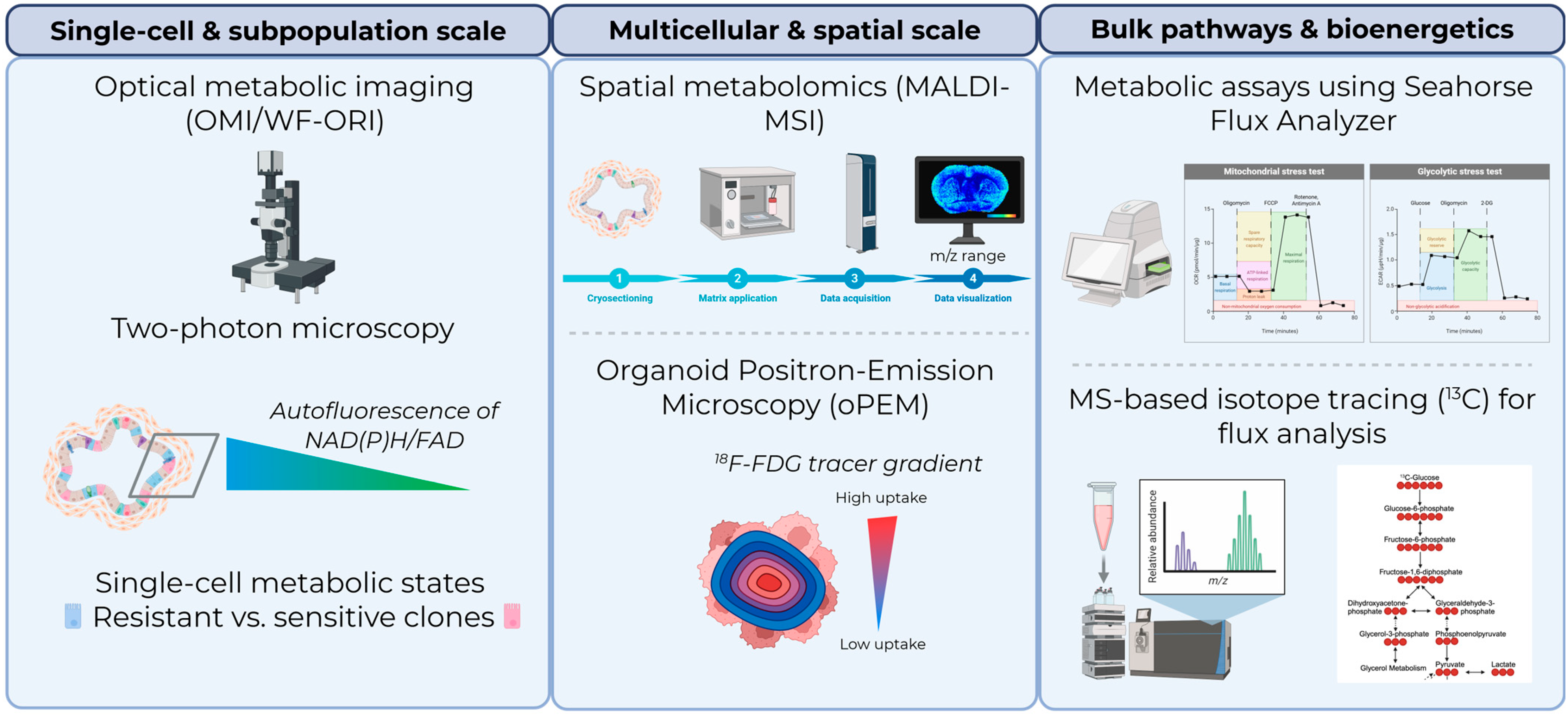
PDOs and related patient-derived 3D systems provide physiologically relevant platforms to investigate tumor metabolism while preserving key features of the original tumor. A wide range of complementary approaches has been developed to interrogate metabolic activity in these models (Figure 1). Beyond descriptive analyses, these technologies collectively support functional applications, including predicting therapeutic response, identifying metabolic biomarkers, and stratifying patients based on basal or treatment-induced metabolic states (Table 1).

2.1. Optical Metabolic Imaging: Resolving Heterogeneity and Predicting Therapy Response

PDOs retain key physicochemical features of the TME, including oxygen and nutrient gradients [47,48,49], making them particularly suitable for studying metabolic heterogeneity. Among the available technologies, optical metabolic imaging (OMI) has emerged as a powerful approach for assessing metabolic states at single-cell resolution.
OMI is based on two-photon fluorescence lifetime imaging microscopy (FLIM) and exploits the intrinsic autofluorescence of metabolic cofactors such as NAD(P)H and flavins (FAD and FMN) [16]. By quantifying fluorescence intensity and lifetime, OMI enables measurement of the optical redox ratio and the relative abundance of free versus protein-bound cofactors, providing insights into cellular metabolic states across pathways including glycolysis, OXPHOS, and lipid metabolism.
This non-invasive, high-resolution approach has been widely applied to tumor organoids, revealing metabolic heterogeneity and enabling early prediction of treatment response across multiple cancer types, including breast [15,18,19], colorectal [21], gastroenteropancreatic neuroendocrine [26], head and neck [20], and pancreatic cancers [17]. Notably, OMI can detect rapid metabolic changes following drug exposure. For example, early decreases in glycolytic activity and shifts in NADH/FAD binding states have been shown to predict subsequent tumor growth inhibition [15]. Importantly, single-cell OMI analyses can identify metabolically distinct subpopulations associated with drug resistance that may not be detectable by bulk measurements [14,15,19]. Beyond cancer cells, OMI has also been applied to characterize metabolic heterogeneity in tumor-associated macrophages within 3D co-culture systems, providing insights into immune–tumor metabolic interactions [22].
More recently, wide-field optical redox imaging (WF-ORI) has emerged as a scalable alternative to two-photon OMI for high-throughput metabolic screening in PDOs. Using autofluorescence measurements of NAD(P)H and FAD, WF-ORI enables rapid quantification of organoid redox states without exogenous labels. Gillette et al. demonstrated that WF-ORI could discriminate treatment responses and mutation-dependent metabolic states in colorectal cancer (CRC) PDOs with higher sensitivity than conventional two-photon imaging approaches, including the identification of resistant subclones in mixed cultures [50]. In parallel, Hsu et al. developed an automated image analysis pipeline integrating Cellpose-based segmentation, single-organoid tracking, and automated background correction for WF-ORI datasets [25]. This workflow substantially improved throughput and reproducibility, addressing a major bottleneck for large-scale PDO drug screening applications.
Optical metabolic imaging has also been integrated into engineered microfluidic tumor-on-chip systems. Ayuso et al. combined OMI with a breast cancer microfluidic platform generating nutrient gradients across a 3D collagen matrix and demonstrated that sensitivity to metabolic inhibitors strongly depended on local nutrient availability and cell density [51]. These findings illustrate how spatial metabolic heterogeneity can determine therapeutic response and highlight the value of integrating metabolic imaging with microengineered control of the microenvironment.
Beyond optical imaging, clinically relevant metabolic imaging modalities are increasingly being adapted to PDO systems. Khan et al. developed organoid positron-emission microscopy (oPEM), enabling high-resolution imaging of radiotracer uptake in PDOs using clinically employed tracers such as 18F-fluorodeoxyglucose [33]. Using head-and-neck cancer organoids, the authors demonstrated that PDOs recapitulate tumor-specific glycolytic activity and treatment responses observed in patients. This approach bridges clinical metabolic imaging and ex vivo functional testing, opening new opportunities for co-clinical studies and radiometabolic precision oncology.
Despite these advantages, OMI approaches face notable constraints, including limited imaging depth in dense organoids, potential phototoxicity during prolonged acquisition, and the need for advanced computational pipelines to handle large imaging datasets.

2.2. Spatial Metabolomics: Mapping Metabolic Heterogeneity In Situ

Beyond global metabolic profiling, spatial metabolomic approaches are particularly relevant for capturing the ecological organization of tumor metabolism, including nutrient gradients, hypoxic niches, and metabolically distinct cellular subpopulations that may contribute to therapy resistance. Matrix-assisted laser desorption/ionization mass spectrometry imaging (MALDI-MSI) provides complementary information by enabling spatial mapping of metabolites within intact samples. Unlike conventional mass spectrometry, MALDI-MSI preserves spatial context by acquiring spectra across defined coordinates, thereby reconstructing the metabolic landscape in situ. This approach has been used to characterize tumor metabolic heterogeneity, identify subtype-specific metabolic signatures, and map drug distribution and metabolism within PDOs [27,29,30,32]. MALDI-MSI studies have revealed lipid metabolic alterations associated with glutaminase inhibition [31] and spatial accumulation of metabolites linked to hypoxia and oxidative stress in spheroid models [28].
While still emerging in organoid research, spatial metabolomics holds strong potential to identify spatial biomarkers and link metabolic organization to therapeutic response. Future applications in assembloid and organ-on-chip systems may further enhance their utility by enabling spatially resolved analyses of multicellular metabolic interactions and nutrient gradients. However, technical challenges, including sensitivity, metabolite annotation, and data complexity, currently limit its widespread implementation. Together, OMI, WF-ORI, oPEM, and spatial metabolomics approaches illustrate how patient-derived 3D systems can provide functional and spatially resolved metabolic information beyond genomic profiling alone, thereby supporting metabolism-informed precision oncology strategies [52].

2.3. Bulk and Flux-Based Metabolomics: Defining Metabolic Pathways and Vulnerabilities

High-throughput analytical techniques, including nuclear magnetic resonance (NMR) and liquid or gas chromatography coupled to mass spectrometry (LC/GC-MS), are widely used to profile metabolites in tumor organoids [35,36,40,41,42]. These approaches can be combined with isotope tracing (e.g., 13C-labeled substrates) to quantify metabolic fluxes and pathway activity.
Recent methodological advances have enabled metabolomics and lipidomics profiling from small organoid samples. For instance, LC-qTOF-MS-based approaches have revealed drug-induced metabolic alterations in CRC organoids treated with 5-fluorouracil [39], while similar strategies have been applied across multiple tumor types [34,38].
High-resolution magic angle spinning magnetic resonance spectroscopy (HR MAS MRS) is a widely used analytical approach for metabolic profiling of intact tissue, but its use has remained limited in organoid studies. Van der Kemp et al. compared HR MAS MRS and extraction-based NMR for metabolic profiling of wild-type and tumor progression organoids derived from human colon cancer. Both approaches reliably identified and quantified sixteen metabolites and captured metabolic alterations associated with sequential oncogenic mutations, including increased lactate levels and reduced myo-inositol and phosphocholine concentrations. Importantly, the nondestructive nature of HR MAS MRS enabled subsequent molecular analyses of the same organoids [37].
Integration of metabolomics with transcriptomics and isotope tracing has provided important insights into tumor-specific metabolic dependencies. In pediatric kidney tumor organoids, multi-omics analyses identified nucleotide biosynthesis as a key vulnerability in malignant rhabdoid tumors that could be therapeutically targeted [43]. Principal component analyses focusing on metabolic genes further showed that tumoroids and corresponding tumor tissues clustered by histopathological subtype, indicating that tumor-specific metabolic programs are largely retained in organoid cultures. Similarly, CRISPR-based functional screens in gastric cancer organoids uncovered lipid metabolic dependencies and context-specific drug sensitivities influenced by enteric neurons [53], further emphasizing the critical role of the microenvironment in shaping metabolic vulnerabilities.
Metabolic profiling has also been used to define tumor subtypes. In triple-negative breast cancer, PDO-based multi-omics analyses revealed distinct metabolic subgroups associated with differential pathway usage and clinical outcomes [54,55]. At the cellular level, combining lineage tracing with metabolic analysis in intestinal tumor organoids uncovered metabolic differences between cancer stem cells and differentiated cells, including the role of lactate in promoting tumor plasticity and therapy resistance [56].
Emerging multicellular 3D systems further extend these approaches by enabling the study of metabolite exchange between tumor cells and stromal or immune compartments, an area that remains difficult to model using conventional monoculture PDOs. These systems may become particularly relevant for investigating metabolic cooperation, nutrient competition, and therapy-induced metabolic rewiring within the TME.

2.4. Bioenergetics, Oxygen Profiling, and Microphysiological Systems

Quantifying cellular bioenergetics provides additional functional insights into tumor metabolism. Technologies such as Seahorse analyzers enable real-time measurement of oxygen consumption rate (OCR) and extracellular acidification rate (ECAR). However, precise interpretation of these readouts requires caution: ECAR reflects total extracellular proton efflux, which includes hydration of respiratory CO2 alongside non-glycolytic proton sources, and thus requires glycolytic stress testing for accurate glycolytic rate attribution. Similarly, baseline OCR reflects total oxygen consumption; establishing ATP-linked respiration, proton leak, and maximal respiratory capacity necessitates sequential mitochondrial perturbations (e.g., using oligomycin, FCCP, and rotenone/antimycin A). These protocols have been optimized for organoid and spheroid systems, allowing assessment of metabolic responses to genetic and pharmacological perturbations [45,46]. Additional approaches have been developed to assess oxygen dynamics in 3D cultures, including microcavity arrays and scanning electrochemical microscopy, which enable measurement of local oxygen consumption and identification of metabolically distinct subpopulations [57,58,59]. These methods are particularly relevant for studying hypoxia-driven metabolic adaptations. Protocols optimized for single-organoid bioenergetic analyses continue to expand the applicability of these approaches. For example, Miguel et al. developed a Seahorse XF96 workflow for individual induced pluripotent stem cell-derived kidney organoids cultured under ALI conditions, demonstrating the feasibility of measuring mitochondrial and glycolytic fluxes in complex 3D structures [44]. Although not developed using tumor organoids, these approaches provide a valuable framework for future application to metabolic studies in PDO models. In a recent study, human neuromuscular organoids were shown to mimic cancer-induced muscle cachexia, and analysis of oxygen flux using an Oroboros O2k revealed that mitochondrial respiration was reduced under basal conditions and upon administration of complex I/II substrates in cachectic human neuromuscular organoids [60]. These findings further illustrate how 3D systems can be used to investigate systemic metabolic alterations associated with cancer.
Organ-on-chip technologies further extend metabolic analyses by enabling dynamic perfusion, multicellular organization, and controlled microenvironmental perturbations. Lacombe et al. recently developed the ASTEROIDS platform, a microphysiological lung tumor model integrating cancer spheroids with stromal and endothelial compartments under perfused conditions [61]. Following irradiation, the system recapitulated complex TME responses, including endothelial disruption, immune recruitment, transcriptomic alterations, and metabolomic rewiring, illustrating the potential of microphysiological systems to study treatment-induced metabolic adaptations under dynamic conditions.
Together, these approaches demonstrate that patient-derived and engineered 3D systems increasingly enable functional assessment of tumor metabolism across multiple scales, ranging from intracellular bioenergetics to tissue-level metabolic organization and therapy-induced remodeling.

3. Modeling Context-Dependent Tumor Metabolism in 3D Systems

The TME is a complex and highly dynamic ecosystem that continuously forces cancer cells to adapt their metabolic machinery to survive and proliferate. Rather than relying solely on cell-autonomous genetic programs, tumors dynamically rewire their metabolism in response to localized physical gradients, nutrient shortages, extracellular acidosis, matrix stiffness, and cellular crosstalk. Crucially, these individual microenvironmental parameters do not act in isolation; rather, they operate synergistically to dictate metabolic plasticity across diverse anatomical niches. A fundamental challenge in functional precision oncology is distinguishing between transient, adaptive metabolic responses and true, therapeutically targetable metabolic vulnerabilities. Application of context-conditioned 3D models enables the unmasking of these context-dependent vulnerabilities, laying the groundwork for targeted metabolic interventions. Table 2 summarizes key microenvironmental stimuli modeled within state-of-the-art 3D platforms, detailing their biological impacts and the distinct, targetable metabolic vulnerabilities they create.

3.1. Microenvironment-Integrated Models: Capturing Metabolic Crosstalk

Tumor metabolism is strongly influenced by interactions with stromal, immune, and vascular components of the TME [72]. To address the limitations of conventional monoculture organoids, co-culture systems incorporating cancer-associated fibroblasts (CAFs) [68,73,74] or peripheral blood lymphocytes [66,75,76,77] have been developed, enabling the study of metabolic crosstalk and its impact on tumor behavior and drug response. For example, stromal cells can provide alternative nutrients such as acetate, supporting tumor growth under metabolic stress [69], or engage in metabolic symbiosis that enhances tumor proliferation [78].
PDO–immune co-culture systems further allow investigation of how metabolic pathways influence immunotherapy responses across multiple cancer types, including CRC [79], renal cell carcinoma [80], lung [81,82], and bladder [83], among others. Recently, Raffo-Romero and colleagues developed three optimized methods for co-culturing human macrophages with breast cancer organoids—a semi-liquid model and two matrix-embedded models—showing that macrophages not only altered the organoid’s molecular profiles but also influenced chemotherapy responses [84]. In addition, vascularized organoid models and engineered systems incorporating endothelial networks offer opportunities to study nutrient delivery and metabolic gradients in a more physiologically relevant context [85,86,87]. Recent studies have reported new modalities of PDO generation, such as the ALI method, to preserve “en bloc” cancer cells with tumor stroma, including CAFs and even functional native immune cells (T and B cells, myeloid cells, macrophages, and NK cells) [88]. Nevertheless, these ALI cultures have been reported to alter the characteristics and functions of monolayers generated from human iPS cell-derived enterocyte-like cell organoids, including their metabolic phenotype [89]. Thus, there is room for further optimization of ALI culture models to maximize their applicability to organoid-based metabolic studies.
More recently, assembloid models have emerged as an important evolution of PDO technology, incorporating multiple patient-derived cellular compartments into organized 3D structures. In esophageal adenocarcinoma, Sharpe et al. established tumor–fibroblast assembloids preserving distinct CAF phenotypes and tumor differentiation states, providing a framework for studying stromal contributions to tumor behavior and metabolism [67]. Similarly, tumor–adipose assembloids developed by Lei et al. revealed dynamic interactions between cancer cells and adipocytes that drive collective invasion through adipocyte dedifferentiation and extracellular matrix remodeling [90]. Although these studies did not directly investigate metabolism, they highlight the increasing capacity of patient-derived 3D systems to model metabolic interactions between tumor and stromal compartments. Immune-integrated assembloids further extend these capabilities. Hanna et al. developed a multicompartment 3D assembloid enabling simultaneous analysis of immune infiltration, stromal migration, and cytotoxic activity [91]. Such systems may become particularly valuable for studying metabolic competition between tumor and immune cells, a major determinant of immunotherapy response. These studies collectively illustrate that tumor metabolism emerges from multicellular interactions rather than from cancer cell-autonomous programs alone.

3.2. Acidosis, Nutrient Availability, and Metabolic Plasticity

Tumor microenvironmental acidosis is a key driver of cancer aggressiveness [92,93]. As tumors outgrow their vascular supply, highly metabolically active cancer cells generate extracellular acidic niches (extracellular pH (pHe) 6.5–7.0) that do not necessarily coincide with hypoxia [94] or nutrient deprivation. Adaptation to this acidity promotes invasive phenotypes [52,95] and chemoresistance, traditionally linked to the sequestration of weak-base drugs [96]. This survival relies on profound metabolic remodeling, creating a specific dependence on fatty acid (FA) metabolism rather than a general requirement [52,62,64,97].
Despite its biological importance, the impact of acidosis on PDOs remains under-explored. In pancreatic ductal adenocarcinoma (PDAC), exposing mouse organoids to pHe 6.8 enhanced viability and resistance to gemcitabine and erlotinib by inducing resistance-associated genes and novel genetic variants [63]. Acid–base transport proteins are differentially expressed following p53 knockout (KO) and during acid adaptation while retaining normal apico–basal localization [98]. While acidic conditions decrease intracellular and luminal pH, an effect reversed by p53 KO, p53-deficient organoids show increased DNA damage and sensitivity to dual ATM/PARP inhibition. Notably, adaptation to acidosis rescues this sensitivity, protecting p53-deficient cells by supporting pH regulation. In CRC PDOs, acidosis forces FA uptake and metabolism independently of genotype [64,97], rendering them highly sensitive to peroxisomal β-oxidation inhibition via ACOX1 [64]. An ex vivo clinical trial using serum from patients taking omega-3 FAs further enhanced this ACOX1 sensitivity, highlighting both the utility of PDOs for systemic interventions and a current culture limitation regarding extracellular lipid availability. Furthermore, acidosis overrides CRC molecular heterogeneity, consistently shifting metabolism toward OXPHOS and PHGDH dependency across 2D lines and primary/metastatic PDOs, revealing a vulnerability to corresponding metabolic inhibitors [65].
Therapeutic interventions targeting acidosis yield complex outcomes. Breast cancer organoids derived from NaHCO3-treated mice exhibited increased proliferation and net acid extrusion capacity [99]. Because systemic buffering risks raising intracellular pH and promoting progression, directly targeting the acidic microenvironment via acid–base transport inhibitors or alternative buffering agents may be more effective. Finally, while protons drive acidity, co-released lactate also shapes tumor biology; extracellular or intrinsic lactate promotes stemness and plasticity in tumor organoids via MYC-mediated epigenetic dedifferentiation, driving resistance and relapse [56].
Beyond local microenvironmental stress, phenotypic heterogeneity and epithelial–mesenchymal plasticity (EMP) represent major drivers of tumor progression, metastasis, and therapeutic resistance. Emerging data reveal that EMP is intrinsically linked to metabolic reprogramming, with distinct epithelial and mesenchymal states exhibiting unique metabolic dependencies [100,101]. Patient-derived 3D models represent powerful, high-throughput platforms for mapping these dynamic metabolic dependencies [102,103,104] and screening for compounds capable of reversing or targeting transitional phenotypic states [105]. Together, these findings underscore that organoid metabolic phenotypes are strictly context-dependent, emphasizing the necessity of physiologically relevant culture media.

3.3. Culture Conditions as Metabolic Determinants

Although PDOs are powerful tools for studying tumor metabolism, culture conditions actively dictate metabolic phenotypes. For instance, while CRC PDOs recapitulate the genetic and transcriptomic features of donor tumors, they often fail to capture the complex metabolic alterations observed in primary tumors or patient-derived xenografts [106]. Most conventional cultures rely on animal-derived extracellular matrices, though synthetic scaffolds are emerging as chemically defined, reproducible alternatives [107,108].
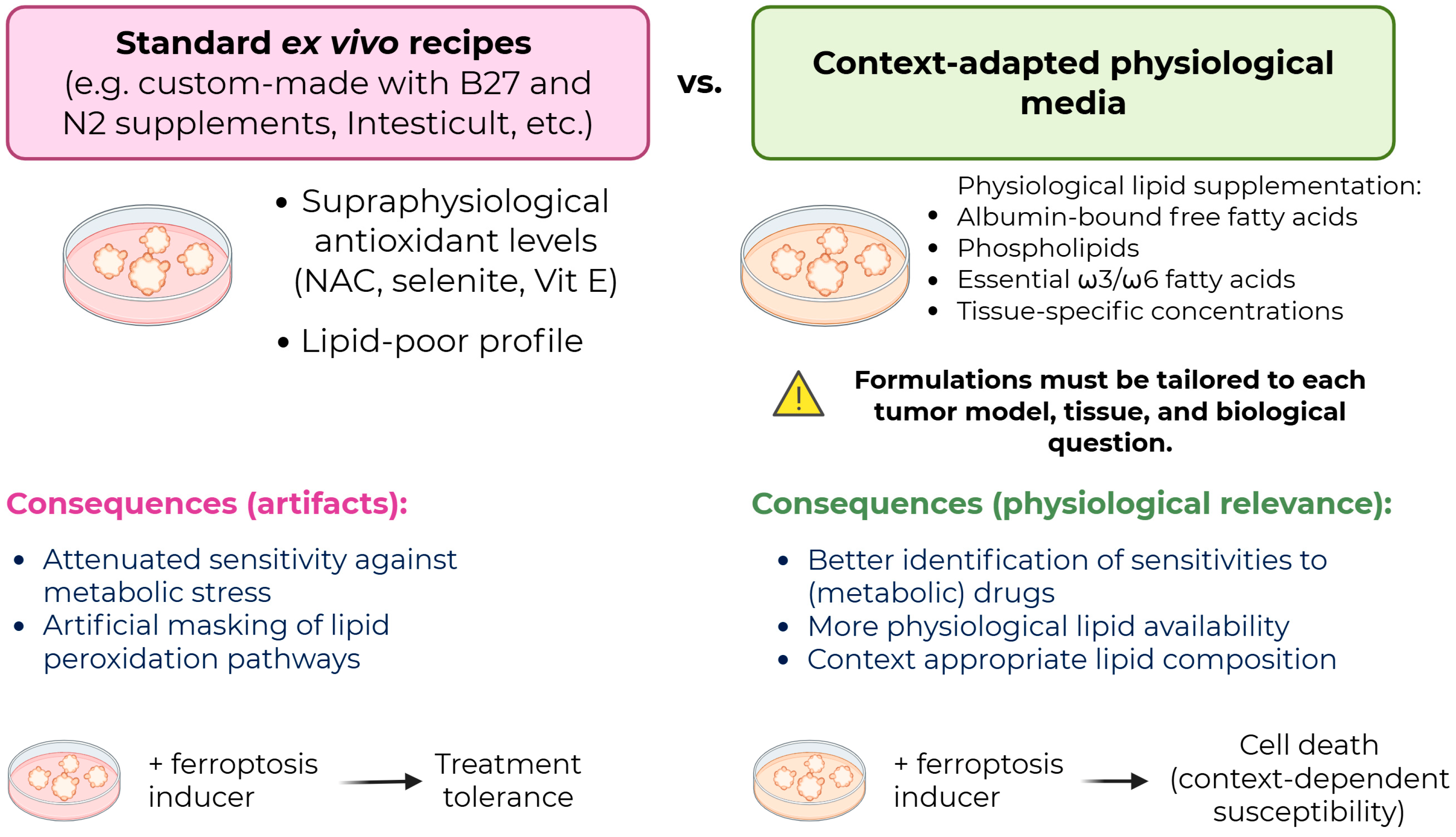
Crucially, standard organoid media contain supraphysiological concentrations of antioxidants, growth factors, and nutrients that distort native metabolic states. The profound influence of culture media on cell fate is particularly evident in studies of ferroptosis, an iron-dependent, lipid peroxidation-driven form of non-apoptotic cell death, which serves as a key example of how microenvironmental factors modulate metabolic drug susceptibility (Figure 2) [109,110,111,112,113]. Because standard media are optimized for organoid expansion rather than metabolic fidelity, they exhibit high antioxidant levels and low essential FA concentrations, which can mask metabolic dependencies and alter drug-screening outcomes [114,115].
Specifically, pioneering formulations rich in antioxidants (including B27, N2, and N-acetylcysteine) have been shown to markedly alter redox responsiveness. Eaton et al. demonstrated that commercial media (e.g., Intesticult) reversibly suppress ferroptosis [115]. Moreover, media containing B27, N2, N-acetylcysteine, selenium, and SB202190 confer prolonged tolerance to ferroptosis, persisting even after media washout [115]. Studies in synovial sarcoma organoids cultured in commercial media similarly report context-dependent variations in ferroptosis sensitivity (often lower in 3D than in 2D due to poorly defined compositions) [116]. Despite these caveats, 3D models remain valuable platforms for pro-ferroptotic drug screening [117]. However, recent studies reporting ferroptosis induction in organoids [118,119] utilized media containing suppressive antioxidants while relying on very small organoids (48–72 h post-seeding in single cells). Whether these observations translate to larger, mature organoids with established structure and diffusion gradients remains unclear. Additionally, in [119], the authors reported that normal organoids were not sensitive to omega-3-FAs and all viability experiments were measured using metabolic activity-based viability assays (in this case, ATP), which complicates the interpretation of results, given the potential impact of stress associated with lipid peroxidation. Pairing these with cytotoxicity assays is necessary to rule out compensatory metabolic mechanisms and confirm that the effects are truly driven (or not) by cell death and ferroptosis. Moreover, these experiments were performed in lipid-poor media, raising the question of what results might occur if they were conducted in media containing physiological levels of lipids.
Extracellular lipid availability further modulates ferroptosis. Lipid limitation drives intracellular FA trafficking to release stored FAs as free polyunsaturated FAs (PUFAs), which are highly susceptible to peroxidation [120]. Conversely, in 2D models, albumin-bound FAs are the primary drivers of proliferation, indicating that the specific nature of the FA matters more than total cellular abundance [121]. Critically, Park et al. showed that lipid composition dictates ferroptosis sensitivity: GPX4 inhibition works in 2D cells but fails in 3D models and xenografts due to depletion of PUFAs from phospholipids and a protective enrichment of monounsaturated FAs [122,123].
Importantly, the concept of a “physiological” culture medium must not be interpreted as a single, static formulation. Physiological concentrations of nutrients, lipids, hormones, and metabolites vary considerably across tissue origins, anatomical sites, disease stages, and individual patients. Consequently, optimizing organoid culture conditions should not necessarily aim to reproduce a universal physiological state, but rather to recapitulate the specific metabolic context relevant to the biological or clinical question under investigation. Achieving this requires context-adapted culture media tailored to tissue-specific nutrient availabilities and metabolic states, moving beyond standard formulations optimized purely for organoid growth while adhering to emerging community-driven guidelines for culture media reporting and standardization.

3.4. Circadian Regulation and Temporal Metabolic States

Tumor metabolism is temporally dynamic, regulated by intrinsic ~24-h circadian clocks that govern nutrient utilization, mitochondrial function, redox homeostasis, and drug responses [124]. Orchestrated by a central clock in the suprachiasmatic nucleus that synchronizes peripheral tissue clocks, the molecular clock relies on transcription–translation feedback loops involving core genes like BMAL1, CLOCK, PER (including PER2), and CRY [124]. While pluripotent stem cell-derived organoids (e.g., human pineal gland organoids displaying rhythmic melatonin secretion) have confirmed that 3D cultures retain tissue-specific circadian outputs [125,126], investigating these dynamics in cancer models remains a nascent field.
Real-time monitoring using bioluminescent reporters (such as BMAL1-driven [71] or PER2-driven [127,128] luciferase) in intestinal organoids reveals that circadian rhythm strength depends on cell state. Stem cell-enriched conditions exhibit weak or no oscillations, while differentiation results in strong rhythmicity [71,127,129]. Mechanistically, niche signals like Paneth cell-secreted Wnt couple circadian oscillations to the cell cycle [129]. Although intestinal tissues harbor autonomous peripheral clocks capable of self-sustained oscillations, they remain sensitive to external synchronization cues, including serum shocks, glucocorticoids, and growth factors [127]. Beyond the gut, microengineered liver organoids exhibit reproducible oscillations useful for high-throughput chronotoxicity testing [130].
In malignancy, circadian regulation is frequently altered or reprogrammed rather than entirely lost. Breast cancer organoids exhibit dampened, highly variable oscillations, where reduced clock strength correlates with heightened invasive potential [70]. In CRC organoids, clock gene disruption drives tumorigenesis by activating Wnt and MYC signaling and boosting glycolysis, resulting in flattened or desynchronized profiles compared to normal tissue [131].
Transcriptomic profiling in PDAC organoids has revealed rhythmic expression of oncogenic pathways (including EGFR and JUN) and cell cycle regulators [132]. This temporal data has been successfully leveraged to optimize chronotherapeutic scheduling, demonstrating that drug efficacy varies significantly by circadian phase [132]. Ultimately, circadian phenotypes are highly sensitive to differentiation states, synchronization protocols, and media composition, necessitating strict experimental control.
A fundamental question for clinical translation is whether these microenvironment-driven metabolic adaptations represent universal pan-cancer responses or tissue-specific phenotypes. Core survival mechanisms, such as HIF-1α stabilization under hypoxia, AMPK activation during energetic stress, and increased reliance on nutrient scavenging, are broadly conserved across solid tumors. However, specific nutrient preferences, FA utilization profiles, and targetable synthetic lethal vulnerabilities are strongly dictated by tissue-of-origin lineage, genetic background, and local microenvironmental context. While pan-cancer pathways offer broad therapeutic targets, context-restricted metabolic dependencies require tailored, patient-specific strategies modeled in high-fidelity 3D systems. Because the magnitude and functional consequences of metabolic rewiring vary across cancer types, vulnerabilities identified in one model cannot be assumed to be universally conserved, reinforcing the essential role of patient-derived 3D models in mapping context-specific metabolic dependencies for precision oncology.

4. Discussion

Patient-derived and engineered 3D tumor models uniquely preserve patient-specific heterogeneity, spatial architecture, and microenvironmental responses compared to 2D systems. However, they also reveal a key challenge for precision oncology: tumor metabolism is not entirely hardwired by cell-intrinsic genetics but is dynamically shaped by extracellular conditions. This plasticity introduces experimental variability, but it also provides an opportunity to actively tune cultures to match patient-specific pathophysiological states.
To bridge the gap between reductionist in vitro experiments and complex human biology, advanced bioengineering approaches must be integrated with PDOs. While PDOs remain the most accessible, high-throughput patient-derived platforms, complementary systems like 3D bioprinting and organ-on-chip models offer superior control over multicellular organization, fluid dynamics, and nutrient gradients [133,134,135,136,137]. Bioprinting allows researchers to isolate environmental constraints from intrinsic cellular programs and better manage nutrient delivery to mitigate non-physiological media artifacts, with integration into machine learning frameworks now underway [138].
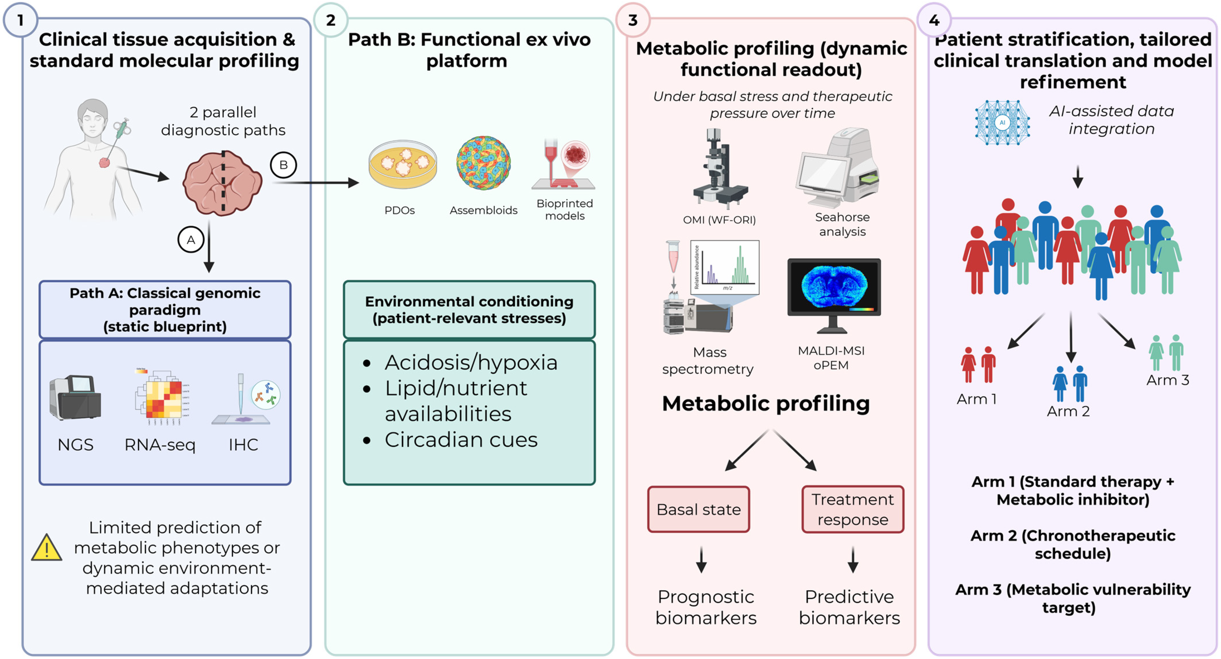
The scale of multidimensional data generated by these models, including high-content imaging, optical metabolic imaging, metabolomics, and spatial transcriptomics, necessitates the implementation of artificial intelligence (AI) and machine learning (ML). Rather than serving as a simple fix for biological limitations, AI/ML frameworks are particularly well-suited to identifying predictive metabolic signatures and classify functional tumor states. To enable clinical use, these methods require standardization of multimodal datasets across different platforms, external validation of predictive models in independent cohorts, and explainable AI systems that deliver biologically interpretable predictions. For example, transformer-based architectures like PharmaFormer integrate multimodal organoid data to predict therapeutic efficacy [139]. By combining metabolic profiling with machine learning, researchers can accelerate biomarker discovery and predict drug responses or toxicities, a strategy already validated in non-oncological settings [140,141,142].
Crucially, AI applications must remain grounded in mechanistic biology. Organoids can serve as experimental platforms for training and validating “hybrid digital twins” that integrate molecular, metabolic, imaging, and clinical profiles to refine patient stratification beyond static genomics. However, most of these applications remain preclinical or at the proof-of-concept stage. Several barriers must be addressed before metabolism-driven functional precision oncology can be routinely implemented, including technical challenges (standardization of organoid derivation, culture media, extracellular matrices and analytical workflows, interlaboratory reproducibility), biological challenges (biopsy availability, organoid establishment success, sampling bias, clonal selection, and limited modeling of systemic metabolism), and clinical challenges (turnaround time, cost, regulatory requirements, and prospective validation). Cross-laboratory protocols for organoid derivation, culture media, extracellular matrices, and analytical workflows remain unstandardized [143,144], although some efforts to standardize are currently underway, for example, in gastrointestinal research [145]. Similar reporting and quality frameworks will be required across tumor types and engineered 3D models.
In addition, current ex vivo systems only partially reproduce the systemic factors that shape tumor metabolism. Some individual components have begun to be modeled: thyroid hormone signaling can alter cancer stem-cell phenotypes and chemotherapy responses in CRC spheroids [146], whereas estradiol can stimulate proliferation in ERα-positive patient-derived thyroid cancer organoids [147]. Likewise, intestinal organoid–microbe co-culture platforms enable controlled investigation of host–microbe interactions [148], and recent studies indicate that microbiota-derived metabolites, such as butyrate, can modulate tumor growth and immunotherapy responses in CRC PDOs [149]. Dietary signals have also been investigated using organoid assays, including the tumor-suppressive effects of β-hydroxybutyrate and the interaction between high-fat diet-associated metabolites and tumor genotype [150,151]. Dietary interventions, including high-fat diet and caloric restriction, have been extensively investigated in mouse models and, to a lesser extent, in organoid systems [152,153]. However, systematic studies reproducing these nutritional contexts in human patient-derived organoids remain limited. Nevertheless, these studies generally examine isolated signals and do not yet reproduce the combined influence of diet, obesity, diabetes, endocrine regulation, microbiota-derived metabolites, hepatic drug metabolism, and inter-organ communication found in patients. Generating more systematic datasets across these contexts will be important not only to improve the physiological relevance of 3D models but also to prevent AI models from learning culture-specific artifacts and to support the development of more reliable patient-specific predictions. Accordingly, the strategy illustrated in Figure 3 should be viewed as a proposed conceptual framework rather than an established clinical workflow.
In conclusion, the convergence of PDOs, advanced bioengineering, metabolomics, and computational modeling is shifting precision oncology toward the functional characterization and targeting of dynamic metabolic vulnerabilities, moving beyond static genomic profiling alone. Future clinical success will depend not only on integrating these technologies but also on accurately defining and controlling the biological context that shapes tumor metabolism, thereby enabling more physiologically relevant experimental models and more reliable AI-assisted clinical predictions.

5. Conclusions

3D tumor models, especially PDOs, have greatly advanced the study of cancer metabolism by enabling functional analyses in physiologically relevant and patient-specific contexts. As discussed throughout this review, these systems preserve key aspects of tumor heterogeneity while allowing investigation of metabolic plasticity, treatment-induced rewiring, and microenvironmental interactions.
Importantly, current evidence demonstrates that metabolic phenotypes in 3D models are highly context-dependent and strongly influenced by experimental conditions, including nutrient availability, extracellular lipid composition, oxygenation, pH, stromal interactions, and circadian regulation. This represents both a limitation and an opportunity. While non-physiological culture conditions may distort metabolic observations and therapeutic responses, careful control and refinement of these parameters could enable more accurate modeling of patient-specific tumor biology.
The field is now evolving toward increasingly sophisticated systems integrating stromal and immune compartments, microfluidics, vascularization, and engineered tissue architectures. In parallel, advances in imaging, metabolomics, spatial profiling, and computational analysis are enabling multidimensional characterization of metabolic states at unprecedented resolution. Together, these developments are progressively moving precision oncology beyond purely genomic classification toward functional and dynamic patient stratification.
Although substantial technical and conceptual challenges remain, integrating patient-derived 3D models with metabolic profiling and computational modeling holds considerable promise for identifying actionable metabolic vulnerabilities, predicting therapeutic responses, and refining personalized treatment strategies. Achieving this potential will necessitate interdisciplinary teamwork, standardized experimental and analytical procedures, reliable metabolic profiling pipelines suitable for clinical use, and validation across multiple centers in prospective studies. Ultimately, these platforms may help transform cancer metabolism from a largely descriptive field into a clinically actionable component of functional precision oncology.

Author Contributions

Conceptualization, M.V.G., O.F. and C.C.; investigation, M.V.G., O.F. and C.C.; writing—original draft preparation, M.V.G., O.F. and C.C.; supervision, O.F. and C.C.; funding acquisition, O.F. and C.C. All authors have read and agreed to the published version of the manuscript.

Funding

This research was supported by grants from the Fonds de la Recherche Scientifique (F.R.S.-FNRS): PDR T.0076.26 (C.C.), WELBIO (Walloon Excellence in Life Sciences and Biotechnology) X.2506.24 (C.C.), the Belgian Foundation against cancer (2022-168) (C.C.), Action de Recherche Concertée (ARC 23/28-129) (C.C.), the Fondation Saint-Luc 41.21220.004 (O.F., C.C.), and the Région wallonne, the Fédération Wallonie-Bruxelles, and the Région Bruxelloise, Belgium: Plateforme technologique d’excellence “Alternatives aux expérimentations animales” (convention n°2310113) (C.C.).

Institutional Review Board Statement

Not applicable.

Informed Consent Statement

Not applicable.

Data Availability Statement

No new data were created or analyzed in this study. Data sharing is not applicable to this article.

Acknowledgments

All figures were created with BioRender.com. The authors have reviewed and edited the output and take full responsibility for the content of this publication.

Conflicts of Interest

The authors declare no conflicts of interest.

Abbreviations

The following abbreviations are used in this manuscript:
AIArtificial intelligence
ALIAir–liquid interface
CAFCancer-associated fibroblast
CRCColorectal cancer
ECARExtracellular acidification rate
EMPEpithelial–mesenchymal plasticity
FAFatty acid
FLIMFluorescence lifetime imaging microscopy
GCGas chromatography
HR MAS-MRSHigh-resolution magic angle spinning magnetic resonance spectroscopy
LCLiquid chromatography
MALDIMatrix-assisted laser desorption/ionization
MLMachine learning
MSMass spectrometry
NMRNuclear Magnetic Resonance
OCROxygen consumption rate
OMIOptical metabolic imaging
oPEMOrganoid positron-emission microscopy
OXPHOSOxidative phosphorylation
PDACPancreatic ductal adenocarcinoma
PDOPatient-derived tumor organoid
PLIMPhosphorescence lifetime imaging microscopy
TMETumor microenvironment
WF-ORIWide-field Optical redox imaging

References

  1. Hanahan, D. Hallmarks of cancer-Then and now, and beyond. Cell 2026, 189, 2254–2277. [Google Scholar] [CrossRef] [Scilit] [PubMed]
  2. Demicco, M.; Liu, X.Z.; Leithner, K.; Fendt, S.M. Metabolic heterogeneity in cancer. Nat. Metab. 2024, 6, 18–38. [Google Scholar] [CrossRef] [Scilit] [PubMed]
  3. Yang, J.; Shay, C.; Saba, N.F.; Teng, Y. Cancer metabolism and carcinogenesis. Exp. Hematol. Oncol. 2024, 13, 10. [Google Scholar] [CrossRef] [Scilit] [PubMed]
  4. Goncalves, A.C.; Richiardone, E.; Jorge, J.; Polonia, B.; Xavier, C.P.R.; Salaroglio, I.C.; Riganti, C.; Vasconcelos, M.H.; Corbet, C.; Sarmento-Ribeiro, A.B. Impact of cancer metabolism on therapy resistance—Clinical implications. Drug Resist. Updat. 2021, 59, 100797. [Google Scholar] [CrossRef] [Scilit] [PubMed]
  5. Elia, I.; Haigis, M.C. Metabolites and the tumour microenvironment: From cellular mechanisms to systemic metabolism. Nat. Metab. 2021, 3, 21–32. [Google Scholar] [CrossRef] [Scilit] [PubMed]
  6. Thorel, L.; Perreard, M.; Florent, R.; Divoux, J.; Coffy, S.; Vincent, A.; Gaggioli, C.; Guasch, G.; Gidrol, X.; Weiswald, L.B.; et al. Patient-derived tumor organoids: A new avenue for preclinical research and precision medicine in oncology. Exp. Mol. Med. 2024, 56, 1531–1551. [Google Scholar] [CrossRef] [Scilit] [PubMed]
  7. Kim, S.Y.; van de Wetering, M.; Clevers, H.; Sanders, K. The future of tumor organoids in precision therapy. Trends Cancer 2025, 11, 665–675. [Google Scholar] [CrossRef] [Scilit] [PubMed]
  8. Gunti, S.; Hoke, A.T.K.; Vu, K.P.; London, N.R., Jr. Organoid and Spheroid Tumor Models: Techniques and Applications. Cancers 2021, 13, 874. [Google Scholar] [CrossRef] [Scilit] [PubMed]
  9. Tuveson, D.; Clevers, H. Cancer modeling meets human organoid technology. Science 2019, 364, 952–955. [Google Scholar] [CrossRef] [Scilit] [PubMed]
  10. LeSavage, B.L.; Suhar, R.A.; Broguiere, N.; Lutolf, M.P.; Heilshorn, S.C. Next-generation cancer organoids. Nat. Mater. 2022, 21, 143–159. [Google Scholar] [CrossRef] [Scilit] [PubMed]
  11. Kim, J.; Koo, B.K.; Knoblich, J.A. Human organoids: Model systems for human biology and medicine. Nat. Rev. Mol. Cell Biol. 2020, 21, 571–584. [Google Scholar] [CrossRef] [Scilit] [PubMed]
  12. Polak, R.; Zhang, E.T.; Kuo, C.J. Cancer organoids 2.0: Modelling the complexity of the tumour immune microenvironment. Nat. Rev. Cancer 2024, 24, 523–539. [Google Scholar] [CrossRef] [Scilit] [PubMed]
  13. Hwangbo, H.; Chae, S.; Kim, W.; Jo, S.; Kim, G.H. Tumor-on-a-chip models combined with mini-tissues or organoids for engineering tumor tissues. Theranostics 2024, 14, 33–55. [Google Scholar] [CrossRef] [Scilit] [PubMed]
  14. Walsh, A.J.; Skala, M.C. Optical metabolic imaging quantifies heterogeneous cell populations. Biomed. Opt. Express 2015, 6, 559–573. [Google Scholar] [CrossRef] [Scilit] [PubMed]
  15. Walsh, A.J.; Cook, R.S.; Sanders, M.E.; Aurisicchio, L.; Ciliberto, G.; Arteaga, C.L.; Skala, M.C. Quantitative optical imaging of primary tumor organoid metabolism predicts drug response in breast cancer. Cancer Res. 2014, 74, 5184–5194. [Google Scholar] [CrossRef] [Scilit] [PubMed]
  16. Walsh, A.J.; Cook, R.S.; Manning, H.C.; Hicks, D.J.; Lafontant, A.; Arteaga, C.L.; Skala, M.C. Optical metabolic imaging identifies glycolytic levels, subtypes, and early-treatment response in breast cancer. Cancer Res. 2013, 73, 6164–6174. [Google Scholar] [CrossRef] [Scilit] [PubMed]
  17. Walsh, A.J.; Castellanos, J.A.; Nagathihalli, N.S.; Merchant, N.B.; Skala, M.C. Optical Imaging of Drug-Induced Metabolism Changes in Murine and Human Pancreatic Cancer Organoids Reveals Heterogeneous Drug Response. Pancreas 2016, 45, 863–869. [Google Scholar] [CrossRef] [Scilit] [PubMed]
  18. Sharick, J.T.; Walsh, C.M.; Sprackling, C.M.; Pasch, C.A.; Pham, D.L.; Esbona, K.; Choudhary, A.; Garcia-Valera, R.; Burkard, M.E.; McGregor, S.M.; et al. Metabolic Heterogeneity in Patient Tumor-Derived Organoids by Primary Site and Drug Treatment. Front. Oncol. 2020, 10, 553. [Google Scholar] [CrossRef] [Scilit] [PubMed]
  19. Sharick, J.T.; Jeffery, J.J.; Karim, M.R.; Walsh, C.M.; Esbona, K.; Cook, R.S.; Skala, M.C. Cellular Metabolic Heterogeneity In Vivo Is Recapitulated in Tumor Organoids. Neoplasia 2019, 21, 615–626. [Google Scholar] [CrossRef] [Scilit] [PubMed]
  20. Shah, A.T.; Heaster, T.M.; Skala, M.C. Metabolic Imaging of Head and Neck Cancer Organoids. PLoS ONE 2017, 12, e0170415. [Google Scholar] [CrossRef] [Scilit] [PubMed]
  21. Pasch, C.A.; Favreau, P.F.; Yueh, A.E.; Babiarz, C.P.; Gillette, A.A.; Sharick, J.T.; Karim, M.R.; Nickel, K.P.; DeZeeuw, A.K.; Sprackling, C.M.; et al. Patient-Derived Cancer Organoid Cultures to Predict Sensitivity to Chemotherapy and Radiation. Clin. Cancer Res. 2019, 25, 5376–5387. [Google Scholar] [CrossRef] [Scilit] [PubMed]
  22. Heaster, T.M.; Humayun, M.; Yu, J.; Beebe, D.J.; Skala, M.C. Autofluorescence Imaging of 3D Tumor-Macrophage Microscale Cultures Resolves Spatial and Temporal Dynamics of Macrophage Metabolism. Cancer Res. 2020, 80, 5408–5423. [Google Scholar] [CrossRef] [Scilit] [PubMed]
  23. Okkelman, I.A.; Neto, N.; Papkovsky, D.B.; Monaghan, M.G.; Dmitriev, R.I. A deeper understanding of intestinal organoid metabolism revealed by combining fluorescence lifetime imaging microscopy (FLIM) and extracellular flux analyses. Redox Biol. 2020, 30, 101420. [Google Scholar] [CrossRef] [Scilit] [PubMed]
  24. Okkelman, I.A.; Foley, T.; Papkovsky, D.B.; Dmitriev, R.I. Multi-Parametric Imaging of Hypoxia and Cell Cycle in Intestinal Organoid Culture. Adv. Exp. Med. Biol. 2017, 1035, 85–103. [Google Scholar] [CrossRef] [Scilit] [PubMed]
  25. Hsu, A.; Samimi, K.; Gillette, A.A.; Udgata, S.; Schmitz, A.E.; Zhao, W.; Deming, D.A.; Skala, M.C. An automated image analysis pipeline for wide-field optical redox imaging of patient-derived cancer organoids. Sci. Rep. 2026, 16, 9757. [Google Scholar] [CrossRef] [Scilit] [PubMed]
  26. Gillette, A.A.; Babiarz, C.P.; VanDommelen, A.R.; Pasch, C.A.; Clipson, L.; Matkowskyj, K.A.; Deming, D.A.; Skala, M.C. Autofluorescence Imaging of Treatment Response in Neuroendocrine Tumor Organoids. Cancers 2021, 13, 1873. [Google Scholar] [CrossRef] [Scilit] [PubMed]
  27. Wang, Y.; Hummon, A.B. MS imaging of multicellular tumor spheroids and organoids as an emerging tool for personalized medicine and drug discovery. J. Biol. Chem. 2021, 297, 101139. [Google Scholar] [CrossRef] [Scilit] [PubMed]
  28. Tucker, L.H.; Hamm, G.R.; Sargeant, R.J.E.; Goodwin, R.J.A.; Mackay, C.L.; Campbell, C.J.; Clarke, D.J. Untargeted Metabolite Mapping in 3D Cell Culture Models Using High Spectral Resolution FT-ICR Mass Spectrometry Imaging. Anal. Chem. 2019, 91, 9522–9529. [Google Scholar] [CrossRef] [Scilit] [PubMed]
  29. Liu, X.; Flinders, C.; Mumenthaler, S.M.; Hummon, A.B. MALDI Mass Spectrometry Imaging for Evaluation of Therapeutics in Colorectal Tumor Organoids. J. Am. Soc. Mass Spectrom. 2018, 29, 516–526. [Google Scholar] [CrossRef] [Scilit] [PubMed]
  30. He, M.J.; Pu, W.; Wang, X.; Zhang, W.; Tang, D.; Dai, Y. Comparing DESI-MSI and MALDI-MSI Mediated Spatial Metabolomics and Their Applications in Cancer Studies. Front. Oncol. 2022, 12, 891018. [Google Scholar] [CrossRef] [Scilit] [PubMed]
  31. Denti, V.; Andersen, M.K.; Smith, A.; Bofin, A.M.; Nordborg, A.; Magni, F.; Moestue, S.A.; Giampa, M. Reproducible Lipid Alterations in Patient-Derived Breast Cancer Xenograft FFPE Tissue Identified with MALDI MSI for Pre-Clinical and Clinical Application. Metabolites 2021, 11, 577. [Google Scholar] [CrossRef] [Scilit] [PubMed]
  32. David, B.P.; Dubrovskyi, O.; Speltz, T.E.; Wolff, J.J.; Frasor, J.; Sanchez, L.M.; Moore, T.W. Using Tumor Explants for Imaging Mass Spectrometry Visualization of Unlabeled Peptides and Small Molecules. ACS Med. Chem. Lett. 2018, 9, 768–772. [Google Scholar] [CrossRef] [Scilit] [PubMed]
  33. Khan, S.; Shin, J.H.; Ferri, V.; Cheng, N.; Noel, J.E.; Kuo, C.; Sunwoo, J.B.; Pratx, G. High-resolution positron emission microscopy of patient-derived tumor organoids. Nat. Commun. 2021, 12, 5883. [Google Scholar] [CrossRef] [Scilit] [PubMed]
  34. Zhou, Q.; Alvarez, M.R.S.; Solakyildirim, K.; Tena, J.; Serrano, L.M.N.; Lam, M.; Nguyen, C.; Tobias, F.; Hummon, A.B.; Nacario, R.C.; et al. Multi-glycomic analysis of spheroid glycocalyx differentiates 2- and 3-dimensional cell models. Glycobiology 2023, 33, 2–16. [Google Scholar] [CrossRef] [Scilit] [PubMed]
  35. Yoshizaki, H.; Ogiso, H.; Okazaki, T.; Kiyokawa, E. Comparative lipid analysis in the normal and cancerous organoids of MDCK cells. J. Biochem. 2016, 159, 573–584. [Google Scholar] [CrossRef] [Scilit] [PubMed]
  36. Weygand, J.; Carter, S.E.; Salzillo, T.C.; Moussalli, M.; Dai, B.; Dutta, P.; Zuo, X.; Fleming, J.B.; Shureiqi, I.; Bhattacharya, P. Can an organoid recapitulate the metabolome of its parent tissue? A pilot NMR spectroscopy study. J. Cancer Prev. Curr. Res. 2017, 8, 425–429. [Google Scholar] [CrossRef] [Scilit]
  37. van der Kemp, W.J.M.; Grinde, M.T.; Malvik, J.O.; van Laarhoven, H.W.M.; Prompers, J.J.; Klomp, D.W.J.; Burgering, B.; Bathen, T.F.; Moestue, S.A. Metabolic profiling of colorectal cancer organoids: A comparison between high-resolution magic angle spinning magnetic resonance spectroscopy and solution nuclear magnetic resonance spectroscopy of polar extracts. NMR Biomed. 2023, 36, e4882. [Google Scholar] [CrossRef] [Scilit] [PubMed]
  38. Reustle, A.; Menig, L.S.; Leuthold, P.; Hofmann, U.; Stuhler, V.; Schmees, C.; Becker, M.; Haag, M.; Klumpp, V.; Winter, S.; et al. Nicotinamide-N-methyltransferase is a promising metabolic drug target for primary and metastatic clear cell renal cell carcinoma. Clin. Transl. Med. 2022, 12, e883. [Google Scholar] [CrossRef] [Scilit] [PubMed]
  39. Neef, S.K.; Janssen, N.; Winter, S.; Wallisch, S.K.; Hofmann, U.; Dahlke, M.H.; Schwab, M.; Murdter, T.E.; Haag, M. Metabolic Drug Response Phenotyping in Colorectal Cancer Organoids by LC-QTOF-MS. Metabolites 2020, 10, 494. [Google Scholar] [CrossRef] [Scilit] [PubMed]
  40. Maddocks, O.D.K.; Athineos, D.; Cheung, E.C.; Lee, P.; Zhang, T.; van den Broek, N.J.F.; Mackay, G.M.; Labuschagne, C.F.; Gay, D.; Kruiswijk, F.; et al. Modulating the therapeutic response of tumours to dietary serine and glycine starvation. Nature 2017, 544, 372–376. [Google Scholar] [CrossRef] [Scilit] [PubMed]
  41. Lindeboom, R.G.; van Voorthuijsen, L.; Oost, K.C.; Rodriguez-Colman, M.J.; Luna-Velez, M.V.; Furlan, C.; Baraille, F.; Jansen, P.W.; Ribeiro, A.; Burgering, B.M.; et al. Integrative multi-omics analysis of intestinal organoid differentiation. Mol. Syst. Biol. 2018, 14, e8227. [Google Scholar] [CrossRef] [Scilit] [PubMed]
  42. Feldman, A.; Mukha, D.; Maor, I.I.; Sedov, E.; Koren, E.; Yosefzon, Y.; Shlomi, T.; Fuchs, Y. Blimp1(+) cells generate functional mouse sebaceous gland organoids in vitro. Nat. Commun. 2019, 10, 2348. [Google Scholar] [CrossRef] [Scilit] [PubMed]
  43. Kes, M.M.G.; Morales-Rodriguez, F.; Zaal, E.A.; de Souza, T.; Proost, N.; van de Ven, M.; van den Heuvel-Eibrink, M.M.; Jansen, J.W.A.; Berkers, C.R.; Drost, J. Metabolic profiling of patient-derived organoids reveals nucleotide synthesis as a metabolic vulnerability in malignant rhabdoid tumors. Cell Rep. Med. 2025, 6, 101878. [Google Scholar] [CrossRef] [Scilit] [PubMed]
  44. Miguel, V.; Reimer, K.C.; Galyga, A.K.; Jansen, J.; Mollmann, J.; Meyer, L.; Schneider, R.K.; Kramann, R. Protocol to analyze bioenergetics in single human induced-pluripotent-stem-cell-derived kidney organoids using Seahorse XF96. STAR Protoc. 2023, 4, 101999. [Google Scholar] [CrossRef] [Scilit] [PubMed]
  45. Ludikhuize, M.C.; Meerlo, M.; Burgering, B.M.T.; Rodriguez Colman, M.J. Protocol to profile the bioenergetics of organoids using Seahorse. STAR Protoc. 2021, 2, 100386. [Google Scholar] [CrossRef] [Scilit] [PubMed]
  46. Campioni, G.; Pasquale, V.; Busti, S.; Ducci, G.; Sacco, E.; Vanoni, M. An Optimized Workflow for the Analysis of Metabolic Fluxes in Cancer Spheroids Using Seahorse Technology. Cells 2022, 11, 866. [Google Scholar] [CrossRef] [Scilit] [PubMed]
  47. Godet, I.; Shin, Y.J.; Ju, J.A.; Ye, I.C.; Wang, G.; Gilkes, D.M. Fate-mapping post-hypoxic tumor cells reveals a ROS-resistant phenotype that promotes metastasis. Nat. Commun. 2019, 10, 4862. [Google Scholar] [CrossRef] [Scilit] [PubMed]
  48. Hubert, C.G.; Rivera, M.; Spangler, L.C.; Wu, Q.; Mack, S.C.; Prager, B.C.; Couce, M.; McLendon, R.E.; Sloan, A.E.; Rich, J.N. A Three-Dimensional Organoid Culture System Derived from Human Glioblastomas Recapitulates the Hypoxic Gradients and Cancer Stem Cell Heterogeneity of Tumors Found In Vivo. Cancer Res. 2016, 76, 2465–2477. [Google Scholar] [CrossRef] [Scilit] [PubMed]
  49. Huang, L.; Holtzinger, A.; Jagan, I.; BeGora, M.; Lohse, I.; Ngai, N.; Nostro, C.; Wang, R.; Muthuswamy, L.B.; Crawford, H.C.; et al. Ductal pancreatic cancer modeling and drug screening using human pluripotent stem cell- and patient-derived tumor organoids. Nat. Med. 2015, 21, 1364–1371. [Google Scholar] [CrossRef] [Scilit] [PubMed]
  50. Gillette, A.; Udgata, S.; Schmitz, A.E.; Stoecker, J.N.; Kratz, J.D.; Deming, D.A.; Skala, M.C. Wide-Field Optical Redox Imaging with Leading-Edge Detection Enables Assessment of Treatment Response and Heterogeneity in Patient-Derived Cancer Organoids. Cancer Res. 2025, 85, 4329–4340. [Google Scholar] [CrossRef] [Scilit] [PubMed]
  51. Ayuso, J.M.; Rehman, S.; Farooqui, M.; Virumbrales-Munoz, M.; Setaluri, V.; Skala, M.C.; Beebe, D.J. Microfluidic Tumor-on-a-Chip Model to Study Tumor Metabolic Vulnerability. Int. J. Mol. Sci. 2020, 21, 9075. [Google Scholar] [CrossRef] [Scilit] [PubMed]
  52. Vander Linden, C.; Corbet, C. Reconciling environment-mediated metabolic heterogeneity with the oncogene-driven cancer paradigm in precision oncology. Semin. Cell Dev. Biol. 2020, 98, 202–210. [Google Scholar] [CrossRef] [Scilit] [PubMed]
  53. Chan, B.K.C.; Zhang, C.; Poon, C.H.; Lee, M.H.Y.; Chu, H.Y.; Wang, B.; Chen, S.G.; Yan, H.H.N.; Leung, S.Y.; Wong, A.S.L. A combined enteric neuron-gastric tumor organoid reveals metabolic vulnerabilities in gastric cancer. Cell Stem Cell 2025, 32, 1595–1613.e1510. [Google Scholar] [CrossRef] [Scilit] [PubMed]
  54. Gong, Y.; Ji, P.; Yang, Y.S.; Xie, S.; Yu, T.J.; Xiao, Y.; Jin, M.L.; Ma, D.; Guo, L.W.; Pei, Y.C.; et al. Metabolic-Pathway-Based Subtyping of Triple-Negative Breast Cancer Reveals Potential Therapeutic Targets. Cell Metab. 2021, 33, 51–64.e9. [Google Scholar] [CrossRef] [Scilit] [PubMed]
  55. Xiao, Y.; Ma, D.; Yang, Y.S.; Yang, F.; Ding, J.H.; Gong, Y.; Jiang, L.; Ge, L.P.; Wu, S.Y.; Yu, Q.; et al. Comprehensive metabolomics expands precision medicine for triple-negative breast cancer. Cell Res. 2022, 32, 477–490. [Google Scholar] [CrossRef] [Scilit] [PubMed]
  56. Nguyen, N.T.B.; Gevers, S.; Kok, R.N.U.; Burgering, L.M.; Neikes, H.; Akkerman, N.; Betjes, M.A.; Ludikhuize, M.C.; Gulersonmez, C.; Stigter, E.C.A.; et al. Lactate controls cancer stemness and plasticity through epigenetic regulation. Cell Metab. 2025, 37, 903–919.e910. [Google Scholar] [CrossRef] [Scilit] [PubMed]
  57. Grun, C.; Pfeifer, J.; Liebsch, G.; Gottwald, E. O(2)-sensitive microcavity arrays: A new platform for oxygen measurements in 3D cell cultures. Front. Bioeng. Biotechnol. 2023, 11, 1111316. [Google Scholar] [CrossRef] [Scilit] [PubMed]
  58. Nashimoto, Y.; Shishido, S.; Onuma, K.; Ino, K.; Inoue, M.; Shiku, H. Oxygen metabolism analysis of a single organoid for non-invasive discrimination of cancer subpopulations with different growth capabilities. Front. Bioeng. Biotechnol. 2023, 11, 1184325. [Google Scholar] [CrossRef] [Scilit] [PubMed]
  59. Ino, K.; Wachi, M.; Utagawa, Y.; Konno, A.; Takinoue, M.; Abe, H.; Shiku, H. Scanning electrochemical microscopy for determining oxygen consumption rates of cells in hydrogel fibers fabricated using an extrusion 3D bioprinter. Anal. Chim. Acta 2024, 1304, 342539. [Google Scholar] [CrossRef] [Scilit] [PubMed]
  60. Chiolerio, P.; Auletta, B.; Pezzini, C.; Sartore, L.; Gregolon, G.; Gagliano, O.; Laterza, C.; Balmaceda Valdez, V.R.; Cacchiarelli, D.; Luni, C.; et al. Human neuromuscular organoids mimic cancer-induced muscle cachexia. Cell Rep. Methods 2026, 6, 101331. [Google Scholar] [CrossRef] [Scilit] [PubMed]
  61. Lacombe, J.; Dunn, S.E.; Layac, M.; Soldevila, M.; Fakrudin, N.M.; Duane, B.; Chen, K.; Barrett, M.W.; Helton, J.; Laiakis, E.C.; et al. A human 3D culture-organ-on-chip platform for investigating the tumor microenvironment response to ionizing radiation. iScience 2026, 29, 114236. [Google Scholar] [CrossRef] [Scilit] [PubMed]
  62. Rolver, M.G.; Holland, L.K.K.; Ponniah, M.; Prasad, N.S.; Yao, J.; Schnipper, J.; Kramer, S.; Elingaard-Larsen, L.; Pedraz-Cuesta, E.; Liu, B.; et al. Chronic acidosis rewires cancer cell metabolism through PPARalpha signaling. Int. J. Cancer 2023, 152, 1668–1684. [Google Scholar] [CrossRef] [Scilit] [PubMed]
  63. Stigliani, A.; Ialchina, R.; Yao, J.; Czaplinska, D.; Dai, Y.; Andersen, H.B.; Rennie, S.; Andersson, R.; Pedersen, S.F.; Sandelin, A. Adaptation to an acid microenvironment promotes pancreatic cancer organoid growth and drug resistance. Cell Rep. 2024, 43, 114409. [Google Scholar] [CrossRef] [Scilit] [PubMed]
  64. Ibanez, S.; Giolito, M.V.; Al-Siyabi, S.; Serafimov, K.; Laloux, F.; Dierge, E.; Aubert, L.; Guilbaud, C.; Kaye, S.; Varon, P.; et al. Acidosis Forces Fatty Acid Uptake and Metabolism in Cancer Cells Regardless of Genotype. Adv. Sci. 2025, 12, e05436. [Google Scholar] [CrossRef] [Scilit] [PubMed]
  65. Richiardone, E.; Giolito, M.V.; Al Roumi, R.; Ambroise, J.; Boidot, R.; Drotleff, B.; Ghesquiere, B.; Lupo, B.; Trusolino, L.; Bardelli, A.; et al. Acidosis overrides molecular heterogeneity to shape therapeutically targetable metabolic phenotypes in colon cancers. Cancer Lett. 2025, 613, 217512. [Google Scholar] [CrossRef] [Scilit] [PubMed]
  66. Tsai, S.; McOlash, L.; Palen, K.; Johnson, B.; Duris, C.; Yang, Q.; Dwinell, M.B.; Hunt, B.; Evans, D.B.; Gershan, J.; et al. Development of primary human pancreatic cancer organoids, matched stromal and immune cells and 3D tumor microenvironment models. BMC Cancer 2018, 18, 335. [Google Scholar] [CrossRef] [Scilit] [PubMed]
  67. Sharpe, B.P.; Nazlamova, L.A.; Tse, C.; Johnston, D.A.; Thomas, J.; Blyth, R.; Pickering, O.J.; Grace, B.; Harrington, J.; Rajak, R.; et al. Patient-derived tumor organoid and fibroblast assembloid models for interrogation of the tumor microenvironment in esophageal adenocarcinoma. Cell Rep. Methods 2024, 4, 100909. [Google Scholar] [CrossRef] [Scilit] [PubMed]
  68. Seino, T.; Kawasaki, S.; Shimokawa, M.; Tamagawa, H.; Toshimitsu, K.; Fujii, M.; Ohta, Y.; Matano, M.; Nanki, K.; Kawasaki, K.; et al. Human Pancreatic Tumor Organoids Reveal Loss of Stem Cell Niche Factor Dependence during Disease Progression. Cell Stem Cell 2018, 22, 454–467.e456. [Google Scholar] [CrossRef] [Scilit] [PubMed]
  69. Murthy, D.; Attri, K.S.; Shukla, S.K.; Thakur, R.; Chaika, N.V.; He, C.; Wang, D.; Jha, K.; Dasgupta, A.; King, R.J.; et al. Cancer-associated fibroblast-derived acetate promotes pancreatic cancer development by altering polyamine metabolism via the ACSS2-SP1-SAT1 axis. Nat. Cell Biol. 2024, 26, 613–627. [Google Scholar] [CrossRef] [Scilit] [PubMed]
  70. Li, S.Y.; Hammarlund, J.A.; Wu, G.; Lian, J.W.; Howell, S.J.; Clarke, R.B.; Adamson, A.D.; Goncalves, C.F.; Hogenesch, J.B.; Anafi, R.C.; et al. Tumor circadian clock strength influences metastatic potential and predicts patient prognosis in luminal A breast cancer. Proc. Natl. Acad. Sci. USA 2024, 121, e2311854121. [Google Scholar] [CrossRef] [Scilit] [PubMed]
  71. Goker, S.; Lee, S.; Hong, C.I. Monitoring Circadian Oscillations with a Bioluminescence Reporter in Organoids. J. Vis. Exp. 2024, 204, e66381. [Google Scholar] [CrossRef] [Scilit] [PubMed]
  72. Li, F.; Simon, M.C. Cancer Cells Don’t Live Alone: Metabolic Communication within Tumor Microenvironments. Dev. Cell 2020, 54, 183–195. [Google Scholar] [CrossRef] [Scilit] [PubMed]
  73. Zhao, H.; Jiang, E.; Shang, Z. 3D Co-culture of Cancer-Associated Fibroblast with Oral Cancer Organoids. J. Dent. Res. 2021, 100, 201–208. [Google Scholar] [CrossRef] [Scilit] [PubMed]
  74. Biffi, G.; Oni, T.E.; Spielman, B.; Hao, Y.; Elyada, E.; Park, Y.; Preall, J.; Tuveson, D.A. IL1-Induced JAK/STAT Signaling Is Antagonized by TGFbeta to Shape CAF Heterogeneity in Pancreatic Ductal Adenocarcinoma. Cancer Discov. 2019, 9, 282–301. [Google Scholar] [CrossRef] [Scilit] [PubMed]
  75. Yuki, K.; Cheng, N.; Nakano, M.; Kuo, C.J. Organoid Models of Tumor Immunology. Trends Immunol. 2020, 41, 652–664. [Google Scholar] [CrossRef] [Scilit] [PubMed]
  76. Dijkstra, K.K.; Cattaneo, C.M.; Weeber, F.; Chalabi, M.; van de Haar, J.; Fanchi, L.F.; Slagter, M.; van der Velden, D.L.; Kaing, S.; Kelderman, S.; et al. Generation of Tumor-Reactive T Cells by Co-culture of Peripheral Blood Lymphocytes and Tumor Organoids. Cell 2018, 174, 1586–1598.e12. [Google Scholar] [CrossRef] [Scilit] [PubMed]
  77. Cattaneo, C.M.; Dijkstra, K.K.; Fanchi, L.F.; Kelderman, S.; Kaing, S.; van Rooij, N.; van den Brink, S.; Schumacher, T.N.; Voest, E.E. Tumor organoid-T-cell coculture systems. Nat. Protoc. 2020, 15, 15–39. [Google Scholar] [CrossRef] [Scilit] [PubMed]
  78. Datta, R.; Sivanand, S.; Lau, A.N.; Florek, L.V.; Barbeau, A.M.; Wyckoff, J.; Skala, M.C.; Vander Heiden, M.G. Interactions with stromal cells promote a more oxidized cancer cell redox state in pancreatic tumors. Sci. Adv. 2022, 8, eabg6383. [Google Scholar] [CrossRef] [Scilit] [PubMed]
  79. Subtil, B.; Iyer, K.K.; Poel, D.; Bakkerus, L.; Gorris, M.A.J.; Escalona, J.C.; van den Dries, K.; Cambi, A.; Verheul, H.M.W.; de Vries, I.J.M.; et al. Dendritic cell phenotype and function in a 3D co-culture model of patient-derived metastatic colorectal cancer organoids. Front. Immunol. 2023, 14, 1105244. [Google Scholar] [CrossRef] [Scilit] [PubMed]
  80. Grassi, L.; Alfonsi, R.; Francescangeli, F.; Signore, M.; De Angelis, M.L.; Addario, A.; Costantini, M.; Flex, E.; Ciolfi, A.; Pizzi, S.; et al. Organoids as a new model for improving regenerative medicine and cancer personalized therapy in renal diseases. Cell Death Dis. 2019, 10, 201. [Google Scholar] [CrossRef] [Scilit] [PubMed]
  81. Balazova, K.; Clevers, H.; Dost, A.F.M. The role of macrophages in non-small cell lung cancer and advancements in 3D co-cultures. Elife 2023, 12, e82998. [Google Scholar] [CrossRef] [Scilit] [PubMed]
  82. Kim, M.; Mun, H.; Sung, C.O.; Cho, E.J.; Jeon, H.J.; Chun, S.M.; Jung, D.J.; Shin, T.H.; Jeong, G.S.; Kim, D.K.; et al. Patient-derived lung cancer organoids as in vitro cancer models for therapeutic screening. Nat. Commun. 2019, 10, 3991. [Google Scholar] [CrossRef] [Scilit] [PubMed]
  83. Lee, S.H.; Hu, W.; Matulay, J.T.; Silva, M.V.; Owczarek, T.B.; Kim, K.; Chua, C.W.; Barlow, L.J.; Kandoth, C.; Williams, A.B.; et al. Tumor Evolution and Drug Response in Patient-Derived Organoid Models of Bladder Cancer. Cell 2018, 173, 515–528.e517. [Google Scholar] [CrossRef] [Scilit] [PubMed]
  84. Raffo-Romero, A.; Ziane-Chaouche, L.; Salome-Desnoulez, S.; Hajjaji, N.; Fournier, I.; Salzet, M.; Duhamel, M. A co-culture system of macrophages with breast cancer tumoroids to study cell interactions and therapeutic responses. Cell Rep. Methods 2024, 4, 100792. [Google Scholar] [CrossRef] [Scilit] [PubMed]
  85. Fernandez-Perianez, R.; Molina-Privado, I.; Rojo, F.; Guijarro-Munoz, I.; Alonso-Camino, V.; Zazo, S.; Compte, M.; Alvarez-Cienfuegos, A.; Cuesta, A.M.; Sanchez-Martin, D.; et al. Basement membrane-rich organoids with functional human blood vessels are permissive niches for human breast cancer metastasis. PLoS ONE 2013, 8, e72957. [Google Scholar] [CrossRef] [Scilit] [PubMed]
  86. Mansour, A.A.; Goncalves, J.T.; Bloyd, C.W.; Li, H.; Fernandes, S.; Quang, D.; Johnston, S.; Parylak, S.L.; Jin, X.; Gage, F.H. An in vivo model of functional and vascularized human brain organoids. Nat. Biotechnol. 2018, 36, 432–441. [Google Scholar] [CrossRef] [Scilit] [PubMed]
  87. Takebe, T.; Sekine, K.; Enomura, M.; Koike, H.; Kimura, M.; Ogaeri, T.; Zhang, R.R.; Ueno, Y.; Zheng, Y.W.; Koike, N.; et al. Vascularized and functional human liver from an iPSC-derived organ bud transplant. Nature 2013, 499, 481–484. [Google Scholar] [CrossRef] [Scilit] [PubMed]
  88. Neal, J.T.; Li, X.; Zhu, J.; Giangarra, V.; Grzeskowiak, C.L.; Ju, J.; Liu, I.H.; Chiou, S.H.; Salahudeen, A.A.; Smith, A.R.; et al. Organoid Modeling of the Tumor Immune Microenvironment. Cell 2018, 175, 1972–1988.e16. [Google Scholar] [CrossRef] [Scilit] [PubMed]
  89. Inui, T.; Uraya, Y.; Ueyama-Toba, Y.; Mizuguchi, H. Air-liquid interface culture alters the characteristics and functions of monolayers generated from human iPS cell-derived enterocyte-like cell organoids. Eur. J. Cell Biol. 2025, 104, 151479. [Google Scholar] [CrossRef] [Scilit] [PubMed]
  90. Lei, M.; Li, J.; Ren, X.; Xie, H.; Li, P.; Qi, F.; Shi, J.; Cai, X.; Liu, Y.; Chen, P.; et al. Tumor-adipose assembloids reveal cell-fate-transition-triggered multistage collective invasions. Cell Syst. 2025, 16, 101353. [Google Scholar] [CrossRef] [Scilit] [PubMed]
  91. Hanna, E.A.; Crawford, A.J.; Du, W.; Johnston, A.; Schell, D.; Wan, Z.; Chen, T.H.; Wu, F.; Ren, K.; Lim, Y.; et al. A 3D multi-compartment assembloid to study combined immune cell infiltration and cytotoxicity. Cell Rep. Methods 2026, 6, 101307. [Google Scholar] [CrossRef] [Scilit] [PubMed]
  92. Swietach, P.; Boedtkjer, E.; Pedersen, S.F. How protons pave the way to aggressive cancers. Nat. Rev. Cancer 2023, 23, 825–841. [Google Scholar] [CrossRef] [Scilit] [PubMed]
  93. Corbet, C.; Feron, O. Tumour acidosis: From the passenger to the driver’s seat. Nat. Rev. Cancer 2017, 17, 577–593. [Google Scholar] [CrossRef] [Scilit] [PubMed]
  94. Glowacka, K.; Ibanez, S.; Renoult, O.; Vermonden, P.; Giolito, M.V.; Ozkan, K.; Degavre, C.; Aubert, L.; Guilbaud, C.; Laloux-Morris, F.; et al. Acid-exposed and hypoxic cancer cells do not overlap but are interdependent for unsaturated fatty acid resources. Nat. Commun. 2024, 15, 10107. [Google Scholar] [CrossRef] [Scilit] [PubMed]
  95. Czaplinska, D.; Ialchina, R.; Andersen, H.B.; Yao, J.; Stigliani, A.; Dannesboe, J.; Flinck, M.; Chen, X.; Mitrega, J.; Gnosa, S.P.; et al. Crosstalk between tumor acidosis, p53 and extracellular matrix regulates pancreatic cancer aggressiveness. Int. J. Cancer 2023, 152, 1210–1225. [Google Scholar] [CrossRef] [Scilit] [PubMed]
  96. Tredan, O.; Galmarini, C.M.; Patel, K.; Tannock, I.F. Drug resistance and the solid tumor microenvironment. J. Natl. Cancer Inst. 2007, 99, 1441–1454. [Google Scholar] [CrossRef] [Scilit] [PubMed]
  97. Severin, M.; Hansen, R.K.; Rolver, M.G.; Hels, T.; Maeda, K.; Pardo, L.A.; Pedersen, S.F. Tumor acidosis supports cancer cell lipid uptake via a rapid transporter-independent mechanism. J. Cell Sci. 2025, 138, jcs263688. [Google Scholar] [CrossRef] [Scilit] [PubMed]
  98. Ialchina, R.; Simonsen, T.R.; Andersen, H.B.; Stigliani, A.; Dai, Y.; Yao, J.; Larsen, T.; Czaplinska, D.; Pardo, L.A.; Sandelin, A.; et al. Microenvironmental acidosis drives PARP- and ATM inhibitor resistance in p53 deficient pancreatic cancer. iScience 2026, 29, 115112. [Google Scholar] [CrossRef] [Scilit] [PubMed]
  99. Voss, N.C.S.; Dreyer, T.; Henningsen, M.B.; Vahl, P.; Honore, B.; Boedtkjer, E. Targeting the Acidic Tumor Microenvironment: Unexpected Pro-Neoplastic Effects of Oral NaHCO(3) Therapy in Murine Breast Tissue. Cancers 2020, 12, 891. [Google Scholar] [CrossRef] [Scilit] [PubMed]
  100. Jia, D.; Park, J.H.; Kaur, H.; Jung, K.H.; Yang, S.; Tripathi, S.; Galbraith, M.; Deng, Y.; Jolly, M.K.; Kaipparettu, B.A.; et al. Towards decoding the coupled decision-making of metabolism and epithelial-to-mesenchymal transition in cancer. Br. J. Cancer 2021, 124, 1902–1911. [Google Scholar] [CrossRef] [Scilit] [PubMed]
  101. Bhattacharya, D.; Azambuja, A.P.; Simoes-Costa, M. Metabolic Reprogramming Promotes Neural Crest Migration via Yap/Tead Signaling. Dev. Cell 2020, 53, 199–211e196. [Google Scholar] [CrossRef] [Scilit] [PubMed]
  102. Zhang, Q.; Bansal, A.; Dunbar, K.B.; Chang, Y.; Zhang, J.; Balaji, U.; Gu, J.; Zhang, X.; Podgaetz, E.; Pan, Z.; et al. A human Barrett’s esophagus organoid system reveals epithelial-mesenchymal plasticity induced by acid and bile salts. Am. J. Physiol. Gastrointest. Liver Physiol. 2022, 322, G598–G614. [Google Scholar] [CrossRef] [Scilit] [PubMed]
  103. Beerling, E.; Seinstra, D.; de Wit, E.; Kester, L.; van der Velden, D.; Maynard, C.; Schafer, R.; van Diest, P.; Voest, E.; van Oudenaarden, A.; et al. Plasticity between Epithelial and Mesenchymal States Unlinks EMT from Metastasis-Enhancing Stem Cell Capacity. Cell Rep. 2016, 14, 2281–2288. [Google Scholar] [CrossRef] [Scilit] [PubMed]
  104. Hahn, S.; Nam, M.O.; Noh, J.H.; Lee, D.H.; Han, H.W.; Kim, D.H.; Hahm, K.B.; Hong, S.P.; Yoo, J.H.; Yoo, J. Organoid-based epithelial to mesenchymal transition (OEMT) model: From an intestinal fibrosis perspective. Sci. Rep. 2017, 7, 2435. [Google Scholar] [CrossRef] [Scilit] [PubMed]
  105. Zhao, N.; Powell, R.T.; Yuan, X.; Bae, G.; Roarty, K.P.; Stossi, F.; Strempfl, M.; Toneff, M.J.; Johnson, H.L.; Mani, S.A.; et al. Morphological screening of mesenchymal mammary tumor organoids to identify drugs that reverse epithelial-mesenchymal transition. Nat. Commun. 2021, 12, 4262. [Google Scholar] [CrossRef] [Scilit] [PubMed]
  106. Schutte, M.; Risch, T.; Abdavi-Azar, N.; Boehnke, K.; Schumacher, D.; Keil, M.; Yildiriman, R.; Jandrasits, C.; Borodina, T.; Amstislavskiy, V.; et al. Molecular dissection of colorectal cancer in pre-clinical models identifies biomarkers predicting sensitivity to EGFR inhibitors. Nat. Commun. 2017, 8, 14262. [Google Scholar] [CrossRef] [Scilit] [PubMed]
  107. Kozlowski, M.T.; Crook, C.J.; Ku, H.T. Towards organoid culture without Matrigel. Commun. Biol. 2021, 4, 1387. [Google Scholar] [CrossRef] [Scilit] [PubMed]
  108. Aisenbrey, E.A.; Murphy, W.L. Synthetic alternatives to Matrigel. Nat. Rev. Mater. 2020, 5, 539–551. [Google Scholar] [CrossRef] [Scilit] [PubMed]
  109. Zhao, M.; Wang, X.; Duan, X.; Wu, C.; Guo, R.; Wen, Y.; Liang, J.; Zhou, J.; Lei, X.; Fu, S.; et al. Ferroptosis as a therapeutic vulnerability in ARID1A-deficient bladder cancer. Commun. Biol. 2026, 9, 776. [Google Scholar] [CrossRef] [Scilit] [PubMed]
  110. Zou, Y.; Zheng, S.; Xie, X.; Ye, F.; Hu, X.; Tian, Z.; Yan, S.M.; Yang, L.; Kong, Y.; Tang, Y.; et al. N6-methyladenosine regulated FGFR4 attenuates ferroptotic cell death in recalcitrant HER2-positive breast cancer. Nat. Commun. 2022, 13, 2672. [Google Scholar] [CrossRef] [Scilit] [PubMed]
  111. Muller, F.; Lim, J.K.M.; Bebber, C.M.; Seidel, E.; Tishina, S.; Dahlhaus, A.; Stroh, J.; Beck, J.; Yapici, F.I.; Nakayama, K.; et al. Elevated FSP1 protects KRAS-mutated cells from ferroptosis during tumor initiation. Cell Death Differ. 2023, 30, 442–456. [Google Scholar] [CrossRef] [Scilit] [PubMed]
  112. Huang, M.; Wu, Y.; Wei, X.; Cheng, L.; Fu, L.; Yan, H.; Wei, W.; Li, B.; Ru, H.; Mo, X.; et al. Trifluridine/tipiracil induces ferroptosis by targeting p53 via the p53-SLC7A11 axis in colorectal cancer 3D organoids. Cell Death Dis. 2025, 16, 255. [Google Scholar] [CrossRef] [Scilit] [PubMed]
  113. Chen, Q.; Jin, Y.; Liao, L.; Shen, R.; Ge, X.; Jiang, Q.; Huang, Y.; Sun, Q.; Liu, D.; Liu, L.; et al. Patients-Derived Organoids Sequencing-based FOXP4 Facilitates Radioresistance by Transcriptionally Modifying GPX4 to Regulate ferroptosis in Colorectal Cancer. Adv. Sci. 2025, 12, e07080. [Google Scholar] [CrossRef] [Scilit] [PubMed]
  114. Shay, J.E.S.; Yilmaz, O.H. Dietary and metabolic effects on intestinal stem cells in health and disease. Nat. Rev. Gastroenterol. Hepatol. 2025, 22, 23–38. [Google Scholar] [CrossRef] [Scilit] [PubMed]
  115. Eaton, J.K.; Chatterji, P.; Sweat, Y.Y.; Victorio, R.A.; Wawer, M.J.; Viswanathan, V.S. Commonly used organoid culture media prevent ferroptosis. bioRxiv 2024. [Google Scholar] [CrossRef] [Scilit]
  116. Wakamatsu, T.; Yamada, Y.; Yoshida, K.; Matsuoka, Y.; Sasagawa, S.; Nagamine, N.; Shizuma, K.; Suzuki, R.; Tamiya, H.; Imura, Y.; et al. Development of synovial sarcoma organoids exhibiting ferroptosis resistance despite low malic enzyme 1 expression. Sci. Rep. 2025, 16, 2369. [Google Scholar] [CrossRef] [Scilit] [PubMed]
  117. Antoszczak, M.; Muller, S.; Caneque, T.; Colombeau, L.; Dusetti, N.; Santofimia-Castano, P.; Gaillet, C.; Puisieux, A.; Iovanna, J.L.; Rodriguez, R. Iron-Sensitive Prodrugs That Trigger Active Ferroptosis in Drug-Tolerant Pancreatic Cancer Cells. J. Am. Chem. Soc. 2022, 144, 11536–11545. [Google Scholar] [CrossRef] [Scilit] [PubMed]
  118. Zheng, H.; Liu, J.; Cheng, Q.; Zhang, Q.; Zhang, Y.; Jiang, L.; Huang, Y.; Li, W.; Zhao, Y.; Chen, G.; et al. Targeted activation of ferroptosis in colorectal cancer via LGR4 targeting overcomes acquired drug resistance. Nat. Cancer 2024, 5, 572–589. [Google Scholar] [CrossRef] [Scilit] [PubMed]
  119. di Blasio, L.; Vara-Messler, M.; Peracino, B.; Masti, E.; Cepas-Lopez, V.; Trusolino, L.; Puliafito, A.; Bertotti, A.; Monica, V.; Primo, L. Omega-3 fatty acid DHA induces ferroptosis in colorectal cancer patient-derived organoids and drug-tolerant cells. Cell Death Dis. 2026, 17, 464. [Google Scholar] [CrossRef] [Scilit] [PubMed]
  120. Sokol, K.H.; Lee, C.J.; Rogers, T.J.; Waldhart, A.; Ellis, A.E.; Madireddy, S.; Daniels, S.R.; House, R.R.J.; Ye, X.; Olesnavich, M.; et al. Lipid availability influences ferroptosis sensitivity in cancer cells by regulating polyunsaturated fatty acid trafficking. Cell Chem. Biol. 2025, 32, 408–422.e6. [Google Scholar] [CrossRef] [Scilit] [PubMed]
  121. Newsom, O.J.; Zheng, E.; Sullivan, L.B. Defined media reveal the essential role of lipid scavenging in supporting cancer cell proliferation. J. Biol. Chem. 2025, 301, 110693. [Google Scholar] [CrossRef] [Scilit] [PubMed]
  122. Park, V.S.; Pope, L.E.; Ingram, J.P.; Alchemy, G.A.; Purkal, J.J.; Murray, M.B.; Jin, S.; Andino-Frydman, E.Y.; Singh, S.; Chen, A.; et al. Lipid Composition Alters Ferroptosis Sensitivity. Cancer Res. 2025, 85, 4380–4397. [Google Scholar] [CrossRef] [Scilit] [PubMed]
  123. Li, W.; Su, Y.; Guo, J.; Wang, M.; Liu, X. Generation of Organoids and Analysis of Ferroptosis in Organoids. Methods Mol. Biol. 2023, 2712, 117–133. [Google Scholar] [CrossRef] [Scilit] [PubMed]
  124. Patke, A.; Young, M.W.; Axelrod, S. Molecular mechanisms and physiological importance of circadian rhythms. Nat. Rev. Mol. Cell Biol. 2020, 21, 67–84. [Google Scholar] [CrossRef] [Scilit] [PubMed]
  125. Umemura, Y.; Koike, N.; Tsuchiya, Y.; Watanabe, H.; Kondoh, G.; Kageyama, R.; Yagita, K. Circadian key component CLOCK/BMAL1 interferes with segmentation clock in mouse embryonic organoids. Proc. Natl. Acad. Sci. USA 2022, 119, e2114083119. [Google Scholar] [CrossRef] [Scilit] [PubMed]
  126. Kiral, F.R.; Yang, W.S.; Iyilikci, O.; Lu, X.; Kim, J.; Choe, M.S.; Lo, C.; Zhong, M.; Kim, K.Y.; Jiang, Y.H.; et al. Generation of human pineal gland organoids with melatonin production for disease modeling. Cell Stem Cell 2026, 33, 91–107.e109. [Google Scholar] [CrossRef] [Scilit] [PubMed]
  127. Moore, S.R.; Pruszka, J.; Vallance, J.; Aihara, E.; Matsuura, T.; Montrose, M.H.; Shroyer, N.F.; Hong, C.I. Robust circadian rhythms in organoid cultures from PERIOD2::LUCIFERASE mouse small intestine. Dis. Model. Mech. 2014, 7, 1123–1130. [Google Scholar] [CrossRef] [Scilit] [PubMed]
  128. Tran, L.; Jochum, S.B.; Shaikh, M.; Wilber, S.; Zhang, L.; Hayden, D.M.; Forsyth, C.B.; Voigt, R.M.; Bishehsari, F.; Keshavarzian, A.; et al. Circadian misalignment by environmental light/dark shifting causes circadian disruption in colon. PLoS ONE 2021, 16, e0251604. [Google Scholar] [CrossRef] [Scilit] [PubMed]
  129. Matsu-Ura, T.; Dovzhenok, A.; Aihara, E.; Rood, J.; Le, H.; Ren, Y.; Rosselot, A.E.; Zhang, T.; Lee, C.; Obrietan, K.; et al. Intercellular Coupling of the Cell Cycle and Circadian Clock in Adult Stem Cell Culture. Mol. Cell 2016, 64, 900–912. [Google Scholar] [CrossRef] [Scilit] [PubMed]
  130. Zhou, J.; Huang, Y.C.; Wang, W.; Li, J.; Hou, Y.; Yi, Z.; Yang, H.; Hu, K.; Zhu, Y.; Wang, Z.; et al. Chronotoxici-Plate Containing Droplet-Engineered Rhythmic Liver Organoids for Drug Toxicity Evaluation. Adv. Sci. 2024, 11, e2305925. [Google Scholar] [CrossRef] [Scilit] [PubMed]
  131. Chun, S.K.; Fortin, B.M.; Fellows, R.C.; Habowski, A.N.; Verlande, A.; Song, W.A.; Mahieu, A.L.; Lefebvre, A.; Sterrenberg, J.N.; Velez, L.M.; et al. Disruption of the circadian clock drives Apc loss of heterozygosity to accelerate colorectal cancer. Sci. Adv. 2022, 8, eabo2389. [Google Scholar] [CrossRef] [Scilit] [PubMed]
  132. Sharma, D.; Adnan, D.; Abdel-Reheem, M.K.; Anafi, R.C.; Leary, D.D.; Bishehsari, F. Circadian transcriptome of pancreatic adenocarcinoma unravels chronotherapeutic targets. JCI Insight 2024, 9, e177697. [Google Scholar] [CrossRef] [Scilit] [PubMed]
  133. Tang, M.; Rich, J.N.; Chen, S. Biomaterials and 3D Bioprinting Strategies to Model Glioblastoma and the Blood-Brain Barrier. Adv. Mater. 2021, 33, e2004776. [Google Scholar] [CrossRef] [Scilit] [PubMed]
  134. Sharifi, M.; Bai, Q.; Babadaei, M.M.N.; Chowdhury, F.; Hassan, M.; Taghizadeh, A.; Derakhshankhah, H.; Khan, S.; Hasan, A.; Falahati, M. 3D bioprinting of engineered breast cancer constructs for personalized and targeted cancer therapy. J. Control Release 2021, 333, 91–106. [Google Scholar] [CrossRef] [Scilit] [PubMed]
  135. Lu, Y.; Du, L.; Sun, M.; Zhang, K.; Pang, M.; Jiang, S.; Dong, J.; Liu, X.; Jin, B.; Xu, F.; et al. Patient-Derived 3D-Bioprinted Intrahepatic Cholangiocarcinoma Models Recapitulate Tumor Autologous Traits and Predict Personalized Adjuvant Therapy. Adv. Sci. 2026, 13, e22025. [Google Scholar] [CrossRef] [Scilit] [PubMed]
  136. Sun, H.; Sun, L.; Ke, X.; Liu, L.; Li, C.; Jin, B.; Wang, P.; Jiang, Z.; Zhao, H.; Yang, Z.; et al. Prediction of Clinical Precision Chemotherapy by Patient-Derived 3D Bioprinting Models of Colorectal Cancer and Its Liver Metastases. Adv. Sci. 2024, 11, e2304460. [Google Scholar] [CrossRef] [Scilit] [PubMed]
  137. Du, L.; Zheng, Z.; Zhang, K.; Sun, H.; Lu, Y.; Sun, M.; Pang, M.; Jiang, S.; He, Y.; Du, S.; et al. Exploring personalized prediction of clinical chemotherapy efficacy and revealing tumor heterogeneity using patient-derived 3D bioprinting gastric cancer models. Mol. Cancer 2025, 24, 259. [Google Scholar] [CrossRef] [Scilit] [PubMed]
  138. Tang, M.; Jiang, S.; Huang, X.; Ji, C.; Gu, Y.; Qi, Y.; Xiang, Y.; Yao, E.; Zhang, N.; Berman, E.; et al. Integration of 3D bioprinting and multi-algorithm machine learning identified glioma susceptibilities and microenvironment characteristics. Cell Discov. 2024, 10, 39. [Google Scholar] [CrossRef] [Scilit] [PubMed]
  139. Zhou, Y.; Dai, Q.; Xu, Y.; Wu, S.; Cheng, M.; Zhao, B. PharmaFormer predicts clinical drug responses through transfer learning guided by patient derived organoid. NPJ Precis. Oncol. 2025, 9, 282. [Google Scholar] [CrossRef] [Scilit] [PubMed]
  140. Pun, F.W.; Ozerov, I.V.; Zhavoronkov, A. AI-powered therapeutic target discovery. Trends Pharmacol. Sci. 2023, 44, 561–572. [Google Scholar] [CrossRef] [Scilit] [PubMed]
  141. Yildirim, Z.; Swanson, K.; Wu, X.; Zou, J.; Wu, J. Next-Gen Therapeutics: Pioneering Drug Discovery with iPSCs, Genomics, AI, and Clinical Trials in a Dish. Annu. Rev. Pharmacol. Toxicol. 2025, 65, 71–90. [Google Scholar] [CrossRef] [Scilit] [PubMed]
  142. Ye, R.; Zhang, Y.; Xu, W.; Lai, L.; Zhang, Z.; Chen, Y.; Qin, S. AI and organoid platforms for brain-targeted theranostics. Theranostics 2026, 16, 876–897. [Google Scholar] [CrossRef] [Scilit] [PubMed]
  143. Ma, C.; Zhang, H.; Rao, Y.; Jiang, X.; Liu, B.; Sun, Z.; Song, Z.; Gao, Y.; Cui, Y.; Liu, X.; et al. AI-driven virtual cell models in preclinical research: Technical pathways, validation mechanisms, and clinical translation potential. NPJ Digit. Med. 2025, 9, 25. [Google Scholar] [CrossRef] [Scilit] [PubMed]
  144. Pushpa Ramesan, S.; Boovadira Poonacha, J.; Pathirana, D.; Merkt, S.; Maass, C.; Hasenauer, J.; Reckzeh, E.S. Next-generation discovery: Empowering organoid research with machine learning, artificial intelligence, and mathematical modeling. Trends Biotechnol. 2026. [Google Scholar] [CrossRef] [Scilit] [PubMed]
  145. VanDussen, K.L.; Karakasheva, T.A.; Mahe, M.M.; Burclaff, J.; Arrieta-Viana, L.F.; Williamson, I.; McCauley, H.A.; Garcia, A.J.; Magness, S.T.; Hamilton, K.E. Guiding Principles: Reporting Elements for Gastrointestinal Organoid Research. Cell Mol. Gastroenterol. Hepatol. 2026, 20, 101772. [Google Scholar] [CrossRef] [Scilit] [PubMed]
  146. Giolito, M.V.; Bodoirat, S.; La Rosa, T.; Reslinger, M.; Guardia, G.D.A.; Mourtada, J.; Claret, L.; Joung, A.; Galante, P.A.F.; Penalva, L.O.F.; et al. Impact of the thyroid hormone T3 and its nuclear receptor TRalpha1 on colon cancer stem cell phenotypes and response to chemotherapies. Cell Death Dis. 2024, 15, 306. [Google Scholar] [CrossRef] [Scilit] [PubMed]
  147. Chen, D.; Tan, Y.; Li, Z.; Li, W.; Yu, L.; Chen, W.; Liu, Y.; Liu, L.; Guo, L.; Huang, W.; et al. Organoid Cultures Derived From Patients with Papillary Thyroid Cancer. J. Clin. Endocrinol. Metab. 2021, 106, 1410–1426. [Google Scholar] [CrossRef] [Scilit] [PubMed]
  148. Puschhof, J.; Pleguezuelos-Manzano, C.; Martinez-Silgado, A.; Akkerman, N.; Saftien, A.; Boot, C.; de Waal, A.; Beumer, J.; Dutta, D.; Heo, I.; et al. Intestinal organoid cocultures with microbes. Nat. Protoc. 2021, 16, 4633–4649. [Google Scholar] [CrossRef] [Scilit] [PubMed]
  149. Zhang, Y.; Song, W.; Zhou, L.; Wei, S.; Xu, L.; Li, X.; Yu, J. Bacteria-metabolite butyrate boosts anti-PD1 efficacy in colorectal cancer patient-derived organoids through activating autologous tumour-infiltrating GNLY (+)CD8(+) T cells. Gut 2025, 74, 1755–1758. [Google Scholar] [CrossRef] [Scilit] [PubMed]
  150. Dmitrieva-Posocco, O.; Wong, A.C.; Lundgren, P.; Golos, A.M.; Descamps, H.C.; Dohnalova, L.; Cramer, Z.; Tian, Y.; Yueh, B.; Eskiocak, O.; et al. beta-Hydroxybutyrate suppresses colorectal cancer. Nature 2022, 605, 160–165. [Google Scholar] [CrossRef] [Scilit] [PubMed]
  151. Morgan, R.; Bayat Tork, M.A.; Lin, Z.; Wild, C.; Frietze, L.R.; Huang, S.; Roh, M.; Olivas, A.; Steele, C.W.; Pan, T.; et al. High-fat diet driven post-operative colon cancer recurrence is dependent upon genetic susceptibility to deoxycholic acid. Cancer Lett. 2025, 631, 217943. [Google Scholar] [CrossRef] [Scilit] [PubMed]
  152. Mana, M.D.; Hussey, A.M.; Tzouanas, C.N.; Imada, S.; Barrera Millan, Y.; Bahceci, D.; Saiz, D.R.; Webb, A.T.; Lewis, C.A.; Carmeliet, P.; et al. High-fat diet-activated fatty acid oxidation mediates intestinal stemness and tumorigenicity. Cell Rep. 2021, 35, 109212. [Google Scholar] [CrossRef] [Scilit] [PubMed]
  153. Beyaz, S.; Mana, M.D.; Roper, J.; Kedrin, D.; Saadatpour, A.; Hong, S.J.; Bauer-Rowe, K.E.; Xifaras, M.E.; Akkad, A.; Arias, E.; et al. High-fat diet enhances stemness and tumorigenicity of intestinal progenitors. Nature 2016, 531, 53–58. [Google Scholar] [CrossRef] [Scilit] [PubMed]
Figure 1. Technological toolbox for interrogating tumor metabolism in 3D tumor models. This multi-scale framework highlights available technologies categorized by spatial and analytical resolution. (Top left) Single-cell and subpopulation-scale assessments using two-photon microscopy and wide-field optical redox imaging (WF-ORI) to evaluate the autofluorescence of NAD(P)H/FAD cofactors, discriminating resistant from sensitive clones. Organoid Positron-Emission Microscopy (oPEM) tracks radiotracer uptake (e.g., 18F-FDG) across a multi-cell gradient. (Top right) Spatial and ecological scale mapping via Matrix-Assisted Laser Desorption/Ionization Mass Spectrometry Imaging (MALDI-MSI), showing a pipeline from cryosectioning and matrix application to data acquisition and localized m/z range visualization. (Bottom) Bulk pathway and bioenergetic characterization using Seahorse Flux Analyzers (Mitochondrial and glycolytic stress tests) alongside mass spectrometry-based bulk isotope tracing (13C) to track active metabolic flux down specific cascades. Created in BioRender. Corbet, C. (2026) https://BioRender.com/pttgpo1 (accessed on 29 June 2026).
Organoids 05 00022 g001
Figure 2. The artifact dilemma: standard vs. physiologically optimized media profiles. Schematic comparison highlighting how ex vivo culture conditions act as active determinants of metabolic phenotypes. (Left) Standard ex vivo recipes (e.g., custom-made formulations with B27 and N2 supplements, Intesticult) feature supraphysiological antioxidant levels (e.g., N-acetylcysteine, selenite, vitamin E) and a lipid-poor profile. These components induce experimental artifacts, including false resistance against metabolic stress, artificial masking of lipid peroxidation pathways, and complete insensitivity to ferroptosis induction. (Right) Physiologically optimized alternatives utilize physiological lipid ratios (such as albumin-bound and omega FAs) and tailored redox balances. This physiological relevance triggers real-world sensitivities, shifting phospholipid compositions containing monounsaturated and polyunsaturated FA (MUFAs/PUFAs) to yield authentic responses to metabolic and pro-ferroptotic therapies. Created in BioRender. Corbet, C. (2026) https://BioRender.com/pttgpo1 (accessed on 29 June 2026).
Organoids 05 00022 g002
Figure 3. Proposed conceptual framework for integrating dynamic metabolic profiling into the functional precision oncology paradigm. Comprehensive translational pipeline comparing static and dynamic diagnostic paths. (Step 1) Clinical tissue acquisition leads into two parallel diagnostic paths. (Path A) The classical genomic paradigm utilizes next-generation sequencing (NGS), RNA-seq, and immunohistochemistry (IHC) to establish a static blueprint; however, genomic alterations frequently fail to predict functional metabolic phenotypes or environment-mediated adaptations. (Path B) The functional ex vivo platform leverages patient-derived organoids (PDOs), assembloids, and bioprinted models subjected to context-adapted environmental conditioning (e.g., acidic pHe niches of 6.5–6.8, physiological lipid/nutrient availabilities, and circadian synchronization cycles). (Step 2) Dynamic metabolic profiling (via OMI/WF-ORI, Seahorse analysis, mass spectrometry, MALDI-MSI, or oPEM) identifies dynamic changes over time under basal stress and therapeutic pressure. This coordinates discovery for both prognostic biomarkers (basal signatures, metabolic tumor subgroups) and predictive biomarkers (treatment-induced rewiring, lipid alterations evading ferroptosis). (Step 3) Patient stratification drives tailored clinical translation across three distinct arms: Arm 1 (combining standard therapy with metabolic inhibitors targeting OXPHOS, PHGDH, or peroxisomal beta-oxidation), Arm 2 (chronotherapeutic schedules optimized to circadian phases), and Arm 3 (targeted metabolic vulnerability therapies such as nucleotide biosynthesis inhibitors). Created in BioRender. Corbet, C. (2026) https://BioRender.com/pttgpo1 (accessed on 29 June 2026).
Organoids 05 00022 g003
Table 1. State-of-the-art technologies for 3D metabolic profiling.
TechnologyPrimary Metabolic Parameters MeasuredInferred Biological ConclusionsSpatial ResolutionApplicable 3D ModelsApproximate ThroughputKey AdvantagesKey Limitations/BottlenecksRefs
Optical Metabolic Imaging (OMI)Fluorescence lifetimes and intensities of autofluorescent coenzymes (NAD(P)H and FAD); fluorescence intensity of exogenous dyes (e.g., TMRM)Optical redox ratio (NAD(P)H/FAD), free vs. bound coenzyme fractions, mitochondrial membrane potential, metabolic heterogeneity, live treatment response kineticsSingle-cell to subcellularSpheroids, PDOs, organ-on-chip platformsLow–mediumNon-invasive, tracks live treatment kinetics, reveals resistant subclones. Compatible with multiplexed imaging (e.g., PLIM)Spectral overlap of NADH and NADPH, global redox proxy requiring targeted perturbations for pathway specificity, depth attenuation. Potential phototoxicity, complex multi-exponential lifetime decay analysis, and shorter lifetimes than PLIM[14,15,16,17,18,19,20,21,22]
Phosphorescence lifetime imaging microscopy (PLIM)Phosphorescence decay lifetime of cell-penetrating O2-sensitive molecular probesLocal intracellular or microenvironmental pericellular oxygenation (pO2), pO2 gradients, local respiratory demandSingle-cell to micro-regionSpheroids, PDOs, assembloids, microfluidic chipsLow–mediumDirect, reversible and non-chemical O2 sensing. Compatible with multiplexed imaging (e.g., FLIM). Highly stable and sensitive signalEmission intensity dependent on probe distribution/uptake. Limited probe selection, complex calibration[23,24]
Wide-Field Optical Redox Imaging (WF-ORI)Bulk intrinsic tissue/organoid autofluorescence intensity (NADH and FAD)Organoid-level metabolic redox state, overall metabolic shift under therapeutic exposureSingle-organoid resolutionHigh-density PDOs, organoid microdropletsHigh (screen-compatible)Rapid acquisition, highly scalable for drug screeningLower subcellular detail than multi-photon OMI[25,26]
Spatial metabolomics (MALDI-MSI)In situ mass-to-charge ratios (m/z) and spatial coordinates of metabolites, lipids, and small moleculesSpatial distribution of metabolic niches, intratumoral metabolite gradients, localized drug penetration/metabolismHigh resolution (5–20 µm)Cryosectioned PDOs, bioprinted constructs, explantsLow–mediumPreserves spatial architecture and tissue microenvironment without label biasComplex ion annotation, destructive sample preparation[27,28,29,30,31,32]
Organoid Positron-Emission Microscopy (oPEM)Emitted positron radiotracer activity (e.g., 18F-FDG uptake)Glucose uptake rates, metabolic viability, translational imaging surrogate for PETMulti-cell/organoid scaleLarge PDOs, organoid clusters, micro-organoidsLow–mediumDirect translation to clinical imaging, ideal for co-clinical studiesRequires specialized radiomedical equipment, and short isotope half-life logistics[33]
Steady-state metabolomics (LC/GC-MS, NMR)Total abundance and relative concentrations of small molecule metabolites at a single timepointMetabolic pool sizes, steady-state metabolic snapshots, pathway biomarker identificationBulk/homogenized sampleDissociated PDOs, pooled organoids, large-scale tissue modelsMedium–highBroad metabolite coverage; high chemical specificity and robust structural annotationStatic measurement only; cannot infer metabolic rates, turnover, or pathway directionality; loss of spatial context[34,35,36,37,38,39,40,41,42]
Isotope-based metabolic flux (LC-MS, NMR)Isotopic incorporation kinetics, enrichment patterns, and mass isotopomer distributions (13C, 15N, 2H) over timeActive metabolic fluxes, pathway utilization rates, nutrient contribution, targeted metabolic dependenciesBulk/homogenized sampleDissociated PDOs, pooled organoids, dynamic microfluidic culturesMediumQuantifies real-time metabolic activity and dynamic pathway rewiring rather than static abundanceRequires isotope-labeled precursors and complex mathematical flux modeling; destructive sample prep; loss of spatial heterogeneity[40,43]
Bioenergetic profiling (Seahorse XF analyzer, Oroboros)OCR (oxygen consumption rate) and ECAR (extracellular acidification rate), proton efflux rateBasal/maximal mitochondrial respiration, ATP production rates, glycolytic activity, bioenergetic capacityIndividual or pooled organoidsIntact PDOs, spheroids, suspended or ECM-embedded organoidsMedium–high (Seahorse 96-well)Real-time and simultaneous functional kinetics of oxygen-consuming and H+-producing reactions (e.g., mitochondrial & glycolytic activityDemanding protocol optimization for complex matrix-embedded structures. No spatial resolution. Use of saturating concentrations of substrates and drugs[44,45,46]
Table 2. Microenvironmental elements and induced metabolic dependencies in 3D models.
TME Stimulus/Context3D System/Cancer Type ModelKey Biological Impact/Rewiring ObservedActionable Target or Vulnerability IdentifiedRefs
Extracellular acidosis (pHe ~6.5–6.8)Mouse and patient-derived pancreatic cancer organoidsEnhanced viability, altered acid–base transport profiles, induction of erlotinib/gemcitabine resistanceCombined ATM and PARP inhibition (interacts with p53 loss status)[62,63]
Colorectal cancer PDOsShifts metabolism toward oxidative phosphorylation, forces extreme FA uptakePeroxisomal β-oxidation (ACOX1 inhibition); PHGDH pathway inhibition[64,65]
Lactate enrichmentIntestinal/colorectal tumor organoidsEpigenetic rewiring via MYC, promotion of cancer stemness, dedifferentiationReversal of tumor plasticity-driven drug resistance pathways[56]
Stromal interaction (CAF co-culture)Esophageal/colorectal assembloidsAlternative nutrient supply (e.g., acetate transfer), metabolic symbiosisDisruption of stroma-to-tumor metabolite exchange networks[66,67,68,69]
Circadian clocks/temporal rhythmsBreast & pancreatic cancer PDOsDampened/flattened BMAL1/PER2 oscillations; rhythmic expression of EGFR/JUNChronotherapeutic targeting (optimizing drug efficacy by treating during specific circadian phases)[70,71]
Disclaimer/Publisher’s Note: The statements, opinions and data contained in all publications are solely those of the individual author(s) and contributor(s) and not of MDPI and/or the editor(s). MDPI and/or the editor(s) disclaim responsibility for any injury to people or property resulting from any ideas, methods, instructions or products referred to in the content.