


Abstract
The complex biology of platelets and their involvement in tissue repair and inflammation have inspired the development of platelet-rich plasma (PRP) therapies for a broad array of medical needs. However, clinical advances are hampered by the fact that PRP products, doses and treatment protocols are far from being standardized. Freeze-drying PRP (FD-PRP) preserves platelet function, cytokine concentration and functionality, and has been proposed as a consistent method for product standardization and fabrication of an off-the-shelf product with improved stability and readiness for future uses. Here, we present the current state of experimental and clinical FD-PRP research in the different medical areas in which PRP has potential to meet prevailing medical needs. A systematic search, according to PRISMA (Preferred Reported Items for Systematic Reviews and Meta-Analyses) guidelines, showed that research is mostly focused on wound healing, i.e., developing combination products for ulcer management. Injectable hydrogels are investigated for lumbar fusion and knee conditions. In dentistry, combination products permit slow kinetics of growth factor release and functionalized membranes for guided bone regeneration.
1. Introduction
The complex biology of platelets and their involvement in inflammation and tissue repair have inspired the development of platelet-rich plasma (PRP) therapies for a broad array of medical needs, expanding from wound healing and dental applications to musculoskeletal medicine, ophthalmology, dermal conditions, gynecology and urology, among others [1].
The clinical benefits of PRP or platelet-rich fibrin (PRF) (MeSH terms since 2007 and 2018, individually) are being investigated in more than 575 and 222 registered clinical trials, respectively (clinicaltrials.gov, accessed 11 August 2020). Furthermore, thousands of research articles have been published in the last decade on different aspects of PRP science, including investigations on the pleiotropic functions of platelet secretome in nonhemostatic events.
Generally, PRP is used fresh and in an autologous fashion. The absence of concerns about PRP safety has fueled its application in unmet medical conditions, i.e., conditions whose treatment are unsatisfactory with current available therapies, including but not limited to osteoarthritis and chronic wounds. However, clinical advances are hampered by the fact that PRP products, doses and treatment protocols are far from being standardized [2]. Additional drawbacks come from an operational perspective, such as the requirement of qualified staff for blood manipulation and medical facilities complying with regulatory requirements to ensure sterility and safety.
In this context, PRP lyophilization, i.e., freezing followed by water sublimation and subsequent removal of water vapor, has been proposed as a consistent method for product standardization and fabrication of an off-the-shelf product with improved stability, ready for future uses. The lyophilization of blood-derived products has been widely studied. Massive transfusion of freeze-dried plasma (FDP) was initiated during the Second World War to overcome logistic problems, such as the cooling and management of bags with high-plasma volumes that often cracked [3,4]. Despite good results in containing traumatic hemorrhages in the military field and catastrophic settings, its allogenic use was interrupted because of disease transmission (i.e., hepatitis, HIV). At present, however, safety can be ensured by using infection screening on donations, leucodepletion or virus inactivation among others. Only a few countries, including Germany, Norway and Denmark, use it for civilian care [3], and, current research is insufficient to encourage the use of FDP [5].
In transfusion science, maintaining platelet integrity and function is paramount to fulfill their hemostatic function [6]. Thus, the quality of FD-PRP is determined by platelet integrity and response to agonists, as well as the activity of coagulation factors, ability to produce thrombin, and clot strength after rehydration [7]. Instead, PRP science is grounded in the interactions of platelet’s secretome with injured tissues (other than vasculature). Indeed, functional proteins released from the a-granules in the platelets are associated with angiogenesis, cell growth and adhesion, inflammation and cell metabolism [8]. Therefore, research to demonstrate equivalence to fresh PRP is mainly based on the assessment of the concentration levels of cytokines that are relevant in healing mechanisms, and the demonstration of the maintenance of their biological activity by in vitro and/or in vivo functional assays.
Lyophilized or freeze-dried PRP (FD-PRP) can be prepared on an autologous basis, which is convenient for treatments requiring multiple applications. In addition, allogeneic FD-PRP from single or pooled donors can be safely prepared using blood from healthy people. FD-PRP can be stored at room temperature for several months, reconstituted easily and used on demand. This review presents the current state of experimental and clinical FD-PRP research in the different medical areas in which PRP has potential to meet current medical needs.
2. Published Articles on Freeze-Dried PRP
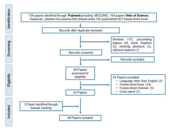
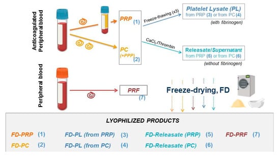
We performed a systematic search, according to PRISMA guidelines from inception until 15 July 2020, using the following data bases: Web of Science (WOS) and PubMed (including MEDLINE) (Figure 1). The following search strategy was used: platelet-rich plasma and (freeze-dried or lyophilized) not freeze-dried bone. Original articles were included if they reported the use of freeze-dried platelet-rich plasma (FD-PRP) or fibrin (FD-PRF) in either experimental or clinical research in applications other than transfusion. Reviews, proceedings, meeting abstracts, book chapters, editorial material and case reports were excluded. Publications were reviewed by at least two researchers, and included articles grouped according to medical field application or those assessing FD-PRP preservation. Of the 46 included articles, skin research was addressed in 17 papers, mainly addressing wound management (15 papers). Other areas of interest were musculoskeletal conditions (10 studies), mainly addressing osteoarticular problems and dentistry (7 studies). Another study examined the feasibility of eye drop preservation for ocular surface conditions [9].
Figure 1.
PRISMA (Preferred Reported Items for Systematic Reviews and Meta-Analyses) workflow.
2.1. Lyophilized PRP Preservation
Platelet-rich plasma preparations: relevant aspects for FD-PRP formulations (resting vs. activated platelets).
Freeze-drying PRP products are encouraged to secure the preservation and standardization of PRP therapies, and thereby boost the clinical benefits that they provide. Quality control of the process is mandatory to guarantee the maintenance of the biological activity of the signaling proteins within each formulation separately.
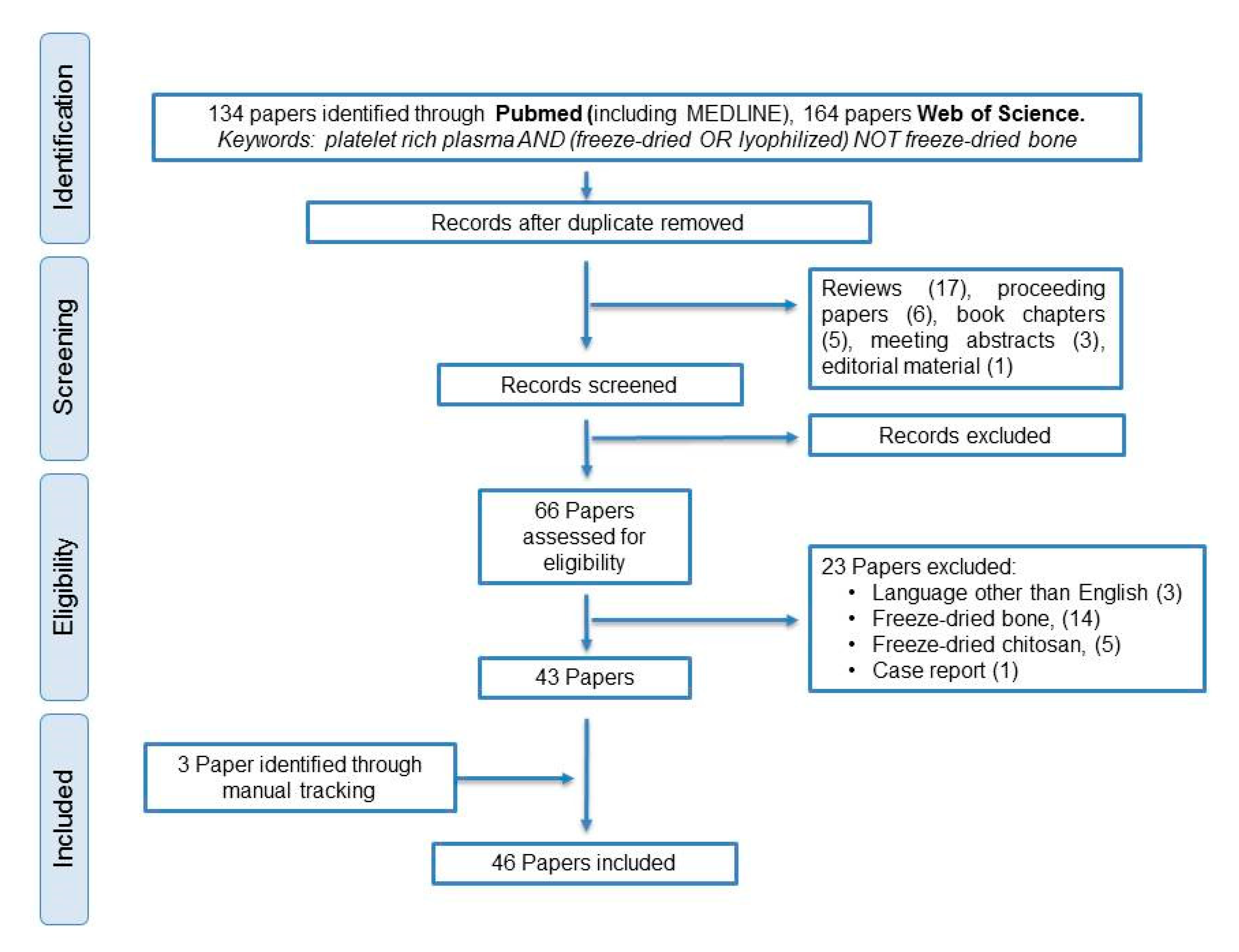
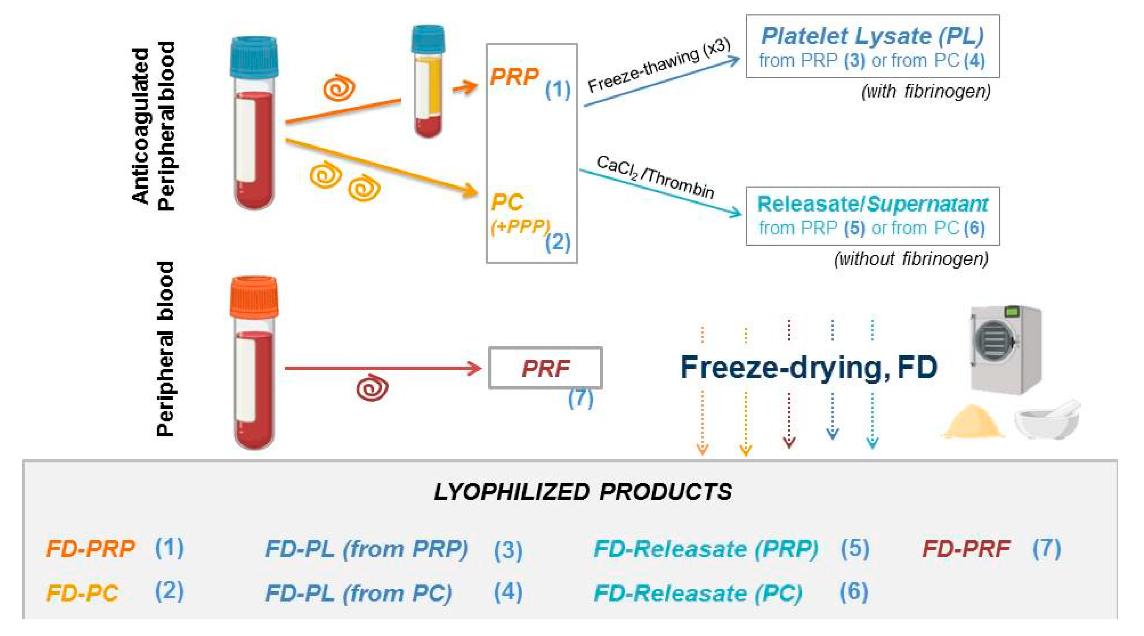
Figure 2 depicts the different PRP formulations that have been lyophilized thus far. For preservation (equivalence) studies, they can be gathered in two groups: (A) products that preserve platelet integrity, (products (1), (2) and (7)), and (B) lysates and releasates mostly composed of platelet secretome (from either PRP or platelet concentrates (PCs), designed as (3)–(6) in Figure 2).
Figure 2.
Preparation of different plasma formulations for lyophilization. Single spinning of anticoagulated whole blood yields platelet-rich plasma (PRP) with moderate concentration of platelets (and optionally leukocytes), for the purpose of this study designed as (1); alternatively, double spinning or plateletpheresis systems produce platelet concentrates (PCs) designed as (2). The pellet can be resuspended in a determined volume of Platelet-Poor Plasma (PPP) and platelet count adjusted, or platelets can be used without plasma. When PRP or PC products are freeze-thawed multiple times (commonly 3) or sonicated, the resultant platelet lysates (PLs) contains platelet secretome along with platelet membranes (ghost platelets) designed as (3) and (4), respectively. Alternatively, the platelet secretome can be obtained after the activation of platelets by adding calcium chloride/ thrombin. The supernatant released from the clot (from PRP (5) or PCs (6)) contains the whole platelet secretome. PRF (7) is obtained by centrifuging noncoagulated peripheral blood and disposing of the formed hydrogel the red blood cells. Terminology: PRP, platelet-rich plasma; PC, platelet concentrate; PPP, platelet-poor plasma; PL, platelet lysate; PRF, platelet-rich fibrin; FD-PRP, freeze-dried platelet-rich plasma; FD-PC, freeze-dried platelet concentrate; FD-PL, freeze-dried platelet lysate; FD-PRF, freeze-dried platelet-rich fibrin.
In lyophilized products from group (A), the platelet membrane should be undamaged after the freeze-drying process [7]: changes in size distribution, altered morphology by SEM, biomarker pattern in flow cytometry, and response to agonists after platelet rehydration are therefore assessed. In this context, platelets can be preserved by introducing trehalose, a commonly used lyoprotectant, into their cytosol, and freeze-drying in trehalose/albumin buffer [10]. Trehalose is also efficient as an extracellular lyoprotectant, preventing protein aggregation and enhancing plasma stability during storage, and is preferred over sucrose and glucose [11].
The comparison of the kinetics of the Growth Factor (GF) release of calcium-activated fresh PRP and calcium-activated rehydrated freeze-dried platelet-rich plasma (FD-PRP) treated with trehalose evidenced differences in the kinetics of platelet-derived growth factor (PDGF) and transforming growth factor (TGF-b) release but not vascular endothelial growth factor (VEGF) [12]. Therefore, Pan et al. suggest that these products should be Ca+2-activated prior to freeze-drying to avoid these differences.
In lyophilized products from group (B), the concentration of alpha-granule proteins are often measured as an index of platelet secretome. In addition, functional assays in vitro are performed to assess signaling proteins’ activities. The rationale for the clinical use of PRP is mainly based on the administration of platelet secretome, particularly the protein pool stored in alpha-granules; each platelet contains approximately 50–80 alpha-granules that release their content upon platelet activation [13]. Instead of the previously identified 300 proteins in a-granules [14,15], new proteomic data revealed that, after collagen and thrombin stimulation of platelets, only 124 proteins constitute the platelet releasate. The discrepancy in protein number is attributed to the subtraction of proteins present in resting platelets [16]. Concentrations differ among proteins, varying from highly abundant (ug/mL), i.e., thrombospondin, PF-4 and von Willebrand factor, to less abundant (pg/mL), such as VEGF. The crucial paradox of platelets is that they contain proteins with opposing functional roles; for example, PF-4 is antiangiogenic while VEGF stimulates endothelial cell proliferation.
Despite containing proteins from different functional categories, most studies focus on the growth factor family (GF). Typically, PDGF, TGF, epidermal growth factor (EGF) and VEGF are involved in tissue repair and assessed in fresh versus FD-PRP. For example, Shiga et al. [17] compared fresh-frozen (−80 ºC) PRP with FD-PRP. They established its stability, up to 8 weeks at room temperature, based on the quantification of PDGF, VEGF, TGF and EGF. Similarly, da Silva et al. [18] verified the stability of PDGF, VEGF, TGF and EGF comparing fresh PRP and FD-PRP (double spinning protocol) and proved the activity by measuring human umbilical endothelial cells (HUVEC) and fibroblast proliferation.
Pooling the PRP of different patients has emerged as a safe option for the standardization of these products. To validate this methodology, Kieb et al. [19] quantified GFs, in particular VEGF, β-FGF, PDGF-AB, TGF-β1, IGF-1, IL-1α, IL-1β and IL-1RA in peripheral blood, fresh PRP prepared using a commercial system, fresh-frozen irradiated PC, and irradiated FD-PC. The different preparations had similar levels of GFs overall, although IGF-1 and IL-1α could not be detected in FD-PCs, since they are plasma proteins, not platelet proteins.
In addition, the safety of FD-PRP is paramount and has to be demonstrated to regulatory bodies by donor testing before donation and one month later to ensure that no virus incubation has escaped from analyses. Sterility can be achieved by gamma irradiation of the extracted product without affecting the efficacy of FD-PL in chondrocytes, fibroblast, osteoblasts and mesenchymal stem cell (BM-MSC) cultures [20]. Indeed, supplementing cultures with 5% FD-PL showed advantages vs. 10% FBS in terms of proliferation, especially at low cell density. These results support the use of human-derived supplements in vitro, moving into a more biomimetic model and avoiding the use of xenogeneic derivatives.
2.2. Skin research
2.2.1. Wound Management
Chronic nonhealing wounds produce a heavy economic and social burden. Their management requires a multimodal approach depending on their etiology. For example, compression therapy is crucial in venous leg ulcers, while adequate foot off-loading is critical in diabetic foot ulcers. Regardless of the etiology of the ulcers, there is consensus regarding the need to optimize the biological conditions of the ulcer bed and its microenvironment. PRPs can provide advantages by augmenting the concepts behind the TIME framework for ulcer care (tissue management, inflammation and infection control, moisture balance and edge (epithelial) advancement) [21]. Indeed, PRPs can help in tissue management by providing a pool of signaling proteins, which can trigger healing mechanisms [22].
Ulcer care requires multiple PRP applications, normally entailing several venipunctures in patients with multimorbidity. In such patients, FD-PRP (either autologous or allogenic), would offer tangible benefits, since levels and biological activities of PRP are conserved through the lyophilization process [23,24] (Table 1).
Many chronic wounds are colonized with bacteria hampering the healing process. The inhibitory actions of PRPs on the growth of various pathogenic bacteria strains is platelet dose-dependent, with platelet-poor plasma showing no impact on bacterial growth [25,26]. Actually, platelets express receptors members of the Toll-like receptor (TLR) family, which bind to bacterial targets, thereby leading to platelet activation and the release of microbicidal proteins and cytokines contained within platelets, including thrombocidins from CTAP-III and NAP-2, kinocidins, beta defensing -1, -2, -3 and thymosin-beta 4 [27]. These proteins exert their role through the recruitment of circulating neutrophils involved in bacterial clearance. Other specific platelet membrane receptors also bind to Escherichia coli or staphylococcal proteins, facilitating neutrophil entry and the debridement of the wound site [28].
In addition, the antibacterial activities of PRP can be improved by optimizing the configuration of combination products. Wang et al. [29] fabricated an antibacterial against S. aureus, P. aeruginosa and C. albicans composite dressing using chitosan/silk fibroin nanosilver loaded with PRP (prepared through double spinning), and assessed the physical and mechanical properties of the device, protein release, biological safety (silver content in organs) and efficacy in infectious wound treatment (CTS-SF/Ag/SA/PRP) in vitro and in vivo. In wound healing studies, Yassin et al. [30] compared PRP wafers (carboxymethyl cellulose, CMC) and powder and antibacterial activities against Gram-negative bacteria. FD-wafers were proposed as PRP delivery systems: they showed better results than PRP powder in a rat wound model, in terms of antimicrobial efficacy, wound size measurements and histopathological analyses. The designed product was proven stable for 3 months at −20 °C.
Moisture balance is beneficial to ensure appropriate wound care and can be achieved by combining PRP with biomaterials commonly used for this purpose in the manufacture of wound dressings. Combination products obtained through the mixture of FD-PRP with alginate [31], CMC [30] or gelatin [32] facilitate moisture preservation in the wound bed while serving as vehicles for sustained release, to match with the biological requirements of the wound [31].
Collagen is commonly used in wound dressings, and Horimizu et al. [33] designed collagen sponges as vehicles for FD-PRP with positive results in a diabetic mice wound model [33]. In another example, different concentrations of FD-platelet lysates were encapsulated in collagen, and the equivalence between fresh and lyophilized products was shown by scratch wound assays, chicken chorioallantoic membrane (CAM) assays, degradation assessment and PDGF-BB and VEGF release [34]. The combination of FD-PRP with collagen products derived from the porcine extracellular matrix (so-called ADM, acellular dermal matrix) promoted neovascularization, collagen deposition and epithelialization [24]. Moreover, FD-PRP can also be combined with other biomaterials. The mechanical and physicochemical properties of silk cocoon combined with FD-PRP offer an advanced wound dressing, which enhanced wound closure in New Zealand rabbits [35].
The use of PRP in specific wound etiologies, such as diabetic foot, is based on high quality clinical evidence [36], but clinical data reporting the efficacy of FD-PRP are limited. FD-PRP powder (1 × 10⁷ platelets/cm2) was used in the management of deep second degree burn wounds in the plantar area showing significant beneficial differences in the healing rate at 3 weeks and bacteria colonization compared to controls [37].
2.2.2. Other Conditions
The high prevalence of androgenetic alopecia and the limited efficacy of current available treatments have prompted the clinical investigation with PRP. A recent meta-analysis has shown that PRP can improve hair density and thickness [38]. Usually, several sessions of microneedling with PRP are needed, often associated to minoxidil [39]. In an initial industrial-scale approach, the lyophilization of activated PRP from pigs was effective in the expansion of human follicle dermal papilla cells. Thus, this can yield an off-the-shelf product with potential for alopecia management [40]. Striae distensae is a skin condition especially common in females, causing cosmetic morbidity and psychological distress. Augmenting fractional CO2 ablational laser procedures with lyophilized growth factors has been shown to improve clinical outcomes [41].
Table 1.
Skin research.
3. Musculoskeletal Applications
Osteoarthritis (OA) is a prevalent degenerative joint condition with no effective treatments available other than surgery. An unsustainable economic burden by 2030 has been predicted due to the continuous growth of joint replacements [47]. Identifying conservative treatments for earlier disease stages is paramount. A recent meta-analysis [48] including 26 randomized studies showed that intra-articular injections of PRP were more effective than hyaluronic acid in terms of pain reduction and functional improvement, with comparable safety profiles. Therefore, FD-PRP can offer important advantages in this context, as most often PRP treatments for knee OA involve multiple injections at each treatment cycle. Preliminary clinical data showed significant improvement in pain, function and quality of life 1, 3 and 6 months after injection of autologous PRP powder resuspended in 6 mL of normosaline [49] (Table 2). Beneficial clinical effects are attributed to the modulation of inflammation.
A common in vitro functional model consists of exposing chondrocytes or cartilage explants to IL-1β and measuring how PRP interferes with the inflammatory response triggered by IL-1β. In this framework, Jain et al. [50] showed that the encapsulation of FD-PRP in polyethylene glycol enhanced PRP effects by rescuing chondrocyte proliferation and decreasing MMP13-induced extracellular matrix catabolism. The latter was achieved through a reduction in common inflammatory molecular pathways, such as NF-kB activation and nitric oxide (NO) synthesis. Similarly, dose-related chondroprotective actions of PRP, without differences between FD-PRP compared to fresh-frozen PRP, were found in equine cartilage cultures [51]. In a similar way, standardized human PRP powder induced a dose- and time-response relationship in human chondrocyte proliferation and metabolic activity [52].
A promising approach is the combination of PRP with biomaterials such as tissue scaffolds. In this way, alginate was functionalized with FD-PRP by creating amide bonds with PRP peptides through carbodiimide chemistry [53] and compared to PRP encapsulated in alginate. The resulting injectable hydrogels were studied in nucleus pulposus cells and FD-PRP-functionalized alginate showed higher levels of cell survival and increased secretion of glycosaminoglycans (GAGs ) over time, compared to encapsulated PRP.
In a rat model of spinal posterolateral fusion, bone union was accelerated by FD-PRP at a comparable rate to fresh PRP and performed better than BMPs (Bone Morphogenetic Proteins). In this study, all were combined with an artificial bone substitute, hydroxyapatite/collagen [54]. In addition, these authors confirmed the pharmacological activity of FD-PRP after a four-week storage by examining the PDGFR/ERK signal transduction in osteoblasts [55].
Tendon and ligament injuries are particularly relevant in sports medicine and orthopedics [56]. Clinical data on the effects of PRP in commonly injured tendons including Achilles and patellar tendons, common extensors and rotator cuffs have been published, but meta-analyses are not conclusive, in part given the lack of PRP standardization. Pioneering studies using commercial trehalose-treated lyophilized platelets in equine research models similar gene activation than fresh-frozen PRP in tendon cell cultures [57]. Furthermore, FD-PRP is an effective carrier for icariin, and accelerated tendon–bone healing in New Zealand rabbits with partial patellectomy, as assessed by histology and mechanical testing [58]. Additionally, the use of polydioxanone fibers containing FD-PRP in volumetric muscle loss caused a dose- and ERK-dependent effect in myogenic differentiation [59]. Recently, the feasibility of dry storage of allogeneic FD-PRP has been illustrated in the treatment of plantar fasciopathy [60].
Table 2.
Studies in musculoskeletal research.
In summary, the equivalence of FD-PRP to fresh-frozen PRP has been reported in the context of bone, muscle, tendon and cartilage research; the clinical efficacy of FD-PRP is endorsed by a preliminary study in knee osteoarthritis [49]. Lyophilized PRP has been combined successfully with alginate and is comparable to fresh-frozen PRP; this combination product is promising in bone fusion with relevant advantages in dosing and precise standardization. Other studies endorse the applicability of FD-PRP in the creation of different combination products for bone–tendon healing or volumetric muscle loss [59].
4. Dentistry Research
The use of PRP in oral implantology dates back to the 1990s [61], initially aiming to stimulate alveolar bone regeneration to help achieve faster dental implant stabilization. Two different formulations were very popular in dentistry: PRP (prepared from anticoagulated blood) and PRF (prepared from blood) [62] (see Figure 2). Their influence in periodontal ligament and alveolar bone regeneration has been the main focus of clinical and experimental research during recent decades. In this way, to leverage the potential of lyophilized plasma products in implantology, FD-PRF was proven to enhance the expression of transcription factors related to alveolar bone regeneration in mice compared to traditional PRF [63] (Table 3). Moreover, the equivalence between fresh-frozen PRP and FD-PRP was shown in terms of PDGF-BB, TGF-β and VEGF concentrations and stimulation of bone formation in immunocompromised mice [64]. However, in an apparent contradiction, rabbit BMSCs differentiation and proliferation was better in CaCl2-activated fresh PRP compared with FD-PRP [65].
In another example, FD-PRP was used to enhance the biological activities of fresh PRP formulations. Supplementing fresh PRF with allogeneic FD-PRF increased the concentration of GFs and other signaling cytokines, thereby enhancing bone regeneration in two different rodent calvarial models [66,67].
To fulfill the needs of bone formation and remodeling over time, a sustained release of GFs is desirable. In this way, the combination of FD-PRP with alginate and chitosan helped to slow down the release of TGF-β, PDGF, IGF, VEGF and TSP-1 [68].
Oral soft tissues have a faster turnover than the alveolar bone. Thus, isolating both compartments using membranes for guided bone regeneration overcomes obstacles caused by the different kinetics of tissue formation. Hence, PRF is investigated as an option for periodontal regeneration. A proprietary closed system, which prepared PRF, showed that membrane freeze-drying was not an obstacle for cell adhesion and proliferation [69]. Accordingly, a randomized clinical trial, comparing FD-PRF vs. fresh PRF in guided bone regeneration surgery did not find any difference in their clinical performance [70]. An optimized membrane, in terms of biological activities and mechanical properties, was elaborated with FD-PRF/chitosan/collagen [71] to be used in periodontal disease. A biodegradable polymer mesh coated with FD-PRP was effective in promoting periodontal ligament cell adhesion and growth as well as stimulating angiogenic mechanisms [72].
Table 3.
Studies in dentistry (9).
In summary, FD-PRP is valuable for promoting alveolar bone regeneration either when used alone or when added to fresh PRP to augment GF concentrations. Moreover, the controlled release of GF and the creation of membranes for guided bone regeneration can be attained when FD-PRP is mixed with biomaterials.
5. The Way Forward
PRP therapies are best known for their benefit in wound healing, osteoarthritis and alveolar bone repair. However, substantial challenges remain to be addressed before PRPs evolve from off-label use to on-label prescription and their costs reimbursed by health insurance. The unanimously identified obstacle, hindering advancement in PRP science, is the lack of standardization in PRP formulations, a challenge that could be met somewhat by lyophilization.
As shown in the present review, freeze-drying preserves platelet function, cytokine concentration and their function. The easiness of combining PRP with biomaterials is guaranteed, and combination products can be designed to control the kinetics of cytokine release and match more specifically the needs of the target tissue. In addition, FD-PRP is promising to functionalize bioprinted constructs [73]).
Implementing efficient freeze-drying procedures for allogeneic platelet-rich plasma formulations are multipurpose, first, guiding scientific investigations, and second leveraging treatment feasibility (Table 4). Actually, the platelet number and the levels of relevant cytokines can be accurately determined, which can inform a research hypothesis about the mechanism of action. Furthermore, product standardization facilitates conducting homogeneous clinical trials informative enough to establish the superiority (or not) of PRP over other treatments. Another fundamental research question is to clarify the biological advantages of PCs over lysates or releasates, i.e., preserving platelet integrity over using platelet’s secretome, in the different clinical conditions.
Table 4.
Advantages and weaknesses of freeze-dried platelet-rich plasma.
On the other hand, expediency is guaranteed, as lyophilized products reduce the need for specific facilities and specialized staff and are readily available on demand.
If we improve our understanding of PRP assets, selecting donors among healthy people might be grounded in the identification of core PRP elements related to clinical efficacy, e.g., application of young PRP in tissues of older patients, or considering the influence of exercise/sport practice on PRP quality.
Freeze-dried allogeneic PRP has to be declared fit as per national guidelines. Grafting FD-PRP across blood groups may need specific guidelines regarding the titration of antibodies but the fact that FD-PRP requirements for regenerative medicine may differ from transfusion medicine needs to be considered.
The manufacturing of freeze-dried allogeneic PRP or PRF has to comply with the specific requirements overseen by the competent regulatory body in each country, in terms of manufacturing establishments and donor suitability. Of note is the presence of blood-type immunoglobulin M antibodies, i.e., specific antiviral IgM.
Preferably, FD-PRP should be used after titration of antibodies, limiting the level of IgM ABO antibody titers.
Author Contributions
I.A.: Study conception, design and writing; C.D.A., A.P.-V.: Acquisition of data, figures preparation; N.M.: Revising it critically for important intellectual content; I.A., A.P.-V., C.D.A., N.M.: Final approval of the article. All authors have read and agreed to the published version of the manuscript.
Funding
This study was supported by Basque Industry Department (KK-2019/BC) and the Basque Health Department (2020333026).
Conflicts of Interest
The authors declare that they have no competing interests.
References
- Andia, I.; Maffulli, N. A contemporary view of platelet-rich plasma therapies: Moving toward refined clinical protocols and precise indications. Regen. Med. 2018, 13, 717–728. [Google Scholar] [CrossRef] [Scilit] [PubMed]
- Harrison, P.; The Subcommittee on Platelet Physiology. The use of platelets in regenerative medicine and proposal for a new classification system: Guidance from the SSC of the ISTH. J. Thromb. Haemost. 2018, 16, 1895–1900. [Google Scholar] [CrossRef] [Scilit] [PubMed]
- Pusateri, A.E.; Butler, F.K.; Shackelford, S.A.; Sperry, J.L.; Moore, E.E.; Cap, A.P.; Taylor, A.L.; Homer, M.J.; Hoots, W.K.; Weiskopf, R.B.; et al. The need for dried plasma—A national issue. Transfusion 2019, 59, 1587–1592. [Google Scholar] [CrossRef] [Scilit] [PubMed]
- Daban, J.; Clapson, P.; Ausset, S.; Deshayes, A.V.; Sailliol, A. Freeze dried plasma: A French army specialty. Crit. Care 2010, 14, 412. [Google Scholar] [CrossRef] [Scilit]
- Feuerstein, S.J.; Skovmand, K.; Møller, A.M.; Wildgaard, K. Freeze-dried plasma in major haemorrhage: A systematic review. Vox Sang. 2020, 115, 263–274. [Google Scholar] [CrossRef] [Scilit]
- Read, M.S.; Reddick, R.L.; Bode, A.P.; Bellinger, D.A.; Nichols, T.C.; Taylor, K.; Smith, S.V.; McMahon, D.K.; Griggs, T.R.; Brinkhous, K.M. Preservation of hemostatic and structural properties of rehydrated lyophilized platelets: Potential for long-term storage of dried platelets for transfusion. Proc. Natl. Acad. Sci. USA 1995, 92, 397–401. [Google Scholar] [CrossRef] [Scilit]
- Valeri, C.R.; Macgregor, H.; Barnard, M.R.; Summaria, L.; Michelson, A.D.; Ragno, G. In vitro testing of fresh and lyophilized reconstituted human and baboon platelets. Transfusion 2004, 44, 1505–1512. [Google Scholar] [CrossRef] [Scilit]
- Nurden, A.T. The biology of the platelet with special reference to inflammation wound healing and immunity. Front. Biosci. 2018, 23, 726–751. [Google Scholar] [CrossRef] [Scilit]
- Anitua, E.; de la Fuente, M.; Muruzábal, F.; Merayo-Lloves, J. Stability of freeze-dried plasma rich in growth factors eye drops stored for 3 months at different temperature conditions. Eur. J. Ophthalmol. 2020. [Google Scholar] [CrossRef] [Scilit]
- Wolkers, W.F.; Walker, N.J.; Tamari, Y.; Tablin, F.; Crowe, J.H. Towards a Clinical Application of Freeze-Dried Human Platelets. Cell Preserv. Technol. 2002, 1, 175–188. [Google Scholar] [CrossRef] [Scilit]
- Brogna, R.; Oldenhof, H.; Sieme, H.; Figueiredo, C.; Kerrinnes, T.; Wolkers, W.F. Increasing storage stability of freeze-dried plasma using trehalose. PLoS ONE 2020, 15, e0234502. [Google Scholar] [CrossRef] [Scilit] [PubMed]
- Pan, L.; Yong, Z.; Yuk, K.S.; Hoon, K.Y.; Yuedong, S.; Xu, J. Growth Factor Release from Lyophilized Porcine Platelet-Rich Plasma: Quantitative Analysis and Implications for Clinical Applications. Aesth. Plast. Surg. 2016, 40, 157–163. [Google Scholar] [CrossRef] [Scilit] [PubMed]
- Andia, I.; Maffulli, N. Platelet-rich plasma for managing pain and inflammation in osteoarthritis. Nat. Rev. Rheumatol. 2013, 9, 721–730. [Google Scholar] [CrossRef] [Scilit] [PubMed]
- Maynard, D.M.; Heijnen, H.F.G.; Horne, M.K.; White, J.G.; Gahl, W.A. Proteomic analysis of platelet α-granules using mass spectrometry: Platelet α-granule proteomics. J. Thromb. Haemost. 2007, 5, 1945–1955. [Google Scholar] [CrossRef] [Scilit]
- Coppinger, J.A.; Cagney, G.; Toomey, S.; Kislinger, T.; Belton, O.; McRedmond, J.P.; Cahill, D.J.; Emili, A.; Fitzgerald, D.J.; Maguire, P.B. Characterization of the proteins released from activated platelets leads to localization of novel platelet proteins in human atherosclerotic lesions. Blood 2004, 103, 2096–2104. [Google Scholar] [CrossRef] [Scilit]
- Wijten, P.; van Holten, T.; Woo, L.L.; Bleijerveld, O.B.; Roest, M.; Heck, A.J.R.; Scholten, A. High Precision Platelet Releasate Definition by Quantitative Reversed Protein Profiling—Brief Report. Arter. Thromb. Vasc. Biol. 2013, 33, 1635–1638. [Google Scholar] [CrossRef] [Scilit]
- Shiga, Y.; Kubota, G.; Orita, S.; Inage, K.; Kamoda, H.; Yamashita, M.; Iseki, T.; Ito, M.; Yamauchi, K.; Eguchi, Y.; et al. Freeze-Dried Human Platelet-Rich Plasma Retains Activation and Growth Factor Expression after an Eight-Week Preservation Period. Asian Spine J. 2017, 11, 329–336. [Google Scholar] [CrossRef] [Scilit]
- Da Silva, L.Q.; Montalvão, S.A.d.L.; Justo-Junior, A.d.S.; Cunha Júnior, J.L.R.; Huber, S.C.; Oliveira, C.C.; Annichino-Bizzacchi, J.M. Platelet-rich plasma lyophilization enables growth factor preservation and functionality when compared with fresh platelet-rich plasma. Regen. Med. 2018, 13, 775–784. [Google Scholar] [CrossRef] [Scilit]
- Kieb, M.; Sander, F.; Prinz, C.; Adam, S.; Mau-Möller, A.; Bader, R.; Peters, K.; Tischer, T. Platelet-Rich Plasma Powder: A New Preparation Method for the Standardization of Growth Factor Concentrations. Am. J. Sports Med. 2017, 45, 954–960. [Google Scholar] [CrossRef] [Scilit]
- Muraglia, A.; Ottonello, C.; Spanò, R.; Dozin, B.; Strada, P.; Grandizio, M.; Cancedda, R.; Mastrogiacomo, M. Biological activity of a standardized freeze-dried platelet derivative to be used as cell culture medium supplement. Platelets 2014, 25, 211–220. [Google Scholar] [CrossRef] [Scilit]
- Perez-Zabala, E.; Basterretxea, A.; Larrazabal, A.; Perez-del-Pecho, K.; Rubio-Azpeitia, E.; Andia, I. Biological approach for the management of non-healing diabetic foot ulcers. J. Tissue Viability 2016, 25, 157–163. [Google Scholar] [CrossRef] [Scilit] [PubMed]
- Burgos-Alonso, N.; Lobato, I.; Hernández, I.; Sebastian, K.; Rodríguez, B.; Grandes, G.; Andia, I. Adjuvant Biological Therapies in Chronic Leg Ulcers. Int. J. Mol. Sci. 2017, 18, 2561. [Google Scholar] [CrossRef] [Scilit] [PubMed]
- Huber, S.C.; Junior, J.L.R.C.; Silva, L.Q.; Montalvão, S.A.L.; Annichino-Bizzacchi, J.M. Freeze-dried versus fresh platelet-rich plasma in acute wound healing of an animal model. Regen. Med. 2019, 14, 525–534. [Google Scholar] [CrossRef] [Scilit] [PubMed]
- Lei, X.; Yang, Y.; Shan, G.; Pan, Y.; Cheng, B. Preparation of ADM/PRP freeze-dried dressing and effect of mice full-thickness skin defect model. Biomed. Mater. 2019, 14, 035004. [Google Scholar] [CrossRef] [Scilit] [PubMed]
- Burnouf, T.; Chou, M.-L.; Wu, Y.-W.; Su, C.-Y.; Lee, L.-W. Antimicrobial activity of platelet (PLT)-poor plasma, PLT-rich plasma, PLT gel, and solvent/detergent-treated PLT lysate biomaterials against wound bacteria: ANTIMICROBIAL ACTIVITY OF PLASMA AND PLT GEL. Transfusion 2013, 53, 138–146. [Google Scholar] [CrossRef] [Scilit]
- Maghsoudi, O.; Ranjbar, R.; Mirjalili, S.H.; Fasihi Ramandi, M. Inhibitory activities of platelet-rich and platelet-poor plasma on the growth of pathogenic bacteria. Iran J. Pathol. 2017, 12, 79–87. [Google Scholar] [CrossRef] [Scilit]
- Kapur, R.; Zufferey, A.; Boilard, E.; Semple, J.W. Nouvelle Cuisine: Platelets Served with Inflammation. J. Immunol. 2015, 194, 5579–5587. [Google Scholar] [CrossRef] [Scilit]
- Watson, C.N.; Kerrigan, S.W.; Cox, D.; Henderson, I.R.; Watson, S.P.; Arman, M. Human platelet activation by Escherichia coli: Roles for FcγRIIA and integrin αIIbβ3. Platelets 2016, 27, 535–540. [Google Scholar] [CrossRef] [Scilit]
- Wang, Q.; Qian, Z.; Liu, B.; Liu, J.; Zhang, L.; Xu, J. In vitro and in vivo evaluation of new PRP antibacterial moisturizing dressings for infectious wound repair. J. Biomater. Sci. Polym. Ed. 2019, 30, 462–485. [Google Scholar] [CrossRef] [Scilit]
- Yassin, G.E.; Dawoud, M.H.S.; Wasfi, R.; Maher, A.; Fayez, A.M. Comparative lyophilized platelet-rich plasma wafer and powder for wound-healing enhancement: Formulation, in vitro and in vivo studies. Drug Dev. Ind. Pharm. 2019, 45, 1379–1387. [Google Scholar] [CrossRef] [Scilit]
- Nardini, M.; Perteghella, S.; Mastracci, L.; Grillo, F.; Marrubini, G.; Bari, E.; Formica, M.; Gentili, C.; Cancedda, R.; Torre, M.L.; et al. Growth Factors Delivery System for Skin Regeneration: An Advanced Wound Dressing. Pharmaceutics 2020, 12, 120. [Google Scholar] [CrossRef] [Scilit] [PubMed]
- Notodihardjo, S.C.; Morimoto, N.; Kakudo, N.; Mitsui, T.; Le, T.M.; Tabata, Y.; Kusumoto, K. Efficacy of Gelatin Hydrogel Impregnated with Concentrated Platelet Lysate in Murine Wound Healing. J. Surg. Res. 2019, 234, 190–201. [Google Scholar] [CrossRef] [Scilit]
- Horimizu, M.; Kawase, T.; Nakajima, Y.; Okuda, K.; Nagata, M.; Wolff, L.F.; Yoshie, H. An improved freeze-dried PRP-coated biodegradable material suitable for connective tissue regenerative therapy. Cryobiology 2013, 66, 223–232. [Google Scholar] [CrossRef] [Scilit] [PubMed]
- Lima, A.C.; Mano, J.F.; Concheiro, A.; Alvarez-Lorenzo, C. Fast and Mild Strategy, Using Superhydrophobic Surfaces, to Produce Collagen/Platelet Lysate Gel Beads for Skin Regeneration. Stem Cell Rev. Rep. 2015, 11, 161–179. [Google Scholar] [CrossRef] [Scilit] [PubMed]
- Liu, J.; Lu, F.; Chen, H.; Bao, R.; Li, Z.; Lu, B.; Yu, K.; Dai, F.; Wu, D.; Lan, G. Healing of skin wounds using a new cocoon scaffold loaded with platelet-rich or platelet-poor plasma. RSC Adv. 2017, 7, 6474–6485. [Google Scholar] [CrossRef] [Scilit]
- del Pino-Sedeño, T.; Trujillo-Martín, M.M.; Andia, I.; Aragón-Sánchez, J.; Herrera-Ramos, E.; Iruzubieta Barragán, F.J.; Serrano-Aguilar, P. Platelet-rich plasma for the treatment of diabetic foot ulcers: A meta-analysis: Platelet-rich plasma for diabetic foot ulcers. Wound Repair Regen. 2019, 27, 170–182. [Google Scholar] [CrossRef] [Scilit] [PubMed]
- Yeung, C.-Y.; Hsieh, P.-S.; Wei, L.-G.; Hsia, L.-C.; Dai, L.-G.; Fu, K.-Y.; Dai, N.-T. Efficacy of Lyophilised Platelet-Rich Plasma Powder on Healing Rate in Patients With Deep Second Degree Burn Injury: A Prospective Double-Blind Randomized Clinical Trial. Ann. Plast. Surg. 2018, 80 (2S Suppl. 1), S66–S69. [Google Scholar] [CrossRef] [Scilit]
- Evans, A.G.; Mwangi, J.M.; Pope, R.W.; Ivanic, M.G.; Botros, M.A.; Glassman, G.E.; Pearce, F.B.; Kassis, S. Platelet-rich plasma as a therapy for androgenic alopecia: A systematic review and meta-analysis. J. Dermatol. Treat. 2020, 1–14. [Google Scholar] [CrossRef] [Scilit]
- Bruce, A.J.; Pincelli, T.P.; Heckman, M.G.; Desmond, C.M.; Arthurs, J.R.; Diehl, N.N.; Douglass, E.J.; Bruce, C.J.; Shapiro, S.A. A Randomized, Controlled Pilot Trial Comparing Platelet-Rich Plasma to Topical Minoxidil Foam for Treatment of Androgenic Alopecia in Women. Dermatol. Surg. 2020, 46, 826–832. [Google Scholar] [CrossRef] [Scilit]
- Lin, Y.-K.; Sugiri, F.; Ma, H.; Chiu, Y.-H.; Yao, C.-L. Industrial-scale processing of activated platelet-rich plasma from specific pathogen-free pigs and its effect on promoting human hair follicle dermal papilla cell cultivation. J. Taiwan Inst. Chem. Eng. 2017, 71, 28–37. [Google Scholar] [CrossRef] [Scilit]
- Abdallah, M.; Fahmy, H.; Abdel Hameed, S.; Mostafa, A.E. Ablative fractional CO2 laser vs lyophilized growth factor intralesional injection vs combination of both modalities for striae distensae treatment. J. Cosmet. Dermatol. 2020. [Google Scholar] [CrossRef] [Scilit] [PubMed]
- Notodihardjo, S.C.; Morimoto, N.; Kakudo, N.; Mitsui, T.; Le, T.M.; Tabata, Y.; Kusumoto, K. Comparison of the efficacy of cryopreserved human platelet lysate and refrigerated lyophilized human platelet lysate for wound healing. Regen. Ther. 2019, 10, 1–9. [Google Scholar] [CrossRef] [Scilit] [PubMed]
- Pietramaggiori, G.; Kaipainen, A.; Czeczuga, J.M.; Wagner, C.T.; Orgill, D.P. Freeze-dried platelet-rich plasma shows beneficial healing properties in chronic wounds. Wound Repair Regen. 2006, 14, 573–580. [Google Scholar] [CrossRef] [Scilit] [PubMed]
- Pietramaggiori, G.; Scherer, S.S.; Mathews, J.C.; Alperovich, M.; Yang, H.-J.; Neuwalder, J.; Czeczuga, J.M.; Chan, R.K.; Wagner, C.T.; Orgill, D.P. Healing modulation induced by freeze-dried platelet-rich plasma and micronized allogenic dermis in a diabetic wound model. Wound Repair Regen. 2008, 16, 218–225. [Google Scholar] [CrossRef] [Scilit]
- Sell, S.A.; Wolfe, P.S.; Spence, A.J.; Rodriguez, I.A.; McCool, J.M.; Petrella, R.L.; Garg, K.; Ericksen, J.J.; Bowlin, G.L. A Preliminary Study on the Potential of Manuka Honey and Platelet-Rich Plasma in Wound Healing. Int. J. Biomater. 2012, 2012, 313781. [Google Scholar] [CrossRef] [Scilit]
- Xu, F.; Zou, D.; Dai, T.; Xu, H.; An, R.; Liu, Y.; Liu, B. Effects of incorporation of granule-lyophilised platelet-rich fibrin into polyvinyl alcohol hydrogel on wound healing. Sci. Rep. 2018, 8, 14042. [Google Scholar] [CrossRef] [Scilit]
- Ackerman, I.N.; Bohensky, M.A.; Zomer, E.; Tacey, M.; Gorelik, A.; Brand, C.A.; de Steiger, R. The projected burden of primary total knee and hip replacement for osteoarthritis in Australia to the year 2030. BMC Musculoskelet. Disord. 2019, 20, 90. [Google Scholar] [CrossRef] [Scilit]
- Tan, J.; Chen, H.; Zhao, L.; Huang, W. Platelet-Rich Plasma Versus Hyaluronic Acid in the Treatment of Knee Osteoarthritis: A Meta-Analysis of 26 Randomized Controlled Trials. Arthrosc. J. Arthrosc. Relat. Surg. 2020. [Google Scholar] [CrossRef] [Scilit]
- Shirata, T.; Kato, Y. Can intra-articular injection of freeze-dried platelet-derived factor concentrate regenerate articular cartilage in the knee joint? Regen. Ther. 2019, 11, 5–7. [Google Scholar] [CrossRef] [Scilit]
- Jain, E.; Chinzei, N.; Blanco, A.; Case, N.; Sandell, L.J.; Sell, S.; Rai, M.F.; Zustiak, S.P. Platelet-Rich Plasma Released from Polyethylene Glycol Hydrogels Exerts Beneficial Effects on Human Chondrocytes. J. Orthop. Res. 2019, 37, 2401–2410. [Google Scholar] [CrossRef] [Scilit]
- Camargo Garbin, L.; McIlwraith, C.W.; Frisbie, D.D. Evaluation of allogeneic freeze-dried platelet lysate in cartilage exposed to interleukin 1-β in vitro. BMC Vet. Res. 2019, 15, 386. [Google Scholar] [CrossRef] [Scilit] [PubMed]
- Hahn, O.; Kieb, M.; Jonitz-Heincke, A.; Bader, R.; Peters, K.; Tischer, T. Dose-Dependent Effects of Platelet-Rich Plasma Powder on Chondrocytes In Vitro. Am. J. Sports Med. 2020, 48, 1727–1734. [Google Scholar] [CrossRef] [Scilit] [PubMed]
- Growney, E.A.; Linder, H.R.; Garg, K.; Bledsoe, J.G.; Sell, S.A. Bio-conjugation of platelet-rich plasma and alginate through carbodiimide chemistry for injectable hydrogel therapies. J. Biomed. Mater. Res. 2020, 108, 1972–1984. [Google Scholar] [CrossRef] [Scilit] [PubMed]
- Shiga, Y.; Orita, S.; Kubota, G.; Kamoda, H.; Yamashita, M.; Matsuura, Y.; Yamauchi, K.; Eguchi, Y.; Suzuki, M.; Inage, K.; et al. Freeze-Dried Platelet-Rich Plasma Accelerates Bone Union with Adequate Rigidity in Posterolateral Lumbar Fusion Surgery Model in Rats. Sci. Rep. 2016, 6, 36715. [Google Scholar] [CrossRef] [Scilit]
- Kinoshita, H.; Orita, S.; Inage, K.; Fujimoto, K.; Shiga, Y.; Abe, K.; Inoue, M.; Norimoto, M.; Umimura, T.; Ishii, T.; et al. Freeze-Dried Platelet-Rich Plasma Induces Osteoblast Proliferation via Platelet-Derived Growth Factor Receptor-Mediated Signal Transduction. Asian Spine J. 2020, 14, 1–8. [Google Scholar] [CrossRef] [Scilit]
- Andia, I.; Maffulli, N. Biological Therapies in Regenerative Sports Medicine. Sports Med. 2017, 47, 807–828. [Google Scholar] [CrossRef] [Scilit]
- McCarrel, T.; Fortier, L. Temporal growth factor release from platelet-rich plasma, trehalose lyophilized platelets, and bone marrow aspirate and their effect on tendon and ligament gene expression. J. Orthop. Res. 2009, 27, 1033–1042. [Google Scholar] [CrossRef] [Scilit]
- Zheng, C.; Lu, H.; Tang, Y.; Wang, Z.; Ma, H.; Li, H.; Chen, H.; Chen, Y.; Chen, C. Autologous Freeze-Dried, Platelet-Rich Plasma Carrying Icariin Enhances Bone-Tendon Healing in a Rabbit Model. Am. J. Sports Med. 2019, 47, 1964–1974. [Google Scholar] [CrossRef] [Scilit]
- McClure, M.J.; Clark, N.M.; Schwartz, Z.; Boyan, B.D. Platelet-rich plasma and alignment enhance myogenin via ERK mitogen activated protein kinase signaling. Biomed. Mater. 2018, 13, 055009. [Google Scholar] [CrossRef] [Scilit]
- Kandil, M.I.; Tabl, E.A.; Elhammady, A.S. Prospective Randomized Evaluation of Local Injection of Allogeneic Growth Factors in Plantar Fasciitis. Foot Ankle Int. 2020. [Google Scholar] [CrossRef] [Scilit]
- Peterson, L.J. Oral and Maxillofacial Surgery. Oral Surg. Oral Med. Oral Pathol. 1998, 85, 9. [Google Scholar]
- Ehrenfest, D.M.D.; Andia, I.; Zumstein, M.A.; Zhang, C.-Q.; Pinto, N.R.; Bielecki, T. Classification of platelet concentrates (Platelet-Rich Plasma-PRP, Platelet-Rich Fibrin-PRF) for topical and infiltrative use in orthopedic and sports medicine: Current consensus, clinical implications and perspectives. Muscles Ligaments Tendons J. 2014, 4, 3–9. [Google Scholar] [CrossRef] [Scilit]
- Li, Q.; Reed, D.; Min, L.; Gopinathan, G.; Li, S.; Dangaria, S.; Li, L.; Geng, Y.; Galang, M.-T.; Gajendrareddy, P.; et al. Lyophilized Platelet-Rich Fibrin (PRF) Promotes Craniofacial Bone Regeneration through Runx2. Int. J. Mol. Sci. 2014, 15, 8509–8525. [Google Scholar] [CrossRef] [Scilit] [PubMed]
- Nakatani, Y.; Agata, H.; Sumita, Y.; Koga, T.; Asahina, I. Efficacy of freeze-dried platelet-rich plasma in bone engineering. Arch. Oral Biol. 2017, 73, 172–178. [Google Scholar] [CrossRef] [Scilit]
- Xie, Y.; Chen, M.; Chen, Y.; Xu, Y.; Sun, Y.; Liang, J.; Fan, Y.; Zhang, X. Effects of PRP and LyPRP on osteogenic differentiation of MSCs. J. Biomed. Mater. Res. 2020, 108, 116–126. [Google Scholar] [CrossRef] [Scilit]
- Li, J.; Chen, M.; Wei, X.; Hao, Y.; Wang, J. Evaluation of 3D-Printed Polycaprolactone Scaffolds Coated with Freeze-Dried Platelet-Rich Plasma for Bone Regeneration. Materials 2017, 10, 831. [Google Scholar] [CrossRef] [Scilit]
- Liu, Z.; Jin, H.; Xie, Q.; Jiang, Z.; Guo, S.; Li, Y.; Zhang, B. Controlled Release Strategies for the Combination of Fresh and Lyophilized Platelet-Rich Fibrin on Bone Tissue Regeneration. BioMed Res. Int. 2019, 2019, 4923767. [Google Scholar] [CrossRef] [Scilit]
- Wang, L.; Wan, M.; Li, Z.; Zhong, N.; Liang, D.; Ge, L. A comparative study of the effects of concentrated growth factors in two different forms on osteogenesis in vitro. Mol. Med. Rep. 2019, 20, 1039–1048. [Google Scholar] [CrossRef] [Scilit]
- Kardos, D.; Hornyák, I.; Simon, M.; Hinsenkamp, A.; Marschall, B.; Várdai, R.; Kállay-Menyhárd, A.; Pinke, B.; Mészáros, L.; Kuten, O.; et al. Biological and Mechanical Properties of Platelet-Rich Fibrin Membranes after Thermal Manipulation and Preparation in a Single-Syringe Closed System. Int. J. Mol. Sci. 2018, 19, 3433. [Google Scholar] [CrossRef] [Scilit]
- Zhang, J.; Qi, X.; Luo, X.; Li, D.; Wang, H.; Li, T. Clinical and immunohistochemical performance of lyophilized platelet-rich fibrin (Ly-PRF) on tissue regeneration. Clin. Implant Dent. Relat. Res. 2017, 19, 466–477. [Google Scholar] [CrossRef] [Scilit]
- Ansarizadeh, M.; Mashayekhan, S.; Saadatmand, M. Fabrication, modeling and optimization of lyophilized advanced platelet rich fibrin in combination with collagen-chitosan as a guided bone regeneration membrane. Int. J. Biol. Macromol. 2019, 125, 383–391. [Google Scholar] [CrossRef] [Scilit] [PubMed]
- Nakajima, Y.; Kawase, T.; Kobayashi, M.; Okuda, K.; Wolff, L.F.; Yoshie, H. Bioactivity of freeze-dried platelet-rich plasma in an adsorbed form on a biodegradable polymer material. Platelets 2012, 23, 594–603. [Google Scholar] [CrossRef] [Scilit] [PubMed]
- Perez-Valle, A.; Del Amo, C.; Andia, I. Overview of Current Advances in Extrusion Bioprinting for Skin Applications. Int. J. Mol. Sci. 2020, 21, 6679. [Google Scholar] [CrossRef] [Scilit] [PubMed]

© 2020 by the authors. Licensee MDPI, Basel, Switzerland. This article is an open access article distributed under the terms and conditions of the Creative Commons Attribution (CC BY) license (http://creativecommons.org/licenses/by/4.0/).